Patients › Hand
ड्यूपुइट्रेन (Dupuytren's) की बीमारी
Dupuytren’s disease causes palm thickening and finger contracture—options range from observation to needle aponeurotomy or surgical excision.
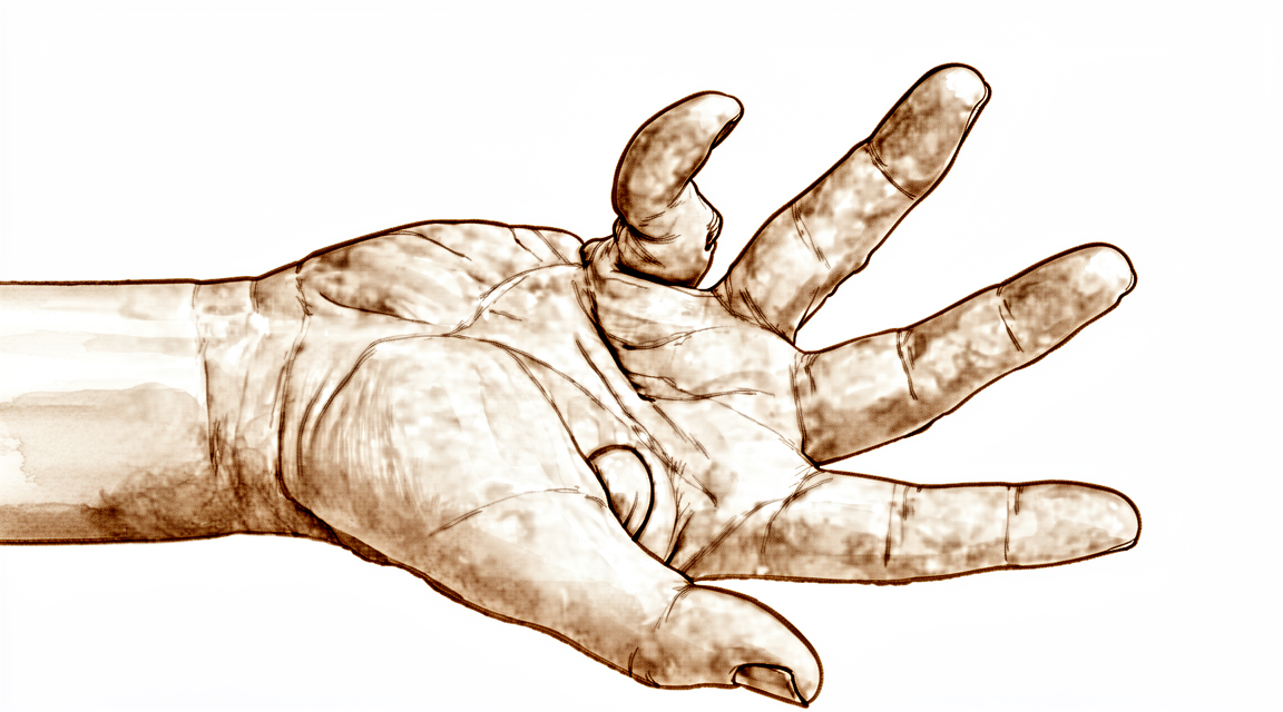
आपको क्या महसूस होता है
ड्यूपुइट्रेन (Dupuytren's) की बीमारी आपकी हथेली में एक छोटी गांठ (नोड्यूल) के रूप में शुरू होती है। शुरू में इसमें आमतौर पर दर्द नहीं होता, या यह छूने पर बस थोड़ी-सी दुखती है। समय के साथ, हथेली की त्वचा के नीचे एक सख्त डोरी बन सकती है, जो एक या ज़्यादा उंगलियों की ओर जाती है। यह डोरी धीरे-धीरे कसती है और प्रभावित उंगली को हथेली की ओर नीचे खींचती है, जिससे उंगली पूरी तरह सीधी नहीं हो पाती। अनामिका और छोटी उंगली सबसे ज़्यादा प्रभावित होती हैं।
यह समस्या बढ़ने वाली (प्रोग्रेसिव) है, यानी यह समय के साथ बदलती रहती है, पर इसकी गति हर किसी में अलग होती है। कुछ लोगों को सालों तक बहुत कम बदलाव दिखता है। दूसरों में उंगली का खिंचना महीनों में बढ़ जाता है। आपको यह सबसे पहले सुबह उठने पर दिख सकता है, जब उंगली जकड़ी हुई लगती है और धीरे-धीरे सीधी होती है। शुरुआत में हाथ को झटककर ढीला करने से मदद मिल सकती है। जैसे-जैसे डोरी कसती है, रोज़ के वे काम कठिन हो जाते हैं जिनमें सपाट हथेली या चौड़ी पकड़ की ज़रूरत होती है: अपना हाथ मेज़ पर सपाट रखना, हाथ मिलाना, स्टीयरिंग व्हील पकड़ना, हथौड़ा या टेनिस रैकेट पकड़ना, या अपना हाथ जेब या दस्ताने में डालना।
मुख्य समस्या आमतौर पर दर्द नहीं होती। समस्या है हाथ को पूरी तरह खोल पाने की क्षमता खो देना, और किसी उंगली को अंदर की ओर मुड़ते देखने की झुंझलाहट। हाथ का काम-काज वापस पाना ही मुख्य कारण है जिसके लिए लोग इलाज करवाते हैं।
कुछ चेतावनी संकेतों पर इंतज़ार करने के बजाय जल्दी कदम उठाने की ज़रूरत होती है। अगर आपका हाथ गर्म, लाल, सूजा हुआ और दर्दभरा हो जाए, खासकर बुखार के साथ, या अगर आपकी उंगलियां फीकी (सफ़ेद पड़ी हुई), ठंडी, सफ़ेद या नीली पड़ जाएं, या आपके हाथ में अचानक महसूस करने या हिलाने-डुलाने की क्षमता चली जाए, तो उसी दिन इमरजेंसी विभाग जाएं। अगर लक्षण ठीक नहीं हो रहे, सप्ताहों में बढ़ रहे हैं, रात को आपको जगा देते हैं, या आपको काम करने या अपना हाथ इस्तेमाल करने से रोकते हैं, तो अपने जीपी (आपके फैमिली डॉक्टर) को दिखाएं या विशेषज्ञ से जांच के लिए कहें।
असल में क्या हो रहा है
आपकी हथेली की त्वचा के नीचे सख्त, लचीले ऊतक की एक परत होती है जिसे पामर फ़ेशिया (palmar fascia) कहते हैं। इसे रेशों के एक नरम जाल की तरह समझें, जो त्वचा को हाथ की गहरी संरचनाओं से जोड़कर टिकाए रखता है और आपकी हथेली को पकड़ने और थामने में मदद करता है। ड्यूपुइट्रेन की बीमारी में यह जाल मोटा होने और कसने लगता है। इस ऊतक की कोशिकाएं बहुत ज़्यादा कोलेजन बनाने लगती हैं, वह रेशा जो सामान्य रूप से फ़ेशिया को उसकी मज़बूती देता है, और ये रेशे फैले रहने के बजाय खिंचने की दिशा में कतार में लग जाते हैं।
जैसे-जैसे मोटा हुआ ऊतक पकता है, वह छोटा होकर एक डोरी बन जाता है। यही वह डोरी है जिसे आप अपनी हथेली की त्वचा के नीचे महसूस कर सकते हैं। यह एक ऐसी रस्सी की तरह व्यवहार करती है जिसे चरखी से धीरे-धीरे अंदर खींचा जा रहा हो। क्योंकि डोरी उंगली में जाकर जुड़ती है, यह उंगली को हथेली की ओर नीचे खींचती है, पहले उंगली की जड़ वाले जोड़ (पोर) पर और फिर उंगली के बीच वाले जोड़ पर। उंगली खुद और उसके टेंडन (कंडरा) स्वस्थ होते हैं। समस्या छोटी हो चुकी फ़ेशिया है, जो उंगली को मुड़ा हुआ रखती है।
डोरी हाथ के हर हिस्से पर एक जैसा असर नहीं डालती। अंगूठे में इस फ़ेशिया की बीच वाली पट्टी नहीं होती, इसीलिए अंगूठा शायद ही कभी प्रभावित होता है। अनामिका और छोटी उंगली उस हिस्से में होती हैं जहां यह बीमारी सबसे ज़्यादा सक्रिय होती है, इसीलिए वे सबसे ज़्यादा प्रभावित होती हैं।
इस समस्या के बारे में कई बातें जानने लायक हैं। इसका ठीक-ठीक कारण अच्छी तरह समझा नहीं गया है, और इसमें कोशिकाओं के स्तर पर जटिल प्रक्रियाएं शामिल हैं। हाथ की चोट या सर्जरी, जिसमें बार-बार पड़ने वाला यांत्रिक दबाव भी शामिल है, इस समस्या के सामने आने से जुड़ी है। सर्जरी उस डोरी को निकाल देती है जो पहले ही बन चुकी है, पर यह बीमारी की अंदरूनी प्रक्रिया को नहीं हटाती, इसलिए समय के साथ डोरी वापस आ सकती है। इसीलिए इलाज अक्सर एक बार में करने के बजाय चरणों में किया जाता है।
डोरी को काटकर ढीला (रिलीज़) कर दिए जाने के बाद भी, कुछ समय तक बदलाव जारी रहते हैं। कॉन्ट्रैक्चर (उंगली का मुड़कर अकड़ जाना) का जो हिस्सा हथेली के ज़्यादा पास है, वह छह महीने तक के समय में शांत हो जाता है। उंगली के ज़्यादा पास वाला हिस्सा शायद कभी पूरी तरह ख़त्म न हो, और अगर वह बचे हुए बीमार ऊतक को खींचता रहे तो फिर से बढ़ सकता है।
हम इसके लिए क्या कर सकते हैं
डॉ. कीरन हिरपरा, माटर प्राइवेट हॉस्पिटल, रॉकहैम्पटन में ऊपरी अंग (कंधे से हाथ तक) के सर्जन हैं, और वे सबसे कम चीर-फाड़ वाले उन विकल्पों से शुरुआत करते हैं जो आपकी समस्या के लिए उपयुक्त हों। मरीज़ आमतौर पर अपने जीपी (आपके फैमिली डॉक्टर) के रेफ़रल से हमारे क्लिनिक आते हैं; अगर किसी फिजियोथेरेपिस्ट ने आपको हमसे मिलने की सलाह दी है, तब भी मेडिकेयर रिबेट पाने के लिए आपको अपने जीपी से रेफ़रल लेना होगा। आपके अपॉइंटमेंट पर हम आपकी पूरी जानकारी लेते हैं, आपके हाथ की जांच करते हैं, और इमेजिंग तभी करवाते हैं जब उससे इलाज की योजना में कोई बदलाव आए। ड्यूपुइट्रेन की बीमारी का निदान आमतौर पर हाथ की जांच से ही हो जाता है।
क्योंकि डोरी धीरे-धीरे कसती है, इलाज अक्सर एक बार में करने के बजाय चरणों में किया जाता है। शुरुआती चरणों में, जब गांठ नरम होती है और उंगली अभी भी सीधी हो जाती है, हम शायद बस इस पर नज़र रखें और देखें कि महीनों में यह कैसा व्यवहार करती है। जब तक बीमारी शांत है, हैंड थेरेपी आपके हाथ को चलता और अच्छी तरह काम करता रखने में मदद कर सकती है। अगर कोई उंगली मुड़ने लगी है, तो उसे स्ट्रेच करते रहना और हिलाते-डुलाते रहना मायने रखता है, क्योंकि विकृति को जितना लंबे समय तक बढ़ने दिया जाए, कॉन्ट्रैक्चर के स्थायी होने की संभावना उतनी ही ज़्यादा होती है।
जब कोई उंगली सीधी नहीं होती, तो ऐसी प्रक्रियाएं हैं जो डोरी को काटकर ढीला (रिलीज़) करती हैं या उसे निकाल देती हैं। सबसे कम चीर-फाड़ वाली प्रक्रिया नीडल रिलीज़ है, जिसमें एक बारीक सुई से त्वचा के आर-पार डोरी को काटा जाता है। इससे लंबे समय तक रहने वाला सुधार मिल सकता है, उन हाथों में भी जिनमें बीमारी ज़्यादा बढ़ चुकी हो। कुछ डोरियों का इलाज एक छोटे चीरे से किए जाने वाले छोटे ओपन रिलीज़ (चीरा लगाकर डोरी काटना) से भी किया जा सकता है। जब बीमारी ज़्यादा जम चुकी हो, तो सर्जरी से डोरी और प्रभावित फ़ेशिया, यानी हथेली की त्वचा के नीचे के ऊतक की परत, निकाल दी जाती है। बढ़ी हुई बीमारी में, प्रभावित त्वचा को निकालकर उसकी जगह स्किन ग्राफ्ट (शरीर के दूसरे हिस्से से ली गई त्वचा) लगाने से ऊतक के दोबारा बनने की संभावना कम हो जाती है। हम आपसे बात करेंगे कि आपके हाथ के लिए कौन सा विकल्प सही है, और आप और हम मिलकर फ़ैसला करेंगे।
समय मायने रखता है। सर्जरी पर आमतौर पर तब विचार किया जाता है जब उंगली की जड़ वाला जोड़ (पोर) 30 डिग्री या उससे ज़्यादा मुड़ा हो, या जब उंगली के बीच वाले जोड़ में ज़रा सा भी मोड़ हो। बीच वाले जोड़ का इलाज जड़ वाले जोड़ से पहले किया जाता है, क्योंकि उसके मुड़ी हुई स्थिति में स्थायी रूप से जकड़ जाने की संभावना खास तौर पर ज़्यादा होती है। उंगली को सीधा करने से हाथ का काम-काज सुधरता है, और इसमें बीच वाले जोड़ का योगदान जड़ वाले जोड़ से ज़्यादा होता है।
इनमें से किसी भी प्रक्रिया के बाद, आपके हाथ को अच्छी तरह काम करता बनाने के लिए हैंड थेरेपी बहुत ज़रूरी है। हाथ की सर्जरी के बाद रिहैबिलिटेशन (पुनर्वास) एक्सटेंड रिहैबिलिटेशन (Extend Rehabilitation) में रूबी डूलन के साथ होता है। रूबी एक हैंड थेरेपिस्ट (हाथ की विशेषज्ञ थेरेपिस्ट) हैं: वे आपकी थेरेपी तय करती हैं और जिस भी स्प्लिंट की ज़रूरत हो, उसे बनाती हैं।
क्या उम्मीद करें
ड्यूपुइट्रेन की बीमारी अपनी ही गति से बढ़ती है। कुछ लोगों को सालों तक बहुत कम बदलाव दिखता है, जबकि दूसरों में उंगली का खिंचना महीनों में बढ़ जाता है। यह अपने आप ठीक नहीं होती, और एक बार डोरी बन जाने के बाद भी यह प्रक्रिया रुकती नहीं। कॉन्ट्रैक्चर का जो हिस्सा हथेली के ज़्यादा पास है, वह छह महीने तक के समय में शांत हो जाता है, पर उंगली के ज़्यादा पास वाला हिस्सा शायद कभी पूरी तरह ख़त्म न हो और फिर से बढ़ सकता है।
उंगली को सीधा करने से आपके हाथ के काम करने में सचमुच फ़र्क पड़ता है। हाथ का काम-काज वापस पाना ही मुख्य कारण है जिसके लिए लोग इलाज करवाते हैं, और विकृति को ठीक करने से आपका हाथ जो कुछ कर सकता है, उसमें काफ़ी सुधार आता है। इसमें उंगली के बीच वाले जोड़ का योगदान जड़ वाले जोड़ (पोर) से ज़्यादा होता है, इसीलिए उसका इलाज पहले किया जाता है।
दोबारा होना (रिकरेंस) इस बीमारी के स्वाभाविक क्रम का हिस्सा है, चाहे आप कोई भी इलाज चुनें। कोई भी मौजूदा इलाज अंदरूनी प्रक्रिया को नहीं रोकता; सभी इलाज केवल उस डोरी को ठीक करते हैं जो यह प्रक्रिया पहले ही बना चुकी है। नीडल रिलीज़ के बाद, जिन लोगों में बीमारी दोबारा होती है, उनमें से ज़्यादातर में यह लगभग 2.3 साल के भीतर होती है। डोरी निकालने की सर्जरी के बाद लगभग एक-चौथाई लोगों में बीमारी दोबारा होती है, औसतन 3.7 साल बाद। जहां प्रभावित त्वचा निकालकर उसकी जगह ग्राफ्ट लगाया जाता है, वहां उस हिस्से में बीमारी के दोबारा होने की संभावना बहुत कम होती है। अगर बीमारी वापस आ जाए, तो उसका फिर से इलाज संभव है, हालांकि दोबारा सर्जरी करना ज़्यादा कठिन होता है और उसमें पहले ऑपरेशन की तुलना में ज़्यादा जोखिम होता है।
इलाज के बाद की ज़्यादातर जटिलताएं (कॉम्प्लिकेशन) जल्दी देखभाल से ठीक की जा सकती हैं। हाथ के सर्जन जिन दो जटिलताओं पर सबसे ज़्यादा बात करते हैं, वे हैं बीमारी का दोबारा होना और जकड़न। जकड़न, खासकर उंगली के बीच वाले जोड़ की, ही वह कारण है जिससे जल्दी इलाज मायने रखता है: जो जोड़ बहुत लंबे समय तक मुड़ा रहे, वह उसी स्थिति में स्थायी रूप से जकड़ सकता है। अगर आपका हाथ गर्म, लाल, सूजा हुआ और दर्दभरा हो जाए, खासकर बुखार के साथ, तो उसी दिन इमरजेंसी विभाग जाएं। अगर लक्षण ठीक नहीं हो रहे या सप्ताहों में बढ़ रहे हैं, तो अपने जीपी को दिखाएं या विशेषज्ञ से जांच के लिए कहें।
किसी भी प्रक्रिया के बाद, आपके हाथ को अच्छी तरह काम करता बनाने के लिए हैंड थेरेपी बहुत ज़रूरी है। हाथ की सर्जरी के बाद रिहैबिलिटेशन (पुनर्वास) एक्सटेंड रिहैबिलिटेशन (Extend Rehabilitation) में रूबी डूलन के साथ होता है। रूबी एक हैंड थेरेपिस्ट (हाथ की विशेषज्ञ थेरेपिस्ट) हैं: वे आपकी थेरेपी तय करती हैं और जिस भी स्प्लिंट की ज़रूरत हो, उसे बनाती हैं।
डॉक्टर को कब दिखाएं
ड्यूपुइट्रेन की बीमारी वाले ज़्यादातर लोग दर्द की वजह से नहीं, बल्कि इसलिए इलाज करवाते हैं क्योंकि हाथ का काम-काज धीरे-धीरे कम होता जा रहा होता है। विशेषज्ञ से जांच के लिए कहें अगर आपकी हथेली में कोई गांठ या डोरी सप्ताहों में बढ़ रही है, अगर कोई उंगली पूरी तरह सीधी नहीं होती, या अगर ये बदलाव आपको काम करने या अपना हाथ इस्तेमाल करने से रोकते हैं। यह समस्या बढ़ने वाली है, पर इसकी गति हर किसी में अलग होती है, इसलिए यह देखने का इंतज़ार करने के बजाय कि यह कितनी बढ़ती है, अपने हाथ की जांच करवाना फ़ायदेमंद है। बच्चों की हथेली में भी गांठ या मुड़ी हुई उंगली हो सकती है, और उसकी भी जांच ज़रूरी है। अगर आपका ट्रिगर फिंगर (उंगली का अटकना) का इलाज हुआ है, तो अपनी हथेली पर नज़र रखें, क्योंकि उसके बाद ड्यूपुइट्रेन की नई बीमारी सामने आ सकती है। अगर आपका हाथ गर्म, लाल, सूजा हुआ और दर्दभरा हो जाए, खासकर बुखार के साथ, या अगर आपकी उंगलियां फीकी (सफ़ेद पड़ी हुई), ठंडी, सफ़ेद या नीली पड़ जाएं, तो उसी दिन इमरजेंसी विभाग जाएं।
विस्तार से
Advanced reading: the deeper science (optional)
यह हिस्सा उससे आगे जाता है जितना आपको अपने इलाज के फ़ैसलों के लिए जानना ज़रूरी है। ड्यूपुइट्रेन के बारे में यह अतिरिक्त जानकारी पढ़ने लायक है, क्योंकि इलाजों के बीच चुनाव असल में इस बात का चुनाव नहीं है कि कौन सा काम करता है, बल्कि इस बात का है कि आप दोबारा होने और जोखिम के किस पैटर्न के साथ जीना पसंद करेंगे।
प्रमाण उतने मज़बूत नहीं हैं जितना इसके बारे में भरोसा दिखाया जाता है
शुरुआत यहीं से करना ठीक है। बोन एंड जॉइंट जर्नल की एक समीक्षा ने निष्कर्ष निकाला कि ड्यूपुइट्रेन कॉन्ट्रैक्चर के इलाज के मार्गदर्शन के लिए अब भी सीमित प्रमाण हैं, और साथ ही यह भी बताया कि परक्यूटेनियस नीडल एपोन्यूरोटॉमी (त्वचा के आर-पार सुई से डोरी काटना) से आमतौर पर मरीज़ ज़्यादा संतुष्ट होते हैं और प्रतिकूल घटनाएं कम होती हैं [1]।
इससे आपको दी गई किसी भी ज़ोरदार सलाह को थोड़ा संभलकर लेना चाहिए। यह बीमारी आम है, इलाज लंबे समय से चले आ रहे हैं, और जो तुलनात्मक ट्रायल इस सवाल को सुलझा सकते थे, वे ज़्यादातर किए ही नहीं गए हैं।
कोलेजिनेज़ असल में क्या देता है
कोलेजिनेज़ इंजेक्शन, जो डोरी को काटने के बजाय घोल देता है, ने इलाज के तरीके को जल्दी बदल दिया। 2,675 मरीज़ों की एक समीक्षा में यह हाथ का काम-काज सुधारने के लिए सुरक्षित और असरदार पाया गया, और ज़्यादातर प्रतिकूल घटनाएं मामूली थीं और अपने आप ठीक हो गईं [2]।
उसी समीक्षा में वह संख्या है जो उम्मीदें तय करने के लिए मायने रखती है: सफलतापूर्वक इलाज किए गए 23% जोड़ों में बीमारी दोबारा हो गई [2]। यहां दोबारा होना इलाज की असफलता नहीं है; यह बीमारी का स्वाभाविक क्रम है जो फिर से सामने आ जाता है। ड्यूपुइट्रेन हथेली में होने वाली एक जैविक प्रक्रिया है, और कोई भी मौजूदा इलाज इस प्रक्रिया को नहीं रोकता; सभी इलाज केवल उस डोरी को ठीक करते हैं जो यह प्रक्रिया पहले ही बना चुकी है।
जोखिमों की ईमानदार तुलना
सर्जरी के मुकाबले तुलना यह नहीं है कि एक सुरक्षित है और दूसरा नहीं, बल्कि यह है कि दोनों अलग तरह से असफल होते हैं। 8,809 मरीज़ों में कोलेजिनेज़ की फेशिएक्टॉमी (कसी हुई डोरी निकालने का ऑपरेशन) से तुलना करने पर, कोलेजिनेज़ के साथ दिखीं और फेशिएक्टॉमी के बाद नहीं बताई गईं प्रतिकूल घटनाओं में हाथ-पैर में सूजन (पेरिफेरल एडिमा), हाथ या पैर में दर्द और इंजेक्शन वाली जगह पर प्रतिक्रिया शामिल थीं [3]।
इसे ध्यान से पढ़ें: ये अतिरिक्त समस्याएं हैं, और ये ज़्यादातर अस्थायी और एक ही जगह तक सीमित होती हैं। इंजेक्शन से हथेली में सर्जरी का घाव नहीं होता, जिसके साथ लंबी रिकवरी, जकड़न का जोखिम और निशान आते हैं। बदले में सर्जरी बीमार ऊतक को ज़्यादा पूरी तरह निकालती है और आमतौर पर बीमारी दोबारा होने में ज़्यादा समय लगता है।
इसलिए यह फ़ैसला सच में निजी है। जो मरीज़ कई सप्ताह तक हाथ के सीमित इस्तेमाल का समय नहीं निकाल सकता, उसके पास इंजेक्शन चुनने का मज़बूत कारण है, भले ही उसे पता हो कि दोबारा होने की संभावना ज़्यादा है। तेज़ी से बढ़ने वाली बीमारी वाला कम उम्र का मरीज़ बार-बार छोटे इलाज के बजाय एक बार बड़ा ऑपरेशन करवाना उचित रूप से स्वीकार कर सकता है।
प्रमाण तय होने से पहले ही इलाज का तरीका बदल गया
कोलेजिनेज़ के आने के समय से लेकर उसके बाद के सालों में, अमेरिका में इसका इस्तेमाल काफ़ी बढ़ गया, और उसी हिसाब से फेशियोटॉमी (डोरी को काटकर अलग करना) और फेशिएक्टॉमी करवाने वाले मरीज़ों का अनुपात घट गया, और कौन सा इलाज चुना गया, इस पर मरीज़ की उम्र और दूसरी बीमारियों का असर पड़ा [4]।
जब कोई डॉक्टर आपको बताए कि "आमतौर पर क्या किया जाता है", तब यह एक उपयोगी संदर्भ है। आमतौर पर जो किया जाता है, वह एक दशक के भीतर काफ़ी बदल गया है, और उन प्रमाणों के आधार पर जिन्हें ऊपर की समीक्षाएं सीमित बताती हैं। यह इस बात का कारण है कि आप पूछें कि आपके लिए कोई खास विकल्प क्यों सुझाया जा रहा है, बजाय यह मान लेने के कि मौजूदा तरीका पक्के विज्ञान को दर्शाता है।
संदर्भ
[1] Soreide E, Murad MH, Denbeigh JM, Lewallen EA, Dudakovic A, Nordsletten L, et al. Treatment of Dupuytren's contracture: a systematic review. Bone Joint J. 2018;100-B(9):1138-45. https://doi.org/10.1302/0301-620X.100B9.BJJ-2017-1194.R2
[2] Sandler AB, Scanaliato JP, Dennis T, Gonzalez Trevizo GA, Raiciulescu S, Nesti L, et al. Treatment of Dupuytren's contracture with collagenase: a systematic review. Hand (N Y). 2021;17(5):815-24. https://doi.org/10.1177/1558944720974119
[3] Peimer CA, Wilbrand S, Gerber RA, Chapman D, Szczypa PP. Safety and tolerability of collagenase Clostridium histolyticum and fasciectomy for Dupuytren's contracture. J Hand Surg Eur Vol. 2014;40(2):141-9. https://doi.org/10.1177/1753193414528843
[4] Lipman MD, Carstensen SE, Deal DN. Trends in the treatment of Dupuytren disease in the United States between 2007 and 2014. Hand (N Y). 2016;12(1):13-20. https://doi.org/10.1177/1558944716647101
Evidence & references
This is the clinical evidence summary written for health professionals. It is technical, and it lists the research this page was built from. You do not need to read it to understand your treatment or to make a decision about it.
Overview
Epidemiology and Prognosis
- Clinically important Dupuytren's disease is common in the general population, with a majority of diagnosed individuals undergoing treatment [8].
- Patients with Dupuytren disease have significantly higher mortality rates 20 years after diagnosis than the population without Dupuytren disease [3].
- Dupuytren disease is progressive, but the pace is unique to each patient [12].
- Dupuytren's contracture of the little finger has many different forms with different prognoses and needing different operations [2].
- Dupuytren's disease in women presents similarly to men with more severe PIP joint involvement but equivalent surgical outcomes regarding final contracture correction, recurrence, and complication rates [17].
Assessment and Staging
- An objective method of evaluating the lesions in Dupuytren’s disease allows accurate preoperative assessment and indicates the amount of improvement achieved by the operation [19].
- The hand is divided into five segments for assessment, each consisting of a finger and corresponding palmar zone [19].
- For each finger, the theoretical range of deformity is from 0 degrees (complete extension) to 200 degrees (contracture of the finger in the palm) [19].
- Six stages can be distinguished for the four fingers, with Stage 0 representing no lesion and Stage 4 representing total flexion deformity exceeding 135° [19].
- The presence of a PIP joint contracture of greater than 70 degrees has severe prognostic importance and is indicated by a ‘+’ after the digital letter (D+) in the assessment coding [19].
- Flexion contracture at the MP joints are more easily corrected than at the PIP [19].
- The thumb’s range of deformity is theoretically from 0 degrees to 160 degrees for the MP and IP joints, and from 0 degrees to more than 45 degrees at the level of the web [19].
Treatment Landscape and Evidence
- Currently there remains limited evidence to guide the management of patients with Dupuytren's contracture [13].
- The literature does not provide evidence in favor of a specific procedure for Dupuytren's disease due to inconsistencies in reporting complications as well as the lack of a standardized definition [5].
- Little agreement exists on treatment recommendations for common presentations of Dupuytren disease among international hand surgeons [9].
- Surgery remains the gold-standard treatment for progressive Dupuytren contractures, with limited palmar fasciectomy being the most common option [81].
- The best available published evidence indicates that surgical treatment in the form of partial or selective fasciectomy remains the most reliable and the most widely used method for treating Dupuytren's disease [47].
- There is low level of evidence that both surgical and nonsurgical treatments provide clinically important improvements for recurrent Dupuytren contracture [44].
- There are several procedural options for the treatment of Dupuytren disease [33].
- Many treatment options exist for Dupuytren contracture, each with its own complication profile [50].
- Dermofasciectomy appears to be a highly effective surgical intervention for advanced Dupuytren disease, offering substantial long-term benefits in terms of function and disease control [27].
- The recurrence rate in a series involving skin replacement for Dupuytren’s disease was 8%, a figure reported as better for disease control than standard approaches [89].
Surgical Considerations
- Patients with Dupuytren's disease of the hand may gain a significant functional benefit following surgical improvement or correction of the deformity [7].
- Primary surgery for Dupuytren's contracture can be performed safely on a day care basis provided that strict criteria are followed [14].
- Advanced Dupuytren's contracture can be treated by day care surgery, but the operations should be performed by surgeons who are skilled in hand surgery [28].
- Surgical treatment of Dupuytren's contracture was well-suited to day surgery, provided an individual judgement concerning suitability is made [62].
- Postoperative stiffness is significantly increased as related to recurrences and staging of the disease, but not to sex [20].
- The indications for arthrodesis in severe recurrent Dupuytren's contracture should be stringent, as only 2.8% of patients operated upon for Dupuytren's contracture required arthrodesis [57].
Non-Operative and Minimally Invasive Options
- Percutaneous needle fasciotomy (PNF) is a minimally invasive intervention with a brief recovery period [195].
- In the only randomized trial on PNF, the extension deficit had improved 6 weeks after the intervention with an average of 63% [195].
- In the short term, there was no statistically significant difference between the results of needle fasciotomy and limited fasciectomy if the contracture was less than 90° preoperatively [195].
- For more severe contractures, limited fasciectomy gave better results than needle fasciotomy [195].
- In experienced hands, the cumulative risk of complications is lower in needle fasciotomy than in limited fasciectomy [195].
- Long-term follow-up of a comparative study showed that the recurrence rate after needle fasciotomy is much higher than after limited fasciectomy, with a rate of 65% after 32 months [195].
- Three-year follow-up data of a randomized comparative study show a recurrence rate of 64% in the PNF group [195].
- Limited fasciectomy is indicated for contractures greater than 30° or painful nodules, with a reported 79% reduction of contracture and a recurrence rate of 41% in 5 years [200].
- Dermofasciectomy is indicated for recurrences or aggressive type in patients younger than 40, with a recurrence rate of 8.4% after 6 years [200].
- Clostridial collagenase is indicated for contracture associated with a palpable cord, with an 87% success rate (correction within 0–5° of full extension) and a recurrence rate of 19% after 2 years [200].
- Radiotherapy is indicated for disease in the initial phase, with mixed results regarding the delay of development of contractures in stage N and Tubiana I.2 [200].
Postoperative Management and Patient Perspective
- Hand therapy is crucial to the effective postoperative management of Dupuytren's contracture and the achievement of optimum hand function [32].
- Most complications following surgical treatment for Dupuytren's contracture can be well controlled and will resolve with appropriate early treatment [1].
- Patients with Dupuytren’s disease want a cure, but as there is no cure, the next best option is a treatment that improves the personal disease situation, such as getting fingers functional again, reducing pain, or reducing/stopping disease progression [46].
- The typical advice given to patients is to wait until they have a real problem and then have surgery, which is considered too simple by patient advocates who seek clear advice on the best treatment for their specific situation [46].
Anatomy & Pathophysiology
Normal Palmar Anatomy and Biomechanics
- The soft tissue of the fingers is mobile and flexible, overlaying fibrous tendon sheaths from the distal phalanges to the metacarpophalangeal joints [66].
- In the normal finger, soft tissue structures pressed together in flexion remain separated by cutaneous flexion creases, which are not stable formations or firm borders to subcutaneous processes [66].
- The palmar aponeurosis is only one part of a functional system of stiff subcutaneous connective tissue that facilitates different mechanisms of grip [105].
- The connective tissue framework between the dermis and musculature is a functional unit that varies with location, including the dorsum of the hand, cranial skin, palm, and fingers [105].
- The palmar fascia is elastic but only half as elastic as palmar skin, absorbing the majority of shear forces applied to the palmar skin via vertical septal fibers [165].
- The plantar fascia has a fixed bony origin and retains a unidirectional orientation, whereas the palmar fascia has a tendinous origin that stretches it to a greater degree and folds in flexion [165].
- Palmar cutaneous nerve branches from the proper digital nerves originate at approximately 19% of the distance from the A1 pulley base to the tip of the finger, or approximately one-fifth of the finger length [123].
- These palmar cutaneous nerve branches converge to form a "chevron shape" and serve a dense concentration of Pacinian corpuscles [122].
- The neurovascular bundles enter the subcutaneous tissue as they leave the space beneath the palmar aponeurosis, with Cleland fibers lying immediately deep to the nerves and arteries [124].
- Grayson fibers cover the arteries and nerves laterally in a crisscrossed configuration, coursing across the tendons to the skin of the contralateral side [124].
- In the flexed finger, digital neurovascular structures are folded into loops above the Cleland fibers, a position observable in many Dupuytren fingers [124].
Disease Pathogenesis and Cellular Biology
- Dupuytren's disease is a fibroproliferative disorder resulting in the formation of nodules and cords in the palmar fascia of the hand [23].
- The disease typically presents over three phases: a proliferative phase with nodular tissue, an involutional phase with myofibroblast alignment along stress lines, and a third stage with deposition of an acellular collagen-rich cord [48].
- Myofibroblasts are the important pathologic cells in the development of Dupuytren's disease [74].
- Myofibroblasts differ from fibroblasts by having actual bundles of contractile actin microfilaments arranged parallel to the long axis of the cell [125].
- Adjacent myofibroblasts connect via extracellular fibrils of fibronectin and act together to generate the contracted tissue seen in Dupuytren disease [125].
- Type III collagen is more prevalent in the extracellular matrix in Dupuytren disease [125].
- Transforming growth factor-β1, TGF-β2, epidermal growth factor, platelet-derived growth factor, and connective tissue growth factor have been suggested to play a role in initiating abnormal cellular proliferation [125].
- Increasing levels of mechanical tension influence fibroblast differentiation into myofibroblasts [125].
- The hallmark feature of Dupuytren's contracture is the localized deposition of excess and abnormal collagen within the palmar fascia, suggesting a perturbed balance of collagen remodeling [115].
- Genomic and proteomic studies indicate a shift in the balance of collagen remodeling in favor of net collagen deposition in Dupuytren's disease tissue [115].
- Dupuytren's disease may be regarded as a manifestation of aberrant wound healing responses, with tissue responses more exaggerated and/or prolonged than those seen in normal healing wounds [174].
- Fibroblasts monitor the degree of static and dynamic force through cell surface β1 integrins and Type I collagen fibrils, transmitting tension to the intracellular cytoskeleton to alter gene expression [174].
- Mechanical tension induces proliferation, transition to myofibroblasts, secretion of TGF-β, generation of contractile force, and increased thickness and stiffness of collagen fibrils in cultured fibroblasts [174].
- Decreased mechanical tension on myofibroblasts results in their disappearance via apoptosis, seen in the resolution phase of wound healing and late stages of Dupuytren's disease [174].
- In fibromatosis, a disorganized hypercellular fibroblastic nodule infiltrates the fibrofatty tissue over the palmar aponeurosis and fibrous tendon sheaths [76].
- Fibromatosis becomes established in the soft tissue of the fingers in its predominant compressed and shortened formation, creating an extension block rather than an active contraction [76].
- Nodular tissue in the stationary compartment of the mid-palm retains its nodular shape over the long term, while tissue in the mobile soft tissue of the fingers is subject to stress adaptation that transforms it into cord-like tissue [76].
- The specific local biomechanical conditions decisively influence the morphogenesis of the fibromatotic tissue mix and the clinical picture of the disorder [76].
- Neurovascular dystopia can be explained as neurovascular loops trapped over hypertrophic Cleland fibers [76].
- The expression of three key collagenases (MMP1, MMP13, and MMP14) is significantly raised in Dupuytren's disease nodules, as is the expression of the collagen biosynthetic enzyme ADAMTS14 [48].
- TIMP1 expression is significantly elevated in Dupuytren's disease nodules compared to normal palmar fascia [48].
- Contraction and fibrosis in Dupuytren's disease may result from increased collagen biosynthesis mediated by ADAMTS-14, elevated TIMP-1 blocking MMP-mediated collagenolysis, and contraction enabled by MMP-14-mediated pericellular collagenolysis [48].
- The expression level of MMP13 and MMP14 positively correlates with poor progression post-fasciectomy [48].
- Structural abnormalities in Dupuytren's cords include increased cross-linking, increased or altered collagen glycation, larger fibril diameter, and more closely packed fascicles with thicker sheaths [115].
- These structural changes result in increased stiffness (decreased elasticity) in the collagen fibrils relative to normal tissue [115].
- The end result of Dupuytren's biology is ischemic fibrosis and microvascular occlusion from endothelial fibroblast infiltration [157].
- Ischemia results in fibroblast proliferation, collagen deposition, and the appearance of myofibroblasts [157].
- Cells from Dupuytren's tissue respond to mechanical stretching forces more than cells from normal fascia [157].
- Mechanical tension increases both fibroblast proliferation and myofibroblast differentiation [157].
- Repetitive mechanical stress promotes collagen synthesis and deposition [157].
- Residual skin tension after fasciotomy increases the likelihood of early disease recurrence [157].
Clinical Presentation and Progression
- Palmar fascial disease may remain confined to the palm and not progress enough to cause digital flexion deformity [75].
- Palmar involvement usually precedes disease extension into the digits, but the disease can begin and remain in the digits [75].
- The ring finger is the most commonly involved digit, followed closely by the small, middle, and index finger, and lastly the thumb [75].
- The disease can be classified into early, intermediate, and late phases [75].
- The early phase entails skin changes including loss of normal architecture and skin pit formation [75].
- The intermediate phase consists of nodule and cord formation [75].
- The late phase is the end point of joint contracture [75].
- In the typical scenario, progressive contracture occurs from the ring to the small and lastly the middle finger, though this progression is not immutable [75].
- Ulmas et al. found a 52% prevalence of spiral nerve intraoperatively and 42% had palpable interdigital soft tissue mass [75].
- The existence of a soft tissue mass was specific (75%) but not sensitive (59%) for the presence of the spiral nerve [75].
- The lesion usually begins on the ulnar side of the hand at the distal palmar crease and progresses to involve the ring and little fingers [168].
- Metacarpophalangeal (MP) and PIP joint flexion contractures gradually develop, with severity depending on the extent and maturity of the fibroplasia [168].
- Lesions are more commonly painless, although some patients may complain of itching or minor discomfort [168].
- Nodules have been shown histologically to have neural entrapment, which may be the reason some individuals present with pain primarily [168].
- Ectopic Dupuytren disease deposits may occur in the medial plantar fascia (Ledderhose disease), penis (Peyronie disease), and over the PIP joints dorsally (knuckle pads) [168].
- Patients with these associated findings are considered to have a Dupuytren diathesis and are prone to a more progressive and recurrent form of the disease [168].
- Dupuytren contracture occurs 6 to 10 times more frequently in men than in women [168].
- The disease occurs significantly earlier in men (33 to 63 years old) than in women (46 to 70 years old) [168].
- The disease is most common in white northern European individuals, although it has been reported occasionally in blacks and rarely in Asians [168].
- The lesion has been reported to be more frequent and severe in individuals with diabetes mellitus or with epilepsy (42%) [168].
- The involvement is often bilateral (45%) but rarely symmetric [168].
- Mortality may be increased in men who develop the disease before age 60 [168].
- Hand trauma and the type of manual labor performed by an individual may be contributing factors to the disease [168].
- The presence of hemosiderin in these lesions suggests hemorrhage from tears, but the nondominant hand is affected as often as the dominant one, making trauma alone an unlikely cause [168].
- A single injury may precipitate the onset of the disease in genetically susceptible individuals [168].
- Surgical trauma, even release of a trigger finger in susceptible patients, may precipitate significant pretendinous cords, palmar thickening, and subsequent troublesome contractures [168].
- Excision of an isolated Dupuytren nodule, especially in younger patients, is rarely warranted as it is more likely than not a more significant lesion will ensue [168].
- A causal relationship between injury and Dupuytren disease may be assumed if consistent histologic changes occur within 2 years after a focal injury in women younger than 50 years and men younger than 40 without a strong diathesis [168].
- In patients with bilateral disease, the disease usually develops in the uninjured hand after age 40 years in men and 50 years in women [168].
- The disease seems to occur earlier and more frequently in some families, with a variable penetrance autosomal dominant pattern [168].
- Vascular insufficiency and cigarette smoking have been linked to Dupuytren disease as possible causative factors [168].
- The prevalence of Dupuytren's disease was significantly lower among Muslim men than among Serbian and Croatian men [31].
- The prevalence of Dupuytren's disease was far higher among those who stated that they suffered from diabetes mellitus [31].
- Changes were equally distributed in the right and left hands in women, while they were found more than three times as often in the right hands as in the left hands of men [31].
- The ring and little finger rays were most often affected, and the thumb and index finger rays least often [31].
- Dupuytren's disease was detected in a total of 775 finger rays, of which 608 were without contracture of the digit (stage 1) [31].
- The prevalence of Dupuytren's disease ranged from 17% for men between 50 and 54 years to 60% in the oldest men [31].
- The prevalence among women was lower than among men [31].
- Dupuytren's disease is a benign, progressive fibrosing disorder of the palmar fascias of the hand and fingers, which leads to the formation of nodules and cords, and may lead to disabling extension deficits of the finger joints [71].
- The reported prevalence of Dupuytren's disease varies from 3% to 42%, with the highest incidence in Caucasians, while it is rarely seen in Africans [71].
- Men are seven times more often affected than women, but in later life the incidence in women increases to the same as men [71].
- Some suggest that the disease symptoms are milder in women and therefore may remain unnoticed for a longer period [71].
- The clustering of Dupuytren's disease in families suggests a genetic influence on the onset of the disease [71].
- Multiple reports describe Dupuytren's disease as an autosomal dominant disease with varying penetrance [71].
- There are many sporadic (nonfamilial) cases of the disease that are not compatible with a Mendelian inheritance pattern [71].
- Recessive inheritance remains a viable hypothesis for Dupuytren's disease [71].
- Dupuytren's disease is considered to be one of the most common hereditary connective tissue disorders in Caucasians [71].
- Factors such as smoking, use of alcohol, antiepilepsy drugs, and medical conditions including liver disease and diabetes are associated with a higher prevalence of the disease [71].
- Dupuytren's Disease is most common among Caucasians living in or originating from North-West Europe [82].
- The prevalence in a country is strongly influenced by the origin of its population and varies from 0.5 to 25% [82].
- Men are most often affected after the age of 50, earlier than women, but by the ninth decade, the gender ratio is nearly 1:1 [82].
- There is a familial predisposition to DD, suggesting a genetic origin [82].
- Work from a consortium of Dupuytren study groups has revealed nine susceptibility loci and a role for the WNT-signaling pathway in the pathogenesis of the disease [82].
- The global prevalence of Dupuytren's contracture among Caucasians is estimated at 3–6% [74].
- Most patients are older than 50 years at presentation [74].
- Among patients with diabetes, the incidence of Dupuytren's disease has been estimated at 10.5%, although other reports suggest the incidence may be much higher [74].
- In patients with thyroid disease, the incidence for Dupuytren's disease has been reported to be 8.8% [74].
- Dupuytren's disease is a disabling fibrotic condition affecting the palmar fascia of the hand, described by Baron Dupuytren in 1831 [48].
- Incidence of DD is age, gender and genetically dependent [48].
- Individuals with a history of alcoholism, smoking and high blood cholesterol are also at a higher risk of developing DD [48].
- The disease is often familial, but the mode of inheritance is currently unknown [48].
- Dupuytren's disease is a fibromatosis of the palmer fascia of the hand and fingers, which often leads to flexion deformity of especially the ulnar fingers [82].
- Patients with digital contracture from DD may develop difficulty using their hands in daily activities [82].
- There is some correlation between DD and smoking, excessive alcohol consumption, male gender, diabetes mellitus, epilepsy, and possibly, performing heavy manual labor [82].
- The cause of DD is not yet fully elucidated [82].
- The exact genetic profile of Dupuytren's Disease is not yet fully unraveled [82].
- Dupuytren's contracture is effected by a multitude of complex processes at the cellular level and the exact relative contribution of each is unknown [67].
- Surgical treatment does not eliminate the disease process and recurrence is frequently noted [67].
- The pathogenesis of Dupuytren's disease remains largely unknown [71].
- The reason for the higher incidence of this disease pattern in the fingers compared with the thumb remains unknown while the pathogenesis of Dupuytren's disease is not fully comprehended [15].
- Dupuytren's disease may have common characteristics with wound healing but it is still fundamentally different [18].
- Regression occurs in Dupuytren's contracture [114].
- An alternative, more mechanical, theory of causation would be that paralysis results in an absence of forces across the Dupuytren’s disease [203].
- The overall extension of the finger was slightly improved by about 10° at each joint following a wasp sting [163].
- The disease affected the insertion of flexor carpi ulnaris at the wrist as well as the palm in a case of Dupuytren's disease affecting the wrist [39].
- The problem of PIP joint contracture is complex and structures on the side and dorsum of the finger are important, as well as those on the palmar aspect [102].
- The spiral nerve occurs most commonly with a proximal interphalangeal joint contracture of 30° and when the small finger is involved [193].
- The current ability to preoperatively identify normal structures and mechanically important cords is crude [64].
- The unpredictable anatomic changes involving neurovascular bundles remain a stumbling block for surgical approaches, particularly for minimally invasive techniques [64].
- An
Classification
Staging Systems and Definitions
- Dupuytren severity is multidimensional, involving biology, anatomy, correction, and recontracture [6].
- Anatomic severity refers to angular contracture severity and determines the likelihood of the finger being straight early after treatment [6].
- Biologic severity refers to disease activity persistence and determines the likelihood of fingers being straight late after treatment [6].
- Historic severity, specifically prior recontracture, is a risk factor for future recontracture [6].
- Initial angular correction depends on pretreatment angular contracture severity rather than biology [6].
- Greater pretreatment angular contracture severity lowers the chance that a corrective procedure will restore full extension [6].
- Failure to achieve complete correction increases the likelihood of some initial correction loss within a year of treatment [6].
- Greater angular contracture correlates with reduced grip strength, which is not improved by contracture release [6].
- Documentation of biologic severity should include family history of Dupuytren's disease in siblings or parents, gender, age of onset, current age, age of first treatment, bilaterality, number of digits involved, thumb involvement, presence of palm nodules, Dupuytren's disease nodes (DDNs), Ledderhose disease, Peyronie disease, and history of frozen shoulder [6].
- An objective method of evaluating lesions in Dupuytren's disease allows accurate preoperative assessment and indicates the amount of improvement achieved by operation [19].
- The Tubiana classification system categorizes total passive extension deficit of each ray into four stages: Stage I (0–45°), Stage II (46–90°), Stage III (91–135°), and Stage IV (136–180°) [178].
- The Tubiana classification system includes a Stage N for nodules, cords, skin retraction, and fixation without flexion deformity [185].
- The Tubiana classification system defines Stage I as an extension deficit of 11–45°, Stage II as 46–90°, Stage III as 91–135°, and Stage IV as >135° [185].
- The Tubiana and Michon classification system scores contracture in a complete finger regardless of the contribution of the metacarpophalangeal (MCP) or proximal interphalangeal (PIP) joint [191].
- The Tubiana and Michon classification system defines Stage 1 as contraction of 0–45°, Stage 2 as 46–90°, Stage 3 as 91–135°, and Stage 4 as over 135° [191].
- The Tubiana classification system does not distinguish between MCP and PIP joint contributions, meaning it cannot deduce whether a finger should be treated conservatively, minimally invasively, or with open surgery [191].
- A revised severity staging system that incorporates total flexion deformity and additional clinical risk factors provides a more objective and precise method for assessing Dupuytren's disease severity and may predict surgical outcomes [37].
- A clinical grading system for Dupuytren's contracture sorts patients into five broad surgical categories, taking into account predisposing factors, risk of recurrence, and sympathetic tone [104].
- Dupuytren staging can be conceptualized in three terms: assessment (measurable aspects), scoring system (quantification via numbers or variables), and classification (subdivisions into non-ordinal types) [41].
- Scoring systems for Dupuytren's disease fall into five proposed categories, including severity according to degree of contracture, detailed scoring of every digit, and systems that score severity or surgical results into arbitrary categories of excellent/good/fair/poor [41].
- Methods of assessment used in the study of Dupuytren's disease include degree of contracture or range of motion, disease type based on localization of pathologic fascia, histology, Dupuytren diathesis, hand function or disability, rate of recovery/time to return to work, recurrence and progression, and complications [41].
- The literature does not provide evidence in favor of a specific procedure for Dupuytren's disease due to inconsistencies in reporting complications and the lack of a standardized definition [5].
- There is no consensus on the most effective operation for primary Dupuytren's contracture [171].
- There is wide disparity in scoring systems, definition of recurrence, and recording of complications in studies of Dupuytren's disease treatment [171].
Histological Classification
- Histological staging is a reliable method for predicting recurrence of Dupuytren's disease, with Type I (proliferative) having the highest risk and Type III (fibrotic) the lowest [113].
- Histological staging was independent of features of Dupuytren’s diathesis [11].
- The clinical course and typical pathological features of Dupuytren's disease are divided into a proliferative phase (increased fibroblasts, nodule formation), an involutional phase (increased myofibroblasts in diseased fiber bundles leading to contracture), and a residual phase (collagenous fibers dominate in connective tissues) [185].
- Luck identified three stages of Dupuytren's disease: the proliferative stage (solitary or multiple soft nodules), the involutional stage (flexion contractures in the palm and fingers), and the residual stage (cord present with joint contractures and joint changes) [98].
- Iselin described five stages of Dupuytren's disease, with Stage 0 characterized by small nodules [98].
Anatomical and Clinical Variants
- Typical Dupuytren’s disease and Non-Dupuytren’s palmar fascial disease are two clinical entities that run different courses and do not share a similar prognosis [4].
- The affected tissues of typical Dupuytren’s disease and Dupuytren’s disease following acute injury show no histological difference [25].
- Dupuytren's disease of the hand is generally considered a disease of adults, but cases have been presented in children who developed the condition before the age of 13 years [10].
- The reason for the higher incidence of Dupuytren's disease in the fingers compared with the thumb remains unknown [15].
- Dupuytren's disease is a progressive condition of genetic origin with a multifactorial pathophysiology involving myofibroblast proliferation and altered collagen matrix composition [29].
- Dupuytren's disease is a deforming, fibrotic condition affecting the palmar fascia with a broad spectrum of involvement from slight thickening to widespread disease associated with severe digital contractures [98].
- Dupuytren's disease is characterized by the development of fibrotic nodules and cords within the palmar fascia and skin, which can lead to finger contractures [170].
- In Dupuytren's disease in isolation, stereotypical cords manifest while demonstrating normal adjacent skin free from the thickening and waxy changes found in limited joint mobility [170].
- The same glycosylation end products, which are more abundant in diabetic patients, are theorized to contribute to cross-linking of collagen and the development of the nodules and cords of Dupuytren’s contracture [170].
- Dupuytren's disease is a common fibroproliferative disorder with a worldwide prevalence of up to 32% [177].
- Dupuytren's disease was detected in 775 finger rays in a population study, with 608 rays without contracture (stage 1) [31].
- In a population study, the ring and little finger rays were most often affected by Dupuytren's disease, while the thumb and index finger rays were least often affected [31].
- Changes in Dupuytren's disease were found more than three times as often in the right hands as in the left hands of men [31].
- Changes in Dupuytren's disease were equally distributed in the right and left hands in women [31].
- There was no significant difference in the prevalence of Dupuytren's disease between smokers and nonsmokers, between those admitting to drinking alcohol and teetotalers, or between those living in urban areas and those living in rural areas [31].
- A meta-analysis showed a prevalence of 31% among diabetic patients, with an odds ratio of 3.9 for type 1 diabetes mellitus and 3.0 for type 2 diabetes mellitus when compared with the nondiabetic population [170].
- A systematic review found a strong association between higher A1c values and the development of Dupuytren’s disease in 27 of the 37 articles evaluated [170].
- A population-based study using International Classification of Diseases codes found that only 1.43% of diabetic patients also carried a diagnosis of Dupuytren’s, for an odds ratio of 1.08 compared with the rest of the study population [170].
- In a population-based study, odds ratios for Dupuytren's disease associated with hemoglobin A1c ranges were 1.19 for 7.0 to 7.9, 1.32 for 8.0 to 8.9, 1.46 for 9.0 to 9.9, and 1.36 for 10.0+ [170].
- All three groups with Dupuytren's disease in a study on alcohol and hepatic pathology had a tendency for the disease to occur towards the ulnar border of the hand, and in particular, the ring finger [158].
- There was no significant difference in the site of the disease between groups with Dupuytren's disease in a study on alcohol and hepatic pathology [158].
- Age-standardized incidence rates of Dupuytren's disease showed no significant differences across individual social classes [161].
- Cumulative incidence rates of Dupuytren's disease were significantly higher in the non-manual than in the manual occupational group beyond 65 years of age [161].
- The age-specific incidence of significant Dupuytren’s disease in men increased with age until 60–69, then declined, with a further increase and second peak at 80–89 [161].
- Consultation rates for Dupuytren's disease were no different across occupational social classes, with the exception of social class 5 (Unskilled), which had a significantly higher rate [161].
- The incidence rate of new cases of Dupuytren’s disease was 34.3 per 100,000 men in a survey population of 502,493 [161].
- Dupuytren's disease is progressive, but the pace is unique to each patient [12].
- Without any therapy, Dupuytren's disease progresses in about 30–50% within 5 years leading to functional deficiencies and requiring surgical correction [185].
- Dupuytren's disease starts usually in the fourth decade and peaks in the fifth to sixth decade with a male to female ratio of 3:1 [185].
- Two-thirds of patients with Dupuytren's disease may develop a bilateral affliction [185].
- A family background is more pronounced among female than male patients with Dupuytren's disease [185].
- The prevalence of Dupuytren's disease was highly age-dependent, ranging from 17% for men between 50 and 54 years to 60% in the oldest men [31].
- The prevalence of Dupuytren's disease among women was lower than among men [31].
- In a population study, the overall prevalence of Dupuytren's disease was 31% in men and 16% in women [31].
- In a population study, the prevalence of Dupuytren's disease at a diabetes clinic was 69% in men and 57% in women [31].
Clinical Presentation
General Characteristics and Epidemiology
- Dupuytren disease is a progressive condition of genetic origin with a multifactorial pathophysiology involving myofibroblast proliferation and altered collagen matrix composition [29].
- The prevalence of Dupuytren's Disease in different geographical locations is extremely variable, and it is not clear whether this is genetic, environmental, or a combination of both [108].
- Dupuytren's disease in patients from the Indian sub-continent, though uncommon, does occur and has essentially the same presentation, distribution and progression as amongst Caucasians [106].
- Dupuytren's disease is a rare entity in the black population, but its predisposing factors and clinical presentation are similar to that seen in white populations [59].
- Dupuytren's disease of the hand is generally considered a disease of adults, but nine children developed the condition before the age of 13 years [10].
- There was no sign of recurrence 27 months after surgery in a case of Dupuytren’s disease that was present at birth [63].
Demographics and Risk Factors
- Men are more likely to have the disease than women, and incidence increases with age [48].
- In a population study of individuals over 50, the prevalence of Dupuytren's disease was 31% in men and 16% in women [31].
- The prevalence of Dupuytren's disease was far higher among those who stated that they suffered from diabetes mellitus compared to non-diabetics [31].
- In patients with diabetes, the incidence of Dupuytren’s disease has been estimated at 10.5%, although other reports suggest the incidence may be much higher [74].
- If a patient has a palmaris longus tendon, then there is a highly significant chance of Dupuytren's disease developing in that hand (p < 0.001) [45].
Anatomical Distribution and Presentation
- The ring and little finger rays were most often affected by Dupuytren's disease, while the thumb and index finger rays were least often affected [31].
- In a surgical series of 153 patients, individual finger involvement was: small finger 46.7%, ring finger 27.3%, long finger 12.0%, index finger 6.4%, and thumb 7.5% [54].
- The disease was bilateral in 76.1% of patients in a surgical series of 153 patients [54].
- Dorsal Dupuytren's nodules are encountered only in Dupuytren's disease patients, especially among those with strong diathesis [58].
- Isolated Dupuytren’s contracture at the wrist is a rare condition that must be included in the differential diagnosis of patients presenting with a mass on the anterior aspect of the wrist, with or without neurological deficits [60].
- The disease affected the insertion of flexor carpi ulnaris at the wrist as well as the palm in a reported case [39].
- A case of severe and long-standing Dupuytren’s contracture presented with an ossified nodule encountered within the diseased tissue intraoperatively [68].
Clinical Progression and Staging
- Typically, DD presents itself over three phases: a proliferative phase showing nodular tissue, an involutional secondary phase characterized by alignment of myofibroblasts along the line of stress, and a third stage represented by the deposition of an acellular collagen-rich cord [48].
- In the early stages of the disease process, nodules form within the palm [74].
- As the condition progresses, collagen cords cause fingers to progressively flex at the metacarpophalangeal (MP) and proximal interphalangeal (PIP) joints, resulting in a fixed-flexion deformity of the fingers and an extension deficit [74].
- Anatomic severity (angular contracture severity) determines how likely the finger is to be straight early after treatment [6].
- Biologic severity (disease activity persistence) determines how likely fingers are to be straight late after treatment [6].
- Prior recontracture is a risk factor for future recontracture [6].
- Greater pretreatment angular contracture severity lowers the chance a corrective procedure will restore full extension [6].
- The revised severity staging system, which incorporates total flexion deformity and additional clinical risk factors, provides a more objective and precise method for assessing Dupuytren's disease severity and may predict surgical outcomes [37].
- A PIP joint contracture of greater than 70 degrees has a severe prognostic importance [19].
- The typical presentation of patients referred for surgery is a male patient, aged 60 or over with a contracture of the MCP and PIP of the small and ring fingers [95].
- Mean flexion contracture at the PIP joint of the ring and small fingers in a surgical cohort were 17.6° and 37.4°, respectively [95].
Associated Conditions and Triggers
- Criteria for recognition of Dupuytren’s contracture after acute injury include objective evidence of injury with no prior disease, injury within the same hand/wrist/forearm, disease appearing within 1 year of injury, and a single nodule or band appearing first in the palm [30].
- Disease following acute injury commonly remains limited to the part of the hand initially involved but may progress within the same hand or to the other hand [30].
- A case of rapid onset of Dupuytren’s disease was presented following acute flexor tenosynovitis [38].
- Patients with Dupuytren's disease may have associated conditions such as Ledderhose disease, Peyronie disease, and frozen shoulder, which are part of the biologic severity assessment [6].
- The physical exam for Peyronie's disease should include evaluation of the hands and feet for indications of systemic fibromatosis, such as Dupuytren’s contracture or Ledderhose Disease [118].
Functional Impact and Patient Experience
- The relationship between digital contracture and functional disability appears to be very weak or even absent [95].
- The DASH score may underestimate the actual functional impact of DD because it does not include difficulties commonly reported by patients like shaking hands, putting on gloves, applying face cream, or appearance-related concerns [95].
- The URAM scale provides the first patient-reported functional measure for Dupuytren's disease [43].
- Only one in five people with a first Dupuytren nodule will need a procedure within a decade of diagnosis [86].
- One in 10 Dupuytren nodules will resolve without any treatment [86].
- Fewer than one in 20 people with Dupuytren have the severe disabling version [86].
- Patients with diathesis don't progress any faster than those without diathesis [86].
- The best result from a procedure is when a joint bend is between 20 and 40 degrees [86].
- High-energy focused extracorporeal shockwave therapy relieved pain in Dupuytren's disease, with mean pain scores decreasing from 8.75 to 2.0 [72].
Investigations
Clinical Assessment and Staging
- An objective method of evaluating lesions in Dupuytren’s disease allows for accurate preoperative assessment and indicates the amount of improvement achieved by surgery [19].
- The Tubiana staging system divides the hand into five segments, each consisting of a finger and its corresponding palmar zone [19].
- Total deformity for each finger is measured by adding the individual flexion deformities of the metacarpophalangeal (MP), proximal interphalangeal (PIP), and distal interphalangeal (DIP) joints [19].
- The theoretical range of deformity for each finger is from 0 degrees to 200 degrees [19].
- Stage 0 indicates no lesion, while Stage N indicates a palmar or digital nodule without established flexion deformity [19].
- Stage 1 corresponds to a total flexion deformity between 0° and 45° [19].
- Stage 2 corresponds to a total flexion deformity between 45° and 90° [19].
- Stage 3 corresponds to a total flexion deformity between 90° and 155° [19].
- Stage 4 corresponds to a total flexion deformity exceeding 135° [19].
- A PIP joint contracture of greater than 70 degrees has severe prognostic importance and is indicated by a ‘+’ after the digital letter (D+) [19].
- Flexion contracture at the MP joints is more easily corrected than at the PIP joints [19].
- The revised severity staging system incorporates total flexion deformity and additional clinical risk factors to provide a more objective and precise method for assessing disease severity [37].
- The revised severity staging system may predict surgical outcomes [37].
- Isolated fifth digit involvement in Dupuytren's disease carries a bad prognosis due to a high recurrence rate of 17.2% and increased complication risk compared to other rays [167].
Imaging and Diagnostic Tools
- An 8-MHz Doppler tone assessment may be used to identify superficially displaced neurovascular bundles when Dupuytren cords lie beneath soft fleshy prominences [64].
- False-negatives are possible with 8-MHz Doppler tone assessment for identifying neurovascular bundles [64].
- MRI is probably most useful in identifying additional pathology such as flexor tendon bowstringing [64].
- MR assessment of Dupuytren’s disease is hindered by the resolution of current equipment, orientation issues due to multiplanar deformities, and lack of intraoperative availability [64].
- MRI may be helpful in providing a quantitative noninvasive measure of cellularity of affected areas, which is an index of biologic activity [64].
- The potential of MRI as a staging tool based on cellularity has not been investigated on a large scale [64].
Patient-Reported Outcomes and Disability
- The Disabilities of the Arm, Shoulder and Hand (DASH) outcome measure is the most commonly used patient-rated activity and participation questionnaire for Dupuytren’s disease [23].
- The DASH has demonstrated acceptable responsiveness with a wide range of hand conditions, including patients undergoing Dupuytren’s surgery [23].
- Engstrand et al. reported no significant correlation between DASH score and flexion contracture in 60 patients undergoing surgery [23].
- Degreef et al. found no significant correlation between DASH score and flexion contracture in 80 patients assessed before surgery [23].
- Zyluk and Jagielski found a weak correlation between total active extension and DASH score that was not statistically significant (Spearman correlation r = 0.26, p > 0.05) in 74 patients undergoing fasciectomy [23].
- The URAM scale provides the first patient-reported functional measure specifically for Dupuytren's disease [43].
Histology and Pathology
- Calcification within Dupuytren's tissue has been reported in two cases [166].
- Heterotopic ossification, specifically an ossified nodule within diseased tissue, has been encountered intraoperatively in severe and long-standing Dupuytren’s contracture [68].
Treatment
General Principles and Evidence Status
- Currently, there is limited evidence to guide the management of patients with Dupuytren's contracture [13].
- There is a wide range of treatment options for patients with Dupuytren’s contracture, but these options have not been compared to each other extensively [36].
- The best treatment for Dupuytren contractures continues to be debated, and best treatments remain uncertain [12].
- The standard intervention for Dupuytren's disease is surgical excision of the diseased tissue to release the contracted finger [23].
- There is debate regarding whether tissue excision should be minimal to ensure a quick recovery or radical to reduce the risk of recurrence [23].
- Dupuytren’s disease is not a surgical disease but a medical condition in need of a medicine [130].
- The ideal treatment is one which is quick to perform, has little risk of complications, allows early recovery of function with the least possible comorbidity, and provides the longest possible disease-free period [36].
- A specialist who has experience in different techniques is the best to advise the patient [36].
- Patients want a treatment that improves their personal disease situation, such as getting fingers functional again, reducing pain, or reducing/stopping disease progression [46].
- The typical advice given to patients is to wait until they have a real problem and then have surgery, which is considered too simple [46].
- Patients need clear advice on what treatment is best in their specific situation from the doctor who treats them [46].
Surgical Treatment
- Limited fasciectomy is considered the gold-standard treatment, satisfying in the short and long run for many patients, especially for those with a contracture of more than 60° [36].
- Surgical intervention for Dupuytren contractures achieves a high rate of full or almost full correction (75%) [151].
- Dermofasciectomy seems more appropriate for patients with a recurrence and for relatively young patients with fast progressing Dupuytren’s Contracture, although comparative research should provide evidence for this [36].
- Segmental aponeurectomy with Z-Plasty has a role in the management of Dupuytren's disease with flexion contracture predominantly involving the MCPJ [24].
- In a French national descriptive analysis from 2002 to 2009, aponeurectomy accounted for 85.2% to 90.0% of procedures related to the treatment of Dupuytren’s disease [101].
- Long-term follow-up of the open palm method revealed a significant rate of recurrence (32%) and extension (48%), as well as flexion loss at one or more joints in 41% of involved hands [34].
- There was a total absence of recurrence of Dupuytren's disease to the grafted area of the palm in a series using dermofasciectomy and full-thickness grafts [56].
- The recurrence rate in a series using skin replacement for Dupuytren’s disease was 8%, which is a much better figure for disease control than has been reported for standard approaches [89].
- Dynamic external fixation has an important role to play as part of the management of the patient with severe Dupuytren’s disease [142].
- The Jacobsen flap technique is considered an excellent method for treatment of severe Dupuytren’s disease of the little finger (Stage III–IV) [140].
- The mini-chevrons incision cutaneous approach appears to be a useful addition to the surgeon’s toolbox, promoting primary skin healing, functional recovery, and immediate postoperative quality of life [137].
Percutaneous Needle Fasciotomy (PNF)
- Percutaneous fasciotomy can produce lasting improvement in patients with Dupuytren's contracture and still has an important role in their overall management [135].
- Overall, PNF is less effective than limited fasciectomy (LF) as a treatment for Dupuytren’s disease, especially in cases with moderate to severe contractures [65].
- Needle fasciotomy is elegant because of its minimal invasive nature but has a high percentage of recurrence, especially in the younger age group [36].
- Needle fasciotomy is not ideal for relatively young patients with a fast progressing Dupuytren’s Contracture [36].
- At 3 months and 1 year, the outcomes of needle fasciotomy and collagenase injection are the same in Dupuytren's disease with predominantly metacarpophalangeal joint involvement [131].
- In a study by van Rijssen et al., the rate of recurrence following percutaneous needle fasciotomy was 85%, compared with 23.8% following limited fasciectomy [199].
- Recurrences following percutaneous needle fasciotomy were significantly earlier than those following limited fasciectomy [199].
Collagenase Clostridium Histolyticum (CCH)
- CCH is a safe, effective treatment to improve hand function in Dupuytren's contracture, with most adverse events being minor and self-resolving [119].
- The recurrence rate of CCH is comparable to other standard treatments, and the absence of long-term adverse events 3 years after initial treatment indicates that CCH is an effective and safe treatment for Dupuytren contracture [88].
- While initially effective, CCH may not provide durable contracture reduction, but remains a viable nonsurgical treatment for Dupuytren's disease [121].
- The role of the injection of collagenase is not yet fully elucidated [36].
Non-Operative and Adjunctive Treatments
- To date, there is no proven non-surgical method of treatment for Dupuytren’s disease [98].
- Many non-surgical techniques have been investigated including physical therapy, therapeutic ultrasound, steroid injections, splinting and vitamin E supplementation, but most have not been proven to be clinically useful [98].
- The role of radiotherapy is not yet fully elucidated [36].
- Radiotherapy is effective in the early stage of the disease but might also help after surgery [46].
- High-energy focused extracorporeal shockwave therapy relieved pain in Dupuytren's disease, with mean pain scores decreasing from 8.75 to 2.0 and tender scores from 8.50 to 2.50 [72].
- Hand grip strength did not show a statistically significant change after high-energy focused extracorporeal shockwave therapy (p=0.145) [72].
- The treatment of Dupuytren's disease is still surgical, although the goal of present research is to find an agent that will interrupt the pathological process [147].
- Numerous non-operative methods have been employed and enthusiastic claims have been made of their efficacy, but the definitive treatment when contracture warrants treatment is surgical [149].
- Hydrocortisone acetate injected into nodules has produced disappointing results, with the exception of several early-stage nodules which were rather soft and were actively growing [149].
- Patients with diathesis do not progress any faster than those without diathesis [86].
- There is no way yet to test whether local nodule treatments such as corticosteroid injections, radiotherapy, or anti-TNF injections prevent contracture over the long term [86].
- Biopsy or excision of nodules as an isolated procedure should not be performed unless there is a specific need to rule out a different diagnosis such as infection or malignancy, as biopsy alone can provoke disease progression [86].
- Regular follow-ups at 6- to 12-month intervals are recommended for patients with new Dupuytren but no contracture [86].
Postoperative Management and Therapy
- Supervised therapy remains an essential component of the treatment algorithm for Dupuytren's disease [156].
- A randomized trial comparing supervised hand therapy plus night extension orthosis or hand therapy alone found no differences for total active extension 3 months after surgical release [156].
- A randomized trial of 154 patients found no difference in total extension deficit, DASH, or patient satisfaction at 12-month follow-up between supervised hand therapy plus night extension orthosis or hand therapy alone [156].
- Routine prescription of nighttime extension splints is not indicated unless the patients present with a recurrence of contracture [156].
- A pragmatic, multi-centre, randomised controlled trial showed no difference between groups at 3, 6, 9 or 12 months follow-up regarding DASH, patient satisfaction and measured range of motion for night splinting after Dupuytren’s contracture release surgery [194].
- The study does not support the routine use of a night splint after Dupuytren’s contracture release surgery [194].
- There is scant and conflicting evidence in the literature supporting the benefits and risks of hand therapy following surgery for Dupuytren’s contracture [196].
- Currently lacking are level 1, evidence-based, randomized controlled studies for physical modalities or exercise protocols following surgery for Dupuytren’s contracture [196].
- Continuous passive motion has been reported to provide no benefit following surgery for Dupuytren’s contracture [196].
- Postoperative therapeutic orthotic use varies widely among hand surgeons and therapists, and may involve relaxed extension, aggressive extension, tension relieved, or no-tension protocols [196].
- There are neither verifiable indications nor risk/benefit analysis data to support routine splinting after any procedure for Dupuytren’s contracture [196].
- Splinting of the Dupuytren finger is a low-risk, low-cost treatment concept that opens up therapeutic options deserving greater attention in clinical practice and research [172].
- The splint with a silicone bed appears to be particularly important in the management of Dupuytren’s disease [172].
- Finger splinting may delay or prevent a recurrent flexion deformity in the finger [199].
- Splinting may lead to partial or complete resolution of an existing extension impairment [199].
- The splint may prevent the Dupuytren nodule from progressing to a finger contracture [199].
- Static night splinting as a non-surgical treatment for Dupuytren’s disease was found to halt progression of the clinical picture and improve initial range of motion in a pilot study of six patients [199].
- The prophylactic effect of the finger splint in preventing a flexion deformity following percutaneous needle fasciotomy is attributable to its maintaining the gap in the Dupuytren tissue created by surgery [199].
Complications
General Complications and Outcomes
- Dupuytren's disease in women presents with equivalent surgical outcomes regarding final contracture correction, recurrence, and complication rates compared to men [17].
Recurrence and Extension
- Recurrence of Dupuytren’s disease occurred in 23 of 75 patients after a mean follow-up period of 9.4 years [69].
- Long-term follow-up of the open palm method revealed a significant rate of recurrence (32%) and extension (48%) [34].
- Subtotal fasciectomy can be relied on to give long-term improvement, even though some recurrence of the disease in the region of the excision can be anticipated in approximately 63 per cent of patients [162].
- Subtotal fasciectomy is associated with spread of the disease to previously uninvolved fascia in 66 per cent of patients [162].
- Recurrence of Dupuytren's disease is a sequela due to the patient's hereditary diathesis rather than a surgical complication [139].
- The extent of the initial aponeurotomy does not reduce fibroblastic activity [139].
- Long-term overall reintervention and perceived recurrence following treatment of Dupuytren contracture affecting a single digit were higher with CCH treatment than surgical fasciectomy when comparing groups with similar baseline characteristics [94].
- The recurrence rate of collagenase Clostridium histolyticum (CCH) is comparable to other standard treatments, and the absence of long-term adverse events 3 years after initial treatment indicates that CCH is an effective and safe treatment [88].
- There was a total absence of recurrence of Dupuytren's disease to the grafted area of the palm following dermofasciectomy and full-thickness grafts [56].
- Extension of the disease to areas outside the graft was seen in only 19 of the 204 hands treated, which equated to 9.3% of the total hands treated between 1970 and 2005 [54].
- There was a 1% higher incidence of recurrence and extensions associated with re-contractures that occurred in the second part of the dermofasciectomy study series [54].
- For the entire cohort in a study of future intervention likelihood, the incidence of subsequent treatment was 42% with a mean follow-up of 5.3 years [150].
- Subsequent treatment on the contralateral hand occurred in 23% with a mean follow-up of 2.3 years [150].
- Subsequent treatment on the same digit occurred in 26% with a mean follow-up of 3.2 years [150].
- Subsequent treatment on a different digit of the same hand occurred in 10% with a mean follow-up of 3.6 years [150].
- Previous authors have reported a rate of recurrence between 2% and 47%, albeit using various definitions of recurrence [42].
- Not all bent fingers following surgery are recurrences of the disease causing contracture of the finger joint; deformity may persist if there is an extensor lag, joint stiffness or arthritis, or a complication, such as infection, swelling or CRPS Type 1 (Reflex Sympathetic Dystrophy, Algodystrophy), which results in excessive scarring of the palmar soft tissues [42].
Stiffness and Functional Deficits
- A review of 103 cases of hands operated on for treatment of Dupuytren's disease demonstrates the significant increase of postoperative stiffness as related to recurrences and staging of the disease, but not to sex [20].
- Long-term follow-up revealed flexion loss at one or more joints in 41% of involved hands following the open palm method [34].
- Fasciotomy in the palm only results in good immediate improvement of the contracture of the metacarpophalangeal joint, but progressive contracture of the proximal interphalangeal joint will eventually cause severe contracture in 72 per cent of patients so treated [162].
- It is essential that the patient understand before operation that no one can predict the degree and duration of stiffness that may occur after operation [162].
Wound Healing and Graft Complications
- In the early part of the dermofasciectomy series, occasional partial loss of the graft occurred because of the inability of immature capillaries nurturing the graft to withstand increased hydrostatic pressure created when a hand inadvertently assumed a dependent position [54].
- Since the mid-1970s, when bolster dressings were left on for 3 weeks, skin graft loss has been a rare occurrence [54].
- There were no infections except for an occasional suture abscess in the dermofasciectomy series [54].
- Decreased sensibility in the grafts was tested for and noted by many patients, but because the grafts were not in critical areas for sensibility, it was not a complaint [54].
- The color match and general appearance of the graft from the upper arm to the palm was satisfactory in 95% of patients [54].
- Hyperpigmentation was not uncommon in grafted areas but was mentioned and not a complaint [54].
- Occasionally, a light growth of hair was reported in the grafts, which was easily handled with a depilatory [54].
- There were no instances of a flare reaction in the dermofasciectomy series [54].
Other Complications
- A 2-year follow-up revealed no recurrence of the pilonidal sinus or Dupuytren’s disease in a case of a pilonidal sinus in a Dupuytren’s pit [80].
Recovery
- Postoperative stiffness is significantly increased in relation to recurrences and staging of the disease, but not to sex [20].
- The operation for segmental aponeurectomy brought a lasting correction of the contracture, with follow-up values in hands without progression being slightly better than immediate postoperative measurements [35].
- The incidence of recurrence at one year for segmental aponeurectomy was no higher than with other techniques [152].
- Long-term overall reintervention and perceived recurrence following treatment of Dupuytren contracture affecting a single digit were higher with collagenase Clostridium histolyticum (CCH) treatment than surgical fasciectomy when comparing groups with similar baseline characteristics [94].
- Initial angular correction depends on pretreatment angular contracture severity, not biology [6].
- Dupuytren's disease in women presents with more severe PIP joint involvement but equivalent surgical outcomes regarding final contracture correction, recurrence, and complication rates compared to men [17].
- The DASH score on disability is not reliable in Dupuytren’s disease because the score is not influenced by the specific impairment of the disease [97].
- The relationship between digital contracture and functional disability as assessed by DASH appears to be very weak or even absent [95].
- The PSFS is a valuable tool to set therapy goals and evaluate the progress over time in patients with Dupuytren's disease [214].
- A 2-year follow-up revealed no recurrence of the pilonidal sinus or Dupuytren’s disease in a case of pilonidal sinus in a Dupuytren’s pit [80].
Key Evidence
- [L5] Most complications following surgical treatment for Dupuytren's contracture can be well controlled and will resolve with appropriate early treatment. [1] (10.1016/s0894-1130(96)80040-8)
- [L4] Dupuytren's contracture of the little finger has many different forms with different prognoses and needing different operations. [2] (10.1016/0266-7681(84)90039-1)
- [L3] Patients with Dupuytren disease have significantly higher mortality rates 20 years after diagnosis than the population without Dupuytren disease. [3] (10.1177/1558944716660555kk)
- [L4] Typical Dupuytren’s disease and Non-Dupuytren’s palmar fascial disease are two clinical entities that run different courses and do not share a similar prognosis. [4] (10.1016/j.jhsb.2005.08.004)
- [L4] The literature does not provide evidence in favor of a specific procedure for Dupuytren's disease due to inconsistencies in reporting complications as well as the lack of a standardized definition. [5] (10.1016/j.hansur.2017.07.002)
- [L4] Patients with Dupuytren's disease of the hand may gain a significant functional benefit following surgical improvement or correction of the deformity. [7] (10.1308/003588406x83104)
- [L3] Clinically important Dupuytren's disease is common in the general population, with a majority of diagnosed individuals undergoing treatment. [8] (10.1177/1753193416687914)
- [L4] Little agreement exists on treatment recommendations for common presentations of Dupuytren disease in this sample of international hand surgeons. [9] (10.1016/j.jhsa.2017.08.023)
- [L4] Dupuytren's disease of the hand is generally considered a disease of adults, but this series presents nine children who developed the condition before the age of 13 years. [10] (10.1016/s0266-7681(96)80024-6)
- [L3] Histological staging was independent of features of Dupuytren’s diathesis. [11] (10.1177/1753193409103729)
- [L5] The best treatment for Dupuytren contractures continues to be fiercely debated; what is known is that Dupuytren disease is progressive, but the pace is unique to each patient, and best treatments remain uncertain. [12] (10.2106/jbjs.18.00282)
- [L2] Currently there remains limited evidence to guide the management of patients with Dupuytren's contracture. [13] (10.1302/0301-620x.100b9.bjj-2017-1194.r2)
- [L4] Primary surgery for Dupuytren's contracture can be performed safely on a day care basis provided that strict criteria are followed. [14] (10.1016/0266-7681(93)90156-a)
- [L4] The reason for the higher incidence of this disease pattern in the fingers compared with the thumb remains unknown while the pathogenesis of Dupuytren's disease is not fully comprehended. [15] (10.1016/0266-7681(86)90017-3)
- [L3] Dupuytren's disease in women presents similarly to men with more severe PIP joint involvement but equivalent surgical outcomes regarding final contracture correction, recurrence, and complication rates. [17] (10.1016/j.jhsa.2007.06.015)
- [L4] This review of 103 cases of hands operated on for treatment of Dupuytren's disease demonstrates the significant increase of postoperative stiffness as related to recurrences and staging of the disease, but not to sex. [20] (10.1016/0363-5023(90)90151-g)
- [Paper] It has a role in the management of Dupuytren's disease with flexion contracture predominantly involving the MCPJ. [24] (10.1016/j.otsr.2019.08.016)
- [L4] However, the affected tissues of typical Dupuytren’s disease and Dupuytren’s disease following acute injury show no histological difference. [25] (10.1016/j.jhse.2007.06.005)
- [L3] Dermofasciectomy appears to be a highly effective surgical intervention for advanced Dupuytren disease, offering substantial long-term benefits in terms of function and disease control. [27] (10.1016/j.jhsa.2025.02.007)
- [L4] Advanced Dupuytren's contracture can be treated by day care surgery, but the operations should be performed by surgeons who are skilled in hand surgery. [28] (10.1016/s0266-7681(97)80060-5)
- [L5] Dupuytren disease is a progressive condition of genetic origin with a multifactorial pathophysiology involving myofibroblast proliferation and altered collagen matrix composition. [29] (10.5435/00124635-201112000-00005)
- [Paper] [30] (10.1016/j.jhsb.2004.08.002)
- [L5] Hand therapy is crucial to the effective postoperative management of Dupuytren's contracture and the achievement of optimum hand function. [32] (10.1097/00130911-200006000-00012)
- [L4] There are several procedural options for the treatment of Dupuytren disease. [33] (10.1177/1558944718787281)
- [L4] Long-term follow-up revealed a significant rate of recurrence (32%) and extension (48%), as well as flexion loss at one or more joints in 41% of involved hands. [34] (10.1016/s0363-5023(86)80094-6)
- [L4] The operation brought a lasting correction of the contracture, with follow-up values in hands without progression being slightly better than immediate postoperative measurements. [35] (10.1016/s0266-7681(96)80195-1)
- [L4] The revised severity staging system, which incorporates total flexion deformity and additional clinical risk factors, provides a more objective and precise method for assessing Dupuytren's disease severity and may predict surgical outcomes. [37] (10.1007/s11552-007-9071-1)
- [L5] The authors present a case of rapid onset of Dupuytren’s disease following acute flexor tenosynovitis. [38] (10.1016/j.jhsb.2006.04.011)
- [L5] The disease affected the insertion of flexor carpi ulnaris at the wrist as well as the palm. [39] (10.1016/s0266-7681(05)80205-0)
- [Paper] [41] (10.1016/j.hcl.2018.03.006)
- [L3] [42] (10.1016/j.jhsb.2006.05.005)
- [L4] We provide the first patient-reported functional measure for Dupuytren's disease. [43] (10.1002/acr.20564)
- [L1] There is low level of evidence that both surgical and nonsurgical treatments provide clinically important improvements for recurrent Dupuytren contracture. [44] (10.1177/1558944721994220)
- [L4] If a patient has a palmaris longus tendon, then there is a highly significant chance of Dupuytren's disease developing in that hand (p < 0.001). [45] (10.1016/0266-7681(86)90162-2)
- [L5] The best available published evidence indicates that surgical treatment in the form of partial or selective fasciectomy remains the most reliable and the most widely used method for treating Dupuytren's disease. [47] (10.1016/j.jhsa.2008.05.027)
- [L5] Many treatment options exist for Dupuytren contracture, each with its own complication profile. [50] (10.1016/j.hcl.2018.03.007)
- [L4] There was a total absence of recurrence of Dupuytren's disease to the grafted area of the palm. [56] (10.1016/s0363-5023(87)80043-6)
- [L4] The indications for this procedure should be stringent, as only 2.8% of patients operated upon for Dupuytren's contracture required arthrodesis. [57] (10.1016/s0363-5023(87)80291-5)
- [L3] Dorsal Dupuytren's nodules are encountered only in Dupuytren's disease patients, especially among those with strong diathesis. [58] (10.1016/j.jhsa.2010.06.001)
- [L5] Dupuytren's disease is a rare entity in the black population, but its predisposing factors and clinical presentation are similar to that seen in white populations. [59] (10.1016/s0363-5023(96)80211-5)
- [L5] Isolated Dupuytren’s contracture at the wrist is a rare condition that must be included in the differential diagnosis of patients presenting with a mass on the anterior aspect of the wrist, with or without neurological deficits. [60] (10.1016/j.main.2014.04.002)
- [L4] Surgical treatment of Dupuytren's contracture was well-suited to day surgery, provided an individual judgement concerning suitability is made. [62] (10.1016/0266-7681(91)90052-p)
- [L5] There was no sign of recurrence 27 months after surgery in this case of Dupuytren’s disease that was present at birth. [63] (10.1053/jhsu.2001.22916)
- [L1] Overall PNF is less effective than LF as a treatment for Dupuytren’s disease, especially in cases with moderate to severe contractures. [65] (10.1016/j.jhsa.2006.02.021)
- [L5] [67] (10.1016/j.jhsa.2006.08.012)
- [L5] We present the case of a severe and long-standing Dupuytren’s contracture where intraoperatively an ossified nodule was encountered within the diseased tissue. [68] (10.1142/s2424835520720194)
- [L4] Recurrence of Dupuytren’s disease occurred in 23 of 75 patients after a mean follow-up period of 9.4 years. [69] (10.1016/j.jhsa.2005.05.008)
- [L4] [72] (10.23736/s1973-9087.18.05498-9)
- [L5] A 2-year follow-up revealed no recurrence of the pilonidal sinus or Dupuytren’s disease. [80] (10.1177/1753193413517368)
- [L4] Surgery remains the gold-standard treatment for progressive Dupuytren contractures, with limited palmar fasciectomy being the most common option. [81] (10.1016/j.jhsa.2011.03.002)
- [L4] The recurrence rate, which is comparable to other standard treatments, and the absence of long-term adverse events 3 years after initial treatment indicate that CCH is an effective and safe treatment for Dupuytren contracture. [88] (10.1016/j.jhsa.2012.09.028)
- [L4] The recurrence rate in this series was 8%, a very much better figure for disease control than has been reported for standard approaches for Dupuytren's disease. [89] (10.1016/s0266-7681(97)80061-7)
- [L4] Long-term overall reintervention and perceived recurrence following treatment of Dupuytren contracture affecting a single digit were higher with CCH treatment than surgical fasciectomy when comparing groups with similar baseline characteristics. [94] (10.1016/j.jhsa.2021.05.022)
- [L4] [98] (10.1177/175899830200700302)
- [L3] [101] (10.1016/j.otsr.2014.05.013)
- [L5] The problem is complex and structures on the side and dorsum of the finger are important, as well as those on the palmar aspect. [102] (10.1016/0266-7681(91)90024-i)
- [L4] The classification system has been used to sort patients with Dupuytren's contracture into five broad surgical categories and takes into account predisposing factors, risk of recurrence and sympathetic tone in the individual patient. [104] (10.1016/s0266-7681(98)80045-4)
- [L4] We believe that Dupuytren's disease in patients from the Indian sub-continent, though uncommon, does occur and has essentially the same presentation, distribution and progression as amongst Caucasians. [106] (10.1016/0266-7681(89)90009-0)
- [L4] The prevalence of Dupuytren's Disease in different geographical locations is extremely variable, and it is not clear whether this is genetic, environmental, or a combination of both. [108] (10.1007/s11552-008-9160-9)
- [L3] Histological staging is a reliable method for predicting recurrence of Dupuytren's disease, with Type I (proliferative) having the highest risk and Type III (fibrotic) the lowest. [113] (10.1177/1753193408103729)
- [L5] [114] (10.1016/s0266-7681(05)80273-6)
- [L2] CCH is a safe, effective treatment to improve hand function in Dupuytren's contracture, with most adverse events being minor and self-resolving. [119] (10.1177/1558944720974119)
- [L4] While initially effective, CCH may not provide durable contracture reduction, but remains a viable nonsurgical treatment for Dupuytren's disease. [121] (10.1007/s11552-013-9524-7)
- [L2] At 3 months and 1 year, the outcomes of needle fasciotomy and collagenase injection are the same in Dupuytren's disease with predominantly metacarpophalangeal joint involvement. [131] (10.1177/1753193415617385)
- [L4] Percutaneous fasciotomy can produce lasting improvement in patients with Dupuytren's contracture and still has an important role in their overall management. [135] (10.1016/s0266-7681(97)80281-1)
- [L5] Recurrence of Dupuytren's disease is a sequela due to the patient's hereditary diathesis rather than a surgical complication, and the extent of the initial aponeurotomy does not reduce fibroblastic activity. [139] (10.1016/s1297-3203(01)00034-8)
- [L4] We believe these results to be encouraging, and that dynamic external fixation has an important role to play as part of the management of the patient with severe Dupuytren’s disease. [142] (10.1016/s0266-7681(96)80229-4)
- [L5] The treatment of Dupuytren's disease is still surgical, although the goal of present research is to find an agent that will interrupt the pathological process. [147] (10.1016/0266-7681(91)90044-o)
- [L3] [150] (10.1016/j.jhsa.2026.05.004)
- [L5] Surgical intervention for Dupuytren contractures achieves a high rate of full or almost full correction (75%). [151] (10.1016/j.hcl.2018.04.002)
- [L2] The incidence of recurrence at one year was no higher than with other techniques, and the procedure is considered a valuable alternative operation for Dupuytren's contracture, particularly in elderly patients. [152] (10.1016/0266-7681(91)90048-s)
- [L5] [156] (10.5435/jaaos-d-20-00190)
- [L4] [158] (10.1016/0266-7681(92)90015-t)
- [L4] [161] (10.1016/j.jhsb.2003.08.012)
- [L4] [162] (10.2106/00004623-197658030-00016)
- [L4] Two cases are presented of calcification within Dupuytren's tissue. [166] (10.1016/0266-7681(87)90033-7)
- [L4] Isolated fifth digit involvement in Dupuytren's disease carries a bad prognosis, mainly due to the high recurrence rate (17.2% in this series) and increased complication risk compared to other rays. [167] (10.1016/s1297-3203(01)00036-1)
- [Paper] [170] (10.1177/15589447231219522)
- [L1] [171] (10.1177/1753193410376286)
- [L3] [177] (10.1177/1753193415601353)
- [L4] The spiral nerve occurs most commonly with a proximal interphalangeal joint contracture of 30° and when the small finger is involved. [193] (10.1016/s0363-5023(82)80020-8)
- [L5] An alternative, more mechanical, theory of causation would be that paralysis results in an absence of forces across the Dupuytren’s disease. [203] (10.1016/j.jhsb.2006.07.005)
- [L3] The PSFS is a valuable tool to set therapy goals and evaluate the progress over time in patients with Dupuytren's disease. [214] (10.1016/j.jht.2020.03.009)
References
[1] Complications Following Surgical Treatment for Dupuytren's Contracture. Journal of Hand Therapy. 1996. DOI: 10.1016/s0894-1130(96)80040-8
[2] Dupuytren’s Disease Arising from the Abductor Digiti Minimi. Journal of Hand Surgery. 1984. DOI: 10.1016/0266-7681(84)90039-1
[3] Dupuytren Disease Predicts Increased Cancer and Cardiovascular Mortality. HAND. 2016. DOI: 10.1177/1558944716660555kk
[4] Non-Dupuytren’s Disease of the Palmar Fascia. Journal of Hand Surgery. 2005. DOI: 10.1016/j.jhsb.2005.08.004
[5] Complications after treating Dupuytren's disease. A systematic literature review. Hand Surgery and Rehabilitation. 2017. DOI: 10.1016/j.hansur.2017.07.002
[6] Green S Operative Hand Surgery. Dupuytren Severity.
[7] Management of Dupuytren's Disease – Clear Advice for an Elusive Condition. The Annals of The Royal College of Surgeons of England. 2006. DOI: 10.1308/003588406x83104
[8] Prevalence and incidence of doctor-diagnosed Dupuytren’s disease: a population-based study. Journal of Hand Surgery (European Volume). 2017. DOI: 10.1177/1753193416687914
[9] Variation in Treatment Recommendations for Dupuytren Disease. The Journal of Hand Surgery. 2017. DOI: 10.1016/j.jhsa.2017.08.023
[10] Dupuytren’s Disease in Children. Journal of Hand Surgery. 1996. DOI: 10.1016/s0266-7681(96)80024-6
[11] Histological Staging and Dupuytren’s Disease Recurrence or Extension after Surgical Treatment: A Retrospective Study of 124 Patients. Journal of Hand Surgery (European Volume). 2009. DOI: 10.1177/1753193409103729
[12] Dupuytren Disease: Is Collagenase Better Than Needling?. Journal of Bone and Joint Surgery. 2018. DOI: 10.2106/jbjs.18.00282
[13] Treatment of Dupuytren’s contracture. The Bone & Joint Journal. 2018. DOI: 10.1302/0301-620x.100b9.bjj-2017-1194.r2
[14] Day Care Surgery for Dupuytren’s Contracture. Journal of Hand Surgery. 1993. DOI: 10.1016/0266-7681(93)90156-a
[15] Dupuytren's disease in the thumb: Two cases of a central cord. The Journal of Hand Surgery: Journal of the British Society for Surgery of the Hand. 1986. DOI: 10.1016/0266-7681(86)90017-3
[17] Results of Surgical Treatment of Dupuytren’s Disease in Women: A Review of 109 Consecutive Patients. The Journal of Hand Surgery. 2007. DOI: 10.1016/j.jhsa.2007.06.015
[18] Dupuytren S Disease And Related Hyperproliferative Disorders. 52. The Patient’s Perspective and the International Dupuytren Society > 52.2 A Personal History > 52.2.1 One Problem and Two Success Stories.
[19] Exam Of The Hand Wrist 2Ed. Aponeurotic lesions > Assessment of deformity in Dupuytren’s disease.
[20] Fasciectomy for treatment of Dupuytren's disease and early complications. The Journal of Hand Surgery. 1990. DOI: 10.1016/0363-5023(90)90151-g
[23] Dupuytren S Disease And Related Hyperproliferative Disorders. 40. Severity of Contracture and Self-Reported Disability in Patients with Dupuytren’s Contracture Referred for Surgery > 40.1 Introduction.
[24] Segmental aponeurectomy with Z-Plasty as a treatment option in Dupuytren's disease: A retrospective cohort study. Orthopaedics & Traumatology: Surgery & Research. 2019. DOI: 10.1016/j.otsr.2019.08.016
[25] Dupuytren’s Disease Following Acute Injury in Japanese Patients: Dupuytren’s Disease or Not?. Journal of Hand Surgery (European Volume). 2007. DOI: 10.1016/j.jhse.2007.06.005
[27] A Longitudinal Analysis of 281 Cases of Dermofasciectomy Efficacy in Advanced Dupuytren Disease Cases: A 20-Year Perspective. The Journal of Hand Surgery. 2025. DOI: 10.1016/j.jhsa.2025.02.007
[28] Day Care Surgery for Advanced Dupuytren’s Contracture. Journal of Hand Surgery. 1997. DOI: 10.1016/s0266-7681(97)80060-5
[29] Dupuytren Disease: An Evolving Understanding of an Age-old Disease. Journal of the American Academy of Orthopaedic Surgeons. 2011. DOI: 10.5435/00124635-201112000-00005
[30] Dupuytren’s Disease Secondary to Acute Injury, Infection or Operation Distal to the Elbow in the Ipsilateral Upper Limb – A Historical Review. Journal of Hand Surgery. 2005. DOI: 10.1016/j.jhsb.2004.08.002
[31] Dupuytren S Disease And Related Hyperproliferative Disorders. 16. The Epidemiology of Dupuytren’s Disease in Bosnia > 16.3 Results.
[32] Postoperative Management of Dupuytrenʼs Contracture. Techniques in Hand & Upper Extremity Surgery. 2000. DOI: 10.1097/00130911-200006000-00012
[33] Dupuytren Disease Management Trends: A Survey of Hand Surgeons. HAND. 2018. DOI: 10.1177/1558944718787281
[34] Surgery of Dupuytren's disease: A review of the open palm method. The Journal of Hand Surgery. 1986. DOI: 10.1016/s0363-5023(86)80094-6
[35] Long-Term Results after Segmental Aponeurectomy for Dupuytren’s Disease. Journal of Hand Surgery. 1996. DOI: 10.1016/s0266-7681(96)80195-1
[36] Dupuytren S Disease And Related Hyperproliferative Disorders. 5. Treatment for Dupuytren’s Disease: An Overview of Options > 5.7 Conclusions.
[37] Revised Tubiana's Staging System for Assessment of Disease Severity in Dupuytren's Disease—Preliminary Clinical Findings. HAND. 2007. DOI: 10.1007/s11552-007-9071-1
[38] Infection as a Trigger to the Onset of Dupuytren’s Disease. Journal of Hand Surgery. 2006. DOI: 10.1016/j.jhsb.2006.04.011
[39] Dupuytren's Disease Affecting the Wrist. Journal of Hand Surgery. 1996. DOI: 10.1016/s0266-7681(05)80205-0
[41] Comparative Outcomes of Dupuytren Disease Treatment. Hand Clinics. 2018. DOI: 10.1016/j.hcl.2018.03.006
[42] Dupuytren’s Contracture: An Audit of the Outcomes of Surgery. Journal of Hand Surgery. 2006. DOI: 10.1016/j.jhsb.2006.05.005
[43] Unité Rhumatologique des Affections de la Main (URAM) scale: Development and validation of a tool to assess Dupuytren's disease–specific disability. Arthritis Care & Research. 2011. DOI: 10.1002/acr.20564
[44] Outcomes of Management of Recurrent Dupuytren Contracture: A Systematic Review and Meta-analysis. HAND. 2021. DOI: 10.1177/1558944721994220
[45] The incidence of a palmaris longus tendon in patients with Dupuytren's disease. The Journal of Hand Surgery: Journal of the British Society for Surgery of the Hand. 1986. DOI: 10.1016/0266-7681(86)90162-2
[46] Dupuytren S Disease And Related Hyperproliferative Disorders. 52. The Patient’s Perspective and the International Dupuytren Society > 52.3 What Do Patients Want? (Generalization).
[47] Nonoperative Treatment of Dupuytren's Disease. The Journal of Hand Surgery. 2008. DOI: 10.1016/j.jhsa.2008.05.027
[48] Dupuytren S Disease And Related Hyperproliferative Disorders. 18. The Expression of Collagen-Degrading Proteases Involved in Dupuytren’s Disease Fibroblast-Mediated Contraction > 18.1 Introduction.
[50] Complications of Treatment for Dupuytren Disease. Hand Clinics. 2018. DOI: 10.1016/j.hcl.2018.03.007
[54] Dupuytren S Disease And Related Hyperproliferative Disorders. 26. Expanded Dermofasciectomies and Full-Thickness Grafts in the Treatment of Dupuytren’s Contracture: A 36-Year Experience > 26.3 Results.
[56] Dermofasciectomy and full-thickness grafts in the treatment of Dupuytren's contracture. The Journal of Hand Surgery. 1987. DOI: 10.1016/s0363-5023(87)80043-6
[57] Salvage of severe recurrent Dupuytren's contracture of the ring and small fingers. The Journal of Hand Surgery. 1987. DOI: 10.1016/s0363-5023(87)80291-5
[58] Dorsal Pads Versus Nodules in Normal Population and Dupuytren's Disease Patients. The Journal of Hand Surgery. 2010. DOI: 10.1016/j.jhsa.2010.06.001
[59] Dupuytren's contracture in the black population: A case report and review of the literature. The Journal of Hand Surgery. 1996. DOI: 10.1016/s0363-5023(96)80211-5
[60] Dupuytren's disease involving Guyon's canal. Chirurgie de la Main. 2014. DOI: 10.1016/j.main.2014.04.002
[62] Dupuytren’s Contracture: The Long-Term Results after Day Surgery. Journal of Hand Surgery. 1991. DOI: 10.1016/0266-7681(91)90052-p
[63] A congenital hand deformity: Dupuytren's disease. The Journal of Hand Surgery. 2001. DOI: 10.1053/jhsu.2001.22916
[64] Dupuytren S Disease And Related Hyperproliferative Disorders. 54. The Future of Dupuytren’s Research and Treatment > 54.4 Mechanical Measurements and Procedures > 54.4.4 Imaging.
[65] A Comparison of the Direct Outcomes of Percutaneous Needle Fasciotomy and Limited Fasciectomy for Dupuytren’s Disease: A 6-Week Follow-Up Study. The Journal of Hand Surgery. 2006. DOI: 10.1016/j.jhsa.2006.02.021
[66] Dupuytren S Disease And Related Hyperproliferative Disorders. 2. Palmar Fibromatosis or the Loss of Flexibility of the Palmar Finger Tissue: A New Insight into the Disease Process of Dupuytren Contracture > 2.2 The Skin Anchoring Fibers in the Normal and Diseased Hand > 2.2.1 The External View of the Soft Tissue of the Fingers.
[67] Factors in the Pathogenesis of Dupuytren’s Contracture. The Journal of Hand Surgery. 2006. DOI: 10.1016/j.jhsa.2006.08.012
[68] A Rare Finding in Dupuytren’s Disease - Heterotopic Ossification. The Journal of Hand Surgery (Asian-Pacific Volume). 2020. DOI: 10.1142/s2424835520720194
[69] The Complications of Dupuytren’s Contracture Surgery. The Journal of Hand Surgery. 2005. DOI: 10.1016/j.jhsa.2005.05.008
[71] Dupuytren S Disease And Related Hyperproliferative Disorders. 15. A Clinical Genetic Study of Familial Dupuytren’s Disease in the Netherlands > 15.1 Introduction.
[72] High-energy focused extracorporeal shockwave therapy relieved pain in Dupuytren's disease: a series of seven hands. European Journal of Physical and Rehabilitation Medicine. 2020. DOI: 10.23736/s1973-9087.18.05498-9
[74] Dupuytren S Disease And Related Hyperproliferative Disorders. 43. Injectable Collagenase (Clostridium histolyticum) for Dupuytren’s Contracture: Results of the CORD I Study > 43.1 Introduction.
[75] Dupuytren S Disease And Related Hyperproliferative Disorders. 1. Dupuytren’s Disease: Anatomy, Pathology, and Presentation > 1.3 Clinical Presentation > 1.3.4 Disease Progression.
[76] Dupuytren S Disease And Related Hyperproliferative Disorders. 2. Palmar Fibromatosis or the Loss of Flexibility of the Palmar Finger Tissue: A New Insight into the Disease Process of Dupuytren Contracture > 2.2 The Skin Anchoring Fibers in the Normal and Diseased Hand > 2.2.3 The Loss of Mobility in Fibromatosis.
[80] ‘An hairy palm’: a pilonidal sinus in a Dupuytren’s pit. Journal of Hand Surgery (European Volume). 2014. DOI: 10.1177/1753193413517368
[81] The Treatment of Dupuytren Disease. The Journal of Hand Surgery. 2011. DOI: 10.1016/j.jhsa.2011.03.002
[82] Dupuytren S Disease And Related Hyperproliferative Disorders. 5. Treatment for Dupuytren’s Disease: An Overview of Options > 5.1 Introduction.
[86] Green S Operative Hand Surgery. TREATMENT: THE DUPUYTREN PATIENT WITHOUT CONTRACTURE.
[88] Dupuytren Contracture Recurrence Following Treatment with Collagenase Clostridium Histolyticum (CORDLESS Study): 3-Year Data. The Journal of Hand Surgery. 2013. DOI: 10.1016/j.jhsa.2012.09.028
[89] Skin Replacement in Dupuytren’s Disease. Journal of Hand Surgery. 1997. DOI: 10.1016/s0266-7681(97)80061-7
[94] Limited Fasciectomy Versus Collagenase Clostridium histolyticum for Dupuytren Contracture: A Propensity Score Matched Study of Single Digit Treatment With Minimum 5 Years of Telephone Follow-Up. The Journal of Hand Surgery. 2021. DOI: 10.1016/j.jhsa.2021.05.022
[95] Dupuytren S Disease And Related Hyperproliferative Disorders. 40. Severity of Contracture and Self-Reported Disability in Patients with Dupuytren’s Contracture Referred for Surgery > 40.4 Discussion.
[97] Dupuytren S Disease And Related Hyperproliferative Disorders. 46. Highly Dosed Tamoxifen in Therapy-Resisting Dupuytren’s Disease > 46.3 Trial Design > 46.3.1 Standards for Clinical Trials in Dupuytren’s Surgical Outcome.
[98] The Use of Splinting as a Non-Surgical Treatment for Dupuytren's Disease: A Pilot Study. The British Journal of Hand Therapy. 2002. DOI: 10.1177/175899830200700302
[101] Hospitalization for Dupuytren's disease: A French national descriptive analysis, 2002 to 2009. Orthopaedics & Traumatology: Surgery & Research. 2014. DOI: 10.1016/j.otsr.2014.05.013
[102] Contracture of the Proximal Interphalangeal Joint in Dupuytren’s Disease. Journal of Hand Surgery. 1991. DOI: 10.1016/0266-7681(91)90024-i
[104] A Clinical Grading System for Dupuytren’s Contracture. Journal of Hand Surgery. 1998. DOI: 10.1016/s0266-7681(98)80045-4
[105] Dupuytren S Disease And Related Hyperproliferative Disorders. 3. Basic Thoughts on Dupuytren’s Contracture > 3.3 Functional Biomechanics of Palmar Fascia.
[106] Dupuytren's Disease in Patients from the Indian Sub-Continent Report of Ten Cases. Journal of Hand Surgery. 1989. DOI: 10.1016/0266-7681(89)90009-0
[108] Epidemiological Evaluation of Dupuytren's Disease Incidence and Prevalence Rates in Relation to Etiology. HAND. 2009. DOI: 10.1007/s11552-008-9160-9
[113] Histological Staging and Dupuytren's Disease Recurrence or Extension after Surgical Treatment: A Retrospective Study of 124 Patients. Journal of Hand Surgery (European Volume). 2009. DOI: 10.1177/1753193408103729
[114] Regression of Dupuytren’s Contracture. Journal of Hand Surgery. 1992. DOI: 10.1016/s0266-7681(05)80273-6
[115] Dupuytren S Disease And Related Hyperproliferative Disorders. 17. A Primer of Collagen Biology: Synthesis, Degradation, Subtypes, and Role in Dupuytren’s Disease > 17.4 Roles of Collagen in Dupuytren’s Disease > 17.4.1 Structural Role of Collagen (Imbalance in Collagen Remodeling).
[118] Dupuytren S Disease And Related Hyperproliferative Disorders. 51. Medical Management of Peyronie’s Disease > 51.2 Clinical Manifestations and Evaluation.
[119] Treatment of Dupuytren’s Contracture With Collagenase: A Systematic Review. HAND. 2021. DOI: 10.1177/1558944720974119
[121] Examining the Efficacy and Maintenance of Contracture Correction after Collagenase Clostridium Histolyticum Treatment for Dupuytren's Disease. HAND. 2013. DOI: 10.1007/s11552-013-9524-7
[122] Dupuytren S Disease And Related Hyperproliferative Disorders. 33. Palmar Cutaneous Branches of the Proper Digital Nerves Encountered in Dupuytren’s Surgery: A Cadaveric Study > 33.3 Results.
[123] Dupuytren S Disease And Related Hyperproliferative Disorders. 33. Palmar Cutaneous Branches of the Proper Digital Nerves Encountered in Dupuytren’s Surgery: A Cadaveric Study > 33.4 Discussion.
[124] Dupuytren S Disease And Related Hyperproliferative Disorders. 2. Palmar Fibromatosis or the Loss of Flexibility of the Palmar Finger Tissue: A New Insight into the Disease Process of Dupuytren Contracture > 2.2 The Skin Anchoring Fibers in the Normal and Diseased Hand > 2.2.2 The Internal View of the Soft Tissue of the Fingers.
[125] Aaos Comprehensive Orthopaedic Review 3. Dupuytren Contracture > II. Pathology.
[130] Dupuytren S Disease And Related Hyperproliferative Disorders. 54. The Future of Dupuytren’s Research and Treatment > 54.6 International Task Force > 54.6.1 Goals.
[131] One-year results of needle fasciotomy and collagenase injection in treatment of Dupuytren’s contracture: A two-centre prospective randomized clinical trial. Journal of Hand Surgery (European Volume). 2015. DOI: 10.1177/1753193415617385
[135] Percutaneous Fasciotomy for Dupuytren’s Contracture. Journal of Hand Surgery. 1997. DOI: 10.1016/s0266-7681(97)80281-1
[137] Dupuytren S Disease And Related Hyperproliferative Disorders. 27. Minimizing Skin Necrosis and Delayed Healing After Surgical Treatment for Dupuytren’s Contracture: The Mini-Chevrons Incision > 27.5 Conclusion.
[139] Traitement des récidives de la maladie de Dupuytren The management of recurrent Dupuytren's disease. Chirurgie de la Main. 2001. DOI: 10.1016/s1297-3203(01)00034-8
[140] Dupuytren S Disease And Related Hyperproliferative Disorders. 29. The “Jacobsen Flap” for the Treatment of Stage III–IV Dupuytren’s Disease at Little Finger: Our Review of 123 Cases > 29.5 Conclusion.
[142] Treatment of primary Dupuytren’s disease using dynamic external fixation. Journal of Hand Surgery. 1996. DOI: 10.1016/s0266-7681(96)80229-4
[147] Progress in Dupuytren’s Disease. Journal of Hand Surgery. 1991. DOI: 10.1016/0266-7681(91)90044-o
[149] Biochemical changes in the collagen of the palmar fascia in patients with Dupuytren's disease.. The Journal of bone and joint surgery. American volume. 1981.
[150] The Likelihood of Future Dupuytren Disease Intervention After Initial Treatment in the Same Digit, Another Digit, and Contralateral Hand. The Journal of Hand Surgery. 2026. DOI: 10.1016/j.jhsa.2026.05.004
[151] Fasciectomy for Dupuytren Contracture. Hand Clinics. 2018. DOI: 10.1016/j.hcl.2018.04.002
[152] Segmental Aponeurectomy for Dupuytren’s Disease: A Prospective Study. Journal of Hand Surgery. 1991. DOI: 10.1016/0266-7681(91)90048-s
[156] Current Concepts in the Management of Dupuytren Disease of the Hand. Journal of the American Academy of Orthopaedic Surgeons. 2021. DOI: 10.5435/jaaos-d-20-00190
[157] Dupuytren S Disease And Related Hyperproliferative Disorders. 39. Hand Therapy for Dupuytren’s Contracture > 39.5 Postoperative Management > 39.5.2 The “No-Tension” Program.
[158] The Association between Alcohol, Hepatic Pathology and Dupuytren’s Disease. Journal of Hand Surgery. 1992. DOI: 10.1016/0266-7681(92)90015-t
[161] The Role of Manual Occupation in the Aetiology of Dupuytren’s Disease in Men in England and Wales. Journal of Hand Surgery. 2004. DOI: 10.1016/j.jhsb.2003.08.012
[162] Treatment of Dupuytrenʼs contracture. Long-term results after fasciotomy and fascial excision. The Journal of Bone & Joint Surgery. 1976. DOI: 10.2106/00004623-197658030-00016
[163] Partial_Improvement_of_Dupuytren_Contracture_Following_a_Wasp_Sting_S0363502312002298. 2002.
[165] Dupuytren S Disease And Related Hyperproliferative Disorders. 54. The Future of Dupuytren’s Research and Treatment > 54.3 Cell Biology > 54.3.2 Disease Models.
[166] Calcification in Dupuytren's disease: A report of two cases. The Journal of Hand Surgery: Journal of the British Society for Surgery of the Hand. 1987. DOI: 10.1016/0266-7681(87)90033-7
[167] L'atteinte isolée du cinquième rayon dans la maladie de Dupuytren. Chirurgie de la Main. 2001. DOI: 10.1016/s1297-3203(01)00036-1
[168] Campbell S Operative Orthopaedics 4 Volume Set. EXCISION OF NECROTIC MUSCLES COMBINED WITH NEUROLYSIS OF MEDIAN AND ULNAR NERVES FOR SEVERE CONTRACTURE > DUPUYTREN CONTRACTURE.
[170] Incidence, Prevalence, and Outcomes of Hand Manifestations in Patients With Diabetes Mellitus: A Comprehensive Literature Review. HAND. 2024. DOI: 10.1177/15589447231219522
[171] The outcome of surgical treatments for primary Dupuytren’s disease – a systematic review. Journal of Hand Surgery (European Volume). 2010. DOI: 10.1177/1753193410376286
[172] Dupuytren S Disease And Related Hyperproliferative Disorders. 42. The Role of Static Night Splinting After Contracture Release for Dupuytren’s Disease: A Preliminary Recommendation Based on Clinical Cases > 42.4 Conclusions.
[174] Dupuytren S Disease And Related Hyperproliferative Disorders. 17. A Primer of Collagen Biology: Synthesis, Degradation, Subtypes, and Role in Dupuytren’s Disease > 17.4 Roles of Collagen in Dupuytren’s Disease > 17.4.2 Functional Role of Collagen (Mechanotransduction and Fibroblast Responses to Force).
[177] Skin involvement in Dupuytren’s disease. Journal of Hand Surgery (European Volume). 2015. DOI: 10.1177/1753193415601353
[178] Dupuytren S Disease And Related Hyperproliferative Disorders. 29. The “Jacobsen Flap” for the Treatment of Stage III–IV Dupuytren’s Disease at Little Finger: Our Review of 123 Cases > 29.2 Patients and Methods.
[185] Dupuytren S Disease And Related Hyperproliferative Disorders. 44. Long-Term Outcome of Radiotherapy for Early Stage Dupuytren’s Disease: A Phase III Clinical Study > 44.1 Introduction.
[191] Dupuytren S Disease And Related Hyperproliferative Disorders. 28. Skin Management in Treatment of Severe PIP Contracture by Homo- or Heterodigital Flaps > 28.1 Introduction.
[193] Prediction of the spiral nerve in Dupuytren's contracture. The Journal of Hand Surgery. 1982. DOI: 10.1016/s0363-5023(82)80020-8
[194] Dupuytren S Disease And Related Hyperproliferative Disorders. 41. Night-Time Splinting After Fasciectomy or Dermofasciectomy for Dupuytren’s Contracture: A Pragmatic, Multi-centre, Randomised Controlled Trial > 41.6 Bullet Points.
[195] Dupuytren S Disease And Related Hyperproliferative Disorders. 5. Treatment for Dupuytren’s Disease: An Overview of Options > 5.5 Surgical treatment > 5.5.3 Percutaneous Needle Fasciotomy or Aponeurotomy.
[196] Dupuytren S Disease And Related Hyperproliferative Disorders. 39. Hand Therapy for Dupuytren’s Contracture > 39.2 Problems Evaluating Therapy for Dupuytren’s > 39.2.3 Lack of Evidence.
[199] Dupuytren S Disease And Related Hyperproliferative Disorders. 42. The Role of Static Night Splinting After Contracture Release for Dupuytren’s Disease: A Preliminary Recommendation Based on Clinical Cases > 42.3 Discussion.
[200] Dupuytren S Disease And Related Hyperproliferative Disorders. 5. Treatment for Dupuytren’s Disease: An Overview of Options > 5.4 Literature of Treatment Options.
[203] Regression of Dupuytren’s Contracture Following a Cerebrovascular Accident. Journal of Hand Surgery. 2006. DOI: 10.1016/j.jhsb.2006.07.005
[214] Content validity and responsiveness of the Patient-Specific Functional Scale in patients with Dupuytren's disease. Journal of Hand Therapy. 2021. DOI: 10.1016/j.jht.2020.03.009