Patients › Hand
म्यूकस सिस्ट
Mucous cysts – common bumps near finger joints, often linked to arthritis, and treatment options.
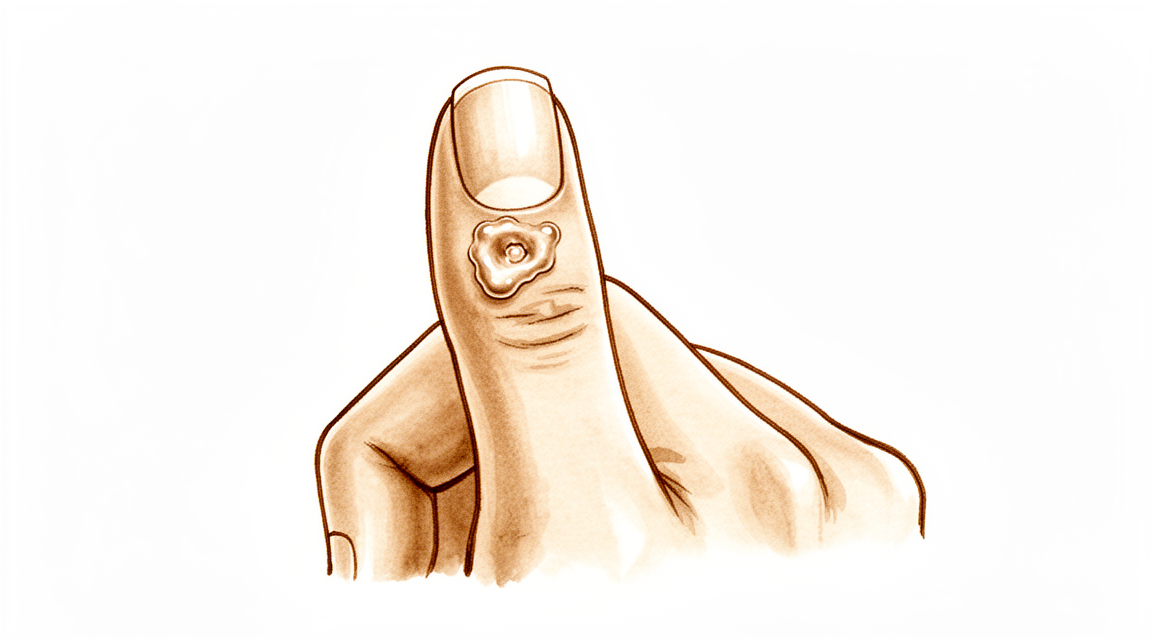
आपको क्या महसूस होता है
म्यूकस सिस्ट (तरल से भरी गांठ) एक छोटी, सख़्त गांठ है जो उंगली के पिछले हिस्से (नाखून वाली तरफ़) पर दिखती है, आमतौर पर नाखून के ठीक पीछे। इसमें गाढ़ा, साफ़ तरल भरा होता है। गांठ में खुद अक्सर दर्द नहीं होता, लेकिन यह आसपास की संरचनाओं पर दबाव डालकर तकलीफ़ पैदा कर सकती है।
आपको नाखून में भी बदलाव दिख सकते हैं। सिस्ट नाखून के तल (नेल बेड) के पास होता है, इसलिए बढ़ते समय यह नाखून पर दबाव डाल सकता है। इससे अक्सर नाखून की पूरी लंबाई में एक खांचा या उभरी लकीर बन जाती है। सिस्ट के ऊपर की त्वचा भी पतली और चमकदार हो सकती है। कुछ मामलों में त्वचा टूट जाती है और सिस्ट से एक छोटे रास्ते के ज़रिए तरल रिसने लगता है।
गांठ अक्सर ठीक वहीं होती है जहां आपकी उंगली नाखून से पहले वाले आख़िरी जोड़ पर मुड़ती है। इससे रोज़मर्रा के कुछ काम मुश्किल हो जाते हैं। तंग जेब में हाथ डालना, जार का ढक्कन पकड़ना, या छोटे सिक्के उठाना तकलीफ़देह हो सकता है। टाइप करने या लिखने से गांठ पर दबाव पड़ सकता है और उसमें दर्द हो सकता है।
यह सिस्ट उसी जोड़ के घिसाव वाले गठिया (आर्थराइटिस) से जुड़ा होता है। हड्डी का एक छोटा उभार, जिसे ऑस्टियोफ़ाइट (बोन स्पर) कहते हैं, जोड़ को परेशान (इरिटेट) करता है, और इससे वह तरल बनता है जो सिस्ट में भरता है। इसी वजह से, हाथ का बहुत इस्तेमाल करने के बाद जोड़ में दुखन हो सकती है, और उस समय गांठ छूने पर ज़्यादा दर्द कर सकती है।
अगर सिस्ट के ऊपर की त्वचा फटकर खुल जाए और तरल बाहर रिसने लगे, तो उस जगह को साफ़ रखें और जल्द डॉक्टर को दिखाएं। त्वचा से उंगली के जोड़ तक बना रास्ता संक्रमण (इन्फेक्शन) को अंदर आने दे सकता है, और संक्रमित जोड़ का इलाज बिना देर किए होना ज़रूरी है। अगर आपकी उंगली गर्म, लाल, सूजी हुई और दर्दनाक हो जाए, ख़ासकर बुखार के साथ, तो उसी दिन इमरजेंसी विभाग जाएं। इसके लिए जीपी (आपके फैमिली डॉक्टर) के रेफ़रल की ज़रूरत नहीं है।
सिस्ट में खुद छेद न करें और उसे न दबाएं। इससे जोड़ में संक्रमण पहुंच सकता है।
अगर गांठ बढ़ रही हो, नाखून का बिगड़ा आकार आपको परेशान कर रहा हो, या कई सप्ताह में लक्षण ठीक न हो रहे हों, तो अपने जीपी से मिलें या विशेषज्ञ (स्पेशलिस्ट) से जांच करवाने के लिए कहें।
असल में क्या हो रहा है
आपकी उंगली का आख़िरी जोड़ एक छोटे कब्ज़े (हिंज) की तरह काम करता है। हर जोड़ की तरह, इसके अंदर आस्तीन जैसी एक नरम परत होती है जो थोड़ा तरल बनाती है, ताकि सतहें आसानी से फिसलती रहें। म्यूकस सिस्ट में उस जोड़ में घिसाव वाला गठिया हो गया होता है। जोड़ की अंदरूनी परत परेशान (इरिटेट) हो जाती है और जोड़ की ज़रूरत से ज़्यादा तरल बनाने लगती है।
साथ ही, गठिया में अक्सर जोड़ के किनारे पर हड्डी का एक छोटा उभार (बोन स्पर) बन जाता है। वह उभार अंदर से जोड़ की परत पर दबाव डालता है और उसे घिसकर उसमें छेद कर देता है। फिर तरल उस घिसी हुई जगह से बाहर रिसता है और आपकी उंगली के पिछले हिस्से पर त्वचा के नीचे जमा हो जाता है। जो गांठ आपको दिखती और महसूस होती है, वह यही फंसा हुआ तरल है, जो जोड़ के एक तरफ़, नाखून के ठीक पीछे होता है।
सिस्ट खुद कोई नई बढ़त (ग्रोथ) नहीं, बल्कि पतली दीवार वाली एक थैली है। इसकी दीवार सहारा देने वाले ऊतक के रेशों से बनी होती है, और इसकी अपनी कोई असली अंदरूनी परत नहीं होती। क्योंकि दीवार पतली होती है और उसके ऊपर की त्वचा अक्सर उससे भी पतली होती है, इसलिए हल्की-सी ठोकर से भी यह फट सकती है। ऐसा होने पर अंदर का साफ़, जेली जैसा तरल बाहर रिस आता है।
इससे वे लक्षण समझ आते हैं जिनके बारे में आपने अभी पढ़ा। गांठ नाखून के तल के पास होती है, इसलिए सिस्ट का दबाव बढ़ते हुए नाखून में खांचा बना सकता है। जोड़ में दुखन इसलिए होती है क्योंकि वहां गठिया भी है, और सिस्ट उसी का एक संकेत है। जोड़ के किनारे का उभार ही तरल को बहता रखता है, इसलिए जब तक वह वहां है, सिस्ट रिसने या उसका तरल निकाले (ड्रेन किए) जाने के बाद भी अक्सर फिर से भर जाता है।
इसीलिए इलाज सिर्फ़ गांठ पर नहीं, बल्कि जोड़ पर केंद्रित होता है। उभार निकालने से रिसाव अपने स्रोत पर ही रुक जाता है, और जब सिस्ट में तरल भरना बंद हो जाता है, तो वह ठीक हो जाता है।
हम इसके लिए क्या कर सकते हैं
माटर प्राइवेट हॉस्पिटल, रॉकहैम्पटन में ऊपरी अंग के सर्जन डॉ. कीरन हिरपरा आपकी समस्या के अनुकूल सबसे कम चीर-फाड़ वाले विकल्पों से शुरुआत करते हैं। मरीज़ आमतौर पर अपने जीपी (आपके फैमिली डॉक्टर) के रेफ़रल से हमारे क्लिनिक आते हैं; अगर किसी फिजियोथेरेपिस्ट ने आपको हमसे मिलने की सलाह दी है, तब भी मेडिकेयर रिबेट पाने के लिए आपको अपने जीपी से रेफ़रल लेना होगा। क्लिनिक की मुलाकात में हम आपकी समस्या का इतिहास लेते हैं, आपकी उंगली की जांच करते हैं, और ज़रूरत हो तो इमेजिंग (एक्स-रे जैसी जांच) करवाते हैं। ज़्यादातर म्यूकस सिस्ट जोड़ के घिसाव वाले गठिया से जुड़े होते हैं, इसलिए हम आमतौर पर पहले बिना ऑपरेशन वाला इलाज आज़माते हैं, और जब उससे काफ़ी सुधार न हो, तब सर्जरी पर विचार करते हैं।
पहला कदम अक्सर बस नज़र रखना और इंतज़ार करना होता है। कुछ सिस्ट छोटे ही रहते हैं और ज़्यादा परेशानी नहीं देते। उंगली को बचाकर रखने से मदद मिलती है: गांठ पर बार-बार चोट लगने से बचें, और जिन कामों में उस पर दबाव पड़ता है, उन्हें सावधानी से करें। अगर ज़्यादा इस्तेमाल के बाद जोड़ में दुखन हो, तो वह काम थोड़ा कम करने से आराम मिल सकता है। हैंड थेरेपी जकड़न में और उंगली को आराम से हिलता-डुलता रखने में मदद कर सकती है। आगे कुछ भी तय करने से पहले इन साधारण उपायों को कई सप्ताह तक ठीक से आज़माएं।
दर्द से राहत आसान है। फ़ार्मेसी से मिलने वाली साधारण दर्द निवारक दवा, बताए गए तरीके से लेने पर, दुखते जोड़ को आराम दे सकती है। सूजन कम करने वाली दवाएं (एंटी-इन्फ्लेमेटरी) सिस्ट के पीछे के गठिया की तकलीफ़ कम कर सकती हैं। ये दवाएं गांठ का नहीं, बल्कि तकलीफ़ का इलाज करती हैं।
एक विकल्प जो हम नहीं देते, वह है सुई से सिस्ट का तरल निकालना (ड्रेन करना)। यह क्लिनिक में आसानी से की जाने वाली प्रक्रिया है, लेकिन इसके बाद लगभग आधे मामलों में सिस्ट वापस आ जाता है। इससे जोड़ में संक्रमण पहुंचने का जोखिम भी होता है, और इसीलिए हम घर पर भी सिस्ट में छेद करने या उसे दबाने से मना करते हैं।
सर्जरी पर तब विचार किया जाता है जब सिस्ट बार-बार भरता रहे, नाखून का बिगड़ा आकार आपको परेशान करे, या सिस्ट के ऊपर की त्वचा पतली हो गई हो या टूटने लगी हो। ऑपरेशन में सिर्फ़ गांठ का नहीं, बल्कि जोड़ का इलाज किया जाता है। जोड़ के किनारे का हड्डी का उभार निकाल दिया जाता है, जिससे तरल का रिसाव अपने स्रोत पर ही रुक जाता है, और जब सिस्ट में तरल भरना बंद हो जाता है, तो वह ठीक हो जाता है। कुछ मामलों में सिस्ट और आसपास की त्वचा का एक छोटा फ़्लैप (त्वचा का टुकड़ा) भी निकाला जाता है, जो ऐसे सिस्ट के लिए ठीक रहता है जिसने अपने ऊपर की त्वचा को पतला कर दिया हो। उभार को सिस्ट की डंडी के साथ निकालने से सिस्ट के वापस आने की संभावना सबसे कम रहती है। हम आपके साथ बात करेंगे कि आपकी उंगली के लिए कौन-सा तरीका ठीक है, और आप और हम मिलकर तय करेंगे।
क्या उम्मीद करें
अगर कुछ न किया जाए, तो म्यूकस सिस्ट गायब होने के बजाय बार-बार लौटता रहता है। जब तक जोड़ के किनारे पर हड्डी का उभार मौजूद है, वह सिस्ट में लगातार तरल भरता रहता है। कुछ सिस्ट लंबे समय तक छोटे रहते हैं और ज़्यादा परेशानी नहीं देते। दूसरे धीरे-धीरे बढ़ते हैं, नाखून के खांचे को और गहरा करते हैं, या गांठ के ऊपर की त्वचा को इतना पतला कर देते हैं कि उससे रिसाव होने लगे।
इलाज के बिना, मुख्य जोखिम वही हैं जिनके बारे में आप पढ़ चुके हैं: जब तक सिस्ट दबाव डालता है, तब तक नाखून पर उभरी लकीर बनी रहना, और त्वचा का टूटकर जोड़ तक एक रास्ता खोल देना। उस रास्ते को गंभीरता से लेना चाहिए। अगर आपकी उंगली गर्म, लाल, सूजी हुई और दर्दनाक हो जाए, ख़ासकर बुखार के साथ, तो उसी दिन इमरजेंसी विभाग जाएं। इसके लिए जीपी के रेफ़रल की ज़रूरत नहीं है।
जोड़ पर केंद्रित इलाज के साथ, आगे की तस्वीर साफ़ है। उभार निकालने से रिसाव अपने स्रोत पर ही रुक जाता है, और जब सिस्ट में तरल भरना बंद हो जाता है, तो वह ठीक हो जाता है। जब उभार निकाल दिया जाता है, तो सिस्ट कम ही वापस आता है। जहां सिस्ट को भी निकाला जाता है, वहां भी दोबारा होने की दर कम रहती है: सर्जरी से इलाज किए गए हर 100 में से लगभग 2 सिस्ट लौटते हैं, और जब उस जगह को बंद करने के लिए आसपास की त्वचा का फ़्लैप इस्तेमाल किया जाता है, तो 100 में से लगभग 1.4। इस तरह इलाज करवाने वाले लोग आमतौर पर निशान के दिखने से संतुष्ट रहते हैं, और यह प्रक्रिया फिर से करवाने को तैयार रहते हैं।
ठीक होना कोई बहुत बड़ा काम नहीं है, लेकिन इसे हल्के में भी नहीं लेना चाहिए। उंगली अक्सर कुछ सप्ताह तक जकड़ी हुई रहती है और छूने पर दर्द करती है, और पकड़ने या चुटकी से पकड़ने जैसे रोज़मर्रा के कामों को फिर से सामान्य लगने में कुछ समय लगता है। सर्जरी के बाद हैंड थेरेपी एक्सटेंड रिहैबिलिटेशन (Extend Rehabilitation) में रूबी डूलन के साथ होती है। रूबी एक हैंड थेरेपिस्ट (हाथ की विशेषज्ञ थेरेपिस्ट) हैं: वे आपके व्यायामों में आपका मार्गदर्शन करती हैं और आपको जिस भी स्प्लिंट की ज़रूरत हो, उसे बनाती हैं। ज़्यादातर लोग कुछ सप्ताह से लेकर कुछ महीनों के अंदर हाथ का सामान्य इस्तेमाल फिर से करने लगते हैं।
नाखून के तल पर से दबाव हटने के बाद नाखून का खांचा भी आमतौर पर ठीक हो जाता है। इसमें धीरज रखें: नाखून के बिगड़े हुए हिस्से को बढ़कर बाहर निकलना होता है, जिसमें कई महीने लगते हैं।
अगर कई सप्ताह में लक्षण ठीक न हो रहे हों, या बिगड़ रहे हों, तो अपने जीपी से मिलें या विशेषज्ञ से जांच करवाने के लिए कहें।
डॉक्टर को कब दिखाएं
ज़्यादातर म्यूकस सिस्ट में जल्दबाज़ी की ज़रूरत नहीं होती, और बहुत से सिस्ट साधारण देखभाल से ठीक हो जाते हैं। कुछ लक्षणों पर कदम उठाना ज़रूरी है।
अगर आपकी उंगली गर्म, लाल, सूजी हुई और दर्दनाक हो जाए, ख़ासकर बुखार के साथ, तो उसी दिन इमरजेंसी विभाग जाएं। यह जोड़ में संक्रमण की ओर इशारा करता है, जिसका इलाज बिना देर किए होना ज़रूरी है। यही बात तब भी लागू होती है जब सिस्ट के ऊपर की त्वचा फटकर खुल गई हो और तरल रिस रहा हो, क्योंकि उस छेद से संक्रमण अंदर जा सकता है।
अगर गांठ बढ़ती जा रही हो, नाखून का खांचा आपको परेशान कर रहा हो, या सिस्ट के ऊपर की त्वचा पतली हो गई हो, तो अपने जीपी से मिलें या विशेषज्ञ से जांच करवाने के लिए कहें। यही वे संकेत हैं जिनमें इलाज पर बात करना फ़ायदेमंद है। अगर कई सप्ताह में लक्षण ठीक न हो रहे हों, या बिगड़ रहे हों, तो आपके जीपी रेफ़रल की व्यवस्था कर सकते हैं।
विस्तार से
Advanced reading: the deeper science (optional)
यह हिस्सा उससे आगे की जानकारी देता है जितनी आपको अपने इलाज के फ़ैसलों के लिए चाहिए। म्यूकस सिस्ट के बारे में अतिरिक्त पढ़ाई सर्जरी से जुड़ी एक छोटी, अच्छे प्रमाण वाली समझ के कारण फ़ायदेमंद है: समस्या सिस्ट नहीं है, और सबसे अच्छा काम करने वाला ऑपरेशन ज़रूरी नहीं कि उसे निकाले।
वजह हड्डी का उभार है, सिस्ट नहीं
म्यूकस सिस्ट उंगली के सिरे के गठिया वाले जोड़ से निकलता है। हड्डी का एक उभार, जिसे ऑस्टियोफ़ाइट कहते हैं, जोड़ के कैप्सूल को परेशान (इरिटेट) करता है और उसमें छेद कर देता है, और जोड़ का तरल उस छेद से बाहर निकलकर त्वचा के नीचे जमा हो जाता है। सिस्ट उस प्रक्रिया का दिखाई देने वाला सिरा है, उसकी शुरुआत नहीं।
इस समझ का सर्जरी पर सीधा असर है, जो एक अध्ययन-श्रृंखला में दिखाया गया जिसमें सिस्ट निकाले बिना ऑस्टियोफ़ाइट निकालने से ज़्यादातर मामलों में समस्या पूरी तरह ठीक हो गई, और इसे एक कम चीर-फाड़ वाला तरीका देने वाला अच्छा इलाज-विकल्प बताया गया [1]।
उभार निकालने से नल बंद हो जाता है। सिस्ट, जिसमें अब तरल नहीं भर रहा, ठीक हो जाता है। यही तर्क कलाई के गैंग्लियन पर भी लागू होता है, जहां थैली के बजाय उसकी डंडी तय करती है कि वह दोबारा होगा या नहीं, और इसी से समझ आता है कि म्यूकस सिस्ट को सिर्फ़ ड्रेन करना या उसमें सुई से छेद करना इतनी नियमितता से क्यों नाकाम होता है।
जहां सिस्ट निकाला जाता है, वहां भी नतीजे अच्छे हैं
दूसरा तरीका सिस्ट को निकालता है और खाली जगह को बंद करने के लिए आसपास की त्वचा का एक फ़्लैप इस्तेमाल करता है। यह भी भरोसेमंद है: 69 मरीज़ों में, लोकल एडवांसमेंट फ़्लैप के साथ सर्जरी से सिस्ट निकालने पर दोबारा होने की दर 1.4% रही, और निशान को लेकर मरीज़ों की संतुष्टि और दोबारा यह प्रक्रिया करवाने की तैयारी ज़्यादा रही [2]।
दोनों तरीके काम करते हैं, और दोनों अंदर के जोड़ का इलाज करते हैं। व्यावहारिक फ़र्क यह है कि कितनी त्वचा शामिल है: लंबे समय से मौजूद सिस्ट ऊपर की त्वचा को पतला कर देता है, कभी-कभी इतना कि उससे रिसाव होने लगे, और उस स्थिति में हड्डी के साथ जो भी किया जाए, पतली हुई त्वचा को निकालकर बदलना पड़ता है।
नाखून क्यों बिगड़ जाता है, और क्या वह ठीक होता है
नाखून की पूरी लंबाई में चलने वाला खांचा या उभरी लकीर अक्सर साथ में होती है, और यह लोगों को गांठ से ज़्यादा चिंतित करती है। इसकी वजह यांत्रिक है: सिस्ट ठीक जर्मिनल मैट्रिक्स (नाखून के तल का वह हिस्सा जो नाखून बनाता है) के ऊपर होता है और उस पर दबाव डालता है, इसलिए नाखून एक ख़राबी के साथ बनता है।
काम की बात यह है कि यह दबाव है, नुकसान नहीं। एक बार सिस्ट का दबाव हट जाए, तो नाखून आमतौर पर सामान्य रूप से बढ़ता है, हालांकि बिगड़े हुए हिस्से को बढ़कर सिरे से निकल जाने में कई महीने लगते हैं। इसलिए नाखून का बिगड़ना सिस्ट का इलाज करने का एक कारण है, न कि उसका स्थायी नतीजा।
इसमें छेद करने को लेकर सावधान रहने की वजह
म्यूकस सिस्ट सीधे जोड़ से जुड़ा होता है। इसमें छेद होना, चाहे जानबूझकर किया जाए या किसी बड़े सिस्ट के ऊपर की त्वचा टूटने से हो, बाहरी दुनिया से उंगली के जोड़ के अंदर तक एक रास्ता बना देता है, और किसी छोटे जोड़ का सेप्टिक आर्थराइटिस (जोड़ के अंदर संक्रमण) सिस्ट से कहीं ज़्यादा गंभीर समस्या है।
यही व्यावहारिक तर्क है कि ऐसे सिस्ट को, जो सिर्फ़ तरल का छाला लगता हो, घर पर ड्रेन न करें, और यही कारण है कि अपने आप रिसने वाले सिस्ट का इलाज बस नज़र रखने के बजाय कुछ जल्दी किया जाता है।
संदर्भ
[1] Lee H, Kim P, Jeon I, Kyung H, Ra I, Kim T. Osteophyte excision without cyst excision for a mucous cyst of the finger. J Hand Surg Eur Vol. 2013;39(3):258-61. https://doi.org/10.1177/1753193413478549
[2] Johnson SM, Treon K, Thomas S, Cox QGN. A reliable surgical treatment for digital mucous cysts. J Hand Surg Eur Vol. 2013;39(8):856-60. https://doi.org/10.1177/1753193413508540
Evidence & references
This is the clinical evidence summary written for health professionals. It is technical, and it lists the research this page was built from. You do not need to read it to understand your treatment or to make a decision about it.
Overview
- The scientific data regarding mucous cysts consist almost entirely of retrospective studies, and much of what is done or recommended is based on expert opinion [3].
- Surgical treatment of mucous cysts is not a totally benign procedure, and complications do occur contrary to literature reporting glowing results [5].
- In-office excision is a safe and effective option for treating digital mucous cysts [6].
- Complications are rare and occur only in cysts associated with fistula, justifying their early surgical treatment [7].
- Surgery provided definitive treatment with no major long-term problems [10].
- Surgical excision was the primary treatment, with no reported recurrences [11].
- Incomplete excisions will invariably result in recurrence [12].
- Surgical excision with a local advancement skin flap is a reliable treatment for digital mucous cysts, demonstrating a low recurrence rate of 1.4% and high patient satisfaction regarding the scar and willingness to undergo the procedure again [13].
- The 2% recurrence rate (2 of 113) obtained in this series illustrates the advantage of this procedure [16].
- Osteophyte excision without cyst excision may be a good treatment choice for mucous cyst of the finger, providing a less invasive method with complete resolution in most cases [20].
- The final recurrence rate of less than 1.5% may be acceptable and comparable with other techniques [21].
- Excision with local flap cover offers benefit in terms of a reduced recurrence rate and is particularly valuable where the cyst has caused marked thinning of the skin [30].
- Excision with local flap cover would seem to offer benefit in terms of a reduced recurrence rate, and is particularly valuable where the cyst has caused marked thinning of the skin [34].
- Excision of the cyst and complete removal of the marginal osteophyte eradicates mucous cysts with extremely rare recurrence [1].
- A total dorsal capsulectomy alone was a simple treatment for mucous cysts and did not lead to any recurrence [8].
Anatomy & Pathophysiology
Clinical Presentation and Location
- Mucous cysts are small, firm, cystic masses that appear just distal to the distal interphalangeal joint [23].
- The lesion is always located to one side of the midline [23].
- Mucous cysts are rarely greater than fifteen millimeters in diameter [23].
- The skin over the mucous cyst is thinned out and occasionally may be ulcerated [23].
- Pressure on the mucous cyst does not usually result in a decrease in its size [23].
- Longitudinal grooving of the nail may be noted, occasionally occurring prior to the appearance of the cyst [23].
- Mucous cysts are usually small and located to one side as they emerge from the joint beside the extensor mechanism [28, 29].
- Occasionally, mucous cysts may spread across the whole of the dorsum of the digit between the distal interphalangeal joint and nail fold [28, 29].
- The overlying skin of a mucous cyst is frequently thin, and minimal trauma may result in rupture [28, 29].
Histology and Ultrastructure
- The histological appearance of a mucous cyst is essentially identical to that of ganglions, synovial cysts, and similar lesions appearing on other areas of the extremities [23].
- The mucous cyst is surrounded by an undemarcated fibrous capsule [23].
- The basic structure of the mucous cyst is myxomatous with interspersed fibroblasts [23].
- Areas of myxomatous degeneration tend to coalesce to form a multiloculated cyst [23].
- An epithelial lining has not been reported for mucous cysts [23].
- Digital mucous cysts and wrist ganglia show no anatomic difference at the light and scanning electron microscopic levels [19].
- The wall of a digital mucous cyst consists of multidirectional strata of collagen fibers with rare fibroblast-like cells on the inner surface [19].
- There are no signs of myxomatous degeneration in the wall of a digital mucous cyst [19].
- The surface of the digital mucous cyst wall is generally flat, with some areas showing localized elevations [19].
- Multifunctional mesenchymal cells are located beneath the lining of the ganglion and digital mucous cyst [19].
- These multifunctional mesenchymal cells contain abundant rough endoplasmic reticulum, smooth-walled vacuoles, and Golgi complexes [19].
- The ganglion fluid may be produced by these multifunctional cells and reaches the ganglion cavity by flowing through the porous collagen matrix of the ganglion wall [19].
- A digital mucous cyst has a large globular dome-shaped interconnecting cavity having a wall of collagen fibers covered by a fenestrated pseudomembrane [19].
- A porous network of collagen fibers is present in the wall of the digital mucous cyst on cross section [19].
Etiology and Pathogenesis
- Degenerative arthritis of the distal interphalangeal joint is a frequent associated finding with mucous cysts [23].
- An incidence of 78 percent for degenerative arthritis of the distal interphalangeal joint has been reported in association with mucous cysts [23].
- The etiology indicated by evidence is that the lesion arises from the joint capsule [23].
- Mucous cysts are often associated with degenerative changes at the distal interphalangeal joints [28, 29].
- Theories on the pathogenesis of mucous cysts include myxoid degeneration, repeated local trauma, vascular insufficiency, and synovial herniation [28, 29].
- Studies suggest that mucous cysts arise due to excessive hyaluronic acid production from a degenerate distal interphalangeal joint [28, 29].
Classification
- The lesion is a small, firm, cystic mass which appears just distal to the distal interphalangeal joint [23].
- The lesion is always located to one side of the mid-line [23].
- The lesion is rarely greater than fifteen millimeters in diameter [23].
- The skin over the lesion is thinned out and occasionally may be ulcerated [23].
- Pressure on the lesion does not usually result in a decrease in its size [23].
- Degenerative arthritis of the distal interphalangeal joint is a frequent associated finding [23].
- An incidence of 78 per cent has been reported for degenerative arthritis of the distal interphalangeal joint associated with mucous cysts [23].
- The histological appearance is essentially identical to that of ganglions, synovial cysts, and similar lesions appearing on other areas of the extremities [23].
- The lesion is surrounded by an undemarcated fibrous capsule [23].
- The basic structure is myxomatous with interspersed fibroblasts [23].
- An epithelial lining has not been reported [23].
- In spite of their different clinical settings, digital mucous cysts and wrist ganglia show no anatomic difference at the light and scanning electron microscopic levels [19].
- The wall of the digital mucous cyst consisted of multidirectional strata of collagen fibers with rare fibroblast-like cells on the inner surface [19].
- There were no signs of myxomatous degeneration in the wall of the digital mucous cyst [19].
- The surface of the digital mucous cyst was generally flat, with some areas showing localized elevations [19].
- Multifunctional mesenchymal cells beneath the lining of the ganglion are postulated to produce collagen fibers, elastic fibers, and the interfibrillary mucopolysaccharide matrix [19].
- The porous nature of the ganglion wall was confirmed in the ultrastructural study of digital mucous cysts [19].
- A previous ultrastructural study of digital mucous cysts demonstrated a large globular dome-shaped interconnecting cavity having a wall of collagen fibers covered by a fenestrated pseudomembrane [19].
- The current study confirmed the anatomic organization of a large globular dome-shaped interconnecting cavity having a wall of collagen fibers covered by a fenestrated pseudomembrane [19].
- A porous network of collagen fibers in the wall was revealed on cross section [19].
Clinical Presentation
- The mucous cyst presents as a small, firm, cystic mass located just distal to the distal interphalangeal joint [23].
- The lesion is easily distinguished from a Heberden's node by its placement distal to the distal interphalangeal joint [23].
- The histological appearance of the mucous cyst is essentially identical to that of ganglions, synovial cysts, and similar lesions appearing on other areas of the extremities [23].
- The basic structure of the lesion is myxomatous with interspersed fibroblasts [23].
- An epithelial lining has not been reported for the mucous cyst [23].
- The wall of the digital mucous cyst consists of a porous network of collagen fibers [19].
- The fluid within the digital mucous cyst is produced by underlying mesenchymal cells [19].
Investigations
- Ultrasound is a powerful modality for evaluation of pathologic conditions in the hand and wrist, providing a cost-effective and expedient alternative and/or adjunct to MRI [33].
- Ultrasound is best used when there is a specific clinical question regarding a well-localized abnormality [33].
- Ultra-sonography provides a new ability to study the complex anatomical structures of the hand, with the technique being quick and simple to perform [38].
- A careful physical examination is essential to direct care and future testing if indicated [35].
- Diagnostic tests such as imaging and serum laboratory studies are useful in this determination but can be expensive, time consuming, and often nonspecific [35].
Treatment
Operative Techniques
- Total dorsal capsulectomy alone is a simple treatment for mucous cysts that did not lead to any recurrence [8].
- Osteophyte-sparing treatment of mucous cysts has a final recurrence rate of less than 1.5% [21].
- Surgical excision was the primary treatment in a series with no reported recurrences [11].
- Surgery provided definitive treatment for mucous cysts with no major long-term problems [10].
- The 2% recurrence rate (2 of 113) obtained in a series of 191 excised cysts illustrates the advantage of the procedure [4, 16].
- The authors of a study on osteophyte excision without cyst excision state that their principle is not to do more damage to the friable skin because the skin is not the culprit of the cyst [15].
Outcomes and Complications
- Complications of surgical treatments include infection, recurrence, nail deformity, swelling, stiffness and pain [28].
- This report provides additional basis for prophylactic extirpation of mucous cyst of the finger [2].
- This case emphasizes the utility of a pathohistological analysis in cases where doubts arise about the initial diagnosis of a benign tumorous lesion [14].
Non-Operative and Adjunctive Considerations
- Aspiration with injection of steroid is a convenient technique that can be performed in an outpatient setting but is followed by a recurrence rate of 50% [28].
- Surgical treatment has a higher cure rate than aspiration with injection of steroid [28].
- Radical treatment with complete excision of the stalk and underlying osteophytes seems to reduce recurrence rates [28].
Complications
- Complications are rare and occur only in cysts associated with fistula [7].
- Aspiration with injection of steroid is followed by a recurrence rate of 50% [28].
- The 2% recurrence rate (2 of 113) was obtained in a series of surgically treated mucous cysts [16].
- Surgical excision with a local advancement skin flap demonstrated a low recurrence rate of 1.4% [13].
- A total dorsal capsulectomy alone did not lead to any recurrence [8].
- Eccrine porocarcinomas have a substantial risk of metastasis, high risk of local recurrence, and are potentially fatal [24].
Recovery
- Surgical excision with a local advancement skin flap demonstrated a low recurrence rate of 1.4% and high patient satisfaction regarding the scar and willingness to undergo the procedure again [13].
Key Evidence
- [L4] Excision of the cyst and complete removal of the marginal osteophyte eradicates mucous cysts with extremely rare recurrence. [1] (10.2106/00004623-197355030-00013)
- [L4] This report provides additional basis for prophylactic extirpation of mucous cyst of the finger. [2] (10.1016/s0363-5023(84)80152-5)
- [L4] The scientific data regarding mucous cysts consist almost entirely of retrospective studies, and much of what is done or recommended is based on expert opinion. [3] (10.1016/j.jhsa.2010.01.029)
- [L5] Of the 191 mucous cysts excised, 113 had at least 6 months of follow-up time. [4] (10.1016/j.jhsa.2010.07.028)
- [L4] Surgical treatment of mucous cysts is not a totally benign procedure, and complications do occur contrary to literature reporting glowing results. [5] (10.1016/s0266-7681(97)80067-8)
- [Paper] In-office excision is a safe and effective option for treating digital mucous cysts. [6] (10.1177/15589447251350168)
- [L4] Complications are rare and occur only in cysts associated with fistula, justifying their early surgical treatment. [7] (10.1016/j.main.2015.06.001)
- [L4] A total dorsal capsulectomy alone was a simple treatment for mucous cysts and did not lead to any recurrence. [8] (10.1016/j.jhsa.2014.03.004)
- [L4] Surgery provided definitive treatment with no major long-term problems. [10] (10.1053/jhsu.2003.50088)
- [L5] Incomplete excisions will invariably result in recurrence. [12] (10.1016/0363-5023(93)90302-j)
- [L4] Surgical excision with a local advancement skin flap is a reliable treatment for digital mucous cysts, demonstrating a low recurrence rate of 1.4% and high patient satisfaction regarding the scar and willingness to undergo the procedure again. [13] (10.1177/1753193413508540)
- [L5] This case emphasizes the utility of a pathohistological analysis in cases where doubts arise about the initial diagnosis of a benign tumorous lesion. [14] (10.1007/s00402-008-0794-4)
- [L5] The authors state that their principle is not to do more damage to the friable skin because the skin is not the culprit of the cyst. [15] (10.1177/1753193414546990)
- [L4] The 2% recurrence rate (2 of 113) obtained in this series illustrates the advantage of this procedure. [16] (10.1016/0363-5023(94)90071-x)
- [L5] [19] (10.1016/s0363-5023(88)80143-6)
- [L4] Osteophyte excision without cyst excision may be a good treatment choice for mucous cyst of the finger, providing a less invasive method with complete resolution in most cases. [20] (10.1177/1753193413478549)
- [L4] The final recurrence rate of less than 1.5% may be acceptable and comparable with other techniques. [21] (10.5435/jaaosglobal-d-21-00164)
- [L4] [23] (10.2106/00004623-197254070-00008)
- [L4] Prompt recognition and appropriate treatment are critical because eccrine porocarcinomas have a substantial risk of metastasis, high risk of local recurrence, and are potentially fatal. [24] (10.1016/j.jhsa.2016.07.112)
- [L4] [28] (10.1177/1753193408103498)
- [L4] [29] (10.1177/1753193409103498)
- [L4] Excision with local flap cover offers benefit in terms of a reduced recurrence rate and is particularly valuable where the cyst has caused marked thinning of the skin. [30] (10.1016/0266-7681(90)90064-b)
- [L5] Ultrasound is a powerful modality for evaluation of pathologic conditions in the hand and wrist, providing a cost-effective and expedient alternative and/or adjunct to MRI, best used when there is a specific clinical question regarding a well-localized abnormality. [33] (10.1016/j.jhsa.2009.02.010)
- [L4] Excision with local flap cover would seem to offer benefit in terms of a reduced recurrence rate, and is particularly valuable where the cyst has caused marked thinning of the skin. [34] (10.1016/0266-7681_90_90064-b)
- [L4] Ultra-sonography provides a new ability to study the complex anatomical structures of the hand, with the technique being quick and simple to perform. [38] (10.1016/0266-7681(90)90133-o)
References
[1] Marginal Osteophyte Excision in Treatment of Mucous Cysts. The Journal of Bone & Joint Surgery. 1973. DOI: 10.2106/00004623-197355030-00013
[2] Infected mucous cyst of the finger. The Journal of Hand Surgery. 1984. DOI: 10.1016/s0363-5023(84)80152-5
[3] Mucous Cysts. The Journal of Hand Surgery. 2010. DOI: 10.1016/j.jhsa.2010.01.029
[4] Mucous Cysts. The Journal of Hand Surgery. 2010. DOI: 10.1016/j.jhsa.2010.07.028
[5] Complications Following Mucous Cyst Excision. Journal of Hand Surgery. 1997. DOI: 10.1016/s0266-7681(97)80067-8
[6] Safety and Cost-Effectiveness of In-Office Digital Mucous Cyst Excisions. HAND. 2025. DOI: 10.1177/15589447251350168
[7] Surgical treatment of mucous cysts by subcutaneous excision and osteophyte resection: Results in 68 cases at a mean 6.63 years’ follow-up. Chirurgie de la Main. 2015. DOI: 10.1016/j.main.2015.06.001
[8] Total Dorsal Capsulectomy for the Treatment of Mucous Cysts. The Journal of Hand Surgery. 2014. DOI: 10.1016/j.jhsa.2014.03.004
[10] Treatment of mucous cysts of the fingers: Review of 134 cases with minimum 2-year follow-up evaluation. The Journal of Hand Surgery. 2003. DOI: 10.1053/jhsu.2003.50088
[11] 10.1177-15589447261433068. n.d..
[12] Juvenile hyaline fibromatosis of the hand in an adult. The Journal of Hand Surgery. 1993. DOI: 10.1016/0363-5023(93)90302-j
[13] A reliable surgical treatment for digital mucous cysts. Journal of Hand Surgery (European Volume). 2013. DOI: 10.1177/1753193413508540
[14] Malignant Natural-Killer cell neoplasm presenting as a mucous cyst on the distal interphalangeal joint of the finger. Archives of Orthopaedic and Trauma Surgery. 2008. DOI: 10.1007/s00402-008-0794-4
[15] Re: Lee, H.-J., Kim, P.-T., Jeon, I.-H., Kyung, H.-S., Ra, I.-H. and Kim, T.-K. Osteophyte excision without cyst excision for a mucous cyst of the finger. J Hand Surg Eur. 2014, 39: 258–61. Journal of Hand Surgery (European Volume). 2014. DOI: 10.1177/1753193414546990
[16] Outcome of surgically treated mucous cysts of the hand. The Journal of Hand Surgery. 1994. DOI: 10.1016/0363-5023(94)90071-x
[19] A surface ultrastructure study of ganglia and digital mucous cysts. The Journal of Hand Surgery. 1988. DOI: 10.1016/s0363-5023(88)80143-6
[20] Osteophyte excision without cyst excision for a mucous cyst of the finger. Journal of Hand Surgery (European Volume). 2013. DOI: 10.1177/1753193413478549
[21] Osteophyte-Sparing Treatment of Mucous Cysts: Case Analysis and Surgical Technique. JAAOS: Global Research and Reviews. 2021. DOI: 10.5435/jaaosglobal-d-21-00164
[23] Etiology and Treatment of the So-Called Mucous Cyst of the Finger. The Journal of Bone & Joint Surgery. 1972. DOI: 10.2106/00004623-197254070-00008
[24] Eccrine Porocarcinoma Presenting as a Hand Cyst. The Journal of Hand Surgery. 2016. DOI: 10.1016/j.jhsa.2016.07.112
[28] Use of Wolfe Graft for the Treatment of Mucous Cysts. Journal of Hand Surgery (European Volume). 2009. DOI: 10.1177/1753193408103498
[29] Use of Wolfe Graft for the Treatment of Mucous Cysts. Journal of Hand Surgery (European Volume). 2009. DOI: 10.1177/1753193409103498
[30] Mucous cyst of the distal interphalangeal joint: Treatment by simple excision or excision and rotation flap. The Journal of Hand Surgery: Journal of the British Society for Surgery of the Hand. 1990. DOI: 10.1016/0266-7681(90)90064-b
[33] Ultrasound of the Hand and Wrist. The Journal of Hand Surgery. 2009. DOI: 10.1016/j.jhsa.2009.02.010
[34] Mucous Cyst of the Distal Interphalangeal Joint: Treatment by Simple Excision or Excision and Rotation Flap. Journal of Hand Surgery. 1990. DOI: 10.1016/0266-7681_90_90064-b
[35] Orthopaedic Knowledge Update 13 Ebook Without Multimedia. Anatomy, Evaluation, Clinical Examination, and Imaging > Evaluation and Clinical Examination: Current Concepts.
[38] Diagnostic medical ultrasound in the management of hand injuries. The Journal of Hand Surgery: Journal of the British Society for Surgery of the Hand. 1990. DOI: 10.1016/0266-7681(90)90133-o