Patients › Elbow
क्यूबिटल टनल रिलीज़ (अल्नर नर्व पर से दबाव हटाने का ऑपरेशन)
Cubital tunnel release relieves ulnar nerve compression—addressing numbness/weakness in the ring & little fingers.
यह ऑपरेशन क्यों सुझाया गया है
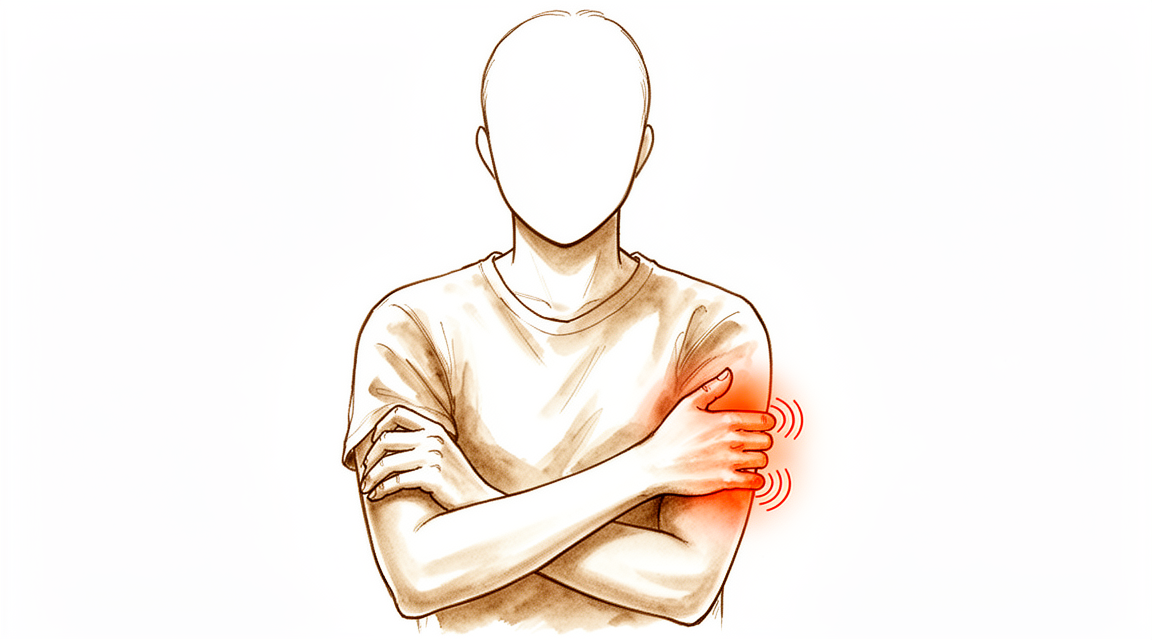
डॉ. कीरन हिरपरा, जो माटर प्राइवेट हॉस्पिटल, रॉकहैम्पटन में ऊपरी अंग (कंधे से हाथ तक) के सर्जन हैं, आपकी समस्या के अनुसार सबसे कम चीर-फाड़ वाले विकल्पों से शुरुआत करते हैं। मरीज़ों को आम तौर पर उनके जीपी (आपके फैमिली डॉक्टर) हमारे क्लिनिक में रेफ़र करते हैं; अगर किसी फिजियोथेरेपिस्ट ने आपको हमसे मिलने की सलाह दी है, तब भी मेडिकेयर की छूट (रिबेट) पाने के योग्य होने के लिए आपको अपने जीपी से रेफ़रल लेना होगा। आपके अपॉइंटमेंट पर हम आपकी मेडिकल हिस्ट्री लेते हैं, आपकी बांह की जांच करते हैं, और ज़रूरत हो तो स्कैन या नर्व की जांच की व्यवस्था करते हैं। क्यूबिटल टनल सिंड्रोम आपकी कोहनी के भीतरी ओर एक नर्व (तंत्रिका) पर दबाव का नाम है। इस दबाव से आपकी छोटी उंगली और हाथ के अनामिका वाले हिस्से में सुन्नपन या झुनझुनी होती है, और जब आपकी कोहनी लंबे समय तक मुड़ी रहती है तो यह बढ़ सकती है। इस तरह की लंबे समय से चली आ रही समस्या के लिए हम आम तौर पर पहले बिना ऑपरेशन वाला इलाज आज़माते हैं: गतिविधियों में बदलाव, फिजियोथेरेपी या हैंड थेरेपी, और स्प्लिंट। जब इन कदमों से आपको पर्याप्त सुधार नहीं मिलता, तब हम सर्जरी पर विचार करते हैं।
इस ऑपरेशन को क्यूबिटल टनल रिलीज़ (नर्व पर से दबाव हटाने का ऑपरेशन) कहते हैं। यह नर्व पर से दबाव कम करता है ताकि आपके लक्षण ठीक हो सकें। यह सर्जरी कराने वाले 90% से ज़्यादा लोग ठीक हो जाते हैं या उनमें सुधार होता है। हम इस बारे में बात करेंगे कि आपके लिए क्या मायने रखता है, और आप और हम मिलकर तय करेंगे कि क्या यह सही अगला कदम है।
ऑपरेशन से पहले
सर्जरी बुक हो जाने पर हम आपको पालन करने के लिए साफ़ निर्देश देंगे। आपको अपने ऑपरेशन से सात घंटे पहले खाना-पीना बंद करना होगा। हम छह के बजाय सात घंटे इसलिए कहते हैं ताकि अगर ऑपरेशन थिएटर की सूची जल्दी चल रही हो तो आपकी सर्जरी का समय पहले किया जा सके। सर्जरी से पहले कुछ दवाओं को रोकना पड़ सकता है, और आपके सर्जन आपको बताएंगे कि कौन-सी और कब। आप जो कुछ भी लेते हैं उसकी सूची साथ लाएं, जिसमें गोलियां, इंजेक्शन और क्रीम शामिल हैं। बाद में आपको घर ले जाने के लिए किसी को गाड़ी चलाने के लिए तय कर लें, क्योंकि आप खुद गाड़ी नहीं चला पाएंगे। ढीले, आरामदायक कपड़े पहनें जिनकी आस्तीन आसानी से उतारी जा सके। आपके ऑपरेशन की योजना बनाने में मदद के लिए एक्स-रे, अल्ट्रासाउंड या एमआरआई स्कैन जैसी जांचें पहले से कराई गई हो सकती हैं। अगर आपको कोई दूसरी बीमारी है, तो आपको खून की जांच या एनेस्थेटिस्ट (बेहोशी के डॉक्टर) से जांच करानी पड़ सकती है, लेकिन ज़्यादातर लोगों को इसकी ज़रूरत नहीं होती।
ऑपरेशन के दिन
आप अस्पताल की सर्जिकल भर्ती यूनिट में आते हैं, जहां आपकी भर्ती की जाती है और आपको ऑपरेशन थिएटर के लिए तैयार किया जाता है। आप एनेस्थेटिस्ट से मिलेंगे, जो एक डॉक्टर हैं और ऑपरेशन के दौरान आपके आराम और सुरक्षा का ध्यान रखते हैं। यह ऑपरेशन जनरल एनेस्थीसिया (जिसमें आप पूरी तरह बेहोश रहते हैं) में किया जाता है। ऑपरेशन के दौरान आप पूरी तरह बेहोश रहेंगे। कुछ मरीज़ों को ऑपरेशन के बाद दर्द से राहत के लिए रीजनल नर्व ब्लॉक (हाथ या बांह को सुन्न करने का इंजेक्शन) भी दिया जा सकता है; एनेस्थेटिस्ट उसी दिन आपकी व्यक्तिगत स्थिति के आधार पर यह तय करते हैं। फिर आपको ऑपरेशन थिएटर में ले जाया जाता है, जहां ऑपरेशन किया जाता है।
आप रिकवरी एरिया में जागेंगे, जहां एनेस्थीसिया का असर खत्म होने तक नर्सें आपकी देखरेख करती हैं। जब आपकी हालत स्थिर हो जाती है, तो प्रक्रिया और आपके ठीक होने के अनुसार आप या तो वार्ड में जाते हैं या घर जाते हैं। कई लोग उसी दिन घर चले जाते हैं। आपके जाने से पहले हम समझाएंगे कि अपनी बांह की देखभाल कैसे करनी है और आने वाले दिनों में क्या उम्मीद करनी है।
ऑपरेशन में क्या किया जाता है
इस ऑपरेशन को क्यूबिटल टनल रिलीज़ कहते हैं। आपके सर्जन आपकी कोहनी के भीतरी ओर, वहां महसूस होने वाले हड्डी के उभार के ठीक पीछे, एक चीरा लगाते हैं। इस चीरे के ज़रिए आपके सर्जन अल्नर नर्व को ढूंढते हैं, यानी वह नर्व जो दब रही है और आपकी छोटी उंगली और अनामिका में झुनझुनी पैदा कर रही है। यह नर्व कोहनी के पास ऊतक से बनी एक सुरंग से होकर गुज़रती है, कुछ वैसे ही जैसे कोई तार एक संकरी नली से होकर जाता है। आपके सर्जन उस सुरंग को खोल देते हैं ताकि नर्व अब दबे नहीं। इसे डीकम्प्रेशन कहते हैं, जिसका मतलब है नर्व पर से दबाव हटाना।
कभी-कभी नर्व को कोहनी के सामने की ओर एक नई जगह पर ले जाने की भी ज़रूरत होती है, ताकि जब आप अपनी बांह मोड़ें तो वह परेशानी से दूर रहे। आपके सर्जन ऑपरेशन के दौरान तय करेंगे कि सिर्फ़ सुरंग खोलना काफ़ी है, या नर्व को दूसरी जगह ले जाना आपके लिए बेहतर रहेगा। यह चुनाव आपकी जांच और आपके सर्जन को ऑपरेशन में जो मिलता है उस पर निर्भर करता है।
नर्व के मुक्त हो जाने के बाद आपके सर्जन चीरे को बंद करते हैं। पहले घाव के ऊपर एक बारीक, अपने आप चिपकने वाली जाली रखी जाती है, जो त्वचा के किनारों को आपस में जोड़े रखती है। फिर जाली के ऊपर एक तरल स्किन ग्लू (त्वचा का गोंद) लगाया जाता है, जो सूखकर जम जाता है और सब कुछ एक साथ सील कर देता है। यह लगभग एक से दो सप्ताह तक लगा रहता है, फिर अपने आप उठकर उतर जाता है, इसलिए कुछ भी निकालना नहीं पड़ता।
ऑपरेशन के बाद
आप रिकवरी एरिया में जागेंगे, जहां एनेस्थीसिया का असर खत्म होने तक नर्सें आपकी देखरेख करती हैं। यह डे-केस ऑपरेशन है, इसलिए आप उसी दिन घर चले जाएंगे। दर्द आम तौर पर हल्के से मध्यम होता है और साधारण दर्द निवारक दवाओं से संभाला जा सकता है; आपके जाने से पहले हम समझाएंगे कि क्या लेना है। आपकी बांह पर घाव के ऊपर एक मुलायम ड्रेसिंग (घाव की पट्टी) होगी, और आप जितना आराम से हो सके उतना अपनी उंगलियां, हाथ और कंधा हिला सकते हैं। कोई प्लास्टर (कास्ट) या ब्रेस नहीं होता, और आपको स्लिंग (बांह को सहारा देने वाली पट्टी) की ज़रूरत नहीं होगी, जब तक हम इसकी सलाह न दें। कृपया घर पहुंचने के बाद पहले 24 घंटे तक किसी को अपने साथ रहने के लिए कहें। हम ड्रेसिंग को लगभग 10 दिन तक लगी रहने देते हैं; कृपया उससे पहले इसे न हटाएं, जब तक हम आपसे न कहें। जब हम आपसे मिलते हैं तब हम इसे बदलते या हटाते हैं।
ठीक होने की प्रक्रिया
ज़्यादातर लोग देखते हैं कि सर्जरी के बाद उनकी छोटी उंगली और अनामिका की झुनझुनी जल्दी कम हो जाती है। कई लोग कुछ ही दिनों में बेहतर नींद आने की बात बताते हैं, क्योंकि रात की तकलीफ़ जल्दी कम हो जाती है। अगर आपके लक्षण गंभीर थे, तो सुन्नपन या कमज़ोरी को जाने में ज़्यादा समय लग सकता है। महसूस करने की क्षमता धीरे-धीरे लौट सकती है, और नर्व कितनी दबी थी इसके आधार पर इसमें कई सप्ताह, कई महीने या उससे ज़्यादा समय लग सकता है। गंभीर लक्षणों वाले ज़्यादातर लोगों में भी सुधार होता है, भले ही ठीक होना धीमा हो।
आपकी बांह पर एक मुलायम ड्रेसिंग होगी, जिसे हम लगभग 10 दिन तक लगी रहने देते हैं। आप जितना आराम से हो सके उतना अपनी उंगलियां, हाथ और कंधा हिला सकते हैं। घाव को सूखा रखें और घर जाने से पहले हम जो देखभाल के निर्देश देते हैं उनका पालन करें। सर्जरी के बाद हैंड थेरेपी एक्सटेंड रिहैबिलिटेशन में रूबी डूलन के साथ होती है। आपके हैंड थेरेपिस्ट (हाथ के विशेषज्ञ थेरेपिस्ट) आपके व्यायाम में मार्गदर्शन करेंगे और ज़रूरत हो तो आपके लिए स्प्लिंट बनाएंगे। हल्का-फुल्का हिलाना-डुलाना नर्व को आसानी से सरकने में मदद करता है और आपकी कोहनी को जकड़ने से बचाता है।
जैसे ही आरामदायक लगे, आप घर पर ज़्यादातर रोज़मर्रा के काम कर सकते हैं, ज़रूरत पड़ने पर अपनी दूसरी बांह का इस्तेमाल करते हुए। ड्रेसिंग हट जाने और घाव ठीक हो जाने के बाद, आप हल्की गतिविधियों पर लौट सकते हैं। आम तौर पर आप तब गाड़ी चला सकते हैं जब कोई भी स्प्लिंट हट चुका हो और आप बिना दर्द के स्टीयरिंग पकड़ सकें और जल्दी प्रतिक्रिया दे सकें; हमारा पेज ऊपरी अंग की सर्जरी के बाद गाड़ी चलाना देखें। काम और खेल पर लौटना इस पर निर्भर करता है कि आपके काम और शौक में क्या-क्या शामिल है, और ऊपर दी गई तालिका में सामान्य समय-सीमा दी गई है।
ठीक होने की गति हर व्यक्ति में अलग होती है। आपकी समय-सीमा अलग हो सकती है, और हम पूरे रास्ते आपका मार्गदर्शन करेंगे।
क्या समस्याएं हो सकती हैं
ज़्यादातर मरीज़ अच्छी तरह ठीक होते हैं, लेकिन कभी-कभी समस्याएं हो सकती हैं। किसी भी समस्या को जल्दी पकड़ने के लिए आपके सर्जन और टीम आप पर करीब से नज़र रखते हैं।
कभी-कभी शुरुआती लक्षण ठीक नहीं होते, या कुछ समय की राहत के बाद लौट आते हैं। आप देख सकते हैं कि आपकी छोटी उंगली और अनामिका में झुनझुनी या सुन्नपन लौट रहा है, या आपकी कोहनी के भीतरी ओर ऐसा दर्द है जो जाता ही नहीं। अगर ऐसा हो, तो अपनी अगली फॉलो-अप जांच में इसके बारे में बताएं। नर्व को दोबारा खोलने के लिए एक और सर्जरी संभव है, और कई लोगों को इससे भी सुधार मिलता है, हालांकि इसके नतीजे पहले ऑपरेशन की तुलना में कम अनुमान लगाने योग्य होते हैं।
घाव में संक्रमण (इन्फेक्शन) हो सकता है। चीरे के आसपास बढ़ते दर्द, उससे बाहर की ओर फैलती लाली, सूजन या रिसाव पर नज़र रखें। अगर आपको इनमें से कुछ भी दिखे, तो जल्दी से क्लिनिक से संपर्क करें। संक्रमण का इलाज आम तौर पर एंटीबायोटिक और घाव की देखभाल से होता है, इसलिए जल्दी जांच ज़रूरी है।
कोहनी के भीतरी हिस्से के पास से गुज़रने वाली एक छोटी नर्व सर्जरी के दौरान परेशान (इरिटेट) हो सकती है। इससे आपकी अग्रबाहु (कोहनी से कलाई तक का हिस्सा) के भीतरी ओर सुन्नपन का एक हिस्सा या छूने पर दुखने वाली जगह रह सकती है, या कभी-कभी एक छोटी दर्दनाक गांठ बन सकती है जहां नर्व भरते समय निशान वाले ऊतक (स्कार टिश्यू) में जुड़ गई हो। अगर आपको उस जगह नया सुन्नपन या छूने पर दर्द महसूस हो, तो अपनी फॉलो-अप जांच के अपॉइंटमेंट में इसके बारे में बताएं।
रिलीज़ के बाद कभी-कभी अल्नर नर्व खुद भी अस्थिर हो सकती है, और कोहनी मोड़ते समय हड्डी के उभार के ऊपर से आगे-पीछे खिसक सकती है। आपको कोहनी के भीतरी हिस्से में कुछ झटके से पलटता हुआ महसूस हो सकता है या दिख सकता है। अगर आपको ऐसा लगे तो अपने सर्जन को बताएं।
धूम्रपान आपके ठीक होने पर असर डाल सकता है, इसलिए सर्जरी से पहले हमसे इस बारे में बात करना अच्छा है। उसी कोहनी में पहले हुआ फ्रैक्चर या डिसलोकेशन (जोड़ का अपनी जगह से खिसक जाना) भी आगे और सर्जरी की संभावना बढ़ा सकता है, जिस पर हम आपके ऑपरेशन की योजना बनाते समय बात करेंगे।
अगर आप विस्तार से जानना चाहते हैं, तो इस पेज पर दी गई जटिलताओं की तालिका में सामान्य दरें दी गई हैं।
हमें कब फ़ोन करें
ज़्यादातर समस्याएं जल्दी सामने आ जाती हैं, और हम उनके बारे में देर से सुनने के बजाय जल्दी सुनना पसंद करेंगे। अगर आपको बुखार हो, घाव के आसपास की लाली फैल रही हो, या उससे रिसाव हो रहा हो, तो हमें फ़ोन करें। अगर आपका दर्द कम होने के बजाय बढ़ता ही जा रहा हो, या आपके हाथ में नया सुन्नपन आ जाए, तब भी हमें फ़ोन करें। अगर आपकी पिंडली में सूजन या दर्द हो, सांस फूले, आपकी बांह में अचानक तेज़ दर्द हो, या आप अपनी बांह बिल्कुल भी न हिला पाएं, तो इमरजेंसी विभाग जाएं।
इस समस्या के बारे में और कहां पढ़ें
यह पेज खुद ऑपरेशन के बारे में है। जिस समस्या का यह इलाज करता है, जिसमें यह भी शामिल है कि प्रमाण क्या दिखाते हैं कि सर्जरी कब मदद करती है और कब नहीं, उसके बारे में क्यूबिटल टनल सिंड्रोम पेज पर ज़्यादा विस्तार से बताया गया है।
Evidence & references
This is the clinical evidence summary written for health professionals. It is technical, and it lists the research this page was built from. You do not need to read it to understand your treatment or to make a decision about it.
Anatomy & Pathophysiology
Epidemiology & Clinical Presentation
- Cubital tunnel syndrome is the second most common entrapment neuropathy of the upper extremity behind carpal tunnel syndrome [1].
- The incidence of cubital tunnel syndrome is reported to be nearly 21 cases per 100,000 people per year [1].
- The prevalence of cubital tunnel syndrome in the United States population is between 1.8% and 5.9% [9].
- Pain is not a common symptom of cubital tunnel syndrome, but an aching pain localized to the elbow or proximal forearm may be reported [1].
- Patients typically present with worsening sensory numbness of the hand and digits in an ulnar distribution [1].
- Clinical progression may involve motor weakness of the hypothenar musculature and clawing of the hand due to loss of intrinsic musculature [1].
- Dysesthesias in the small finger and ulnar side of the ring finger are exacerbated by prolonged elbow flexion [8].
- Advanced clinical findings include intrinsic muscle weakness of the hand, hypothenar wasting, and resulting functional impairment [8].
- Patients with cubital tunnel release often have a history of trauma to the anatomic site of the cubital tunnel [9].
- Male gender is a risk factor for cubital tunnel syndrome [9].
Anatomical & Biomechanical Factors
- The ulnar nerve’s posterior location and superficial course make it susceptible to irritation, compression, and traction with elbow motion [8].
- Elbow flexion diminishes the volume of the cubital tunnel and elongates the nerve [8].
- Both compression and nerve tension can contribute to cubital tunnel syndrome [8].
- Ulnar nerve compression may stem from space-occupying lesions [8].
- Compression may occur proximally at the ligament of Struthers (medial intermuscular septum) [8].
- Compression may occur distally from fascial bands between the ulnar and humeral heads of the flexor carpi ulnaris [8].
- Compression may occur about the roof of the cubital tunnel in patients with an anconeus epitrochlearis [8].
- Subluxation or dislocation of the ulnar nerve during elbow flexion can be palpated and may be associated with increased symptoms [1].
- Ulnar nerve mobility may be associated with dislocation of the medial head of the triceps [1].
Diagnostic Localization
- The dorsal ulnar cutaneous nerve innervates the ulnar aspect of the dorsum of the hand and arises from the ulnar nerve approximately 6 cm proximal to the wrist [1].
- Diminished sensation in the dorsal ulnar cutaneous nerve territory localizes the lesion proximal to this branch, likely within the cubital tunnel [1].
- Preserved dorsal sensation may localize the lesion to Guyon canal in the wrist [1].
- Ultrasonography sensitivity for diagnosing ulnar neuropathy at the elbow ranges from 46% to 100% [1].
- Ultrasonography specificity for diagnosing ulnar neuropathy at the elbow ranges from 43% to 97% [1].
- MRI sensitivity for diagnosing ulnar neuropathy at the elbow is as high as 95% [1].
- MRI specificity for diagnosing ulnar neuropathy at the elbow is 80% [1].
- MRI can demonstrate enlarged, T2-hyperintense nerve lesions proximal to a point of compression with distal muscle atrophy [1].
- Ultrasonography can demonstrate hypoechoic, enlarged nerve fascicles [1].
- Electrodiagnostic studies are critical in the differential diagnosis and localization of the level of compression [7].
- If sensory loss far exceeds motor complaints, a sensory neuropathy should be considered [7].
- If motor complaints exceed sensory complaints, compression in the Guyon canal should be considered, especially if proximal extrinsic hand muscles are normal and there is no thenar atrophy [7].
- If there is additional thenar atrophy, radiculopathy or compression in the thoracic outlet should be considered [7].
- If extrinsic motors are equally involved, brachial plexus neuritis or upper motor neuron disease may be considered if deep tendon reflexes are increased [7].
Clinical Presentation
- Cubital tunnel syndrome is the second most common peripheral nerve entrapment disorder of the upper extremity after carpal tunnel syndrome [8].
- Pain is not a common symptom of cubital tunnel syndrome, but an aching pain localized to the elbow or proximal forearm may be reported by patients [1].
- Clinical manifestations include dysesthesias in the small finger and ulnar side of the ring finger exacerbated by prolonged elbow flexion [8].
- Patients may report numbness and tingling in the ulnar one and one-half digits of the affected upper extremity [8].
- Patients may report pain and numbness along the ulnar forearm and elbow [8].
- The disease progresses to involve motor weakness of the hypothenar musculature [1].
- Advanced findings include intrinsic muscle weakness of the hand, hypothenar wasting, and resulting functional impairment [8].
- Later in the disease process, patients may complain of grip weakness and hand atrophy [8].
- Clawing of the hand due to loss of intrinsic musculature is a typical presentation feature [1].
- In mild-to-moderate cubital tunnel syndrome, patients are instructed to avoid prolonged elbow flexion for sleeping [9].
- The severity of cubital tunnel syndrome is divided into three categories: mild dysfunction (intermittent paresthesias and subjective weakness), moderate dysfunction (intermittent paresthesias and measurable weakness), and severe dysfunction (persistent paresthesias and measurable weakness) [9].
- A detailed ulnar nerve examination should assess the presence or absence of sensation in the distribution of the dorsal ulnar cutaneous nerve [1].
- If sensation is diminished in the dorsal ulnar cutaneous nerve territory, the localization is proximal to this nerve and likely within the cubital tunnel [1].
- With preserved dorsal sensation, the lesion may be localized to Guyon canal in the wrist [1].
- Identification of dislocating structures is important as it may affect surgical decision making [1].
- Many conditions can mimic cubital tunnel syndrome, including amyotrophic lateral sclerosis and Pancoast tumors [7].
- If there is additional thenar atrophy, radiculopathy or rarely compression in the thoracic outlet with the very rare Gilliatt “true” neurogenic thoracic outlet syndrome should be considered [7].
- If extrinsic motors are equally involved, brachial plexus neuritis can be considered or upper motor neuron disease if the deep tendon reflexes are increased [7].
- Electrodiagnostic studies by an experienced examiner are critical in the differential diagnosis and localization of the level of compression [7].
- EMG/NCS may be ordered to confirm the extent of compression as well as sites, including identifying additional sites of compression such as above the elbow in the cervical spine [8].
- Electrophysiology is helpful to ensure proper diagnosis and targeted treatment [1].
Investigations
- Diagnosis of cubital tunnel syndrome is made by clinical history and physical examination, with adjunct electrophysiology and imaging as needed [1].
- Diminished sensation in the dorsal ulnar cutaneous nerve territory localizes the lesion proximal to this branch and likely within the cubital tunnel [1].
- Ultrasonography demonstrates hypoechoic, enlarged nerve fascicles in ulnar neuropathy at the elbow [1].
- Ultrasonography sensitivity for diagnosing ulnar neuropathy at the elbow is reported between 46% and 100% [1].
- Ultrasonography specificity for diagnosing ulnar neuropathy at the elbow is reported between 43% and 97% [1].
- MRI demonstrates enlarged, T2-hyperintense nerve lesions proximal to a point of compression with distal muscle atrophy in ulnar neuropathy at the elbow [1].
- MRI and ultrasonography can be used to evaluate recurrent or persistent symptoms after surgery [1].
Treatment
Non-Operative Management
- Conservative management for cubital tunnel syndrome includes activity modification, anti-inflammatory medications, and therapy [1].
- Many cases of cubital tunnel syndrome require surgical intervention despite attempts at conservative management [1].
Surgical Techniques: In Situ Decompression
- In situ decompression is a commonly utilized technique for ulnar nerve surgery within the cubital tunnel [1].
- Techniques for in situ decompression vary, with some surgeons unroofing the cubital tunnel and others performing circumferential neurolysis from the distal third of the arm (arcade of Struthers) through the cubital tunnel to distal structures (Osborne fascia) [1].
- The reported success rate for in situ decompression is 65.3% to 94.1% [1].
- The complication rate for in situ decompression is reported to be 3% [1].
- The rate of secondary surgery for in situ decompression is 2.5% [1].
- In patients with ulnar neuropathy and no evidence of active subluxation, in situ decompression is feasible and cost-effective [1].
- A recent trend favors performing in situ decompression rather than ulnar nerve transposition due to similar outcomes but higher complication and revision surgery rates associated with transposition [1].
Surgical Techniques: Anterior Transposition
- Anterior transposition is a commonly utilized technique for ulnar nerve surgery within the cubital tunnel [1].
- Anterior transposition can be performed subcutaneously, intramuscularly, or in a submuscular fashion [1].
- The reported success rate for subcutaneous transposition is 77.7% to 94% [1].
- Complication rates for anterior transposition are reported to be up to 14%, likely due to the more extensive dissection required [1].
- The rate of secondary surgery for anterior transposition is 11.1% [1].
- Anterior transposition involves more mobilization of the nerve and more extensive degrees of dissection of the soft tissues around the elbow compared to in situ decompression [1].
- Anterior transposition should be considered in patients where ulnar nerve subluxation or dislocation is apparent and reproducible during elbow flexion on physical examination [1].
- Structures that may tether the nerve distally after transposition include branches of the medial antebrachial cutaneous nerve, vascular branches from the ulnar artery, Osborne fascia, ulnar motor branches to the flexor carpi ulnaris, the distal intermuscular septum, the flexor-pronator muscle origin, and the investing fascia of the flexor digitorum superficialis overlying the ulnar nerve [1].
Surgical Techniques: Endoscopic Release
- Endoscopic techniques are being increasingly utilized for cubital tunnel release [1].
- Outcomes for endoscopic cubital tunnel release have been similar to in situ decompression for symptom relief and return to work [1].
- Patients undergoing endoscopic cubital tunnel release report higher scar satisfaction compared to other techniques [1].
- For endoscopic cubital tunnel release, the patient is placed supine with the shoulder abducted and externally rotated, and the arm on an arm table [4].
- A tourniquet is placed high on the brachium to avoid interfering with the surgical release during endoscopic cubital tunnel release [4].
- The incision for endoscopic cubital tunnel release is 2 cm long, made through the skin over the cubital tunnel just posterior to the medial epicondyle [4].
- During endoscopic cubital tunnel release, the dissection is carried down to the medial epicondyle while protecting superficial nerves and avoiding violation of the deep fascia during initial exposure [4].
- The ulnar nerve is identified by palpation directly posterior to the medial epicondyle, and an incision is made through the roof of the canal [4].
- A spatula is used to open the space between the ulnar nerve and the roof of the canal (fascia) during endoscopic release [4].
- A trocar/cannula is placed into the canal, with an attached retractor sliding on the external surface of the fascia to elevate superficial nerves [4].
- The roof of the canal (fascia) is divided with a blade along the superior slot of the cannula during endoscopic release [4].
Surgical Techniques: Open Decompression with Epicondylectomy
- An 8-cm skin incision is made along the course of the ulnar nerve centered over the posterior aspect of the medial epicondyle for open decompression with epicondylectomy [4].
- The medial epicondyle is exposed subperiosteally, incising the common flexor-pronator origin while protecting the ulnar collateral ligament [4].
- The entire medial epicondyle and a portion of the supracondylar ridge are removed with an osteotome or rongeur to release the insertion of the medial intermuscular septum [4].
- The medial intermuscular septum is exposed and excised proximally to the insertion of the coracobrachialis muscle to release the arcade of Struthers [4].
- A bone rasp is used to ensure that no bony ridges remain in the area of the osteotomy [4].
- The periosteum is reattached to the common flexor-pronator tendon to separate the raw cancellous surface from the ulnar nerve [4].
- The ulnar nerve is allowed to seek its own position adjacent to the medial humeral condyle after decompression [4].
- Postoperative care involves protecting the wound in a soft bulky dressing and allowing early range of motion as tolerated [4].
Comparative Outcomes and Complications
- A Cochrane review demonstrated no difference in symptom severity scores at 6 and 12 months follow-up between in situ decompression and anterior transposition techniques [1].
- There was insufficient evidence in the Cochrane review to recommend a best treatment between in situ decompression and anterior transposition [1].
- Complications from both in situ decompression and anterior transposition include ulnar instability, infection, and medial antebrachial cutaneous nerve injury [1].
- Cohorts reporting success rates for in situ decompression and subcutaneous transposition are small and subject to significant publication bias [1].
Revision Surgery
- Recurrent or persistent symptoms after cubital tunnel surgery can be difficult to treat [1].
- In patients with recurrent symptoms, 77% can experience either motor and/or sensory improvement after revision cubital tunnel decompression [1].
- 23% of patients with recurrent symptoms achieve complete recovery from symptoms at final follow-up after revision cubital tunnel decompression [1].
- Improvement in recurrent symptoms after revision surgery may be due to pain relief even when neurologic findings do not improve [1].
Complications
- Complications from in situ decompression and anterior transposition include ulnar instability, infection, and medial antebrachial cutaneous nerve injury [1].
- The reported complication rate for in situ decompression is 3% [1].
- The reported complication rate for anterior transposition is up to 14% [1].
- Higher complication rates for anterior transposition are likely due to the more extensive dissection required to complete the nerve transposition [1].
- Rates of secondary surgery were higher in patients undergoing anterior transposition compared to in situ release (11.1% vs 2.5%) [1].
- In patients with recurrent symptoms after surgery, 77% can experience either motor and/or sensory improvement after revision cubital tunnel decompression [1].
- In patients with recurrent symptoms after surgery, 23% achieve complete recovery from symptoms at final follow-up after revision cubital tunnel decompression [1].
References
[1] Orthopaedic Knowledge Update 13 Ebook Without Multimedia. Neuropathies, Vascular Conditions: Buerger’s, Raynaud’s; Degenerative Conditions > Upper Extremity Neuropathies > Cubital Tunnel Syndrome.
[4] Campbell S Operative Orthopaedics 4 Volume Set. COMPRESSIVE NEUROPATHIES OF THE HAND, FOREARM, AND ELBOW > ENDOSCOPIC CUBITAL TUNNEL RELEASE.
[7] Green S Operative Hand Surgery. Median Nerve Compression at the Elbow and Forearm > Recurrent Cubital Tunnel Syndrome.
[8] Orthopaedic Knowledge Update 13 Ebook Without Multimedia. Elbow Degenerative Conditions and Nerve Disorders > Nerve Disorders > Cubital Tunnel Syndrome.
[9] Campbell S Operative Orthopaedics 4 Volume Set. COMPRESSIVE NEUROPATHIES OF THE HAND, FOREARM, AND ELBOW > CUBITAL TUNNEL SYNDROME AND TARDY ULNAR NERVE PALSY.