Patients › Hand
अंगूठे के UCL (अल्नर कोलैटरल लिगामेंट) की चोट
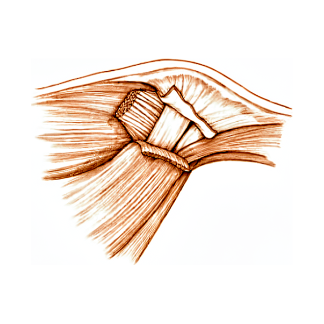
आपको क्या महसूस होता है
अंगूठे की यूसीएल चोट (अल्नर कोलैटरल लिगामेंट की चोट) आपके अंगूठे के भीतरी किनारे पर मौजूद लिगामेंट (स्नायुबंधन) को प्रभावित करती है, उस जोड़ पर जहां अंगूठा आपके हाथ से मिलता है। यह चोट आमतौर पर तब लगती है जब आपका अंगूठा झटके से ज़ोर से एक तरफ़ मुड़ जाता है, अक्सर फैले हुए हाथ के बल गिरने पर। स्कीइंग में ऐसा तब हो सकता है जब आप स्की पोल (छड़ी) पकड़े-पकड़े गिर जाएं, और स्नोबोर्ड करने वालों के अंगूठे में गिरने और मुड़ने के दौरान चोट लग सकती है।
दर्द आपके अंगूठे के भीतरी किनारे पर, हथेली के सबसे पास वाले जोड़ पर होता है। यह आमतौर पर किसी अचानक चोट से शुरू होता है, और साथ में उस जगह सूजन होती है और छूने पर दर्द होता है। चुटकी भरने, पकड़ने और ऐसी किसी भी हरकत से दर्द होगा जो आपके अंगूठे को एक तरफ़ धकेलती है। रोज़ के वे काम मुश्किल हो जाते हैं जो अंगूठे पर इस तरह भार डालते हैं: चाबी घुमाना, जार खोलना, भारी पैन (कड़ाही) को उसके किनारे से पकड़ना। आपका अंगूठा अस्थिर भी लग सकता है, मानो किसी चीज़ पर दबाने पर वह जवाब दे देता हो।
कुछ लोगों को अंगूठे के जोड़ के भीतरी किनारे पर एक छोटी, सख्त गांठ दिखती है। दूसरे अंगूठे की तुलना में आपका अंगूठा थोड़ा टेढ़ा बैठा या थोड़ा घूमा हुआ भी लग सकता है। अगर चोट कुछ समय पहले लगी थी, तो दर्द शायद उतना साफ़ न हो, लेकिन चुटकी भरते या पकड़ते समय अंगूठा फिर भी कमज़ोर या ढीला लग सकता है।
इन लक्षणों पर नज़र रखें और उसी दिन कदम उठाएं: कोई उंगली, हाथ या बांह जो गर्म, लाल, सूजी हुई और दर्दनाक हो जाए, ख़ासकर बुखार के साथ; चोट वाले जोड़ के ऊपर त्वचा कटी या फटी हुई हो, हड्डी दिख रही हो, या साफ़ दिखने वाला टेढ़ापन हो; अंगूठा या हाथ जो फीका (सफ़ेद पड़ा हुआ), ठंडा, सफ़ेद या नीला पड़ जाए; या चोट के बाद अचानक महसूस करने की क्षमता या हिलने-डुलने की ताकत चली जाए। इनके लिए तुरंत इमरजेंसी विभाग में इलाज की ज़रूरत है, और इसके लिए जीपी के रेफ़रल की ज़रूरत नहीं है।
अगर आपके लक्षण शांत नहीं हो रहे हैं, सप्ताहों में बिगड़ते जा रहे हैं, रात में आपको जगा देते हैं, या आपको काम करने या अपना हाथ इस्तेमाल करने से रोकते हैं, तो अपने जीपी को दिखाएं या विशेषज्ञ से जांच के लिए कहें।
असल में क्या हो रहा है
आपके अंगूठे के भीतरी किनारे पर, जहां वह आपके हाथ से जुड़ता है, लिगामेंट का एक मज़बूत पट्टा होता है जिसे अल्नर कोलैटरल लिगामेंट कहते हैं। इसे तंबू के खंभे को स्थिर रखने वाली खिंची हुई रस्सी की तरह समझें: जब आप चुटकी भरते या पकड़ते हैं, तो यह अंगूठे के जोड़ को एक तरफ़ मुड़ने से रोकता है। हर बार जब आप अपने अंगूठे को किसी उंगली से दबाते हैं, तो यही पट्टा भार उठाता है। इसीलिए यहां फटाव होने पर ठीक इन्हीं कामों में कमज़ोरी और दर्द दिखाई देता है।
जब आपका अंगूठा ज़ोर से एक तरफ़ मुड़ता है, तो यह पट्टा खिंच सकता है, आंशिक रूप से फट सकता है, या पूरी तरह आर-पार फट सकता है। डॉक्टर इसे इस हिसाब से ग्रेड (दर्जों) में बांटते हैं कि यह कितना फटा है: खिंचाव या आंशिक फटाव, और पूरा फटाव जिसमें लिगामेंट हड्डी से अलग हो जाता है। कभी-कभी लिगामेंट हड्डी के एक छोटे टुकड़े के साथ हड्डी से उखड़ जाता है। आंशिक फटाव आमतौर पर स्प्लिंट में भर सकता है। पूरा फटाव अक्सर नहीं भर पाता, क्योंकि फटा हुआ सिरा पलटकर पीछे जा सकता है और जोड़ के ऊपर मौजूद ऊतक की एक परत के नीचे फंस सकता है। जब ऐसा होता है, तो लिगामेंट उस हड्डी से दूर पड़ा रह जाता है जिससे उसे दोबारा जुड़ना है, इसलिए वह अपने-आप वापस नहीं जुड़ पाता।
अगर फटाव का इलाज न किया जाए, तो जोड़ ढीला रहता है। जब अंगूठे पर एक तरफ़ से भार पड़ता है, तो वह बार-बार जवाब दे देता है, और सालों में यह लगातार हिलना-डुलना जोड़ को घिस सकता है और गठिया (आर्थराइटिस) का कारण बन सकता है। इसीलिए इस चोट को गंभीरता से लिया जाता है, भले ही यह एक साधारण मोच जैसी लगे।
हम इसके लिए क्या कर सकते हैं
डॉ. कीरन हिरपरा, माटर प्राइवेट हॉस्पिटल, रॉकहैम्पटन में ऊपरी अंग (कंधे से हाथ तक) के सर्जन, इलाज को आपकी खास चोट के हिसाब से तय करते हैं। अंगूठे के लिगामेंट के कुछ फटाव बिना ऑपरेशन के भर जाते हैं, और कुछ में जल्द ही सर्जरी की ज़रूरत होती है, इसलिए जल्दी जांच करवाना मायने रखता है। मरीज़ों को आमतौर पर उनके जीपी (आपके फैमिली डॉक्टर) हमारे क्लिनिक में रेफ़र करते हैं; अगर किसी फिजियोथेरेपिस्ट ने आपको हमसे मिलने की सलाह दी है, तब भी मेडिकेयर रिबेट पाने के योग्य होने के लिए आपको अपने जीपी से रेफ़रल की ज़रूरत होगी। क्लिनिक में हम आपकी पूरी जानकारी लेते हैं, आपके अंगूठे की जांच करते हैं, और जहां ज़रूरत हो वहां इमेजिंग (स्कैन या एक्स-रे) करवाते हैं। इसमें एक्स-रे, अल्ट्रासाउंड स्कैन या एमआरआई शामिल हो सकते हैं; एमआरआई एक तेज़ चुंबक की मदद से लिगामेंट की विस्तृत तस्वीरें लेता है। इन स्कैन से पता चलता है कि लिगामेंट पूरी तरह आर-पार फटा है या नहीं, और फटा हुआ सिरा पलटकर अपनी जगह से बाहर तो नहीं चला गया है।
अगर फटाव आंशिक है, या लिगामेंट हड्डी के एक ऐसे छोटे टुकड़े के साथ उखड़ गया है जो अपनी जगह से खिसका नहीं है, तो हम आमतौर पर अंगूठे को 4 सप्ताह तक प्लास्टर (कास्ट) में स्थिर रखते हैं। उसके बाद आप 3 और सप्ताह तक एक निकाला जा सकने वाला स्प्लिंट पहनते हैं और हिलाने-डुलाने के हल्के व्यायाम शुरू करते हैं। आपकी हैंड थेरेपिस्ट (हाथ की विशेषज्ञ थेरेपिस्ट), एक्सटेंड रिहैबिलिटेशन में रूबी डूलन, ये व्यायाम तय करती हैं और आपको जिस भी स्प्लिंट की ज़रूरत हो, वह बनाती हैं। जिस पूरे फटाव में फटा हुआ सिरा पलटकर अपनी जगह से बाहर नहीं गया है, उसमें आमतौर पर पहले स्थिर रखकर (इमोबिलाइज़ेशन) इलाज आज़माया जाता है: लिगामेंट को वापस जुड़ने देने के लिए जोड़ को 4 से 6 सप्ताह तक स्थिर रखा जाता है।
सर्जरी पर तब विचार किया जाता है जब फटाव पूरा और अस्थिर हो, जब फटे हुए लिगामेंट का सिरा पलटकर अपनी जगह से बाहर चला गया हो, या जब हड्डी का कोई टुकड़ा अपनी सीध से खिसक गया हो। ऐसे मामलों में लिगामेंट अपने-आप हड्डी पर वापस नहीं जुड़ सकता, इसलिए ऑपरेशन से उसे वापस जोड़ा जाता है। अगर चोट कुछ समय पहले लगी थी और लिगामेंट का अब सीधे रिपेयर नहीं किया जा सकता, तो हम आपकी अपनी बांह के टेंडन (कंडरा) की एक पट्टी से या दान किए गए ऊतक से उसे दोबारा बना सकते हैं। इस ऑपरेशन का अपना अलग पेज है, और आपके कोई भी फ़ैसला लेने से पहले हम आपसे बात करेंगे कि इसमें क्या-क्या होता है।
क्या उम्मीद करें
इलाज से अंगूठे के लिगामेंट के ज़्यादातर फटाव अच्छी तरह ठीक हो जाते हैं। स्प्लिंट या प्लास्टर में स्थिर रखा गया आंशिक फटाव आमतौर पर भर जाता है, और चुटकी भरने और पकड़ने के लिए आपके अंगूठे की ताकत वापस आ जाती है। अगर आपको ऑपरेशन की ज़रूरत हो, तो नतीजे आमतौर पर अच्छे होते हैं: ज़्यादातर लोगों में अंगूठे का हिलना-डुलना, चुटकी और पकड़ की ताकत बनी रहती है, और सर्जरी के बाद समस्याएं कम ही होती हैं। जिन लोगों की यह सर्जरी हुई, वे अपने पहले वाले स्तर पर खेल खेलने लौटे हैं, और पेशेवर खिलाड़ियों ने बाद में लगभग उतने ही मैच खेले और उनका करियर लगभग उतना ही लंबा रहा।
ठीक होने में धैर्य चाहिए। शुरुआत में आपके अंगूठे को स्थिर रखा जाएगा, क्योंकि शुरुआती जकड़न ही वह मुख्य चीज़ है जिससे आगे चलकर बचना है। एक बार हिलाने की अनुमति मिल जाने पर हल्के, धीरे-धीरे बढ़ने वाले व्यायाम ज़रूरी हैं: भर रहे लिगामेंट पर धीरे-धीरे भार डालने और उसे स्ट्रेच करने (खींचने) से हिलना-डुलना और अंगूठे का काम बेहतर होता है। मरीज़ों के खुद बताए गए नतीजे सर्जरी से पहले की तुलना में सर्जरी के तीन और 12 महीने बाद काफ़ी बेहतर होते हैं। अगर आपकी चोट कुछ समय पहले लगी थी, तब भी रिपेयर कारगर होता है, हालांकि जल्दी किए गए रिपेयर की तुलना में देर से किए गए रिपेयर में अंगूठे का हिलना-डुलना आमतौर पर थोड़ा ज़्यादा कम हो जाता है।
बिना इलाज के, आगे की तस्वीर अलग होती है। चार में से तीन पूरे फटाव आराम देने पर भी अपने-आप नहीं भरते। जोड़ ढीला रहता है, भार पड़ने पर आपका अंगूठा बार-बार जवाब देता रहता है, और चुटकी कमज़ोर और दर्दनाक बनी रहती है। सालों में यह लगातार ढीलापन जोड़ को घिस देता है: जिन लोगों के पुराने (क्रॉनिक) फटाव का बाद में रिपेयर हुआ, उनमें से 88% के अंगूठे के जोड़ में पहले से ही कुछ गठिया था। चोट के समय ज़्यादा उम्र का संबंध उस जोड़ में ज़्यादा गंभीर गठिया से है।
आगे की योजना बनाते समय कुछ बातें जानना फ़ायदेमंद है। अगर आपका अंगूठा गर्म, लाल, सूजा हुआ और दर्दनाक हो जाए, ख़ासकर बुखार के साथ, तो उसी दिन इमरजेंसी विभाग जाएं; इसके लिए जीपी के रेफ़रल की ज़रूरत नहीं है। अगर लक्षण शांत नहीं हो रहे हैं या सप्ताहों में बिगड़ते जा रहे हैं, तो अपने जीपी को दिखाएं या विशेषज्ञ से जांच के लिए कहें।
डॉक्टर को कब दिखाएं
किसी भी ऐसी चोट के बाद जिसमें आपका अंगूठा ज़ोर से एक तरफ़ मुड़ गया हो, जल्द ही उसकी जांच करवाएं, भले ही वह एक साधारण मोच जैसी लगे। पूरे फटाव की जांच जितनी जल्दी होती है, आपके पास उतने ज़्यादा विकल्प होते हैं, क्योंकि बहुत लंबे समय तक छोड़े गए फटाव में बड़े ऑपरेशन की ज़रूरत पड़ सकती है या उससे स्थायी जकड़न और कमज़ोरी रह सकती है। विशेषज्ञ से जांच के लिए कहें अगर चुटकी भरते या पकड़ते समय आपका अंगूठा अब भी जवाब दे देता है, अगर कुछ सप्ताह बाद भी चुटकी कमज़ोर या दर्दनाक बनी रहती है, या अगर आपको जोड़ के भीतरी किनारे पर एक छोटी, सख्त गांठ महसूस होती है। अगर चोट कुछ समय पहले लगी थी और आपका अंगूठा ढीला है या घिस रहा है, तब भी जांच करवाना फ़ायदेमंद है। और अगर आपका अंगूठा गर्म, लाल, सूजा हुआ और दर्दनाक हो जाए, ख़ासकर बुखार के साथ, तो उसी दिन इमरजेंसी विभाग जाएं।
विस्तार से
Advanced reading: the deeper science (optional)
यह हिस्सा उससे आगे जाता है जितना आपको अपने इलाज के फ़ैसलों के लिए जानना ज़रूरी है। अंगूठे के अल्नर कोलैटरल लिगामेंट की चोट के बारे में ज़्यादा पढ़ना इसलिए फ़ायदेमंद है क्योंकि पूरा फ़ैसला शरीर की बनावट से जुड़े एक ही सवाल पर टिका है, कि फटा हुआ लिगामेंट पलटकर पहुंच से बाहर तो नहीं चला गया, और क्योंकि अब इसका साफ़ जवाब है कि इसे तय करने के लिए कौन-सा स्कैन इस्तेमाल करना चाहिए।
स्टेनर लीज़न, और यह सब कुछ क्यों बदल देता है
अल्नर कोलैटरल लिगामेंट हर चुटकी और पकड़ के दौरान लगने वाले बगल के ज़ोर के खिलाफ़ अंगूठे को स्थिर रखता है। जब यह अपने जुड़ने की जगह से फटता है, तो आमतौर पर ऐसी जगह रहता है जहां यह भर सके।
कभी-कभी ऐसा नहीं होता। टेंडन की एक परत, जिसे एडक्टर एपोन्यूरोसिस कहते हैं, लिगामेंट के ऊपर (त्वचा की ओर) होती है, और अगर फटा हुआ सिरा सिकुड़कर उस परत के ऊपर चला जाए, तो एपोन्यूरोसिस लिगामेंट और हड्डी के बीच आ जाती है। इसे स्टेनर लीज़न कहते हैं, और यही कारण है कि इस चोट का इलाज ज़्यादातर मोचों से अलग तरह से किया जाता है: लिगामेंट अब उस हड्डी को छू नहीं रहा होता जिससे उसे जुड़कर भरना है, इसलिए कितना भी स्प्लिंट लगाया जाए, वह उसे वापस नहीं जोड़ेगा।
इलाज से जुड़ी हर बात इसी पर निर्भर करती है कि ऐसा हुआ है या नहीं।
इसका जवाब देने के लिए अल्ट्रासाउंड काफ़ी है
क्योंकि फ़ैसला हां-या-ना वाला है और उसके नतीजे बड़े हैं, इसलिए इमेजिंग का सवाल असामान्य रूप से साफ़ तौर पर तय है — और इसका जवाब मिल चुका है। 422 मरीज़ों में, स्टेनर लीज़न का पता लगाने में अल्ट्रासाउंड और एमआरआई दोनों की सटीकता ऊंची पाई गई, और अल्ट्रासाउंड पहली जांच के रूप में इस्तेमाल करने के लिए उचित इमेजिंग तरीका है [1]।
यह व्यवहार में काम आने वाली जानकारी है। अल्ट्रासाउंड एमआरआई से जल्दी होता है, सस्ता है और आसानी से उपलब्ध है, और जहां कोई अनुभवी जांच करने वाला मौजूद हो, वहां जो इसने पहले ही दिखा दिया है उसकी पुष्टि के लिए बाद में एमआरआई की ज़रूरत नहीं होती। यह डायनेमिक (हरकत के दौरान) भी होता है, स्कैन के दौरान अंगूठे पर ज़ोर डालकर जांच की जा सकती है।
जहां स्टेनर लीज़न नहीं है, वहां ढीलापन फ़ैसला तय करता है
ब्रिटिश सोसाइटी फ़ॉर सर्जरी ऑफ़ द हैंड की गाइडलाइन इसका रास्ता सीधे-सीधे बताती है। नई (एक्यूट) चोट वाले मरीज़ों की जांच उनकी जानकारी (हिस्ट्री), शारीरिक जांच और एक्स-रे से की जानी चाहिए। जिनमें जोड़ का कोई खास ढीलापन नहीं है, उनका इलाज बिना सर्जरी के किया जा सकता है, जबकि जिनमें खास ढीलापन है, उनका इलाज आपस में मिलकर लिए गए फ़ैसले के बाद, या तो बिना सर्जरी के स्थिर रखकर या सर्जिकल रिपेयर से किया जा सकता है [2]।
यहां दो बातें ध्यान देने लायक हैं। पहली यह कि एक्स-रे उन्नत इमेजिंग से पहले किए जाते हैं, कुछ हद तक एवल्शन फ्रैक्चर (लिगामेंट के साथ हड्डी का टुकड़ा उखड़ जाना) को जांचकर अलग करने के लिए, जो इलाज को फिर से बदल देता है। दूसरी यह कि खास ढीलापन होने पर भी सर्जरी अनिवार्य नहीं है: गाइडलाइन साफ़ तौर पर इसे स्थिर रखने और रिपेयर के बीच एक साझा फ़ैसला बताती है, जो ऑपरेशन को एकमात्र रास्ता बताने से ज़्यादा ईमानदार रुख है।
गलती होने के नतीजे खास तरह के क्यों होते हैं
काम न कर पाने वाला अल्नर कोलैटरल लिगामेंट आराम के समय दर्द या ज़्यादातर गतिविधियों में दिक्कत पैदा नहीं करता। इससे ऐसा अंगूठा बनता है जो बगल से भार पड़ने पर जवाब दे देता है, चाबी घुमाते समय, जार खोलते समय, भारी कड़ाही को किनारे से पकड़ते समय, क्योंकि चुटकी के लिए दबाने को एक स्थिर टेक चाहिए।
इलाज न होने पर, लंबे समय तक रहने वाली अस्थिरता से सालों में उस जोड़ में गठिया हो जाता है। यही कारण है कि जो चोट एक साधारण मोच जैसी लगती है, उसकी भी पक्की जांच होनी चाहिए: इसे नज़रअंदाज़ करने की कीमत चोट के बाद के सप्ताहों में महसूस नहीं होती, जब अंगूठा ठीक हो जाता है और सही लगता है, बल्कि बहुत बाद में होती है।
संदर्भ
[1] Qamhawi Z, Shah K, Kiernan G, Furniss D, Teh J, Azzopardi C. Diagnostic accuracy of ultrasound and magnetic resonance imaging in detecting Stener lesions of the thumb: systematic review and meta-analysis. J Hand Surg Eur Vol. 2021;46(9):946-53. https://doi.org/10.1177/1753193421993015
[2] Dean B, Rodrigues J, Riley N, Rabey N, Donnison E, Challen K, et al. Guideline on managing thumb ulnar collateral ligament injuries: the British Society for Surgery of the Hand. J Hand Surg Eur Vol. 2024;49(10):1195-201. https://doi.org/10.1177/17531934241274612
Evidence & references
This is the clinical evidence summary written for health professionals. It is technical, and it lists the research this page was built from. You do not need to read it to understand your treatment or to make a decision about it.
Overview
Diagnosis and Imaging
- Ultrasonography has evolved as a reliable adjunct to clinical examination in the evaluation of the ulnar collateral ligament (UCL) of the thumb [1].
- MRI evaluation reveals greater details enabling better understanding and management of ulnar collateral ligament injuries of the thumb metacarpophalangeal (MCP) joint [2].
- A 4-stage, treatment-oriented classification of thumb UCL injury is based on the degree of UCL displacement, with correlation with the likelihood of success with either immobilization or operative intervention [10].
- The presence of a displaced fleck sign has implications for offering surgery to patients with thumb UCL injuries because of a high likelihood of a Stener lesion [17].
Clinical Indications and Natural History
- Valgus laxity greater than 30 degrees or more than 15 degrees more than in the noninjured thumb with the MCP joint in extension is an indication for operative management [3].
- Untreated ulnar collateral ligament injury of the thumb metacarpophalangeal joint can lead to long-term pain and functional limitations [5].
- 75% of cases with complete rupture of the ulnar collateral ligament of the thumb will fail to heal with conservative treatment [13].
- The rate of surgery for acute thumb MP UCL injury varies based on patient characteristics and the individual treating surgeon [6].
Treatment Outcomes
- Surgical management of thumb UCL injuries produces overall favorable results [4].
- Patient-reported outcomes improve significantly at three and 12 months after open surgical repair of the thumb UCL compared to baseline [24].
- Patients with both acute and chronic thumb UCL injuries have similarly acceptable functional outcomes, postoperative pain, and satisfaction [11].
- Return-to-play rates after surgical treatment of thumb UCL injuries are high, with reassuring return to preinjury level of play with few complications [9].
- Thumb ulnar collateral ligament injuries can be safely and effectively managed by football position demands [14].
- Players who underwent thumb UCL repair played in a similar number of games per season and had similar career lengths in the MLB as controls [16].
- Treatment of a thumb UCL complete rupture (Grade III) using a custom-made hinged splint shows promise in the conservative management of compliant patients [19].
Surgical Techniques and Rehabilitation
- A controlled active motion therapy protocol after suture anchor repair of a ruptured UCL of the thumb is safe from a biomechanical point of view [8].
- Intraosseous suture anchor repair is a safe and effective method for repair of complete tears of the ulnar collateral ligament of the thumb MP joint [12].
- Both pull-out sutures and bone anchor techniques are safe and effective for treating thumb ulnar collateral ligament injuries [23].
- Thumb UCL repair with suture tape augmentation demonstrates short-term outcomes comparable to what has been reported for other methods of repair [26].
- Mini hook plate fixation could be an alternative treatment technique for thumb UCL avulsion fractures [21].
- The surgical treatment of an ulnar collateral ligament lesion of the thumb using the Fiji Anchor can lead to an excellent clinical outcome with a minor complication rate [48].
- An accelerated rehabilitation protocol has been clinically applied in athletes following thumb MCP UCL repair with suture tape augmentation [22].
- The patient is placed in the supine position with a small bump on which to rest the thumb for open repair of the UCL [28].
- The surgeon should position themselves between the shoulder and head to allow easier access to the ulnar aspect of the thumb during UCL repair [28].
- A lazy S approach is used on the ulnar aspect of the thumb for UCL repair [28].
- Careful identification of the radial sensory nerve is required during UCL repair, with prior documentation of sensibility [28].
- The adductor aponeurosis is incised on the ulnar side during UCL repair [28].
- The site of rupture for the UCL is identified as distal off the phalanx, midsubstance, or proximal [28].
- Nonabsorbable suture with pullout button, bone anchors, or figure-of-eight imbrication of midsubstance repair are options for repairing the UCL [28].
- Motion is limited for the first 4 to 6 weeks depending on chronicity of the injury, with more chronic injuries immobilized longer [28].
- Guided therapy at 6 weeks in a removable splint is part of the postoperative protocol for UCL repair [28].
Anatomy & Pathophysiology
Bony Anatomy and Joint Mechanics
- The thumb metacarpophalangeal (MCP) articulation is of a condylar type [52].
- Flexion of the thumb MCP joint is always accompanied by radial deviation and pronation, which stretches the ulnar metacarpophalangeal ligament [52].
- The stability of the thumb MCP joint is considered more important than movement from a functional viewpoint [52].
- The carpometacarpal ray of the thumb is anterior to the plane of the other metacarpals and makes an angle of about 47 degrees with the second ray [52].
- The distal articular surface of the trapezium is likened to a saddle whose concave curvature lies in a dorsopalmar direction [52].
- The corresponding surface on the base of the first metacarpal presents a grooved surface in the radioulnar direction [52].
- The trapeziometacarpal joint has two longitudinal axes and two degrees of freedom [52].
- The shape of the trapeziometacarpal articular surface does not make for good stability except in anteposition and pronation when the two surfaces are congruent [52].
- The necessary stability of the trapeziometacarpal joint is provided by a complex ligamentous system which does not limit mobility [52].
- This ligamentous system essentially consists of a strong ulnar ligament from the base of the first metacarpal to the second metacarpal and to the trapezoid [52].
- The ulnar ligament of the trapeziometacarpal joint is the structure that resists in a Bennett fracture [52].
- The radial side of the trapeziometacarpal joint has a much weaker ligament [52].
- There is an intrinsic instability at the level of the trapeziometacarpal joint where the pressure is very high [52].
- This pressure is proportional to the force of the grip at the distal end of the thumb and to the length of the lever arm [52].
- Cooney and Chao (1977) found that joint compression forces during simple pinch averaged 3.0 kg of force at the interphalangeal joint, 5.4 kg at the metacarpophalangeal joint, and 12.0 kg at the carpometacarpal joint [52].
- Compression forces of up to 120 kg may occur at the carpometacarpal joint during strong grasp [52].
- Stability of the trapeziometacarpal joint is dynamically provided by a single structure, the tendon of the abductor pollicis longus [52].
- The abductor pollicis longus tendon has stabilizing function only when the first metacarpal is abducted [52].
- In adduction, the long abductor increases the risk of subluxation of the trapeziometacarpal joint [52].
Ligamentous Anatomy and Injury Patterns
- The ulnar collateral ligament (UCL) serves as the primary stabilizer against valgus stress at the thumb MCP joint [20].
- The UCL plays an essential role in pinch strength, grasping function, and dexterity [20].
- The mechanism of injury to the UCL is sudden, forced radial deviation (abduction), often resulting from a fall on an outstretched hand with the thumb abducted [44].
- In skiing, if a person falls while gripping the ski pole, the handle abducts the thumb [44].
- Associated injuries to UCL tears include tears of the dorsal capsule and ulnar aspect of the volar plate [44].
- Associated injuries to UCL tears occasionally include a rent in the adductor aponeurosis [44].
- Volar subluxation of the MCP joint may result from concomitant tears of the dorsal capsule and UCL [44].
- Distal tears of the UCL at its insertion in the proximal phalangeal base are more common than proximal tears from the metacarpal [44].
- Ruptures within the substance of the ligament occasionally occur [44].
- The collateral ligaments of the thumb MP joint afford lateral stability and resist volar subluxation [44].
- If one collateral ligament is torn, the proximal phalanx tends to rotate volarly on the side of the tear, with the opposite intact ligament serving as the axis [44].
- In the case of an isolated UCL rupture, the proximal phalanx rotates in supination around the intact radial collateral ligament [44].
- An avulsion fracture of the ulnar base of the proximal phalanx at the insertion of the ligament is the most common fracture pattern associated with UCL injuries [44].
- Typically, the avulsion fracture fragment is small and includes little of the articular surface [44].
- Fractures involving more than 10% of the articular surface can occur and may require fixation if they are displaced 2 mm or more and associated with articular incongruity [44].
- Avulsion fractures from the metacarpal head and intraarticular shearing fractures of the volar surface of the radial condyle of the metacarpal head have also been reported [44].
- A rare but potentially problematic fracture pattern involves a rupture of the UCL from the proximal phalanx and a simultaneous articular shear fracture of the proximal phalangeal base [44].
- In the skeletally immature individual, isolated rupture of the UCL without a Salter fracture of the proximal phalanx is rare but does occur [44].
- The UCL ruptures off the base of the proximal phalanx [62].
- The avulsed ligament, with or without a bony fragment, can become displaced above the adductor aponeurosis, preventing healing [62].
- This displacement is known as a Stener lesion [62].
- In 25 of 39 patients with complete ruptures of the UCL, Stener found the adductor aponeurosis interposed between the ruptured ulnar collateral ligament and its site of insertion on the base of the proximal phalanx [47].
- On clinical examination, a prominent lump can be palpated that represents the ulnar collateral ligament being proximally and superficially displaced by the adductor aponeurosis [47].
- Pathologic rotation of the thumb may be evident in Stener lesions [47].
- If left uncorrected, a Stener lesion prevents proper healing and leads to chronic instability and subsequent arthrosis [47].
- Other injuries associated with tears of the UCL include avulsion fractures, dorsal capsular tears, and volar plate tears [47].
- The adductor aponeurosis interposition prevents direct ligament healing without surgery [42].
- The UCL is typically avulsed distally [35].
- Although the injury is often at the distal portion where it attaches to the proximal volar aspect of the phalanx, midsubstance and metacarpal-based tears can occur [35].
- The anatomic attachment site for a distal avulsion of the UCL is 25% dorsal from the volar surface of the proximal phalanx [94].
- A single anchor is placed volar to the axis of the joint for distal avulsion repair [94].
- The radial collateral ligament (RCL) ruptures from its origin, from its insertion, or at the midsubstance [62].
- RCL ruptures are frequently associated with dorsal or dorsoradial capsular tears and with extensor pollicis brevis avulsions or tears [62].
- No equivalent of the Stener lesion exists for the RCL [62].
- RCL ruptures seem susceptible to chronic instability [62].
- The radial-based abductor cannot create a Stener-type lesion [63].
Pathophysiology and Biomechanics
- Sequential tearing of the thumb UCL leads to progressive instability of the MCP joint [7].
- Untreated complete ruptures may result in chronic instability, weakness of pinch, pain, and functional impairment [20].
- Causes of chronic instability of the UCL include an untreated acute tear, failure to recognize a Stener lesion, or progressive attenuation of the ligament [100].
- Patients with chronic instability usually present with pain, swelling, and weakness of the involved thumb [100].
- The pain tends to be exacerbated by forceful pinch and activities requiring torsional motions of the hand, such as unscrewing jar tops [100].
- Holding large objects like a half-gallon container of milk may be painful and may cause a feeling of instability or lack of power because of the thumb’s inability to resist counterpressure [100].
- Chronic instability is manifested by gross instability to valgus stress with no endpoint [100].
- Crepitus with active motion suggests degenerative disease in chronic UCL instability [100].
- The resting posture of the thumb should be observed for volar subluxation or radial deviation of the MP joint in chronic instability [100].
- Biomechanically, there are no treatments of repair or reconstruction using native tissues that provide equivalent strength to the preinjured ligament [105].
- No static ligament reconstruction restores the normal stability characteristics of the thumb UCL [15].
- Single- and double-bundle UCL reconstructions of the thumb MCP joint have comparable biomechanical properties in regard to joint congruity under valgus load [40].
- The reconstruction tunnel positioning of triangular configuration with apex proximal stabilizes the thumb MCP joint while maintaining flexion/extension range of motion [60].
- Although both suture tape and suture anchor constructs recapitulate native joint stiffness, repair with suture tape demonstrated the greatest biomechanical strength in stiffness and load-to-failure [90].
- In round joints, only the non-anatomic Glickel procedure restores normal range of motion and stability [111].
- In flat joints, the Glickel procedure and a modified Fairhurst configuration with origins dorsal in the metacarpal head restore range of motion and stability [111].
- Nonanatomic repositioning of the UCL might affect joint motion [30].
- Palmar placement of the UCL origin on the metacarpal increased radial deviation from a mean of 18° to a mean of 27° compared with the intact joint [30].
- Proximal placement of the UCL origin on the metacarpal decreased radial deviation from a mean of 18° to a mean of 10° compared with the intact joint [30].
- Dorsal placement of the UCL insertion on the phalanx increased radial deviation from a mean of 18° to a mean of 25° compared with the intact joint [30].
- Distal positioning of the UCL insertion on the phalanx decreased radial deviation from a mean of 18° to a mean of 11° compared with the intact joint [30].
- Performing a physical examination to assess the amount of instability of an ulnar collateral ligament injury did not create a Stener lesion if the exam was performed in a controlled, gentle manner with the thumb held without rotation [112].
- It was possible to create a Stener lesion when the thumb was tested in a flexed and supinated position, but only if there was sectioning of the proper UCL, the accessory UCL, and the origin of the ulnar sagittal band at the adductor aponeurosis [112].
- Supination and flexion of the thumb provided less stability when both components of the UCL were sectioned [112].
- Pronation and supination play a role in stability of the thumb MCP joint [112].
Classification
- The BSSH BEST guideline defines significant UCL laxity as no firm endpoint, OR > 20 degrees laxity vs. the other side, OR > 30 degrees laxity [39].
- The term ‘Stener lesion’ is used to describe when the ligament is completely torn and is displaced superficial to the adductor aponeurosis [18].
- In a Stener lesion, the ruptured ligament becomes displaced superficial to the adductor aponeurosis and is unable to heal anatomically without surgical intervention [20].
- Presence of a displaced fleck sign has implications for offering surgery to patients with thumb UCL injuries because of a high likelihood of a Stener lesion [17].
Clinical Presentation
Symptoms and Signs
- Patients commonly report pain, swelling, and ecymosis around the thumb metacarpophalangeal joint [47].
- Tenderness is greatest over the ulnar aspect of the thumb metacarpophalangeal joint [47].
- A prominent lump can be palpated representing the ulnar collateral ligament being proximally and superficially displaced by the adductor aponeurosis in the presence of a Stener lesion [47].
- Pathologic rotation of the thumb may be evident in cases of Stener lesion [47].
- Incomplete acute tears and nondisplaced avulsion fractures are managed with a thumb spica cast for up to 4 weeks, followed by removable protective splinting for 3 more weeks with active range-of-motion exercises [42].
- Skier's thumb can lead to chronic pain and instability when diagnosed incorrectly [83].
Clinical Examination
- The metacarpophalangeal joint is generally examined by applying a radial force in extension and with approximately 30° of MCPJ flexion to relax the palmar plate [18].
- Physical examination is performed with a valgus stress applied in neutral rotation to the thumb in both extension (testing the accessory ligament) and 30° to 35° flexion (proper ligament) [42].
- An injured thumb that shows more than 30 degrees of instability compared with the uninjured side indicates a complete rupture [47].
- Valgus instability of more than 30°—or more than 10° compared with the contralateral thumb in both flexion and extension—indicates complete rupture of the proper and accessory UCL [80].
- Instability of the MCP joint in flexion indicates rupture of the proper UCL only [80].
- Comparison with the uninjured contralateral thumb is unreliable in many individuals due to significant variation between right and left thumbs [32].
- New evidence suggests that a lack of definite end point rather than comparison with the uninjured side should be used to define complete UCL rupture [42].
- If a hard endpoint is not encountered and there is increased deviation (>20 degrees) at the MP joint, surgery is recommended [35].
- For those patients with significant guarding, a digital block may be necessary to carry out the stress test [35].
- The ulnar side of the joint should be palpated for a Stener lesion, where both the proper and accessory ligament are retracted and lie on the adductor aponeurosis [42].
- Physical examination is required to determine stability with injuries to the ulnar structures of the thumb metacarpophalangeal joint [31].
- Clinical examination is recommended to assess for significant laxity of the UCL, defined as no firm endpoint, or > 20 degrees laxity vs. the other side, or > 30 degrees laxity [39].
Imaging and Diagnostic Tests
- Plain radiographs should be obtained prior to any stress examinations to avoid displacing an otherwise nondisplaced fracture [42].
- Plain radiographs should be obtained prior to any stress examinations to exclude obvious fractures and dislocation or subluxation [58].
- Anteroposterior stress radiographs can be obtained of both thumbs for comparison purposes [47].
- Stress radiographs are useful for identifying Stener lesions, however, these are often uncomfortable for the patient and guarding may lead to false-negative results [42].
- Fluoroscopy can aid the diagnosis to see if the joint becomes incongruent and to measure the deviation compared to the contralateral limb [35].
- Ultrasonography has evolved as a reliable adjunct to clinical examination in evaluation of the UCL of the thumb [1].
- MRI evaluation reveals greater details enabling better understanding and management of ulnar collateral ligament injuries of the thumb MCP joint [2].
- Ultrasound had a positive predictive value for rupture of 94% when compared with the operative finding [58].
- The overall agreement between specialist clinic examination and ultrasound for injury to the ulnar collateral ligament was 85% (kappa 0.647) [58].
- Senior House Officers in the A&E department were able to correctly diagnose only 45% of the ruptures of the ulnar collateral ligament [58].
- In a study of 49 patients, ultrasound identified Stener lesions with a sensitivity of 36% and specificity of 61% [51].
- MRI and US of suspected UCL injury did not demonstrate good enough sensitivity for the authors to recommend their implementation and use in clinical situations [51].
- A magnetic resonance imaging scan is recommended for additional preoperative planning because it provides a detailed assessment of tear location, injury grade, and ligament quality [80].
- Clinical assessment of a thumb ulnar collateral ligament injury should be supplemented with radiographs, as underlying pathology, such as an enchondroma, may be a factor [61].
Investigations
Clinical Examination
- The MCP joint is generally examined by applying a radial force in extension and with approximately 30° of MCP joint flexion to relax the palmar plate, which is thought to isolate the UCL proper [18].
- Careful stress testing of the injured and uninjured side in both extension and flexion can often elucidate those with a significant tear of their UCL/RCL [35].
- The absence of a normal “endpoint” with radial stress is a key diagnostic finding signifying a complete ligament tear [35].
- For patients with significant guarding, a digital block may be necessary to carry out the stress test [35].
- Patients with a soft endpoint, incongruent joint, or Stener lesion require surgical intervention [35].
Imaging
- Ultrasonography (US) has evolved as a reliable adjunct to clinical examination in evaluation of the UCL of the thumb [1].
- Diagnostic ultrasound is a useful, noninvasive tool for assessing the extent of rupture of the ulnar collateral ligament [114].
- Ultrasound or MRI can be helpful to gather more information when it is difficult to ascertain if there is a Stener lesion based on palpation [35].
- Ultrasound and MRI findings do not have 100% accuracy and their findings should only be one component of the decision-making algorithm [35].
Treatment
Non-Operative Management
- A thumb spica cast or functional brace is recommended for 4 to 6 weeks for incomplete ruptures of the ulnar collateral ligament [47].
- Tears of the UCL of the thumb MCP joint without a Stener lesion are believed to heal with 4 to 6 weeks of immobilization [62].
- In patients having less than 30 degrees of laxity of the UCL, less than a 15-degree differential in laxity compared with the contralateral side, and a discrete endpoint to joint opening, nonoperative treatment consists of immobilization of the MP joint for 4 weeks in neutral alignment and slight flexion [104].
- Untreated ulnar collateral ligament (UCL) injury of the thumb metacarpophalangeal joint can lead to long-term pain and functional limitations [5].
Indications for Surgery
- Injuries that are unstable (usually 30° more opening with radial stress than the opposite uninjured side) are believed to have Stener lesions and are treated surgically [62].
- Complete tears require open repair with suture anchors or bone tunnels placed at the site of the avulsion, which most commonly is the proximal phalanx [42].
- The tear will not heal in the setting of a Stener lesion because of adductor aponeurosis interposition [42].
- Acute complete rupture of the ulnar collateral ligament should be surgically repaired [47].
- Ulnar collateral ligament retraction more than 3 mm and interposed soft tissue are reasonable guides to surgical intervention [47].
Operative Techniques
- Repair of the UCL of the thumb is one of the most common ligamentous repairs in the hand, with options including a pullout suture and button, bone anchors, or a combination of the two with local tissue augmentation or imbrication [63].
- A K-wire across the joint is often employed to allow proper ligament tensioning and to maintain joint stability postoperatively [63].
- The surgical approach involves a lazy S incision on the ulnar aspect of the thumb, with careful identification and retraction of the dorsal sensory branch of the radial nerve [28].
- The adductor aponeurosis is incised longitudinally to allow repair after the collateral ligament repair [28].
- Direct repair of the ligament should be obtained with the use of one or two bone anchors, preferably small metallic anchors with stout nonabsorbable suture material [35].
- Additional sutures are used to augment the repair to the volar plate distally and capsule dorsally [35].
- The capsule and adductor aponeurosis should be repaired in separate layers [35].
- For chronic tears with excessive laxity, the adductor insertion can be advanced to increase dynamic stability [35].
- Both pull-out suture and bone anchor techniques are safe and effective for treating thumb ulnar collateral ligament injuries [23].
- In a biomechanical model, thumb UCL repair with suture tape augmentation demonstrated greater maximum and clinical failure loads compared with nonaugmented repair at time 0 [41].
- Arthroscopic treatment of acute complete thumb metacarpophalangeal ulnar collateral ligament tears resulted in key pinch, tip pinch, grip strength, and range of motion equal to or greater than in the thumb on the unaffected side in most cases [46].
Chronic Injury and Reconstruction
- If the diagnosis is delayed for 1 month or longer, fibrosis makes ligament identification and repair more difficult, although repair can be done by dissecting out the ligament from within the fibrotic mass and reattaching it appropriately [47].
- The detached tendinous insertion of the adductor muscle can be advanced and reattached to furnish a dynamic reinforcement in delayed repairs [47].
- If the repair is done several months after the injury, a graft can be used [47].
- Chronic ruptures of the UCL can be challenging to reconstruct, but most often there is enough local tissue present to allow for direct repair with the existing native ligament [35].
- Graft reconstruction of the UCL can often be accomplished with either an autograft palmaris/plantaris tendon or with an allograft [35].
- Newer techniques for chronic reconstruction involve the use of 3.0-mm drills, the graft, fibertape, and interference screws [35].
- An X or Y type configuration is often needed to appropriately reconstruct the surface area of the ligament to prevent recurrent instability in chronic cases [35].
- If a Y construct is used for chronic reconstruction, the single limb is placed distally and the dual limb proximally to prevent iatrogenic injury [35].
- A K-wire is highly recommended in chronic reconstruction to allow adequate healing and provide sufficient stability to permit early mobilization of the adjacent IP and CMC joints [35].
- The pin is typically removed in the office at 6 weeks following chronic reconstruction [35].
- Nonbiological ligament reconstruction of the thumb ulnar collateral ligament generates short-term outcomes comparable with those of biological reconstruction, potentially allowing for expedited recovery and rehabilitation [45].
- Patient-reported outcomes, including pain and function, improved after thumb UCL reconstruction with a tendon autograft [36].
- Increased time from injury to treatment (beyond 6 weeks) may decrease the likelihood of direct repair [104].
- If no local tissue is available for chronic repair, UCL reconstruction is most commonly performed with a free tendon graft [104].
- The graft is tensioned at 45 degrees of flexion with some ulnar deviation placed on the MCP joint during reconstruction [104].
- A relative contraindication to ligament reconstruction is marked volar subluxation and/or supination of the MP joint [104].
- The only absolute contraindication to reconstruction is osteoarthritis of the MP joint, in which case arthrodesis is a very dependable option [104].
Outcomes and Rehabilitation
- Collegiate football athletes treated for thumb UCL injuries with suture anchor repair had quick return to play, reliable return to the same level of activity, and excellent long-term clinical outcomes [33].
- Complications are rare and most patients show preservation of motion, key pinch, and grip strength [25].
- Thumb MCP joint collateral ligament injuries have a good outcome with appropriate diagnosis and treatment [34].
- There was no significant change in pinch strength in patients with chronic UCL reconstructions, but significantly better results were achieved in grip strength, flexion, extension, ulnar and radial deviation [29].
- This is the first clinical application of an accelerated rehabilitation protocol in athletes following thumb MCP UCL repair [22].
- Strenuous activity is avoided for 3 months, with unrestricted return to sport usually at 2 to 3 months [42].
- Limit motion for the first 4 to 6 weeks depending on chronicity of the injury; more chronic injuries are immobilized longer [28].
- Guided therapy at 6 weeks in a removable splint is part of the postoperative protocol [28].
- A thumb spica cast is worn for 4 weeks postoperatively after reconstruction, followed by hand therapy and protection for an additional 5 weeks in a splint [104].
Complications
Non-Operative
Operative
- Long-term dangers and the cost effectiveness of the procedure using the Fiji Anchor® are not known yet [48].
- Thumb spica casting risks placing additional stress upon adjacent joints and causing adjacent injury [37].
Recovery
Outcomes and Function
- Recovery of pinch grip strength measured using two point palmar pinch is on average within 92 per cent of expected values after normalization for dominance [66].
Return to Sport and Work
- Players who underwent thumb UCL surgery played in a similar number of games per season and had similar career lengths in the NFL as controls [67].
Rehabilitation Protocols
Key Evidence
- [L4] Ultrasonography (US) has evolved as a reliable adjunct to clinical examination in evaluation of the UCL of the thumb. [1] (10.1148/rg.264055117)
- [L4] MRI evaluation reveals greater details enabling better understanding and management of ulnar collateral ligament injuries of the thumb MCP joint. [2] (10.1016/s0363-5023(09)60139-8)
- [L5] Valgus laxity greater than 30 degrees or more than 15 degrees more than in the noninjured thumb with the MCP joint in extension is an indication for operative management. [3] (10.5435/00124635-199707000-00006)
- [L1] Surgical management of thumb UCL injuries produces overall favorable results. [4] (10.5435/jaaosglobal-d-25-00082)
- [L5] Untreated ulnar collateral ligament (UCL) injury of the thumb metacarpophalangeal joint can lead to long-term pain and functional limitations. [5] (10.2106/jbjs.k.01024)
- [L3] The rate of surgery for acute thumb MP UCL injury varies based on patient characteristics and the individual treating surgeon. [6] (10.1177/1558944716681974)
- [L5] Sequential tearing of the thumb UCL leads to progressive instability of the MCP joint. [7] (10.1177/1558944719868518)
- [L5] A controlled active motion therapy protocol after suture anchor repair of a ruptured UCL of the thumb is safe from a biomechanical point of view. [8] (10.1016/j.jhsa.2004.04.017)
- [L4] Return-to-play rates after surgical treatment of thumb UCL injuries are high, with reassuring return to preinjury level of play with few complications. [9] (10.1016/j.jhsg.2023.03.005)
- [L4] Our 4-stage, treatment-oriented classification of thumb UCL injury is based on the degree of UCL displacement, with correlation with the likelihood of success with either immobilization or operative intervention. [10] (10.1016/j.jhsa.2014.08.033)
- [L3] Patients with both acute and chronic thumb UCL injuries have similarly acceptable functional outcomes, postoperative pain, and satisfaction. [11] (10.1016/j.jhsg.2022.02.008)
- [L3] The authors concluded that this is a safe and effective method for repair of complete tears of the ulnar collateral ligament of the thumb MP joint. [12] (10.1016/s0363-5023(97)80113-x)
- [L5] 75% of cases with complete rupture of the ulnar collateral ligament of the thumb will fail to heal with conservative treatment. [13] (10.1007/s11552-008-9145-8)
- [L4] Thumb ulnar collateral ligament injuries can be safely and effectively managed by football position demands. [14] (10.1177/2325967114s00092)
- [L5] No static ligament reconstruction restores the normal stability characteristics of the thumb UCL. [15] (10.1016/j.jhsa.2004.09.012)
- [L3] Players who underwent thumb UCL repair played in a similar number of games per season and had similar career lengths in the MLB as controls. [16] (10.1177/2325967117747268)
- [L4] Presence of a displaced fleck sign has implications for offering surgery to patients with thumb UCL injuries because of a high likelihood of a Stener lesion. [17] (10.1016/j.jhsa.2024.12.003)
- [L1] [18] (10.1177/17531934241274612)
- [L4] Treatment of a thumb UCL complete rupture (Grade III) using a custom-made hinged splint shows promise in the conservative management of compliant patients. [19] (10.1016/j.jht.2009.10.001)
- [L4] [20] (10.1016/j.injury.2026.113711)
- [L4] It could be an alternative treatment technique for thumb UCL avulsion fractures. [21] (10.1016/j.otsr.2019.01.008)
- [L4] This is the first clinical application of an accelerated rehabilitation protocol in athletes following thumb MCP UCL repair. [22] (10.1016/j.jht.2022.07.007)
- [L3] Both repair methods are safe and effective for treating thumb ulnar collateral ligament injuries. [23] (10.1097/prs.0b013e3181882163)
- [L2] Patient-reported outcomes improve significantly at three and 12 months after open surgical repair of the thumb UCL compared to baseline. [24] (10.1016/j.jhsa.2023.05.003)
- [L5] Complications are rare and most patients show preservation of motion, key pinch, and grip strength. [25] (10.1016/j.ocl.2014.11.007)
- [L4] Thumb UCL repair with suture tape augmentation demonstrates short-term outcomes comparable to what has been reported for other methods of repair. [26] (10.1142/s2424835520500046)
- [L3] There was no significant change in pinch strength in patients with chronic UCL reconstructions, but significantly better results were achieved in grip strength, flexion, extension, ulnar and radial deviation. [29] (10.1016/j.hansur.2020.03.002)
- [L5] [30] (10.1053/jhsu.1999.0283)
- [L5] This suggests that physical examination is required to determine stability with injuries to the ulnar structures of the thumb metacarpophalangeal joint. [31] (10.1016/s0363-5023(98)80158-5)
- [L4] Comparison with the uninjured contralateral thumb is unreliable in many individuals due to significant variation between right and left thumbs. [32] (10.1177/1753193408100957)
- [L4] Collegiate football athletes treated for thumb UCL injuries with suture anchor repair had quick return to play, reliable return to the same level of activity, and excellent long-term clinical outcomes. [33] (10.1016/j.jhsa.2014.06.132)
- [L5] Thumb MCP joint collateral ligament injuries have a good outcome with appropriate diagnosis and treatment. [34] (10.1016/j.csm.2014.09.004)
- [L4] Patient-reported outcomes, including pain and function, improved after thumb UCL reconstruction with a tendon autograft. [36] (10.1016/j.jhsa.2024.05.005)
- [L4] Thumb spica casting protects the surgically repaired thumb UCL and allows for earlier return to play, but risks placing additional stress upon adjacent joints and causing adjacent injury. [37] (10.1177/1558944718788644)
- [L5] [39] (10.1177/17531934241311826)
- [L5] Single- and double-bundle UCL reconstructions of the thumb MCP joint have comparable biomechanical properties in regard to joint congruity under valgus load. [40] (10.1177/1558944717744338)
- [L5] In this model, thumb UCL repair with suture tape augmentation demonstrated greater maximum and clinical failure loads compared with nonaugmented repair at time 0, that is, without any biological healing. [41] (10.1016/j.jhsa.2018.02.002)
- [L4] Nonbiological ligament reconstruction of the thumb ulnar collateral ligament generates short-term outcomes comparable with those of BLR, potentially allowing for expedited recovery and rehabilitation. [45] (10.1016/j.jhsa.2024.01.008)
- [L4] In most cases, key pinch, tip pinch, grip strength, and range of motion were equal to or greater than in the thumb on the unaffected side. [46] (10.1016/s0363-5023(05)80156-x)
- [Paper] The surgical treatment of an ulnar collateral ligament lesion of the thumb using the Fiji Anchor® can lead to an excellent clinical outcome with a minor complication rate; however, long-term dangers and the cost effectiveness of the procedure are not known yet. [48] (10.1007/s00402-020-03625-x)
- [L4] [51] (10.1177/1753193420932496)
- [L2] [58] (10.1054/jhsb.1999.0283)
- [L5] The reconstruction tunnel positioning of triangular configuration with apex proximal stabilizes the thumb MCP joint while maintaining flexion/extension range of motion. [60] (10.1016/j.jhsa.2005.05.004)
- [L5] This case highlights that clinical assessment of a thumb ulnar collateral ligament injury should be supplemented with radiographs, as underlying pathology, such as an enchondroma, may be a factor. [61] (10.1177/17531934251315313)
- [L4] Recovery of pinch grip strength measured using two point palmar pinch is on average within 92 per cent of expected values after normalization for dominance. [66] (10.1016/0020-1383(94)90179-1)
- [L4] Players who underwent thumb UCL surgery played in a similar number of games per season and had similar career lengths in the NFL as controls. [67] (10.1177/1558944718760001)
- [L5] [80] (10.1016/j.eats.2025.103957)
- [L5] Skier's thumb is an often-encountered injury and can lead to chronic pain and instability when diagnosed incorrectly. [83] (10.1186/1865-1380-6-31)
- [L5] Although both ST and SA constructs recapitulate native joint stiffness, repair with ST demonstrated the greatest biomechanical strength in stiffness and load-to-failure. [90] (10.1016/j.jhsa.2021.09.028)
- [L5] Biomechanically, there are no treatments of repair or reconstruction using native tissues that provide equivalent strength to the preinjured ligament. [105] (10.5435/jaaos-d-22-00112)
- [L5] In round joints, only the non-anatomic Glickel procedure restores normal ROM and stability, while in flat joints, the Glickel procedure and a modified Fairhurst configuration with origins dorsal in the metacarpal head restore ROM and stability. [111] (10.1177/17531934231164260)
- [L5] [112] (10.1142/s0218810417500411)
- [L5] Diagnostic ultrasound is a useful, noninvasive tool for assessing the extent of rupture of the ulnar collateral ligament. [114] (10.1016/0363-5023(94)90025-6)
References
[1] US Diagnosis of UCL Tears of the Thumb and Stener Lesions: Technique, Pattern-based Approach, and Differential Diagnosis. RadioGraphics. 2006. DOI: 10.1148/rg.264055117
[2] MRI Classification of Ulnar Collateral Ligament Injuries of the Thumb MCP Joint. The Journal of Hand Surgery. 2009. DOI: 10.1016/s0363-5023(09)60139-8
[3] Injuries to the Ulnar Collateral Ligament of the Thumb Metacarpophalangeal Joint. Journal of the American Academy of Orthopaedic Surgeons. 1997. DOI: 10.5435/00124635-199707000-00006
[4] Surgical Management of Thumb Ulnar Collateral Ligament Injuries: A Systematic Review and Meta-analysis of 614 Patients With a Minimum 1 Year Follow-up. JAAOS: Global Research and Reviews. 2025. DOI: 10.5435/jaaosglobal-d-25-00082
[5] Management of Thumb Metacarpophalangeal Ulnar Collateral Ligament Injuries. Journal of Bone and Joint Surgery. 2012. DOI: 10.2106/jbjs.k.01024
[6] Variation in the Rate of Surgery for Ulnar Collateral Ligament Injury of the Metacarpophalangeal Joint of the Thumb. HAND. 2016. DOI: 10.1177/1558944716681974
[7] Biomechanical Analysis of Thumb Ulnar Collateral Ligament Tear Kinematics. HAND. 2019. DOI: 10.1177/1558944719868518
[8] A biomechanical modeling of injury, repair, and rehabilitation of ulnar collateral ligament injuries of the thumb. The Journal of Hand Surgery. 2004. DOI: 10.1016/j.jhsa.2004.04.017
[9] Return to Play After Thumb Ulnar Collateral Ligament Injuries Managed Surgically in Athletes—A Systematic Review. Journal of Hand Surgery Global Online. 2023. DOI: 10.1016/j.jhsg.2023.03.005
[10] Gamekeeper’s Thumb—A Treatment-Oriented Magnetic Resonance Imaging Classification. The Journal of Hand Surgery. 2015. DOI: 10.1016/j.jhsa.2014.08.033
[11] A Comparison of Acute Versus Chronic Thumb Ulnar Collateral Ligament Surgery Using Primary Suture Anchor Repair and Local Soft Tissue Advancement. Journal of Hand Surgery Global Online. 2022. DOI: 10.1016/j.jhsg.2022.02.008
[12] Repair of acute ulnar collateral ligament injuries of the thumb metacarpophalangeal joint with an intraosseous suture anchor. The Journal of Hand Surgery. 1997. DOI: 10.1016/s0363-5023(97)80113-x
[13] Injury to the Ulnar Collateral Ligament of the Thumb. HAND. 2008. DOI: 10.1007/s11552-008-9145-8
[14] Return to Play and Long-Term Clinical Outcomes after Suture Anchor Repair of Thumb Ulnar Collateral Ligament Injuries in Collegiate Football Athletes. Orthopaedic Journal of Sports Medicine. 2014. DOI: 10.1177/2325967114s00092
[15] Reconstruction of the ulnar collateral ligament of the thumb metacarpophalangeal joint: A cadaver study. The Journal of Hand Surgery. 2005. DOI: 10.1016/j.jhsa.2004.09.012
[16] Performance and Return to Sport After Thumb Ulnar Collateral Ligament Repair in Major League Baseball Players. Orthopaedic Journal of Sports Medicine. 2018. DOI: 10.1177/2325967117747268
[17] The Displaced Fleck Sign: Description of a Radiographic Finding Consistent with Grade III Thumb Ulnar Collateral Ligament Tears with Stener Lesions. The Journal of Hand Surgery. 2025. DOI: 10.1016/j.jhsa.2024.12.003
[18] Guideline on managing thumb ulnar collateral ligament injuries: the British Society of Surgery for the Hand Evidence for Surgical Treatment (BEST) findings and recommendations. Journal of Hand Surgery (European Volume). 2024. DOI: 10.1177/17531934241274612
[19] Treatment of Grade III Thumb Metacarpophalangeal Ulnar Collateral Ligament Injuries with Early Controlled Motion Using a Hinged Splint. Journal of Hand Therapy. 2010. DOI: 10.1016/j.jht.2009.10.001
[20] Return to Duty After Suture Anchor Repair of Thumb Metacarpophalangeal Ulnar Collateral Ligament Injuries in Active Duty Soldiers. Injury. 2026. DOI: 10.1016/j.injury.2026.113711
[21] Mini hook plate fixation for thumb ulnar collateral ligament avulsion fracture: A technical report. Orthopaedics & Traumatology: Surgery & Research. 2019. DOI: 10.1016/j.otsr.2019.01.008
[22] Accelerated rehabilitation following thumb ulnar collateral ligament repair with suture tape augmentation: A case series of professional basketball players. Journal of Hand Therapy. 2023. DOI: 10.1016/j.jht.2022.07.007
[23] Repair of Acute Ulnar Collateral Ligament Injuries of the Thumb Metacarpophalangeal Joint: A Retrospective Comparison of Pull-Out Sutures and Bone Anchor Techniques. Plastic and Reconstructive Surgery. 2008. DOI: 10.1097/prs.0b013e3181882163
[24] Patient-Reported Outcomes and Function After Surgical Repair of the Ulnar Collateral Ligament of the Thumb. The Journal of Hand Surgery. 2023. DOI: 10.1016/j.jhsa.2023.05.003
[25] Ulnar Collateral Ligament Injuries of the Thumb. Orthopedic Clinics of North America. 2015. DOI: 10.1016/j.ocl.2014.11.007
[26] Thumb Ulnar Collateral Ligament Repair with Suture Tape Augmentation. The Journal of Hand Surgery (Asian-Pacific Volume). 2020. DOI: 10.1142/s2424835520500046
[28] Rockwood And Green S Fractures In Adults. 42: Fractures of the Distal Radius and Ulna > Open Repair of the Ulnar or Radial Collateral Ligament of the MCP Joint of the Thumb: Preoperative Planning Checklist.
[29] The effects of rehabilitation on the outcomes of surgically treated acute and chronic thumb metacarpophalangeal ulnar collateral ligament ruptures. Hand Surgery and Rehabilitation. 2020. DOI: 10.1016/j.hansur.2020.03.002
[30] The effect of thumb metacarpophalangeal ulnar collateral ligament attachment site on joint range of motion: An in vitro study. The Journal of Hand Surgery. 1999. DOI: 10.1053/jhsu.1999.0283
[31] The stener lesion revisited: A case report. The Journal of Hand Surgery. 1998. DOI: 10.1016/s0363-5023(98)80158-5
[32] Clinical Testing of Ulnar Collateral Ligament Injuries of the Thumb. Journal of Hand Surgery (European Volume). 2009. DOI: 10.1177/1753193408100957
[33] Return to Football and Long-Term Clinical Outcomes After Thumb Ulnar Collateral Ligament Suture Anchor Repair in Collegiate Athletes. The Journal of Hand Surgery. 2014. DOI: 10.1016/j.jhsa.2014.06.132
[34] Thumb Ulnar Collateral and Radial Collateral Ligament Injuries. Clinics in Sports Medicine. 2015. DOI: 10.1016/j.csm.2014.09.004
[35] Rockwood And Green S Fractures In Adults. 42: Fractures of the Distal Radius and Ulna > Thumb MCP Ligament Repair.
[36] Patient-Reported and Clinical Outcomes After Tendon Autograft Reconstruction of the Thumb Ulnar Collateral Ligament. The Journal of Hand Surgery. 2025. DOI: 10.1016/j.jhsa.2024.05.005
[37] Early Return to Play With Thumb Spica Gauntlet Casting for Ulnar Collateral Ligament Injuries Complicated by Adjacent Joint Dislocations in Collegiate Football Linemen. HAND. 2018. DOI: 10.1177/1558944718788644
[39] Re. Dean B, Rodrigues J, Riley N, Rabey N, Donnison E, Challen K, Bradford S; BSSH BEST UCL Guideline Development Group. Guideline on managing thumb ulnar collateral ligament injuries: the British Society of Surgery for the Hand Evidence for Surgical Treatment (BEST) findings and recommendations. J Hand Surg Eur. 2024, 49: 1195–201. Journal of Hand Surgery (European Volume). 2025. DOI: 10.1177/17531934241311826
[40] Single-Bundle vs Double-Bundle (Anatomical) Reconstruction of the Thumb Ulnar Collateral Ligament: Biomechanical Study. HAND. 2017. DOI: 10.1177/1558944717744338
[41] Suture Tape Augmentation of the Thumb Ulnar Collateral Ligament Repair: A Biomechanical Study. The Journal of Hand Surgery. 2018. DOI: 10.1016/j.jhsa.2018.02.002
[42] Orthopaedic Knowledge Update Sports Medicine 6. Hand and Wrist Injuries > Hand Injuries > Skier’s Thumb.
[44] Green S Operative Hand Surgery. Dislocations and Ligament Injuries of the Digits > Acute Ulnar Collateral Ligament Injuries (Skier’s Thumb).
[45] Biological Versus Nonbiological Reconstruction of the Ulnar Collateral Ligament of the Thumb Metacarpophalangeal Joint: A Retrospective Study. The Journal of Hand Surgery. 2024. DOI: 10.1016/j.jhsa.2024.01.008
[46] Arthroscopic treatment of acute complete thumb metacarpophalangeal ulnar collateral ligament tears. The Journal of Hand Surgery. 1995. DOI: 10.1016/s0363-5023(05)80156-x
[47] Campbell S Operative Orthopaedics 4 Volume Set. FRACTURES, DISLOCATIONS, AND LIGAMENTOUS INJURIES OF THE HAND AND WRIST > THUMB METACARPOPHALANGEAL JOINT ULNAR COLLATERAL LIGAMENT RUPTURE.
[48] Clinical results of the BoneWelding®Fiji® anchor for the treatment of Stener lesions of the thumb. Archives of Orthopaedic and Trauma Surgery. 2020. DOI: 10.1007/s00402-020-03625-x
[51] The value of magnetic resonance imaging and ultrasound in diagnosing displaced rupture of the thumb ulnar collateral ligament. Journal of Hand Surgery (European Volume). 2020. DOI: 10.1177/1753193420932496
[52] Exam Of The Hand Wrist 2Ed. Movements of the thumb.
[58] The Use of Ultrasound in the Diagnosis of Injuries of the Ulnar Collateral Ligament of the Thumb. Journal of Hand Surgery. 2000. DOI: 10.1054/jhsb.1999.0283
[60] Thumb Metacarpophalangeal Ulnar Collateral Ligament Injuries: A Biomechanical Simulation Study of Four Static Reconstructions. The Journal of Hand Surgery. 2005. DOI: 10.1016/j.jhsa.2005.05.004
[61] Clinical thumb ulnar collateral ligament injury owing to a pathological fracture through an enchondroma of the proximal phalanx. Journal of Hand Surgery (European Volume). 2025. DOI: 10.1177/17531934251315313
[62] Aaos Comprehensive Orthopaedic Review 3. Hand Trauma > VI. Thumb Metacarpophalangeal Ligament Injuries and Dislocations.
[63] Rockwood And Green S Fractures In Adults. 42: Fractures of the Distal Radius and Ulna > Open Repair of the Ulnar or Radial Collateral Ligament of the MCP Joint of the Thumb.
[66] Gamekeeper's thumb: a quantitative evaluation of acute surgical repair. Injury. 1994. DOI: 10.1016/0020-1383(94)90179-1
[67] Performance and Return to Sport After Thumb Ulnar Collateral Ligament Surgery in National Football League Players. HAND. 2018. DOI: 10.1177/1558944718760001
[80] Thumb Ulnar Collateral Ligament Repair Using Suture Anchors and Suture Tape Augmentation. Arthroscopy Techniques. 2025. DOI: 10.1016/j.eats.2025.103957
[83] Rupture of the ulnar collateral ligament of the thumb – a review. International Journal of Emergency Medicine. 2013. DOI: 10.1186/1865-1380-6-31
[90] Biomechanical Comparison of 3 Thumb Ulnar Collateral Ligament Repair Methods. The Journal of Hand Surgery. 2023. DOI: 10.1016/j.jhsa.2021.09.028
[94] Green S Operative Hand Surgery. Dislocations and Ligament Injuries of the Digits > Types of Operations > Technique of open repair of the thumb metacarpophalangeal ulnar collateral ligament..
[100] Green S Operative Hand Surgery. Dislocations and Ligament Injuries of the Digits > Chronic Ulnar Collateral Ligament Injuries (Gamekeeper’s Thumb) > Preoperative Evaluation.
[104] Green S Operative Hand Surgery. Dislocations and Ligament Injuries of the Digits > Chronic Ulnar Collateral Ligament Injuries (Gamekeeper’s Thumb) > Types of Operations.
[105] Thumb Metacarpophalangeal Joint Ulnar Collateral Ligament Injuries: Management and Biomechanical Evaluation. Journal of the American Academy of Orthopaedic Surgeons. 2023. DOI: 10.5435/jaaos-d-22-00112
[111] Thumb metacarpophalangeal joint morphology and reconstruction of the ruptured ulnar collateral ligament. Journal of Hand Surgery (European Volume). 2023. DOI: 10.1177/17531934231164260
[112] Can Physical Examination Create a Stener Lesion?. The Journal of Hand Surgery (Asian-Pacific Volume). 2017. DOI: 10.1142/s0218810417500411
[114] Ultrasonographic detection of thumb ulnar collateral ligament injuries: A cadaveric study. The Journal of Hand Surgery. 1994. DOI: 10.1016/0363-5023(94)90025-6