Patients › Elbow
ओलेक्रेनन फ्रैक्चर
Olecranon fractures — patterns, non-operative care, and tension-band or plate fixation.
आपको क्या महसूस होता है
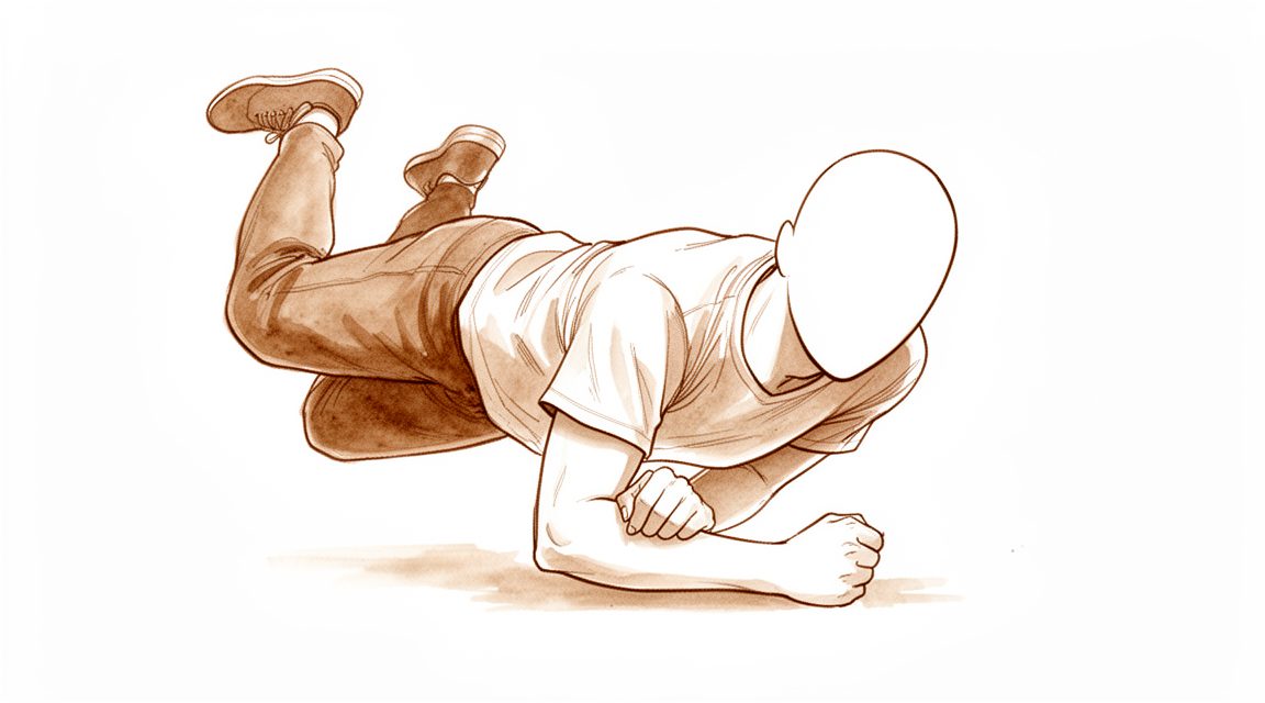
ओलेक्रेनन फ्रैक्चर (हड्डी टूटना) आपकी कोहनी के पीछे की हड्डी की नोक का टूटना है। यह आम तौर पर दो में से किसी एक तरीके से होता है। कम उम्र के लोगों में यह अक्सर ज़ोर से गिरने या कोहनी पर सीधी ठोकर लगने के बाद होता है। उम्रदराज़ लोगों में यह अक्सर खड़े-खड़े साधारण रूप से गिर जाने से होता है। बांह को अचानक और ज़ोर से सीधा करने से भी कोहनी की नोक से हड्डी का एक टुकड़ा खिंचकर अलग हो सकता है।
आम तौर पर आपको ठीक कोहनी के पीछे दर्द महसूस होगा, और यह पूरे हिस्से में नहीं, बल्कि एक ही जगह पर ज़्यादा दुखता है। कोहनी के पीछे सूजन होना आम है और यह काफ़ी ज़्यादा हो सकती है। क्योंकि हड्डी की वह नोक त्वचा के ठीक नीचे होती है, इसलिए कभी-कभी आप त्वचा के ऊपर से फ्रैक्चर का किनारा महसूस कर सकते हैं। अगर गिरने में त्वचा फट गई थी, तो घाव आम तौर पर कोहनी के पीछे होता है, जहां वह ज़मीन से टकराई थी। बांह का इस्तेमाल करना मुश्किल हो सकता है, और किसी चीज़ के ख़िलाफ़ उसे सीधा करना, जैसे कुर्सी से धक्का देकर उठना, कमज़ोर या नामुमकिन लग सकता है।
पहले कुछ दिनों में, कोहनी को हिलाने की कोशिश करने पर दर्द अक्सर बढ़ जाता है, और इससे आपकी नींद में ख़लल पड़ सकता है। रोज़ के जिन कामों में कोहनी मुड़ती है या उस पर भार पड़ता है, जैसे केतली उठाना, कपड़े पहनना, या अपनी अग्रबाहु (कोहनी से कलाई तक का हिस्सा) के बल टिकना, उनमें आम तौर पर दर्द होगा। पहले दो-एक सप्ताहों में सूजन और नील (चोट का नीला निशान) कम हो जाते हैं, और जैसे-जैसे हड्डी जुड़ने लगती है, दर्द धीरे-धीरे कम होता जाता है।
कुछ हालात में तुरंत इलाज की ज़रूरत होती है। अगर कोहनी के ऊपर की त्वचा फट गई है, अगर हड्डी दिख रही है, या अगर बांह साफ़ तौर पर बेढंगी (अपनी सामान्य शक्ल से हटी हुई) दिखती है, तो उसी दिन इमरजेंसी विभाग जाएं। यही बात तब भी लागू होती है जब आपका हाथ या उंगलियां फीकी (सफ़ेद पड़ी हुई), ठंडी, सफ़ेद या नीली पड़ जाएं, या अगर चोट के बाद आपकी बांह में अचानक एहसास (महसूस करने की क्षमता) या हिलने-डुलने की ताक़त चली जाए। अगर लक्षण ठीक नहीं हो रहे हैं, सप्ताहों के साथ बिगड़ रहे हैं, या रात में आपकी नींद खोल रहे हैं, तो अपने जीपी (आपके फैमिली डॉक्टर) से मिलें या विशेषज्ञ से जांच के लिए कहें। अगर आप क्लिनिक से संपर्क नहीं कर पा रहे हैं, तो अपने सबसे नज़दीकी इमरजेंसी विभाग जाएं।
असल में क्या हो रहा है
आपकी कोहनी के पीछे की हड्डी की नोक को ओलेक्रेनन कहते हैं। यह अग्रबाहु की हड्डियों में से एक का ऊपरी सिरा है, और यह कब्ज़े (हिंज) के ढक्कन जैसा काम करती है। जब आप अपनी बांह सीधी करते हैं, तो यह ढक्कन बांह की ऊपरी हड्डी के सिरे पर बनी एक खांच में फिसलता है। यह फ्रैक्चर इसलिए अहम है क्योंकि इससे क्या जुड़ा है: आपकी ट्राइसेप्स, यानी बांह के ऊपरी हिस्से के पीछे की मांसपेशी, का टेंडन (कंडरा) ठीक उसी जगह पर जुड़ा होता है। यही टेंडन आपको भार के ख़िलाफ़ अपनी कोहनी सीधी करने देता है।
इसे ऐसे समझें जैसे एक दरवाज़ा हो, जिसे एक मज़बूत स्प्रिंग खींचकर बंद रखती है। ओलेक्रेनन वह ब्रैकेट (कड़ा) है जो उस स्प्रिंग को थामे रहता है। जब हड्डी टूटती है, तो स्प्रिंग खींचती रहती है, और वह टूटे हुए टुकड़े को ऊपर और दूर खींच ले जाती है, जिससे जोड़ की सतह में एक खाली जगह (गैप) रह जाती है। इसीलिए अभी आपकी बांह सामान्य रूप से काम नहीं करती: मांसपेशी अब भी मज़बूत है, लेकिन वह आपकी अग्रबाहु को हिलाने के बजाय हड्डी के एक ढीले टुकड़े को खींच रही है। रुकावट के ख़िलाफ़ बांह सीधी करना कमज़ोर या नामुमकिन हो जाता है।
हड्डी फिर से आपस में जुड़कर ठीक होती है, और टूटकर अलग हुआ टुकड़ा, जिससे टेंडन जुड़ा हो, हड्डी जुड़ने के साथ वापस जुड़ सकता है। लेकिन यह अच्छी तरह हो, इसके लिए टुकड़ों का सही स्थिति में होना ज़रूरी है। जब फ्रैक्चर खिसका नहीं होता, तो जुड़ने तक टुकड़ों को बस स्थिर रखा जा सकता है। जब टुकड़े खिंचकर अलग हो गए हों, या जोड़ की सतह कई टुकड़ों में टूट गई हो, तो कोहनी अस्थिर (जोड़ का ढीलापन) हो सकती है, और ऐसे फ्रैक्चर में आम तौर पर जुड़ने के दौरान टुकड़ों को अपनी जगह पर पकड़कर रखने के लिए सर्जरी की ज़रूरत होती है।
सर्जन इन फ्रैक्चर को इस आधार पर ग्रेड करते हैं कि टुकड़े कितनी दूर खिसके हैं और कोहनी का जोड़ स्थिर रहता है या नहीं। ग्रेड एक का फ्रैक्चर खिसका नहीं होता। ग्रेड दो का फ्रैक्चर खिसका होता है, लेकिन जोड़ फिर भी काम करता है। ग्रेड तीन का मतलब है कि कोहनी का जोड़ खुद अस्थिर है। खिसके हुए और अस्थिर ग्रेड ही वे हैं जिनमें सबसे ज़्यादा बार सर्जरी की ज़रूरत पड़ती है।
इनमें से ज़्यादातर फ्रैक्चर अच्छी तरह जुड़ जाते हैं, और कोहनी के पूरी तरह सीधा होने में थोड़ी कमी रह जाना आम है।
हम इसके लिए क्या कर सकते हैं
डॉ. कीरन हिरपरा, माटर प्राइवेट हॉस्पिटल, रॉकहैम्पटन में ऊपरी अंग (कंधे से हाथ तक) के सर्जन, इलाज को आपकी ख़ास चोट के हिसाब से तय करते हैं। इनमें से कुछ फ्रैक्चर बिना ऑपरेशन के जुड़ जाते हैं, और कुछ में चोट के कुछ ही समय बाद सर्जरी की ज़रूरत होती है, इसलिए जल्दी जांच करवाना अहम है। मरीज़ों को आमतौर पर उनके जीपी हमारे क्लिनिक में भेजते हैं; अगर किसी फिजियोथेरेपिस्ट ने आपको हमसे मिलने की सलाह दी है, तब भी मेडिकेयर रिबेट पाने के योग्य होने के लिए आपको अपने जीपी से रेफ़रल लेना होगा। क्लिनिक में हम आपकी पूरी जानकारी लेते हैं, आपकी बांह की जांच करते हैं, और जहां ज़रूरत हो वहां इमेजिंग (एक्स-रे जैसी जांच) करवाते हैं।
अगर फ्रैक्चर खिसका नहीं है, या बस थोड़ा-सा खिसका है, और आपकी कोहनी स्थिर है, तो हम जुड़ने तक उसे स्थिर रखने की सलाह दे सकते हैं। इसका मतलब है कुछ समय के लिए स्प्लिंट या प्लास्टर (कास्ट), और बार-बार एक्स-रे करके जांच, ताकि पक्का हो सके कि टुकड़े अपनी जगह पर ही रहें। यह रास्ता उम्रदराज़ मरीज़ों में, और उन लोगों में आम है जिनकी बांह भारी काम के बजाय रोज़ के कामों में इस्तेमाल होती है। इससे ऑपरेशन और उसके साथ आने वाले जोखिमों से बचा जा सकता है। इसका नुकसान यह है कि अंत में कोहनी शायद पूरी तरह सीधी न हो पाए, और कुछ लोगों को यह स्वीकार करना मुश्किल लगता है। आपके हैंड थेरेपिस्ट (हाथ के विशेषज्ञ थेरेपिस्ट) के साथ हिलना-डुलना चरणों में वापस लाया जाता है, और वही आपके लिए ज़रूरी कोई भी स्प्लिंट बनाएंगे।
अगर टुकड़े खिंचकर अलग हो गए हैं, या कोहनी का जोड़ अस्थिर है, तो हम आम तौर पर शुरू से ही सर्जरी की सलाह देंगे। इसका मक़सद टुकड़ों को सही स्थिति में पकड़कर रखना है, ताकि हड्डी जुड़ सके और आप जल्दी हिलाना-डुलाना शुरू कर सकें। हड्डी को किस तरीके से पकड़कर रखा जाए, यह फ्रैक्चर के पैटर्न पर निर्भर करता है, और कोई एक तरीका हर फ्रैक्चर के लिए सही नहीं होता। कभी-कभी दोनों रास्ते सचमुच खुले होते हैं, उदाहरण के लिए किसी उम्रदराज़ मरीज़ में खिसका हुआ फ्रैक्चर। हम उन बातों पर बात करेंगे जो आपके लिए मायने रखती हैं: आप कितना दर्द सहने को तैयार हैं, और क्या कोहनी के सीधा होने के आख़िरी कुछ डिग्री खो देना आपको खलेगा।
आप जो भी रास्ता चुनें, पहले कुछ सप्ताह मिलते-जुलते होते हैं। सूजन कम होने तक दर्द निवारक दवाएं आपको आराम में रखती हैं। हड्डी जुड़ने तक चोट को सुरक्षित रखना ज़रूरी है, और हम आपको बताएंगे कि आप बांह से क्या कर सकते हैं और क्या नहीं। आपके हैंड थेरेपिस्ट सही चरण पर आपके हिलने-डुलने का मार्गदर्शन करेंगे, पहले धीरे-धीरे, और फिर जैसे-जैसे हड्डी जुड़ती है, उसे बढ़ाते हुए। इनमें से ज़्यादातर फ्रैक्चर अच्छी तरह जुड़ जाते हैं, और कोहनी के पूरी तरह सीधा होने में थोड़ी कमी रह जाना आम है। कोहनी के पीछे त्वचा के ठीक नीचे रहने वाला धातु का इम्प्लांट तकलीफ़ दे सकता है, और कुछ लोगों को बाद में उसे निकालने के लिए एक छोटे ऑपरेशन की ज़रूरत पड़ती है।
क्या उम्मीद करें
इनमें से ज़्यादातर फ्रैक्चर अच्छी तरह जुड़ जाते हैं। कोहनी के पूरी तरह सीधा होने में थोड़ी कमी आम है, और ज़्यादातर लोगों की बांह अच्छी तरह काम करती रहती है। जटिल फ्रैक्चर की सर्जरी के लंबे समय बाद के नतीजे अच्छे होते हैं, तब भी जब बाद के एक्स-रे में जोड़ में कुछ घिसाव वाला गठिया (आर्थराइटिस) दिखाई देता है।
अगर आपके फ्रैक्चर को स्प्लिंट या प्लास्टर (कास्ट) में स्थिर रखा जाता है, तो हड्डी दिनों में नहीं, बल्कि सप्ताहों में जुड़ती है। टुकड़े अपनी सही जगह पर ही रहें, यह पक्का करने के लिए आपके बार-बार एक्स-रे होंगे। यह रास्ता उम्रदराज़ मरीज़ों और उन लोगों के लिए सही है जिनकी बांह भारी काम के बजाय रोज़ के काम करती है। इस समूह में कम समय और लंबे समय, दोनों के नतीजे संतोषजनक होते हैं। इसका नुकसान यह है कि अंत में कोहनी शायद पूरी तरह सीधी न हो पाए।
अगर आपकी सर्जरी होती है, तो हड्डी जुड़ने तक उसे धातु के इम्प्लांट से अपनी जगह पर पकड़कर रखा जाता है। हिलाना-डुलाना जल्दी शुरू होता है और आपके हैंड थेरेपिस्ट के साथ चरणों में बढ़ता है। आप कोई भी रास्ता चुनें, इस चोट के बाद कोहनी के पूरी तरह सीधा होने में कुछ कमी की उम्मीद रहती है। खिसके हुए फ्रैक्चर के लिए सर्जरी से इलाज किए गए लोगों पर लंबे समय तक नज़र रखने वाले एक अध्ययन में, चोट के 25 साल बाद तक 96% लोगों के नतीजे अच्छे या बहुत अच्छे थे।
ठीक होने का मतलब है धीरे-धीरे अपने सामान्य कामों पर लौटना। पहले रोज़ के हल्के काम लौटते हैं, फिर जैसे-जैसे हड्डी मज़बूत होती है, भार उठाना और ज़्यादा ज़ोर वाला इस्तेमाल। आप कितनी जल्दी गाड़ी चला सकते हैं, काम कर सकते हैं या खेल सकते हैं, यह आपके काम, आपकी बांह और हड्डी के जुड़ने पर निर्भर करता है, और हम हर चरण में आपको समझाएंगे।
जो चीज़ें गलत हो सकती हैं, उनके बारे में जानना अच्छा है। धातु का इम्प्लांट कोहनी के पीछे त्वचा के ठीक नीचे होता है, और यह इतनी तकलीफ़ दे सकता है कि कुछ लोग बाद में उसे निकलवाने का फ़ैसला करते हैं। इस सर्जरी के बाद यह बार-बार सामने आने वाली समस्या है। दूसरी समस्याएं, जैसे हड्डी का गलत स्थिति में जुड़ना या कोहनी का जकड़ा रह जाना, कम आम हैं। अगर आपके लक्षण ठीक नहीं हो रहे हैं, सप्ताहों के साथ बिगड़ रहे हैं, या रात में आपकी नींद खोल रहे हैं, तो अपने जीपी से मिलें या विशेषज्ञ से जांच के लिए कहें। अगर आप क्लिनिक से संपर्क नहीं कर पा रहे हैं, तो अपने सबसे नज़दीकी इमरजेंसी विभाग जाएं।
डॉक्टर को कब दिखाएं
अगर आपकी कोहनी साफ़ तौर पर बेढंगी (अपनी सामान्य शक्ल से हटी हुई) दिखे, अगर चोट के ऊपर की त्वचा फट गई हो, या अगर आपका हाथ या उंगलियां फीकी (सफ़ेद पड़ी हुई), ठंडी, सफ़ेद या नीली पड़ जाएं, तो तुरंत इलाज करवाएं। चोट के बाद बांह में अचानक एहसास (महसूस करने की क्षमता) या हिलने-डुलने की ताक़त चली जाए, तो उसके लिए भी उसी दिन इमरजेंसी विभाग जाना ज़रूरी है। इन लक्षणों का मतलब है कि चोट की जांच फ़ौरन होनी चाहिए।
बाकी सब के लिए, अपने जीपी से शुरुआत करें। वे आपकी बांह की जांच कर सकते हैं और एक्स-रे करवा सकते हैं, और इस तरह के फ्रैक्चर को दिखाने के लिए आम तौर पर बस इतना ही काफ़ी होता है। अगर आपके जीपी को लगता है कि किसी विशेषज्ञ को इलाज संभालना चाहिए, तो वे आपको जांच के लिए रेफ़र करेंगे।
अगर दर्द कम नहीं हो रहा है, या हड्डी जुड़ने के दौरान सूजन, हिलना-डुलना या बांह का इस्तेमाल हर सप्ताह बेहतर नहीं हो रहा है, तो दोबारा आएं, या विशेषज्ञ से जांच के लिए कहें। जो लक्षण सप्ताहों के साथ बिगड़ रहे हैं, रात में आपकी नींद खोल रहे हैं, या आपको काम करने या बांह का इस्तेमाल करने से रोक रहे हैं, उन्हें भी दोबारा दिखाना चाहिए। अगर आप क्लिनिक से संपर्क नहीं कर पा रहे हैं, तो अपने सबसे नज़दीकी इमरजेंसी विभाग जाएं।
विस्तार से
Advanced reading: the deeper science (optional)
यह हिस्सा आपके अपने इलाज से जुड़े फ़ैसलों के लिए ज़रूरी जानकारी से आगे जाता है। ओलेक्रेनन फ्रैक्चर के बारे में यह अतिरिक्त जानकारी पढ़ने लायक है, क्योंकि एक ऐसा निष्कर्ष है जो फ्रैक्चर सर्जरी के सामान्य तर्क को उलट देता है: उम्रदराज़ मरीज़ों में, ओलेक्रेनन के खिसके हुए फ्रैक्चर को बिना जोड़े छोड़ा जा सकता है, और कोहनी फिर भी काम करती है।
खिसका हुआ फ्रैक्चर जिसे जोड़ने की ज़रूरत नहीं होती
ओलेक्रेनन के आर-पार फ्रैक्चर ट्राइसेप्स (बांह के पिछले हिस्से की मांसपेशी) को अग्रबाहु (कोहनी से कलाई तक का हिस्सा) से अलग कर देता है, इसलिए सामान्य तर्क यह है कि इसे फिर से जोड़ना ही होगा, वरना कोहनी को रुकावट के ख़िलाफ़ सीधा नहीं किया जा सकेगा। 70 साल से ज़्यादा उम्र के मरीज़ों में यह तर्क टिकता नहीं है। एक व्यवस्थित समीक्षा (सिस्टमैटिक रिव्यू) में पाया गया कि इस समूह में खिसके हुए ओलेक्रेनन फ्रैक्चर को बिना ऑपरेशन के असरदार तरीके से संभाला जा सकता है, जिससे मरीज़ों की संतुष्टि ज़्यादा रहती है और जोड़ इतना हिल-डुल पाता है कि रोज़ का काम चल सके (फ़ंक्शनल रेंज ऑफ मोशन) [1]।
इसके बदले जो खोया जाता है वह मापा जा सकता है: एक्सटेंशन लैग, यानी कोहनी को पूरी तरह सीधा न कर पाना, और सीधा करने की ताक़त में कमी। जिससे बचा जाता है वह है पतली, अक्सर नाज़ुक त्वचा में, त्वचा के ठीक नीचे की हड्डी के ऊपर एक घाव, और उस त्वचा के नीचे धातु का इम्प्लांट। क्योंकि उभरा हुआ धातु का इम्प्लांट ओलेक्रेनन को जोड़ने के बाद दूसरे ऑपरेशन की सबसे आम वजह है, इसलिए इससे बचने पर सबसे आम जटिलता पूरी तरह ख़त्म हो जाती है।
किसी उम्रदराज़ व्यक्ति के लिए, जिसकी बांह ज़ोर से धक्का देने के बजाय रोज़ के कामों में इस्तेमाल होती है, ऐसी कोहनी जो आख़िरी कुछ डिग्री तक सीधी न हो पाए, घाव की किसी जटिलता से कम बड़ी परेशानी है। किसी कम उम्र या ज़्यादा मेहनत वाले मरीज़ के लिए ऐसा नहीं है, और इसीलिए यह निष्कर्ष सामान्य नहीं, बल्कि उम्र से जुड़ा है।
जहां हड्डी जोड़ी जाती है, वहां इम्प्लांट का चुनाव साफ़ होने लगा है
आम, खिसके हुए लेकिन सीधे-सादे पैटर्न के लिए, दो इम्प्लांट के बीच मुक़ाबला है: तारों से बनी टेंशन बैंड व्यवस्था और एक प्लेट। मेयो टाइप II फ्रैक्चर वाले 827 मरीज़ों में, टेंशन बैंड वायरिंग की तुलना में प्लेट लगाने से बेहतर असर और सुरक्षा दिखी, और लेखकों ने यह भी बताया कि इसमें कम अध्ययन शामिल थे और ज़्यादा उच्च गुणवत्ता वाले रैंडमाइज़्ड ट्रायल की अब भी ज़रूरत है [2]। 472 मरीज़ों की 2025 की एक समीक्षा, जिसमें आधुनिक फिक्सेशन तकनीकों की तुलना पारंपरिक टेंशन बैंड वायरिंग से की गई, भी उसी दिशा में इशारा करती है [3]।
यह इस साइट पर उन गिनी-चुनी जगहों में से है जहां तकनीकों की तुलना से कंधे उचकाने के बजाय एक दिशा मिलती है। लेकिन जिस प्रमाण आधार की कमी की ओर ख़ुद लेखकों ने ध्यान दिलाया है, उसे देखते हुए इस पर ज़्यादा ज़ोर न दें।
गठिया होता है, और ज़्यादातर मायने नहीं रखता
लंबे समय बाद के इमेजिंग निष्कर्ष बिना संदर्भ के बताए जाएं तो डरावने लग सकते हैं। सिर्फ़ ओलेक्रेनन के फ्रैक्चर के बाद, चोट के बाद होने वाले ऑस्टियोआर्थराइटिस की मीडियन दर 41 महीने के मीडियन फॉलो-अप पर 19% थी, और फ्रैक्चर का प्रकार कुछ भी हो या गठिया हो या न हो, मरीज़ों द्वारा बताए गए आख़िरी नतीजे अच्छे से लेकर बहुत अच्छे तक रहे [4]।
उस वाक्य के दोनों हिस्से अहम हैं। इस चोट के बाद फॉलो-अप एक्स-रे में गठिया दिखना आम है। साथ ही, इस समय-सीमा में, यह काफ़ी हद तक इस बात से अलग है कि लोग अपनी कोहनी के काम करने के बारे में क्या बताते हैं। एक्स-रे में दिखी कोई बात अपने आप कोई क्लिनिकल समस्या नहीं है, और सिर्फ़ उसके आधार पर आगे कोई दख़ल नहीं दिया जाना चाहिए।
संदर्भ
[1] Alvara CA, Biedron G, Dunn JC. Nonoperative management of olecranon fractures in elderly patients: a systematic review. Hand (N Y). 2020;17(4):734-9. https://doi.org/10.1177/1558944720944261
[2] Jia Y, Liu A, Guo T, Chen J, Yu W, Zhai J. Efficacy and safety of tension band wire versus plate for Mayo II olecranon fractures: a systematic review and meta-analysis. J Orthop Surg Res. 2022;17(1). https://doi.org/10.1186/s13018-022-03262-7
[3] Wang C, Li C. Modern fixation techniques versus traditional tension band wiring for olecranon fractures: a systematic review and meta-analysis. J Orthop Surg Res. 2025;20(1). https://doi.org/10.1186/s13018-025-06061-y
[4] Wiersma JP, de Klerk HH, Priester-Vink S, Doornberg JN, Bhasyam AR, van den Bekerom MP. Incidence of post-traumatic osteoarthritis in olecranon fractures and the role of fracture type: a systematic review. J Shoulder Elbow Surg. 2026. https://doi.org/10.1016/j.jse.2026.02.024
Evidence & references
This is the clinical evidence summary written for health professionals. It is technical, and it lists the research this page was built from. You do not need to read it to understand your treatment or to make a decision about it.
Overview
General Outcomes and Complications
- A majority of olecranon fractures heal uneventfully with good or excellent results, with a small loss of motion expected [11].
- 96% of individuals with a closed displaced fracture of the olecranon treated by open reduction and internal fixation had excellent or good outcomes up to 25 years after the fracture [4].
- Surgical treatment of olecranon fractures is associated with a high rate of complications [18].
- Patients undergoing revisions beyond implant removal after olecranon fracture fixation had poorer functional outcomes [18].
- No significant differences in functional outcomes or secondary operations were found with respect to fracture type, gender, or surgical method for olecranon fractures [18].
- The incidence of post-traumatic osteoarthritis following isolated olecranon fractures is 19% at a median follow-up of 41 months [22].
Operative Management
- No single technique is suitable for the management of all olecranon fractures [21].
- Low-profile double-plate osteosynthesis is a safe and effective alternative treatment for olecranon fractures with excellent subjective and objective clinical outcome measures [1].
- Plating of the olecranon leads to predictable union, though the most common complication was lack of full extension in 39% of patients [16].
- Both Kirschner wire tension band and anatomical locking plate procedures effectively treat Mayo type II olecranon fractures [9].
- Current randomized evidence suggests that tension band wiring and plate fixation may yield similar functional outcomes for displaced olecranon fractures, based on a limited number of randomized controlled trials [24].
- Tension band wiring remains an effective treatment for appropriately selected olecranon fractures and outperformed plate osteosynthesis in one cohort [40].
- The timing of fixation of displaced olecranon fractures does not significantly increase the rate of early complications or reoperation [15].
- Excision of the olecranon fragment with repair of the triceps mechanism is supported as a preferred method of treatment due to significantly less morbidity compared with internal fixation [65].
Non-Operative Management
- Close radiographic follow-up is recommended for nonsurgically treated olecranon fractures in children [2].
- Some isolated displaced (>2 mm) olecranon fractures in elderly patients can be successfully treated nonoperatively [6].
- Satisfactory short-term and long-term outcomes were found following the nonoperative management of isolated displaced olecranon fractures in older, lower-demand patients [13].
- Displaced olecranon fractures in patients older than 70 years may be effectively managed with nonoperative measures to produce high satisfaction and functional range of motion [23].
- Nonoperative management of olecranon fractures in elderly patients offers comparable functional outcomes to surgical intervention while significantly reducing exposure to potential complications and healthcare costs [41].
- Nonoperative treatment is a reasonable option for displaced stable olecranon fractures in elderly patients [29].
Elderly Population Specifics
- Olecranon fractures in the elderly have higher than expected 1-year mortality rates [5].
- Patients with olecranon fractures have essentially similar demographic characteristics compared to patients with distal radius fractures [10].
- The literature on the treatment of olecranon fractures in elderly patients is limited [7].
Anatomy & Pathophysiology
Bony Anatomy
- The olecranon and the coronoid process form the greater sigmoid notch, which articulates with the trochlea of the distal humerus [37].
- The intrinsic anatomy of the ulnohumeral articulation allows flexion/extension movement of the elbow joint and provides stability for the elbow [37].
- The olecranon serves as the insertion for the triceps tendon, which blends with the periosteum of the proximal ulna [37].
- The exposed, subcutaneous position of the olecranon renders it vulnerable to direct trauma and violent muscular contractions from the triceps [37].
- In children, the olecranon is predominantly cartilage, particularly in younger children, which reduces the chance of fracture occurring with a direct blow [48].
- In children, the thick periosteum and relatively thin metaphyseal cortex of the olecranon predispose it to minimally displaced greenstick fractures [48].
- Radiographic measurements of normal elbows guide the surgeon to obtain a more reliable length of the olecranon and maintain articular geometry during fixation [69].
Mechanisms of Injury
- Olecranon fractures can result from a direct blow, a fall on an outstretched hand with the elbow in flexion, or high-energy trauma associated with radial head fractures or elbow dislocation [37].
- Sudden and violent triceps muscle contraction can produce an avulsion fracture of varying size of the olecranon tip [37].
- A bimodal distribution of olecranon fractures is seen in young patients with high-energy trauma and elderly patients with low-energy trauma such as a fall from standing [37].
- Olecranon fractures typically occur as low-energy falls from standing in older patients or high-energy injury mechanisms in younger patients [38].
- The bone typically fails from a direct blow to the elbow or as a tensile force from the pull of the triceps tendon [38].
- Regardless of the mechanism of injury, the triceps acts as the main deforming mechanism pulling the fractured fragment proximally, creating a gap at the articular surface, and an incompetent extensor mechanism [38].
- In children, olecranon fractures are usually the result of a hyperextension injury [48].
- In children, flexion injuries are usually caused by a fall on an outstretched hand with the elbow flexed, resulting from a strong eccentric contraction of the triceps pulling the olecranon over the fulcrum of the distal humerus [48].
- In children, shear injuries result from a force to the proximal ulna just anterior to the humeral condyles, causing the olecranon to fracture through metaphyseal bone with anterior displacement of the distal fragment [48].
- Olecranon fractures commonly occur with a direct blow, generally resulting in comminuted fractures, or as an avulsion injury with triceps contracture [54].
- Contraction of the triceps often results in transverse or short oblique types of olecranon fractures [54].
- In children, hyperextension injuries are frequently associated with other elbow injuries, where the direction of the associated coronal plane force determines the corresponding injuries [48].
- In children, a valgus hyperextension force may produce an associated radial neck or medial epicondyle fracture [48].
- In children, a varus hyperextension injury may be associated with lateral dislocation of the radial head or a Bado type III Monteggia lesion [48].
- In children, flexion injuries are generally transverse (perpendicular to the axis of the ulna), displaced posteriorly rather than anteriorly, and rarely associated with other injuries [48].
Associated Injuries
- Olecranon fractures typically occur in isolation; however, given the subcutaneous location of the injury, there is a risk to soft tissues as well as risk of the fracture being open [38].
- In an epidemiologic study of olecranon fractures, 22% of patients had injuries to the ipsilateral limb and 6.4% of fractures were open [38].
- In children, olecranon fractures are associated with other elbow injuries (usually the medial epicondyle) in 20% to 50% of cases [48].
Classification
- The Mayo classification is the most common classification used for olecranon fractures, based on the amount of fracture displacement, the presence of comminution, and stability of the ulnohumeral joint [38].
- In the Mayo classification, Type I fractures are nondisplaced, Type II fractures are displaced, and Type III fractures have an unstable ulnohumeral joint [38].
- In the Mayo classification, modifier A indicates simple (noncomminuted) fractures and modifier B indicates comminuted fractures [38].
- The Mayo classification is simple to use, can help guide fracture management, and has good interobserver reliability [38].
- Displaced olecranon fracture lines entered along the medial side of the trochlear notch and exited at the base of the coronoid, while minimally displaced fractures entered and exited the trochlear notch at the base of the coronoid [38].
- The Schatzker and Colton classification attempted to classify olecranon fractures based on different fracture pattern morphology; however, its application was limited [38].
- A proposed fragment-specific classification system for complex olecranon fractures is anatomically based and considers the deforming forces from ligaments and tendons [64].
- In a study of 2462 fractures, the distribution included a larger share of comminuted central fractures (29%) compared to a single-center report from Scotland [8].
- In a study of 2462 fractures, an almost threefold larger share of distal olecranon fractures was observed compared to the 6.2% reported in a Scottish study of 64 olecranon fractures [8].
- In a study of 2462 fractures, the distribution differed from a single-center report from Scotland where 74% of the fractures were simple central fractures and 81% were central fractures [8].
Classification
Classification Systems and Reproducibility
- The Mayo classification is the most commonly used classification system in clinical practice for olecranon fractures [43].
- The Mayo classification consists of 3 types with a modifier to indicate comminution [43].
- Type I fractures in the Mayo classification are nondisplaced [43].
- Type II fractures in the Mayo classification are displaced [43].
- Type III fractures in the Mayo classification have accompanying injuries [43].
- The Mayo classification was designed to simplify categorization of olecranon fractures but does not achieve this goal due to poor reproducibility [27].
- Multiple classification systems for olecranon fractures exist, including AO, Mayo, Schatzker and Colton, but none are widely accepted or provide direct and reliable advice on operative strategies [88].
- The low reproducibility rates of existing olecranon fracture classification systems raise questions about their use in clinical and research contexts [88].
- The Colton classification had the best intra- and inter-observer agreement among the Mayo, AO/OTA, and Colton & Schatzker classifications [111].
- The AO classification subsumes olecranon fractures to proximal forearm injuries [88].
- Schatzker and Colton divide olecranon fractures into groups by quantity of fragments and fracture lines [88].
Fracture Morphology and Distribution
- Quantitative 3-dimensional computed tomography analysis further clarified the fracture morphology of Mayo type I, II, and III fractures [62].
- In a study of 2462 fractures from the Swedish Fracture Register, 29% of olecranon fractures were comminuted central fractures [8].
- The share of distal olecranon fractures in the Swedish Fracture Register was almost threefold larger than the 6.2% reported in a Scottish study of 64 olecranon fractures [8].
- In a study of 64 olecranon fractures, the most frequent injury according to the AO fracture classification was the 21-B1.1 type [94].
- In a study of 64 olecranon fractures, a simple isolated displaced olecranon fracture (Mayo type 2A) was the most common according to the Mayo classification [94].
- In a study of 44 patients treated with tension band wiring, Type IIA fractures were the most common pattern, accounting for 91% of cases [107].
- In a study of 57 patients treated with a shape-memory alloy concentrator, the Mayo classification system was used to distinguish fracture displacement, presence of comminution, and ulnohumeral stability [60].
- Mayo type III fractures are categorized into anterior and posterior olecranon fracture-dislocations based on the radio-ulnar relationship [106].
- Articular impaction is a common feature of geriatric olecranon fractures [32].
Pediatric and Adolescent Considerations
- Fracture of the olecranon in a child is often only part of a complex injury to the bone and soft tissues around the elbow [30].
- The olecranon apophyseal ossification system, when combined with age and sex, successfully predicts peak height velocity within a year in 90% of cases [93].
Clinical Presentation
Mechanisms and Epidemiology
- The triceps acts as the main deforming mechanism pulling the fractured fragment proximally, creating a gap at the articular surface, and an incompetent extensor mechanism [38].
- The incidence of olecranon fractures increased by 29% over a 20-year study period in Denmark [12].
- In a Swedish study of 2462 fractures, 29% were comminuted central fractures and the share of distal olecranon fractures was almost threefold larger than in a Scottish study [8].
- In a Swedish study, high-energy injury mechanisms accounted for 5.7% of female and 11.6% of male patients overall, while low-energy mechanisms accounted for 87.4% of female and 77.8% of male patients overall [8].
- Fractures of the ipsilateral olecranon associated with the radial neck are not as rare as previously reported [42].
Physical Examination
- Pain is usually localized to the posterior part of the elbow [37].
- Given the subcutaneous location of the olecranon, the fracture itself may be palpable [37].
- Extensive posterior swelling is typical [37].
- A careful examination of the integrity of the extensor mechanism with gravity eliminated can aid surgical decision making [37].
- If present, open wounds are typically posterior and result from the direct impact of the posterior surface of the elbow against an unyielding structure [37].
- The affected extremity from shoulder to hand should be thoroughly examined for any soft tissue compromise [38].
- There is a low suspicion for open fractures given the subcutaneous nature of the olecranon [38].
- The elbow will often present with a large fluid collection subcutaneously over the olecranon [38].
- Examination of the extensor mechanism both with and without gravity eliminated should be performed to assess competency [38].
Imaging
- Plain radiographs are usually sufficient for isolated fractures of the olecranon [37].
- A true lateral radiograph is necessary to accurately identify the plane of the fracture and the number of fracture fragments [37].
- The examiner should assess for fracture comminution and impaction on radiographs [37].
- In more complex cases, CT may help delineate the comminution or impaction better, but this is not routinely required [37].
- Anteroposterior and lateral radiographs of the elbow are required for olecranon fractures [38].
- If there is concern for a radial head fracture, a radiocapitellar view of the elbow can be obtained [38].
- In the setting of an elbow dislocation, post-reduction radiographs should be obtained [38].
- Computed tomography (CT) can be used for preoperative planning for comminuted fractures of the olecranon if there is an associated radial head or coronoid fracture, but this is not routinely utilized [38].
Classification
- The Mayo classification is based on the amount of fracture displacement, the presence of comminution, and stability of the ulnohumeral joint [38].
- The modifier A (simple) and B (comminuted) in the Mayo classification indicate the presence of comminution [38].
- The Schatzker and Colton classification attempted to classify olecranon fractures based on different fracture pattern morphology, but its application was limited [38].
Prognosis and Outcomes
- A majority of olecranon fractures heal uneventfully with good or excellent results with a small loss of motion to be expected [11].
- Primary elbow instability and fracture morphology are prognostic factors for elbow function and development of arthrosis after operative treatment of olecranon fractures [45].
- The median incidence of post-traumatic osteoarthritis following isolated olecranon fractures is 19% at a median follow-up of 41 months [22].
- Patients aged 50 years or more with olecranon fracture should be evaluated for osteoporosis and fracture risks for secondary prevention of subsequent fractures [36].
Investigations
Clinical Examination
- The history may help distinguish a triceps avulsion from an actual direct blow to the elbow [37].
- Extensive posterior swelling is typical of olecranon fractures [37].
- Open wounds, if present, are typically posterior and result from the direct impact of the posterior surface of the elbow against an unyielding structure [37].
- The affected extremity from shoulder to hand should be thoroughly examined to identify associated ipsilateral injuries [38].
- The arm should be examined for soft tissue compromise, with a low suspicion for open fractures maintained given the subcutaneous nature of the olecranon [38].
Imaging
- In more complex cases, CT may help delineate comminution or impaction better, though this is not routinely required [37].
- Quantitative analysis of olecranon fractures via CT further clarified fracture morphology of Mayo type I, II, and III fractures [62].
Classification
- The distribution of olecranon fractures in a Swedish register study showed a larger share of comminuted central fractures (29%) compared to a single-center report from Scotland [8].
- The Swedish register study observed an almost threefold larger share of distal olecranon fractures than the 6.2% reported in a Scottish study of 64 olecranon fractures [8].
Pediatric Specifics
- Olecranon fractures are associated with other elbow injuries (usually the medial epicondyle) in 20% to 50% of pediatric cases [48].
- In younger children, the olecranon is predominantly cartilage, which reduces the chance of a fracture occurring with a direct blow [48].
- The thick periosteum and relatively thin metaphyseal cortex of the pediatric olecranon predispose it to minimally displaced greenstick fractures [48].
Treatment
General Principles and Indications
- Conventional indications for surgical treatment of olecranon fractures include open fractures, displaced fractures, and fractures causing disruption to the extensor mechanism affecting the insertion of the triceps muscle into the olecranon [28].
- The aims of treatment for all olecranon fractures are restoration of function and stability to the elbow joint to allow early mobilization, while minimizing associated complications [113].
- Fixation must be secure enough to permit early motion to avoid significant stiffness of the elbow joint [20].
- Good results overall are to be expected after surgical management of olecranon fractures [43].
Non-Operative Management
- Mayo type I stable undisplaced fractures can be managed effectively with nonoperative management [113].
- Some authors suggest splinting the elbow in 45° to 90° of flexion for three to four weeks followed by supervised mobilization for stable undisplaced fractures [113].
- Early active mobilization is recommended where pain allows, and time in a cast should be minimized to reduce stiffness [113].
- The acceptable degree of fracture displacement for nonoperative management is commonly quoted as < 2 mm of articular displacement on the lateral radiograph [113].
- Aggregate data support the non-operative treatment of isolated undisplaced olecranon fractures in children with good results [33].
- Nonoperative management of isolated displaced olecranon fractures in older, lower-demand patients yields satisfactory short-term and long-term outcomes [13].
Operative Management: Techniques and Outcomes
- There are four common methods in the surgical management of olecranon fractures: tension band wiring, plate fixation, intramedullary fixation, and excision of the proximal fractured fragment with re-attachment of triceps tendon [28].
- Both operative procedures (Kirschner wire tension band and anatomical locking plate) effectively treat Mayo type II olecranon fractures [9].
- Current randomized evidence suggests that tension band wiring (TBW) and plate fixation (PF) may yield similar functional outcomes for displaced olecranon fractures, based on a limited number of RCTs [24].
- Among active patients with a simple isolated, displaced fracture of the olecranon, no difference was found between TBW and plate fixation in patient-reported outcome at 1 year following surgery [35].
- Plate fixation has better efficacy and safety for Mayo II olecranon fractures compared to tension band wiring [51].
- TBW remains an effective treatment for appropriately selected olecranon fractures and outperformed plate osteosynthesis in one cohort [40].
- Internal fixation by cable pin system (CPS) is associated with a shorter healing time, fewer complications, and better function than TBW [34].
- Low-profile double-plate osteosynthesis is a safe and effective alternative treatment of olecranon fractures with excellent subjective and objective clinical outcome measures [1].
- Low-profile double-plate osteosynthesis for treating olecranon fractures resulted in good clinical outcomes [85].
- Suture tension band fixation reduces hardware complications in olecranon fractures [3].
- In all patients treated with percutaneously placed K-wires and absorbable tension-band sutures for displaced olecranon fractures, fracture united within 6 weeks and full painless range of movement was regained within 6 months [47].
- Fractures through an ununited olecranon apophysis are successfully treated with plate and screw fixation with bone grafting [71].
- The Nickel-Titanium olecranon memory connector (OMC) can be an effective alternative to treat olecranon fractures [77].
- The study supports the rationale for excision of the fragment of the olecranon with repair of the triceps mechanism as the preferred method of treatment due to significantly less morbidity compared with internal fixation [65].
- Functional results after at least 2 years were equally good with tension band and screwing techniques, but there was a considerably higher rate of separation at the fracture after screwing than after inserting a tension band [46].
Complications and Risk Factors
- Patients undergoing revisions beyond implant removal had poorer functional outcomes after olecranon fracture fixation [18].
- No significant differences in functional outcomes or secondary operations were found with respect to fracture type, gender, or surgical method for olecranon fracture fixation [18].
- Patients who have operative fixation of a fracture of the olecranon can be counseled that most patients keep their implants, that only 3% experience implant migration, and that technical factors such as the type or configuration of an implant seem less important than personal factors in determining who requests a second surgery for implant removal [25].
- Risk factors associated with a poorer outcome following operative treatment of displaced olecranon fractures are patient factors, fracture morphology, and associated elbow instability or fractures [113].
Complications
General Outcomes and Mortality
- ORIF for olecranon fractures demonstrates low short-term complication rates [101].
- Specific comorbidities such as smoking and ascites were associated with increased risk of short-term complications following ORIF for olecranon fractures [101].
Hardware and Fixation Complications
- Symptomatic hardware requiring removal is a recurring and frequent problem in olecranon fracture management [43].
- The most common complication following plating of the olecranon was lack of full extension in 39% of patients [16].
- Only 3% of patients who undergo operative fixation of an olecranon fracture experience implant migration [25].
- Technical factors such as the type or configuration of an implant seem less important than personal factors in determining who requests a second surgery for implant removal [25].
- Internal fixation by cable pin system (CPS) is associated with fewer complications than tension band wiring (TBW) for olecranon fractures [34].
- Suture tension band fixation reduces hardware complications in olecranon fractures compared to traditional techniques [3].
Long-Term and Specific Complications
- Articular impaction in olecranon fractures is associated with worse postoperative outcomes [67].
- Inadequate reduction of articular impaction contributes to increased step-off and poorer function in olecranon fractures [67].
Recovery
General Outcomes
- Articular impaction in olecranon fractures is associated with worse postoperative outcomes, with inadequate impaction reduction contributing to increased step-off and poorer function [67].
Operative Recovery
- Surgical treatment of olecranon fractures is associated with a high rate of complications, and patients undergoing revisions beyond implant removal had poorer functional outcomes [18].
- No significant differences in functional outcomes or secondary operations were found with respect to fracture type, gender, or surgical method following open reduction and fixation of olecranon fractures [18].
- Both tension band wiring and plate fixation yield excellent or good clinical outcomes with minimal loss of physical capacity, little pain, and disability in the majority of patients with simple and comminuted displaced olecranon fractures [68].
- Among active patients with a simple isolated, displaced fracture of the olecranon, no difference was found between tension band wiring and plate fixation in the patient-reported outcome at 1 year following surgery [35].
- Internal fixation by cable pin system is an effective method for olecranon fracture and is associated with a shorter healing time, fewer complications, and better function than tension band wiring [34].
- Tension band wiring remains an effective treatment for appropriately selected olecranon fractures and in one cohort outperformed plate osteosynthesis [40].
- Both Kirschner wire tension band combined with anatomical locking plate and standard operative procedures effectively treat Mayo type II olecranon fractures [9].
Non-Operative Recovery
- Close radiographic follow-up for nonsurgically treated olecranon fractures is recommended due to the risk of further displacement after initial nonsurgical treatment in children [2].
- All 10 cases of olecranon fractures in children treated with a sling were free of symptoms and had a full range of elbow movement at 3 weeks [17].
Elderly-Specific Outcomes
Key Evidence
- [L3] Low-profile double-plate osteosynthesis is a safe and effective alternative treatment of olecranon fractures with excellent subjective and objective clinical outcome measures. [1] (10.1016/j.otsr.2019.08.019)
- [L4] Close radiographic follow-up for nonsurgically treated olecranon fractures is recommended. [2] (10.5435/jaaos-d-25-00821)
- [L3] Future studies should focus on the long-term outcomes of this technique, as compared to traditional and more established techniques to treat olecranon fractures. [3] (10.1016/j.jseint.2026.101734)
- [L3] 96% of individuals with a closed displaced fracture of the olecranon treated by open reduction and internal fixation had excellent or good outcomes up to 25 years after the fracture. [4] (10.1067/mse.2002.124548)
- [L3] Olecranon fractures in the elderly have higher than expected 1 year mortality rates. [5] (10.1177/1758573221994860)
- [L4] Some isolated displaced (>2 mm) olecranon fractures in elderly patients can be successfully treated nonoperatively. [6] (10.1016/j.ocl.2016.08.011)
- [L4] The literature on the treatment of olecranon fractures in elderly patients is limited. [7] (10.1007/s11678-018-0488-7)
- [L4] [8] (10.1007/s00068-021-01765-2)
- [L3] Both operative procedures effectively treat Mayo type II olecranon fractures. [9] (10.1186/s12891-025-08843-1)
- [L3] Patients with olecranon fractures have essentially similar demographic characteristics compared to patients with distal radius fractures. [10] (10.1177/17585732221124301)
- [L4] A majority of olecranon fractures heal uneventfully with good/excellent results with a small loss of motion to be expected. [11] (10.1016/j.hcl.2015.07.003)
- [L3] The incidence of olecranon fractures increased by 29% over the 20-year study period. [12] (10.1186/s13018-025-05970-2)
- [L4] We found satisfactory short-term and long-term outcomes following the nonoperative management of isolated displaced olecranon fractures in older, lower-demand patients. [13] (10.2106/jbjs.l.01137)
- [L3] The timing of fixation of displaced olecranon fractures does not significantly increase the rate of early complications or reoperation. [15] (10.1016/j.jhsg.2023.09.002)
- [L3] Plating of the olecranon leads to predictable union, though the most common complication was lack of full extension in 39% of patients. [16] (10.1016/j.injury.2016.04.015)
- [L4] All 10 cases treated with a sling were free of symptoms and had a full range of elbow movement at 3 weeks. [17] (10.1016/0020-1383(75)90056-x)
- [L4] Surgical treatment of olecranon fractures is associated with a high rate of complications, and patients undergoing revisions beyond implant removal had poorer functional outcomes; however, no significant differences in functional outcomes or secondary operations were found with respect to fracture type, gender, or surgical method. [18] (10.1016/j.xrrt.2025.08.004)
- [L5] Fixation must be secure enough to permit early motion to avoid significant stiffness of the elbow joint. [20] (10.5435/00124635-200007000-00007)
- [Paper] No one technique is suitable for the management of all olecranon fractures. [21] (10.1016/j.injury.2008.12.013)
- [L4] This review identified a median OA incidence of 19% at a median follow-up of 41 months following isolated olecranon fractures. [22] (10.1016/j.jse.2026.02.024)
- [L4] Displaced olecranon fractures in patients older than 70 years may be effectively managed with nonoperative measures to produce high satisfaction and functional range of motion. [23] (10.1177/1558944720944261)
- [L1] Current randomized evidence suggests that TBW and PF may yield similar functional outcomes for displaced olecranon fractures; however, these findings are based on a limited number of RCTs, with several secondary outcomes derived from only 2 studies. [24] (10.1016/j.xrrt.2026.100817)
- [L3] Patients who have operative fixation of a fracture of the olecranon can be counseled that most patients keep their implants, that only 3% experience implant migration, and that technical factors such as the type or configuration of an implant seem less important than personal factors in determining who requests a second surgery for implant removal. [25] (10.1007/s11999-015-4488-2)
- [L5] The Mayo classification was designed to simplify categorization of olecranon fractures but does not achieve this goal due to poor reproducibility. [27] (10.1097/corr.0000000000000614)
- [L1] [28] (10.1002/14651858.cd010144.pub2)
- [L1] This supports nonoperative treatment as a reasonable option for displaced stable olecranon fractures in elderly patients. [29] (10.2106/jbjs.24.00655)
- [L4] Fracture of the olecranon in a child is often only part of a complex injury to the bone and soft tissues around the elbow. [30] (10.1016/0020-1383(80)90009-1)
- [L4] Articular impaction is a common feature of geriatric olecranon fractures. [32] (10.5435/jaaos-d-20-01293)
- [L4] Aggregate data support the non-operative treatment of isolated undisplaced olecranon fractures with good results, and support the operative treatment of fractures displaced ≥4 mm. [33] (10.1302/2058-5241.5.190082)
- [L1] Internal fixation by CPS is an effective method for olecranon fracture and is associated with a shorter healing time, fewer complications and better function than TBW. [34] (10.1177/147323001204000324)
- [L1] Among active patients with a simple isolated, displaced fracture of the olecranon, no difference was found between TBW and plate fixation in the patient-reported outcome at 1 year following surgery. [35] (10.2106/jbjs.16.00773)
- [L4] Our results suggest that patients aged 50 years or more with olecranon fracture should be evaluated for osteoporosis and fracture risks for secondary prevention of subsequent fractures. [36] (10.11005/jbm.2017.24.3.175)
- [L4] TBW remains an effective treatment for appropriately selected olecranon fractures and in this cohort outperformed plate osteosynthesis. [40] (10.1007/s00590-015-1724-0)
- [L5] Nonoperative management of olecranon fractures in elderly patients offers comparable functional outcomes to surgical intervention while significantly reducing exposure to potential complications and healthcare costs. [41] (10.1016/j.injury.2025.112579)
- [L3] Fractures of the ipsilateral olecranon associated with the radial neck are not so rare as previously reported. [42] (10.1186/s13018-021-02373-x)
- [L4] [43] (10.1016/j.jhsa.2012.12.036)
- [L3] Primary elbow instability and fracture morphology are prognostic factors for elbow function and development of arthrosis after operative treatment of olecranon fractures. [45] (10.1016/j.injury.2003.12.002)
- [L4] [46] (10.1016/0020-1383(87)90386-x)
- [L4] In all patients, fracture was united within 6 weeks and all patients regained full painless range of movement of elbow within 6 months. [47] (10.1097/bte.0b013e318254642f)
- [L1] Plate has better efficacy and safety for Mayo II olecranon fractures. [51] (10.1186/s13018-022-03262-7)
- [L4] [60] (10.1186/s13018-020-01982-2)
- [L4] Quantitative analysis of olecranon fractures further clarified fracture morphology of Mayo type I, II, and III fractures. [62] (10.1016/j.jse.2015.10.002)
- [L4] This proposed classification system is anatomically based and considers the deforming forces from ligaments and tendons. [64] (10.1016/j.jse.2023.12.021)
- [L3] The study supports the rationale for excision of the fragment of the olecranon with repair of the triceps mechanism as the preferred method of treatment of fractures of the olecranon due to significantly less morbidity compared with internal fixation. [65] (10.2106/00004623-198163050-00005)
- [L3] Articular impaction in olecranon fractures is associated with worse postoperative outcomes, with inadequate impaction reduction contributing to increased step-off and poorer function. [67] (10.1302/0301-620x.108b1.bjj-2025-0444.r1)
- [L3] Both TBW and plate fixation yield excellent/good clinical outcomes with minimal loss of physical capacity, little pain and disability in the majority of patients with simple and comminuted displaced olecranon fractures. [68] (10.1007/s00402-014-2021-9)
- [L4] The authors conclude that these measurements guide the surgeon to obtain a more reliable length of the olecranon and maintain articular geometry during fixation. [69] (10.1002/ca.20431)
- [L5] Fractures through an ununited olecranon apophysis are successfully treated with plate and screw fixation with bone grafting. [71] (10.1142/s2424835520720017)
- [L2] The study showed that OMC could be an effective alternative to treat olecranon fractures. [77] (10.1007/s00264-013-1878-5)
- [L3] Low-profile double-plate osteosynthesis for treating olecranon fractures resulted in good clinical outcomes. [85] (10.1016/j.jse.2020.01.091)
- [L4] [88] (10.1016/j.jor.2019.09.017)
- [L3] When combined with age and sex, the olecranon system successfully predicts PHV within a year in 90% of cases, establishing a single lateral view of the olecranon as a simple alternative to more complex grading systems. [93] (10.2106/jbjs.20.01856)
- [L3] [94] (10.1016/j.injury.2011.10.017)
- [L4] ORIF for olecranon fractures demonstrates low short-term complication rates; however, specific comorbidities such as smoking and ascites were associated with increased risk. [101] (10.1016/j.jseint.2025.06.017)
- [Paper] [106] (10.1007/s00402-016-2593-7)
- [L4] [107] (10.1016/j.jhsa.2013.05.012)
- [L4] [111] (10.1016/j.injury.2024.111496)
- [L4] [113] (10.1302/0301-620x.105b2.bjj-2022-0703.r1)
References
[1] Clinical evaluation of double-plate osteosynthesis for olecranon fractures: A retrospective case-control study. Orthopaedics & Traumatology: Surgery & Research. 2019. DOI: 10.1016/j.otsr.2019.08.019
[2] Further Displacement After Initial Nonsurgical Treatment of Minimally Displaced Olecranon Fractures in Children. Journal of the American Academy of Orthopaedic Surgeons. 2026. DOI: 10.5435/jaaos-d-25-00821
[3] Suture tension band fixation reduces hardware complications in olecranon fractures: a comparative study. JSES International. 2026. DOI: 10.1016/j.jseint.2026.101734
[4] Comparison of tension-band and figure-of-eight wiring techniques for treatment of olecranon fractures. Journal of Shoulder and Elbow Surgery. 2002. DOI: 10.1067/mse.2002.124548
[5] Complications and mortality associated with olecranon fractures in the elderly: a retrospective cohort comparison from a large level one trauma centre. Shoulder & Elbow. 2021. DOI: 10.1177/1758573221994860
[6] Controversies in Fractures of the Proximal Ulna. Orthopedic Clinics of North America. 2017. DOI: 10.1016/j.ocl.2016.08.011
[7] Nonoperative treatment of olecranon fractures in the elderly—a systematic review. Obere Extremität. 2018. DOI: 10.1007/s11678-018-0488-7
[8] Epidemiology, classification and treatment of olecranon fractures in adults: an observational study on 2462 fractures from the Swedish Fracture Register. European Journal of Trauma and Emergency Surgery. 2021. DOI: 10.1007/s00068-021-01765-2
[9] Efficacy evaluation of Kirschner wire tension band combined with anatomical locking plate in the treatment of Mayo type II olecranon fractures. BMC Musculoskeletal Disorders. 2025. DOI: 10.1186/s12891-025-08843-1
[10] Mortality and subsequent fractures of patients with olecranon fractures compared to other upper extremity osteoporotic fractures. Shoulder & Elbow. 2022. DOI: 10.1177/17585732221124301
[11] Olecranon Fractures. Hand Clinics. 2015. DOI: 10.1016/j.hcl.2015.07.003
[12] Epidemiology and Treatment of Olecranon Fractures: a nationwide register-based analysis of 27,880 cases in Denmark from 1999 to 2018. Journal of Orthopaedic Surgery and Research. 2025. DOI: 10.1186/s13018-025-05970-2
[13] Nonoperative Management of Displaced Olecranon Fractures in Low-Demand Elderly Patients. Journal of Bone and Joint Surgery. 2014. DOI: 10.2106/jbjs.l.01137
[15] Timing of Olecranon Fracture Fixation Does Not Affect Early Complication or Reoperation Rates. Journal of Hand Surgery Global Online. 2024. DOI: 10.1016/j.jhsg.2023.09.002
[16] Outcomes after plating of olecranon fractures: A multicenter evaluation. Injury. 2016. DOI: 10.1016/j.injury.2016.04.015
[17] Olecranon fractures in children. Injury. 1975. DOI: 10.1016/0020-1383(75)90056-x
[18] Risk factors for complications and poor function after open reduction and fixation of olecranon fractures. JSES Reviews, Reports, and Techniques. 2025. DOI: 10.1016/j.xrrt.2025.08.004
[20] Olecranon Fractures: Treatment Options. Journal of the American Academy of Orthopaedic Surgeons. 2000. DOI: 10.5435/00124635-200007000-00007
[21] Olecranon fractures. Injury. 2009. DOI: 10.1016/j.injury.2008.12.013
[22] Incidence of Post-traumatic Osteoarthritis in Olecranon Fractures and the Role of Instability and Comminution in its Development: A Systematic Review. Journal of Shoulder and Elbow Surgery. 2026. DOI: 10.1016/j.jse.2026.02.024
[23] Nonoperative Management of Olecranon Fractures in Elderly Patients: A Systematic Review. HAND. 2020. DOI: 10.1177/1558944720944261
[24] Comparative efficacy and safety of tension band wiring versus plate fixation for olecranon fractures: a meta-analysis and trial sequential analysis of randomized clinical trials. JSES Reviews, Reports, and Techniques. 2026. DOI: 10.1016/j.xrrt.2026.100817
[25] Factors Associated With Reoperation After Fixation of Displaced Olecranon Fractures. Clinical Orthopaedics & Related Research. 2016. DOI: 10.1007/s11999-015-4488-2
[27] Classifications in Brief: Mayo Classification of Olecranon Fractures. Clinical Orthopaedics & Related Research. 2018. DOI: 10.1097/corr.0000000000000614
[28] Surgical interventions for treating fractures of the olecranon in adults. Cochrane Database of Systematic Reviews. 2014. DOI: 10.1002/14651858.cd010144.pub2
[29] Surgery for Olecranon Fractures in the Elderly (SOFIE). Journal of Bone and Joint Surgery. 2025. DOI: 10.2106/jbjs.24.00655
[30] Fractures of the olecranon in children. Injury. 1980. DOI: 10.1016/0020-1383(80)90009-1
[32] Incidence and Management of Articular Impaction in Geriatric Olecranon Fractures. Journal of the American Academy of Orthopaedic Surgeons. 2021. DOI: 10.5435/jaaos-d-20-01293
[33] Paediatric olecranon fractures: a systematic review. EFORT Open Reviews. 2020. DOI: 10.1302/2058-5241.5.190082
[34] Randomized Prospective Study of Olecranon Fracture Fixation: Cable Pin System versus Tension Band Wiring. Journal of International Medical Research. 2040. DOI: 10.1177/147323001204000324
[35] Plate Versus Tension-Band Wire Fixation for Olecranon Fractures. Journal of Bone and Joint Surgery. 2017. DOI: 10.2106/jbjs.16.00773
[36] Olecranon Fractures Have Features of Osteoporotic Fracture. Journal of Bone Metabolism. 2017. DOI: 10.11005/jbm.2017.24.3.175
[37] Aaos Comprehensive Orthopaedic Review 3. Fractures of the Elbow > II. Olecranon Fractures.
[38] Rockwood And Green S Fractures In Adults. 40: Fractures of the Proximal Forearm: Olecranon, Proximal Radius, and Radial Head > Assessment of Olecranon Fractures.
[40] Outcome after olecranon fracture repair: Does construct type matter?. European Journal of Orthopaedic Surgery & Traumatology. 2015. DOI: 10.1007/s00590-015-1724-0
[41] How should displaced olecranon fractures in the elderly be treated - nonoperatively or operatively?. Injury. 2025. DOI: 10.1016/j.injury.2025.112579
[42] Management strategies of pediatric ipsilateral olecranon with associated radial neck fractures based on multicenter experience. Journal of Orthopaedic Surgery and Research. 2021. DOI: 10.1186/s13018-021-02373-x
[43] Olecranon Fractures. The Journal of Hand Surgery. 2013. DOI: 10.1016/j.jhsa.2012.12.036
[45] Olecranon fractures in adults: factors influencing outcome. Injury. 2004. DOI: 10.1016/j.injury.2003.12.002
[46] The complications of surgical treatment of displaced fractures of the olecranon. Injury. 1987. DOI: 10.1016/0020-1383(87)90386-x
[47] Outcome of Open Reduction and Internal Fixation With Percutaneously Placed K-wires and Absorbable Tension-band Sutures for Displaced Olecranon Fractures in Children. Techniques in Shoulder & Elbow Surgery. 2012. DOI: 10.1097/bte.0b013e318254642f
[48] Tachdjian S Pediatric Orthopaedics From The Texas Scottish Rite Hospital For Children E Book. Pigmented Villonodular Synovitis and Giant Cell Tumor of the Tendon Sheath > Olecranon Fractures.
[51] Efficacy and safety of tension band wire versus plate for Mayo II olecranon fractures: a systematic review and meta-analysis. Journal of Orthopaedic Surgery and Research. 2022. DOI: 10.1186/s13018-022-03262-7
[54] A Lange Medical Book Current Diagnosis Treatment In Orthopedics Fifth Edition. 2Musculoskeletal Trauma Surgery > OLECRANON FRACTURES (ICD-9:813.01).
[60] Application of a novel shape-memory alloy concentrator in displaced olecranon fractures: a report of the technique and mid-term clinical results. Journal of Orthopaedic Surgery and Research. 2020. DOI: 10.1186/s13018-020-01982-2
[62] Quantitative 3-dimensional computed tomography analysis of olecranon fractures. Journal of Shoulder and Elbow Surgery. 2016. DOI: 10.1016/j.jse.2015.10.002
[64] A novel fragment specific classification of complex olecranon fractures: 3-dimensional model design, radiological validation, and proposed surgical algorithm. Journal of Shoulder and Elbow Surgery. 2024. DOI: 10.1016/j.jse.2023.12.021
[65] Operative treatment of olecranon fractures. Excision or open reduction with internal fixation.. The Journal of Bone & Joint Surgery. 1981. DOI: 10.2106/00004623-198163050-00005
[67] Articular impaction of olecranon fracture is associated with poor postoperative clinical outcome. The Bone & Joint Journal. 2026. DOI: 10.1302/0301-620x.108b1.bjj-2025-0444.r1
[68] Simple and comminuted displaced olecranon fractures: a clinical comparison between tension band wiring and plate fixation techniques. Archives of Orthopaedic and Trauma Surgery. 2014. DOI: 10.1007/s00402-014-2021-9
[69] Radiographic measurements of normal elbows: Clinical relevance to olecranon fractures. Clinical Anatomy. 2006. DOI: 10.1002/ca.20431
[71] Olecranon Fracture through Persistent Olecranon Apophysis in a 21-Year-Old Male: A Case Report and Systematic Review of the Literature. The Journal of Hand Surgery (Asian-Pacific Volume). 2020. DOI: 10.1142/s2424835520720017
[77] Design and application of Nickel-Titanium olecranon memory connector in treatment of olecranon fractures: a prospective randomized controlled trial. International Orthopaedics. 2013. DOI: 10.1007/s00264-013-1878-5
[85] Can low-profile double-plate osteosynthesis for olecranon fractures reduce implant removal? A retrospective multicenter study. Journal of Shoulder and Elbow Surgery. 2020. DOI: 10.1016/j.jse.2020.01.091
[88] Plate fixation and tension band wiring after isolated olecranon fracture comparison of outcome and complications. Journal of Orthopaedics. 2020. DOI: 10.1016/j.jor.2019.09.017
[93] The Relationship of Olecranon Apophyseal Ossification and Sanders Hand Scores with the Timing of Peak Height Velocity in Adolescents. Journal of Bone and Joint Surgery. 2021. DOI: 10.2106/jbjs.20.01856
[94] The epidemiology of fractures of the proximal ulna. Injury. 2012. DOI: 10.1016/j.injury.2011.10.017
[101] Short-term complications of open reduction and internal fixation of olecranon fractures: a national database study. JSES International. 2025. DOI: 10.1016/j.jseint.2025.06.017
[106] Fracture line distribution of olecranon fractures. Archives of Orthopaedic and Trauma Surgery. 2016. DOI: 10.1007/s00402-016-2593-7
[107] Use of Kirschner Wires With Eyelets for Tension Band Wiring of Olecranon Fractures. The Journal of Hand Surgery. 2013. DOI: 10.1016/j.jhsa.2013.05.012
[111] Olecranon fractures: An old fixation device for a new surgical technique. Injury. 2024. DOI: 10.1016/j.injury.2024.111496
[113] Olecranon fractures. The Bone & Joint Journal. 2023. DOI: 10.1302/0301-620x.105b2.bjj-2022-0703.r1