Patients › Wrist
कलाई की गैंग्लियन सिस्ट निकालने का ऑपरेशन
Ganglion excision — when to consider surgery, arthroscopic vs open approaches, and expected recovery.
यह ऑपरेशन क्यों सुझाया गया है
डॉ. कीरन हिरपरा, माटर प्राइवेट हॉस्पिटल, रॉकहैम्पटन में ऊपरी अंग (कंधे से हाथ तक) के सर्जन हैं, और वे उन सबसे कम चीर-फाड़ वाले विकल्पों से शुरुआत करते हैं जो आपकी समस्या के लिए सही हों। कलाई की गैंग्लियन सिस्ट, कलाई के जोड़ के पास तरल से भरी एक गांठ है। इसे निकालने को एक्सिज़न कहते हैं। मरीज़ों को आमतौर पर उनके जीपी (आपके फैमिली डॉक्टर) हमारे क्लिनिक में रेफ़र करते हैं; अगर किसी फिजियोथेरेपिस्ट ने आपको हमसे मिलने का सुझाव दिया है, तब भी मेडिकेयर रिबेट पाने के योग्य होने के लिए आपको अपने जीपी से रेफ़रल लेना होगा। आपकी मुलाक़ात में हम आपकी पूरी जानकारी लेते हैं, आपकी कलाई की जांच करते हैं और ज़रूरत हो तो स्कैन या इमेजिंग करवाते हैं।
कलाई की ज़्यादातर गैंग्लियन सिस्ट का इलाज पहले बिना सर्जरी के किया जाता है। हम आमतौर पर कामकाज में बदलाव, हैंड थेरेपी, स्प्लिंट या सुई से गांठ का तरल निकालना आज़माते हैं। जब इन क़दमों से आपको पर्याप्त सुधार नहीं मिलता, तब सर्जरी की बारी आती है। कलाई की लगभग 40% गैंग्लियन सिस्ट पहले 6 साल में अपने आप छोटी हो जाती हैं, इसलिए शुरुआत में इंतज़ार करना अक्सर उचित होता है। सुई से गांठ का तरल निकालने से आमतौर पर उसका लौटना नहीं रुकता, जबकि सर्जरी से निकालने के बाद लगभग 10% मामलों में यह लौटती है। ऑपरेशन का मक़सद गांठ को निकालना और आपका दर्द कम करना है, ताकि आपकी कलाई फिर से अच्छी तरह काम करे।
ऑपरेशन से पहले
ज़्यादातर लोगों को बहुत कम तैयारी की ज़रूरत होती है। आपसे कहा जाएगा कि सर्जरी से 7 घंटे पहले तक कुछ न खाएं और न पिएं। हम सामान्य 6 घंटे की बजाय 7 घंटे इसलिए कहते हैं ताकि अगर ऑपरेशन थिएटर की सूची जल्दी चल रही हो तो आपको पहले बुलाया जा सके। आपके सर्जन आपको बताएंगे कि कौन-सी दवाएं कब बंद करनी हैं, और आप जो भी दवाएं लेते हैं उन सबकी सूची साथ लाएं। बाद में आपको घर ले जाने के लिए किसी के गाड़ी चलाने का इंतज़ाम कर लें, और ढीले, आरामदायक कपड़े पहनें। ऑपरेशन की योजना बनाने के लिए एक्स-रे, अल्ट्रासाउंड या एमआरआई स्कैन जैसी जांच पहले ही हो चुकी हो सकती है। अगर आपको कोई और बीमारी है, तो आपको ख़ून की जांच या एनेस्थेटिस्ट (बेहोशी के डॉक्टर) से जांच करवानी पड़ सकती है।
ऑपरेशन के दिन
आप अस्पताल की सर्जिकल एडमिशन यूनिट में पहुंचते हैं, जहां आपकी भर्ती की जाती है और आपको ऑपरेशन थिएटर के लिए तैयार किया जाता है। वहां आप एनेस्थेटिस्ट से मिलते हैं। यह ऑपरेशन जनरल एनेस्थीसिया (जिसमें आप पूरी तरह बेहोश रहते हैं) में किया जाता है। ऑपरेशन के दौरान आप पूरी तरह बेहोश रहेंगे। कुछ मरीज़ों को ऑपरेशन के बाद के दर्द से राहत के लिए नर्व ब्लॉक (शरीर के एक हिस्से को सुन्न करने का इंजेक्शन) भी दिया जा सकता है; एनेस्थेटिस्ट उसी दिन आपकी व्यक्तिगत स्थिति के आधार पर यह तय करते हैं। फिर आपको ऑपरेशन थिएटर में ले जाया जाता है, जहां ऑपरेशन किया जाता है।
आपकी आंख रिकवरी एरिया में खुलती है, जहां एनेस्थीसिया का असर खत्म होने तक नर्सें आप पर नज़र रखती हैं। जब आपकी हालत स्थिर हो जाती है, तो ऑपरेशन और आपके ठीक होने के आधार पर आप या तो वार्ड में जाते हैं या घर चले जाते हैं। ज़्यादातर लोग उसी दिन घर चले जाते हैं। अगर आप घर जाते हैं, तो सर्जरी से पहले किए गए इंतज़ाम के अनुसार किसी और को ही गाड़ी चलाकर आपको ले जाना होगा। जाने से पहले आपको अपनी कलाई की देखभाल और अपनी फॉलो-अप जांच के बारे में निर्देश दिए जाएंगे।
ऑपरेशन में क्या किया जाता है
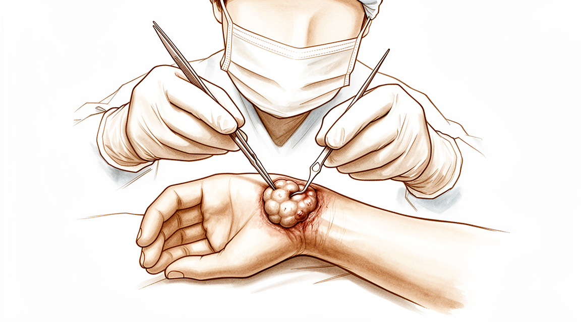
ऑपरेशन में गैंग्लियन, यानी तरल से भरी गांठ, को उस छोटे डंठल के साथ निकाला जाता है जो उसे कलाई के जोड़ से जोड़ता है। उस डंठल को निकालना ज़रूरी है, क्योंकि उसे छोड़ देने से गांठ के फिर से उगने की संभावना बढ़ जाती है।
सर्जन कहां काम करेंगे, यह इस पर निर्भर करता है कि गांठ कहां है। कलाई के पिछले हिस्से की गांठ के लिए, ऑपरेशन दो या तीन छोटे कीहोल चीरों से किया जाता है, हर चीरा लगभग 1 सेमी लंबा। एक पतला कैमरा कलाई के जोड़ के अंदर डाला जाता है, ताकि सर्जन अंदर देख सकें और गांठ और उसके डंठल को निकाल सकें। कलाई की हथेली वाली तरफ़ की गांठ के लिए, ऑपरेशन गांठ के ऊपर एक खुले चीरे से किया जाता है। सर्जन गांठ को आसपास की संरचनाओं से सावधानी से अलग करते हैं। हथेली वाली तरफ़, गांठ अक्सर हाथ को ख़ून पहुंचाने वाली एक धमनी के पास होती है, और कभी-कभी उसके चारों ओर लिपटी होती है, इसलिए ऑपरेशन का यह हिस्सा धीरे-धीरे और हल्के हाथ से किया जाता है। आपके सर्जन पहले से यह भी जांच लेंगे कि आपके हाथ की दोनों मुख्य धमनियों से ख़ून का बहाव ठीक है।
गांठ निकल जाने के बाद, चीरों को टांकों से बंद कर दिया जाता है। ऊपर ड्रेसिंग (घाव की पट्टी) लगाई जाती है, और आप उस ड्रेसिंग को लगभग 10 दिन तक लगाए रखते हैं। आपके सर्जन आपको जल्दी, सर्जरी के बाद पहले 2 सप्ताह के भीतर, कलाई हिलाना शुरू करने के लिए प्रोत्साहित करेंगे, क्योंकि हल्की-फुल्की हरकत जकड़न को रोकने में मदद करती है।
ऑपरेशन के बाद
यह आमतौर पर डे-केस होता है, इसलिए आप उसी दिन घर जाने की उम्मीद कर सकते हैं, हालांकि कभी-कभी मरीज़ रात भर रुकते हैं। पहले 24 घंटे किसी को आपके साथ रहना चाहिए। चीरों के आसपास आपको कुछ दर्द महसूस हो सकता है; आमतौर पर साधारण दर्द निवारक दवाओं से यह ठीक हो जाता है। सूजन कम करने के लिए आराम करते समय अपना हाथ तकिए पर ऊपर उठाकर रखें। जैसे ही आप ख़ुद को स्थिर महसूस करें, आप घर में चल-फिर सकते हैं, और जल्दी ही कलाई की हल्की-फुल्की हरकत करने को बढ़ावा दिया जाता है। हम ड्रेसिंग को लगभग 10 दिन तक लगी रहने देते हैं; जब तक हम न कहें, कृपया उससे पहले इसे न उतारें। जब हम आपसे मिलते हैं तब हम इसे बदलते हैं या हटा देते हैं। ज़्यादातर लोग दो से तीन सप्ताह के भीतर, जब वे स्टीयरिंग व्हील आराम से पकड़ पाते हैं, फिर से गाड़ी चलाने लगते हैं।
ठीक होने की प्रक्रिया
पहले कुछ दिनों तक आपकी कलाई में दर्द रहेगा और चीरों के आसपास थोड़ी सूजन रहेगी। आमतौर पर साधारण दर्द निवारक दवाओं से यह ठीक हो जाता है, और हाथ को तकिए पर ऊपर उठाकर रखने से सूजन कम होने में मदद मिलती है। पहले दो-एक सप्ताह में तकलीफ़ धीरे-धीरे कम होती जाती है।
आप ड्रेसिंग को लगभग 10 दिन तक लगाए रखेंगे, और जब हम आपसे मिलते हैं तब हम इसे बदलते हैं या हटा देते हैं। कलाई की हल्की-फुल्की हरकत जल्दी, पहले 2 सप्ताह के भीतर, शुरू होती है, क्योंकि जल्दी हिलाने से जकड़न को रोकने में मदद मिलती है। सर्जरी के बाद हैंड थेरेपी एक्सटेंड रिहैबिलिटेशन (Extend Rehabilitation) में रूबी डूलन के साथ होती है। आपके हैंड थेरेपिस्ट (हाथ के विशेषज्ञ थेरेपिस्ट) आपकी एक्सरसाइज़ में मार्गदर्शन करेंगे और जैसे-जैसे आपकी हरकत और पकड़ लौटेगी, उसकी जांच करेंगे। जैसे ही आप ख़ुद को स्थिर महसूस करें, आप घर में चल-फिर सकते हैं, और जितना आराम से हो सके, हल्के कामों के लिए अपने हाथ का इस्तेमाल करेंगे।
जैसे-जैसे सूजन कम होती है, रोज़ के काम आसान होते जाते हैं। जब आप बिना दर्द के पकड़ पाएंगे, तो आपको लिखना, खाना और कपड़े पहनना फिर से सामान्य लगने लगेगा। गाड़ी चलाना आमतौर पर दो से तीन सप्ताह के भीतर फिर से शुरू होता है, जब घाव में तकलीफ़ न रहे और आप स्टीयरिंग व्हील आराम से पकड़ सकें; ऊपरी अंग की सर्जरी के बाद गाड़ी चलाने के बारे में हमारा पेज देखें। जैसे-जैसे हरकत और ताक़त लौटती है, ज़्यादातर लोग अपने सामान्य काम और गतिविधियों पर लौट आते हैं।
ठीक होने में हर व्यक्ति के लिए अलग समय लगता है, इसलिए आपकी समय-सीमा अलग हो सकती है। हर मुलाक़ात में आपके सर्जन और आपके हैंड थेरेपिस्ट आपका मार्गदर्शन करेंगे, और जैसे-जैसे आपकी कलाई ठीक होती है, आप लगातार सुधार की उम्मीद कर सकते हैं।
क्या समस्याएं हो सकती हैं
ज़्यादातर मरीज़ अच्छी तरह ठीक होते हैं, लेकिन कभी-कभी समस्याएं हो सकती हैं। आपके सर्जन और टीम किसी भी समस्या को जल्दी पकड़ने के लिए आप पर बारीकी से नज़र रखते हैं।
सर्जरी के बाद गांठ फिर से उग सकती है। आपको अपनी कलाई के पास वही नरम उभार लौटता दिख सकता है, कभी-कभी सप्ताहों या महीनों बाद। अगर आपको यह लौटता दिखे या महसूस हो, तो अपनी अगली फॉलो-अप जांच में इसके बारे में बताएं। अगर इसे और इलाज की ज़रूरत हो, तो आपके सर्जन आपको विकल्पों के बारे में समझा सकते हैं।
कुछ लोगों को ऑपरेशन के बाद कलाई में दर्द बना रहता है। इसकी संभावना ज़्यादा है अगर सर्जरी से पहले आपकी कलाई में दर्द था, या अगर आपके काम या खेल में कलाई को मज़बूती और ज़ोर से पीछे की ओर मोड़ना पड़ता है। यह दर्द एक गहरे दर्द जैसा महसूस हो सकता है, जो काम करने पर बढ़ जाता है और साधारण दर्द निवारक दवाओं से पूरी तरह शांत नहीं होता। कुछ लोगों को यह भी लगता है कि उनका हाथ उतनी आसानी से काम नहीं करता जितनी उन्होंने उम्मीद की थी, और पकड़ना या रोज़ के काम पहले से ज़्यादा मुश्किल लगते हैं। अगर शुरुआती ठीक होने के समय के बाद भी दर्द या कमज़ोरी बनी रहती है, तो अपनी फॉलो-अप जांच में अपने सर्जन को बताएं, ताकि हैंड थेरेपी या दूसरे क़दमों पर विचार किया जा सके।
कलाई के पिछले हिस्से पर दूरबीन से ऑपरेशन के दौरान, उंगलियों को सीधा करने वाले टेंडनों को चोट लगना एक दुर्लभ समस्या है। आपको इसका पता एक या ज़्यादा उंगलियों को सीधा करने में दिक़्क़त से, या किसी उंगली के बाक़ी उंगलियों से नीचे लटके रहने से चलेगा। अगर आप किसी उंगली को ठीक से सीधा नहीं कर पा रहे हैं, तो तुरंत क्लिनिक से संपर्क करें।
कलाई की हथेली वाली तरफ़ की गांठ के लिए, कुछ दुर्लभ समस्याएं इस वजह से होती हैं कि गांठ हाथ को ख़ून पहुंचाने वाली एक धमनी के पास होती है। चेतावनी के संकेतों में ऐसा हाथ शामिल है जो फीका (सफ़ेद पड़ा हुआ), नीला या सामान्य से ठंडा दिखे, या ऐसा सुन्नपन और झुनझुनी जो शांत न हो। अगर आपको इनमें से कुछ भी दिखे, तो इंतज़ार करने की बजाय इमरजेंसी विभाग जाएं।
अगर आप बारीक जानकारी चाहते हैं, तो इस पेज पर जटिलताओं की तालिका में आम दरें दी गई हैं।
हमें कब फ़ोन करें
अगर आपको बुखार है, या आपके चीरों के आसपास की त्वचा ज़्यादा लाल हो जाती है, सूज जाती है या उसमें से तरल रिसने लगता है, तो हमें फ़ोन करें। अगर आपका दर्द कम होने की बजाय लगातार बढ़ रहा है, या आप किसी उंगली को सीधा नहीं कर पा रहे हैं, तो हमें फ़ोन करें। अगर आपका हाथ फीका (सफ़ेद पड़ा हुआ) या नीला हो जाता है या ठंडा लगता है, अगर आपके हाथ में महसूस होना बंद हो जाता है, या अगर आप उसे हिला नहीं पा रहे हैं, तो इमरजेंसी विभाग जाएं। अगर आपकी पिंडली में सूजन है या आपकी सांस फूल रही है, तब भी इमरजेंसी विभाग जाएं।
इस समस्या के बारे में और कहां पढ़ें
यह पेज ख़ुद ऑपरेशन के बारे में है। जिस समस्या का यह इलाज करता है, और उसमें यह भी कि प्रमाण क्या बताते हैं कि सर्जरी कब मदद करती है और कब नहीं, उसके बारे में कलाई की गैंग्लियन सिस्ट पेज पर ज़्यादा विस्तार से बताया गया है।
Evidence & references
This is the clinical evidence summary written for health professionals. It is technical, and it lists the research this page was built from. You do not need to read it to understand your treatment or to make a decision about it.
Anatomy & Pathophysiology
Bony Anatomy
- The wrist includes the distal radioulnar, radiocarpal, and ulnocarpal joints and the eight carpal bones and their proximal and distal articulations [4].
- The proximal row of carpal bones consists of the scaphoid, lunate, triquetrum, and pisiform [4].
- The distal row of carpal bones consists of the trapezium, trapezoid, capitate, and hamate [4].
- The pisiform and trapezoid are the smallest carpal bones, while the capitate is the largest [4].
- The radiocarpal joints are formed by the articulation of the distal radius with the scaphoid and lunate through their respective concave facets on the distal radius [4].
- The triquetrum articulates with the triangular fibrocartilage at the radiocarpal joint [4].
- The distal concave articular surfaces of the proximal carpal row form the midcarpal articulations with the distal row [4].
- The distal row articulates with the metacarpals, allowing mobility in the thumb, stability in the index and long finger metacarpals, and increased mobility in the ring and little finger metacarpals [4].
- The distal ulnar convexity articulates at the lesser sigmoid notch of the distal radius [4].
- The sigmoid notch articular surface accommodates the ulnar head through two thirds of its arc [4].
- There is about a 20-degree inclination of the distal ulna at its articulation with the radius [4].
- The ulnar styloid lies dorsal to the ulnar head and extends distally [4].
- The distal radius has three articular components: the scaphoid fossa, the lunate fossa, and the sigmoid notch [8].
- A ridge corresponding with the scapholunate interval is located between the scaphoid and lunate fossae on the distal radius [8].
- The radial styloid allows attachment of the brachioradialis tendon [8].
- The radial styloid is the origin of the radial scapholunate and radial lunocapitate ligaments [8].
- The concave elliptical distal radius is oriented in the sagittal plane with an average of 11 degrees of volar tilt [8].
- In the frontal plane, the average radial inclination of the distal radius is 23 degrees [8].
- Radial length is measured from the tip of the radial styloid to the ulnar articular surface and averages 13 mm [8].
- The ulnar shaft remains fixed in its rotation at the ulnohumeral joint, while the radius rotates around the ulna in pronation and supination [8].
- The radius has a lateral bow that is crucial to the maintenance of full pronation and supination [8].
Ligamentous Anatomy
- The triangular fibrocartilage complex (TFCC) attaches to the base of the ulnar styloid and separates the hyaline cartilage–covered ulnar head from the styloid [4].
- The TFCC includes the ulnar collateral ligament, dorsal and volar radioulnar ligaments, articular disc, meniscal homologue, extensor carpi ulnaris sheath, and ulnolunate and ulnotriquetral ligaments [4].
- Interosseous intrinsic ligaments connect the carpal bones in the proximal and distal carpal rows [4].
- The scapholunate and lunotriquetral interosseous ligaments connect the proximal carpal row [4].
- Ligaments connecting the trapezium to the trapezoid, trapezoid to the capitate, and capitate to the hamate connect the distal carpal row [4].
- Extrinsic ligaments extend from the radius and ulna distally across the carpal rows [4].
- The radial collateral ligament extends from the radial styloid to the scaphoid waist [4].
- The ulnar collateral ligament extends from the base of the ulnar styloid to the pisiform [4].
- The transverse carpal ligament is an extrinsic ligament [4].
- Volar extrinsic ligaments include the radioscapocapitate, radiolunotriquetral, and radioscapolunate ligaments on the radial side [4].
- The ulnolunate and ulnotriquetral components of the TFCC are volar extrinsic ligaments on the ulnar side [4].
- The space of Poirier is a relatively thin area on the palmar side of the carpus between the radiolunotriquetral and radioscapocapitate ligaments [4].
- The space of Poirier overlies the palmar surface of the lunate [4].
- Dorsal extrinsic ligaments include the dorsal radiocarpal and dorsal intercarpal ligaments [4].
- The trapezoidal dorsal radiocarpal ligament attaches along the dorsal radial articular margin of the lunate fossa, from the Lister tubercle to the lesser sigmoid notch [4].
- The trapezoidal dorsal radiocarpal ligament spans the lunotriquetral joint and inserts on the dorsal surface of the triquetrum [4].
- The dorsal intercarpal ligament is attached to the distal, dorsal surface of the triquetrum and passes across the midcarpal joint to attach to the dorsal surfaces of the scaphoid waist and the trapezoid [4].
- Extrinsic carpal ligaments connect the radius or the ulna to the carpus [9].
- Volar ligaments are generally stronger than dorsal ligaments [9].
- The radioscaphocapitate ligament connects to the waist of the scaphoid and limits ulnar translation of the carpus [9].
- The long radiolunate ligament helps limit ulnar translocation of the carpus [9].
- The short radiolunate ligament helps control lunate position [9].
- The radioscapholunate ligament is a vascular conduit and not a true ligament, also known as the ligament of Testut [9].
- The ulnolunate ligament attaches to the palmar radioulnar ligament and the lunate [9].
- The ulnocapitate ligament attaches to the ulnar head and is the most superficial or palmar palmar ulnocarpal ligament [9].
- The ulnotriquetral ligament attaches to the palmar radioulnar ligament and the triquetrum [9].
- The dorsal radiocarpal ligament has a trapezoidal shape and passes from the dorsal rim of the distal radius to the lunate and triquetrum [9].
- Fibers of the dorsal radiocarpal ligament insert onto the dorsal lunotriquetral interosseous ligament [9].
- The scapholunate interosseous ligament is a major stabilizer of the wrist and the most commonly injured wrist ligament [9].
- The scapholunate interosseous ligament is C-shaped, consisting of dorsal, palmar, and interosseous portions, with the dorsal portion being the strongest and thickest [9].
- The scapholunate interosseous ligament provides a flexion force on the lunate given its attachment to the scaphoid [9].
- The lunotriquetral interosseous ligament is C-shaped, with the volar portion being the thickest and strongest [9].
- The lunotriquetral interosseous ligament provides an extension moment on the lunate given its attachment to the triquetrum [9].
- The capitohamate ligament is a thick ligament with a 5 × 5 mm cross section and extensions to the third or fourth metacarpals [9].
- The dorsal intercarpal ligament passes from the dorsal tubercle of the triquetrum to the distal pole of the scaphoid [9].
- The dorsal intercarpal ligament reinforces the elastic dorsal wrist capsule and helps stabilize the scapholunate articulation with a contribution to the dorsal scapholunate interosseous ligament from its deep fibers [9].
- The space of Poirier is an area adjacent to the proximal capitate without ligamentous attachment, situated ulnar to the radioscaphocapitate ligament and radial to the long radiolunate in the floor of the carpal tunnel [9].
- The space of Poirier is a weak area vulnerable to instability, through which the distal carpal row separates from the lunate during a perilunate dislocation [9].
Soft Tissue Anatomy
- Six dorsal compartments contain wrist and digital extensor tendons on the dorsal surface of the wrist [8].
- The contents of the carpal tunnel reside on the volar surface, including nine flexor tendons and the median nerve [8].
- The flexor carpi ulnaris tendon can be palpated near its insertion on the pisiform on the ulnar surface [8].
- The boundaries of Guyon’s canal are the volar carpal ligament and transverse carpal ligament, the hook of the hamate radially, and the pisiform ulnarly [8].
- Guyon’s canal contains the ulnar artery and nerve [8].
- The interosseous membrane connects the shafts of the radius and ulna in the interosseous space [8].
- The central portion of the interosseous membrane is thickened and important in force transmission between the radius and ulna [8].
Ganglion Pathophysiology
- The dorsal wrist ganglion accounts for 60% to 70% of all hand and wrist ganglions [3].
- The main cyst of a dorsal wrist ganglion is usually located directly over the scapholunate ligament [3].
- Dorsal wrist ganglions may occur anywhere else between the extensor tendons [3].
- Failure to identify and excise the pedicle attachment to the scapholunate ligament increases the likelihood of recurrence for dorsal wrist ganglions [3].
- A small, mucin-filled duct is invariably seen piercing the transverse fibers of the scapholunate ligament, connecting the underlying scapholunate joint with the main cyst [3].
- The volar wrist ganglion is the second most common ganglion of the hand and wrist, accounting for 18% to 20% [1].
- The majority of volar ganglions occur either directly over the distal edge of the radius or slightly more distally over the scaphoid tubercle [1].
- Volar ganglions arising over the distal edge of the radius arise from the capsular and ligamentous fibers of the radiocarpal joint [1].
- Volar ganglions arising over the distal edge of the radius occur under the volar wrist crease between the flexor carpi radialis and abductor pollicis longus tendons [1].
- The main cyst of a volar wrist ganglion arising from the radiocarpal joint may be intertwined with bifurcating branches of the radial artery [1].
- Another type of volar ganglion arises from the capsule of the scaphotrapezial joint [1].
- Volar ganglions may appear small clinically but can be surprisingly extensive at surgery [1].
- Multiloculated volar ganglion cysts may extend under the thenar muscles, along the flexor carpi radialis tendon, into the carpal canal, and under the first extensor compartment adjacent to the dorsal branch of the radial artery [1].
- Extensions of volar ganglions may reach as far dorsally as the first web space [1].
- The radial artery is frequently intimately attached to the wall of a volar ganglion and may be completely encircled by it [1].
Biomechanics and Kinematics
- The eight carpal bones represent the most complex articular system in the human body [5].
- The human wrist allows precise positioning of the hand and optimization of power and prehensile tasks throughout a nearly hemispherical arc of wrist motion [5].
- The wrist joint’s motion planes include flexion, extension, radial deviation, ulnar deviation, and circumduction [9].
- There is minimal carpal motion with pronosupination [9].
- Approximately 62° of wrist extension occurs through the radiocarpal joint [9].
- 62% of wrist flexion occurs through the midcarpal joint [9].
- The midcarpal joint is mostly responsible for 20° of radial deviation and 40° of ulnar deviation [9].
- The midcarpal joint is responsible for the “dart thrower’s motion,” which involves moving from radial extension into ulnar flexion positioning of the wrist [9].
- The radius bears 80% of the axial load transmitted through the radiocarpal joint in neutral ulnar variance [9].
- The ulna bears 20% of the axial load transmitted through the radiocarpal joint in neutral ulnar variance [9].
- The proximal row of carpal bones forms an intercalated segment between the distal carpal row and the distal radius, bound into a functional unit by the scapholunate and lunotriquetral interosseous ligaments [9].
- The distal row is rigid with little motion between its bones due to stout intercarpal ligaments, acting as a functional unit with the scaphoid bridging both rows [9].
- During wrist flexion from neutral, the distal row flexes and ulnarly deviates slightly while the scaphoid pronates [9].
- During wrist flexion from neutral, the proximal row flexes differentially, with more rotation through the scaphoid, followed by the triquetrum and the lunate [9].
- The proximal row translates dorsally during wrist flexion [9].
- During wrist extension from neutral, the distal row extends and radially deviates slightly while the scaphoid supinates [9].
- During wrist extension from neutral, the proximal row extends differentially, with more motion in the scaphoid, followed by the triquetrum and then the lunate [9].
- The proximal row translates palmarly during wrist extension [9].
Clinical Presentation
- Volar wrist ganglions are the second most common ganglion of the hand and wrist, accounting for 18% to 20% of cases [1].
- The majority of volar ganglions occur directly over the distal edge of the radius or slightly more distally over the scaphoid tubercle [1].
- Ganglions arising from the distal edge of the radius originate from capsular and ligamentous fibers of the radiocarpal joint [1].
- Radiocarpal volar ganglions occur under the volar wrist crease between the flexor carpi radialis and abductor pollicis longus tendons [1].
- The main cyst of a radiocarpal volar ganglion may be intertwined with bifurcating branches of the radial artery [1].
- A second type of volar ganglion arises from the capsule of the scaphotrapezial joint [1].
- Multiloculated cysts may extend under the thenar muscles, along the flexor carpi radialis tendon, into the carpal canal, and under the first extensor compartment adjacent to the dorsal branch of the radial artery [1].
- Extensions of volar ganglions can reach as far dorsally as the first web space [1].
- Extensions of volar ganglions can often be appreciated preoperatively by careful palpation and digital compression [1].
- The patency of the radial and ulnar arteries must be assessed prior to surgery [1].
- The Allen test should be performed routinely to exclude ulnar artery occlusion [1].
- Preservation of the radial artery is particularly important in patients with a radial-dominant circulation [1].
Investigations
Magnetic Resonance Imaging
- MRI for hand and wrist imaging was first reported in 1986 [10].
- The primary advantages of MRI compared with CT and radiography are improved tissue characterization, especially of soft tissues such as ligamentous structures in the wrist and synovium in the hand, and the lack of ionizing radiation [10].
- MRI is the modality of choice for imaging radiographically occult fractures of the hand and wrist [10].
- Early MRI was limited by low magnetic field strength (0.15 T) and limited image options and image processing [10].
- Modern MRI is generally at 1.5T or 3T with a wide variety of imaging options and powerful image processing and postprocessing available [10].
- 3T is much preferred for hand and wrist imaging, especially for imaging small fields of view [10].
- Dedicated extremity magnets have been marketed, but image quality is poor compared with conventional MR imaging [10].
- 7T MRI has recently become approved for clinical use [10].
- 7T MRI has the potential to become a powerful tool for hand and wrist imaging as applications are developed [10].
- MRI with contrast enhancement is most commonly used to determine whether soft-tissue lesions are solid or cystic [10].
- In the case of rheumatologic imaging, MRI with contrast enhancement is used to better visualize erosions and synovial burden [10].
- Dynamic contrast enhancement has been used with inconsistent results to assess for the presence of avascular necrosis in the lunate or scaphoid after injury [10].
- MR angiography of the hand and wrist can be helpful in situations such as diagnosis of the hypothenar hammer syndrome [10].
- MR arthrography can be performed for evaluation of the triangular fibrocartilage and intercarpal ligament tears [10].
- MR arthrography is generally unnecessary with the increasing availability of high field MRI [10].
Computed Tomography and Other Imaging Modalities
- Hybrid SPECT/CT has been used for the diagnosis of radiographic occult fractures of the wrist [6].
- Cone-beam CT has been used in the diagnosis of scaphoid fractures [6].
- CT and MRI have a diagnostic impact on wrist injuries in young adults [6].
- Cine MRI is a new approach to the diagnosis of scapholunate dissociation [6].
- Cone-beam computed tomography arthrography is an innovative modality for the evaluation of wrist ligament and cartilage injuries [6].
- 3D analysis of the wrist has been described [6].
- Normal and variant anatomy of the wrist and hand can be evaluated on MR imaging [6].
- A practical guide to diagnostic imaging of the upper extremity has been published [7].
- 3-Tesla imaging of the wrist and hand has specific techniques and applications [7].
- MR imaging of the human hand and wrist at 7 T has been described [7].
- Magnetic resonance imaging can be used for the anterior radiocarpal ligaments [7].
- The carpal contents/canal ratio can be established by means of magnetic resonance imaging [7].
- MR imaging can be used for trauma of the elbow and wrist [7].
- Magnetic resonance imaging of the wrist has been described [7].
- MR imaging can be used for ulnar impaction [7].
- Rupture of the distal biceps tendon can be evaluated with MR imaging [7].
- MR imaging can be used for the wrist and elbow in clinical sports medicine [7].
- Magnetic resonance imaging of the musculoskeletal system, including the elbow, has been described [7].
- The efficacy of magnetic resonance imaging of the elbow has been evaluated [7].
- Sports and occupational injuries of the elbow can be evaluated with MR imaging findings [7].
- Magnetic resonance imaging of the elbow has been described [7].
- Magnetic resonance imaging can be used in Kienbock disease [7].
- Magnetic resonance imaging of the elbow, including normal anatomy, imaging technique, and osseous abnormalities, has been described [7].
- Magnetic resonance imaging of the elbow, including abnormalities of the ligaments, tendons, and nerves, has been described [7].
- Low-field MRI can be used for scaphoid fracture [7].
- MR imaging can be used for traumatic and overuse injuries of the wrist and hand in athletes [7].
- MR imaging diagnosis of TFC pathology can be correlated with arthroscopy [7].
- MR imaging of the elbow in baseball pitchers has been described [7].
- Overuse syndromes and injuries involving the elbow can be evaluated with MR imaging findings [7].
- Occult wrist fractures can be detected by magnetic resonance imaging [7].
- Ulnar collateral ligament injury in the throwing athlete can be evaluated with saline-enhanced MR arthrography [7].
- Magnetic resonance imaging of the triangular fibrocartilage complex has been described [7].
- Preoperative evaluation of the ulnar collateral ligament by magnetic resonance imaging and computed tomography arthrography has been evaluated in baseball players with surgical confirmation [7].
- Histologic and magnetic resonance imaging correlations in Kienbock’s disease have been described [7].
- MRI can be used for the diagnosis of occult dorsal wrist ganglion [7].
Radiography and Stress Views
- Distal radioulnar joint stress radiography can be used for detecting radioulnar ligament injury [6].
- Radiographic stress views can be compared for scapholunate dynamic instability in a cadaver model [6].
- Imaging of radial wrist pain involves various imaging modalities and anatomy [6].
- Intrinsic ligament and triangular fibrocartilage complex tears of the wrist can be compared using MDCT arthrography, conventional 3-T MRI, and MR arthrography [6].
- Simple plain radiographic signs and measures can be used to diagnose acute scapholunate ligament injuries of the wrist [11].
- Radiographic clues can be used for determining carpal instability and treatment protocol for scaphoid fractures [11].
- The role of imaging in diagnosing diseases of the distal radioulnar joint, triangular fibrocartilage complex, and distal ulna has been described [11].
Diagnostic Accuracy and Systematic Reviews
- The efficacy of magnetic resonance imaging and clinical tests in diagnostics of wrist ligament injuries has been evaluated in a systematic review [11].
- The role of magnetic resonance imaging in scaphoid fractures has been described [11].
Treatment
Dorsal Wrist Ganglion
- Failure to identify and excise the pedicle attachment to the scapholunate ligament increases the likelihood of recurrence [3].
- Careful preoperative palpation of the cyst with digital compression often reveals its extent and the direction of the pedicle [3].
- Transillumination or aspiration confirms the diagnosis preoperatively [3].
- Review of preoperative radiographs to rule out an interosseous component is recommended before dissection [3].
- Most dorsal ganglions can be approached through a transverse incision over the proximal carpal row [3].
- A modified incision or second transverse incision may be necessary for ganglions not directly over the scapholunate ligament [3].
- The diagnosis of ganglion cyst should be made before commitment to a transverse incision because this incision is not readily incorporated into a limb-sparing incision if a subsequent diagnosis of a malignant soft tissue tumor is made [3].
- The extensor pollicis longus and extensor digitorum communis tendons are retracted radially and ulnarly, respectively, to expose the dorsal ganglion [3].
- The joint capsule is opened along the border of the radius and scaphoid's proximal pole with the wrist in volar flexion [3].
- The capsule is elevated and retracted distally to expose the capsular attachments to the scapholunate ligament [3].
- Smaller intraarticular cysts are often seen attached to the scapholunate ligament [3].
- The capsular incision is continued around the ganglion, leaving all capsular attachments to the ligament intact [3].
- The capsular incision is extended laterally if capsular ducts are encountered, which can be identified by small amounts of mucin drainage [3].
- The ganglion and its capsular attachments are tangentially excised off the scapholunate ligament [3].
- A small, mucin-filled duct is invariably seen piercing the transverse fibers of the scapholunate ligament [3].
- This duct appears to connect the underlying scapholunate joint with the main cyst [3].
- Synovial and capsular attachments along the distal margin of the scapholunate ligament are excised to give an unobstructed view of the head and neck of the capitate [3].
Volar Wrist Ganglion
- Volar ganglions arising from the distal edge of the radius occur under the volar wrist crease between the flexor carpi radialis and abductor pollicis longus tendons [1].
- The main cyst of a volar ganglion may be intertwined with bifurcating branches of the radial artery [1].
- Extensions of volar ganglions can be appreciated preoperatively by careful palpation and digital compression [1].
- The Allen test should be performed routinely to assess the patency of the radial and ulnar arteries [1].
- Ulnar artery occlusion should be excluded preoperatively [1].
- The radial artery must be preserved, particularly in patients with a radial-dominant circulation [1].
- Exposure and precise identification of the capsular attachments of a volar ganglion are more difficult than for a dorsal ganglion [1].
- The incision for volar ganglion excision must be planned to allow for extension into the carpal tunnel or base of the thenar muscles [1].
- Longitudinal incisions allow for optimal visualization during volar ganglion excision [1].
- The forearm fascia is incised longitudinally to identify and mobilize the dome of the cyst [1].
- The radial artery is frequently intimately attached to the wall of the ganglion and may be completely encircled by it [1].
- Loupe magnification aids in the dissection to protect the radial artery [1].
- The pedicle is traced to the volar joint capsule, usually the scaphotrapezial or radiocarpal ligament [1].
- The joint is opened and explored, and the ganglion attachments are excised approximately 3 ± 4 mm [1].
- Surrounding tissues are compressed digitally after excision to rule out further mucin-filled pockets [1].
- Unidentified extensions of the ganglion must be excised if present [1].
- Capsular closure is unnecessary and only delays early mobilization [1].
- A bulky bandage and elevation of the hand ensure early postoperative comfort [1].
- Motion of the wrist should begin within the first 2 weeks after surgery [1].
- Unexpected branches of the radial sensory or lateral antebrachial cutaneous nerves may be injured, leading to troublesome neuromas [1].
- Extensions of the routine incision into the carpal canal must avoid injury to the palmar cutaneous branch of the median nerve [1].
- Injuries to the radial artery can be repaired microscopically [1].
- Some authors recommend leaving a portion of the cyst wall attached to the artery to avoid arterial injury [1].
- Stiffness of the wrist is less common than with dorsal ganglions but can occur if early motion is not encouraged [1].
- Curved incisions appear to consistently provide more attractive scars, especially near the volar wrist creases [1].
References
[1] Green S Operative Hand Surgery. BOX 59.1 Ganglions of the Hand and Wrist > Operative Treatment > Volar Wrist Ganglion.
[3] Green S Operative Hand Surgery. BOX 59.1 Ganglions of the Hand and Wrist > Operative Treatment > Dorsal Wrist Ganglion.
[4] Campbell S Operative Orthopaedics 4 Volume Set. NERVE INJURIES AT THE LEVEL OF THE HAND AND WRIST > ANATOMY.
[5] Green S Operative Hand Surgery. INTRODUCTION.
[6] Campbell S Operative Orthopaedics 4 Volume Set. ANATOMIC RECONSTRUCTION OF THE DISTAL RADIOULNAR LIGAMENTS > RADIOGRAPHIC TECHNIQUES.
[7] Campbell S Operative Orthopaedics 4 Volume Set. ELBOW, WRIST, AND HAND.
[8] A Lange Medical Book Current Diagnosis Treatment In Orthopedics Fifth Edition. 2Musculoskeletal Trauma Surgery > FRACTURES AND DISLOCATIONS OF THE DISTAL AND MID-FOREARM.
[9] Aaos Comprehensive Orthopaedic Review 3. Carpal Instability* > II. Anatomy and Biomechanics (See Chapter 92).
[10] Orthopaedic Knowledge Update 13 Ebook Without Multimedia. Anatomy, Evaluation, Clinical Examination, and Imaging > Imaging: Advances in Imaging of the Hand and Upper Extremity > Magnetic Resonance Imaging.
[11] Campbell S Operative Orthopaedics 4 Volume Set. ANATOMIC RECONSTRUCTION OF THE DISTAL RADIOULNAR LIGAMENTS > DIAGNOSIS AND EVALUATION.