Patients › Wrist
कार्पल टनल रिलीज़ (नर्व पर से दबाव हटाने का ऑपरेशन)
Carpal tunnel release surgery relieves median nerve compression—when it’s needed and what to expect.
यह ऑपरेशन क्यों सुझाया गया है
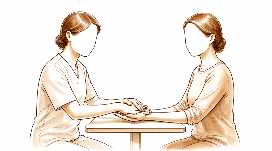
डॉ. कीरन हिरपरा, माटर प्राइवेट हॉस्पिटल, रॉकहैम्पटन में ऊपरी अंग (कंधे से हाथ तक) के सर्जन, सबसे पहले वे सबसे कम चीर-फाड़ वाले विकल्प अपनाते हैं जो आपकी समस्या के लिए सही हों। मरीज़ों को आम तौर पर उनके जीपी (आपके फैमिली डॉक्टर) हमारे क्लिनिक में रेफ़र करते हैं; अगर किसी फिजियोथेरेपिस्ट ने आपको हमसे मिलने की सलाह दी है, तब भी मेडिकेयर रिबेट पाने के लिए आपको अपने जीपी से रेफ़रल लेना होगा। आपके अपॉइंटमेंट में हम आपकी पूरी जानकारी (हिस्ट्री) लेते हैं, आपके हाथ की जांच करते हैं, और ज़रूरत हो तो इमेजिंग (स्कैन) या नर्व के टेस्ट करवाते हैं। कार्पल टनल सिंड्रोम तब होता है जब आपकी कलाई में एक नर्व (तंत्रिका) दब जाती है, जिससे आपके हाथ में झुनझुनी, सुन्नपन और दर्द होता है। हम आम तौर पर बिना ऑपरेशन वाले इलाज से शुरुआत करते हैं, जैसे स्प्लिंट, हैंड थेरेपी या इंजेक्शन। सर्जरी पर तब विचार किया जाता है जब इन इलाजों से पर्याप्त सुधार नहीं हुआ हो। इस ऑपरेशन को कार्पल टनल रिलीज़ कहते हैं। यह कलाई के आर-पार फैले एक कसे हुए पट्टे को काटकर नर्व पर से दबाव कम करता है। इस ऑपरेशन के बाद लक्षणों की गंभीरता और हाथ के काम-काज में अच्छे-ख़ासे सुधार की उम्मीद की जा सकती है, चाहे उम्र, दूसरी बीमारियां या वर्कर्स कंपेंसेशन (काम पर लगी चोट का मुआवज़ा) की स्थिति कुछ भी हो। हम आपसे विकल्पों पर बात करेंगे, और आप और हम मिलकर तय करेंगे कि सर्जरी आपके लिए सही है या नहीं।
ऑपरेशन से पहले
आपके सर्जन आपको साफ़ निर्देश देंगे, लेकिन यहां बताया गया है कि क्या उम्मीद करें। सर्जरी से 7 घंटे पहले आपको खाना और पीना बंद करना होगा। हम 6 के बजाय 7 घंटे इसलिए कहते हैं ताकि अगर ऑपरेशन की सूची जल्दी चल रही हो तो आपको पहले बुलाया जा सके। अपनी मौजूदा दवाओं की सूची साथ लाएं, और अपने सर्जन को खून पतला करने वाली किसी भी दवा के बारे में बताएं, क्योंकि कुछ दवाओं को पहले से रोकना पड़ सकता है। बाद में आपको घर ले जाने के लिए किसी के गाड़ी चलाकर आने का इंतज़ाम करें। ढीले, आरामदायक कपड़े पहनें जिनकी आस्तीनें आसानी से उतर जाएं। ऑपरेशन की योजना बनाने के लिए कुछ इमेजिंग, जैसे एक्स-रे, एमआरआई या अल्ट्रासाउंड, की ज़रूरत पड़ सकती है। अगर आपको दूसरी बीमारियां हैं, तो आपको खून की जांच या एनेस्थेटिस्ट (बेहोशी के डॉक्टर) से जांच की ज़रूरत पड़ सकती है, लेकिन ज़्यादातर लोगों को इसकी ज़रूरत नहीं होती।
ऑपरेशन के दिन
सर्जरी के दिन आप अस्पताल की सर्जिकल एडमिशन यूनिट (ऑपरेशन के लिए भर्ती विभाग) में आते हैं। वहां का स्टाफ़ आपको भर्ती करता है और ऑपरेशन थिएटर के लिए तैयार करता है। फिर आप एनेस्थेटिस्ट से मिलते हैं, जो डॉक्टर एनेस्थीसिया देते हैं और ऑपरेशन के दौरान आपकी देखभाल करते हैं। यह ऑपरेशन लोकल एनेस्थीसिया (एक इंजेक्शन जो सिर्फ़ ऑपरेशन वाले हिस्से को सुन्न करता है, और आप जागते रहते हैं) या जनरल एनेस्थीसिया (जिसमें आप पूरी तरह बेहोश रहते हैं) में किया जा सकता है। ज़्यादातर लोग लोकल चुनते हैं: इसमें रिकवरी जल्दी होती है और आप जल्द ही घर जा सकते हैं। अगर आप बेहोश रहना पसंद करें, तो यह भी एक सही विकल्प है; इस बारे में अपने सर्जन और एनेस्थेटिस्ट से बात करें। फिर आपको ऑपरेशन थिएटर में ले जाया जाता है, जहां ऑपरेशन किया जाता है। बाद में आप रिकवरी वाले हिस्से में होश में आते हैं, जहां एनेस्थीसिया का असर खत्म होने तक नर्सें आप पर नज़र रखती हैं। जब आपकी हालत स्थिर हो जाती है, तो प्रक्रिया और आपकी रिकवरी के हिसाब से आप या तो घर चले जाते हैं या थोड़े समय के लिए वार्ड में रहते हैं।
ऑपरेशन में क्या किया जाता है
इस ऑपरेशन को कार्पल टनल रिलीज़ कहते हैं। आपके सर्जन ऑपरेशन वाले हिस्से के ऊपर, कलाई और हथेली पर, एक ही चीरा लगाते हैं। इस चीरे के रास्ते आपके सर्जन ऊतक के उस कसे हुए पट्टे तक पहुंचते हैं जो कार्पल टनल की छत बनाता है, यानी आपकी कलाई में वह संकरा रास्ता जिससे होकर नर्व गुज़रती है। उस पट्टे को काटने से टनल खुल जाती है और नर्व पर से दबाव हट जाता है। खुद उस पट्टे को रिपेयर करने की ज़रूरत नहीं होती; वह इस तरह भर जाता है कि अंदर नर्व के लिए ज़्यादा जगह रहती है।
चीरे को टांकों से बंद किया जाता है। फिर आपके हाथ और कलाई के चारों ओर एक मोटी, गद्दीदार पट्टी लपेटी जाती है। आप यह ड्रेसिंग (घाव की पट्टी) लगभग 10 दिन तक लगाए रखेंगे, जैसा कि अगले हिस्से में बताया गया है।
ऑपरेशन खुद छोटा होता है, और आप उसी दिन घर चले जाते हैं। सहमति फ़ॉर्म पर हस्ताक्षर करने से पहले आपके सर्जन आपको ये कदम फिर से समझाएंगे, और आप किसी भी समय सवाल पूछ सकते हैं।
ऑपरेशन के बाद
आप रिकवरी वाले हिस्से में होश में आएंगे, जहां एनेस्थीसिया का असर खत्म होने तक नर्सें आप पर नज़र रखती हैं। आपका हाथ एक मोटी, गद्दीदार पट्टी में होगा, और आपको आराम में रखने के लिए हम आपको दर्द की दवा देंगे। यह डे-केस (उसी दिन छुट्टी वाला) ऑपरेशन है, इसलिए आप उसी दिन घर जाएंगे। कृपया पहले 24 घंटों के लिए किसी को अपने साथ रहने का इंतज़ाम करें। हम ड्रेसिंग को लगभग 10 दिन तक लगी रहने देते हैं; कृपया उससे पहले इसे न हटाएं, जब तक हम आपसे ऐसा करने को न कहें। जब हम आपको देखते हैं तब हम इसे बदलते हैं या हटाते हैं। आराम करते समय अपना हाथ ऊपर उठाकर रखें, और जैसे ही आपको ठीक लगे, हल्के कामों के लिए इसका धीरे-धीरे इस्तेमाल करें। ज़्यादातर लोग एक से दो सप्ताह के भीतर फिर से गाड़ी चलाने लगते हैं, जब घाव में आराम हो और वे ऑपरेशन वाले हाथ को बचाए बिना स्टीयरिंग व्हील को पकड़ और घुमा सकें। देखें: ऊपरी अंग की सर्जरी के बाद गाड़ी चलाना।
ठीक होने की प्रक्रिया
पहले कुछ दिनों तक आपके हाथ में दर्द और सूजन रहेगी, और गद्दीदार पट्टी भारी-भरकम लगेगी। आराम करते समय अपना हाथ ऊपर उठाकर रखें, क्योंकि इससे सूजन और तकलीफ़ कम होती है। आम तौर पर साधारण दर्द की दवा काफ़ी होती है। इस ऑपरेशन के बाद कई लोगों को तेज़ दर्द निवारक दवा की बिल्कुल ज़रूरत नहीं पड़ती।
जैसे ही आपको ठीक लगे, आप हल्के कामों के लिए अपने हाथ का धीरे-धीरे इस्तेमाल करेंगे। ड्रेसिंग तब तक लगी रहती है जब तक हम आपको नहीं देखते, और उसके हटने के बाद आप सामान्य रूप से हाथ धो सकते हैं और नहा सकते हैं। आपके टांके आपकी फॉलो-अप जांच में निकाले जाते हैं। सर्जरी के बाद हैंड थेरेपी एक्सटेंड रिहैबिलिटेशन में रूबी डूलन के साथ होती है। रूबी आपकी एक्सरसाइज़ में मार्गदर्शन करेंगी और आराम के लिए ज़रूरत हो तो स्प्लिंट बनाएंगी। धीरे-धीरे हिलाने-डुलाने से नर्व को शांत होने में मदद मिलती है, इसलिए जैसे-जैसे सप्ताह बीतेंगे, आप अपने हाथ का ज़्यादा इस्तेमाल करने लगेंगे।
जैसे-जैसे सूजन कम होगी, आप देखेंगे कि झुनझुनी और सुन्नपन कम हो रहे हैं और आपकी पकड़ लौट रही है। जब आप बिना दर्द के पकड़ और घुमा सकें, तो रोज़ के काम फिर से सामान्य लगने लगते हैं। नर्व पर से समय रहते दबाव हटाने से हाथ में महसूस करने की क्षमता और काम-काज लौट आते हैं। अगर आपके लक्षण दोनों हाथों में थे, तो हम एक ही बार में दोनों हाथों का ऑपरेशन करने पर बात कर सकते हैं।
ठीक होने का समय हर व्यक्ति में अलग होता है। आपकी समय-सीमा अलग हो सकती है, और आपके सर्जन और थेरेपिस्ट हर कदम पर आपका मार्गदर्शन करेंगे।
क्या समस्याएं हो सकती हैं
ज़्यादातर मरीज़ अच्छी तरह ठीक हो जाते हैं, लेकिन कभी-कभी समस्याएं हो सकती हैं। किसी भी समस्या को जल्दी पकड़ने के लिए आपके सर्जन और टीम आप पर बारीकी से नज़र रखते हैं।
संक्रमण (इन्फेक्शन)। घाव के संक्रमण में चीरे से बाहर की ओर फैलती लाली, गर्माहट या तरल का रिसाव दिख सकता है। आपको गहरा, धड़कता हुआ (टीस मारता) दर्द महसूस हो सकता है जो साधारण दर्द निवारक दवाओं से कम नहीं होता, या आपको बुखार हो सकता है। अगर आपको इनमें से कुछ भी दिखे, तो उसी दिन क्लिनिक को फ़ोन करें। अगर आपकी तबीयत ख़राब लगे या लाली तेज़ी से फैल रही हो, तो इमरजेंसी विभाग जाएं। कुछ बातें संक्रमण की संभावना बढ़ा सकती हैं, जैसे सर्जरी से कुछ ही समय पहले कलाई में स्टेरॉयड इंजेक्शन, या कुछ ख़ास बीमारियां। ऑपरेशन से पहले हम आपसे जुड़ी किसी भी ज़रूरी बात पर चर्चा करेंगे।
घाव की समस्याएं। कभी-कभी घाव के किनारे भरने में ज़्यादा समय लेते हैं या थोड़ा खुल जाते हैं, या त्वचा के नीचे खून इकट्ठा होकर सूजन पैदा कर देता है। आपको टांके खिंचकर अलग होते दिख सकते हैं, या चीरे के पास एक सख़्त गांठ महसूस हो सकती है जिसे छूने पर दर्द हो। अपनी फॉलो-अप जांच में इसके बारे में बताएं, या अगर यह ज़्यादा बिगड़ता दिखे तो उससे पहले ही क्लिनिक को फ़ोन करें।
नर्व का इरिटेशन। जिस नर्व को रिलीज़ किया जा रहा है, वह ऑपरेशन वाली जगह के पास होती है, इसलिए सर्जरी के दौरान उस पर चोट (नील) लग सकती है या वह परेशान (इरिटेट) हो सकती है। यह आम तौर पर हाथ में नई झुनझुनी या सुन्नपन जैसा महसूस होता है, या त्वचा का कोई हिस्सा पहले से अलग महसूस होता है। नर्व का ज़्यादातर इरिटेशन समय के साथ ठीक हो जाता है, लेकिन कुछ बदलाव बने रह सकते हैं। अगर आपको नया सुन्नपन दिखे जो सुधर नहीं रहा है, तो अगली जांच में अपने सर्जन को बताएं।
लक्षण जो लौट आएं या पूरी तरह ठीक न हों। कभी-कभी नर्व पर से दबाव पूरी तरह नहीं हटता, या महीनों या सालों में टनल फिर से कस जाती है। आपको शुरुआती झुनझुनी, सुन्नपन या रात का दर्द लौटता हुआ महसूस हो सकता है। अगर ऐसा हो, तो अपनी जांच में इसके बारे में बताएं। कभी-कभी दोबारा सर्जरी की ज़रूरत पड़ती है, और आपके सर्जन इसकी वजह जानने के लिए टेस्ट करवाएंगे।
अगर आप विस्तार से जानना चाहते हैं, तो इस पेज पर दी गई जटिलताओं (कॉम्प्लिकेशन) की तालिका में आम दरें दी गई हैं।
हमें कब फ़ोन करें
ज़्यादातर लोग बिना किसी समस्या के ठीक हो जाते हैं, लेकिन कुछ लक्षणों पर तुरंत ध्यान देने की ज़रूरत होती है। अगर आपको बुखार हो, घाव में बढ़ती लाली या घाव से रिसाव हो, या दर्द लगातार बढ़ता जा रहा हो, तो हमें फ़ोन करें। अगर आपको हाथ में नया सुन्नपन दिखे जो सुधर नहीं रहा है, या आप अपनी उंगलियां नहीं हिला पा रहे हैं, तो हमें फ़ोन करें। अगर आपको अचानक तेज़ दर्द हो, पिंडली में सूजन या दर्द हो, या सांस फूले, तो इमरजेंसी विभाग जाएं। अगर कोई शक हो, तो क्लिनिक को फ़ोन करें और हम आपका मार्गदर्शन करेंगे।
इस समस्या के बारे में और कहां पढ़ें
यह पेज खुद ऑपरेशन के बारे में है। जिस समस्या का यह इलाज करता है, और प्रमाण इस बारे में क्या बताते हैं कि सर्जरी कब मदद करती है और कब नहीं, इसकी विस्तार से जानकारी कार्पल टनल सिंड्रोम पेज पर दी गई है।
Evidence & references
This is the clinical evidence summary written for health professionals. It is technical, and it lists the research this page was built from. You do not need to read it to understand your treatment or to make a decision about it.
Overview
- Symptoms of pain, numbness, and tingling in the hands are common in the general population [1].
Anatomy & Pathophysiology
Epidemiology & Symptoms
- Carpal tunnel syndrome with compression of the median nerve at the wrist is the most commonly diagnosed site of nerve compression in the upper extremity [10].
- Symptoms include paresthesia or numbness (or both) in the median nerve distribution, specifically the thumb, index finger, middle finger, and radial side of the ring finger [10].
- Nocturnal paresthesias in the radial three digits of the hand is nearly pathognomonic for carpal tunnel syndrome [10].
- Paresthesias occur characteristically in "fixed wrist activities" such as reading a book or a newspaper, driving, or use of a computer keyboard or mouse [10].
- Patients rarely describe aching in the thenar eminence [10].
- With advanced nerve compression, weakness and atrophy of the abductor pollicis brevis and opponens pollicis muscles occur [10].
- Because of the slow onset of thenar weakness, patients typically adapt to this loss without functional impairments [10].
Pathogenesis
- Bony abnormalities of the carpal bones can decrease the size of the carpal tunnel [5].
- Acromegaly can decrease the size of the carpal tunnel [5].
- Flexion or extension of the wrist can decrease the size of the carpal tunnel [5].
- Forearm and wrist fractures, including Colles fracture and scaphoid fracture, can increase the contents of the carpal canal [5].
- Dislocations and subluxations, including scaphoid rotary subluxation and lunate volar dislocation, can increase the contents of the carpal canal [5].
- Posttraumatic arthritis with osteophytes can increase the contents of the carpal canal [5].
- Musculotendinous variants can increase the contents of the carpal canal [5].
- Aberrant muscles, including lumbrical, palmaris longus, and palmaris profundus, can increase the contents of the carpal canal [5].
- Local tumors, including neuroma, lipoma, multiple myeloma, and ganglion cysts, can increase the contents of the carpal canal [5].
- A persistent medial artery, whether thrombosed or patent, can increase the contents of the carpal canal [5].
- Hypertrophic synovium can increase the contents of the carpal canal [5].
- Hematoma resulting from hemophilia, anticoagulation therapy, or trauma can increase the contents of the carpal canal [5].
- Diabetes mellitus is a neuropathic condition involved in the pathogenesis of carpal tunnel syndrome [5].
- Alcoholism is a neuropathic condition involved in the pathogenesis of carpal tunnel syndrome [5].
- Double-crush syndrome is a neuropathic condition involved in the pathogenesis of carpal tunnel syndrome [5].
- Exposure to industrial solvents is a neuropathic condition involved in the pathogenesis of carpal tunnel syndrome [5].
- Rheumatoid arthritis is an inflammatory condition involved in the pathogenesis of carpal tunnel syndrome [5].
- Gout is an inflammatory condition involved in the pathogenesis of carpal tunnel syndrome [5].
- Nonspecific tenosynovitis is an inflammatory condition involved in the pathogenesis of carpal tunnel syndrome [5].
- Infection is an inflammatory condition involved in the pathogenesis of carpal tunnel syndrome [5].
- Pregnancy involves alterations of fluid balance in the pathogenesis of carpal tunnel syndrome [5].
- Menopause involves alterations of fluid balance in the pathogenesis of carpal tunnel syndrome [5].
- Eclampsia involves alterations of fluid balance in the pathogenesis of carpal tunnel syndrome [5].
- Thyroid disorders, especially hypothyroidism, involve alterations of fluid balance in the pathogenesis of carpal tunnel syndrome [5].
- Renal failure involves alterations of fluid balance in the pathogenesis of carpal tunnel syndrome [5].
- Long-term hemodialysis involves alterations of fluid balance in the pathogenesis of carpal tunnel syndrome [5].
- Raynaud disease involves alterations of fluid balance in the pathogenesis of carpal tunnel syndrome [5].
- Obesity involves alterations of fluid balance in the pathogenesis of carpal tunnel syndrome [5].
- Lupus erythematosus involves alterations of fluid balance in the pathogenesis of carpal tunnel syndrome [5].
- Scleroderma involves alterations of fluid balance in the pathogenesis of carpal tunnel syndrome [5].
- Amyloidosis involves alterations of fluid balance in the pathogenesis of carpal tunnel syndrome [5].
- Paget disease involves alterations of fluid balance in the pathogenesis of carpal tunnel syndrome [5].
- Vibration is an external force involved in the pathogenesis of carpal tunnel syndrome [5].
- Direct pressure is an external force involved in the pathogenesis of carpal tunnel syndrome [5].
Anatomical Structures & Surgical Landmarks
- The palmar cutaneous sensory branch of the median nerve lies in the interval between the palmaris longus and the flexor carpi radialis tendons [4].
- The palmar cutaneous branch of the median nerve is located deep to the thenar crease and radial to the palmaris longus [11].
- The superficial palmar arterial arch is 5 to 8 mm distal to the distal margin of the transverse carpal ligament [4].
- The flexor retinaculum includes the distal deep fascia of the forearm proximally, the transverse carpal ligament, and the aponeurosis between the thenar and hypothenar muscles [4].
- Fibers of the transverse carpal ligament can extend distally farther than expected [4].
- The recurrent branch of the median nerve may perforate the transverse carpal ligament and leave the median nerve on the volar side [4].
- Potential anatomical anomalies include connections between the flexor pollicis longus and the index flexor digitorum profundus tendons [4].
- Potential anatomical anomalies include anomalous flexor digitorum superficialis [4].
- Potential anatomical anomalies include palmaris longus, hypothenar, and lumbrical muscle bellies [4].
- Potential anatomical anomalies include median and ulnar nerve branches and interconnections [4].
- The thenar crease takes a variable course [4].
- A crossing cutaneous branch from the ulnar nerve is identified in a distal portion of the incision about 15% of the time [11].
Diagnostic & Prognostic Factors
- Carpal tunnel syndrome is a clinical diagnosis based on a combination of symptoms and characteristic physical findings [10].
- Electrodiagnostic studies are useful to stage the degree of nerve compression and assist in anticipating the time needed for recovery of nerve function [10].
- Patients with long-standing symptoms, severe atrophy of the thenar musculature, and dense sensory loss should be cautioned that release may not lead to complete recovery of sensation or thenar strength [10].
- Ultrasound sensitivity for carpal tunnel syndrome has been reported to be over 97% when the median nerve diameter is greater than 10 mm² at the level of the pisiform [5].
- In patients with negative electrodiagnostic studies but a clinical diagnosis of carpal tunnel syndrome, high-resolution ultrasonography has a sensitivity of 73% if the cutoff of 9.4 mm² at the inlet of the carpal tunnel is used [5].
- The diagnosis of carpal tunnel syndrome should be based on clinical acumen and physical examination in the vast majority of patients [5].
- Ancillary tests should be reserved for patients without clear presentations [5].
- MRI is not routinely used for diagnosis of carpal tunnel syndrome [5].
- A major advantage of MRI is its high soft-tissue contrast, which gives detailed images of bones and soft tissues [5].
- Reported false-negative rates of electrodiagnostic testing are 10% [5].
- Postoperative electrodiagnostic testing may be helpful in assessing recurrent symptoms [5].
Clinical Presentation
Epidemiology and Risk Factors
- Carpal tunnel syndrome is the most common compression neuropathy of the upper extremity [9].
- The mean age at diagnosis for carpal tunnel syndrome is 50 years [9].
- Carpal tunnel syndrome is more common in women than men by nearly four times [9].
- By the age of 65 years, the prevalence of carpal tunnel syndrome is approximately 5.1% for women and 1.3% for men [9].
- Risk factors for carpal tunnel syndrome include obesity, pregnancy, hypothyroidism, diabetes mellitus, and menopause [9].
- The American Academy of Orthopaedic Surgeons guidelines list body mass index (BMI) and high hand repetition rate as factors with strong evidence of increased risk for development of carpal tunnel syndrome [9].
Clinical Symptoms and Signs
- Classically, carpal tunnel syndrome presents with nocturnal paresthesias in a median nerve distribution that gradually worsen as nerve injury progresses [9].
- Late in the disease course, carpal tunnel syndrome leads to sensory loss and thenar muscle atrophy [9].
- Many patients report pain in the hand and may report symptoms that are not directly referable to the median nerve [9].
- A positive Tinel sign at the wrist can aid in the diagnosis of carpal tunnel syndrome [9].
- Development of symptoms after a provocative Phalen maneuver can aid in the diagnosis of carpal tunnel syndrome [9].
- The reported specificity of the Tinel sign at the wrist varies from 55% to 100% [9].
- The reported specificity of the Phalen test varies from 54% to 98% [9].
- Tinel sign and Phalen maneuver are most reliable as adjuncts to other diagnostic tests [9].
Diagnostic Testing
- Nerve conduction studies are a useful diagnostic tool for carpal tunnel syndrome, assessing focal demyelination by delayed conduction velocities of the median nerve at the wrist [9].
- Needle electromyography is considered an optional adjunct to nerve conduction studies and is mostly used to differentiate carpal tunnel syndrome from other possible causes [9].
- Documenting muscle atrophy and fibrillations on needle EMG can assist with identifying severity of the disease and help with prognostication [9].
- Thenar atrophy and abductor pollicis brevis weakness can often be detected on physical examination [9].
- Ultrasonography allows rapid diagnosis of carpal tunnel syndrome by identification of enlarged, hypoechoic median nerve fascicles proximal to the carpal tunnel [9].
- Controversy remains as to whether ultrasonography evaluation could replace electrophysiology in the diagnosis of carpal tunnel syndrome [9].
- MRI and/or ultrasonography imaging should be considered in patients who have new, persistent, or recurrent symptoms after surgery to delineate the etiology of the symptoms [9].
- Ancillary tests for carpal tunnel syndrome should be reserved for patients without clear presentations [5].
- Reports of MRI in carpal tunnel syndrome are promising, especially with newer techniques such as diffusion tensor imaging [5].
Pathogenesis Factors
- Bony abnormalities of the carpal bones are a factor involved in the pathogenesis of carpal tunnel syndrome [5].
- Acromegaly is a factor involved in the pathogenesis of carpal tunnel syndrome [5].
- Flexion or extension of the wrist is a factor involved in the pathogenesis of carpal tunnel syndrome [5].
- Forearm and wrist fractures, such as Colles fracture and scaphoid fracture, are factors involved in the pathogenesis of carpal tunnel syndrome [5].
- Dislocations and subluxations, such as scaphoid rotary subluxation and lunate volar dislocation, are factors involved in the pathogenesis of carpal tunnel syndrome [5].
- Posttraumatic arthritis with osteophytes is a factor involved in the pathogenesis of carpal tunnel syndrome [5].
- Musculotendinous variants are a factor involved in the pathogenesis of carpal tunnel syndrome [5].
- Aberrant muscles, including lumbrical, palmaris longus, and palmaris profundus, are factors involved in the pathogenesis of carpal tunnel syndrome [5].
- Local tumors, including neuroma, lipoma, multiple myeloma, and ganglion cysts, are factors involved in the pathogenesis of carpal tunnel syndrome [5].
- A persistent medial artery, whether thrombosed or patent, is a factor involved in the pathogenesis of carpal tunnel syndrome [5].
- Hypertrophic synovium is a factor involved in the pathogenesis of carpal tunnel syndrome [5].
- Hematoma from hemophilia, anticoagulation therapy, or trauma is a factor involved in the pathogenesis of carpal tunnel syndrome [5].
- Pregnancy is an alteration of fluid balance involved in the pathogenesis of carpal tunnel syndrome [5].
- Menopause is an alteration of fluid balance involved in the pathogenesis of carpal tunnel syndrome [5].
- Eclampsia is an alteration of fluid balance involved in the pathogenesis of carpal tunnel syndrome [5].
- Thyroid disorders, especially hypothyroidism, are alterations of fluid balance involved in the pathogenesis of carpal tunnel syndrome [5].
- Renal failure is an alteration of fluid balance involved in the pathogenesis of carpal tunnel syndrome [5].
- Long-term hemodialysis is an alteration of fluid balance involved in the pathogenesis of carpal tunnel syndrome [5].
- Raynaud disease is an alteration of fluid balance involved in the pathogenesis of carpal tunnel syndrome [5].
- Obesity is an alteration of fluid balance involved in the pathogenesis of carpal tunnel syndrome [5].
- Lupus erythematosus is an alteration of fluid balance involved in the pathogenesis of carpal tunnel syndrome [5].
- Scleroderma is an alteration of fluid balance involved in the pathogenesis of carpal tunnel syndrome [5].
- Amyloidosis is an alteration of fluid balance involved in the pathogenesis of carpal tunnel syndrome [5].
- Paget disease is an alteration of fluid balance involved in the pathogenesis of carpal tunnel syndrome [5].
Investigations
Clinical Presentation and Prevalence
- The American Academy of Orthopaedic Surgeons (AAOS) guidelines list body mass index (BMI) and high hand repetition rate as factors with strong evidence of increased risk for development of carpal tunnel syndrome [9].
Physical Examination
Electrodiagnostic Testing
- Nerve conduction studies remain a useful diagnostic tool in carpal tunnel syndrome [9].
- Focal demyelination in carpal tunnel syndrome can be assessed by delayed conduction velocities of the median nerve at the wrist [9].
- Needle electromyography is currently considered an optional adjunct to nerve conduction studies [9].
- Needle electromyography is mostly used to differentiate carpal tunnel syndrome from other possible causes [9].
- Pre-operative electrodiagnostic testing predicts time to resolution of symptoms after carpal tunnel release [3].
- Reported false-negative rates of 10% limit the usefulness of electrodiagnostic testing to determine treatment [5].
- Electrodiagnostic testing does not increase the diagnostic value of the four commonly used clinical tests (abnormal hand diagram, abnormal Semmes-Weinstein, positive Durkan compression, and night pain) [5].
- Electrodiagnostic testing does not predict functional recovery or reemployment after carpal tunnel release [5].
Imaging
- MRI is not routinely used for the diagnosis of carpal tunnel syndrome [5].
- Advances in ultrasonography technology have allowed rapid diagnosis of carpal tunnel syndrome by identification of enlarged, hypoechoic median nerve fascicles proximal to the carpal tunnel [9].
- Imaging can help identify causes of postoperative symptoms such as incomplete ligament division, iatrogenic injury, or other causes [9].
Diagnostic Principles
- The diagnosis of carpal tunnel syndrome is made by clinical history, physical examination, and supportive diagnostic testing with exclusion of other possible disorders [9].
Treatment
Operative Technique
- Palmar incisions for carpal tunnel release should be placed well ulnar to the thenar crease to avoid injury to the median nerve palmar cutaneous branch [4].
- A curved incision ulnar and parallel to the thenar crease is not advisable because the palmar cutaneous branch of the median nerve may be at higher risk of injury proximally [4].
- The incision should be extended proximally to the wrist flexion crease and angled toward the ulnar side of the wrist to avoid crossing flexor creases at a right angle [4].
- The palmar cutaneous sensory branch lies in the interval between the palmaris longus and the flexor carpi radialis tendons [4].
- If the palmar sensory branch is severed, it frequently causes a painful neuroma that may later require excision from the scar [4].
- When the palmar sensory branch is severed, repair is not attempted; instead, the nerve is sectioned more proximally to be covered by the middle finger sublimis muscle [4].
- The transverse carpal ligament (TCL) must be carefully divided while avoiding damage to the median nerve and its recurrent branch, which may perforate the ligament and leave the median nerve on the volar side [4].
- Fibers of the TCL can extend distally farther than expected [4].
- The flexor retinaculum includes the distal deep fascia of the forearm proximally, the TCL, and the aponeurosis between the thenar and hypothenar muscles [4].
- A successful carpal tunnel release usually requires division of all components of the flexor retinaculum [4].
- Potential anatomical anomalies to be aware of include connections between the flexor pollicis longus and index flexor digitorum profundus tendons, anomalous flexor digitorum superficialis, palmaris longus/hypothenar/lumbrical muscle bellies, and median/ulnar nerve branches and interconnections [4].
- The superficial palmar arterial arch is located 5 to 8 mm distal to the distal margin of the TCL and must be avoided [4].
- Tenosynovectomy is occasionally indicated, especially in patients with rheumatoid arthritis [4].
- Closure involves only the skin, with wound drainage as needed [4].
Postoperative Care
- A light compression dressing and a volar splint may be applied postoperatively [4].
- The hand is actively used as soon as possible after surgery, but the dependent position is avoided [4].
- The dressing can usually be removed by the patient at home 2 or 3 days after surgery [4].
- Gentle washing and showering of the hand are permitted after dressing removal [4].
- Gradual resumption of normal hand use is encouraged [4].
- Sutures are removed after 10 to 14 days [4].
- A splint may be continued for comfort as needed for 14 to 21 days [4].
Complications
- Transection of the motor branch of the ulnar nerve is a complication of two-portal endoscopic carpal tunnel release [2].
- Scar discomfort can occur after carpal tunnel surgery [2].
- Recurrent carpal tunnel syndrome is a recognized clinical entity following carpal tunnel release [2].
- Poor outcome is associated with neural surgery (epineurotomy or neurolysis) for carpal tunnel syndrome compared with carpal tunnel release alone [2].
- Symptoms may return after carpal tunnel surgery [2].
- Complications related to carpal tunnel release are a documented clinical concern [2].
- Carpal tunnel release in patients with diabetes results in poor outcomes in long-term study [3].
Key Evidence
- [L4] Symptoms of pain, numbness, and tingling in the hands are common in the general population. [1] (10.1001/jama.282.2.153)
References
[1] Prevalence of Carpal Tunnel Syndrome in a General Population. JAMA. 1999. DOI: 10.1001/jama.282.2.153
[2] Campbell S Operative Orthopaedics 4 Volume Set. COMPRESSIVE NEUROPATHIES OF THE HAND, FOREARM, AND ELBOW > CARPAL TUNNEL SYNDROME.
[3] Campbell S Operative Orthopaedics 4 Volume Set. COMPRESSIVE NEUROPATHIES OF THE HAND, FOREARM, AND ELBOW > REFERENCES > CARPAL TUNNEL SYNDROME.
[4] Campbell S Operative Orthopaedics 4 Volume Set. COMPRESSIVE NEUROPATHIES OF THE HAND, FOREARM, AND ELBOW > EXTENDED OPEN CARPAL TUNNEL RELEASE.
[5] Campbell S Operative Orthopaedics 4 Volume Set. COMPRESSIVE NEUROPATHIES OF THE HAND, FOREARM, AND ELBOW > CARPAL TUNNEL SYNDROME > BOX 77.1.
[9] Orthopaedic Knowledge Update 13 Ebook Without Multimedia. Neuropathies, Vascular Conditions: Buerger’s, Raynaud’s; Degenerative Conditions > Upper Extremity Neuropathies > Carpal Tunnel Syndrome.
[10] Green S Operative Hand Surgery. Median Nerve Compression at the Elbow and Forearm > COMPRESSION OF THE MEDIAN NERVE > Median Nerve Compression at the Wrist: Carpal Tunnel Syndrome.
[11] Green S Operative Hand Surgery. Median Nerve Compression at the Elbow and Forearm > AUTHORS’ PREFERRED METHOD OF TREATMENT: OPEN CARPAL TUNNEL RELEASE.