Patients › Shoulder
आपका कंधा कैसे काम करता है
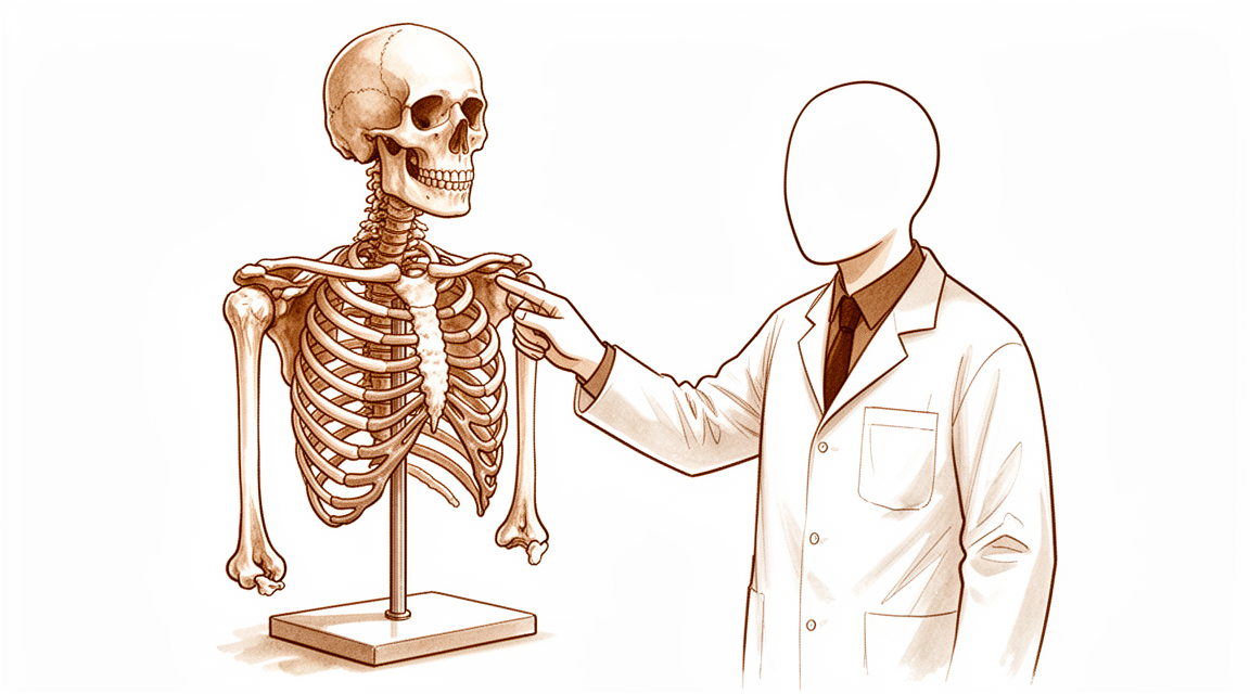
Shoulder anatomy overview – bones (clavicle, scapula, humerus) and how they enable wide range of motion.
मुख्य हिस्से
आपका कंधा तीन हड्डियों से बना है। हंसली (कॉलरबोन, क्लेविकल) आपकी छाती की बीच वाली हड्डी (ब्रेस्टबोन) से बाहर की ओर आपके कंधे की चपटी हड्डी तक जाती है। कंधे की चपटी हड्डी (स्कैपुला) एक सपाट हड्डी है जो आपकी छाती के पीछे की ओर रहती है और 17 मांसपेशियों को जुड़ने की जगह देती है। बांह के ऊपरी हिस्से की हड्डी (ह्यूमरस) का ऊपरी सिरा गोल होता है, जिसे ह्यूमरल हेड कहते हैं, और यह कंधे की चपटी हड्डी पर बने एक उथले गड्ढे (सॉकेट) में बैठता है।
यह बॉल-एंड-सॉकेट जोड़ (गेंद और गड्ढे वाला जोड़, जिसे ग्लेनोह्यूमरल जोड़ कहते हैं) आपके कंधे को आपके शरीर के किसी भी दूसरे जोड़ से ज़्यादा हिलने-डुलने की क्षमता (रेंज ऑफ मोशन) देता है। क्योंकि सॉकेट उथला है, इसलिए उसके किनारे के चारों ओर नरम कार्टिलेज (उपास्थि) का एक छल्ला (लैब्रम) उसे गहरा बनाता है और गेंद को बीच में टिकाए रखने में मदद करता है। जहां आपकी हंसली कंधे की चपटी हड्डी से मिलती है वहां एक छोटा जोड़ होता है (एक्रोमियोक्लेविकुलर जोड़), और जहां यह छाती की बीच वाली हड्डी से मिलती है वहां एक और जोड़ होता है (स्टर्नोक्लेविकुलर जोड़)। स्टर्नोक्लेविकुलर जोड़ ही वह अकेला असली जोड़ है जो आपकी बांह को आपके बाकी कंकाल से जोड़ता है।
लिगामेंट (स्नायुबंधन), जो ऊतक की मज़बूत पट्टियां होती हैं, इन हड्डियों को आपस में जोड़े रखते हैं। ऊतक की एक ढीली थैली (जॉइंट कैप्सूल) बॉल-एंड-सॉकेट जोड़ को घेरे रहती है, और इसके अंदर के कई लिगामेंट तभी कसते हैं जब आपकी बांह अपनी हिलने की सीमा के आखिरी छोर तक पहुंचती है। ये लगाम की तरह काम करते हैं, और गेंद को किसी भी दिशा में बहुत दूर सरकने से रोकते हैं। कैप्सूल आमतौर पर खुला-खुला और ढीला होता है, और इसी वजह से आपका कंधा इतनी आज़ादी से हिल पाता है।
हिलाने का काम मांसपेशियां और टेंडन (कंडरा) करते हैं। कंधे की चपटी हड्डी पर मौजूद चार मांसपेशियां रोटेटर कफ़ (कंधे को घुमाने वाली मांसपेशियों और टेंडनों का समूह) बनाती हैं, और उनके टेंडन ह्यूमरल हेड के चारों ओर लिपटे रहते हैं, जो बांह उठाते और हाथ बढ़ाते समय उसे सॉकेट में कसकर थामे रखते हैं। सबस्कैपुलेरिस आगे की ओर होती है, सुप्रास्पाइनेटस ऊपर से होकर जाती है, और इन्फ्रास्पाइनेटस और टेरीस माइनर पीछे के हिस्से को ढकती हैं। बड़ी डेल्टॉइड मांसपेशी कंधे को बाहर की ओर से ढकती है और आपकी बांह को सिर के ऊपर उठाने की ताकत देती है। बाइसेप्स का एक टेंडन भी जोड़ के बीच से गुज़रता है, जिसे ऊतक की छोटी पट्टियां उसके खांचे में थामे रखती हैं, और यह कंधे को स्थिर रखने में मदद करता है।
ये सभी हिस्से मिलकर काम करते हैं। जब इनमें से किसी एक को चोट लगती है या वह घिस जाता है, तो आपके कंधे के हिलने और महसूस होने का तरीका बदल सकता है।
सब कुछ मिलकर कैसे काम करता है
अपने कंधे को टी पर टिकी गोल्फ़ की गेंद की तरह समझें। गेंद आपकी बांह की हड्डी का ऊपरी सिरा है, और टी आपके कंधे की चपटी हड्डी पर बना उथला सॉकेट है। टी पर रखी गोल्फ़ की गेंद लुढ़क जाएगी, इसलिए आपका शरीर सहायक जोड़ता है। सॉकेट के किनारे के चारों ओर कार्टिलेज का एक छल्ला उसे थोड़ा गहरा बनाता है, और रोटेटर कफ़ के टेंडन गेंद के चारों ओर लिपटकर हिलते समय उसे धीरे से सॉकेट में दबाते हैं। यही दबाव हाथ बढ़ाते, कुछ उठाते और फेंकते समय गेंद को बीच में टिकाए रखता है।
ये हिस्से एक-दूसरे पर आसानी से सरकते हैं। आपकी बांह की हड्डी का गोल ऊपरी सिरा अपने ऊपर हड्डी और मांसपेशी से बने एक मेहराब के नीचे सरकता है, और उनके बीच एक चिकनी परत सतहों को बिना अटके सरकने देती है। जोड़ के आसपास के लिगामेंट केवल तब कसते हैं जब आपकी बांह अपनी सीमा के बिल्कुल आखिरी छोर तक घूमती है, लगाम की तरह। हिलने के बीच के हिस्से में गेंद को मांसपेशियां ही स्थिर रखती हैं।
यही तालमेल आपको काम करने देता है। आप ऊंची शेल्फ़ तक हाथ पहुंचा सकते हैं, कोई जार पकड़ सकते हैं, खुले पैसे लेने के लिए हथेली ऊपर की ओर घुमा सकते हैं, या रैकेट घुमा सकते हैं। कंधा अकेले काम नहीं करता। यह अच्छे पॉश्चर (शरीर की सही मुद्रा) पर और आपकी छाती, पीठ और बांह की मांसपेशियों के अपना हिस्सा निभाने पर निर्भर करता है, इसीलिए कंधे के व्यायामों में अक्सर सिर्फ़ कंधे से ज़्यादा हिस्से शामिल होते हैं।
नर्व (तंत्रिका) और खून की नलियां इन सभी काम करने वाले हिस्सों तक पहुंचती हैं। गर्दन के निचले हिस्से से निकलने वाली नर्व वे संकेत ले जाती हैं जो आपके कंधे की मांसपेशियों को बताते हैं कि कब हिलना है, और वे दर्द और स्थिति की जानकारी भी वापस भेजती हैं। खून की नलियां मांसपेशियों, टेंडनों और हड्डी तक ऑक्सीजन पहुंचाती हैं, और एक ही हिस्से तक कई रास्तों से खून पहुंचता है, ताकि अगर एक रास्ता दब भी जाए तो खून का बहाव जारी रहे।
जब ये सभी हिस्से तालमेल से काम करते हैं, तो आपका कंधा बिना किसी मेहनत के चलता महसूस होता है। जब किसी एक हिस्से को चोट लगती है या वह घिस जाता है, तो बाकी हिस्सों को ज़्यादा मेहनत करनी पड़ती है, और अक्सर तभी दर्द या जकड़न शुरू होती है।
आम तौर पर कहां गड़बड़ होती है
आपके कंधे के कुछ हिस्सों पर दूसरों की तुलना में ज़्यादा ज़ोर पड़ता है। यह जानना कि किन हिस्सों पर सबसे ज़्यादा भार पड़ता है, यह समझाने में मदद करता है कि कुछ खास समस्याएं आम क्यों हैं।
रोटेटर कफ़ के टेंडन एक तंग जगह में होते हैं। जहां सुप्रास्पाइनेटस (ऊपर से जाने वाला टेंडन) और सबस्कैपुलेरिस (आगे वाला टेंडन) हड्डी से जुड़ते हैं, वहां जोड़ के आगे के पास वे दूसरे नरम ऊतकों के रास्ते को काटते हैं। वह भीड़ भरा कोना टेंडन के फटने की एक आम जगह है। अगर वहां बाइसेप्स टेंडन को उसके खांचे में थामने वाली छोटी पट्टियां भी फट जाएं, तो बाइसेप्स टेंडन अस्थिर होकर अपनी जगह से खिसक सकता है।
लैब्रम, यानी कार्टिलेज का वह छल्ला जो आपके सॉकेट को गहरा बनाता है, भी आसानी से चोटिल हो सकता है। यह आपकी हिलने की सीमा के बीच वाले हिस्से में सबसे ज़्यादा मेहनत करता है, और गेंद को बीच में टिकाए रखता है। लेकिन जब आप कंधे के ऊपर से हाथ घुमाकर फेंकते हैं, तो आपकी बांह ऊपर और पीछे की ओर उस स्थिति में जाती है जिसे कॉकिंग पोज़िशन (फेंकने से पहले बांह को पीछे खींचने की स्थिति) कहते हैं। उस स्थिति में सॉकेट का किनारा रोटेटर कफ़ के टेंडनों में दबता है। इस दबाव से कफ़ की निचली सतह पर फटाव हो सकता है। जो फेंकने वाले खिलाड़ी अपनी बांह को ज़्यादा पीछे तक घुमा सकते हैं, उनमें ऐसा होने की संभावना ज़्यादा होती है। लैब्रम का ऊपरी और पिछला हिस्सा, और साथ में जॉइंट कैप्सूल (जोड़ के चारों ओर की ढीली थैली) का पिछला हिस्सा, फेंकने वाले कंधे में चोट की आम जगहें हैं। अगर कंधे की चपटी हड्डी और बॉल-एंड-सॉकेट जोड़ एक-दूसरे के साथ तालमेल में हिलना बंद कर दें, तो इन्हीं हिस्सों पर खिंचाव आ सकता है।
एक्रोमियोक्लेविकुलर जोड़ (जहां आपकी हंसली कंधे की चपटी हड्डी के ऊपरी हिस्से से मिलती है) को आमतौर पर सीधी टक्कर से चोट लगती है। इसका आम कारण है कंधे के सिरे के बल गिरना, जब बांह शरीर से सटी हुई हो। जब ताकत हंसली को नीचे की ओर धकेलती है, तो उस जोड़ के लिगामेंट सबसे कमज़ोर कड़ी होते हैं। इससे ज़्यादा मज़बूत सहारा खुद हंसली से और छाती की बीच वाली हड्डी पर बने जोड़ से मिलता है। उनके पीछे बहुत मज़बूत लिगामेंट होते हैं जो बचाव की आखिरी पंक्ति की तरह काम करते हैं। अगर वे भी ताकत के आगे हार जाएं, तो ताकत उस जगह तक फैल जाती है जहां डेल्टॉइड और ट्रेपेज़ियस मांसपेशियां जुड़ती हैं।
रोज़मर्रा का काम भी आपके कंधे पर ज़ोर डाल सकता है। ज़ोर लगाकर काम करना, अटपटी मुद्राएं, बार-बार एक ही तरह की हरकतें, कंपन करने वाले बिजली के औज़ार और ठंडा माहौल, ये सब काम की जगह पर होने वाली कंधे की समस्याओं में भूमिका निभाते हैं। जिन कामों में बांह को लगातार हिलाना पड़ता है, वे कंधे पर लगातार भार डालते हैं। ऐसे भार में कंधे की मांसपेशियां जल्दी थक जाती हैं, और बांह को एक ही स्थिति में स्थिर रखने के लगभग 1 घंटे के भीतर दर्द शुरू हो सकता है।
मुख्य बात
आपका कंधा आज़ादी से हिलने के लिए स्थिरता से समझौता करता है। सॉकेट उथला है, इसलिए कार्टिलेज का एक छल्ला उसे गहरा बनाता है और हिलते समय रोटेटर कफ़ के टेंडन गेंद को धीरे से अपनी जगह पर दबाते हैं। यह दबाव बांह की हर स्थिति में काम करता है, इसीलिए कफ़ इतना अहम है। लिगामेंट केवल आपकी हिलने की सीमा के बिल्कुल आखिरी छोर पर कसते हैं, इसलिए हिलने के बीच के हिस्से में स्थिर रखने का काम मांसपेशियां करती हैं। जोड़ के आगे के पास जहां टेंडन और लिगामेंट एक-दूसरे के रास्ते को काटते हैं, वहां ऊतक भीड़ भरे होते हैं और उनके फटने की संभावना रहती है। कंधे के ऊपर से हाथ घुमाकर फेंकना, कंधे के सिरे पर सीधी टक्कर, और ऐसा काम जो आपकी बांह पर लगातार भार डालता है, ये सभी इन्हीं हिस्सों पर ज़ोर डालते हैं।
Evidence & references
This is the clinical evidence summary written for health professionals. It is technical, and it lists the research this page was built from. You do not need to read it to understand your treatment or to make a decision about it.
Overview
Rotator Cuff Mechanics and Stability
- The lines of action of the supraspinatus, infraspinatus, and subscapularis do not create any substantial inferiorly directed force [1].
- The rotator cuff muscles cannot function as humeral head "depressors" [1].
- The rotator cuff embraces the humeral head and compresses it into the glenoid fossa [1].
- The rotator cuff centers the humeral head in the glenoid by the mechanism known as concavity compression [1].
- Concavity compression is effective with the arm in any position [1].
- The stabilizing effect of ligaments is active only in extreme positions where they are under tension [1].
- Contact between the proximal humeral convexity and the coracoacromial arch is not "impingement" [1].
- Contact between the proximal humeral convexity and the coracoacromial arch is the way the humeral head is normally stabilized to resist superiorly directed forces [1].
- The anterior and posterior glenoid labra deepen the glenoid fossa [1].
- The deepening of the glenoid fossa by the labra facilitates the concavity compression mechanism that centers the humeral head in the shallow glenoid during mid-range of shoulder motion [1].
- Internal abutment occurs between the edge of the glenoid and the cuff insertion when the arm is in abduction and external rotation [1].
- Internal abutment between the glenoid edge and cuff insertion is particularly likely to be prominent in pitchers with a large range of external rotation [1].
- Internal abutment between the glenoid edge and cuff insertion may result in deep surface tears of the cuff [1].
Glenohumeral Joint Anatomy
- The articular cartilage covering the humeral head and glenoid is thin [1].
- The complex intersection of the insertions of the supraspinatus and subscapularis joins the superior glenohumeral capsule and the transverse humeral ligament [1].
- Tears of the important fibers of the anterior supraspinatus and upper subscapularis can occur at the intersection of their insertions with the superior glenohumeral capsule and transverse humeral ligament [1].
- Tears of the transverse humeral ligament can result in instability of the long head tendon of the biceps [1].
- Vessels visible with arteriography can guide to the location of nerves that run along with them [1].
- The vascular anatomy is useful for locating nerves in cases where anatomy has been distorted by previous fracture or surgery [1].
Radiographic Evaluation
- The purpose of imaging of the shoulder is to help establish the diagnosis, determine the severity of the pathoanatomy, assist in surgical planning, and enable the surgeon to illustrate the condition of the shoulder to the patient [3].
- Standardized plain films are almost always sufficient to garner the information needed for care [3].
- The anteroposterior (AP) view in the plane of the scapula shows the superoinferior position of the humeral head relative to the glenoid [3].
- The anteroposterior (AP) view in the plane of the scapula shows the presence of osteophytes on the humeral head and glenoid [3].
- The anteroposterior (AP) view in the plane of the scapula shows narrowing of the joint space [3].
- The anteroposterior (AP) view in the plane of the scapula shows the degree of medial displacement of the humerus in relation to the lateral acromial line [3].
- The anteroposterior (AP) view in the plane of the scapula shows the quality of the humeral and glenoid bone [3].
- The anteroposterior (AP) view in the plane of the scapula shows the presence of loose bodies [3].
- The anteroposterior (AP) view in the plane of the scapula shows whether there is humeral head collapse or deformity [3].
- The axillary view is taken with the arm in the functional position of elevation in the plane of the scapula [3].
- The axillary view is oriented so that both the spinoglenoid notch and the scapular neck are visible [3].
- The axillary view shows the amount of glenoid bone [3].
- The axillary view shows the shape of the glenoid [3].
- The axillary view shows the version of the glenoid in relation to the plane of the scapula [3].
- The axillary view shows the relationship of the humeral head to the glenoid fossa [3].
- The axillary view is referred to as the "truth view" because it demonstrates glenohumeral relationships in the functional position of elevation [3].
- CT scans are taken with the arm in the adducted position [3].
- Joint space narrowing is most evident on the axillary truth view as opposed to images made with the arm at the side [3].
- The axillary truth view shows posterior subluxation or "functional decentering" that is not evident in images taken with the arm at the side [3].
- The degree of posterior subluxation can be measured as the position of the center of the humeral head in relation to the plane of the scapula [3].
- The degree of posterior subluxation can be measured as the position of the center of the humeral head in relation to the glenoid face [3].
- The degree of posterior subluxation can be measured as the point of contact of the humeral articular surface on the glenoid articular surface [3].
- The point of contact of the humeral articular surface on the glenoid articular surface reflects the degree of centering of the net humeral joint reaction force on the glenoid [3].
- Malcentering of the joint reaction force leads to posterior instability, posterior glenoid wear, and "rocking horse" loosening of prosthetic glenoid components [3].
Patient-Reported Outcomes
- The Western Ontario Rotator Cuff Index (WORC) assesses sharp pain in the shoulder over the last week [4].
- The Western Ontario Rotator Cuff Index (WORC) assesses constant, nagging pain in the shoulder over the last week [4].
- The Western Ontario Rotator Cuff Index (WORC) assesses weakness in the shoulder over the last week [4].
- The Western Ontario Rotator Cuff Index (WORC) assesses stiffness in the shoulder over the last week [4].
- The Western Ontario Rotator Cuff Index (WORC) assesses clicking, grinding, or crunching in the shoulder over the last week [4].
- The Western Ontario Rotator Cuff Index (WORC) assesses discomfort in the neck because of the shoulder over the last week [4].
- The Western Ontario Rotator Cuff Index (WORC) assesses how much the shoulder has affected fitness level over the past week [4].
- The Western Ontario Rotator Cuff Index (WORC) assesses how much the shoulder has affected the ability to throw hard or far over the past week [4].
- The Western Ontario Rotator Cuff Index (WORC) assesses difficulty with someone or something coming in contact with the affected shoulder over the past week [4].
- The Western Ontario Rotator Cuff Index (WORC) assesses difficulty doing push-ups or other strenuous shoulder exercises because of the shoulder over the past week [4].
- The Rotator Cuff Quality-of-Life Measure (RC-QOL) assesses how often a patient has to concern themselves with general safety with respect to the injured shoulder over the last 3 months [6].
- The Rotator Cuff Quality-of-Life Measure (RC-QOL) assesses how much enjoyment of life has been limited by the shoulder problem over the last 3 months [6].
- The Rotator Cuff Quality-of-Life Measure (RC-QOL) assesses how often a patient is aware of their shoulder problem over the last 3 months [6].
- The Rotator Cuff Quality-of-Life Measure (RC-QOL) assesses how often a patient is concerned about their shoulder with respect to lifestyle and family over the last 3 months [6].
- The Rotator Cuff Quality-of-Life Measure (RC-QOL) assesses if a patient has modified their lifestyle to avoid potentially damaging activities to the shoulder over the last 3 months [6].
Osseous Anatomy
Scapula
- The scapula spans the second through seventh ribs and serves as an attachment site for 17 muscles [44].
- The scapula is anteverted on the chest wall approximately 30 degrees relative to the body [44].
- The glenoid is retroverted approximately 5 degrees relative to the scapular body [44].
- Os acromiale is an incomplete fusion of secondary ossification centers, most commonly occurring between the mesoacromion and meta-acromion [44].
- The coracoid process serves as an attachment site for the coracoacromial ligament, coracoclavicular ligaments (conoid and trapezoid), conjoined tendon (coracobrachialis and short head of biceps), and pectoralis minor [44].
- The suprascapular artery passes superior to the superior transverse scapular ligament, while the suprascapular nerve passes inferior to the ligament through the suprascapular notch [44].
- In the spinoglenoid notch, both the suprascapular artery and nerve pass inferior to the inferior transverse scapular ligament [44].
- The acromial branch of the thoracoacromial artery runs on the medial aspect of the coracoacromial ligament [44].
Clavicle
- The clavicle is the first bone in the body to ossify, occurring at 5 weeks gestation [44].
- The clavicle is the last bone to fuse, with the medial epiphysis fusing at 25 years of age [44].
- Clavicle fracture is the most common musculoskeletal birth injury [44].
Proximal Humerus
- The humeral head is retroverted 30 degrees relative to the transepicondylar axis of the humerus [44].
- The humeral head height is approximately 5.6 cm above the superior border of the pectoralis major tendon [44].
- The anatomic neck, located directly below the humeral head, serves as an attachment site for the shoulder capsule [44].
- The surgical neck is located more distally than the anatomic neck and is more often involved in fractures [44].
- The transverse humeral ligament is an important stabilizer of the biceps tendon [44].
Glenohumeral Joint
- The glenohumeral joint is a ball-and-socket articulation with the greatest range of motion in the body [44].
- The fibrocartilaginous glenoid labrum deepens the socket by 50% and provides a bumper to translation [44].
- Labral anatomic variants include the sublabral foramen (anterosuperior) and the Buford complex, which is characterized by the absence of the anterosuperior labrum and a cordlike middle glenohumeral ligament [44].
- The middle glenohumeral ligament is absent in up to 30% of shoulders [44].
Sternoclavicular Joint
- The sternoclavicular joint is a double gliding joint with an articular disc [44].
- The sternoclavicular joint is the only true joint connecting the upper extremity with the axial skeleton [44].
- The posterior sternoclavicular ligament is the strongest and primary restraint to anteroposterior instability [44].
- The sternoclavicular joint rotates 30 degrees with shoulder motion [44].
Acromioclavicular Joint
- The acromioclavicular joint is a plane/gliding joint with a fibrocartilaginous disc [44].
- The acromioclavicular ligaments prevent anteroposterior displacement of the distal clavicle [44].
- The posterior and superior acromioclavicular ligaments are considered the strongest [44].
- The coracoclavicular ligaments prevent superior displacement of the distal clavicle [44].
- The trapezoid ligament is located anterolaterally, approximately 25 mm from the acromioclavicular joint [44].
- The conoid ligament is located posteromedially, approximately 45 mm from the acromioclavicular joint, and is stronger than the trapezoid ligament [44].
Ligaments and Joint Capsule
Glenohumeral Ligaments
- The glenohumeral ligaments are collagenous reinforcements to the shoulder capsule that are not visible on its external surface [21].
- The glenohumeral ligaments are best appreciated in situ arthroscopically without distension by air or saline [21].
- The function of the glenohumeral ligaments depends on their collagenous integrity, their attachment sites, and the position of the arm [21].
- The superior glenohumeral ligament is present in 97% of shoulders examined in the classic anatomic study by DePalma [21].
- The superior glenohumeral ligament is present in 26% to 90% of specimens in an anatomic study conducted at the authors' institution [21].
- The superior glenohumeral ligament can arise from a common origin with the biceps tendon [21].
- The superior glenohumeral ligament can arise from the labrum, slightly anterior to the biceps tendon [21].
- The superior glenohumeral ligament can originate with the middle glenohumeral ligament [21].
- The superior glenohumeral ligament inserts into the fovea capitis and lies just superior to the lesser tuberosity [21].
- The size and integrity of the superior glenohumeral ligament are variable, ranging from a thin wisp of capsular tissue to a thickening similar to the patellofemoral ligaments in the knee [21].
- Biomechanical studies show that the superior glenohumeral ligament contributes very little to the static stability of the glenohumeral joint [21].
- Selective cutting of the superior glenohumeral ligament did not significantly affect translation either anteriorly or posteriorly in the abducted shoulder [21].
- The contribution of the superior glenohumeral ligament to stability is best demonstrated with the arm in the dependent position, where it helps keep the humeral head suspended [21].
- The superior glenohumeral ligament is the primary restraint to inferior humeral subluxation in 0 degrees of abduction [30].
- The superior glenohumeral ligament is the primary stabilizer to anterior and posterior stress in 0 degrees of abduction [30].
- Tightening of the rotator interval, which includes the superior glenohumeral ligament, decreases posterior and inferior translation [30].
- The middle glenohumeral ligament limits external rotation when the arm is in the lower and middle ranges of abduction [30].
- The middle glenohumeral ligament has little effect on external rotation when the arm is in 90 degrees of abduction [30].
- The middle glenohumeral ligament originates anterosuperiorly on the glenoid and inserts midway along the anterior humeral articular surface adjacent to the lesser tuberosity [26].
- The middle glenohumeral ligament is tensioned by external rotation when the humerus is abducted to 45 degrees [26].
- The middle glenohumeral ligament can act as an important secondary restraint to anterior translation if the anterior portion of the inferior glenohumeral ligament is damaged [21].
- The inferior glenohumeral ligament is a complex structure that is the main static stabilizer of the abducted shoulder [15].
- The inferior glenohumeral ligament is a hammock-like structure originating from the glenoid and inserting into the anatomic neck of the humerus [15].
- The inferior glenohumeral ligament complex consists of an anterior band, a posterior band, and an axillary pouch lying in between [15].
- The anterior and posterior bands of the inferior glenohumeral ligament complex are most clearly defined with the arm abducted [15].
- With abduction and external rotation, the anterior band of the inferior glenohumeral ligament fans out to support the head, and the posterior band becomes cord-like [15].
- With abduction and internal rotation, the posterior band of the inferior glenohumeral ligament fans out to support the head, and the anterior band becomes cord-like [15].
- The inferior glenohumeral ligament complex is the main stabilizer to anterior and posterior stresses when the shoulder is abducted 45 degrees or more [30].
- The inferior glenohumeral ligament attaches to the glenoid margin from the 2- to 3-o’clock positions anteriorly to the 8- to 9-o’clock positions posteriorly [33].
- The humeral attachment of the inferior glenohumeral ligament is below the level of the horizontally oriented physis into the inferior aspect of the anatomic and surgical neck of the humerus [33].
- The anterosuperior edge of the inferior glenohumeral ligament is usually quite thickened [33].
- The glenohumeral ligaments play important stabilizing roles only at the extremes of motion [31].
- The glenohumeral ligaments are lax and relatively ineffectual in most functional positions of the joint [31].
Joint Capsule
- The shoulder capsule is large and has twice the surface area of the humeral head [32].
- The shoulder capsule typically accepts approximately 28 to 35 mL of fluid [32].
- In patients with adhesive capsulitis, the shoulder capsule accepts only 5 mL or less of fluid [32].
- The capsule is lined by synovium and extends from the glenoid neck (or occasionally the labrum) to the anatomic neck and the proximal shaft of the humerus [32].
- The capsule often extends and attaches to the coracoid process superiorly via the coracohumeral ligament [32].
- The capsule can extend down along the biceps tendon for variable length and across the intertubercular groove of the humerus [32].
- The joint capsule blends with ligamentous structures that arise on nearby bony landmarks and contains within its substance the glenohumeral ligaments [32].
- The thickness of the capsule decreases as it nears the humerus [26].
- The capsule is thickest in the inferior pouch at 2.8 mm [26].
- The capsule is 2.4 mm thick in its anterior portion [26].
- The capsule is 2.2 mm thick in its posterior portion [26].
- The thickness of the glenohumeral joint capsule ranges from 1.3 to 4.5 mm in cadaveric specimens [26].
- The glenohumeral joint capsule is normally large, loose, and redundant, which allows full and free range of motion of the shoulder [26].
- By virtue of their mandatory redundancy, the capsule and its ligaments are lax throughout much of the range of joint motion [26].
- The capsule and its ligaments act as checkreins when they come under tension as the joint approaches the limits of its range of motion [26].
- In the midrange of shoulder motion, the center of the humeral head remains within 2.2 mm of the center of the glenoid on magnetic resonance imaging [26].
- On all sides of the shoulder capsule, except for the inferior portion, the capsule is reinforced and strengthened by the tendons of the rotator cuff muscles [32].
- The rotator cuff tendons blend into the capsule over varying lengths and average approximately 2.5 cm [32].
- The most prominent capsular reinforcement is the anterior tendinous portion of the subscapularis [32].
- A recent cadaveric study demonstrated diminished tensile behavior of the glenohumeral capsule in patients with anterior instability [32].
Coracohumeral and Transverse Humeral Ligaments
- The coracohumeral ligament is a strong band that originates from the base and lateral border of the coracoid process just below the origin of the coracoacromial ligament [32].
- The coracohumeral ligament is directed transversely and inserts on the greater tuberosity [32].
- The anterior border of the coracohumeral ligament is often distinct medially and merges with the capsule laterally [32].
- The posterior border of the coracohumeral ligament is usually indistinct from the rest of the capsule [32].
- The coracohumeral ligament appears to have a static suspensory function for the humeral head in the glenoid cavity when the arm is in the dependent position [32].
- With abduction, the coracohumeral ligament relaxes and loses its ability to support the humerus [32].
- The transverse humeral ligament consists of a few transverse fibers of capsule that extend between the greater and lesser tuberosities [32].
- The transverse humeral ligament helps contain the long head of the biceps tendon in its groove [32].
- The coracohumeral ligament originates at the base of the coracoid, blends into the cuff tendons, and inserts into the greater and lesser tuberosities [26].
- The superior glenohumeral ligament crosses the rotator interval capsule and lies between the supraspinatus and subscapularis tendons [26].
- The superior glenohumeral ligament, the coracohumeral ligament, and the rotator interval capsule come under tension with glenohumeral flexion, extension, external rotation, and adduction [26].
- When under tension, the superior glenohumeral ligament, coracohumeral ligament, and rotator interval capsule resist posterior and inferior displacement of the humeral head [26].
- Releasing or surgically tightening the rotator interval capsule increases or decreases the allowed posterior and inferior translational laxity, respectively [26].
Stability Mechanics
- The primary passive stabilizers of the glenohumeral joint are the capsule and the scapulohumeral ligaments [26].
- The capsule and scapulohumeral ligaments play a primary role in positions near the extremes of the allowed range of motion [26].
- The glenohumeral joint does not offer isometric articular ligaments that provide stability as the joint is flexed around a defined anatomic axis [31].
- The net humeral joint reaction force is the vector sum of all forces acting on the head of the humerus relative to the glenoid fossa [31].
- The glenohumeral joint will not dislocate as long as the net humeral joint reaction force is directed within the effective glenoid arc [31].
- If the net humeral joint reaction force passes outside the effective glenoid arc, the joint becomes unstable [31].
- The effective glenoid arc is the arc of the glenoid able to support the net humeral joint reaction force [31].
- The balance stability angle is the maximal angle that the net humeral joint reaction force can make with the glenoid center line before dislocation occurs [31].
- The shape of the bone, cartilage, and labrum all contribute to the effective glenoid arc and the balance stability angle [31].
- The deltoid and cuff muscle forces maintain the net humeral joint reaction force within the balance stability angle [31].
- The superior and inferior balance stability angles are greater than the anterior and posterior balance stability angles for a normal glenoid [31].
Muscles and Tendons
Rotator Cuff Anatomy and Function
- The rotator cuff consists of four muscles arising from the scapula whose tendons blend with the subjacent capsule as they attach to the humeral tuberosities [25].
- The subscapularis arises from the anterior aspect of the scapula and attaches over much of the lesser tuberosity [25].
- The supraspinatus arises from the fossa superior to the scapular spine, passes beneath the acromion and acromioclavicular joint, and attaches to the superior aspect of the greater tuberosity [25].
- The infraspinatus arises from the fossa below the scapular spine and attaches to the posterolateral aspect of the greater tuberosity [25].
- The teres minor arises from the lower lateral aspect of the scapula and attaches to the lower portion of the greater tuberosity [25].
- The tendons of the rotator cuff blend together to form a continuous cuff around the humeral head [25].
- The lines of action of the supraspinatus, infraspinatus, and subscapularis do not create substantial inferiorly directed force, meaning they cannot function as humeral head "depressors" [1].
- The rotator cuff embraces the humeral head and compresses it into the glenoid fossa via a mechanism known as concavity compression [1].
- Concavity compression is effective with the arm in any position, unlike ligaments which are only active in extreme positions where they are under tension [1].
- The anterior and posterior glenoid labra deepen the glenoid fossa, facilitating the concavity compression mechanism that centers the humeral head during mid-range shoulder motion [1].
- The infraspinatus is one of the two main external rotators of the humerus and accounts for as much as 60% of the external rotation force [22].
- The infraspinatus stabilizes the shoulder against posterior subluxation in internal rotation by creating a forward force on the humeral head [22].
- The infraspinatus has a line of pull posteriorly and stabilizes against anterior subluxation when the shoulder is in abduction–external rotation [22].
- The infraspinatus is a pennate muscle with a median raphe covered by a fat stripe that can be mistaken at surgery for the gap between the infraspinatus and teres minor muscles [22].
- The superior portion of the subscapularis tendon has significantly higher stiffness and ultimate load than its inferior portion [24].
- The capsule is relatively thin between the supraspinatus and subscapularis (the rotator interval), allowing the cuff to slide back and forth around the coracoid process as the arm is elevated and lowered [24].
- Histologic studies describe the rotator cuff tendons as having five distinct layers, with the most superficial layer being coracohumeral ligament fibers and the fifth layer being the continued sheet of collagen fibrils composing the superior joint capsule [25].
- There is significant overlap and interdigitation of the supraspinatus and infraspinatus tendons occurring near the footprint on the greater tuberosity [25].
- The portion of the greater tuberosity occupied by the supraspinatus insertion is much smaller than previously believed, with the infraspinatus insertion occupying the preponderance of the footprint [25].
Biceps Tendon
- The tendon of the long head of the biceps originates from the supraglenoid tubercle and runs beneath the cuff in the area of the rotator interval [24].
- The long head of the biceps tendon exits the shoulder beneath the transverse humeral ligament and between the greater and lesser tuberosities [24].
- The long head of the biceps tendon is subject to injury when incising the upper subscapularis from the lesser tuberosity [24].
- In the bicipital groove of the humerus, the long head of the biceps tendon is endangered by procedures that involve lateral transfer of the subscapularis tendon across the groove [24].
- The long head of the biceps tendon may be considered a functional part of the rotator cuff [25].
- The coracohumeral ligament and the transverse humeral ligament keep the biceps tendon aligned in the groove [25].
- Electromyographic studies demonstrate that the long head of the biceps may be inactive during shoulder movements but may serve as a passive restraint during most shoulder motions [25].
- The long head of the biceps contributes to anterior stability in abduction and external rotation [25].
- The bicipital groove travels on the biceps tendon like a monorail on its track, helping explain why the humerus is capable of substantial rotation when adducted and allows very little rotation when maximally abducted [25].
Innervation
- The subscapularis is innervated by the upper and lower subscapular nerves [25].
- The supraspinatus is innervated by the suprascapular nerve after it passes through the suprascapular notch [25].
- The infraspinatus is innervated by the suprascapular nerve after it passes through the spinoglenoid notch [25].
- The teres minor is innervated by a branch of the axillary nerve [25].
- The suprascapular nerve arises from the superior lateral aspect of the upper trunk shortly after its formation at Erb’s point [11].
- The suprascapular nerve passes below the transverse scapular or suprascapular ligament and enters the supraspinatus muscle, which it innervates through two branches [11].
- The suprascapular nerve innervates the infraspinatus muscle through two branches after inferiorly passing around the base of the spine of the scapula [11].
- The motor branch to the supraspinatus branches within 1 cm of the suprascapular notch in all specimens studied [11].
- The nerve medially curves and innervates the infraspinatus muscle within 1 cm of the base of the scapular spine in nearly 90% of cases [11].
- The distance from the midline of the posterior glenoid rim to the suprascapular nerve at the base of the scapular spine averages 1.8 cm, with some as close as 1.4 cm [11].
- The safe zone for the suprascapular nerve is described as 2.1 cm from the supraglenoid tubercle to the nerve at the scapular notch and 1.1 cm from the midline of the posterior glenoid rim [11].
- The axillary nerve is a terminal branch coming off the posterior cord of the brachial plexus just proximal to the coracoid process [10].
- The axillary nerve passes beneath the conjoined tendon anterior to the subscapularis 3 to 5 mm medial to the musculotendinous junction and then adjacent to the inferior capsule before entering the quadrilateral space posteriorly [10].
- The axillary nerve splits into anterior and posterior branches within the quadrangular space [10].
- The anterior and middle deltoid muscle receives sole innervation from the anterior branch of the axillary nerve [10].
- Posterior deltoid muscle innervation varies, with supply only from the anterior branch in 2.3% of cases, from the posterior branch in 8.5%, and from both branches in 89.1% [10].
- The posterior branch of the axillary nerve branches to supply the teres minor muscle and then terminates as the superior lateral brachial cutaneous nerve [10].
- The musculocutaneous nerve lies on the deep surface of the coracoid muscles and penetrates the coracobrachialis with one or more branches lying a variable distance distal to the coracoid [5].
- The often-described 5-cm safe zone for the musculocutaneous nerve beneath the coracoid process refers only to the average position of the main trunk and not to an area that can be entered recklessly [5].
Vascular Supply
- The subscapular artery originates in the third part of the axillary artery and runs caudally on the subscapularis muscle [8].
- The circumflex scapular artery passes under the inferior edge of the subscapularis and then medial to the long head of the triceps through the triangular space, supplying a branch to the infraspinatus fossa [8].
- The thoracodorsal artery runs with the thoracodorsal nerve toward the latissimus dorsi on the subscapularis, teres major, and latissimus dorsi [8].
- The posterior humeral circumflex artery descends into the quadrilateral space with the axillary nerve and supplies the anterior two-thirds of the deltoid via its anterior branch [8].
- The anterior humeral circumflex artery travels laterally at the inferior border of the subscapularis tendon, marking the border between the upper tendinous insertion and lower muscular insertion of the subscapularis [8].
- The anterolateral ascending branch of the anterior humeral circumflex artery supplies the majority of the humeral head [8].
- The infraspinatus blood supply is generally described as coming from two large branches of the suprascapular artery [22].
- In two-thirds of specimens, the subscapular artery through its dorsal or circumflex scapular branch supplied the greater portion of the circulation of the infraspinatus muscle [22].
Surgical Landmarks and Interfaces
- The humeroscapular motion interface lies between the inner structures of the proximal humerus, rotator cuff, coracohumeral ligament, and biceps tendon sheath and the superficial layer of the acromion, deltoid, coracoacromial ligament, coracoid process, and conjoined tendon [10].
- Smooth, unrestricted motion at the humeroscapular motion interface is vital to shoulder mobility [10].
- The axillary nerve runs in the humeroscapular motion interface, superficial to the humerus and cuff and deep to the deltoid and coracoid muscles [5].
- In the anterior deltopectoral approach, the axillary nerve can be palpated by sweeping a finger inferiorly across the subscapularis muscle tendon interface [10].
- In the anterolateral deltoid splitting approach, the axillary nerve crosses approximately 5 cm inferior to the anterolateral acromial corner [10].
- Shoulder abduction brings the axillary nerve closer to the acromion landmark in the anterolateral approach [10].
- In the posterior deltoid splitting approach, the axillary nerve is approximately 7 cm from the posterior acromial corner [10].
- The coracoacromial arch provides a concavity that articulates with the proximal humeral convexity, with the center of this articulation identical to the center of the glenohumeral articulation in the normal shoulder [5].
- The radii of the coracoacromial and glenohumeral articulations differ by the thickness of the rotator cuff and tuberosity [5].
- The concentricity of the coracoacromial and glenohumeral spheres provides stable centering of the normal shoulder through an extremely wide range of positions [5].
- Contact between the proximal humeral convexity and the coracoacromial arch is not "impingement," but rather the way the humeral head is normally stabilized to resist superiorly directed forces [1].
- The intersection of the insertions of the supraspinatus and subscapularis as they join the superior glenohumeral capsule and the transverse humeral ligament is a key spot for tears of anterior supraspinatus, upper subscapularis, and transverse humeral ligament [1].
- Internal abutment between the edge of the glenoid and the cuff insertion occurs in the cocking position of the overarm throw and may result in deep surface tears of the cuff [1].
Neurovascular Anatomy
Arterial Supply
- Bartlett et al. found no important branches of the subscapular artery before the origin of the circumflex scapular artery [8].
- The circumflex scapular artery passes posteriorly under the inferior edge of the subscapularis and then medial to the long head of the triceps through the triangular space [8].
- The circumflex scapular artery supplies a branch to the inferior angle of the scapula and a branch to the infraspinatus fossa [8].
- The posterior humeral circumflex artery descends into the quadrilateral space with the axillary nerve [8].
- The anterior branch of the posterior humeral circumflex artery travels with the axillary nerve approximately 2 inches below the level of the acromion and supplies the anterior two-thirds of the deltoid [8].
- The anterior humeral circumflex artery laterally travels at the inferior border of the subscapularis tendon, marking the border between the upper tendinous insertion and lower muscular insertion of the subscapularis [8].
- One branch of the anterior humeral circumflex artery crosses the subscapularis tendon anteriorly and is regularly encountered during anterior glenohumeral reconstruction [8].
- The anterolateral ascending branch of the anterior humeral circumflex artery runs parallel to the lateral aspect of the long head of the biceps tendon [8].
- The terminal end branch of the anterior humeral circumflex artery is called the arcuate artery [8].
- Brooks et al. found significant intraosseous anastomoses between the arcuate artery and posteromedial vessels from the posterior humeral circumflex [8].
- The thoracoacromial artery pierces the clavipectoral fascia and gives off four branches: deltoid, clavicular, acromial, and pectoral [17].
- The pectoral branch of the thoracoacromial artery travels in the space between the pectoralis minor and pectoralis major [17].
- The deltoid artery of the thoracoacromial trunk supplies the clavicular head of the pectoralis major and much of the anterior deltoid [17].
- The acromial artery is generally a branch of the deltoid artery that proceeds up to the acromioclavicular joint [17].
- The lateral thoracic artery runs deep to the pectoralis minor and supplies blood to the pectoralis minor, serratus anterior, and intercostal spaces 3 to 5 [17].
- The subclavian artery communicates with the third portion of the axillary artery through anastomosis with the transverse cervical, dorsal scapular, and suprascapular arteries [23].
- Communications exist between the posterior humeral circumflex artery and the anterior circumflex, deltoid, suprascapular, and profunda brachii arteries [23].
- A limb may survive on a flow pressure as low as 20 mm Hg due to collateral circulation around the shoulder [23].
- Collateral vessels are fewer in number when compact and mobile tissues span the joint [23].
Nerve Supply
- The axillary nerve passes beneath the conjoined tendon anterior to the subscapularis 3 to 5 mm medial to the musculotendinous junction [10].
- The axillary nerve enters the quadrilateral space posteriorly and splits into anterior and posterior branches within this space [10].
- In 89.1% of cases, the posterior deltoid muscle receives innervation from both the anterior and posterior branches of the axillary nerve [10].
- The posterior branch of the axillary nerve supplies the teres minor muscle and terminates as the superior lateral brachial cutaneous nerve [10].
- Shoulder abduction brings the axillary nerve closer to the acromion landmark [10].
- Gardner et al. demonstrated no accessory motor branches to the anterior deltoid crossing the tendinous raphe between the anterior and lateral deltoid [10].
- The suprascapular nerve passes below the transverse scapular or suprascapular ligament and enters the supraspinatus muscle [11].
- The suprascapular nerve provides an articular branch in the supraspinatus fossa to the acromioclavicular and superior glenohumeral joints [11].
- The suprascapular nerve provides an articular branch in the infraspinatus fossa to the posterior superior glenohumeral joint [11].
- Bigliani et al. found that the motor branch to the supraspinatus branches within 1 cm of the suprascapular notch in all specimens [11].
- Gumina et al. describe the safe zone to be a distance 2.1 cm from the supraglenoid tubercle to the suprascapular nerve at the scapular notch [11].
- Gumina et al. describe the safe zone to be a distance 1.1 cm from the midline of the posterior glenoid rim to the suprascapular nerve [11].
- The upper subscapular nerves (C5) take origin off the posterior cord and supply two-thirds to four-fifths of the upper portion of the subscapularis muscle [29].
- The lower subscapular nerves (C5 and C6) innervate the lower portion of the subscapularis muscle and the teres major [29].
- The palpable anterior border of the glenoid rim deep to the subscapularis along with the medial border of the conjoined tendon can serve as a safe zone for surgical dissection of the subscapular nerves [29].
- The superficial and deep structures of the shoulder are innervated by a network of nerve fibers mainly derived from the C5, C6, and C7 nerve roots [18].
- The axillary nerve and suprascapular nerve provide most of the nerve supply to the anterior capsule and glenohumeral joint [18].
- Superiorly, two branches of the suprascapular nerve make the primary contributions to the joint innervation [18].
- Posteriorly, the chief nerves supplying the joint are the suprascapular nerve in the upper region and the axillary nerve in the lower region [18].
- Alpantaki and colleagues found that thinly myelinated or unmyelinated sensory neurons provide innervation to the long head of the biceps tendon [18].
Anatomical Landmarks and Spaces
- The axillary space contains the brachial plexus and its branches, the axillary artery and vein, and major lymphatic drainage [28].
- The posterior wall of the axillary space consists of the subscapularis, teres major, and latissimus dorsi muscles from top to bottom [28].
- The anterior boundary of the axillary space is the pectoralis minor muscle and the clavipectoral fascia [28].
- The clavipectoral fascia continues medially to the first rib as the costocoracoid membrane [28].
- A deposit of adipose tissue lies posteriorly, deep to the deep fascia, inferomedial to the medial border of the posterior deltoid, lateral to the trapezius, and superior to the latissimus dorsi [28].
- This posterior adipose tissue contains the cutaneous continuations of the circumflex scapular artery [28].
- A third deep deposit of adipose tissue lies between the supraspinatus tendon and the overlying clavicle and acromioclavicular joint [28].
- This superior adipose tissue cushions and protects the branches of the acromial artery [28].
- The cephalic vein and the deltoid branches of the thoracoacromial artery lie in the interval between the deltoid and pectoralis major [7].
- The musculocutaneous nerve lies beneath the short head of the biceps in the distal part of the anteromedial approach wound [7].
- The axillary nerve that supplies the deltoid is not disturbed in the anteromedial approach where deltoid fibers are not divided [7].
- Cetik et al. found that the average distance from the anterior edge of the acromion to the course of the axillary nerve is 6.08 cm, with 5.2 cm being the closest distance [29].
- Stecco et al. found the mean distance between the point where the axillary nerve enters into the deltoid muscle and the acromion is 6.8 cm [29].
- Cheung reported the average distance from the mid-acromion to the superior border of the axillary nerve as 66.6 mm with the shoulder in neutral position [29].
- Vertical abduction to 60 degrees moves the superior borders of the axillary nerve to a distance of 53.9 mm from the mid-acromion [29].
Biomechanics and Function
Rotator Cuff Mechanics
- The anterior and posterior glenoid labra deepen the glenoid fossa to facilitate the concavity compression mechanism [1].
- The concavity compression mechanism centers the humeral head in the shallow glenoid during functional activities in the mid-range of shoulder motion [1].
Humeral Head and Coracoacromial Arch Interaction
- The smooth outer surface of the proximal humeral convexity forms a normal articulation with the coracoacromial arch [1].
- The humeroscapular motion interface consists of two articular surfaces: the proximal humeral convexity on the inside and the coracoacromial arch and deltoid on the outside [1].
Specific Positional Biomechanics
- In the cocking position of the overarm throw (abduction and external rotation), there is internal abutment between the edge of the glenoid and the cuff insertion [1].
- Internal abutment between the edge of the glenoid and the cuff insertion is particularly likely to be prominent in pitchers with a large range of external rotation [1].
- Internal abutment between the edge of the glenoid and the cuff insertion may result in deep surface tears of the cuff [1].
General Functional Principles
- The shoulder does not work in isolation [19].
- Rehabilitation requires incorporating functional movement patterns and reinforcing good postural control [19].
- Shoulder exercises are not isolated to the shoulder joint itself [19].
Common Sites of Injury
Rotator Cuff and Labrum
- Concavity compression can be effective with the arm in any position [1].
- The complex intersection of the insertions of the supraspinatus and subscapularis as they join the superior glenohumeral capsule and the transverse humeral ligament is a key spot for tears of the anterior supraspinatus and upper subscapularis [1].
- Tears of the transverse humeral ligament at the intersection of the supraspinatus and subscapularis insertions result in instability of the long head tendon of the biceps [1].
- The anterior and posterior glenoid labra deepen the glenoid fossa, facilitating the concavity compression mechanism that centers the humeral head in the shallow glenoid during mid-range of shoulder motion [1].
- Internal abutment between the edge of the glenoid and the cuff insertion occurs in the cocking position of the overarm throw [1].
- Typical sites of pathoanatomy in the throwing shoulder include the superior and posterosuperior labrum [16].
- Typical sites of pathoanatomy in the throwing shoulder include the articular surface of the supraspinatus and infraspinatus [16].
- Typical sites of pathoanatomy in the throwing shoulder include the posterior capsule [16].
- Injury to the thrower’s shoulder joint occurs most commonly in the late cocking or early acceleration phases [16].
- Abnormal scapulothoracic and glenohumeral motion can injure the superior and posterosuperior labrum [16].
- Abnormal scapulothoracic and glenohumeral motion can injure the undersurface of the rotator cuff [16].
- Abnormal scapulothoracic and glenohumeral motion can injure the posterior capsule [16].
Acromioclavicular Joint
- The most common mechanism for injury to the ACJ is a direct force applied to the superior aspect of the acromion process [14].
- Direct force to the superior aspect of the acromion process occurs with a fall onto the outer aspect of the shoulder with the upper limb in an adducted position [14].
- A falling object or a deliberate blow striking the superior acromion is a rarer mechanism of direct injury to the ACJ [14].
- The AC ligaments offer the weakest resistance to forces that push the acromion inferiorly and medially [14].
- Stronger resistance to forces pushing the acromion inferiorly and medially is afforded by the intact clavicle and SCJ [14].
- AC injuries that occur in conjunction with clavicle fractures or SCJ injuries have been reported [14].
- The CC ligaments act as an extremely strong buffer after force advances through the ACJ [14].
- If the integrity of the CC ligaments is overcome, remaining forces are likely to be dissipated at the attachment sites of the deltoid and trapezius muscle [14].
- An inferiorly directed force acting upon the superior lateral clavicle can cause more damage when the upper limb is abducted and the scapula is retracted [14].
- An inferiorly directed force acting upon the superior lateral clavicle with an abducted upper limb and retracted scapula can result in an inferior dislocation of the clavicle beneath the coracoid process [14].
- Indirect mechanisms of ACJ injury are exceedingly rare [14].
- A fall onto the adducted upper limb is apt to drive the humeral head into the inferior aspect of the acromion, subjecting the ACJ to variable degrees of injury [14].
- The integrity of the acromion process and glenohumeral stability is risked when extremely high superiorly directed forces are encountered [14].
- The ACJ may be injured by pulling or traction-like forces applied to the upper limb [14].
- In type I ACJ injuries, the AC ligaments sustain a mild-to-moderate sprain, maintaining the ACJ integrity [14].
- In type I ACJ injuries, the CC ligaments and the deltoid and trapezius muscles are normal [14].
Occupational Stressors
- Excessive or sustained exertion of force is a physical stressor playing a role in the etiology of occupational shoulder disorders [20].
- Awkward postures, mainly shoulder flexion/extension or abduction, are physical stressors playing a role in the etiology of occupational shoulder disorders [20].
- Repetitive motions of the shoulder and neck are physical stressors playing a role in the etiology of occupational shoulder disorders [20].
- Contact with vibrating power tools is a physical stressor playing a role in the etiology of occupational shoulder disorders [20].
- Low ambient temperature is a physical stressor playing a role in the etiology of occupational shoulder disorders [20].
- Work tasks that demand continuous arm movements generate load patterns with a static load component at the shoulder level [20].
- The low fatigue tolerance of shoulder muscles may cause pain sooner, within about 1 hour of static contraction [20].
- Although required muscle loads decrease as the arm-torso angle exceeds 90 degrees, the pressure on soft tissues continues to increase [20].
Surgical Anatomy
Bony Landmarks and Articulations
- The coracoacromial (CA) arch provides a concavity that articulates with the proximal humeral convexity [5].
- The center of the CA arch articulation is identical to the center of the glenohumeral articulation in the normal shoulder [5].
- The radii of the CA arch and glenohumeral articulations differ by the thickness of the rotator cuff and tuberosity [5].
- The concentricity of the CA and glenohumeral spheres provides stable centering of the normal shoulder through an extremely wide range of positions [5].
- The coracoid process serves as a palpable guide to the deltopectoral groove, a locator for the CA arch, and an anchor for the coracoid muscles [5].
- The coracoid process separates the lateral "safe side" from the medial "suicide" area where the brachial plexus and major vessels lie [5].
- The clavipectoral fascia covers the floor of the deltopectoral groove [5].
- The deltoid muscle arises from the clavicle, acromion, and scapular spine [37].
- The anterior deltoid extends to a line running approximately from the midclavicle to the midlateral portion of the humerus [37].
- This line passes over the cephalic vein, the anterior venous drainage of the deltoid, and the coracoid process [37].
- The posterior corner of the acromion marks the junction of the middle and posterior thirds of the deltoid [37].
- The glenoid track is calculated as 84% of the actual glenoid width measured on the sagittal oblique MR image [27].
- The distance from the rotator cuff footprint to the medial margin of the Hill-Sachs lesion is measured on the coronal MR image to determine engagement risk [27].
- Percent glenoid bone loss is calculated using the formula (B - A) / (2 x B) x 100%, where A is the distance from the bare spot to the anterior rim and B is the distance from the bare spot to the posterior rim [27].
Neurovascular Anatomy
- The axillary nerve hangs like a watch chain across the subscapularis muscle belly [5].
- Tracing the axillary nerve proximally and medially leads to the bulk of the brachial plexus [5].
- Tracing the axillary nerve laterally and posteriorly leads beneath the shoulder capsule toward the quadrangular space [5].
- From a posterior vantage, the axillary nerve exits the quadrangular space beneath the teres minor and extends laterally to the deep surface of the deltoid muscle [5].
- The axillary nerve is the most commonly injured structure in shoulder surgery due to its prominent location in close proximity to the shoulder joint anteriorly, inferiorly, and posteriorly [5].
- The musculocutaneous nerve lies on the deep surface of the coracoid muscles and penetrates the coracobrachialis [5].
- The musculocutaneous nerve is vulnerable to injury from retractors placed under the coracoid muscles and to traction injury during coracoid transfer [5].
- The commonly described 5-cm safe zone for the musculocutaneous nerve beneath the coracoid process refers only to the average position of the main trunk and not to an area that can be entered recklessly [5].
- The deltoid is innervated by the axillary nerve, whose branches swoop upward as they extend anteriorly [37].
- Anterior branches of the axillary nerve can come as close as 2 cm to the acromion [37].
- The radial nerve originates off of the posterior cord of the brachial plexus and travels through the triangular space bounded by the teres minor, humeral shaft, and long head of the triceps [43].
- The radial nerve is vulnerable as it runs in the spiral groove intimate with the humeral cortex [43].
- The radial nerve is increasingly vulnerable as it courses distally along the middle 1/3 of the humeral shaft from 21 cm proximal to the medial epicondyle to 14 cm proximal to the lateral epicondyle where it pierces the lateral intermuscular septum [43].
- The radial nerve can be identified in the distal third of the arm within the interval between the brachialis and brachioradialis [43].
- In a cadaver study, the radial nerve was measured to be an average of 13 cm distal to the coracoid [43].
- In a cadaver study, the radial nerve was measured to be 10 cm from the inferior border of the subscapularis insertion onto the lesser tuberosity [43].
- In a cadaver study, the radial nerve was measured to be 8 cm from the superior insertion and 4 cm from the inferior insertion of the latissimus [43].
- The radial nerve was distal to the inferior border of the latissimus in all eight cadavers studied [43].
- The axillary nerve can be found approximately 63 mm from the anterolateral tip of the acromion in the anterolateral deltoid splitting approach [43].
Soft Tissue and Fascial Layers
- The subscapularis muscle moves beneath the clavipectoral fascia when the humerus is rotated [5].
- Incising the clavipectoral fascia up to but not through the CA ligament preserves the stabilizing function of the CA arch [5].
- The deltoid has an important tendon of origin between its anterior and middle thirds arising from the anterior lateral corner of the acromion [13].
- This deltoid tendon is split longitudinally for 3 to 4 cm distal to the acromion in line with its fibers for the deltoid-on approach [13].
- The humeroscapular motion interface is mobilized during mini-open rotator cuff repair [13].
- All scar tissue in the humeroscapular motion interface is released from the axillary nerve medially as it crosses the subscapularis, then under the coracoid, then under the coracoacromial ligament, then under the acromion, and finally under the deltoid to the axillary nerve posteriorly [13].
- The deltoid meets the clavicular head of the pectoralis major at the deltopectoral groove [37].
- Splitting the deltopectoral interval just medial to the cephalic vein preserves the deltoid’s venous drainage [37].
- Extension of the shoulder tightens the pectoralis major, anterior deltoid, and coracoid muscles, compromising exposure [37].
- The subdeltoid and subacromial spaces are bluntly released of adhesions during deltopectoral approach surgery [38].
- The clavipectoral fascia may be incised longitudinally if not already traumatized [38].
- The long head of the biceps tendon is used to locate the tuberosities [38].
Radiographic Anatomy and Imaging
- The purpose of shoulder imaging is to establish diagnosis, determine pathoanatomy severity, assist in surgical planning, and illustrate the condition to the patient [3].
- The first key radiographic view is the anteroposterior (AP) view in the plane of the scapula taken so that the x-ray beam passes through the glenohumeral joint [3].
- The AP view in the plane of the scapula shows the superoinferior position of the humeral head relative to the glenoid, osteophytes, joint space narrowing, medial displacement of the humerus, bone quality, loose bodies, and humeral head collapse or deformity [3].
- The second key radiographic view is the axillary view taken with the arm in the functional position of elevation in the plane of the scapula [3].
- The axillary view shows the amount of glenoid bone, shape of the glenoid, its version in relation to the plane of the scapula, and the relationship of the humeral head to the glenoid fossa [3].
- CT scans have the disadvantage of being taken with the arm in the adducted position [3].
- The axillary truth view can show posterior subluxation or "functional decentering" that is not evident in images taken with the arm at the side [3].
- The degree of posterior subluxation can be measured by the position of the center of the humeral head in relation to the plane of the scapula [3].
- The degree of posterior subluxation can be measured by the position of the center of the humeral head in relation to the glenoid face [3].
- The degree of posterior subluxation can be measured by the point of contact of the humeral articular surface on the glenoid articular surface [3].
- The point of contact reflects the degree of centering of the net humeral joint reaction force on the glenoid [3].
- The AP radiograph in the plane of the scapula is most easily taken by positioning the patient’s scapula flat on the cassette and passing the x-ray beam at right angles to the film, aimed at the coracoid process [41].
- The axillary radiograph is obtained with the patient’s arm in abduction, the cassette on the superior aspect of the shoulder, and the x-ray beam passing up the axilla, aimed at the coracoid [41].
Surgical Approach Anatomy
- Anterior shoulder stabilization incisions are placed in the lower part of the anterior axillary crease [37].
- Posterior shoulder stabilization incisions are placed in line with the extended posterior axillary crease [37].
- The deltopectoral approach is internervous and extensile [43].
- The deltopectoral approach is the gold standard for primary and revision shoulder arthroplasty [43].
- The anterolateral deltoid splitting approach uses the natural raphe between the anterior and lateral heads of the deltoid [43].
- The superolateral approach requires disruption of the deltoid origin and is not extensile [43].
- The deltoid-on approach allows direct assessment of the cuff tear, tissue quality, and tendon edge restoration [13].
- The transosseous technique for tendon attachment creates a trough that increases surface area, stimulates local stem cells and growth factors, excludes joint fluid, and places sutures laterally to avoid rubbing beneath the coracoacromial arch [13].
- The desired attachment site for rotator cuff repair is at the sulcus near the base of the tuberosity [13].
- The top of the humeral head should be 56 mm proximal to the upper border of the pectoralis major tendon insertion [36].
- The top of the humeral head should be 7 to 8 mm proximal to the top of the greater tuberosity [36].
- Anatomic components should be placed in approximately 30 to 40 degrees of retroversion [36].
References
[1] Rockwood And Matsen S The Shoulder. Developmental Anatomy of the Shoulder and Anatomy of the Glenohumeral Joint > SENIOR EDITOR COMMENTARY.
[3] Rockwood And Matsen S The Shoulder. Arthroscopic Management of Prearthritic and Arthritic Conditions of the Shoulder and the Postarthroplasty Shoulder > Radiographic Evaluation.
[4] Classifications And Scores Of The Shoulder. THE WESTERN ONTARIO ROTATOR CUFF INDEX (WORC).
[5] Rockwood And Matsen S The Shoulder. Fractures, Dislocations, and Acquired Problems of the Shoulder in Children > Coracoacromial Arch and Clavipectoral Fascia.
[6] Classifications And Scores Of The Shoulder. 19.18 The rotator cuff quality-of-life measure (RC-QOL) [59]* > Section D.
[7] Campbell S Operative Orthopaedics 4 Volume Set. ANTEROMEDIAL APPROACHES TO THE SHOULDER.
[8] Rockwood And Matsen S The Shoulder. Developmental Anatomy of the Shoulder and Anatomy of the Glenohumeral Joint > Axillary Artery > Third Portion.
[10] Rockwood And Matsen S The Shoulder. Developmental Anatomy of the Shoulder and Anatomy of the Glenohumeral Joint > EDITOR COMMENTARY.
[11] Rockwood And Matsen S The Shoulder. Developmental Anatomy of the Shoulder and Anatomy of the Glenohumeral Joint > Suprascapular Nerve.
[13] Rockwood And Matsen S The Shoulder. Fractures, Dislocations, and Acquired Problems of the Shoulder in Children > MINI-OPEN ROTATOR CUFF REPAIR.
[14] Rockwood And Matsen S The Shoulder. Shoulder and Elbow Specialty Clinic Workers’ Survey > Mechanism of Injury.
[15] Rockwood And Matsen S The Shoulder. Developmental Anatomy of the Shoulder and Anatomy of the Glenohumeral Joint > Inferior Glenohumeral Ligament.
[16] Rockwood And Matsen S The Shoulder. Arthroscopic Management of Prearthritic and Arthritic Conditions of the Shoulder and the Postarthroplasty Shoulder > THROWING SHOULDER CONDITIONS.
[17] Rockwood And Matsen S The Shoulder. Developmental Anatomy of the Shoulder and Anatomy of the Glenohumeral Joint > Axillary Artery > Second Portion.
[18] Rockwood And Matsen S The Shoulder. Developmental Anatomy of the Shoulder and Anatomy of the Glenohumeral Joint > Innervation.
[19] Funk Shoulder Rehab Book. BEFORE YOU GET STARTED WITH YOUR EXERCISES....
[20] Rockwood And Matsen S The Shoulder. Risk Factors for Failure of Arthroscopic Stabilization > PREVENTION.
[21] Rockwood And Matsen S The Shoulder. Developmental Anatomy of the Shoulder and Anatomy of the Glenohumeral Joint > Glenohumeral Ligaments.
[22] Rockwood And Matsen S The Shoulder. Developmental Anatomy of the Shoulder and Anatomy of the Glenohumeral Joint > Infraspinatus.
[23] Rockwood And Matsen S The Shoulder. Developmental Anatomy of the Shoulder and Anatomy of the Glenohumeral Joint > Collateral Circulation.
[24] Rockwood And Matsen S The Shoulder. Fractures, Dislocations, and Acquired Problems of the Shoulder in Children > Rotator Cuff.
[25] Rockwood And Matsen S The Shoulder. Fractures, Dislocations, and Acquired Problems of the Shoulder in Children > RELEVANT SHOULDER ANATOMY > Rotator Cuff.
[26] Rockwood And Matsen S The Shoulder. Fractures, Dislocations, and Acquired Problems of the Shoulder in Children > Rotator Cuff > Scapulohumeral Ligaments.
[27] Campbell S Operative Orthopaedics 4 Volume Set. POSTERIOR SURGICAL APPROACH FOR QUADRILATERAL SPACE SYNDROME > ANTERIOR INSTABILITY OF THE SHOULDER.
[28] Rockwood And Matsen S The Shoulder. Developmental Anatomy of the Shoulder and Anatomy of the Glenohumeral Joint > Adipose Tissue.
[29] Rockwood And Matsen S The Shoulder. Developmental Anatomy of the Shoulder and Anatomy of the Glenohumeral Joint > SPECIFIC TERMINAL BRANCHES.
[30] Campbell S Operative Orthopaedics 4 Volume Set. ANTERIOR CRUCIATE LIGAMENT RECONSTRUCTION WITH BONE-PATELLAR TENDON-BONE GRAFT > SHOULDER INJURIES > ANATOMY AND BIOMECHANICS.
[31] Rockwood And Matsen S The Shoulder. Fractures, Dislocations, and Acquired Problems of the Shoulder in Children > MECHANICS OF GLENOHUMERAL STABILITY.
[32] Rockwood And Matsen S The Shoulder. Developmental Anatomy of the Shoulder and Anatomy of the Glenohumeral Joint > Shoulder Capsule.
[33] Campbell S Operative Orthopaedics 4 Volume Set. POSTERIOR SURGICAL APPROACH FOR QUADRILATERAL SPACE SYNDROME > SHOULDER > NORMAL FUNCTIONAL ANATOMY.
[36] Rockwood And Green S Fractures In Adults. 29: Principles of Nonunion and Bone Defect Treatment > ORIF of Periprosthetic Humeral Fractures About Shoulder Arthroplasty: Key Surgical Steps.
[37] Rockwood And Matsen S The Shoulder. Fractures, Dislocations, and Acquired Problems of the Shoulder in Children > RELEVANT ANATOMY.
[38] Rockwood And Matsen S The Shoulder. Shoulder and Elbow Specialty Clinic Workers’ Survey > Surgical Technique.
[41] Rockwood And Matsen S The Shoulder. Arthroscopic Management of Prearthritic and Arthritic Conditions of the Shoulder and the Postarthroplasty Shoulder > Surgical Technique for Anatomic Arthroplasty > Preparation of the Patient..
[43] Rockwood And Green S Fractures In Adults. 29: Principles of Nonunion and Bone Defect Treatment > Pathoanatomy and Applied Anatomy Related to Periprosthetic Humeral Fractures About Shoulder Arthroplasty.
[44] Miller S Review Of Orthopaedics. Genetics of musculoskeletal conditions and abnormalities are summarized in Table 1.27 > UPPER EXTREMITY > SHOULDER.