Patients › Hand
पीआईपी जोड़ (उंगली के बीच वाले जोड़) का फ्यूज़न
PIP joint fusion stabilizes a severely damaged middle finger joint, relieving pain when other treatments fail.
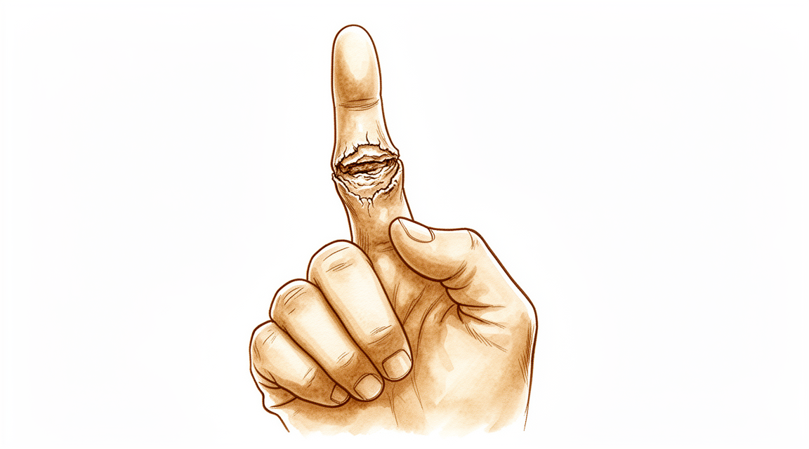
यह ऑपरेशन क्यों सुझाया गया है
डॉ. कीरन हिरपरा, माटर प्राइवेट हॉस्पिटल, रॉकहैम्पटन में ऊपरी अंग (कंधे से हाथ तक) के सर्जन, आपकी समस्या के अनुसार सबसे कम चीर-फाड़ वाले विकल्पों से शुरुआत करते हैं। मरीज़ों को आमतौर पर उनके जीपी (आपके फैमिली डॉक्टर) हमारे क्लिनिक में भेजते हैं; अगर किसी फिजियोथेरेपिस्ट ने आपको हमसे मिलने की सलाह दी है, तब भी मेडिकेयर रिबेट के योग्य होने के लिए आपको अपने जीपी से रेफ़रल की ज़रूरत होगी। आपके अपॉइंटमेंट पर हम आपकी पूरी जानकारी लेते हैं, आपके हाथ की जांच करते हैं, और समस्या क्या है यह पता लगाने के लिए ज़रूरत हो तो इमेजिंग करवाते हैं।
यह ऑपरेशन आपकी उंगली के बीच वाले जोड़ का फ्यूज़न (दो हड्डियों को जोड़कर एक कर देना) है। उस जोड़ को बनाने वाली दो हड्डियों को आपस में जोड़ दिया जाता है ताकि वे एक होकर भरें, और जोड़ फिर हिलता नहीं। हम आमतौर पर इसकी सलाह तब देते हैं जब जोड़ में दर्द हो, जकड़न हो या वह टेढ़ा हो गया हो, और जब गतिविधियों में बदलाव, फिजियोथेरेपी या हैंड थेरेपी, और स्प्लिंट जैसे आसान इलाज से आपको पर्याप्त सुधार न मिला हो। कुछ चोटों या बनावट की समस्याओं में, सर्जरी की सलाह सीधे भी दी जा सकती है। इसका लक्ष्य दर्द खत्म करके और जोड़ को स्थिर रखकर हाथ का काम-काज वापस लाना है।
ऑपरेशन से पहले
सर्जरी से पहले के सप्ताहों में हम आपकी उंगली की नई तस्वीरों, जैसे एक्स-रे, और कभी-कभी एमआरआई या अल्ट्रासाउंड स्कैन से योजना पक्की करते हैं। इनसे सर्जन को पता चलता है कि जोड़ कैसा दिखता है और फ्यूज़न का कोण तय करने में मदद मिलती है। ज़्यादातर लोगों को इससे ज़्यादा कुछ नहीं चाहिए। अगर आपको दूसरी बीमारियां हैं, तो आपको खून की जांच या एनेस्थेटिस्ट (बेहोशी के डॉक्टर) से जांच की भी ज़रूरत पड़ सकती है, लेकिन यह हर किसी के लिए नहीं होता। ऑपरेशन से पहले के दिनों में हम आपको बताएंगे कि आपकी नियमित दवाओं में से कौन-सी और कब बंद करनी हैं। सर्जरी से सात घंटे पहले तक कुछ न खाएं और न पिएं; हम थोड़ा अतिरिक्त समय इसलिए मांगते हैं ताकि अगर ऑपरेशन थिएटर की सूची जल्दी चले तो आपका समय पहले किया जा सके। घर जाने के लिए किसी से गाड़ी का इंतज़ाम करें, क्योंकि आप खुद गाड़ी नहीं चला पाएंगे। अपनी मौजूदा दवाओं की लिखी हुई सूची साथ लाएं और ढीले, आरामदायक कपड़े पहनें।
ऑपरेशन के दिन
आप अस्पताल की सर्जिकल एडमिशन यूनिट में पहुंचते हैं, जहां आपका नाम दर्ज किया जाता है और आपको ऑपरेशन थिएटर के लिए तैयार किया जाता है। वहां आप एनेस्थेटिस्ट से मिलेंगे। यह ऑपरेशन जनरल एनेस्थीसिया में किया जाता है। ऑपरेशन के दौरान आप पूरी तरह बेहोश रहेंगे। कुछ मरीज़ों को ऑपरेशन के बाद दर्द से राहत के लिए रीजनल नर्व ब्लॉक (हाथ या बांह को सुन्न करने का इंजेक्शन) भी दिया जा सकता है; एनेस्थेटिस्ट आपकी व्यक्तिगत स्थिति के आधार पर उसी दिन यह तय करते हैं। इसके बाद आपको ऑपरेशन थिएटर में ले जाया जाता है, जहां ऑपरेशन किया जाता है।
आप रिकवरी एरिया में होश में आते हैं, जहां एनेस्थीसिया का असर खत्म होने तक नर्सें आप पर नज़र रखती हैं। जब आपकी हालत स्थिर हो जाती है, तो प्रक्रिया और आपके ठीक होने के हिसाब से आप या तो वार्ड में जाते हैं या घर।
ऑपरेशन में क्या किया जाता है
आपके सर्जन आपकी उंगली के बीच वाले जोड़ के ऊपर एक चीरा लगाते हैं। इस चीरे से जोड़ को खोला जाता है और दोनों हड्डियों की घिसी हुई, खराब सतहों को इस तरह तैयार किया जाता है कि वे आपस में जुड़कर बढ़ सकें। हड्डियों के सिरों को एक-दूसरे से सटकर बैठने लायक आकार दिया जाता है, और उंगली को ऐसी मुड़ी हुई स्थिति में जमाया जाता है जिससे आपका हाथ मुट्ठी से पकड़ने और चुटकी से पकड़ने के लिए अच्छी तरह काम कर सके।
इसके बाद हड्डियों को भरने तक आपस में थामकर रखा जाता है। आपके सर्जन दोनों हड्डियों को स्थिर और सही स्थिति में रखने के लिए धातु के छोटे इम्प्लांट, जैसे तार या स्क्रू (पेंच), इस्तेमाल करते हैं। इसी तरह थामे रखने से जोड़ जुड़कर एक ठोस हड्डी बन पाता है। हड्डियों के सीध में आकर फिक्स हो जाने के बाद, घाव को टांकों से बंद करके उस पर ड्रेसिंग (घाव की पट्टी) लगा दी जाती है।
इस सबका लक्ष्य सीधा है: दर्द वाला, हिलने वाला जोड़ हटा दिया जाता है और उसकी जगह दोनों हड्डियों के बीच एक स्थिर, दर्द-रहित जुड़ाव बना दिया जाता है। क्योंकि जोड़ अब हिलता नहीं, उंगली पहले से ज़्यादा जकड़ी हुई रहेगी, लेकिन इस्तेमाल करते समय उसे अब न अटकना चाहिए और न दुखना चाहिए।
ऑपरेशन के बाद
इस ऑपरेशन के बाद ज़्यादातर मरीज़ एक रात अस्पताल में रुकते हैं, हालांकि कुछ उसी दिन घर जा पाते हैं। आप रिकवरी वार्ड में होश में आते हैं, जहां एनेस्थीसिया का असर खत्म होने तक नर्सें आप पर नज़र रखती हैं। आपके हाथ पर ड्रेसिंग लगी होगी और उसे स्प्लिंट का सहारा दिया जा सकता है, और आपको आराम में रखने के लिए हम दर्द निवारक दवा देंगे। जैसे ही आप खुद को स्थिर महसूस करें, आप उठकर चल-फिर सकते हैं। कृपया पहले 24 घंटे अपने साथ किसी के रहने का इंतज़ाम करें। हम ड्रेसिंग को लगभग 10 दिन तक लगी रहने देते हैं; जब तक हम न कहें, कृपया उससे पहले इसे न हटाएं। जब हम आपसे मिलेंगे तब हम इसे बदलेंगे या हटाएंगे।
ठीक होने की प्रक्रिया
पहले कुछ दिनों और सप्ताहों तक आपकी उंगली में दर्द और सूजन रहेगी। यह सामान्य है और धीरे-धीरे कम हो जाती है। अपने हाथ को तकिये पर ऊंचा रखने से, सोते समय भी, सूजन कम करने में मदद मिलती है। इस दौरान हम जो दर्द निवारक दवा देते हैं वह आपको आराम में रखती है।
हड्डियों के जुड़ने तक उंगली की रक्षा के लिए आप उस पर स्प्लिंट पहनेंगे। सर्जरी के बाद हैंड थेरेपी एक्सटेंड रिहैबिलिटेशन (Extend Rehabilitation) में रूबी डूलन के साथ होती है। रूबी एक हैंड थेरेपिस्ट (हाथ की विशेषज्ञ थेरेपिस्ट) हैं: वह आपकी एक्सरसाइज़ तय करती हैं और आपको ज़रूरत हो तो स्प्लिंट बनाती हैं। आपकी थेरेपी का ध्यान आपके बाकी हाथ और उंगलियों को हिलाते-डुलाते रखने पर होगा, ताकि जब तक फ्यूज़ किया गया जोड़ जमता है, आपकी मुट्ठी और चुटकी की पकड़ मज़बूत बनी रहे।
दूसरे हाथ की मदद से आप घर पर ज़्यादातर हल्के रोज़मर्रा के काम तुरंत कर सकते हैं। जब तक हम अनुमति न दें, भारी सामान उठाने, ज़ोर से पकड़ने, या ऐसे किसी भी काम से बचें जिससे भरती हुई उंगली पर ज़ोर पड़े। ड्रेसिंग हटने और घाव भर जाने के बाद, आप रोज़मर्रा के कामों के लिए हाथ का ज़्यादा खुलकर इस्तेमाल कर सकते हैं। जब तक स्प्लिंट की वजह से आप स्टीयरिंग व्हील को ठीक से नहीं पकड़ पाते, तब तक गाड़ी चलाना सुरक्षित नहीं है। स्प्लिंट हटने और आपके सर्जन की अनुमति मिलने के बाद, हमारा पेज ऊपरी अंग की सर्जरी के बाद गाड़ी चलाना देखें।
ठीक होने का समय हर व्यक्ति में अलग होता है। आपकी समय-सीमा अलग हो सकती है, और हर फॉलो-अप जांच में आपके सर्जन और थेरेपिस्ट आपका मार्गदर्शन करेंगे।
क्या समस्याएं हो सकती हैं
ज़्यादातर मरीज़ ठीक रहते हैं, लेकिन कभी-कभी समस्याएं हो सकती हैं। किसी भी परेशानी को जल्दी पकड़ने के लिए आपके सर्जन और टीम आप पर करीब से नज़र रखते हैं।
फ्यूज़न का मुख्य जोखिम यह है कि दोनों हड्डियां आपस में न जुड़ें। अगर ऐसा होता है, तो उंगली में दर्द बना रह सकता है, और जहां कोई हलचल नहीं होनी चाहिए वहां आपको कट की आवाज़ या हलचल महसूस हो सकती है। अगर दर्द उम्मीद के मुताबिक शांत नहीं होता, तो अपनी अगली फॉलो-अप जांच में हमें बताएं।
हड्डियों को थामने वाले धातु के इम्प्लांट भी परेशानी पैदा कर सकते हैं। कोई तार या स्क्रू ढीला हो सकता है, टूट सकता है, या त्वचा पर दबाव डाल सकता है। आपको त्वचा के नीचे कोई नुकीला किनारा महसूस हो सकता है, या उंगली टेढ़ी दिख सकती है। अपनी फॉलो-अप जांच में इसके बारे में बताएं ताकि हम इसे जांच सकें।
संक्रमण (इन्फेक्शन) कम होता है, लेकिन इस पर जल्दी ध्यान देना ज़रूरी है। गहरे, धड़कन जैसे (टीस वाले) दर्द पर नज़र रखें जो साधारण दर्द निवारक दवाओं से कम न हो, घाव से बाहर की ओर फैलती लाली, या घाव से तरल का रिसाव। आपको गर्मी और आमतौर पर बीमार महसूस हो सकता है। अगर आपको इनमें से कोई भी संकेत दिखे, तो तुरंत क्लिनिक को फ़ोन करें। अगर क्लिनिक का समय खत्म हो चुका है या लाली तेज़ी से फैल रही है, तो इमरजेंसी विभाग जाएं।
संक्रमण उंगली के किसी जोड़ तक उंगली में लगे कट या छेद वाले घाव से, पास के त्वचा के संक्रमण से, या शरीर में कहीं और से खून के रास्ते भी पहुंच सकता है। हाथ के किसी छोटे जोड़ में संक्रमण के संकेत अक्सर चोट के कुछ ही दिनों में शुरू हो जाते हैं। अगर किसी चोट या घाव के बाद कोई उंगली गर्म, सूजी हुई और दर्द वाली हो जाए, तो बिना देर किए अपने जीपी को दिखाएं, ताकि उसका जल्दी इलाज हो सके।
फ्यूज़न के अच्छी तरह जुड़ जाने पर भी जकड़न और लगातार दर्द हो सकता है। अगर ठीक होने की अपेक्षित अवधि के बाद भी आपकी उंगली बहुत जकड़ी हुई या दर्द वाली रहती है, तो हम उसकी जांच करेंगे और इस पर बात करेंगे कि क्या किया जा सकता है।
अगर आप विस्तार से जानना चाहते हैं, तो इस पेज पर जटिलताओं (कॉम्प्लिकेशन) की तालिका में आम दरें दी गई हैं।
हमें कब फ़ोन करें
हमें तुरंत फ़ोन करें अगर आपको बुखार हो, अगर आपके घाव के आसपास की त्वचा ज़्यादा लाल हो जाए या उससे तरल रिसने लगे, या अगर आपको अचानक ऐसा तेज़ दर्द हो जो साधारण दर्द निवारक दवाओं से शांत न हो। इमरजेंसी विभाग जाएं अगर आपकी पिंडली में सूजन या दर्द हो, या सांस फूल रही हो, क्योंकि ये खून के थक्के का संकेत हो सकते हैं। इमरजेंसी विभाग तब भी जाएं अगर आपकी उंगलियां सुन्न हो जाएं, उनका रंग उड़ जाए (फीकी, सफ़ेद पड़ जाएं), या आप उन्हें बिल्कुल न हिला पा रहे हों। अगर क्लिनिक का समय खत्म हो चुका है और आप चिंतित हैं, तो क्लिनिक खुलने का इंतज़ार करने के बजाय इमरजेंसी विभाग जाएं।
इस समस्या के बारे में और कहां पढ़ें
यह पेज खुद ऑपरेशन के बारे में है। जिस समस्या का यह इलाज करता है, और प्रमाण इस बारे में क्या बताते हैं कि सर्जरी कब मदद करती है और कब नहीं, इसकी ज़्यादा विस्तार से जानकारी पीआईपी जोड़ का गठिया पेज पर दी गई है।
Evidence & references
This is the clinical evidence summary written for health professionals. It is technical, and it lists the research this page was built from. You do not need to read it to understand your treatment or to make a decision about it.
Anatomy & Pathophysiology
General Hand Architecture
- The hand is composed of 19 bones, 17 articulations, and 19 muscles situated entirely within the hand [2].
- The hand contains approximately the same number of tendons activated by forearm muscles as it has intrinsic muscles [2].
- The hand functions as an organ designed to obtain information and an organ of execution [2].
- The dorsal aspect of the hand is convex, while the palmar or volar aspect is concave [2].
- The dorsal surface of the hand is usually visible and aesthetically important, whereas the palmar surface is usually hidden and functional [2].
- The hand moves within a large volume of space with the shoulder as the apex, allowing it to reach any part of the body fairly easily due to the mobility of the shoulder, elbow, and wrist [2].
- Distal to the elbow, the wrist and forearm function as a single physiological unit that places the hand in a position for grasping [2].
- The hand is capable of conforming to the shape of objects to be grasped or studied [2].
Digits and Phalanges
- The digits are divided into the thumb and four fingers [2].
- The thumb has a more proximal and lateral position, allowing movement inward and outward from the palm [2].
- The four fingers are the distal extension of the carpometacarpal part of the hand [2].
- Each finger ray is a polyarticular chain composed of a metacarpal and three phalanges, except the thumb which has only two phalanges [2].
- When fingers are extended and separated, their tips lie on the circumference of a circle whose center is the head of the third metacarpal [2].
- The web space of the thumb is the largest and deepest among the digital web spaces [2].
Cutaneous Units
- The dorsal covering of the interphalangeal articulations of the digits forms a unique cutaneous unit characterized by a considerable excess of skin when the digits are in extension [3].
- The fine tight skin of the dorsal aspect of the middle phalanx forms a distinct cutaneous unit [3].
- The dorsal integument of the distal phalanx is characterized by the nail bed with its matrix [3].
- The palm forms a cutaneous unit extending from the distal transverse crease of the wrist up to the transverse crease at the base of the digits [3].
- The skin of the radial portion of the palm covers the thenar eminence and is relatively well vascularized and mobile [3].
- The skin of the ulnar and distal portion of the palm covers the hypothenar eminence and has poor mobility [3].
- The central triangular part of the palm has fixed, poorly vascularized skin that covers almost directly the superficial palmar aponeurosis [3].
- The integument of the palmar face of the digits is subdivided into phalangeal units separated by digital flexion folds [3].
- When a digit is completely flexed, the integument of adjacent phalanges comes into contact in the zones of the flexion creases, establishing areas of cutaneous contact in the form of a diamond [3].
- The sides of the diamond-shaped cutaneous contact zones do not undergo variations in length during movements of flexion and extension [3].
- Incisions made along the lines of the diamond-shaped cutaneous contact zones present a minimal chance of retraction [3].
- The dorsal slope of the web spaces has a gradual incline and its supple skin is not adherent to the subjacent region [3].
- The palmar surface of the web spaces is flat and precipitously interrupted, with skin densely adherent to the commissural skeleton [3].
Intrinsic Muscles
- There are seven interosseous muscles in the hand, consisting of four dorsal and three volar muscles [4].
- The dorsal interossei are abductors [4].
- The volar interossei are adductors [4].
- The middle finger has two dorsal interossei and no volar interossei because the central axis of the hand lies within it [4].
- Each dorsal interosseous muscle, with the exception of the third, has two muscle heads [4].
- The superficial head of the dorsal interosseous muscles abducts and weakly flexes the proximal phalanx [4].
- The deep head of the dorsal interosseous muscles flexes and weakly abducts the proximal phalanx while extending the middle and distal phalanges [4].
- Transverse fibers arch dorsally from each lateral band to join over the dorsum of the finger, flexing the proximal phalanx [4].
- Oblique fibers from the lateral bands insert onto the lateral tubercles at the base of the middle phalanx to extend the middle phalanx at the PIP joint [4].
- The lateral bands are joined by the lateral slips of the extensor tendon to form the conjoined lateral band [4].
- The two conjoined lateral bands to each finger unite at the distal third of the middle phalanx to form the terminal tendon [4].
- The terminal tendon inserts at the base of the distal phalanx to extend it [4].
- Each volar interosseous muscle has only one muscle head and none insert onto the proximal phalanx [4].
- The volar interossei form the ulnar lateral band of the index finger and the radial lateral band of the ring and little fingers [4].
- The abductor digiti quinti and flexor digiti quinti brevis are similar in structure and function to the superficial and deep heads of the dorsal interossei, respectively [4].
- The opponens digiti quinti arises from the pisohamate ligament and the hook of the hamate and inserts onto the ulnar side of the diaphysis of the fifth metacarpal [4].
Metacarpal Arch and Stability
- The metacarpal arch is endowed with a great deal of adaptability because of the mobility of the peripheral metacarpals [7].
- The index metacarpal is the most firmly fixed [7].
- The ring metacarpal has about 10 degrees of mobility in flexion and extension [7].
- The fifth metacarpal has a range of flexion–extension of approximately 20 degrees [7].
- The second to fifth metacarpals are bound together by various fibrous structures, the most distal of which is the deep transverse intermetacarpal ligament [7].
- The deep transverse intermetacarpal ligament is also known as the interglenoid ligament because it ties together the anterior glenoid ligaments of the metacarpophalangeal articulations [7].
- The metacarpophalangeal articulations are the keystones of the longitudinal arches of the hand [7].
- The thick anterior glenoid capsules, known as volar plates, prevent hyperextension at the metacarpophalangeal joints [7].
- The stability of the metacarpophalangeal joints is essential to the support of the longitudinal arch as well as of the transverse metacarpal arch [7].
Vascular Anatomy
- The arteries of the thumb vary in both size and number, making surgical reconstruction delicate [8].
- The layout of the thumb arteries is the result of innumerable variations regarding origin, transit, connections, and size [8].
- In anatomical studies, only 15% of dissections of the palmar arteries of the thumb fall into the classical "typical" category [8].
- The princeps pollicis artery is the terminal branch of the radial artery that crosses the first intermetacarpal space [8].
- The princeps pollicis artery runs along the ulnar side of the first metacarpal bone and along the volar surface of the adductor muscle [8].
- The princeps pollicis artery emerges onto the subcutaneous palmar tissue at the level of the cutaneous flexion crease of the metacarpophalangeal joint [8].
- At the metacarpophalangeal joint level, the princeps pollicis divides into two terminal rami known as the collateral palmar arteries of the thumb [8].
- The collateral palmar arteries run along the digital tunnel symmetrically and are of equal caliber [8].
- The collateral palmar arteries head distally to finally unite in the pulp arcade [8].
- An arcade located deep in the flexor tendon joins together the two arteries at the level of the distal metaphysis of the first phalanx [8].
- Vessels originating from the subtendinous arcade enter the vincula and irrigate the flexor tendon [8].
- In the second segment of the thumb, the main artery is the ulnar collateral artery [8].
- The subtendinous anastomosis situated at the level of the neck of the first phalanx acts as a moderator between the two collateral arteries [8].
- In cases where the palmar ulnar collateral artery is absent, the dorsal artery takes its place by means of a branch through the subtendinous arcade [8].
- In the pulp segment, the two arteries are of similar size and run through the thick fatty subcutaneous padding [8].
- The dorsal arteries of the thumb originate from the palmar arteries at the level of the first metacarpal [8].
- The dorsal arteries run laterally along the metacarpophalangeal joint and continue obliquely from volar to dorsal [8].
- At the level of the neck of the first phalanx, an anastomosis can be found which originates from the palmar arteries [8].
- The dorsal arteries are joined by three arcades: one inconstant arcade under the extensor tendon at the neck of the first phalanx, the arcade of the nail matrix, and the arcade of the nailbed [8].
Surgical Approaches and Incisions
- Distal palmar incisions are transverse, while proximal palmar incisions tend to be more longitudinal with the distal end curving radially [9].
- Proximal palmar incisions should parallel the thenar crease [9].
- Incisions extended proximal to the wrist should not cross the flexor wrist creases at a right angle [9].
- The most important structure in the thenar area is the recurrent branch (motor) of the median nerve [9].
- Anatomic studies have shown that there is no single longitudinal incision in the proximal palm that completely avoids the palmar cutaneous branches of the median and ulnar nerves [9].
- Midlateral incisions described for the fingers are also suitable for the thumb [9].
- The radial side of the thumb is more accessible for midlateral incisions [9].
- Care should be taken to avoid the dorsal branch of the superficial radial nerve to the radial side of the thumb during midlateral incisions [9].
- The volar midoblique incision crosses the flexion creases obliquely in the midline of the finger between the neurovascular bundles [9].
Investigations
- Clinical evaluation of the hand and wrist requires combining patient history with a careful physical examination to pinpoint or narrow the scope of possible pathologic processes [1].
- Diagnostic tests such as imaging and serum laboratory studies are useful in determining pathology but can be expensive, time consuming, and often nonspecific [1].
- A careful physical examination is essential to direct care and future testing if indicated [1].
- A systematic method for approaching the physical examination of the hand is essential due to the number of structures in a small space [1].
- Clinicians may organize the hand examination by anatomic location or region, or by organ system or pathology [1].
- The skeleton of the hand and wrist consists of 27 bones, of which 19 are long bones [12].
- The skeleton is divided into five rays, each comprising a polyarticulated chain of metacarpals and phalanges [12].
- The base of each metacarpal articulates with the distal row of the carpus [12].
- The wrist has three axes of movement, permitting the hand to be positioned in any spatial configuration for grasping [12].
- The radial ray (first ray) is the shortest, made up of a metacarpal and two phalanges [12].
- The other four digital rays are formed by four skeletal segments: a metacarpal and three phalanges [12].
- The thumb metacarpal is the shortest, while the index metacarpal is the longest [12].
- The proximal and middle phalanges of the middle and ring fingers are longer than those of the index finger [12].
- The transverse axis of the palm forms an acute angle of approximately 75 degrees with the longitudinal axis [12].
- Epiphyseal plates are located at the proximal ends of the phalanges and the first metacarpal [12].
- Epiphyseal plates are located at the distal ends of the other metacarpals [12].
References
[1] Orthopaedic Knowledge Update 13 Ebook Without Multimedia. Anatomy, Evaluation, Clinical Examination, and Imaging > Evaluation and Clinical Examination: Current Concepts.
[2] Exam Of The Hand Wrist 2Ed. INTRODUCTION.
[3] Exam Of The Hand Wrist 2Ed. Functional cutaneous units.
[4] Green S Operative Hand Surgery. Interosseous and Hypothenar Muscles.
[7] Exam Of The Hand Wrist 2Ed. The arches of the hand > The metacarpal arch.
[8] Exam Of The Hand Wrist 2Ed. Techniques of investigation of the arterial supply by J P Melki > Vascularization of the thumb > Palmar aspect.
[9] Campbell S Operative Orthopaedics 4 Volume Set. RESULTS OF SUTURE OF THE SCIATIC NERVE > PALMAR INCISIONS.
[12] Exam Of The Hand Wrist 2Ed. 1.1 SKELETON OF THE HAND > The osseous skeleton.