Patients › Hand
ट्रिगर फिंगर रिलीज़
Trigger finger release — understanding the condition, conservative treatments, and surgical options for a stuck or clicking finger.
यह ऑपरेशन क्यों सुझाया गया है
डॉ. कीरन हिरपरा, माटर प्राइवेट हॉस्पिटल, रॉकहैम्पटन में ऊपरी अंग (कंधे से हाथ तक) के सर्जन हैं, और वे आपकी समस्या के अनुकूल सबसे कम चीर-फाड़ वाले विकल्पों से शुरुआत करते हैं। मरीज़ों को आमतौर पर उनके जीपी (आपके फैमिली डॉक्टर) हमारे क्लिनिक में रेफ़र करते हैं; अगर किसी फिजियोथेरेपिस्ट ने आपको हमसे मिलने की सलाह दी है, तब भी मेडिकेयर रिबेट पाने के योग्य होने के लिए आपको अपने जीपी से रेफ़रल लेना होगा। आपके अपॉइंटमेंट में हम आपकी पूरी जानकारी (हिस्ट्री) लेते हैं, आपके हाथ की जांच करते हैं, और ज़रूरत हो तो इमेजिंग (एक्स-रे, स्कैन आदि) करवाते हैं। इससे हमें पता चलता है कि आपकी उंगली या अंगूठा किस वजह से अटक रहा है या लॉक हो रहा है।
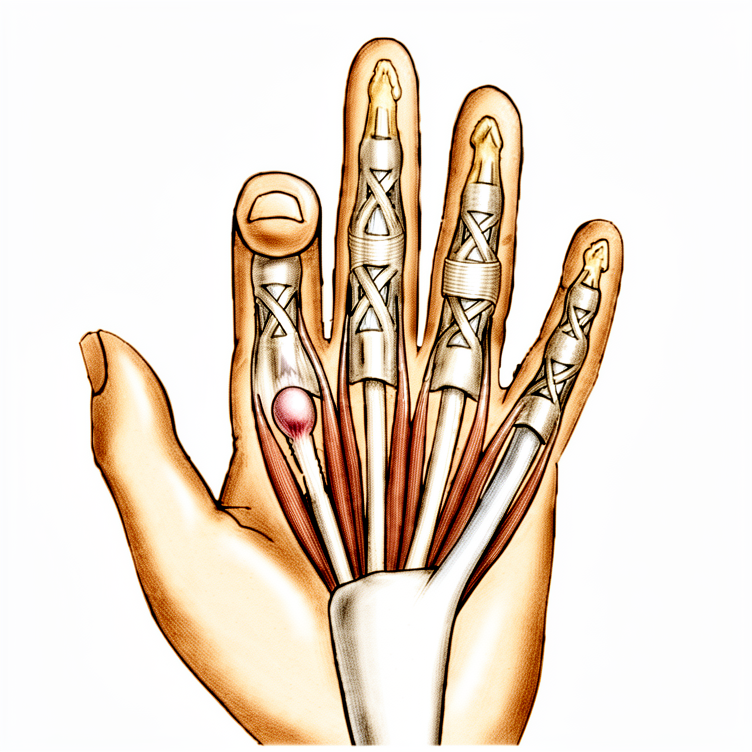
ट्रिगर फिंगर (उंगली का अटकना) तब होता है जब उंगली या अंगूठे का एक टेंडन (कंडरा) हथेली की एक कसी हुई सुरंग से सरकते समय अटक जाता है। आमतौर पर पहले बिना ऑपरेशन वाला इलाज किया जाता है। इसमें स्प्लिंट, हैंड थेरेपी, या स्टेरॉयड इंजेक्शन (टेंडन के पास लगाई जाने वाली दवा, जो सूजन (इन्फ्लेमेशन) को शांत करती है) शामिल हो सकते हैं। हम सर्जरी पर तब विचार करते हैं जब इनसे पर्याप्त सुधार न हुआ हो, या जब उंगली मुड़ी हुई स्थिति में ऐसे फंस गई हो कि उसे अब सीधा न किया जा सके।
यह ऑपरेशन अपने आप में एक रिलीज़ है: ऑपरेशन वाली जगह के ऊपर एक ही चीरा लगाया जाता है, और सुरंग का कसा हुआ हिस्सा खोल दिया जाता है ताकि टेंडन आसानी से सरक सके। सर्जरी के बाद लगभग 97% लोगों में उंगली का अटकना पूरी तरह ठीक हो जाता है। लक्ष्य है ऐसी उंगली या अंगूठा जो बिना अटके, बिना लॉक हुए और बिना दर्द के आसानी से चले।
ऑपरेशन से पहले
ज़्यादातर लोगों को बहुत कम तैयारी की ज़रूरत होती है। आपको सर्जरी से सात घंटे पहले से कुछ भी न खाने और न पीने के लिए कहा जाएगा। हम छह की जगह सात घंटे इसलिए कहते हैं ताकि अगर ऑपरेशन थिएटर की सूची जल्दी चल रही हो, तो आपका समय पहले किया जा सके। आपके सर्जन बताएंगे कि कौन-सी दवाएं कब बंद करनी हैं, और आप जो भी दवाएं लेते हैं उनकी एक लिखित सूची साथ लाएं। बाद में आपको घर ले जाने के लिए किसी गाड़ी चलाने वाले का इंतज़ाम करें, और उस दिन ढीले, आरामदायक कपड़े पहनें। ऑपरेशन की योजना बनाने में मदद के लिए कभी-कभी पहले से एक्स-रे, एमआरआई या अल्ट्रासाउंड जैसी इमेजिंग करवाई जाती है। अगर आपको दूसरी बीमारियां हैं, तो आपको खून की जांच या एनेस्थेटिस्ट (बेहोशी के डॉक्टर, जो एनेस्थीसिया देते हैं) से जांच की ज़रूरत पड़ सकती है, लेकिन ज़्यादातर लोगों को इनमें से किसी की ज़रूरत नहीं होती।
ऑपरेशन के दिन
आप अस्पताल की सर्जिकल एडमिशन यूनिट में पहुंचते हैं, जहां आपकी भर्ती की जाती है और आपको ऑपरेशन थिएटर के लिए तैयार किया जाता है। आप एनेस्थेटिस्ट (वे डॉक्टर जो एनेस्थीसिया देते हैं) से मिलेंगे। यह ऑपरेशन लोकल एनेस्थीसिया (एक इंजेक्शन जो केवल ऑपरेशन वाली जगह को सुन्न करता है, और आप जागे रहते हैं) या जनरल एनेस्थीसिया (जिसमें आप पूरी तरह बेहोश रहते हैं) में किया जा सकता है। ज़्यादातर लोग लोकल चुनते हैं: इसमें रिकवरी जल्दी होती है और आप जल्द ही घर जा सकते हैं। अगर आप बेहोश रहना पसंद करें, तो वह भी एक उचित विकल्प है; इस बारे में अपने सर्जन और एनेस्थेटिस्ट से बात करें।
इसके बाद आपको ऑपरेशन थिएटर ले जाया जाता है, जहां ऑपरेशन किया जाता है। बाद में आप रिकवरी एरिया में होश में आते हैं, जहां एनेस्थीसिया का असर खत्म होने तक नर्सें आप पर नज़र रखती हैं। आपकी हालत स्थिर हो जाने पर, प्रक्रिया और आपकी रिकवरी के अनुसार, आप या तो वार्ड में जाते हैं या घर जाते हैं। यह ऑपरेशन करवाने वाले ज़्यादातर लोग उसी दिन घर चले जाते हैं। पिछले भाग में जिस ड्राइवर का इंतज़ाम किया था, उसे साथ लाएं, और जाने से पहले नर्सें आपके हाथ के बारे में जो भी निर्देश दें, उनका पालन करें।
ऑपरेशन में क्या किया जाता है
इस ऑपरेशन को ट्रिगर फिंगर रिलीज़ कहते हैं। आपके सर्जन ऑपरेशन वाली जगह के ऊपर एक ही चीरा लगाते हैं, जो आमतौर पर लगभग 2 सेमी लंबा होता है। इस चीरे के ज़रिए आपके सर्जन हथेली की उस सुरंग के कसे हुए हिस्से तक पहुंचते हैं जिसमें से टेंडन सरकता है। ऊतक के कसे हुए पट्टे को सावधानी से, एक बार में लगभग 1 सेमी, तब तक खोला जाता है जब तक टेंडन आसानी से न चलने लगे। फिर आपके सर्जन जांचेंगे कि उंगली या अंगूठा बिना अटके आसानी से मुड़ता और सीधा होता है।
ट्रिगर थंब (अंगूठे के अटकने) में केवल एक कसे हुए पट्टे को रिलीज़ करने की ज़रूरत होती है। उंगली में, पास का एक दूसरा पट्टा भी खोला जा सकता है। आपके सर्जन पास की नर्वों (तंत्रिकाओं) को बचाने के लिए, और उंगली के अंदर गहराई में सहारा देने वाले ऊतक के पट्टों को बिना छुए छोड़ने के लिए सावधानी से काम करते हैं, क्योंकि ये टेंडन को ठीक से काम करते रहने में मदद करते हैं।
रिलीज़ पूरी होने के बाद घाव को टांकों से बंद किया जाता है। हाथ पर एक दबाव वाली ड्रेसिंग (घाव की कसी हुई पट्टी) लगाई जाती है। आप यह ड्रेसिंग लगभग 10 दिन तक लगाए रखेंगे, और टांके 10 से 14 दिन पर निकाले जाते हैं। अपनी उंगली या अंगूठे का सामान्य इस्तेमाल तुरंत शुरू करने को कहा जाता है।
अगर आपको रूमेटॉइड आर्थराइटिस (एक बीमारी जिसमें जोड़ों में सूजन (इन्फ्लेमेशन) होती है) है, तो योजना थोड़ी अलग हो सकती है। उस स्थिति में आपके सर्जन पट्टे को रिलीज़ करने की बजाय टेंडन की एक छोटी पट्टी (स्लिप) निकाल सकते हैं, क्योंकि पट्टे को रिलीज़ करने से समय के साथ उंगली एक तरफ़ खिसक सकती है। आपकी सहमति लेने से पहले आपके सर्जन आपके हाथ के लिए सटीक योजना समझाएंगे।
ऑपरेशन के बाद
आप रिकवरी एरिया में होश में आएंगे, जहां एनेस्थीसिया का असर खत्म होने तक नर्सें आप पर नज़र रखती हैं। यह डे-केस है, यानी आप उसी दिन घर जाएंगे। आपके हाथ पर एक दबाव वाली ड्रेसिंग (कसी हुई पट्टी) होगी, और आप अपनी उंगली या अंगूठे को तुरंत हिला सकते हैं। पहले एक-दो दिन कुछ दर्द होना सामान्य है; आमतौर पर आपके फार्मासिस्ट या जीपी से मिलने वाली साधारण दर्द निवारक दवाएं काफ़ी होती हैं। पहले 24 घंटे किसी को अपने साथ रहने के लिए कहें। हम ड्रेसिंग को लगभग 10 दिन तक लगी रहने देते हैं; जब तक हम न कहें, कृपया उससे पहले इसे न उतारें। जब हम आपसे मिलते हैं, तब हम इसे बदलते या हटाते हैं। ज़्यादातर लोग एक से दो सप्ताह के अंदर फिर से गाड़ी चलाने लगते हैं, जब घाव आरामदायक हो जाए और वे हाथ को बचाए बिना स्टीयरिंग व्हील को पकड़ और घुमा सकें।
ठीक होने की प्रक्रिया
ज़्यादातर लोग यह देखकर हैरान होते हैं कि हाथ कितनी जल्दी ठीक हो जाता है। पहले कुछ दिन चीरे के आसपास का हिस्सा दुखेगा और थोड़ा सूजा रहेगा। आमतौर पर आपके फार्मासिस्ट या जीपी से मिलने वाली साधारण दर्द निवारक दवाएं आपको आराम में रखने के लिए काफ़ी होती हैं, और बैठते या आराम करते समय अपने हाथ को तकिये पर ऊंचा रखने से सूजन कम होने में मदद मिलती है। निशान के आसपास छूने पर दर्द बाकी हिस्से से ज़्यादा समय तक रह सकता है, और घाव के परिपक्व होने के साथ यह कम हो जाता है।
आप अपनी उंगली या अंगूठे को तुरंत हिलाएंगे, और शुरू से ही रोज़मर्रा का सामान्य इस्तेमाल करने को कहा जाता है। कोई प्लास्टर (कास्ट) या ब्रेस नहीं पहनना होता, केवल ड्रेसिंग होती है, जिसे हम लगभग 10 दिन तक लगी रहने देते हैं। जब हम आपसे मिलेंगे, तब हम इसे बदलेंगे या हटाएंगे और टांके निकालेंगे। सर्जरी के बाद हैंड थेरेपी एक्सटेंड रिहैबिलिटेशन में रूबी डूलन के साथ होती है। रूबी आपको आसान व्यायाम करवाएंगी जिनसे उंगली आसानी से मुड़ती और सीधी होती रहे, और अगर आपके हाथ को ज़रूरत हो तो वे स्प्लिंट भी बना सकती हैं।
जैसे ही आप सक्षम महसूस करें, आप घर के ज़्यादातर काम कर सकते हैं: खाना खाना, कपड़े पहनना, टाइप करना और घर के हल्के काम। जब तक घाव भर न जाए, भारी चीज़ें कसकर पकड़ने, वज़न उठाने और गंदी या गीली जगहों से बचें। जब आप हाथ को बचाए बिना स्टीयरिंग व्हील को पकड़ और घुमा सकें, तब आमतौर पर गाड़ी चलाना ज़्यादा दूर नहीं होता; हमारा पेज ऊपरी अंग की सर्जरी के बाद गाड़ी चलाना देखें।
ठीक होने में हर व्यक्ति का समय अलग होता है, और आपकी समय-सीमा अलग हो सकती है। आपके सर्जन और आपके थेरेपिस्ट पूरे समय आपका मार्गदर्शन करेंगे।
क्या समस्याएं हो सकती हैं
ज़्यादातर मरीज़ ठीक रहते हैं, लेकिन कभी-कभी समस्याएं हो सकती हैं। किसी भी समस्या को जल्दी पकड़ने के लिए आपके सर्जन और टीम आप पर करीबी नज़र रखते हैं।
सबसे आम समस्याएं घाव के आसपास की छोटी-मोटी दिक्कतें होती हैं, या ऐसी उंगली जो कुछ समय तक जकड़ी हुई लगे। शुरुआती सप्ताहों में आपको कुछ लगातार दर्द या सूजन महसूस हो सकती है, या लग सकता है कि उंगली उतनी आसानी से नहीं मुड़ रही जितनी आपने उम्मीद की थी। अगर यह ठीक नहीं हो रहा, तो अपनी अगली फॉलो-अप जांच में इसके बारे में बताएं।
संक्रमण (इन्फेक्शन) आम नहीं है, लेकिन इसके बारे में जानना ज़रूरी है। घाव पर ध्यान रखें: चीरे से बाहर की ओर फैलती लाली, बढ़ता दर्द, गर्माहट, या रिसाव। गहरा संक्रमण गहरा, धड़कता हुआ दर्द पैदा कर सकता है जो साधारण दर्द निवारक दवाओं से कम नहीं होता, और आप बीमार महसूस कर सकते हैं। अगर आपको इनमें से कोई भी लक्षण दिखे, तो अपने अगले अपॉइंटमेंट का इंतज़ार करने की बजाय तुरंत क्लिनिक को फ़ोन करें।
कभी-कभी सर्जरी के पास की कोई नर्व परेशान (इरिटेट) हो सकती है। यह उंगली या अंगूठे के किसी हिस्से में सुन्नपन या झुनझुनी जैसा महसूस होता है। शुरू में घाव के किनारे पर कुछ सुन्नपन होना अपेक्षित है, लेकिन अगर यह बना रहे या फैले, तो अपनी फॉलो-अप जांच में इसका ज़िक्र करें।
कुछ थोड़े-से लोगों में उंगली का अटकना वापस आ सकता है। अगर ठीक हो जाने के बाद अटकना या लॉक होना फिर से शुरू हो जाए, तो अपने फॉलो-अप में हमें बताएं।
कुछ लोगों की उंगली में जकड़न आ जाती है, जिसमें अटकना खत्म हो जाने के बावजूद उंगली पूरी तरह सीधी नहीं होती या पूरी तरह नहीं मुड़ती। हैंड थेरेपी के व्यायाम इससे बचाने में मदद करते हैं, इसलिए रूबी ने जो हरकतें सिखाई हैं उन्हें करते रहें। अगर लगे कि उंगली ढीली होने की बजाय और जकड़ रही है, तो जल्दी ही अपने थेरेपिस्ट या सर्जन को बताएं।
अगर सर्जरी से पहले आपकी उंगली लंबे समय तक अटकती रही थी, तो रिलीज़ के बाद भी उंगली के बीच वाले जोड़ में दर्द बना रह सकता है। यह तुरंत ठीक होने की बजाय धीरे-धीरे सुधरता है।
अंत में, ट्रिगर फिंगर की सर्जरी का संबंध ड्यूपुइट्रेन (Dupuytren's) बीमारी होने की थोड़ी-सी संभावना से है; यह एक समस्या है जिसमें हथेली में सख़्त डोरियां बन जाती हैं जो उंगलियों को मोड़कर खींच लेती हैं। अगर बाद में आपको अपनी हथेली में मोटापन या गांठें दिखें, तो उनकी जांच करवाएं।
अगर आप विस्तार से जानना चाहते हैं, तो इस पेज पर दी गई जटिलताओं की तालिका में आम दरें दी गई हैं।
हमें कब फ़ोन करें
अगर आपको बुखार हो, या घाव के आसपास लाली, गर्माहट या रिसाव बढ़ रहा हो, तो तुरंत क्लिनिक को फ़ोन करें। अगर दर्द अचानक बहुत तेज़ हो जाए, या सुन्नपन या झुनझुनी फैले और ठीक न हो, तो हमें फ़ोन करें। अगर आपकी पिंडली में सूजन या दर्द हो, या सांस फूल रही हो, तो इमरजेंसी विभाग जाएं, क्योंकि ये खून के थक्के के लक्षण हो सकते हैं। अगर आप उंगली या अंगूठे को बिल्कुल भी न हिला पाएं, या हाथ ठंडा या फीका (सफ़ेद पड़ा हुआ) हो जाए, तो इमरजेंसी विभाग जाएं।
इस समस्या के बारे में और कहां पढ़ें
यह पेज ऑपरेशन के बारे में है। जिस समस्या का यह इलाज करता है, उसके बारे में, जिसमें यह भी शामिल है कि प्रमाण क्या बताते हैं कि सर्जरी कब मदद करती है और कब नहीं, ट्रिगर फिंगर पेज पर ज़्यादा विस्तार से बताया गया है।
Evidence & references
This is the clinical evidence summary written for health professionals. It is technical, and it lists the research this page was built from. You do not need to read it to understand your treatment or to make a decision about it.
Overview
Operative Technique
- Local anesthetic infiltration in the palm proximal to the incision site is preferred for trigger finger release [5].
- A pneumatic arm tourniquet may be helpful, although a high forearm Esmarch wrap usually is sufficient [5].
- For middle, ring, and small trigger finger releases, a transverse incision about 2 cm long is made several millimeters distal to the distal palmar crease [5].
- For index trigger finger releases, a transverse incision about 2 cm long is made several millimeters distal to the proximal palmar crease [5].
- Trigger thumb releases can be performed through incisions either distal or proximal to the metacarpophalangeal joint flexion crease [5].
- Alternative incisions for fingers can be made obliquely or longitudinally between the metacarpophalangeal and distal palmar creases [5].
- Alternative incisions for the thumb can be made obliquely across the thumb metacarpophalangeal flexion crease [5].
- The digital nerves on the thumb are more palmar and closer to the flexor sheath than might be anticipated [5].
- The thumb radial digital nerve is especially vulnerable during trigger thumb release [5].
- Subcutaneous tissues are spread away from the underlying annular pulley system to ensure digital nerves are safely protected [5].
- Trigger thumbs require release of only the A1 pulley [5].
- Trigger digits require division of the A1 and A0, or proximal palmar pulley [5].
- Pulley division is usually accomplished with an initial opening of the pulley with a No. 15 knife blade and a pair of tenotomy scissors [5].
- For trigger thumb release, cutting too far distally should be avoided to prevent disrupting the oblique pulley [5].
- The sheath is incised from proximal to distal, approximately 1 cm, and reassessed for triggering [5].
- Persistent triggering after initial release implies that either the A1 and palmar pulleys are incompletely released or an alternate site of triggering is present [5].
- When the distal A1 pulley edge is released, the divided pulley leaves are parallel rather than ending in a V-shaped pattern [5].
- The distinction between the A1 and A2 pulleys may not be apparent during surgery [5].
- Other fingers can be found to trigger at the same surgical setting and can be managed at the same time [5].
Postoperative Care
- The compression dressing is removed after 48 hours [5].
- Sutures are removed at 10 to 14 days [5].
- Normal use of the finger or thumb is encouraged postoperatively [5].
Anatomy & Pathophysiology
Demographics & Epidemiology
- Trigger finger occurs in 2% to 3% of the general population [9].
- Women are more commonly affected than men [9].
- Women older than 50 years of age are the primary demographic for trigger finger [1, 2].
- Middle and ring finger involvement is most common in adults [1, 2].
- The digits are affected in the following order of decreasing prevalence: thumb, ring, long, little, and index [9].
- Stenosing tenosynovitis is more common in diabetic patients than in nondiabetic patients [4].
- Trigger finger is more common in patients with diabetes mellitus, with a 10% to 20% lifetime incidence [9].
Comorbidities & Etiology
- Trigger finger is associated with diabetes and inflammatory arthropathy [1, 2].
- Trigger finger is seen in patients with hypothyroidism, sarcoidosis, rheumatoid arthritis, and septic tenosynovitis [9].
- The etiology of trigger finger is possibly associated with repetitive grasping activities [1, 2].
- Gout can present as trigger finger due to monosodium urate precipitation eliciting a fulminant inflammatory reaction in the tenosynovium [9].
- Calcific tendinitis can result in triggering due to calcium salt deposition in the tenosynovium [9].
- Pseudogout can cause triggering via calcium pyrophosphate dihydrate crystal deposition [9].
- Amyloidosis, characterized by beta-2-microglobulin deposition, can cause trigger finger in patients with renal failure undergoing dialysis [9].
- Trigger finger is considered an early indication of Dupuytren’s disease by some authors [12].
- The incidence of concurrent trigger finger and Dupuytren’s disease is higher in the middle and ring fingers than expected by statistical coincidence [11].
Histology & Pathology
- Histology of the affected pulley demonstrates fibrocartilaginous metaplasia of the pulley and/or FDS tendon [1, 2].
- Pathologic examination of affected pulleys demonstrates a proliferation of chondrocytes and increased type III collagen [9].
- The flexor digitorum profundus tendon often demonstrates a pathologic nodule, while the flexor digitorum superficialis is often unaffected [9].
- A nodule or fusiform swelling of the flexor tendon just distal to the first annular pulley may be palpable [7].
- The tendon nodule is usually located just proximal to the anulus at the metacarpophalangeal joint level [7].
- In rheumatoid patients, a nodule distal to the metacarpophalangeal joint level may cause triggering [7].
- A volar retinacular ganglion cyst between the A1 and A2 pulleys may be present [9].
- In congenital trigger digits, the pathologic anatomy includes narrowing and thickening of the sheath with occasional formation of a ganglion cyst [10].
- An intratendinous nodule proximal to the first annular pulley, often referred to as Notta’s nodule, may be present in congenital trigger digits [10].
Clinical Presentation
- Patients present with pain and tenderness in the palm at the proximal edge of the digital A1 pulley [4].
- Patients frequently note catching or triggering of the affected finger or thumb after forceful flexion [4].
- In more severe cases, the opposite hand must be used to force the finger or thumb passively into extension [4].
- In the most severe cases, the finger becomes locked in a flexed position [4].
- Triggering is often more pronounced in the morning than later in the day [4].
- A common complaint is referred pain at the dorsal MCP/PIP area [1, 2].
- Patients frequently state that the problem is in the proximal interphalangeal joint with trigger finger or trigger thumb [7].
- Concomitant trigger finger and carpal tunnel syndrome occurs in 40% to 60% of patients [1, 2].
- Local tenderness may be present but is not a prominent complaint [7].
- Pressure accentuates the apparent snapping or triggering of the more distal joints [7].
Anatomical Variations & Specific Structures
- Newer evidence has found a fourth pulley (variable annular pulley) in 75% of patients with thumb trigger finger, which may contribute to stenosis [1, 2].
- The radial digital nerve is at risk of iatrogenic injury during thumb trigger finger release due to its superficial location [1, 2].
- On the thumb, digital nerves are more palmar and closer to the flexor sheath than might be anticipated [5].
- The thumb radial digital nerve is especially vulnerable during surgical release [5].
- Trigger thumbs require release of only the A1 pulley, whereas trigger digits require division of the A1 and A0 (or proximal palmar) pulley [5].
- The distinction between the A1 and A2 pulleys may not be apparent during surgery; however, when the distal A1 pulley edge is released, the divided pulley leaves are parallel rather than ending in a V-shaped pattern [5].
- The fibers of the A2 pulley must be spared to preserve effective digital flexion [4].
- In patients with rheumatoid arthritis, the entire annular pulley system should be preserved to prevent further ulnar drift of the fingers [4].
Classification
- Green Classification Grade I is defined as pain and tenderness at the A1 pulley [1, 2, 9].
- Green Classification Grade II is defined as catching of the finger [1, 2] or mechanical catching of the digit without locking [9].
- Green Classification Grade III is defined as locking of the finger that is passively correctable [1, 2] or mechanical locking of the digit which is passively correctable [9].
- Green Classification Grade IV is defined as a fixed, locked finger [1, 2, 9].
Classification
- The Green classification of trigger finger consists of four grades [1].
- Grade I is defined as pain and tenderness at the A1 pulley [1].
- Grade II is defined as catching of the finger [1].
- Grade III is defined as locking of the finger that is passively correctable [1].
- Grade IV is defined as a fixed, locked finger [1].
Clinical Presentation
Demographics and Epidemiology
- Trigger finger is most common in women older than 50 years of age [1, 2].
- Trigger finger is more common in patients with systemic diseases such as diabetes mellitus, hypothyroidism, sarcoidosis, rheumatoid arthritis, and septic tenosynovitis [9].
- Diabetes mellitus has a 10% to 20% lifetime incidence of trigger finger [9].
- When multiple digits are involved, the possibility of diabetes should be considered [4].
Etiology and Pathology
- Trigger finger is possibly associated with repetitive grasping activities [1, 2].
- Histology of the affected pulleys demonstrates fibrocartilaginous metaplasia of the pulley and/or FDS tendon [1, 2].
- Pathologic examination of the affected pulleys demonstrates a proliferation of chondrocytes and increased type III collagen [9].
- The flexor digitorus profundus tendon often demonstrates a pathologic nodule, while the flexor digitorus superficialis is often unaffected [9].
- A fourth pulley (variable annular pulley) is found in 75% of patients with thumb trigger finger, which may contribute to stenosis [1, 2].
Symptoms and Signs
- Patients present with pain and tenderness in the distal palm [1, 2].
- Pain and tenderness are located at the proximal edge of the digital A1 pulley [4].
- Symptoms progress to mechanical catching or locking of the finger [1, 2].
- In severe cases, the opposite hand must be used to force the finger or thumb passively into extension [4].
- Patients may note a lump or knot in the palm [7].
- The lump may be a thickened area in the first annular pulley or a nodule/fusiform swelling of the flexor tendon just distal to it [7].
- The tendon nodule can be palpated by the examiner’s fingertip and moves with the tendon [7].
- The tendon nodule is usually just proximal to the anulus at the metacarpophalangeal joint level [7].
- In rheumatoid patients, a nodule distal to the MCP joint level may cause triggering [7].
- Patients frequently state that the problem is in the proximal interphalangeal joint [7].
- Concomitant trigger finger and carpal tunnel syndrome occur in 40% to 60% of patients [1, 2].
Physical Examination Findings
- Tenderness to palpation of the flexor tendon at the level of the A1 pulley is a physical examination finding [9].
- Palpable triggering or pain with flexion and extension of the finger is a physical examination finding [9].
- Nodularity of the flexor tendon just proximal to the A1 pulley is a physical examination finding [9].
- Presence of a volar retinacular ganglion cyst between the A1 and A2 pulleys is a physical examination finding [9].
- Presence of a fixed flexion deformity of the proximal interphalangeal (PIP) joint is a physical examination finding [9].
Classification
- Green classification Grade II is defined as catching of the finger [1, 2].
- Green classification Grade II is defined as mechanical catching of the digit without locking [9].
- Green classification Grade III is defined as locking of the finger that is passively correctable [1, 2].
- Green classification Grade III is defined as mechanical locking of the digit which is passively correctable [9].
Differential Diagnosis Considerations
- Intraarticular disorders such as loose bodies, degenerative joint disease, and fractures can cause symptoms similar to trigger finger [7].
- Common extensor tendon subluxation can cause symptoms similar to trigger finger [7].
- Gout can mimic infectious tenosynovitis with marked pain, erythema, swelling, and warmth [9].
- Calcific tendinitis can resemble an infection and result in triggering [9].
- Pseudogout can present with calcium pyrophosphate dihydrate crystal deposition localized to the triangular fibrocartilage or within the carpal tunnel [9].
- Amyloidosis can present with beta-2-microglobulin deposition along flexor tendons, most commonly in patients with renal failure undergoing dialysis [9].
Investigations
Clinical Presentation and History
- Trigger finger is characterized by pain and tenderness in the palm at the proximal edge of the digital A1 pulley [4].
- Pain and tenderness in the distal palm progress to mechanical catching or locking, and may become fixed [1].
- A common complaint is referred pain at the dorsal MCP or PIP area [1].
Physical Examination Findings
- Physical examination may reveal tenderness to palpation of the flexor tendon at the level of the A1 pulley [9].
- Nodularity of the flexor tendon just proximal to the A1 pulley may be present on examination [9].
- A volar retinacular ganglion cyst between the A1 and A2 pulleys may be present on examination [9].
- A fixed flexion deformity of the proximal interphalangeal (PIP) joint may be present on examination [9].
Classification
Associated Conditions and Demographics
- The lifetime incidence of trigger finger in patients with diabetes mellitus is 10% to 20% [9].
- Calcific tendinitis can result in triggering and is affected five times more frequently in males than females [9].
- Pseudogout involves calcium pyrophosphate dihydrate crystal deposition often localized to the triangular fibrocartilage or within the carpal tunnel [9].
- Amyloidosis is characterized by the deposition of beta-2-microglobulin in thick, plaque-like accumulations along the flexor tendons [9].
Imaging and Histology
- MR imaging findings of trigger thumb have been described [3].
Treatment
Nonoperative
- Corticosteroid injection into the flexor tendon sheath is curative in about 60% of patients initially [1].
- Diabetic patients are generally less responsive to corticosteroid injection [1].
- There is no difference between soluble and insoluble corticosteroid preparations for trigger finger injection [1].
- In a study of 292 corticosteroid injections, repeat injections provided symptomatic relief for a year or more in 50% of patients [7].
- Corticosteroid injections may elevate serum glucose levels for 5 days or more in patients with diabetes mellitus [7].
- Patients with unstable diabetes may be better treated without corticosteroid injection [7].
- Preoperative hypoglycemia increases infection risk after trigger finger injection and release [7].
- Immediate surgical release in the clinic was identified as the most cost-effective treatment strategy for trigger finger in diabetic patients [7].
- Nonoperative methods for trigger digits include stretching, night splinting, and combinations of heat and ice [7].
Operative
- Surgical release of the A1 pulley is curative in digits refractory to steroid injection [4].
- Surgical release reliably relieves the problem for most patients, with approximately 97% of patients having complete resolution after operative treatment [7].
- Persistence of triggering is more common than recurrence after surgical release [7].
- Trigger release should be performed with a local block so that the cessation of triggering can be evaluated intraoperatively [7].
- Adjacent finger triggering may become obvious only after a given finger is released and can be managed at the same surgical setting [7].
- In patients with rheumatoid arthritis, the preference is to excise a slip of the FDS tendon rather than to release the A1 pulley to prevent exacerbation of ulnar drift at the MCP joint [1].
- The fibers of the A2 pulley must be spared during surgical release to preserve effective digital flexion [4].
- Minor complications of surgical release include wound dehiscence, scar tenderness, and decreased range of motion [1].
- Incomplete pulley release and damage to the flexor tendons and digital nerves remain concerns, especially in the index finger and thumb with limited exposure techniques [7].
- Percutaneous release of the A1 pulley may be accomplished with a needle on the middle and ring fingers, especially if they actively lock [4].
- The safety and effectiveness of percutaneous trigger finger release using a needle or a push knife have literature support [7].
- For percutaneous release, an 18- or 19-gauge needle may suffice [8].
- During percutaneous release, the bevel of the needle should be oriented longitudinally parallel to the flexor tendons [8].
- Injection of corticosteroid is optional during percutaneous trigger finger release [8].
- Postoperative compression dressing for open trigger finger release is removed after 48 hours [5].
- Sutures for open trigger finger release are removed at 10 to 14 days [5].
- Normal use of the finger or thumb is encouraged after open trigger finger release [5].
- Active hand and finger use with stretching exercises is encouraged after percutaneous trigger finger release [8].
Complications
Operative
- Digital nerves on the thumb are more palmar and closer to the flexor sheath than might be anticipated, making the thumb radial digital nerve especially vulnerable [5].
- Minor complications of trigger finger release are relatively high and include wound dehiscence, scar tenderness, and decreased range of motion [1].
- Incomplete pulley release remains a concern with limited exposure techniques [7].
- Damage to flexor tendons remains a concern with limited exposure techniques, especially in the index finger and thumb [7].
- Damage to digital nerves remains a concern with limited exposure techniques, especially in the index finger and thumb [7].
- Patients may experience triggering after operative release because of catching of the tendon on the palmar aponeurosis transverse fibers [7].
- Triggering caused by catching on palmar aponeurosis transverse fibers usually resolves with time [7].
- A partially lacerated flexor tendon at the metacarpophalangeal joint level may heal with a nodule sufficiently large to cause triggering [7].
- Persistence of triggering is more common than recurrence after operative treatment [7].
- Preoperative hypoglycemia increases infection risk after trigger finger release [3].
- Preoperative hypoglycemia increases infection risk after trigger finger injection [3].
- In patients with rheumatoid arthritis, release of the A1 pulley carries a chance of exacerbating ulnar drift at the MCP joint [1].
Non-Operative
- Corticosteroid injections may elevate serum glucose levels for 5 days or more [7].
References
[1] Miller S Review Of Orthopaedics. SECTION 16 PATELLAR TRACKING IN TOTAL KNEE ARTHROPLASTY > 2. Flexor tendon injury > 3. Stenosing tenosynovitis (trigger finger).
[2] Miller S Review Of Orthopaedics. 2. Flexor tendon injury > 3. Stenosing tenosynovitis (trigger finger).
[3] Campbell S Operative Orthopaedics 4 Volume Set. EXCISION OF NECROTIC MUSCLES COMBINED WITH NEUROLYSIS OF MEDIAN AND ULNAR NERVES FOR SEVERE CONTRACTURE > REFERENCES > TRIGGER THUMB AND TRIGGER FINGER.
[4] A Lange Medical Book Current Diagnosis Treatment In Orthopedics Fifth Edition. 9Hand Surgery > 2. Flexor Tenosynovitis (Trigger Finger and Trigger Thumb).
[5] Campbell S Operative Orthopaedics 4 Volume Set. EXCISION OF NECROTIC MUSCLES COMBINED WITH NEUROLYSIS OF MEDIAN AND ULNAR NERVES FOR SEVERE CONTRACTURE > TRIGGER FINGER AND THUMB > SURGICAL RELEASE OF TRIGGER FINGER.
[7] Campbell S Operative Orthopaedics 4 Volume Set. EXCISION OF NECROTIC MUSCLES COMBINED WITH NEUROLYSIS OF MEDIAN AND ULNAR NERVES FOR SEVERE CONTRACTURE > TRIGGER FINGER AND THUMB.
[8] Campbell S Operative Orthopaedics 4 Volume Set. EXCISION OF NECROTIC MUSCLES COMBINED WITH NEUROLYSIS OF MEDIAN AND ULNAR NERVES FOR SEVERE CONTRACTURE > PERCUTANEOUS RELEASE OF TRIGGER FINGER.
[9] Aaos Comprehensive Orthopaedic Review 3. Tendinopathy of the Hand and Wrist* > II. Trigger Finger.
[10] Campbell S Operative Orthopaedics 4 Volume Set. MULTIPLE Z-PLASTY RELEASE OF A CONGENITAL RING > TRIGGER FINGERS.
[11] Dupuytren S Disease And Related Hyperproliferative Disorders. 31. The Influence of Dupuytren’s Disease on Trigger Fingers and Vice Versa > 31.5 Discussion.
[12] Dupuytren S Disease And Related Hyperproliferative Disorders. 31. The Influence of Dupuytren’s Disease on Trigger Fingers and Vice Versa > 31.5 Discussion > 31.5.1 Why Is DD Concurring with TF?.