Patients › Elbow
डिस्टल बाइसेप्स रप्चर
Distal biceps rupture causes sudden elbow pain, bruising, and weakness—often needing surgical repair.
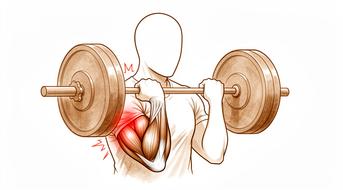
आपको क्या महसूस होता है
डिस्टल बाइसेप्स रप्चर एक ही पल में होता है, महीनों में धीरे-धीरे नहीं। यह आम तौर पर 90 डिग्री पर मुड़ी हुई बांह पर अचानक और अनपेक्षित रूप से सीधा करने वाले ज़ोर से होता है, जैसे गिरती हुई किसी भारी चीज़ को थामना, या कसकर पकड़े रहना जबकि आपकी बांह खिंचकर सीधी हो जाती है। ऐसा होते समय बहुत से लोगों को कोहनी के सामने 'पॉप' जैसा महसूस होता है या उसकी आवाज़ सुनाई देती है। टेंडन (कंडरा) आम तौर पर अग्रबाहु (कोहनी से कलाई तक का हिस्सा) की एक हड्डी पर अपने जुड़ने की जगह से उखड़ जाता है। आपको मांसपेशी का आकार बदला हुआ दिख सकता है, क्योंकि वह सिकुड़कर बांह में ऊपर चली जाती है और बीच के पास इकट्ठा हो जाती है।
चोट लगते ही आपकी कोहनी का सामने वाला हिस्सा दर्द भरा और सूजा हुआ होने की संभावना है। नील पड़ना (चोट का नीला निशान) आम है और अगले एक-दो दिनों में कोहनी और अग्रबाहु के आसपास दिख सकता है। बांह को मोड़ने और घुमाने में दर्द होता है, इसलिए हो सकता है कि आप केतली उठाने, दरवाज़े का नॉब घुमाने, या स्टीयरिंग व्हील को कसकर पकड़ने से बचने लगें। रुकावट के ख़िलाफ़ हथेली को ऊपर की ओर घुमाना, जैसे पेचकस चलाना, अक्सर सबसे मुश्किल हरकत होती है। कुछ लोग अब भी कोहनी मोड़ पाते हैं, क्योंकि उसमें दूसरी मांसपेशियां मदद करती हैं, और इससे चोट असल से कम गंभीर लग सकती है।
पहले कुछ दिनों और सप्ताहों में दर्द अक्सर हिलाने-डुलाने पर बढ़ जाता है और रात में दुख सकता है। जैसे-जैसे शरीर फटे हुए टेंडन के आसपास ठीक होना शुरू करता है, दर्द धीरे-धीरे शांत होता जाता है। सूजन और नील एक-दो सप्ताह में कम हो जाते हैं, लेकिन ताकत की कमी अपने आप ठीक नहीं होती, क्योंकि टेंडन हड्डी से उखड़ चुका है और अपनी सही जगह पर खुद दोबारा नहीं जुड़ सकता।
कुछ चेतावनी वाले लक्षणों पर जल्दी कदम उठाना ज़रूरी है। अगर आपका हाथ या उंगलियां फीकी (सफ़ेद पड़ी हुई), ठंडी, सफ़ेद या नीली पड़ जाएं, या चोट के बाद आपके हाथ में अचानक महसूस करने की क्षमता या हिलने-डुलने की ताकत चली जाए, तो उसी दिन इमरजेंसी विभाग जाएं। अगर दर्द शांत नहीं हो रहा है, सप्ताहों के साथ बढ़ता जा रहा है, या आपको रात में जगा देता है, तो अपने जीपी (आपके फैमिली डॉक्टर) को दिखाएं या विशेषज्ञ से जांच करवाने के लिए कहें।
असल में क्या हो रहा है
बाइसेप्स मांसपेशी आपकी बांह के ऊपरी हिस्से के सामने नीचे की ओर जाती है और कोहनी से ठीक पहले एक मज़बूत डोरी, यानी टेंडन, में बदल जाती है। यह टेंडन अग्रबाहु की एक हड्डी, रेडियस, के एक छोटे उभार पर जुड़ता है। इसका काम आपकी अग्रबाहु को घुमाना है ताकि हथेली ऊपर की ओर मुड़ जाए, और कोहनी मोड़ने में मदद करना है।
जब टेंडन फटता है, तो वह हड्डी के उस उभार से उखड़ जाता है और सिकुड़कर बांह में ऊपर चला जाता है। मांसपेशी अब भी स्वस्थ होती है, लेकिन अब वह उस हड्डी से जुड़ी नहीं रहती जिसे वह हिलाती है, इसलिए उसकी खींचने की ताकत कहीं नहीं पहुंचती। हथेली को ऊपर की ओर घुमाने वाली हरकत सबसे ज़्यादा प्रभावित होती है, क्योंकि कोई दूसरी मांसपेशी यह काम नहीं संभाल सकती। कोहनी मोड़ना आम तौर पर बेहतर ठीक होता है, क्योंकि बाइसेप्स के नीचे की एक दूसरी मांसपेशी इसमें मदद करती है।
चरखी (विंच) से बंधी एक रस्सी के बारे में सोचें। अगर रस्सी ड्रम से टूटकर अलग हो जाए, तो चरखी की मोटर चलती रहती है, लेकिन कुछ नहीं घूमता। रस्सी अपने आप वापस आकर बंध नहीं जाती। उसे दोबारा जोड़ना पड़ता है।
अगर कुछ न किया जाए, तो फटा हुआ सिरा हड्डी पर अपनी सही जगह से दोबारा नहीं जुड़ता। इसके बजाय खाली जगह निशान वाले ऊतक (स्कार टिश्यू) से भर जाती है, जो चीज़ों को जोड़े तो रखता है, लेकिन ताकत वापस नहीं लाता। सर्जरी टेंडन के सिरे को उसकी मूल जुड़ने की जगह पर दोबारा जोड़ती है, ताकि यह जुड़ाव फिर से मज़बूती से भर सके।
यह फटाव आम तौर पर चालीस से साठ साल की उम्र के पुरुषों की ज़्यादा इस्तेमाल होने वाली बांह में होता है, ज़्यादातर मुड़ी हुई बांह और ऊपर की ओर हथेली के साथ लगाए गए एक ही भारी ज़ोर के दौरान, जैसे वज़न उठाना। टेंडन लगभग हमेशा अपनी लंबाई में चिरने के बजाय सीधे हड्डी से उखड़ जाता है, हालांकि आंशिक रूप से फटना भी हो सकता है, जिसमें टेंडन का कुछ हिस्सा जुड़ा रहता है।
हम इसके लिए क्या कर सकते हैं
माटर प्राइवेट हॉस्पिटल, रॉकहैम्पटन में ऊपरी अंग (कंधे से हाथ तक) के सर्जन डॉ. कीरन हिरपरा इलाज को आपकी खास चोट के अनुसार तय करते हैं। डिस्टल बाइसेप्स के फटने के कुछ मामलों का इलाज बिना ऑपरेशन के किया जा सकता है, और कुछ में चोट के जल्द बाद सर्जरी की ज़रूरत होती है, इसलिए जल्दी जांच करवाना मायने रखता है। मरीज़ आम तौर पर अपने जीपी के रेफ़रल से हमारे क्लिनिक आते हैं; अगर किसी फिजियोथेरेपिस्ट ने आपको हमसे मिलने की सलाह दी है, तब भी मेडिकेयर रिबेट पाने के लिए आपको अपने जीपी से रेफ़रल लेना होगा। क्लिनिक में हम आपकी पूरी जानकारी लेते हैं, दोनों कोहनियों की जांच करते हैं, और जहां मददगार हो वहां इमेजिंग करवाते हैं। आम तौर पर पहली जांच एक्स-रे होती है, और एमआरआई स्कैन से पुष्टि हो सकती है कि टेंडन फटा है या नहीं और कितना बुरी तरह।
पूरी तरह फटने पर, जहां टेंडन हड्डी से पूरी तरह उखड़ गया हो, शुरू से ही सर्जरी की सलाह दी जाती है। उन लोगों के लिए यह आम तौर पर सही विकल्प होता है जिनके काम या खेल में अग्रबाहु को ज़ोर से घुमाना पड़ता है, जैसे मेहनत-मज़दूरी करने वाले और खिलाड़ी। ऑपरेशन में टेंडन को उसकी मूल जुड़ने की जगह पर दोबारा जोड़ा जाता है, ताकि ताकत और कामकाज वापस आ सकें। एक छोटे बटन से जोड़ने (बटन फ़िक्सेशन) वाली रिपेयर लगभग सामान्य ताकत और कामकाज वापस लाती है, और सर्जरी में गंभीर जटिलताओं की दर कम है। रिपेयर के बाद ज़्यादातर छोटी समस्याएं सुन्नपन वाली जगहें होती हैं, जो समय के साथ ठीक हो जाती हैं। रिपेयर चोट के जल्द बाद सबसे अच्छा काम करती है, आदर्श रूप से लगभग 3 सप्ताह के भीतर, क्योंकि टेंडन सिकुड़कर बांह में ऊपर चला जाता है और जिस जगह से वह आया था, वहां निशान वाला ऊतक बन जाता है। देर से रिपेयर कभी-कभी संभव होती है, लेकिन यह कहीं ज़्यादा मुश्किल होती है और लंबाई वापस लाने के लिए टेंडन ग्राफ्ट (शरीर के दूसरे हिस्से से लिया गया टेंडन) की ज़रूरत पड़ सकती है।
आंशिक रूप से फटने पर, जहां टेंडन का कुछ हिस्सा अभी भी जुड़ा हो, बिना ऑपरेशन वाला इलाज कभी-कभी एक उचित विकल्प होता है। इसमें बांह को स्लिंग (बांह को सहारा देने वाली पट्टी) या स्प्लिंट में आराम देना, ठीक होने तक फटी हुई जगह को बचाकर रखना, और अपने हैंड थेरेपिस्ट (हाथ के विशेषज्ञ थेरेपिस्ट) के साथ चरणों में धीरे-धीरे हिलाना-डुलाना शुरू करना शामिल है। इस रास्ते में सर्जरी से बचने के बदले कुछ ताकत छोड़नी पड़ती है, और यह लेन-देन सचमुच ऐसा फ़ैसला है जो आप और हम मिलकर करते हैं। अगर हथेली को ऊपर की ओर घुमाने में कमज़ोरी आपके काम या रोज़मर्रा की ज़िंदगी के लिए स्वीकार्य नहीं होगी, तो सर्जरी आपके लिए बेहतर हो सकती है।
आप चाहे कोई भी रास्ता चुनें, शुरुआती सप्ताहों की बुनियादी बातें एक जैसी रहती हैं। दर्द पर काबू, ठीक होने तक बांह को बचाकर रखना, और सही चरण पर हैंड थेरेपी। सर्जरी के बाद आपके हैंड थेरेपिस्ट आपके ठीक होने की प्रक्रिया की दिशा तय करते हैं और आपको जिस भी स्प्लिंट की ज़रूरत हो, उसे बनाते हैं; एक्सटेंड रिहैबिलिटेशन (Extend Rehabilitation) में रूबी डूलन के साथ रिहैबिलिटेशन (पुनर्वास) ऑपरेशन के जल्द बाद शुरू होता है, और शुरुआत में हल्का-हल्का हिलाना-डुलाना अच्छी तरह सहन हो जाता है।
क्या उम्मीद करें
डिस्टल बाइसेप्स रप्चर के बाद ठीक होना धीरे-धीरे बनने वाली प्रक्रिया है, कोई झटपट इलाज नहीं। टेंडन को हड्डी से दोबारा जुड़ने के लिए समय चाहिए, और ताकत दिनों में नहीं बल्कि महीनों में धीरे-धीरे लौटती है। पहले सप्ताहों में ध्यान ठीक होने तक बांह को बचाकर रखने पर रहता है, और दर्द और सूजन लगभग वैसे ही शांत होते हैं जैसे चोट के बाद के शुरुआती दिनों में हुए थे।
अगर आपकी सर्जरी होती है, तो ज़्यादातर लोग अपने काम की ज़िम्मेदारियों में बिना किसी बदलाव के काम पर लौट आते हैं, और काम पर लौटने का औसत समय लगभग 14 सप्ताह है। खेल में ज़्यादा समय लगता है। लगभग 95% खिलाड़ी खेल में लौटते हैं, और 82% चोट से पहले वाले मुकाबले के स्तर पर लौट पाते हैं, खेल के अनुसार लगभग 40 सप्ताह में। बांह को मोड़ने और घुमाने की ताकत लगभग सामान्य स्तर तक लौट सकती है। कुछ लोगों को चोट से पहले वाली बांह की तुलना में थोड़ा अंतर महसूस होता है, खासकर बार-बार घुमाने में।
अगर आपके फटे टेंडन का इलाज बिना सर्जरी के किया जाता है, तो ठीक होने तक बांह को स्लिंग या स्प्लिंट में आराम दिया जाता है, और आप अपने हैंड थेरेपिस्ट के साथ चरणों में हिलना-डुलना फिर से बढ़ाते हैं। इस रास्ते से हथेली को ऊपर की ओर घुमाने में कुछ स्थायी कमज़ोरी रह जाती है, और यही वह लेन-देन है जिसकी चर्चा इस पेज पर पहले की गई है।
ज़्यादातर लोग अच्छी तरह ठीक हो जाते हैं, लेकिन यह जानना ईमानदारी की बात है कि क्या गलत हो सकता है। रिपेयर के बाद मुख्य जोखिम हैं नर्व (तंत्रिका) की चोट, जो सबसे अधिक एक जगह पर सुन्नपन होती है जो समय के साथ ठीक हो जाती है, कोहनी में जकड़न, और रिपेयर का दोबारा फट जाना। रिपेयर के दोबारा फटने की संभावना कम है, 1.5%, और जब ऐसा होता है तो आम तौर पर सर्जरी के 3 सप्ताह के भीतर होता है। कभी-कभी अग्रबाहु के आर-पार नई हड्डी बन सकती है और घुमाव को सीमित कर सकती है, हालांकि ऐसा होने पर भी ताकत बनी रह सकती है, बिना किसी मापे जा सकने वाले अंतर के।
कुछ लक्षणों पर जल्दी कदम उठाना ज़रूरी है। अगर आपका हाथ या उंगलियां फीकी (सफ़ेद पड़ी हुई), ठंडी, सफ़ेद या नीली पड़ जाएं, या चोट के बाद आपके हाथ में अचानक महसूस करने की क्षमता या हिलने-डुलने की ताकत चली जाए, तो उसी दिन इमरजेंसी विभाग जाएं। अगर दर्द या जकड़न शांत नहीं हो रही है, सप्ताहों के साथ बढ़ती जा रही है, या आपको अपनी बांह इस्तेमाल करने से रोकती है, तो अपने जीपी को दिखाएं या विशेषज्ञ से जांच करवाने के लिए कहें।
डॉक्टर को कब दिखाएं
अगर आपकी बांह साफ़ तौर पर टेढ़ी या बेढंगी दिखे, चोट के ऊपर खुला घाव हो, या आप बांह का बिल्कुल भी इस्तेमाल न कर पाएं, तो तुरंत चिकित्सा सहायता लें। अगर आपका हाथ या उंगलियां फीकी (सफ़ेद पड़ी हुई), ठंडी, सफ़ेद या नीली पड़ जाएं, या चोट के बाद आपके हाथ में अचानक महसूस करने की क्षमता या हिलने-डुलने की ताकत चली जाए, तो उसी दिन इमरजेंसी विभाग जाएं। इन लक्षणों के बारे में इस पेज पर पहले बताया गया है।
बाकी सब के लिए, शुरुआत अपने जीपी से करें। अगर दर्द शांत नहीं हो रहा है, या ठीक होने के साथ-साथ सूजन, हिलना-डुलना या ताकत हर सप्ताह बेहतर नहीं हो रहे हैं, तो अपने जीपी को दिखाएं या विशेषज्ञ से जांच करवाने के लिए कहें। कोहनी के सामने अचानक 'पॉप' के साथ मांसपेशी के आकार में बदलाव हो, तो इसकी जल्दी जांच करवाना ठीक है, क्योंकि रिपेयर चोट के लगभग 3 सप्ताह के भीतर सबसे अच्छा काम करती है।
विस्तार से
Advanced reading: the deeper science (optional)
यह भाग आपके अपने इलाज के फ़ैसलों के लिए ज़रूरी जानकारी से आगे जाता है। डिस्टल बाइसेप्स रप्चर के बारे में यह अतिरिक्त जानकारी पढ़ने लायक है, क्योंकि यह इस साइट पर उन गिनी-चुनी चोटों में से है जिनमें सर्जरी साबित तौर पर वह चीज़ वापस लाती है जो उसके बिना ठीक होने पर नहीं लौटती, जिससे फ़ायदे और नुकसान का हिसाब असामान्य रूप से ठोस हो जाता है, और जटिलताओं के आंकड़े असामान्य रूप से प्रासंगिक।
यहां, ऑपरेशन सचमुच कामकाज बदलता है
इस साइट पर ज़्यादातर तुलनाओं का नतीजा होता है "कोई खास अंतर नहीं"। यह तुलना ऐसी नहीं है। 2,481 मरीज़ों के आंकड़ों को मिलाकर देखने पर, बिना ऑपरेशन वाले इलाज की तुलना में ऑपरेशन से कोहनी और अग्रबाहु की ताकत और सहनशक्ति बेहतर रही, साथ ही DASH और Mayo Elbow Performance स्कोर भी बेहतर रहे [1]।
इसका कारण आंकड़ों से ज़्यादा शरीर की बनावट में है। डिस्टल बाइसेप्स अग्रबाहु का मुख्य सुपिनेटर है, यानी वह मांसपेशी जो रुकावट के ख़िलाफ़ हथेली को ऊपर की ओर घुमाती है। जब टेंडन रेडियस हड्डी से अलग हो जाता है, तो वह सिकुड़कर बांह में ऊपर चला जाता है, और आसपास चाहे जो भी ठीक हो जाए, मांसपेशी वह घुमाव पैदा नहीं कर पाती। कोहनी मोड़ने का काम कुछ हद तक ब्रेकियालिस मांसपेशी भी करती है, इसलिए कोहनी मोड़ना काफ़ी हद तक ठीक हो जाता है; सुपिनेशन (हथेली ऊपर घुमाना) के लिए ऐसी कोई तुलनीय सहायक मांसपेशी नहीं है।
इसीलिए यह कमी एक बार के पूरे ज़ोर के बजाय सबसे साफ़ तौर पर सहनशक्ति में दिखती है। बार-बार घुमाना, जैसे पेचकस, दरवाज़े का हैंडल, या लंबी ड्राइव में स्टीयरिंग व्हील, वहीं यह अंतर महसूस होता है।
जटिलताओं की दर फ़ैसले का दूसरा आधा हिस्सा है
जो ऑपरेशन भरोसेमंद तरीके से ताकत लौटाता है, वह फिर भी एक ऑपरेशन है, और इस ऑपरेशन के जोखिमों की तस्वीर अच्छी तरह जानी-पहचानी है। 3,091 मरीज़ों को शामिल करने वाले सबसे बड़े विश्लेषण में, बड़ी जटिलताओं की दर 4.6% थी [2]।
जो होता है उसमें से ज़्यादातर मामूली और अस्थायी होता है; अग्रबाहु के पिछले हिस्से पर सुन्नपन, जो संवेदना वाली एक छोटी नर्व पर खिंचाव से होता है, सबसे आम है, और आम तौर पर ठीक हो जाता है। बड़ी जटिलताएं वे हैं जो ऑपरेशन की तकनीक तय करती हैं: नर्व की स्थायी चोट, हेटेरोटॉपिक ऑसिफ़िकेशन (अग्रबाहु में नई हड्डी बनना, जिससे घुमाव सीमित हो जाता है), और दोबारा फटना।
दो तरीके, और फ़ायदे-नुकसान का फ़र्क असली है
रिपेयर या तो कोहनी के सामने एक चीरे से की जाती है, या दो छोटे चीरों से, एक सामने और एक पीछे। दोनों तरीके अलग-अलग दिशाओं से एक ही हड्डी तक पहुंचते हैं, और हर तरीका एक अलग खतरे से बचाता है।
2,622 मरीज़ों में कामकाज के स्कोर में कोई खास अंतर नहीं था। एक चीरे वाले तरीके से कोहनी मोड़ने और हथेली नीचे घुमाने (प्रोनेशन) की रेंज ज़्यादा मिली और हेटेरोटॉपिक ऑसिफ़िकेशन और दोबारा ऑपरेशन का जोखिम कम रहा, जबकि दो चीरों वाला तरीका दूसरे मापदंडों पर बेहतर रहा [3]। 2,833 मरीज़ों के 2025 के एक मेटा-विश्लेषण में भी यही तस्वीर मिली: एक चीरे वाला तरीका मापे जा सकने वाले कामकाज में बेहतर और बनावट से जुड़ी कम जटिलताओं से जुड़ा था, जबकि दो चीरों वाला तरीका संवेदना वाली कुछ नर्व की चोटों के कम जोखिम से [4]।
दोनों को साथ पढ़ें, तो चुनाव सामने की नर्व और पीछे की हड्डी के बीच है। कोई भी तरीका गलत नहीं है, और दोनों अध्ययनों के लेखक इस सवाल को सुलझा हुआ मानने से पहले बेहतर गुणवत्ता वाले रैंडमाइज़्ड ट्रायल की ज़रूरत बताते हैं।
यहां समय दूसरी जगहों से ज़्यादा मायने क्यों रखता है
क्योंकि टेंडन सिकुड़कर ऊपर चला जाता है और जिस जगह वह था वहां निशान वाला ऊतक (स्कार टिश्यू) बन जाता है, देर करने से उसे वापस जोड़ना मुश्किल हो जाता है, और जहां सीधी रिपेयर संभव नहीं रहती, वहां ग्राफ्ट (शरीर के दूसरे हिस्से से लिया गया ऊतक) से पुनर्निर्माण ज़रूरी हो जाता है। यही व्यावहारिक कारण है कि डिस्टल बाइसेप्स रप्चर का इलाज ज़्यादातर टेंडन चोटों की तुलना में ज़्यादा जल्दी किया जाता है, और क्यों कोहनी के सामने अचानक दर्द भरा 'पॉप', साथ में मांसपेशी के आकार में दिखने वाला बदलाव, इंतज़ार करके देखने के बजाय जल्दी जांच करवाने लायक है।
बिना ऑपरेशन वाला इलाज एक जायज़ विकल्प बना रहता है, खासकर उन बांहों के लिए जिनसे कम मेहनत का काम लिया जाता है और उन लोगों के लिए जिन्हें बार-बार ज़ोर लगाकर हथेली ऊपर घुमाने की ज़रूरत नहीं पड़ती। यह एक मापी जा सकने वाली कमी को सोच-समझकर स्वीकार करने का फ़ैसला है, न कि उसी नतीजे का कोई कमतर रूप।
संदर्भ
[1] Looney AM, Day J, Bodendorfer BM, Wang D, Fryar CM, Murphy JP, et al. Operative vs. nonoperative treatment of distal biceps ruptures: a systematic review and meta-analysis. J Shoulder Elbow Surg. 2022;31(4):e169-e189. https://doi.org/10.1016/j.jse.2021.12.001
[2] Amarasooriya M, Bain GI, Roper T, Bryant K, Iqbal K, Phadnis J. Complications after distal biceps tendon repair: a systematic review. Am J Sports Med. 2020;48(12):3103-11. https://doi.org/10.1177/0363546519899933
[3] Castioni D, Mercurio M, Fanelli D, Cosentino O, Gasparini G, Galasso O. Single- versus double-incision technique for the treatment of distal biceps tendon rupture: a systematic review and meta-analysis of comparative studies. Bone Joint J. 2020;102-B(12):1608-17. https://doi.org/10.1302/0301-620X.102B12.BJJ-2020-0822.R2
[4] Awad G, Boutros M, Hajj Youssef M, Elhassan B. Comparing single-incision and double-incision techniques in distal biceps tendon repair: a systematic review and meta-analysis. Shoulder Elbow. 2025. https://doi.org/10.1177/17585732251399838
Evidence & references
This is the clinical evidence summary written for health professionals. It is technical, and it lists the research this page was built from. You do not need to read it to understand your treatment or to make a decision about it.
Overview
- Distal biceps tendon injuries typically occur in middle-aged, higher-demand patients who often require surgical reconstruction to optimize outcome and performance [24].
- Surgical repair of distal biceps tendon ruptures has an overall low rate of serious complications regardless of approach or technique [8, 9, 11, 12].
- Distal biceps repair is associated with a 7.5% major complication rate and a 4.5% reoperation rate [7].
- A systematic review indicates a major complication rate of 4.6% after distal biceps repair [31].
- Patients should be counseled that 1 in 5 patients will have a minor complication and 1 in 20 patients will have a major complication after surgery on the distal biceps tendon [14].
- The overall frequency of reported complications is higher for single-incision distal biceps repair than for double-incision repair [1].
- Nerve injury is the most common complication following distal biceps tendon repair [38].
- The early complication rate following partial distal biceps tendon repair was 20.5% [23].
- Surgical treatment of chronic distal biceps injuries yields favorable objective and subjective outcomes [3].
- Although there may be a slightly higher immediate complication rate for chronic distal biceps ruptures, functional outcomes remain comparable with those seen in the patient population with acute distal biceps [6].
- Patients who underwent distal biceps reconstruction with a graft had similar failure rates, reoperation rates, and final range of motion compared to primary repair [18].
- Even in the setting of a distal biceps reconstruction with graft augmentation, patients can expect low complications and good functional results [10].
- Surgical repair of the distal biceps insertion reliably regains both supination and flexion strength in appropriate patients [55].
- Athletes sustaining distal biceps tendon rupture have a high postoperative return to sport rate, independently of selected surgical technique or rehabilitation program [50].
- Surgical management of distal biceps tears was a predictor of improved patient-reported outcomes only for patients with complete tears [16].
Anatomy & Pathophysiology
Anatomy
- The biceps brachii is the primary supinator and a secondary flexor of the elbow [52].
- The short head of the biceps muscle arises from the coracoid process of the scapula [52].
- The long head of the biceps originates from the supraglenoid tubercle of the scapula and the glenoid labrum [52].
- The long head merges with the short head to form the distal biceps tendon and its medial extension, the lacertus fibrosus [52].
- The distal biceps tendon rotates 90 degrees such that the medial side of the tendon attaches to the distal portion of the radial tuberosity [52].
- The distal biceps tendon is a paratenon-covered extrasynovial tendon with three zones of blood supply [120].
- Zone 1 of the distal biceps tendon blood supply receives vessels from the brachial artery via branches extending past the musculotendinous junction to supply the bulk of the tendon [120].
- Zone 3 of the distal biceps tendon blood supply receives contributions from the posterior interosseous recurrent artery and is limited to the tendon enthesis on the radial tuberosity [120].
- Zone 2 of the distal biceps tendon is covered in a significantly thinner paratenon and receives vessels from both Zone 1 and Zone 3 sources through its extratendinous paratenon cover [120].
- The junction among the three zones of blood supply in the distal biceps tendon may be a transition area where tendon repair mechanisms are limited [120].
- The narrow passage between the lateral ulnar border and the radial tuberosity decreases by roughly 50% in pronation as compared with supination [120].
- The brachialis tendon is an intramuscular tendon at the level of the antecubital fossa and cannot be hooked away from an underlying muscle belly [37].
- The normal lacertus fibrosus often remains intact even with biceps rupture and must be discriminated from the biceps tendon during examination [34].
Pathophysiology
- Distal biceps injuries frequently occur in middle-aged males after an eccentric load to the elbow in flexion [17].
- Distal biceps tendon ruptures typically occur in the dominant arm of males during the fifth to sixth decade of life [37].
- The incidence of distal biceps tendon ruptures is 1.2 per 100,000 persons per year [37].
- Distal biceps tendon ruptures are associated with a traumatic event causing a sudden, eccentric load to a flexed and supinated forearm [37].
- Common risk factors for distal biceps tendon rupture include anabolic steroid use, body building, and smoking [37].
- Distal biceps tendon ruptures were most commonly observed in weightlifting with a slightly flexed or isometrically extended elbow and forearm supination [98].
- In a series of competitive strength athletes, the most common strength sport activities leading to distal biceps tendon rupture were deadlifting (20%) and biceps curls (13%) [119].
- Outside of sporting activities, the most common mechanisms of injury for distal biceps tendon rupture were lifting (34%) or carrying (11%) a heavy object and falling onto the outstretched arm (13%) [119].
- A hypovascular zone near the tendon insertion on the tuberosity can limit repair mechanisms in this area [52].
- A 50% narrowing of the space available for the tendon between the radius and ulna occurs in pronation as opposed to supination [52].
- Preexisting inflammatory or degenerative changes involving the distal biceps tendon predispose to rupture from the radial tuberosity [120].
- The junction among the three zones of blood supply in the distal biceps tendon is more prone to attritional injury and rupture [120].
- Rupture of the distal insertion of the biceps tendon necessarily completely eliminates the function of the muscle [52].
- Distal biceps tendon rupture has been found to result in a 30–50% decrease in supination endurance and strength [52].
- Distal biceps tendon rupture has been found to result in an approximate 20% decrease in flexion strength [52].
- Partial distal biceps tendon tears encompass a spectrum of disease from tendinopathic micro-tears to near-full-thickness rupture from the radial tuberosity [2].
- The mechanism of full-thickness distal biceps tendon tears is well reported with a defined episode of injury, deformity, pain, and weakness [2].
- Partial distal biceps tendon tears may have a variable presentation with either an insidious or acute traumatic history, pain, mechanical symptoms, and subtle examination findings [2].
- The condition of partial distal biceps tendon tears may remain undiagnosed or mismanaged, leading to a delay in treatment [2].
- The insertion site of the distal biceps tendon on the radial tuberosity undergoes degenerative changes that begin with partial tearing of the tendon [40].
- Degenerative changes at the distal biceps insertion can be painful but also clearly can be asymptomatic [40].
- Patients with distal biceps ruptures often show signs of significant chronic degeneration with partial tearing before the acute rupture [40].
- Many patients with distal biceps ruptures deny having any symptoms referable to their distal biceps insertion before the acute event that caused them to present for treatment [40].
- Partial biceps tendon tears may result from an acute event or tearing in conjunction with chronic degeneration [40].
Classification
- Partial distal biceps tendon tears encompass a spectrum of disease ranging from tendinopathic micro-tears to near-full-thickness rupture from the radial tuberosity [2].
- Full-thickness distal biceps tendon tear patients tend to present acutely [2].
- Recommended management algorithms for partial distal biceps tendon tears typically focus on patient factors and a tear percentage of 50% as a guide for conservative treatment versus repair [2].
- The '50%-rule' on a 2-dimensional MRI measurement is likely an over-simplified parameter for a complex three-dimensional structure with known anatomical variants [2].
- Isolated short head ruptures are a rare and distinct form of distal biceps tear that present with consistent clinical findings that can aid in diagnosis [19].
- The incidence of women sustaining a distal biceps tendon tear is 3.2% [20].
- Partial tears are statistically more common than complete ruptures in women sustaining a distal biceps tendon tear [20].
- Injury to the distal biceps typically occurs in middle aged, higher demand patients who often require surgical reconstruction to optimize outcome and performance [24].
- Distal biceps tendon ruptures occur at a rate of 1.2 ruptures per 100,000 persons per year [47].
- Distal biceps tendon ruptures account for approximately 3% of all biceps tendon injuries [47].
- Distal biceps tendon ruptures remain a predominately male injury, possibly due to the larger cross-sectional area of biceps in men [47].
- Distal biceps tendon injuries are most likely traumatic events arising when sudden extension load is applied to a flexed, supinated arm [47].
- The aetiology of biceps tendon ruptures is most likely multifactorial, involving a variety of mechanical, degenerative and hypovascular factors [47].
- Seiler et al. discovered three vascular zones in the tendon and reported an approximately 2 cm zone of hypovascularity between the proximal and distal zones [47].
- A more than 50% decrease in the cross sectional area available for the tendon from full supination to full pronation leads to an increased risk for rupture [47].
- Kannus and colleagues reported degenerative tendinopathy in ruptured tendons, based on histopathologic findings [47].
- Smokers have a 7.5 times higher risk of a total rupture compared with non smokers [47].
- Smoking along with anabolic steroids may be related to tendon degeneration and rupture [47].
- There was a 7-fold increase in the incidence of distal biceps tendon rupture surgery in Finnish men during 1997 to 2016 [25].
- There was a 28-fold increase in the incidence of distal biceps tendon rupture surgery in Swedish men during 1997 to 2016 [25].
Clinical Presentation
Epidemiology and Risk Factors
- Distal biceps tendon ruptures typically occur in the dominant arm of males during the fifth to sixth decade of life at a rate of 1.2 per 100,000 persons per year [37].
- The vast majority of complete distal biceps tendon ruptures occurs in men between 40 and 60 years of age [46].
- The dominant limb is involved in 52% of complete distal biceps tendon rupture cases [46].
- An 8% cumulative incidence of bilateral biceps tendon ruptures has been reported [46].
- The incidence of women sustaining a distal biceps tendon tear is 3.2%, with partial tears being statistically more common than complete ruptures in women [20].
- In women, distal biceps tendon ruptures are described as having a more gradual onset of symptoms and a higher incidence of partial tears [46].
- There was a 7-fold increase in the incidence of distal biceps tendon rupture surgery in Finnish men between 1997 and 2016 [25].
- There was a 28-fold increase in the incidence of distal biceps tendon rupture surgery in Swedish men between 1997 and 2016 [25].
History and Mechanism
- Patients with full-thickness distal biceps tendon tears tend to present acutely [2].
- Distal biceps tendon ruptures are associated with a traumatic event which causes a sudden, eccentric load to a flexed and supinated forearm [37].
- A patient typically relates a history of lifting an object or a forced extension of a flexed and loaded forearm [40].
- The patient may recall an audible pop over the anterior elbow [37].
- Patients with chronic and slowly degenerative partial tears often complain of nonspecific elbow pain that is worsened with daily activities [40].
Physical Examination Findings
- A patient with biceps pathologic conditions exhibits pain and weakness in flexion and supination [34].
- Ecchymoses in the antecubital fossa may be present in patients with biceps pathologic conditions [34].
- An abnormal muscle contour of the biceps may be observed in patients with biceps pathologic conditions [34].
- Inspection of the soft tissues tends to demonstrate swelling of the soft tissues in the antecubital fossa [37].
- Retraction of the muscle belly with a visible bulge, known as a reverse Popeye deformity, is a clinical finding supporting the diagnosis of distal biceps rupture [37].
- Weakness in flexion and supination is a clinical finding supporting the diagnosis of distal biceps rupture [37].
- Bruising is a clinical finding supporting the diagnosis of distal biceps rupture [37].
- If seen a few days after the injury episode, ecchymosis is commonly present over the elbow and proximal forearm [40].
- Patients often point out the asymmetry and the "Pop-eye" deformity in the biceps muscle belly [40].
- In partial tears, the biceps may still be palpable [40].
- Patients with chronic partial tears often point to the anterior middle aspect of the antecubital fossa but do not specifically point to the posterolateral aspect of the elbow or the area of the radial tuberosity [40].
- Pain elicited by deep palpation of the radial tuberosity from the posterolateral aspect while passively pronating the forearm is a good indication of tendinopathy involving the insertion of the distal biceps [40].
- Isolated short head ruptures present with consistent clinical findings that can aid in diagnosis [19].
Diagnostic Tests
- The hook test involves having the patient's elbow in 90° of flexion and full supination, then hooking a finger around the lateral edge of the distal biceps tendon as it spans the antecubital fossa [37].
- The hook test has proven 100% sensitivity and specificity for making a prompt diagnosis of distal biceps rupture [37].
- The hook test allows for the distinction between complete avulsion, which results in an abnormal hook test, and partial avulsion, which yields a painful hook test [37].
- O'Driscoll hook test sensitivity is not 100% but remains a useful clinical test to assess patients with suspected distal biceps tendon tears [81].
- A negative O'Driscoll hook test does not exclude rupture [81].
- The evidence-based diagnostic algorithm using the combination of the Flexion Initiation Test (FIT) and hook test demonstrates high accuracy for the diagnosis of both complete and high-grade partial distal biceps tendon tears [33].
- Missed injuries have been attributed to the examiner palpating an intact brachialis tendon, which is an intramuscular tendon at the level of the antecubital fossa and cannot be hooked away from an underlying muscle belly [37].
- Occasionally, some fibers or a fascial sleeve may remain intact, giving an appearance of an intact tendon [34].
- The examiner must discriminate between the normal lacertus fibrosus, which often remains intact even with biceps rupture, and the biceps tendon [34].
Imaging
- Plain film radiographs are most commonly normal and do not show pathologic changes in distal biceps ruptures [34].
- Biceps ruptures typically do not involve bony ruptures, and minimal changes are seen on standard radiographs of the elbow [34].
- MRI may show rupture but is unnecessary in most cases and occasionally may be read as falsely negative [34].
- Some authors suggest a 100% correlation between MRI findings and intraoperative findings for distal biceps ruptures [34].
- MRI did not affect the operative plan in the setting of rupture of a previously repaired ruptured distal biceps tendon [34].
- MRI is useful to exclude alternative diagnoses and to evaluate the extent of suspected partial rupture [34].
- MRI is useful in cases where the history suggests a biceps rupture but the clinical examination is unclear [34].
- MRI tells the surgeon the expected location of the distal tendon end and whether it has retracted proximally [34].
- MRI findings suggestive of complete rupture include absence of the tendon insertion or a fluid-filled sheath [34].
- Partial ruptures on MRI are typically seen as high signal intensity, fluid within the tendon sheath, or thinning or thickening of the tendon distally [34].
- The FABS view (shoulder abducted over the head, elbow in 90 degrees of flexion, forearm in supination) has been suggested to improve visualization of the distal biceps insertion into the radial tuberosity [34].
- Ultrasound is a lower-cost diagnostic tool that is usually unnecessary but has been shown to be accurate in the diagnosis of complete or partial tears [34].
- Ultrasound findings suggestive of complete rupture include tendon absence, fluid, and mass in the antecubital fossa [34].
- Incomplete rupture on ultrasound may be represented by a focal hypoechogenic area or thinning of the tendon [34].
- Advanced imaging with MRI or ultrasonography is helpful in cases of partial avulsions as it can assess the biceps insertion [37].
- The finding that distal biceps MRI signal changes consistent with tendinopathy are common even in asymptomatic elbows reduces the probability that symptoms correlate with pathology on imaging [42].
- The prevalence of distal biceps tendon signal changes on MRI in asymptomatic patients is very low [43].
Investigations
Clinical Examination
- The hook test has proven 100% sensitivity and specificity for making a prompt diagnosis of distal biceps tendon rupture [37].
- Missed injuries have been attributed to the examiner palpating an intact brachialis tendon, which is an intramuscular tendon at the level of the antecubital fossa and cannot be hooked away from an underlying muscle belly as in the case of an intact distal biceps tendon [37].
- On clinical examination, a patient with biceps pathologic conditions exhibits pain and weakness in flexion and supination and sometimes ecchymoses in the antecubital fossa or an abnormal muscle contour of the biceps [34].
- With the elbow at 90 degrees of flexion, the examiner may ask the patient to rotate the pronated forearm into supination; normally, the examiner can hook the tip of his or her thumb around the biceps tendon in the cubital fossa, but with distal avulsion of the tendon, this may be impossible [34].
- Deep palpation of the radial tuberosity from the posterolateral aspect while passively pronating the forearm sometimes elicits pain, which is a very good indication of tendinopathy involving the insertion of the distal biceps [40].
Imaging
- Plain film radiographs should be obtained but are most commonly normal and do not show pathologic changes, as biceps ruptures typically do not involve bony ruptures [34].
- MRI may show rupture, but it is unnecessary in most cases and occasionally may be read as falsely negative [34].
- Some authors suggest a 100% correlation between MRI findings and intraoperative findings for distal biceps rupture [34].
- If obtained, the surgeon should view the MRI images themselves, as the radiologic interpretation is just an interpretation [34].
- In the setting of rupture of a previously repaired ruptured distal biceps tendon, MRI did not affect the operative plan [34].
- MRI may be useful to exclude alternative diagnoses and to evaluate the extent of suspected partial rupture [34].
- MRI may be useful in cases in which the history suggests a biceps rupture but the clinical examination is unclear [34].
- Positioning the prone patient with the shoulder abducted over the head and the elbow in 90 degrees of flexion and the forearm in supination (the FABS view) has been suggested to improve visualization of the distal biceps insertion into the radial tuberosity [34].
- Obtaining an MRI view of the elbow during flexion, abduction, and supination allows for optimal evaluation of the tendon along its long axis and minimizes error by reducing volume averaging [37].
Treatment
Non-Operative Management
- Nonoperative management of complete distal biceps tendon ruptures does not restore function to the elbow [45].
- Conservative treatment is a viable option for very low-demand patients who have concerns about problems connected to surgery [86].
- For partial distal biceps tears, nonoperative treatment is recommended initially if less than 50% of the tendon is involved [86].
- Patients with partial distal biceps tears should be treated nonoperatively for the first 3 to 6 months [87].
- Patients with partial distal biceps tears are encouraged to avoid heavy lifting and use the arm primarily for activities of daily living only [87].
- Strengthening exercises and physical therapy are unnecessary for patients with partial biceps tendon ruptures [87].
- In a cohort study, 82% of participants with partial distal biceps tears had failed non-surgical treatment [86].
- In a study by Bauer et al., 55.7% of patients who tried a nonoperative course for partial distal biceps tears ultimately underwent surgery [36].
- High-need patients, as defined by occupation, were more likely to report ideal recovery if they underwent surgery compared to those who did not [36].
- An MRI-diagnosed tear of greater than 50% was a predictor of needing surgery for partial distal biceps tears [36].
- A meta-analysis showed the superiority of surgical management over the nonoperative approach for distal biceps tendon detachment, with superior flexion and supination strength and better patient-reported outcomes [73].
- In two case reports of nonoperative management for distal biceps tendon rupture, flexion and supination strength returned to normal limits when compared with the opposite upper extremity [22].
Operative Management
- Surgical reinsertion of the biceps tendon to the radial tuberosity is considered the treatment of choice for most complete ruptures [112].
- Although there may be a slightly higher immediate complication rate for chronic distal biceps repairs, functional outcomes remain comparable with those seen in the patient population with acute distal biceps [6].
- Patients undergoing distal biceps reconstruction with a graft had similar failure rates, reoperation rates, and final range of motion compared to primary repair [18].
- Patients can be consulted that direct repair in the acute setting is preferred; however, even in the setting of a distal biceps reconstruction with graft augmentation, they can expect low complications and good functional results [10].
- Distal biceps tendon repair surgery can be safely and successfully executed with either a single- or double-incision approach [48].
- Single anterior incision with suture anchor repair may be utilized for repair of ruptures of the distal biceps tendon with good clinical and functional outcomes and minimal loss of range of motion and strength [80].
- Bioabsorbable interference screw fixation of distal biceps ruptures through a single anterior incision is a safe and successful technique [15].
- Anatomic reattachment of the distal biceps tendon is a successful operative treatment option [29].
- In chronic biceps tendon rupture, anatomical reinsertion is the desired surgical approach due to a high unsatisfactory results rate of 40% in non-anatomical insertion [86].
- Morrey et al. suggest performing primary distal biceps tendon repairs in up to 90 degrees of flexion [86].
- Operative treatment for significant partial distal biceps tears that do not improve after 6 months of nonoperative treatment involves taking down the remaining fibers of the distal biceps and repair to the insertion site [87].
- Operative management for significant partial distal biceps tears has been found to be satisfactory with restoration of function and resolution of symptoms through a single posterior incision [87].
- Patients who present with a classic history of a “pop” in the arm with resisted motion and sudden onset of pain should be treated as having complete biceps tears regardless of MRI findings suggestive of “partial tear” [87].
- The average time to surgery for the operative group in a study of distal biceps tendon ruptures was 9 days, ranging from 3 days to 4 weeks [69].
Complications and Outcomes
- Complication rates after distal biceps repair are low [5].
- Distal biceps repair is associated with a 7.5% major complication rate and 4.5% reoperation rate [7].
- Workers' compensation patients who underwent distal biceps tendon repair took longer to return to work and had worse DASH scores than non-workers' compensation patients [56].
Complications
- The short-term clinical influence of biceps complications on shoulder outcome is very limited [4].
- Although there may be a slightly higher immediate complication rate, the functional outcomes remain comparable with those seen in the patient population with acute distal biceps [6].
- The study examined clinical and surgical outcomes for distal biceps tendon repairs in a large cohort, finding that anterior single incision techniques were used in the majority of cases with varying fixation methods [13].
- This is a safe and successful technique for the management of distal biceps tendon ruptures [15].
- Patients that underwent distal biceps reconstruction with a graft had similar failure rates, reoperation rates, and final range of motion compared to primary repair [18].
- Minor complications were common after distal biceps tendon repair; however, most were sensory nerve injuries that resolved with time [26].
- This is the largest analysis of complications after distal biceps repair, indicating a major complication rate of 4.6% [31].
- Complication rates after distal biceps tendon repair performed by newly trained surgeons were similar to those previously reported in large cohort studies, with nerve injury as the most common complication [38].
Complication Rates and Outcomes
Specific Complications and Risk Factors
- Timing significantly impacts surgical technique selection in distal biceps tendon ruptures, with direct repair favored for acute injuries and high flexion angle repair or allograft reconstruction for chronic cases [39].
Functional Recovery and Strength
- Distal biceps repair/reconstruction can restore near-normal flexion and supination strength [108].
Key Evidence
- [L1] The overall frequency of reported complications is higher for single-incision distal biceps repair than for double-incision repair. [1] (10.1177/2325967116668137)
- [L5] [2] (10.1177/17585732241245054)
- [L1] Surgical treatment of chronic distal biceps injuries yields favorable objective and subjective outcomes. [3] (10.1016/j.xrrt.2022.02.007)
- [L3] Nevertheless, the short-term clinical influence of biceps complications on shoulder outcome is very limited. [4] (10.1177/2325967121s00362)
- [L3] Complication rates after distal biceps repair are low. [5] (10.5397/cise.2021.00472)
- [L4] Although there may be a slightly higher immediate complication rate, the functional outcomes remain comparable with those seen in the patient population with acute distal biceps. [6] (10.1177/23259671211065772)
- [L3] Distal biceps repair is associated with a 7.5% major complication rate and 4.5% reoperation rate. [7] (10.1016/j.jse.2018.06.028)
- [L3] Surgical repair of distal biceps tendon ruptures has an overall low rate of serious complications regardless of approach or technique. [8] (10.1016/j.jse.2017.06.021)
- [L3] Surgical repair of distal biceps tendon ruptures has an overall low rate of serious complications regardless of approach or technique. [9] (10.1016/j.jse.2017.06.020)
- [L3] Patients can be consulted that direct repair in the acute setting is preferred; however, even in the setting of a distal biceps reconstruction with graft augmentation, they can expect low complications and good functional results. [10] (10.1016/j.jseint.2020.10.023)
- [L3] The surgical repair of distal biceps tendon ruptures has an overall low rate of serious complications, regardless of approach or technique. [11] (10.1177/0363546517720200)
- [L3] The surgical repair of distal biceps tendon ruptures has an overall low rate of serious complications, regardless of approach or technique. [12] (10.1016/j.arthro.2017.08.065)
- [L3] The study examined clinical and surgical outcomes for distal biceps tendon repairs in a large cohort, finding that anterior single incision techniques were used in the majority of cases with varying fixation methods. [13] (10.1016/j.jse.2016.12.053)
- [L4] Patients should be counseled that 1 in 5 patients will have a minor complication and 1 in 20 patients will have a major complication after surgery on the distal biceps tendon. [14] (10.1016/j.jse.2016.02.032)
- [Paper] This is a safe and successful technique for the management of distal biceps tendon ruptures. [15] (10.1007/s00402-009-0974-x)
- [L2] Surgical management of distal biceps tears was a predictor of improved patient-reported outcomes only for patients with complete tears. [16] (10.1016/j.jhsa.2025.12.027)
- [Paper] Distal biceps injuries frequently occur in middle-aged males after an eccentric load to the elbow in flexion. [17] (10.1016/j.injury.2025.112556)
- [L3] Patients that underwent distal biceps reconstruction with a graft had similar failure rates, reoperation rates, and final range of motion compared to primary repair. [18] (10.1016/j.jse.2020.01.062)
- [L4] Isolated short head ruptures are a rare and distinct form of distal biceps tear that present with consistent clinical findings that can aid in diagnosis. [19] (10.1016/j.jse.2020.04.038)
- [L4] The incidence of women sustaining a distal biceps tendon tear is 3.2%, with partial tears being statistically more common than complete ruptures. [20] (10.1016/j.jse.2014.02.006)
- [L5] In both patients, flexion and supination strength returned to normal limits when compared with the opposite upper extremity. [22] (10.1016/j.jht.2020.03.013)
- [L4] The early complication rate following partial distal biceps tendon repair was 20.5%. [23] (10.1016/j.jhsa.2025.04.012)
- [Paper] Injury to the distal biceps typically occurs in middle aged, higher demand patients who often require surgical reconstruction to optimize outcome and performance. [24] (10.1016/j.csm.2020.02.004)
- [L4] There was a 7-fold and a 28-fold increase in the incidence of distal biceps tendon rupture surgery in Finnish and Swedish men during 1997 to 2016. [25] (10.1016/j.jhsa.2020.07.024)
- [L4] Minor complications were common after distal biceps tendon repair; however, most were sensory nerve injuries that resolved with time. [26] (10.1016/j.jhsa.2012.06.022)
- [Case_report] Anatomic reattachment of the distal biceps tendon is a successful operative treatment option. [29] (10.1186/s12891-020-03304-3)
- [L2] This is the largest analysis of complications after distal biceps repair, indicating a major complication rate of 4.6%. [31] (10.1177/0363546519899933)
- [L4] The evidence-based diagnostic algorithm using the combination of the FIT and hook test demonstrates high accuracy for the diagnosis of both complete and high-grade partial distal biceps tendon tears. [33] (10.1016/j.asmr.2021.01.010)
- [L4] [36] (10.1016/j.jse.2017.12.010)
- [L4] Complication rates after distal biceps tendon repair performed by newly trained surgeons were similar to those previously reported in large cohort studies, with nerve injury as the most common complication. [38] (10.1016/j.jse.2022.09.014)
- [L3] Timing significantly impacts surgical technique selection in distal biceps tendon ruptures, with direct repair favored for acute injuries and high flexion angle repair or allograft reconstruction for chronic cases. [39] (10.1016/j.jse.2025.07.027)
- [L4] The finding that distal biceps MRI signal changes consistent with tendinopathy are common even in asymptomatic elbows reduces the probability that symptoms correlate with pathology on imaging. [42] (10.5397/cise.2023.00164)
- [L3] The prevalence of distal biceps tendon signal changes on MRI in asymptomatic patients is very low. [43] (10.1016/j.jhsa.2022.01.020)
- [L5] Nonoperative management does not restore function to the elbow. [45] (10.1007/s11552-013-9551-4)
- [L4] [46] (10.1302/2058-5241.6.200145)
- [Paper] [47] (10.1016/j.injury.2013.01.003)
- [L4] Distal biceps tendon repair surgery can be safely and successfully executed with either a single- or double-incision approach. [48] (10.1016/j.jse.2010.11.009)
- [L4] Athletes sustaining distal biceps tendon rupture have a high postoperative return to sport rate, independently of selected surgical technique or rehabilitation program. [50] (10.1016/j.jse.2022.02.027)
- [L4] [52] (10.1097/00130911-199812000-00005)
- [L4] Surgical repair of the distal biceps insertion reliably regains both supination and flexion strength in appropriate patients. [55] (10.5435/00124635-201003000-00003)
- [L3] WC patients who underwent distal biceps tendon repair took longer to return to work and had worse DASH scores than non-WC patients. [56] (10.1016/j.jse.2012.11.011)
- [L4] [69] (10.1016/s1058-2746(00)90059-4)
- [L1] The results of this meta-analysis showed the superiority of surgical management over the nonoperative approach for distal biceps tendon detachment, with superior flexion and supination strength and better patient-reported outcomes. [73] (10.1177/23259671211037311)
- [L4] Single anterior incision with suture anchor repair may be utilized for repair of ruptures of the distal biceps tendon with good clinical and functional outcomes and minimal loss of range of motion and strength. [80] (10.1097/bco.0000000000000529)
- [L3] O'Driscoll hook test sensitivity is not 100% but remains a useful clinical test to assess patients with suspected distal biceps tendon tears; a negative test does not exclude rupture. [81] (10.1177/1758573219847146)
- [L5] [86] (10.1530/eor-23-0035)
- [L4] Distal biceps tendon ruptures were most commonly observed in weightlifting with a slightly flexed or isometrically extended elbow and forearm supination. [98] (10.1016/j.jse.2023.02.061)
- [L5] Distal biceps repair/reconstruction can restore near-normal flexion and supination strength. [108] (10.1016/j.jhsa.2019.09.014)
- [Paper] Surgical reinsertion of the biceps tendon to the radial tuberosity is considered the treatment of choice for most complete ruptures. [112] (10.1097/bte.0000000000000011)
- [L4] [119] (10.1177/23259671251322700)
- [L5] [120] (10.1016/s1058-2746(05)80044-8)
References
[1] Complications of Distal Biceps Tendon Repair. Orthopaedic Journal of Sports Medicine. 2016. DOI: 10.1177/2325967116668137
[2] Current concepts in the aetiology, assessment and management of partial distal biceps tendon tears. Shoulder & Elbow. 2024. DOI: 10.1177/17585732241245054
[3] Outcomes and complications after different surgical techniques for the treatment of chronic distal biceps tendon ruptures: a systematic review and quantitative synthesis. JSES Reviews, Reports, and Techniques. 2022. DOI: 10.1016/j.xrrt.2022.02.007
[4] Biceps-related complications after tenotomy associated with arthroscopic rotator cuff repair: risk factors and clinical impact. Orthopaedic Journal of Sports Medicine. 2022. DOI: 10.1177/2325967121s00362
[5] Determining the incidence and risk factors for short-term complications following distal biceps tendon repair. Clinics in Shoulder and Elbow. 2022. DOI: 10.5397/cise.2021.00472
[6] Surgical Results of Chronic Distal Biceps Ruptures: A Systematic Review. Orthopaedic Journal of Sports Medicine. 2022. DOI: 10.1177/23259671211065772
[7] Major complications after distal biceps tendon repairs: retrospective cohort analysis of 970 cases. Journal of Shoulder and Elbow Surgery. 2018. DOI: 10.1016/j.jse.2018.06.028
[8] Analysis of 784 surgically treated distal biceps tendon ruptures. Journal of Shoulder and Elbow Surgery. 2017. DOI: 10.1016/j.jse.2017.06.021
[9] The distal biceps deficient arm, “the short arc syndrome”. Journal of Shoulder and Elbow Surgery. 2017. DOI: 10.1016/j.jse.2017.06.020
[10] Outcomes of chronic distal biceps reconstruction with tendon grafting: a matched comparison with primary repair. JSES International. 2021. DOI: 10.1016/j.jseint.2020.10.023
[11] Surgical Treatment of Distal Biceps Tendon Ruptures: An Analysis of Complications in 784 Surgical Repairs. The American Journal of Sports Medicine. 2017. DOI: 10.1177/0363546517720200
[12] Paper #76: All‐Endoscopic Distal Biceps Repair: Cadaveric Portal Safety Analysis and Technical Feasibility Using 2 Fixation Techniques. Arthroscopy. 2017. DOI: 10.1016/j.arthro.2017.08.065
[13] The distal biceps deficient arm, “the short arc syndrome”. Journal of Shoulder and Elbow Surgery. 2017. DOI: 10.1016/j.jse.2016.12.053
[14] Factors associated with adverse events after distal biceps tendon repair or reconstruction. Journal of Shoulder and Elbow Surgery. 2016. DOI: 10.1016/j.jse.2016.02.032
[15] Bioabsorbable interference screw fixation of distal biceps ruptures through a single anterior incision: a single-surgeon case series and review of the literature. Archives of Orthopaedic and Trauma Surgery. 2009. DOI: 10.1007/s00402-009-0974-x
[16] Thirty-Six Percent Conversion to Surgery Rate for Nonsurgical Treatment of Complete and Partial Distal Biceps Tears. The Journal of Hand Surgery. 2026. DOI: 10.1016/j.jhsa.2025.12.027
[17] Distal biceps injuries: an overview. Injury. 2025. DOI: 10.1016/j.injury.2025.112556
[18] Chronic Distal Biceps Repair with Tendon Grafting: A Matched Comparison to Primary Repair. Journal of Shoulder and Elbow Surgery. 2020. DOI: 10.1016/j.jse.2020.01.062
[19] Distal biceps short head tears: repair, reconstruction, and systematic review. Journal of Shoulder and Elbow Surgery. 2020. DOI: 10.1016/j.jse.2020.04.038
[20] Re-rupture rate of primarily repaired distal biceps tendon injuries. Journal of Shoulder and Elbow Surgery. 2014. DOI: 10.1016/j.jse.2014.02.006
[22] Distal biceps tendon rupture: Is surgery the best course of treatment? Two case reports. Journal of Hand Therapy. 2021. DOI: 10.1016/j.jht.2020.03.013
[23] Early Postoperative Complications Following Partial Distal Biceps Tendon Surgical Repair. The Journal of Hand Surgery. 2026. DOI: 10.1016/j.jhsa.2025.04.012
[24] Distal Biceps Injuries. Clinics in Sports Medicine. 2020. DOI: 10.1016/j.csm.2020.02.004
[25] Distal Biceps Tendon Rupture Surgery: Changing Incidence in Finnish and Swedish Men Between 1997 and 2016. The Journal of Hand Surgery. 2020. DOI: 10.1016/j.jhsa.2020.07.024
[26] Complications Following Distal Biceps Repair. The Journal of Hand Surgery. 2012. DOI: 10.1016/j.jhsa.2012.06.022
[29] No functional differences in anatomic reconstruction with one vs. two suture anchors after non-simultaneous bilateral distal biceps brachii tendon rupture: a case report and review of the literature. BMC Musculoskeletal Disorders. 2020. DOI: 10.1186/s12891-020-03304-3
[31] Complications After Distal Biceps Tendon Repair: A Systematic Review. The American Journal of Sports Medicine. 2020. DOI: 10.1177/0363546519899933
[33] The Flexion Initiation Test and an Evidence‐Based Diagnostic Algorithm for Distal Biceps Tendon Tears. Arthroscopy, Sports Medicine, and Rehabilitation. 2021. DOI: 10.1016/j.asmr.2021.01.010
[34] Green S Operative Hand Surgery. DISTAL BICEPS RUPTURE > Diagnosis.
[36] Is nonoperative management of partial distal biceps tears really successful?. Journal of Shoulder and Elbow Surgery. 2018. DOI: 10.1016/j.jse.2017.12.010
[37] Orthopaedic Knowledge Update 13 Ebook Without Multimedia. Tendinopathy, Elbow Ligament Reconstruction, and Throwing Injuries > Tendinopathy > Distal Biceps Injury.
[38] Trends and complications of distal biceps tendon repair among American Board of Orthopaedic Surgery part II oral examination candidates. Journal of Shoulder and Elbow Surgery. 2023. DOI: 10.1016/j.jse.2022.09.014
[39] Effect of time from injury to surgery on surgical technique and complication rate in distal biceps tendon repair. Journal of Shoulder and Elbow Surgery. 2026. DOI: 10.1016/j.jse.2025.07.027
[40] Green S Operative Hand Surgery. DISTAL BICEPS RUPTURE.
[42] Prevalence of incidental distal biceps signal changes on magnetic resonance imaging. Clinics in Shoulder and Elbow. 2023. DOI: 10.5397/cise.2023.00164
[43] Evaluation of MRI Signal Changes of the Distal Biceps Tendon in Asymptomatic Patients. The Journal of Hand Surgery. 2022. DOI: 10.1016/j.jhsa.2022.01.020
[45] Treatment of Chronic Biceps Tendon Ruptures. HAND. 2013. DOI: 10.1007/s11552-013-9551-4
[46] Acute complete and partial distal biceps tendon ruptures: what have we learned? A review. EFORT Open Reviews. 2021. DOI: 10.1302/2058-5241.6.200145
[47] Distal biceps and triceps ruptures. Injury. 2013. DOI: 10.1016/j.injury.2013.01.003
[48] Controversies in the surgical treatment of distal biceps tendon ruptures: single versus double-incision repairs. Journal of Shoulder and Elbow Surgery. 2011. DOI: 10.1016/j.jse.2010.11.009
[50] Systematic review of distal biceps tendon rupture in athletes: treatment and rehabilitation. Journal of Shoulder and Elbow Surgery. 2022. DOI: 10.1016/j.jse.2022.02.027
[52] Single Incision Repair of Distal Biceps Tendon Rupture. Techniques in Hand & Upper Extremity Surgery. 1998. DOI: 10.1097/00130911-199812000-00005
[55] Surgical Treatment of Distal Biceps Rupture. American Academy of Orthopaedic Surgeon. 2010. DOI: 10.5435/00124635-201003000-00003
[56] Outcomes after distal biceps repair in patients with workers' compensation claims. Journal of Shoulder and Elbow Surgery. 2013. DOI: 10.1016/j.jse.2012.11.011
[69] Repair of distal biceps brachii tendon ruptures. Journal of Shoulder and Elbow Surgery. 2000. DOI: 10.1016/s1058-2746(00)90059-4
[73] Operative Versus Nonoperative Management for Distal Biceps Brachii Tendon Lesions: A Systematic Review and Meta-analysis. Orthopaedic Journal of Sports Medicine. 2021. DOI: 10.1177/23259671211037311
[80] Clinical outcomes of single-incision suture anchor repair of distal biceps tendon rupture. Current Orthopaedic Practice. 2017. DOI: 10.1097/bco.0000000000000529
[81] Distal biceps hook test – Sensitivity in acute and chronic tears and ability to predict the need for graft reconstruction. Shoulder & Elbow. 2019. DOI: 10.1177/1758573219847146
[86] Distal biceps tendon rupture: a comprehensive overview. EFORT Open Reviews. 2023. DOI: 10.1530/eor-23-0035
[87] Green S Operative Hand Surgery. AUTHORS’ PREFERRED METHOD OF TREATMENT: PARTIAL BICEPS TEARS.
[98] Distal Biceps Tendon Ruptures Occur With The Almost Extended Elbow And Supinated Forearm – An Online Video Analytic Study. Journal of Shoulder and Elbow Surgery. 2023. DOI: 10.1016/j.jse.2023.02.061
[108] Distal Biceps Tendon Repair and Reconstruction. The Journal of Hand Surgery. 2020. DOI: 10.1016/j.jhsa.2019.09.014
[112] Repair and Rehabilitation of Distal Biceps Ruptures. Techniques in Shoulder & Elbow Surgery. 2014. DOI: 10.1097/bte.0000000000000011
[119] Distal Biceps Tendon Repair in Competitive Strength Athletes: A Retrospective Series of 183 Athletes. Orthopaedic Journal of Sports Medicine. 2025. DOI: 10.1177/23259671251322700
[120] The distal biceps tendon. Journal of Shoulder and Elbow Surgery. 1995. DOI: 10.1016/s1058-2746(05)80044-8