Patients › Shoulder
रिवर्स शोल्डर आर्थ्रोप्लास्टी (उल्टा कंधा बदलने का ऑपरेशन)
Reverse shoulder replacement for severe rotator cuff tears and arthritis—when a traditional replacement isn’t ideal.
यह ऑपरेशन क्यों सुझाया गया है
डॉ. कीरन हिरपरा, जो माटर प्राइवेट हॉस्पिटल, रॉकहैम्पटन में ऊपरी अंग (कंधे से हाथ तक) के सर्जन हैं, आपकी समस्या के अनुसार सबसे कम चीर-फाड़ वाले विकल्पों से शुरुआत करते हैं। मरीज़ों को आम तौर पर उनके जीपी (आपके फैमिली डॉक्टर) हमारे क्लिनिक में रेफ़र करते हैं; अगर किसी फिजियोथेरेपिस्ट ने आपको हमसे मिलने की सलाह दी है, तब भी मेडिकेयर रिबेट पाने के लिए आपको अपने जीपी से रेफ़रल लेना होगा। हम आपकी बीमारी का पूरा इतिहास (हिस्ट्री) आपसे बात करके समझते हैं, आपके कंधे की जांच करते हैं, और जहां ज़रूरत हो वहां इमेजिंग (एक्स-रे या स्कैन) करवाकर आपका आकलन करते हैं।
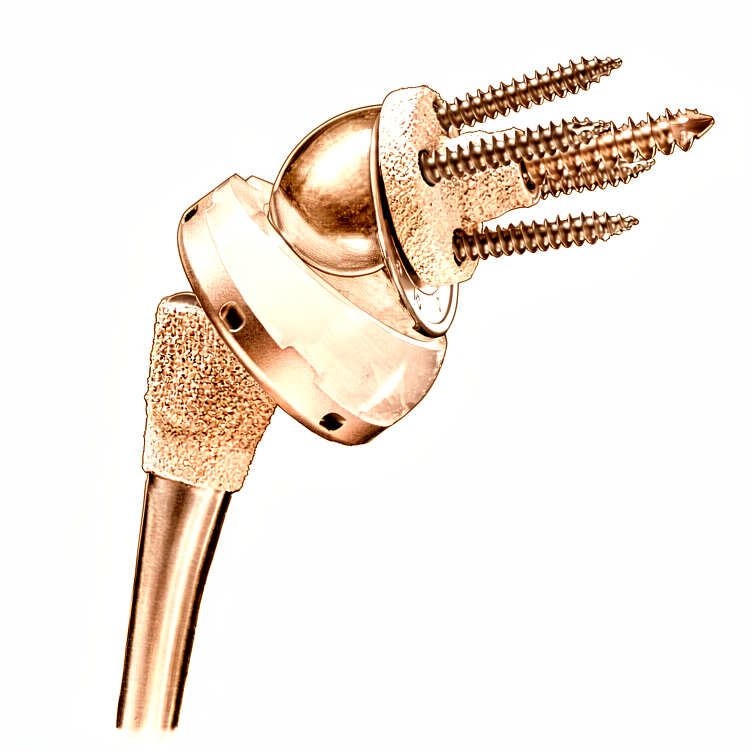
रिवर्स शोल्डर रिप्लेसमेंट जोड़ बदलने का एक ऑपरेशन है, जिसमें कंधे की सामान्य बॉल-और-सॉकेट (गेंद और कटोरी) व्यवस्था को उलट दिया जाता है, ताकि बॉल वहां आ जाए जहां सॉकेट था और सॉकेट वहां आ जाए जहां बॉल थी। हम आम तौर पर इसकी सलाह तब देते हैं जब रोटेटर कफ़, यानी टेंडनों (कंडरा) का वह समूह जो कंधे को स्थिर रखता है, बुरी तरह घिस गया हो या फट गया हो, या जब कंधे की दूसरी समस्याएं बिना ऑपरेशन वाले इलाज से ठीक न हुई हों, जैसे गतिविधियों में बदलाव, फिजियोथेरेपी, स्प्लिंट लगाना या इंजेक्शन। कुछ चोटों में तुरंत सर्जरी की सलाह दी जा सकती है। ऑपरेशन का उद्देश्य दर्द से राहत देना और आपके कंधे की हरकत और काम करने की क्षमता को बेहतर बनाना है। इसके क्लिनिकल फ़ायदे 10 साल तक बने रहते हैं। हम आपके साथ फ़ायदों और जोखिमों पर बात करेंगे, और यह फ़ैसला आप और हम मिलकर लेते हैं।
ऑपरेशन से पहले
जब आप और आपके सर्जन रिवर्स शोल्डर रिप्लेसमेंट पर सहमत हो जाते हैं, तो हम आपके ऑपरेशन की योजना बनाने के लिए ज़रूरी जांचें करवाएंगे। आम तौर पर इसका मतलब होता है आपके कंधे का एक्स-रे, और कभी-कभी सीटी स्कैन, जो एक विस्तृत स्कैन है और हड्डी की त्रि-आयामी (थ्री-डी) तस्वीर बनाता है। इन तस्वीरों से हमें नए जोड़ के हिस्सों की जगह की योजना बनाने और सर्जरी के दिन से पहले आपके कंधे को सावधानी से मापने में मदद मिलती है। आपसे आपके सामान्य स्वास्थ्य के बारे में भी पूछा जाएगा, जिसमें आपके कंधे के पिछले ऑपरेशन, स्टेरॉयड जैसी दवाएं, और ऑस्टियोपोरोसिस जैसी समस्याएं शामिल हैं, यानी हड्डियों का पतला और कमज़ोर होना। कृपया अपनी मौजूदा दवाओं की सूची अपने अपॉइंटमेंट पर साथ लाएं। अगर आप कुछ खास दवाएं लेते हैं, तो आपके सर्जन आपसे सर्जरी से पहले थोड़े समय के लिए उन्हें बंद करने को कह सकते हैं। आपको ऑपरेशन से पहले सात घंटे तक फ़ास्टिंग (खाली पेट रहना) करनी होगी; हम सामान्य से थोड़ा ज़्यादा समय मांगते हैं ताकि अगर ऑपरेशन थिएटर की सूची जल्दी चले तो आपको पहले बुलाया जा सके। किसी ऐसे व्यक्ति का इंतज़ाम करें जो बाद में आपको गाड़ी से घर ले जाए, और ढीले, आरामदायक कपड़े पहनें। अगर आपको दूसरी बीमारियां हैं, तो आपको खून की जांच करवाने या एनेस्थेटिस्ट, यानी एनेस्थीसिया देने वाले डॉक्टर, से मिलकर जांच करवाने की ज़रूरत पड़ सकती है।
ऑपरेशन के दिन
सर्जरी के दिन आप अस्पताल की सर्जिकल एडमिशन यूनिट (सर्जरी के मरीज़ों की भर्ती वाला विभाग) में आते हैं। वहीं आपकी भर्ती की जाती है और आपको ऑपरेशन थिएटर के लिए तैयार किया जाता है। आप पहले किसी वार्ड में नहीं जाते।
यह ऑपरेशन जनरल एनेस्थीसिया (जिसमें आप पूरी तरह बेहोश रहते हैं) और उसके साथ रीजनल नर्व ब्लॉक (शरीर के एक हिस्से को सुन्न करने का इंजेक्शन) देकर किया जाता है। एनेस्थेटिस्ट ऑपरेशन से पहले आपसे मिलेंगे और आपको दोनों हिस्सों के बारे में समझाएंगे।
इसके बाद आपको ऑपरेशन थिएटर में ले जाया जाता है, जहां ऑपरेशन किया जाता है। ऑपरेशन पूरा होने पर आप रिकवरी एरिया में जागते हैं। वहां नर्सें आप पर नज़र रखती हैं, जब तक एनेस्थीसिया का असर खत्म होता है। जब आपकी हालत स्थिर हो जाती है, तो आपको वार्ड में ले जाया जाता है।
ऑपरेशन में क्या किया जाता है
कंधे के जोड़ तक पहुंचने के लिए आपके सर्जन ऑपरेशन वाली जगह के ऊपर एक ही चीरा लगाते हैं। जोड़ की घिसी हुई सतहों को निकाल दिया जाता है और उनकी जगह धातु और प्लास्टिक के नए हिस्से लगाए जाते हैं। सामान्य बॉल-और-सॉकेट व्यवस्था को उलट दिया जाता है, ताकि बॉल वहां आ जाए जहां सॉकेट था और सॉकेट वहां आ जाए जहां बॉल थी। इसी वजह से इसे "रिवर्स" (उल्टा) रिप्लेसमेंट कहा जाता है। नए हिस्से इस तरह बनाए गए हैं कि आपके कंधे को ऊपर से ढकने वाली मज़बूत मांसपेशी, डेल्टॉइड, घिसे या फटे रोटेटर कफ़ टेंडनों की जगह बांह को उठा सके।
ऑपरेशन के दौरान आपके सर्जन कंधे के टेंडनों और दूसरे नरम ऊतकों के आसपास सावधानी से काम करते हैं। अगर इनमें से कुछ ऊतक अच्छी हालत में हों, तो नया इम्प्लांट अपनी जगह लग जाने के बाद उन्हें टांकों से वापस उस पर जोड़ दिया जाता है। नए हिस्सों की जगह और आकार उस योजना के अनुसार तय किए जाते हैं जो सर्जरी के दिन से पहले आपके एक्स-रे और स्कैन से बनाई गई थी।
जब नया जोड़ अपनी जगह लग जाता है, तो घाव बंद कर दिया जाता है। पहले बंद घाव के ऊपर एक बारीक, खुद चिपकने वाली जाली रखी जाती है, जो त्वचा के किनारों को आपस में जोड़े रखती है। फिर जाली के ऊपर एक तरल स्किन एडहेसिव (त्वचा पर लगाया जाने वाला गोंद) लगाया जाता है, जो जमकर पूरे हिस्से को सील कर देता है। यह लगभग एक से दो सप्ताह तक लगा रहता है और फिर अपने आप उठकर उतर जाता है, इसलिए कुछ भी निकलवाने की ज़रूरत नहीं होती।
ऑपरेशन के बाद
आप रिकवरी एरिया में जागेंगे, और हालत स्थिर होने पर आपको वार्ड में ले जाया जाएगा। वहां नर्सें आपकी देखभाल करती हैं और आपको आराम में रखने के लिए दवा देती हैं। सहारे के लिए आपकी बांह एक साधारण स्लिंग (बांह को सहारा देने वाली पट्टी) में रहती है; व्यायाम और नहाने-धोने के समय इसे उतारा जाता है। इस ऑपरेशन के बाद ज़्यादातर मरीज़ एक या दो रात अस्पताल में रहते हैं। हम ड्रेसिंग (घाव की पट्टी) को लगभग 10 दिन तक लगी रहने देते हैं; कृपया उससे पहले इसे न हटाएं, जब तक हम आपसे ऐसा करने को न कहें। जब हम आपसे मिलते हैं, तब हम इसे बदलते या हटाते हैं। आम तौर पर घर जाने से पहले एक फिजियोथेरेपिस्ट आपसे मिलकर हल्की हरकतें शुरू करवाते हैं। कृपया इंतज़ाम करें कि घर पहुंचने के बाद पहले 24 घंटे कोई आपके साथ रहे।
ठीक होने की प्रक्रिया
घर पर शुरुआती दिन आराम और सुकून के होते हैं। आपके कंधे में दर्द और सूजन रहेगी, और व्यायाम के बीच के समय में बांह को सहारा देने के लिए स्लिंग है। बताए गए तरीके से ली गई साधारण दर्द की दवा से आम तौर पर यह संभालने लायक रहता है। जैसे-जैसे सप्ताह बीतते हैं, तकलीफ़ धीरे-धीरे कम होती जाती है, और ज़्यादातर लोग पाते हैं कि जकड़न से पहले सूजन कम हो जाती है।
आपके फिजियोथेरेपिस्ट आपके व्यायाम करवाएंगे, जो हल्की हरकतों से शुरू होकर आपके कंधे की क्षमता के अनुसार बढ़ते जाएंगे। इन सत्रों के लिए और नहाने-धोने के लिए स्लिंग उतारी जाती है। जब तक ऑपरेशन वाली बांह ठीक हो रही है, आप अपने दूसरे हाथ से घर के हल्के काम कर सकेंगे। शुरुआत में सोना असुविधाजनक हो सकता है; बहुत से लोगों को कुर्सी पर टेक लगाकर या तकियों के सहारे ऊंचा होकर आराम करना आसान लगता है, जब तक लेटना फिर से आरामदायक न लगने लगे।
प्रगति के पड़ाव तय तारीखों पर नहीं, बल्कि घटनाओं के रूप में आते हैं। जब आपके सर्जन आपको गाड़ी चलाने की अनुमति दे दें, जो आम तौर पर छह सप्ताह वाली फॉलो-अप जांच में होता है, तब आप फिर से गाड़ी चला सकते हैं; हमारी अलग गाइड ऊपरी अंग की सर्जरी के बाद गाड़ी चलाना इसके बारे में बताती है। जैसे-जैसे हरकत लौटती है, कपड़े पहनना और हाथ बढ़ाकर चीज़ें लेना जैसे रोज़ के काम आसान होते जाते हैं। जब आपके फिजियोथेरेपिस्ट आपकी ताकत से संतुष्ट हों, तो आप उन गतिविधियों पर लौट सकते हैं जिन्हें आप पहले पसंद करते थे, और जो लोग इस ऑपरेशन से पहले सक्रिय थे उनमें से ज़्यादातर बाद में उन गतिविधियों पर लौट आते हैं।
ठीक होने की प्रक्रिया हर व्यक्ति में अलग होती है। आपकी समय-सीमा अलग हो सकती है, और आपके सर्जन और फिजियोथेरेपिस्ट हर कदम पर आपका मार्गदर्शन करेंगे।
क्या समस्याएं हो सकती हैं
ज़्यादातर मरीज़ अच्छी तरह ठीक हो जाते हैं, लेकिन कभी-कभी समस्याएं हो सकती हैं। किसी भी समस्या का जल्दी पता लगाने के लिए आपके सर्जन और टीम आप पर करीब से नज़र रखते हैं।
कभी-कभी नया जोड़ अपनी जगह से खिसक सकता है। आपको अचानक "खट" जैसा झटका महसूस हो सकता है, जिसके बाद दर्द होता है और कंधा सामान्य रूप से नहीं हिलता। अगर ऐसा हो, तो तुरंत क्लिनिक से संपर्क करें या इमरजेंसी विभाग जाएं।
नए जोड़ के आसपास संक्रमण (इन्फेक्शन) हो सकता है। इन बातों पर ध्यान दें: गहरा, धड़कता हुआ दर्द जो साधारण दर्द निवारक दवाओं से कम न हो, घाव से बाहर की ओर फैलती लाली, या बुखार। अगर आपको इनमें से कोई भी संकेत दिखे, तो बिना देर किए क्लिनिक को बताएं। संक्रमण का इलाज कई चरणों में किया जाता है, और शोल्डर रिप्लेसमेंट के आसपास के ज़्यादातर संक्रमण इस तरीके से खत्म किए जा सकते हैं।
नए हिस्से समय के साथ ढीले हो सकते हैं। यह आम तौर पर ऐसे दर्द के रूप में सामने आता है जो लौट आता है या बढ़ जाता है, कभी-कभी कट-कट की आवाज़ या रगड़ के एहसास के साथ। अपनी अगली फॉलो-अप जांच में इसके बारे में बताएं ताकि एक्स-रे से इसकी जांच की जा सके।
नर्व (तंत्रिका) कंधे के पास से होकर गुज़रती हैं, और सर्जरी के दौरान उन्हें चोट लग सकती है या वे परेशान (इरिटेट) हो सकती हैं। आपको बांह में सुन्नपन, झुनझुनी या कमज़ोरी महसूस हो सकती है। अक्सर यह अपने आप ठीक हो जाता है, लेकिन अपनी फॉलो-अप जांच में इसके बारे में बताएं ताकि इस पर नज़र रखी जा सके।
नए जोड़ के आसपास की हड्डी में, या कंधे के सबसे ऊपर वाले हड्डी के उभरे हिस्से में, छोटे फ्रैक्चर (हड्डी टूटना) हो सकते हैं। आपको तेज़ दर्द महसूस होगा, अक्सर गिरने के बाद या ज़ोर से धक्का देने के बाद। अगर ऐसा हो तो क्लिनिक से संपर्क करें।
कंधे की सर्जरी के बाद कभी-कभी खून का थक्का बन सकता है, जिससे आम तौर पर पिंडली में अचानक सूजन और छूने पर दर्द होता है। अगर आपको ऐसा दिखे, तो बिना देर किए डॉक्टरी मदद लें।
कुछ बातें समस्याओं की संभावना बढ़ा देती हैं। ऊपरी बांह की हड्डी टूटने पर यह ऑपरेशन कराने वाले पुरुषों में जटिलताओं (कॉम्प्लिकेशन) की संभावना ज़्यादा होती है। अगर पहले रोटेटर कफ़ रिपेयर (टेंडन को वापस जोड़ना) हो चुका हो, तो भी नए जोड़ के आसपास संक्रमण की संभावना बढ़ जाती है। अगर कंधे में स्टेरॉयड इंजेक्शन लगा हो, तो सर्जरी से पहले कम से कम 4 सप्ताह इंतज़ार करना होता है, और हम उसी के अनुसार योजना बनाएंगे।
अगर आप विस्तृत आंकड़े जानना चाहते हैं, तो इस पेज पर दी गई जटिलताओं की तालिका में आम दरें दी गई हैं।
हमें कब फ़ोन करें
इस ऑपरेशन के बाद ज़्यादातर समस्याएं ऐसे चेतावनी संकेतों के साथ सामने आती हैं जिन्हें आप घर पर पहचान सकते हैं। अगर आपको बुखार हो, घाव के आसपास लाली या रिसाव बढ़ रहा हो, या दर्द कम होने के बजाय लगातार बढ़ता जा रहा हो, तो हमें फ़ोन करें। अगर आपको अचानक तेज़ दर्द हो, आपका कंधा अपने सामान्य आकार से बिगड़ा हुआ दिखे, या आप बांह हिला न सकें, तो इमरजेंसी विभाग जाएं। पिंडली में सूजन के साथ छूने पर दर्द, या सांस फूलना, इनके लिए तुरंत इमरजेंसी इलाज की ज़रूरत है। अगर आपकी बांह या हाथ में कुछ महसूस होना बंद हो जाए, या नया सुन्नपन और झुनझुनी हो जो ठीक न हो, तब भी हमें फ़ोन करें।
विस्तार से
Advanced reading: the deeper science (optional)
यह हिस्सा उससे आगे की जानकारी देता है जो आपको अपने इलाज के फ़ैसलों के लिए चाहिए। रिवर्स शोल्डर रिप्लेसमेंट के बारे में ज़्यादा पढ़ना फ़ायदेमंद है, क्योंकि यही वह ऑपरेशन है जिसने एक ऐसी समस्या का हल निकाला जिसे पहले हल न हो सकने वाला माना जाता था, यानी ऐसा कंधा जिसमें रोटेटर कफ़ काम ही नहीं करता, और क्योंकि इसकी खास जटिलता ऐसी है जिसके बारे में ज़्यादातर मरीज़ों ने कभी सुना नहीं होता।
जोड़ को उलटना क्यों काम करता है
सामान्य कंधे में बांह पर एक बॉल और कंधे की चपटी हड्डी (स्कैपुला) पर एक सॉकेट होता है, और यह इस बात पर निर्भर करता है कि जब डेल्टॉइड बांह उठाती है तब रोटेटर कफ़ बॉल को बीच में टिकाए रखे। जहां कफ़ बचा ही नहीं है, वहां सामान्य (कन्वेंशनल) रिप्लेसमेंट को स्थिर रखने वाला कुछ नहीं होता और बॉल बस ऊपर की ओर खिसक जाती है।
रिवर्स डिज़ाइन इन्हें आपस में बदल देता है: बॉल कंधे की चपटी हड्डी पर और सॉकेट बांह पर लगाया जाता है। इससे घूमने का केंद्र अंदर और नीचे की ओर खिसक जाता है, जिससे डेल्टॉइड का लीवर आर्म (उठाने में मदद करने वाली दूरी) लंबा हो जाता है और वह अकेले ही बांह उठा पाती है। यह जैविक नहीं बल्कि यांत्रिक (मैकेनिकल) हल है: यह किसी चीज़ की मरम्मत नहीं करता, बल्कि बची हुई मांसपेशी को काम के लिए काफ़ी बना देता है।
इसी से यह भी समझ आता है कि यह ऑपरेशन क्या-क्या वापस लाता है। बांह को सिर के ऊपर तक उठाना आम तौर पर अच्छी तरह लौट आता है। घुमाव उन मांसपेशियों पर निर्भर करता है जिन्हें यह ऑपरेशन नहीं बदलता, इसलिए पीठ के पीछे हाथ ले जाना अक्सर सीमित रहता है, और यह बात सर्जरी से पहले की उम्मीदों में शामिल होनी चाहिए, न कि बाद में निराशा बने।
जहां इसने पुराने ऑपरेशनों की जगह ले ली है
फ्रैक्चर के मामले में यह बदलाव निर्णायक रहा है। 228,523 मरीज़ों के आंकड़ों को मिलाकर देखने पर, ऊपरी बांह की हड्डी के ऊपरी सिरे के फ्रैक्चर (प्रॉक्सिमल ह्यूमरस फ्रैक्चर) वाले बुज़ुर्ग मरीज़ों में, रिवर्स टोटल शोल्डर आर्थ्रोप्लास्टी से हेमीआर्थ्रोप्लास्टी (केवल बॉल वाला हिस्सा बदलना) की तुलना में बेहतर कार्यक्षमता के नतीजे और जटिलताओं की बेहतर दरें मिलीं, और प्लेट लगाकर हड्डी जोड़ने की तुलना में दोबारा ऑपरेशन (रिवीज़न) के मामले में स्थिति ज़्यादा अनुकूल रही [1]।
इम्प्लांट से जुड़ी एक बात जानने लायक है, क्योंकि यह अक्सर पूछी जाती है: इन फ्रैक्चर के लिए रिवर्स रिप्लेसमेंट में सामान्य (स्टैंडर्ड) हिस्सों की तुलना फ्रैक्चर के लिए खास तौर पर बने हिस्सों से करने पर, 436 मरीज़ों में क्लिनिकल नतीजों या जटिलताओं की दरों में कोई खास अंतर नहीं मिला [2]।
जिस जटिलता के बारे में जानना चाहिए
यहां जो जटिलता मायने रखती है, वह है एक्रोमियन (कंधे के सबसे ऊपर का हड्डी का सिरा) या स्कैपुला के उभरे हुए किनारे (स्कैपुलर स्पाइन) का स्ट्रेस फ्रैक्चर (दबाव से आई दरार), यानी जोड़ के ऊपर की हड्डी में डेल्टॉइड के उस बढ़े हुए तनाव से दरार आ जाना जिसे यह डिज़ाइन जानबूझकर पैदा करता है। यह सीधे उसी तंत्र का नतीजा है जिसकी वजह से यह ऑपरेशन काम करता है।
एक मेटा-एनालिसिस (कई अध्ययनों का साथ में विश्लेषण) में जोखिम के कारण पहचाने गए: जिन मरीज़ों को ये फ्रैक्चर हुए, वे ज़्यादा उम्र के थे और उनका बीएमआई (लंबाई के हिसाब से वज़न) कम था, और दूसरे जोखिमों में ऑस्टियोपोरोसिस, सूजन वाला गठिया (इन्फ्लेमेटरी आर्थराइटिस), महिला होना और पहले हुआ रोटेटर कफ़ रिपेयर शामिल थे [3]। एक अलग समीक्षा में पाया गया कि शोध-साहित्य में इन फ्रैक्चर को एक जैसे तरीके से दर्ज नहीं किया जाता, और इन्हें परिभाषित करने, पहचानने और गिनने के तरीकों में काफ़ी अंतर है [4], इसलिए बताई गई दरों को सावधानी से देखना चाहिए।
इसके मुकाबले, कम समय की तस्वीर भरोसा दिलाने वाली है: पहले 90 दिनों में मृत्यु और मेडिकल व सर्जिकल जटिलताओं की कुल दरें कम हैं, और केवल 6% मरीज़ों को दोबारा अस्पताल में भर्ती होना पड़ा [5]।
दो बातें जिनका प्रमाण समर्थन नहीं करते
यह कि सबस्कैपुलैरिस का रिपेयर ज़रूरी है। 267 मरीज़ों में, रिवर्स रिप्लेसमेंट के बाद सबस्कैपुलैरिस (कंधे के सामने वाली रोटेटर कफ़ की मांसपेशी) का रिपेयर करने या न करने से बांह को बगल में उठाने (एब्डक्शन), अंदर की ओर घुमाने या बाहर की ओर घुमाने की ताकत में कोई अंतर नहीं था, हालांकि लेखक बताते हैं कि ताकत के नतीजों पर शोध सीमित है [6]।
यह कि ऑपरेशन किस वजह से हो रहा है, इससे नतीजे पर असर नहीं पड़ता। असर पड़ता है। दो या उससे ज़्यादा साल बाद, कॉन्स्टेंट स्कोर (कंधे के काम को मापने का एक मानक स्कोर) उन मरीज़ों में काफ़ी ज़्यादा थे जिनका रिवर्स रिप्लेसमेंट प्राइमरी ऑस्टियोआर्थराइटिस के लिए हुआ और जिनका कफ़ सही-सलामत था, उनकी तुलना में जिनके प्राइमरी ऑस्टियोआर्थराइटिस के साथ कफ़ फटा हुआ था या जिन्हें सेकेंडरी ऑस्टियोआर्थराइटिस था [7]। एक ही ऑपरेशन के नतीजे इस पर निर्भर करते हैं कि आपको इसकी ज़रूरत क्यों पड़ी, और जब आपको कोई औसत आंकड़ा बताया जाए, तब यह बात जानने लायक है।
संदर्भ
[1] Mekhail J, Mullan R, Cross JL, Jahagirdar O, Luo X, Salameh M. Outcomes of reverse total shoulder arthroplasty vs. other surgical fixation methods for proximal humerus fractures: a systematic review and meta-analysis. JSES Rev Rep Tech. 2026;6(2):100644. https://doi.org/10.1016/j.xrrt.2025.100644
[2] Apivatgaroon A, Kongmalai T, Kongmalai P. Standard compared with fracture-specific components in reverse shoulder arthroplasty for proximal humeral fractures: a systematic review and meta-analysis. Bone Joint J. 2025;107-B(9):931-41. https://doi.org/10.1302/0301-620X.107B9.BJJ-2024-1508.R2
[3] Elmenawi KA, Sperling JW, Sanchez-Sotelo J, Barlow JD. Risk factors for acromial and scapular fractures following reverse shoulder arthroplasty: a meta-analysis. JSES Rev Rep Tech. 2026;6(1):100578. https://doi.org/10.1016/j.xrrt.2025.08.015
[4] Davie RA, Nathan K, Persaud SG, Oladeji LO, Taylor SA, Dines JS, et al. Inconsistent reporting of risk factors for acromial stress fractures following reverse total shoulder arthroplasty: a systematic review. J Shoulder Elbow Surg. 2025;34(11):e975-e984. https://doi.org/10.1016/j.jse.2025.02.032
[5] Kent LM, Hurley ET, Davey MS, Klifto CS, Mullett H. Low complication rate following reverse total shoulder arthroplasty at 90-days follow-up: a systematic review. J ISAKOS. 2024;9(2):205-10. https://doi.org/10.1016/j.jisako.2023.11.006
[6] Preuss FR, Eble SK, Peebles AM, Osuna-Garcia A, Provencher CMT. Shoulder strength outcomes after reverse total shoulder arthroplasty: a systematic review. JSES Rev Rep Tech. 2022;2(2):131-4. https://doi.org/10.1016/j.xrrt.2021.11.004
[7] Nové-Josserand L, Nerot C, Colotte P, Guery J, van Rooij F, Hibon A, et al. Reverse shoulder arthroplasty for primary glenohumeral osteoarthritis: significantly different characteristics and outcomes in shoulders with intact vs. torn rotator cuff. J Shoulder Elbow Surg. 2024;33(4):850-62. https://doi.org/10.1016/j.jse.2023.07.027
Evidence & references
This is the clinical evidence summary written for health professionals. It is technical, and it lists the research this page was built from. You do not need to read it to understand your treatment or to make a decision about it.
Anatomy & Pathophysiology
Glenoid and Scapular Anatomy
- The glenoid is suspended from the body of the scapula by the neck and fixed to the clavicle by the acromioclavicular and coracoclavicular ligaments [3].
- As the face of the glenoid transitions into the neck, the glenoid vault narrows [3].
- The scapular spine is subcutaneous posteriorly and widens gradually as it transitions into the base of the acromion laterally [3].
- The acromion curves anteriorly and meets the clavicle at the acromioclavicular joint and the coracoid via the coracoacromial ligament, which originates under the anterior margin of the acromion [3].
- The suprascapular nerve arises from the C4–C5 nerve roots off of the supraclavicular brachial plexus at “Erb’s point” [3].
- The suprascapular nerve runs just medial to the base of the coracoid, under the transverse scapular ligament within the suprascapular notch, and gives off branches to the supraspinatus within 1 cm of the notch [3].
- The suprascapular nerve continues through the supraspinatus fossa heading laterally and distally on the under surface of the supraspinatus [3].
- The suprascapular nerve runs under the ill-defined spinoglenoid ligament around the lateral base of the scapula within spinoglenoid notch before terminating in posterior capsular sensory branches heading laterally and an infraspinatus motor branch heading medially within 1 cm of the lateral margin of the scapular spine [3].
- Cadaver studies show the suprascapular nerve is present 29 mm (23 to 35 mm) from the superior rim of the glenoid at the suprascapular notch [3].
- Cadaver studies show the suprascapular nerve is present 18 mm (14 to 24 mm) from the posterior rim at the spinoglenoid notch [3].
- Injury to the suprascapular nerve can cause pain and denervation of the supraspinatus and infraspinatus [3].
Biomechanics and Pathophysiology
- Anatomic total shoulder arthroplasties and hemiarthroplasties are dependent on the rotator cuff to center the head in the glenoid and to optimize larger periscapular muscle function [7].
- In the absence of a functional rotator cuff, the deltoid pulls the humerus proximally, shearing along the glenoid and resulting in eccentric glenoid wear, acetabularization of the acromion, and pseudoparalysis [7].
- The reverse shoulder arthroplasty does not require the rotator cuff for function but is dependent on an intact deltoid neuromuscular unit [7].
- The semiconstrained nature of the reverse prosthesis provides a stable fulcrum that allows the deltoid to elevate the shoulder even in the absence of a functional rotator cuff [4].
- In the Grammont reverse prosthesis, the center of rotation is medial to the glenoid component–bone interface to decrease shear stress and provide compressive stress, aiming to decrease the chances of glenoid loosening [4].
- The humeral component in the Grammont design is inset, resting almost completely inside the proximal humerus metaphysis [4].
- The opening angle of the polyethylene in the Grammont design is relatively horizontal at 155 degrees compared with conventional arthroplasty [4].
- Once articulated, the humerus is more medial and more distal than preoperatively, providing a mechanical advantage to the deltoid for active elevation in the absence of a rotator cuff [4].
- The traditional Grammont style decreases shear forces seen by the glenoid and lowers baseplate failure by medializing the center of rotation, but this is associated with the risk of inferior scapular notching in adduction [7].
- Distalization doubles the lever arm of the deltoid and optimizes the length–tension curve of its sarcomeres, increasing its efficiency by 30% at the cost of rotational strength [7].
- Lateralized glenosphere and lateralized humerus designs have gained popularity to improve the rotational profile, deltoid function, implant stability, and decrease impingement such as scapular notching [7].
- Early reverse designs had a high failure rate due to the profound lever arm on the glenoid and baseplate bone [7].
- Recent reverse designs have a better track record but increased forces are seen by the scapula and acromion [7].
Periprosthetic Scapular Fracture Pathophysiology
- Periprosthetic scapular fractures are universally associated with stable glenoid implants [2].
- Fracture is unlikely in the face of dislocation, glenosphere dissociation, or baseplate pullout at the bone–baseplate interface [2].
- Periprosthetic scapular fracture has been noted to result in new glenohumeral instability due to the change of the orientation of the glenosphere and loss of deltoid tension [2].
- Postoperative periprosthetic scapular fracture is a challenge unique to reverse shoulder arthroplasty and occurs more commonly than humeral fractures [7].
- Postoperative periprosthetic scapular fractures are an effect of nonphysiologic forces transferred from the implant to the scapula, often in a suboptimal host [7].
- Fatigue fracture has been found to occur through already weakened acromiums or those with preexisting lesions [7].
- Acromial thinning and eventual fragmentation occur at the final stages of rotator cuff-tear arthropathy as the humeral head acetabularizes [7].
Clinical Presentation
Periprosthetic Scapular Fractures
- The weak point in periprosthetic scapular fracture injuries is the scapular bone [2].
- Periprosthetic scapular fractures are unlikely in the presence of dislocation, glenosphere dissociation, or baseplate pullout at the bone–baseplate interface [2].
- Periprosthetic scapular fractures have been noted to rarely result in new glenohumeral instability [2].
- New glenohumeral instability following periprosthetic scapular fracture is due to a change in the orientation of the glenosphere and loss of deltoid tension [2].
- Diagnosis of periprosthetic fractures is often subtle and requires a high index of suspicion [2].
- Workup for periprosthetic fractures should begin with a complete history and examination [2].
- Past medical history should elucidate the underlying diagnosis for the index surgery and subsequent surgeries [2].
- Complications including infection should be accounted for in the past medical history [2].
- The examiner should understand the patient's shoulder function and level of disability before surgery, after surgery, and at present [2].
- The examiner should understand the time course of changes in shoulder function and disability [2].
- In the case of a stress reaction, new pain at the base of the acromion may be the only finding [2].
- New pain at the base of the acromion in a stress reaction should raise suspicion and spark further imaging or a period of rest [2].
- Stress fractures can be more painful than after they propagate into a displaced fracture [2].
- Patients typically present around their 8th decade of life after a sudden increase in pain or loss of function [2].
- Patients typically present after a sudden increase in pain or loss of function in an otherwise smooth postoperative course [2].
- Presentation for periprosthetic scapular fracture is generally within 1 year but up to 2 years from surgery [2].
- Patients who go on to have periprosthetic scapular fractures initially outperform those who do not [2].
- Past medical history must identify risk factors including a history of steroid use, osteoporosis, subacromial decompression, or rotator cuff tear arthropathy [2].
- Previous operative reports, clinic notes, and imaging can help provide a thorough understanding of any previous surgeries on the shoulder or history of radiation [2].
- Physical examination starts with inspection [2].
- Deformity is concerning for dislocation, hematoma, or displaced fracture [2].
- Erythema or incisional dehiscence is concerning for infection [2].
- Tenderness along the acromion or scapular spine raises suspicion for fracture which should be confirmed with imaging [2].
- A complete neurovascular examination is performed as part of the physical examination [2].
- Assessment of active and passive motion is performed as part of the physical examination [2].
- Fracture can result in motion limited by pain, new weakness, or loss of function [2].
- Infection should be investigated with laboratory tests [2].
- A sudden loss of function or increase in pain is consistent with both scapular fracture and infection [2].
- A sudden loss of function or increase in pain should trigger further workup [2].
Investigations
Periprosthetic Scapular Fracture Diagnosis and Workup
- Diagnosis of periprosthetic scapular fractures is often subtle and requires a high index of suspicion [2].
- The workup for periprosthetic scapular fractures should begin with a complete history and examination [2].
- The examiner should understand the patient's shoulder function and level of disability before surgery, after surgery, and at present, as well as the time course of these changes [2].
- New pain at the base of the acromion may be the only finding in a stress reaction and should raise suspicion for further imaging or a period of rest [2].
- Patients with periprosthetic scapular fractures typically present around their 8th decade of life after a sudden increase in pain or loss of function in an otherwise smooth postoperative course [2].
- Periprosthetic scapular fractures generally occur within 1 year but up to 2 years from surgery [2].
- Inspection for deformity is concerning for dislocation, hematoma, or displaced fracture [2].
- A complete neurovascular examination is performed as well as assessment of active and passive motion [2].
- A sudden loss of function or increase in pain is consistent with both scapular fracture and infection and should trigger further workup [2].
Preoperative Imaging and Planning
- Careful assessment of the preoperative radiographs and CT with three-dimensional reconstruction is extremely useful in preparation for surgery [5].
- The main goals of preoperative imaging include understanding the fracture pattern and anticipating the ideal height of stem implantation [5].
- Radiographs of both humeri (affected and unaffected) with magnifier markers may be used to understand where the stem should be positioned in reference to the fracture line on the humeral shaft [5].
- The glenoid should be assessed in radiographs and CT to plan for component positioning, version, inclination, and rotation, as well as anticipated screw length [5].
- Associated fractures of the rim of the glenoid in anterior or posterior fracture-dislocations are rare [5].
- Most of the times, the size of the fractured glenoid rim is small enough not to interfere with secure baseplate placement [5].
- If the size of the fractured glenoid rim is large enough to interfere with the stability of the glenoid baseplate, fixation with small fragment screws may be performed [5].
- The glenoid should be assessed on CT for glenoid component implantation [5].
- Most surgeons do not use fluoroscopy for this procedure [5].
Treatment
Indications and Rationale
- Reverse shoulder arthroplasty is the replacement procedure of choice when arthroplasty is considered for proximal humeral fractures [4].
- Tuberosity and rotator cuff-related complications are the main reason for poor functional outcome when a humeral head replacement is implanted for management of a proximal humeral fracture [4].
- Shoulder arthroplasty is considered for proximal humeral nonunion in the presence of severe cavitation and bone loss at the humeral head and metaphysis or collapse and degenerative change of the humeral articular surface [6].
- Severe tuberosity malunion in the setting of a proximal humeral nonunion is more reliably compensated for with reverse arthroplasty than with osteotomy and internal fixation [6].
- Reverse shoulder arthroplasty may improve shoulder function in patients with nonunions associated with severe tuberosity malunions [6].
Prosthesis Design and Biomechanics
- The Grammont reverse prosthesis features an articulating glenoid component shaped as a third of a sphere [4].
- In the Grammont design, the center of rotation is medial to the glenoid component–bone interface to decrease shear stress and provide compressive stress, aiming to decrease the chances of glenoid loosening [4].
- The opening angle of the polyethylene in the Grammont design is 155 degrees [4].
- Articulation of the Grammont implants results in the humerus being more medial and more distal than preoperatively, providing a mechanical advantage to the deltoid for active elevation [4].
- The 155-degree opening angle in the Grammont design was selected to decrease the chances of dislocation [4].
- The humeral component in the Grammont design was recommended to be implanted in more anteversion (0 to 10 degrees of retroversion) than conventional arthroplasty [4].
- Subsequent reverse designs with a larger portion of a sphere place the center of rotation more lateral than the Grammont prosthesis [4].
- Subsequent reverse designs with a larger portion of a sphere utilize an opening angle of 135 degrees for the humeral component [4].
- Later reverse designs introduced onlay humeral bearings that lateralize the position of the humerus without changing the center of rotation [4].
- Later reverse designs with onlay humeral bearings utilize a 145-degree opening angle for the bearing [4].
- There is very little published on reverse arthroplasty biomechanics in the setting of a proximal humeral fracture [4].
Surgical Technique and Tuberosity Management
- Some surgeons initially elected to implant a reverse arthroplasty in proximal humeral fractures without repair, or sometimes with excision, of the greater tuberosity and/or lesser tuberosity [4].
- Healing of at least the greater tuberosity in good position provides a higher chance of restoration of active external rotation [4].
- Not performing a tuberosity repair at the time of reverse arthroplasty for proximal humeral nonunion has been correlated with a higher rate of dislocation [4].
- Technical principles for reverse arthroplasty in fracture may need to be modified to enhance tuberosity healing by avoiding translating the humeral shaft too lateral or too distal, allowing the tuberosities to overlap a few millimeters with the shaft [4].
- Use of a stem with fracture-dedicated features, such as a proximal ingrowth surface, small cross section, and holes for suture fixation, may be beneficial [4].
Comparison with Hemiarthroplasty
- Hemiarthroplasty is less commonly considered than reverse arthroplasty for proximal humeral nonunion [6].
- The functional outcome of hemiarthroplasty for nonunion is particularly concerning when tuberosity osteotomies need to be added [6].
- Studies reporting on hemiarthroplasty for nonunion suggest the procedure may be effective in reducing or eliminating pain but is associated with a high rate of complications that often require further surgery and disappointing functional recovery [6].
Complications
- Scapular notching is a complication of reverse shoulder arthroplasty [1].
- The clinical impact of scapular notching on outcomes after reverse total shoulder arthroplasty has been analyzed in a study of 476 shoulders [1].
- Humeral version in reverse shoulder arthroplasty affects impingement during activities of daily living [1].
- Subscapularis tendon integrity impacts shoulder function after reverse shoulder arthroplasty [1].
- Component positioning affects the intrinsic stability of the reverse shoulder arthroplasty [1].
- Humeral component lateralization in reverse shoulder arthroplasty affects rotator cuff torque [1].
References
[1] Campbell S Operative Orthopaedics 4 Volume Set. RECONSTRUCTIVE PROCEDURES OF THE SHOULDER AND ELBOW IN ADULTS > REVERSE SHOULDER ARTHROPLASTY.
[2] Rockwood And Green S Fractures In Adults. 29: Principles of Nonunion and Bone Defect Treatment > Injuries Associated with Periprosthetic Scapular Fractures About Reverse Shoulder Arthroplasty.
[3] Rockwood And Green S Fractures In Adults. 29: Principles of Nonunion and Bone Defect Treatment > Pathoanatomy and Applied Anatomy Related to Periprosthetic Scapular Fractures About Reverse Shoulder Arthroplasty.
[4] Rockwood And Green S Fractures In Adults. 29: Principles of Nonunion and Bone Defect Treatment > Reverse Shoulder Arthroplasty.
[5] Rockwood And Green S Fractures In Adults. 29: Principles of Nonunion and Bone Defect Treatment > Preoperative Planning > Reverse Shoulder Arthroplasty for Fracture: Preoperative Planning Checklist.
[6] Rockwood And Green S Fractures In Adults. 29: Principles of Nonunion and Bone Defect Treatment > Reverse Shoulder Arthroplasty and Hemiarthroplasty.
[7] Rockwood And Green S Fractures In Adults. 29: Principles of Nonunion and Bone Defect Treatment > Periprosthetic Scapular Fractures About Reverse Shoulder Arthroplasty.