Patients › Elbow
जकड़ी हुई कोहनी को खोलने का ऑपरेशन (आर्थ्रोलाइसिस)
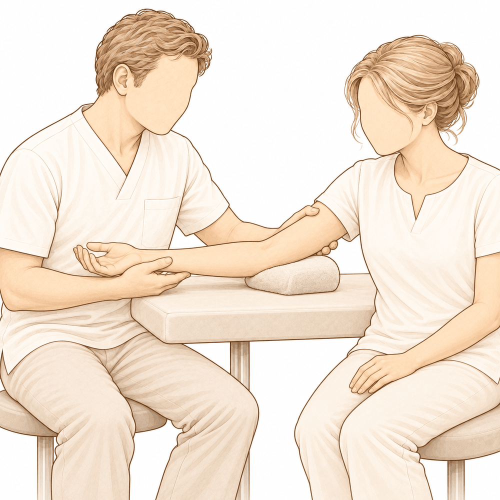
यह ऑपरेशन क्यों सुझाया गया है
डॉ. कीरन हिरपरा, माटर प्राइवेट हॉस्पिटल, रॉकहैम्पटन में ऊपरी अंग (कंधे से हाथ तक) के सर्जन हैं, और वे आपकी समस्या के हिसाब से सबसे कम चीर-फाड़ वाले विकल्पों से शुरुआत करते हैं। मरीज़ आम तौर पर अपने जीपी (आपके फैमिली डॉक्टर) के रेफ़रल से हमारे क्लिनिक आते हैं; अगर किसी फिजियोथेरेपिस्ट ने आपको हमसे मिलने की सलाह दी है, तब भी मेडिकेयर रिबेट पाने के लिए आपको अपने जीपी से रेफ़रल लेना होगा। आपके अपॉइंटमेंट पर हम आपकी बीमारी का इतिहास लेते हैं, आपकी कोहनी की जांच करते हैं और ज़रूरत हो तो इमेजिंग (एक्स-रे, स्कैन) का इंतज़ाम करते हैं। जकड़ी हुई कोहनी में अक्सर समय और इस्तेमाल के साथ सुधार हो जाता है, इसलिए हम आम तौर पर पहले बिना ऑपरेशन वाला इलाज आज़माते हैं। इसमें 6 से 12 सप्ताह तक फिजियोथेरेपी, स्प्लिंट या इंजेक्शन शामिल हो सकते हैं। जब इस इलाज से पर्याप्त सुधार नहीं होता, तब सर्जरी पर विचार किया जाता है।
जकड़ी हुई कोहनी को खोलने का ऑपरेशन, जिसे आर्थ्रोलाइसिस भी कहते हैं, एक ऐसा ऑपरेशन है जिसमें आपकी कोहनी के आसपास के कसे हुए, निशान वाले ऊतक को ढीला करके मुक्त किया जाता है, ताकि जोड़ फिर से हिल-डुल सके। हम इसकी सलाह तब देते हैं जब बिना ऑपरेशन वाले इलाज के बाद भी जकड़न आपको सीमित कर रही हो। यह तब सबसे अच्छा काम करता है जब जोड़ की सतह अब भी चिकनी हो और अपने सामान्य आकार में हो। इसका उद्देश्य हरकत में लंबे समय तक टिकने वाला सुधार और कम दर्द है, ताकि रोज़ के काम आसान हो जाएं।
ऑपरेशन से पहले
सर्जरी तय हो जाने के बाद कुछ व्यावहारिक बातों का इंतज़ाम करना होता है। आपको बताया जाएगा कि कब खाना-पीना बंद करना है: यह आपके ऑपरेशन से सात घंटे पहले है। हम छह के बजाय सात घंटे मांगते हैं, ताकि अगर ऑपरेशन थिएटर की सूची जल्दी चल रही हो तो हम आपको पहले बुला सकें। आपके सर्जन बताएंगे कि आपकी रोज़ की कौन-सी दवाएं कब बंद करनी हैं, और आपको उन सबकी पूरी सूची अपने साथ लानी चाहिए। ऑपरेशन के बाद आपको घर ले जाने के लिए किसी का इंतज़ाम करें। ऑपरेशन के दिन ढीले, आरामदायक कपड़े पहनें। ऑपरेशन की योजना बनाने के लिए हमेशा एक्स-रे लिए जाते हैं, और अगर जकड़न में हड्डी शामिल हो तो सीटी स्कैन का इस्तेमाल किया जा सकता है। अगर आपको दूसरी बीमारियां हैं, तो आपको खून की जांच या एनेस्थेटिस्ट (बेहोशी के डॉक्टर) से जांच करवाने की ज़रूरत पड़ सकती है।
ऑपरेशन के दिन
आप अस्पताल की सर्जिकल एडमिशन यूनिट में पहुंचते हैं, जहां आपका नाम दर्ज किया जाता है और आपको ऑपरेशन थिएटर के लिए तैयार किया जाता है। वहां आप एनेस्थेटिस्ट से मिलते हैं। यह ऑपरेशन जनरल एनेस्थीसिया (जिसमें आप पूरी तरह बेहोश रहते हैं) में किया जाता है। ऑपरेशन के बाद दर्द से राहत के लिए कभी-कभी रीजनल नर्व ब्लॉक (हाथ या बांह को सुन्न करने का इंजेक्शन) भी जोड़ा जाता है; एनेस्थेटिस्ट उसी दिन आपसे इस पर बात करेंगे। इसके बाद आपको ऑपरेशन थिएटर में ले जाया जाता है, जहां ऑपरेशन किया जाता है।
आप रिकवरी एरिया में होश में आते हैं, जहां एनेस्थीसिया का असर खत्म होने तक नर्सें आपकी निगरानी करती हैं। जब आपकी हालत स्थिर हो जाती है, तो प्रक्रिया और आपकी रिकवरी के आधार पर आप या तो वार्ड में जाते हैं या घर चले जाते हैं।
ऑपरेशन में क्या किया जाता है
जकड़ी हुई कोहनी को खोलने के एक से ज़्यादा तरीके हैं, और आपके सर्जन वह तरीका चुनेंगे जो आपकी कोहनी के लिए सही हो। कुछ ऑपरेशन दूरबीन से ऑपरेशन (आर्थ्रोस्कोपी) के ज़रिए किए जाते हैं, जिसमें कुछ छोटे चीरे और एक पतला कैमरा इस्तेमाल होता है। दूसरे ऑपरेशन कोहनी के भीतरी या बाहरी तरफ़ एक या दो खुले चीरों से किए जाते हैं, जो कभी-कभी लगभग 3 से 5 सेमी लंबे होते हैं। बहुत ज़्यादा जकड़ी हुई कोहनियों में, दोनों तरफ़ पहुंचने के लिए कोहनी के पीछे एक चीरा लगाया जा सकता है।
अंदर पहुंचकर आपके सर्जन कसे हुए कैप्सूल को ढीला करते हैं, जो जोड़ के चारों ओर ऊतक की एक आस्तीन जैसी परत है। हरकत को रोकने वाले हड्डी के उभार (बोन स्पर) काटकर हटा दिए जाते हैं, और जोड़ में तैर रहे हड्डी या कार्टिलेज (उपास्थि) के किसी भी ढीले टुकड़े को निकाल दिया जाता है। अगर कोहनी के भीतरी तरफ़ की किसी नर्व (तंत्रिका) पर निशान वाले ऊतक (स्कार टिश्यू) से दबाव पड़ रहा हो, तो उसे ऊतक की उस सुरंग से मुक्त किया जा सकता है, या किसी नई जगह पर खिसकाया जा सकता है जहां वह खिंच न सके। अगर जोड़ की सतह खुद बुरी तरह घिस चुकी हो, तो दूसरे विकल्प भी हैं, जैसे हड्डी पर नरम ऊतक की परत लगाना, या जोड़ की सतहों को धातु और प्लास्टिक के हिस्सों से बदलना। कभी-कभी कोहनी पर एक कब्ज़ेदार (हिंज वाला) फ्रेम लगाया जाता है, ताकि ठीक होते समय जोड़ अच्छी स्थिति में रहे।
आखिर में चीरों को टांकों से बंद किया जाता है और उन पर ड्रेसिंग (घाव की पट्टी) लगाई जाती है। जैसा कि ठीक होने वाले हिस्से में बताया गया है, आप यह ड्रेसिंग लगभग 10 दिन तक लगी रहने देंगे।
ऑपरेशन के बाद
आप रिकवरी एरिया में होश में आते हैं, फिर वार्ड में जाते हैं। नर्सें आपकी कोहनी, आपके हाथ और आप कैसा महसूस कर रहे हैं, इसकी जांच करेंगी। आपकी ज़रूरत के हिसाब से दर्द से राहत की दवा दी जाती है; अगर आपकी कोहनी में तकलीफ़ हो तो नर्सों को बताएं। आपकी कोहनी एक नरम ड्रेसिंग में लिपटी होगी, और आराम के लिए आप अपनी बांह स्लिंग (बांह को सहारा देने वाली पट्टी) में रखकर घर जा सकते हैं। आप जल्दी ही बिस्तर से उठकर चल-फिर सकते हैं, और पहली बार नर्सें आपकी मदद करेंगी। जब तक आपकी कोहनी शांत होती है, आपका हाथ, कलाई और कंधा आज़ादी से हिल-डुल सकते हैं। घर जाने के बाद पहले 24 घंटे किसी को आपके साथ रहना चाहिए। आपकी टीम आपको बताएगी कि आप उसी दिन घर जाएंगे या एक रात अस्पताल में रुकेंगे। हम ड्रेसिंग को लगभग 10 दिन तक लगी रहने देते हैं; कृपया तब तक इसे न हटाएं, जब तक हम आपसे ऐसा करने को न कहें। जब हम आपसे मिलेंगे, तब हम इसे बदलेंगे या हटाएंगे।
ठीक होने की प्रक्रिया
पहले कुछ दिनों और सप्ताहों तक आपकी कोहनी में दर्द और सूजन रहेगी। जकड़न खोलने के ऑपरेशन के बाद यह सामान्य है। आराम करने, अपने हाथ को ऊपर उठाकर रखने और दर्द निवारक दवाएं बताए अनुसार लेने से तकलीफ़ कम होगी। सूजन धीरे-धीरे कम होती है, और उसके साथ कोहनी आम तौर पर कम कसी हुई महसूस होती है।
आप आराम के लिए अपनी बांह स्लिंग में रखकर घर जाएंगे। जब इससे मदद मिलनी बंद हो जाए, तो आप इसका इस्तेमाल बंद कर सकते हैं। आपका हाथ, कलाई और कंधा शुरू से ही आज़ादी से हिलते-डुलते रहने चाहिए। ऑपरेशन के बाद हैंड थेरेपी एक्सटेंड रिहैबिलिटेशन में रूबी डूलन के साथ होती है। रूबी आपके व्यायामों में मार्गदर्शन करेंगी और आपको ज़रूरत का कोई भी स्प्लिंट बनाएंगी। व्यायाम ही आपकी रिकवरी का मुख्य काम हैं, और उन्हें बताए अनुसार करना बाकी किसी भी चीज़ से ज़्यादा मायने रखता है। आप ड्रेसिंग को लगभग 10 दिन तक लगी रहने देंगे, और जब हम आपसे मिलेंगे, तब हम इसे बदलेंगे या हटाएंगे।
रोज़ के काम धीरे-धीरे, चरणों में वापस आएंगे। शुरुआत में आपको खाना बनाने, कपड़े पहनने और सामान उठाकर ले जाने में मदद की ज़रूरत होगी। जैसे-जैसे हरकत लौटेगी और सूजन कम होगी, आप ऑपरेशन वाली बांह से ज़्यादा काम कर पाएंगे। जब आपके सर्जन आपको गाड़ी चलाने की अनुमति दे दें, तब आप फिर से गाड़ी चला सकते हैं; ऊपरी अंग की सर्जरी के बाद गाड़ी चलाना पर हमारी गाइड लागू होने वाले नियम बताती है, जिनमें यह भी शामिल है कि जब तक आपकी बांह स्लिंग में है तब तक गाड़ी न चलाएं, और तेज़ दर्द निवारक दवाएं बंद हो चुकी हों।
ठीक होने की प्रक्रिया हर व्यक्ति में अलग होती है। आपकी समय-सीमा अलग हो सकती है, और आपके सर्जन और थेरेपिस्ट पूरे रास्ते आपका मार्गदर्शन करेंगे।
क्या समस्याएं हो सकती हैं
ज़्यादातर मरीज़ ठीक रहते हैं, लेकिन कभी-कभी समस्याएं हो सकती हैं। आपके सर्जन और टीम किसी भी समस्या को जल्दी पकड़ने के लिए आप पर करीब से नज़र रखते हैं।
कोहनी जितने लंबे समय से जकड़ी हुई है, जकड़न खोलने के ऑपरेशन के बाद समस्याओं की संभावना उतनी ही ज़्यादा होती है। अगर आपकी कोहनी लंबे समय से जकड़ी हुई है, तो आपके फ़ैसला करने से पहले आपके सर्जन इस बारे में आपसे बात करेंगे।
कुछ लोगों को ऑपरेशन के बाद छोटी उंगली और अनामिका में नई झुनझुनी, सुन्नपन या कमज़ोरी महसूस होती है। यह कोहनी के भीतरी तरफ़ की नर्व से आता है। यह पहले से बताना हमेशा संभव नहीं होता कि किसे ऐसा होगा। अगर आपको ऐसा महसूस हो, तो अपनी अगली फॉलो-अप जांच में इसके बारे में बताएं, या अगर यह अचानक शुरू हो तो क्लिनिक को फ़ोन करें।
पहले चोट खा चुकी कुछ कोहनियों में ऑपरेशन के बाद गहरे संक्रमण (इन्फेक्शन) की संभावना ज़्यादा होती है। घाव पर नज़र रखें कि कहीं उससे बाहर की ओर फैलती लाली, गर्माहट, बढ़ती सूजन, या गहरा, धक-धक करता दर्द तो नहीं है जो सामान्य दर्द निवारक दवाओं से कम नहीं होता। आपको बुखार जैसा या आम तौर पर अस्वस्थ महसूस हो सकता है। अगर आपको इनमें से कोई भी लक्षण दिखे, तो तुरंत क्लिनिक को फ़ोन करें, या अगर क्लिनिक का समय खत्म हो चुका हो तो इमरजेंसी विभाग जाएं।
अगर आपके ऑपरेशन में कोहनी पर कब्ज़ेदार फ्रेम लगाया जाता है, तो कुछ अतिरिक्त बातें जानना ज़रूरी है। फ्रेम से सर्जरी के दौरान खून का नुकसान, ऑपरेशन थिएटर में लगने वाला समय और अस्पताल में रहने का समय बढ़ जाता है। इससे जोड़ के ढीला या अस्थिर हो जाने की थोड़ी संभावना भी रहती है। आपके सर्जन समझाएंगे कि आपके अस्पताल में रहने और आपकी रिकवरी के लिए इसका क्या मतलब है।
अगर इस कोहनी पर पहले भी ऑपरेशन हो चुके हैं, तो गहरे संक्रमण की संभावना और भी ज़्यादा होती है। यही बात तब भी लागू होती है जब ऑपरेशन खुद ज़्यादा जटिल हो। आपके सर्जन पहले से ही आपके साथ इन सब बातों को तौलते हैं।
कोहनी की पुरानी चोटें जोड़ के काम करने के तरीके को भी बदल सकती हैं। निशान वाली त्वचा, गलत जगह पर जुड़ी हड्डियां, खराब हुआ कार्टिलेज, अतिरिक्त हड्डी, ढीले या कसे हुए लिगामेंट (स्नायुबंधन), दबी हुई नर्व और निशान वाली मांसपेशी, ये सब कोहनी के हिलने-डुलने के तरीके को बदल सकते हैं। आपके ऑपरेशन की योजना बनाते समय आपके सर्जन इन पर ध्यान देंगे।
अगर आप ठीक-ठीक आंकड़े जानना चाहते हैं, तो इस पेज पर दी गई जटिलताओं की तालिका में सामान्य दरें दी गई हैं।
हमें कब फ़ोन करें
अगर आपको बुखार हो, घाव के आसपास लाली फैल रही हो, घाव से रिसाव हो, या दर्द लगातार बढ़ता जा रहा हो, तो हमें फ़ोन करें। अगर आपको अचानक तेज़ दर्द हो, पिंडली में सूजन या दर्द हो, या सांस फूल रही हो, तो इमरजेंसी विभाग जाएं। अगर आपका हाथ या उंगलियां सुन्न या फीकी (सफ़ेद पड़ी हुई) हो जाएं, या आप अपनी बांह न हिला पाएं, तो तुरंत हमें फ़ोन करें। अगर छोटी उंगली और अनामिका में झुनझुनी या सुन्नपन अचानक शुरू हो, तो अपनी अगली फॉलो-अप जांच का इंतज़ार करने के बजाय क्लिनिक को फ़ोन करें।
विस्तार से
Advanced reading: the deeper science (optional)
यह हिस्सा आपके अपने इलाज के फ़ैसलों के लिए ज़रूरी जानकारी से आगे जाता है। जकड़ी हुई कोहनी को खोलने के ऑपरेशन के बारे में यह अतिरिक्त जानकारी पढ़ना इसलिए फ़ायदेमंद है क्योंकि प्रमाण एक असामान्य दिशा की ओर इशारा करते हैं: ज़्यादातर मरीज़ जिस चुनाव के बारे में पूछते हैं, यानी दूरबीन से या खुला ऑपरेशन, वह इस बात से कम मायने रखता दिखता है कि कितनी सर्जरी की जाती है, और इस ऑपरेशन का असली विरोधी वही प्रक्रिया है जिसने सबसे पहले जकड़न पैदा की थी।
खुला या दूरबीन वाला, यह ज़रूरी सवाल नहीं है
चोट के बाद कोहनी की जकड़न वाले 4,311 मरीज़ों की एक व्यवस्थित समीक्षा और मेटा-एनालिसिस ने खुले और दूरबीन वाले आर्थ्रोलाइसिस की तुलना की और कुल नतीजे एक जैसे पाए [1]। इसका निष्कर्ष यह था कि तकनीक से ज़्यादा ज़रूरी बातें सर्जन का अनुभव और मरीज़ की अपनी खास समस्या हैं [1]।
किसी व्यवस्थित समीक्षा के लिए यह असामान्य रूप से सीधी बात है, और इसे जैसा कहा गया है वैसा ही मान लेना ठीक है। यह तकनीकी रूप से मुश्किल ऑपरेशन है, जो तीन बड़ी नर्व के पास, ऐसे जोड़ में किया जाता है जिसकी जगह इलाज की जा रही जकड़न के कारण पहले से ही कम हो चुकी है। तरीके से ज़्यादा काम तरीके से परिचित होना करता है।
कम सर्जरी, कम समस्याएं
798 मरीज़ों की एक पुरानी व्यवस्थित समीक्षा एक ऐसे निष्कर्ष पर पहुंची जो आज भी सही साबित होता है: जटिलताओं की संख्या ऑपरेशन की हद के साथ बढ़ती दिखती है, और इससे निकलने वाली सलाह यह है कि जितना हो सके उतनी कम चीर-फाड़ से इलाज किया जाए [2]। लेखकों ने इस सलाह को सीमित दर्जा दिया, शोध-साहित्य किसी मज़बूत सांख्यिकीय निष्कर्ष के लिए पर्याप्त मज़बूत नहीं है, लेकिन दिशा एक जैसी बनी रहती है।
यही इस ऑपरेशन के केंद्र में मौजूद दुविधा है। ज़्यादा पूरा रिलीज़ ऑपरेशन टेबल पर ज़्यादा हरकत दिलाता है, और इसकी कीमत होती है नरम ऊतकों को ज़्यादा चोट, जोड़ में ज़्यादा खून बहना, और सूजन (इन्फ्लेमेशन) को ज़्यादा बढ़ावा, यही वे कच्चे माल हैं जिनसे जकड़न लौटती है।
हेटरोटोपिक ऑसिफ़िकेशन ही वह चीज़ है जिसे रोकना है
हेटरोटोपिक ऑसिफ़िकेशन, जिसमें उस नरम ऊतक में हड्डी बन जाती है जिसे लचीला रहना चाहिए, वही प्रक्रिया है जिससे खोली गई कोहनी फिर से जकड़ जाती है। सूजन कम करने वाली दवाओं (एंटी-इन्फ्लेमेटरी) से इसकी रोकथाम सामान्य तरीका है, हालांकि इसके पक्ष में प्रमाण इस चलन से कमज़ोर हैं।
कोहनी की चोट की सर्जरी के बाद COX-2 सेलेक्टिव और नॉन-सेलेक्टिव सूजन कम करने वाली दवाओं की तुलना करने वाले 622 मरीज़ों के एक मेटा-एनालिसिस में पाया गया कि दोनों हेटरोटोपिक ऑसिफ़िकेशन का जोखिम असरदार ढंग से कम करती हैं, जबकि आमने-सामने की अलग-अलग तुलनाओं में दोनों के बीच कोई सांख्यिकीय रूप से महत्वपूर्ण अंतर नहीं दिखा, और प्रमाण की कुल ताकत कम थी [3]। तो: रोकथाम फ़ायदेमंद लगती है, कौन-सी दवा चुनी जाए इसका कोई साफ़ असर नहीं दिखता, और निश्चितता सीमित है।
संदर्भ से उम्मीदें तय करने में मदद मिलती है। कोहनी का पूरा जोड़ बदलने के ऑपरेशन (टोटल एल्बो आर्थ्रोप्लास्टी) के बाद हेटरोटोपिक ऑसिफ़िकेशन एक असामान्य जटिलता है, और जहां यह होता है वहां ज़्यादातर मरीज़ों में कोई लक्षण नहीं होते और इसके लिए सर्जरी की ज़रूरत नहीं पड़ती, यहां तक कि उस ऑपरेशन के बाद नियमित रोकथाम शोध-साहित्य से समर्थित नहीं है [4]। कोहनी की हर सर्जरी में जोखिम एक जैसा नहीं होता; यह चोट के मामलों और बड़े रिलीज़ ऑपरेशनों में केंद्रित होता है।
एक इलाज जो वैसा नहीं करता जैसी आप उम्मीद करेंगे
ट्रानेक्सामिक एसिड कई हड्डी के ऑपरेशनों में खून बहना कम करता है, और हाल ही में खोली गई कोहनी में खून बहना कम करने से लगता है कि जकड़न कम होनी चाहिए। 660 मरीज़ों के एक मेटा-एनालिसिस में पाया गया कि ट्रानेक्सामिक एसिड खुले कोहनी आर्थ्रोलाइसिस के दौरान खून बहने की मात्रा सच में कम कर सकता है, लेकिन इससे आखिर में जोड़ कितना हिल-डुल सकता है (रेंज ऑफ मोशन) या दर्द के स्कोर पर कोई असर नहीं पड़ा [5]।
यह इस बात का साफ़ उदाहरण है कि एक समझ में आने वाला तंत्र उस नतीजे में नहीं बदलता जिसकी किसी को परवाह है, और यह याद दिलाता है कि "कम खून बहना" एक बीच का पैमाना है, नतीजा नहीं।
असल में आपका नतीजा किस पर निर्भर करता है
इस शोध-साहित्य में कुछ भी किसी तकनीकी शॉर्टकट की ओर इशारा नहीं करता। लगातार मिलने वाले संकेत ये हैं कि ऑपरेशन ज़रूरत से बड़ा नहीं होना चाहिए, कि हेटरोटोपिक ऑसिफ़िकेशन से बचाव का इलाज लेना फ़ायदेमंद है, और कि ऑपरेशन थिएटर में मिली हरकत सिर्फ़ उसके बाद होने वाले रिहैबिलिटेशन (व्यायाम और थेरेपी) से ही बनी रहती है। कोहनी असामान्य रूप से आसानी से जकड़ जाती है; रिलीज़ के बाद के महीने कम से कम उतने ही मायने रखते हैं जितना खुद रिलीज़।
संदर्भ
[1] Khorram R, Ghayyad K, Vafadar R, Borazjani R, Nezameslami A, Huffman GR, et al. Surgical treatments of post-traumatic elbow stiffness: a systematic review and meta-analysis. J Shoulder Elbow Surg. 2026;35(1):387-407. https://doi.org/10.1016/j.jse.2025.05.004
[2] Kodde IF, van Rijn J, van den Bekerom MP, Eygendaal D. Surgical treatment of post-traumatic elbow stiffness: a systematic review. J Shoulder Elbow Surg. 2013;22(4):574-80. https://doi.org/10.1016/j.jse.2012.11.010
[3] Ahmad A, Khorram R, Ghayyad K, Amin V, Kachooei AR, Huffman GR, et al. Postoperative nonsteroidal anti-inflammatory drug prophylaxis for elbow heterotopic ossification: a systematic review and meta-analysis comparing COX-2 selective and nonselective inhibitors. JSES Rev Rep Tech. 2026;6(2):100628. https://doi.org/10.1016/j.xrrt.2025.100628
[4] Liu EY, Hildebrand A, Horner NS, Athwal GS, Khan M, Alolabi B. Heterotopic ossification after total elbow arthroplasty: a systematic review. J Shoulder Elbow Surg. 2019;28(3):587-95. https://doi.org/10.1016/j.jse.2018.10.003
[5] Nejat MH, Khayami A, Daliri M, Ebrahimzadeh MH, Sadeghi M, Moradi A. Does tranexamic acid diminish hemorrhage and pain in open elbow arthrolysis? A systematic review and meta-analysis. BMC Musculoskelet Disord. 2023;24(1). https://doi.org/10.1186/s12891-023-06835-7
Evidence & references
This is the clinical evidence summary written for health professionals. It is technical, and it lists the research this page was built from. You do not need to read it to understand your treatment or to make a decision about it.
Overview
- In the setting of failed nonsurgical treatment of the stiff elbow, surgical release can yield sustained improvement of range of motion and patient quality of life [1].
- Treatment choices for elbow stiffness must consider non-surgical management and various surgical options including arthroscopic or open capsular release, arthroplasty, and elbow replacement [2].
- Arthroscopic capsular release of the elbow is effective for restoring a functional arc of motion in the short term in most patients with extrinsic contractures [3].
- Arthroscopic elbow contracture release can improve function and range of motion, though outcomes may vary based on preoperative patient characteristics [4].
- Current literature provides state-of-the-art guidance on the management regarding prevention, evaluation, and treatment of elbow stiffness [5].
- Patients with stiff elbows who underwent arthroscopic arthrolysis achieved satisfactory clinical outcomes very early postoperatively [7].
- Current treatment options for post-traumatic elbow stiffness range from conservative to surgical, with varying rates of success, invasiveness, and complications [8].
- A multinational initiative provides the first comprehensive clinical practice guideline for open arthrolysis in adult posttraumatic elbow stiffness [9].
- A subset of pediatric patients with persistent stiffness following medial epicondyle fractures may benefit from additional interventions, including intensive therapy, transposition of the ulnar nerve, and open capsular release [13].
- In most cases, the current use of arthroscopy by experienced surgeons will produce results superior to those of open release given the proper indications [14].
- The use of a hinged external fixator in open arthrolysis for posttraumatic elbow stiffness may result in short-term improvements in flexion-extension range of motion [16].
- The use of a hinged external fixator in open arthrolysis for posttraumatic elbow stiffness is accompanied by increased blood loss, longer operative time, extended hospitalization, and higher costs [16].
- The dual mediolateral mini-open technique allows for a safe and effective release of stiff elbows through small incisions of 3–5 cm in length [18].
Anatomy & Pathophysiology
Bony Anatomy
- The elbow is a trocho-ginglymoid joint consisting of medial and lateral articulations that provide bony stability [28].
- The medial articulation involves the trochlea and the ulna within the greater sigmoid notch, forming the ulnohumeral hinged portion [28].
- The ulnohumeral articulation features highly congruent anatomy through almost 180° of articular contact, except for a bare area of the greater sigmoid notch devoid of cartilage [28].
- The coronoid process has medial and lateral facets that buttress the trochlea anteriorly [28].
- The sublime tubercle is located just distal and medial to the coronoid and serves as the attachment site for the anterior bundle of the medial ulnar collateral ligament [28].
- The lateral articulation involves the capitellum and radial head, forming the radiocapitellar joint [28].
- The radius is held in close approximation to the ulna at the proximal radioulnar joint by the annular ligament [28].
- The radial head is a concave elliptical structure covered with articular cartilage along the radiocapitellar joint and approximately 270° of the articular margin [28].
- The distal humeral articulation is angled 30° from the longitudinal axis [28].
- The axis of rotation is angulated 5° to 7° in the coronal plane relative to the epicondylar axis, with the medial side more distal than the lateral side [28].
- The ulna medially bends approximately 8° at 8 cm from the tip of the olecranon [28].
- The articulation to the tip of the coronoid is approximately 30° from the long axis of the ulna in the sagittal plane [28].
Ligaments and Soft Tissue
- Elbow stability is determined by primary stabilizers (ulnohumeral articulation, MUCL, LUCL complex) and secondary stabilizers (radiocapitellar articulation, common flexor tendon, common extensor tendon, joint capsule) [25].
- The medial ulnar collateral ligament is the primary valgus stabilizer of the elbow [29].
- The anterior bundle of the medial ulnar collateral ligament is the most important component for stability [29].
- The posterior bundle of the medial ulnar collateral ligament becomes taut at flexion beyond 120 degrees [29].
- The lateral ulnar collateral ligament serves as the posterolateral stabilizer of the elbow [29].
- Osborne’s ligament stabilizes the ulnar nerve in the cubital tunnel [29].
- The ligament of Struthers is a variant anatomy arising from the supracondylar process to attach to the medial epicondyle and is a potential site of median nerve compression [29].
- The joint capsule allows maximum distension at approximately 70 to 80 degrees of flexion [29].
- The anterior capsule attaches at a point approximately 6 mm distal to the tip of the coronoid [29].
- Tensile forces are present at the medial elbow, while compressive forces are present at the lateral elbow [29].
Muscles
- The brachialis is the strongest elbow flexor and attaches to the coronoid 11 mm distal to the tip [29].
- The biceps brachii inserts at the ulnar margin of the radial tuberosity and acts as a powerful supinator of the forearm [29].
- The triceps brachii is the primary elbow extensor and inserts on the olecranon process [29].
- The mobile wad consists of the brachioradialis, extensor carpi radialis longus, and extensor carpi radialis brevis [29].
- The flexor-pronator mass includes the pronator teres, flexor carpi radialis, palmaris longus, flexor carpi ulnaris, and flexor digitorum superficialis [29].
Pathophysiology and Functional Range
- The normal elbow has a range of motion from 0° to 140° from extension to flexion [25].
- The normal elbow has a range of motion of 75° in pronation and 85° in supination [25].
- A functional arc of motion for the elbow is 100° for flexion and extension [25].
- A functional arc of motion for the elbow is 100° for forearm rotation [25].
- Successful intervention for a stiff elbow depends on the quality of the joint and the extent of soft tissue damage [11].
- The functional arc of motion serves as a frame of reference rather than an absolute goal for stiff elbow treatment [11].
- Treatment of the stiff elbow requires a thorough understanding of normal anatomy and etiological factors to develop effective strategies [10].
Classification
- The S.T.I.F. classification system highlights the aetiology of elbow stiffness [51].
- The S.T.I.F. classification system provides a framework to better understand the natural history of elbow stiffness [51].
- The S.T.I.F. classification system provides a framework to direct surgical management of elbow stiffness [51].
- The S.T.I.F. classification system provides a framework to predict clinical outcomes for elbow stiffness [51].
- Influencing factors must be taken into consideration for proper surgical indication and prognosis when using the S.T.I.F. classification system [51].
- The S.T.I.F. classification system is beneficial for research because identifying homogenous patterns of stiffness allows for more appropriate comparison between different treatment options [51].
- Based on the S.T.I.F. classification system, a treatment algorithm can be proposed that includes the severity of the disorders as a guide to surgical treatment [51].
- Based on the S.T.I.F. classification system, a treatment algorithm can be proposed that includes the severity of the disorders as a guide to the preferred operative approach (open or scope) [51].
- The prevalent types of stiffness identified by the S.T.I.F. classification system could be applied in the future to Artificial Intelligence to help in diagnosing the cause of elbow stiffness [51].
Clinical Presentation
- Elbow stiffness is a challenging problem with no ideal management solution [6].
- Elbow contracture is challenging to treat, and therefore prevention is of paramount importance [17].
- Restoration of joint motion in the posttraumatic stiff elbow can be a difficult, time-consuming, and costly challenge [15].
- Successful intervention for stiff elbow depends on the quality of the joint and the extent of soft tissue damage [11].
- The functional arc of motion serves as a frame of reference rather than an absolute goal for stiff elbow intervention [11].
- Conservative treatment is indicated for patients that have elbow stiffness due to heterotopic ossification for less than six months [22].
- Current non-surgical treatment measures for elbow stiffness due to heterotopic ossification include physical therapy and manipulation under anesthesia in order to restore the range of motion of the involved joint [22].
- Non-surgical options for heterotopic ossification have limited effects and can be used mainly in cases that cause a small limitation of range of motion [22].
- Surgical options are necessary if nonoperative management fails to restore the elbow function and range of motion after 6 months in patients with heterotopic ossification [22].
- Surgical excision of ectopic bone and contracture release, if present, are the most common surgical options in elbow heterotopic ossification management [22].
- Arthroscopic elbow contracture release can improve function and range of motion, although outcomes may vary based on preoperative patient characteristics [4].
- Although both open and arthroscopic techniques may produce satisfactory results, the current use of arthroscopy by experienced surgeons will produce results superior to those of open release given the proper indications [14].
- Operative management of pediatric elbow contractures is effective [23].
- Patients undergoing operative management of elbow stiffness secondary to heterotopic ossification maintained substantial reductions in pain, improvement in elbow range of motion, and increased overall elbow function [12].
- Treatment of bony encasement of the ulnar nerve secondary to heterotopic ossification of the elbow leads to superior range of motion, improved or resolved ulnar neuropathy, and good to excellent long-term functional outcomes [24].
- Delayed-onset anterior interosseous nerve palsy can develop one week after open elbow contracture release, likely due to a stretch injury [26].
Investigations
History and Physical Examination
- The history for elbow stiffness evaluation includes the duration of the contracture, initial injury, previous surgical procedures, trials of splinting/therapy/injections, surgical complications, and patient work/life demands [34].
- Physical examination must assess the function of the shoulder, wrist, and hand [34].
- The soft tissue surrounding the elbow should be examined for previous skin incisions, grafts, eschar, or infection [34].
- Active and passive flexion, extension, supination, and pronation should be evaluated using a goniometer for accurate measurement [34].
- The contralateral elbow should be examined for comparison during range of motion assessment [34].
- If the elbow has less than 90° to 100° of flexion, the posterior bundle of the medial collateral ligament is contracted and must be released to restore flexion [34].
- Pain assessment during the mid-arc or at terminal ends of motion helps distinguish intrinsic disease from contracture, as mid-arc pain is more common with intrinsic disease and may not improve with contracture release alone [34].
- The ulnar nerve is of utmost importance in the neurovascular examination due to its anatomic proximity to the elbow [34].
- Electromyography and nerve conduction velocity studies should be performed if there is any question about neurologic dysfunction [34].
- An assessment for ulnar nerve subluxation should be performed, as subluxation is a relative contraindication for arthroscopic procedures due to the risk of iatrogenic nerve injury [34].
- The surgeon must verify if the ulnar nerve has been transposed if there is a history of prior surgical procedures [34].
- Successful intervention for stiff elbow depends on the quality of the joint and the extent of soft tissue damage, with the functional arc of motion serving as a frame of reference rather than an absolute goal [11].
Imaging
- Radiographs should always be obtained for the evaluation of elbow stiffness [34].
- Standard radiographic views include AP, lateral, and oblique views, with serial radiography used as follow-up when heterotopic ossification is present [34].
- Primary bony landmarks assessed on radiographs include the ulnohumeral joint, coronoid process, radial head, capitellum, radiocapitellar joint, olecranon tip, coronoid/olecranon fossae, and trochlear ridge [34].
- CT is helpful for assessing malunion architecture and the location and pattern of osteophytes or loose bodies [34].
- Three-dimensional CT is used to check for heterotopic ossification [34].
- CT is not necessary when the stiffness is entirely soft-tissue related, but is beneficial if any joint incongruity or abnormal bony anatomy is present [34].
- MRI can be used to evaluate ligaments and tendons but is rarely indicated for elbow stiffness [34].
- The normal elbow has a range of motion from 0° to 140° from extension to flexion and 75° and 85° in pronation and supination respectively [25].
- A functional arc in each plane is 100° for flexion and extension and forearm rotation [25].
- Elbow stability is determined by primary stabilizers (ulnohumeral articulation, MUCL, LUCL) and secondary stabilizers (radiocapitellar articulation, common flexor tendon, common extensor tendon, joint capsule) [25].
Treatment
General Principles and Indications
- Elbow stiffness is a challenging problem with no ideal management solution; however, functional improvements can be achieved with both nonsurgical and surgical strategies [6].
Arthroscopic Release
- Arthroscopic elbow contracture release can improve function and range of motion; however, outcomes may vary based on preoperative patient characteristics [4].
- Although both open and arthroscopic techniques may produce satisfactory results, the authors believe that in most cases the current use of arthroscopy by experienced surgeons will produce results superior to those of open release given the proper indications [14].
- Additional peripheral nerve block combined with a postoperative nerve block catheter in arthroscopic arthrolysis in cases of elbow stiffness may be an opportunity to enhance postoperative outcomes by achieving better functional ROM, perhaps through reduced postoperative pain [48].
Open Release and Techniques
- This multinational initiative provides the first comprehensive clinical practice guideline for open arthrolysis in adult posttraumatic elbow stiffness [9].
- The use of a hinged external fixator in open arthrolysis for posttraumatic elbow stiffness may result in short-term improvements in flexion-extension range of motion but is accompanied by increased blood loss, longer operative time, extended hospitalization, and higher costs [16].
- The most common reason for a failed contracture release is a failure on the part of the surgeon to abandon an orthodox method in favor of a less assured technique or to use a single anatomic approach to the joint when the potential gain in motion is not achieved at the time of surgery [40].
- The operative contracture release requires a progressive, adaptive surgical approach, and the surgeon must safely increase the exposure in order to excise the offending fibrosis on nearly all aspects of the joint, anteroposterior and mediolateral [40].
- If an uncomplicated, simple dislocation has caused a 30-degree flexion contracture, arthroscopic capsular excision may be sufficient [40].
- The best care for a given patient with a contracture is the most minimal necessary surgery, including minimal anatomic exposure, and rehabilitation to achieve the desired result [40].
- A patient with a range of 40 to 105 degrees and heterotopic ossification will usually require exposure of the capsule for complete excision from both the medial and lateral approaches but not necessarily hinged fixation [40].
- In a circumstance with more massive heterotopic ossification, hinged external fixation may be necessary to stabilize the elbow postoperatively and permit immediate motion with intermittent passive stretch [40].
- The surgeon should be prepared to extend the exposure or approach the joint from an additional direction if full passive motion is not achieved on the operating table [40].
- In older patients with suspected cartilage damage or avascular bone, total elbow replacement should be available and discussed before surgery [40].
Outcomes and Complications
- Overall, patients maintained substantial reductions in pain, improvement in elbow range of motion, and increased overall elbow function after operative management of elbow stiffness secondary to heterotopic ossification [12].
- The gain in range of motion for open arthrolysis was 51° [19].
- The gain in range of motion for arthroscopic arthrolysis was 40° [19].
- The gain in range of motion for open arthrolysis with external fixation was 88° [19].
- The gain in range of motion for open arthrolysis with distraction arthroplasty was 56° [19].
- The average percentage of complications for open arthrolysis was 23% [19].
- The average percentage of complications for arthroscopic arthrolysis was 5% [19].
- The average percentage of complications for open arthrolysis with external fixation was 73% [19].
- The average percentage of complications for open arthrolysis with distraction arthroplasty was 58% [19].
Complications
- The use of a hinged external fixator in open arthrolysis is accompanied by increased blood loss [16].
- The use of a hinged external fixator in open arthrolysis is accompanied by longer operative time [16].
- The use of a hinged external fixator in open arthrolysis is accompanied by extended hospitalization [16].
- The use of a hinged external fixator in open arthrolysis is accompanied by higher costs [16].
- Current treatment options for post-traumatic elbow stiffness have varying rates of complications [8].
Recovery
- Surgical release of the stiff elbow can yield sustained improvement of range of motion and patient quality of life in the setting of failed nonsurgical treatment [1].
- Arthroscopic capsular release is effective for restoring a functional arc of motion in the short term in most patients with extrinsic contractures [3].
- Functional improvements can be achieved with both nonsurgical and surgical strategies for elbow stiffness [6].
- Treatment for bony encasement of the ulnar nerve secondary to heterotopic ossification of the elbow leads to superior range of motion, improved or resolved ulnar neuropathy, and good to excellent long-term functional outcomes [24].
Key Evidence
- [L5] In the setting of failed nonsurgical treatment of the stiff elbow, surgical release can yield sustained improvement of ROM and patient quality of life. [1] (10.5435/jaaos-d-14-00051)
- [L5] Treatment choices must consider non-surgical management and various surgical options including arthroscopic or open capsular release, arthroplasty, and elbow replacement. [2] (10.1016/j.jisako.2023.10.009)
- [L5] Arthroscopic capsular release of the elbow is effective for restoring a functional arc of motion in the short term in most patients with extrinsic contractures. [3] (10.5435/00124635-201105000-00004)
- [L4] Arthroscopic elbow contracture release can improve function and range of motion; however, outcomes may vary based on preoperative patient characteristics. [4] (10.1016/j.jseint.2026.101621)
- [L4] This paper reviews the current literature and provides state-of-the-art guidance on the management regarding prevention, evaluation, and treatment of elbow stiffness. [5] (10.1530/eor-23-0039)
- [L4] Elbow stiffness is a challenging problem with no ideal management solution; however, functional improvements can be achieved with both nonsurgical and surgical strategies, and recent advancements in biology and pathology may lead to future breakthroughs in prevention and treatment. [6] (10.1016/j.jhsa.2013.06.007)
- [L1] Patients with stiff elbows who underwent arthroscopic arthrolysis achieved satisfactory clinical outcomes very early postoperatively. [7] (10.1016/j.jse.2024.06.009)
- [L5] Current treatment options for post-traumatic elbow stiffness range from conservative to surgical, with varying rates of success, invasiveness, and complications. [8] (10.1177/1758573218793903)
- [L5] This multinational initiative provides the first comprehensive clinical practice guideline for open arthrolysis in adult posttraumatic elbow stiffness. [9] (10.1016/j.jse.2025.07.015)
- [L5] Treatment of the stiff elbow requires a thorough understanding of normal anatomy and etiological factors to develop effective strategies. [10] (10.1016/j.jisako.2023.10.006)
- [L5] Successful intervention for stiff elbow depends on the quality of the joint and the extent of soft tissue damage, with the functional arc of motion serving as a frame of reference rather than an absolute goal. [11] (10.1016/j.jisako.2023.09.002)
- [L4] Overall, patients maintained substantial reductions in pain, improvement in elbow range of motion, and increased overall elbow function. [12] (10.1016/j.jse.2024.11.019)
- [L4] A subset of pediatric patients with persistent stiffness following medial epicondyle fractures may benefit from additional interventions, including intensive therapy, transposition of the ulnar nerve, and open capsular release. [13] (10.1016/j.jhsg.2023.07.002)
- [L5] Although both open and arthroscopic techniques may produce satisfactory results, the authors believe that in most cases the current use of arthroscopy by experienced surgeons will produce results superior to those of open release given the proper indications. [14] (10.1016/j.jse.2010.11.029)
- [L4] Restoration of joint motion in the posttraumatic stiff elbow can be a difficult, time-consuming, and costly challenge. [15] (10.1016/j.jhsa.2007.09.015)
- [L3] The use of a hinged external fixator in open arthrolysis for posttraumatic elbow stiffness may result in short-term improvements in flexion-extension range of motion but is accompanied by increased blood loss, longer operative time, extended hospitalization, and higher costs. [16] (10.1186/s12891-024-08167-6)
- [L5] Elbow contracture is challenging to treat, and therefore prevention is of paramount importance. [17] (10.1016/j.jhsa.2009.02.020)
- [L4] The dual mediolateral mini-open technique allows for a safe and effective release of stiff elbows through small incisions of 3–5 cm in length. [18] (10.1186/s13018-025-06288-9)
- [L4] [19] (10.1016/j.jse.2012.11.010)
- [L4] [22] (10.3390/life13122358)
- [L1] Operative management of pediatric elbow contractures is effective. [23] (10.1016/j.jhsa.2024.01.010)
- [L4] This treatment approach leads to superior range of motion, improved or resolved ulnar neuropathy, and good to excellent long-term functional outcomes. [24] (10.1016/j.jse.2023.12.003)
- [Case_report] The authors describe a case of delayed-onset anterior interosseous nerve palsy developing one week after open elbow contracture release, likely due to a stretch injury. [26] (10.5397/cise.2022.00899)
- [L3] Additional peripheral nerve block combined with a postoperative nerve block catheter in arthroscopic arthrolysis in cases of elbow stiffness may be an opportunity to enhance postoperative outcomes by achieving better functional ROM, perhaps through reduced postoperative pain. [48] (10.1016/j.jseint.2024.10.009)
- [L5] [51] (10.1016/j.jisako.2023.10.011)
References
[1] Open Surgical Release for Contractures of the Elbow. Journal of the American Academy of Orthopaedic Surgeons. 2015. DOI: 10.5435/jaaos-d-14-00051
[2] Elbow stiffness: Arthritis and heterotopic ossification. Journal of ISAKOS. 2024. DOI: 10.1016/j.jisako.2023.10.009
[3] Arthroscopic Management of the Stiff Elbow. American Academy of Orthopaedic Surgeon. 2011. DOI: 10.5435/00124635-201105000-00004
[4] Preoperative risk factors associated with patient outcomes following arthroscopic elbow contracture release. JSES International. 2026. DOI: 10.1016/j.jseint.2026.101621
[5] Management of the stiff elbow: a literature review. EFORT Open Reviews. 2023. DOI: 10.1530/eor-23-0039
[6] Prevention and Treatment of Elbow Stiffness: A 5-Year Update. The Journal of Hand Surgery. 2013. DOI: 10.1016/j.jhsa.2013.06.007
[7] Does tranexamic acid reduce elbow swelling and improve early function following arthroscopic arthrolysis? A double-blind randomized controlled trial. Journal of Shoulder and Elbow Surgery. 2024. DOI: 10.1016/j.jse.2024.06.009
[8] Post-traumatic elbow stiffness: Pathogenesis and current treatments. Shoulder & Elbow. 2018. DOI: 10.1177/1758573218793903
[9] Clinical guideline on the open arthrolysis for post-traumatic elbow stiffness in adult patients. Journal of Shoulder and Elbow Surgery. 2026. DOI: 10.1016/j.jse.2025.07.015
[10] The examination and treatment of soft tissue contracture of the elbow. Journal of ISAKOS. 2024. DOI: 10.1016/j.jisako.2023.10.006
[11] Elbow stiffness: Interview with professor Bernard Morrey. Journal of ISAKOS. 2024. DOI: 10.1016/j.jisako.2023.09.002
[12] Assessing long-term outcomes after operative management of elbow stiffness secondary to heterotopic ossification. Journal of Shoulder and Elbow Surgery. 2025. DOI: 10.1016/j.jse.2024.11.019
[13] Outcomes of Therapy and Ulnar Nerve Transposition for Elbow Stiffness After Pediatric Medial Epicondyle Fractures. Journal of Hand Surgery Global Online. 2023. DOI: 10.1016/j.jhsg.2023.07.002
[14] Arthroscopic management of the post-traumatic stiff elbow. Journal of Shoulder and Elbow Surgery. 2011. DOI: 10.1016/j.jse.2010.11.029
[15] The Posttraumatic Stiff Elbow: A Review of the Literature. The Journal of Hand Surgery. 2007. DOI: 10.1016/j.jhsa.2007.09.015
[16] Comparative study of open elbow arthrolysis with and without hinge external fixation for the treatment of post-traumatic elbow stiffness. BMC Musculoskeletal Disorders. 2024. DOI: 10.1186/s12891-024-08167-6
[17] Prevention and Treatment of Elbow Stiffness. The Journal of Hand Surgery. 2009. DOI: 10.1016/j.jhsa.2009.02.020
[18] Dual mediolateral mini-open technique for the release of elbow contracture. Journal of Orthopaedic Surgery and Research. 2025. DOI: 10.1186/s13018-025-06288-9
[19] Surgical treatment of post-traumatic elbow stiffness: a systematic review. Journal of Shoulder and Elbow Surgery. 2013. DOI: 10.1016/j.jse.2012.11.010
[22] Heterotopic Ossification around the Elbow Revisited. Life. 2023. DOI: 10.3390/life13122358
[23] Clinical Outcomes Following Surgical Management of Post-Traumatic Elbow Contractures in the Pediatric Age Group: A Meta-Analysis and Systematic Review. The Journal of Hand Surgery. 2025. DOI: 10.1016/j.jhsa.2024.01.010
[24] Bony encasement of the ulnar nerve secondary to heterotopic ossification of the elbow: an evaluation of long-term outcomes. Journal of Shoulder and Elbow Surgery. 2024. DOI: 10.1016/j.jse.2023.12.003
[25] Orthopaedic Knowledge Update 13 Ebook Without Multimedia. Anatomy, Biomechanics, Physical Examination, and Imaging of the Elbow > Summary and Conclusions.
[26] Anterior interosseous nerve palsy in the early postoperative period after open capsular release for elbow stiffness: a case report. Clinics in Shoulder and Elbow. 2023. DOI: 10.5397/cise.2022.00899
[28] Orthopaedic Knowledge Update 13 Ebook Without Multimedia. Anatomy, Biomechanics, Physical Examination, and Imaging of the Elbow > Anatomy > Bony Anatomy.
[29] Miller S Review Of Orthopaedics. SECTION 16 PATELLAR TRACKING IN TOTAL KNEE ARTHROPLASTY > TABLE 2.3 Shoulder Spaces.
[34] Aaos Comprehensive Orthopaedic Review 3. Elbow Stiffness* > IV. Evaluation.
[40] Green S Operative Hand Surgery. Classifying the Contracture and Matching the Operative Plan.
[48] A comparative analysis of short-term results in range of motion following arthroscopic arthrolysis with vs. without peripheral nerve block in cases of elbow stiffness. JSES International. 2025. DOI: 10.1016/j.jseint.2024.10.009
[51] Classification of elbow stiffness. Journal of ISAKOS. 2024. DOI: 10.1016/j.jisako.2023.10.011