Patients › Elbow
रेडियल टनल रिलीज़
Radial tunnel release relieves pain from PIN compression – distinct from tennis elbow, and how surgery helps.
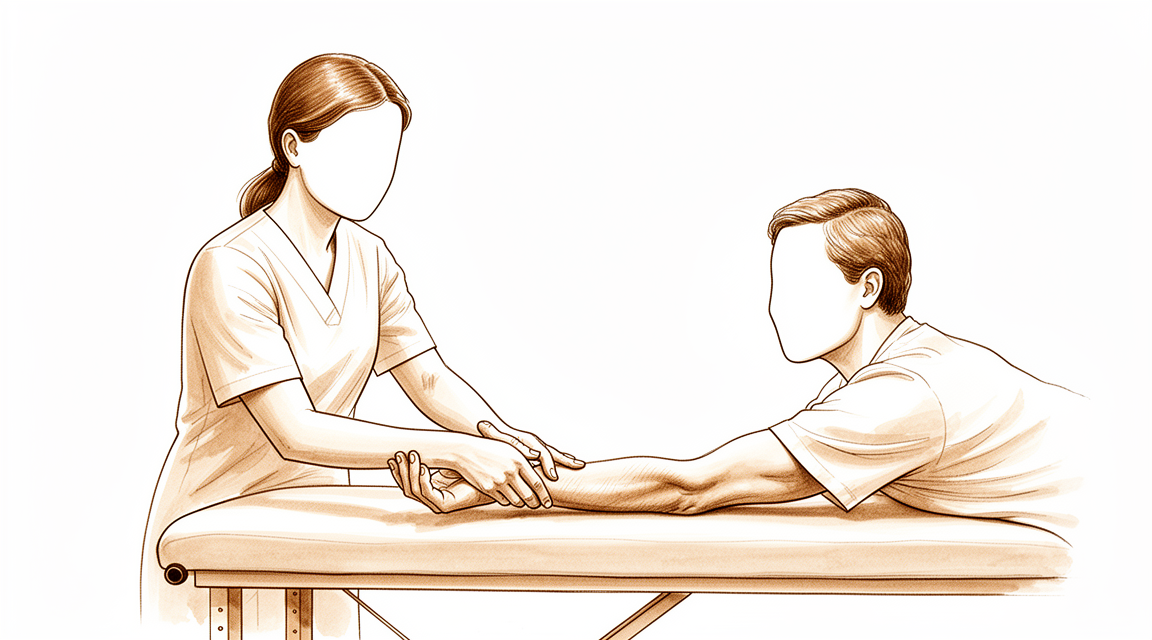
यह ऑपरेशन क्यों सुझाया गया है
डॉ. कीरन हिरपरा, माटर प्राइवेट हॉस्पिटल, रॉकहैम्पटन में ऊपरी अंग (कंधे से हाथ तक) के सर्जन हैं, और वे आपकी समस्या के हिसाब से सबसे कम चीर-फाड़ वाले विकल्पों से शुरुआत करते हैं। मरीज़ आम तौर पर अपने जीपी (आपके फैमिली डॉक्टर) के रेफ़रल से हमारे क्लिनिक आते हैं; अगर किसी फिजियोथेरेपिस्ट ने आपको हमसे मिलने की सलाह दी है, तब भी मेडिकेयर रिबेट पाने के लिए आपको अपने जीपी से रेफ़रल लेना होगा। आपकी बीमारी के इतिहास, शारीरिक जांच और ज़रूरत पड़ने पर इमेजिंग (एक्स-रे, स्कैन) के आधार पर हमारी जांच से निदान तय होता है।
रेडियल टनल सिंड्रोम में आपकी कोहनी और अग्रबाहु (कोहनी से कलाई तक का हिस्सा) के बाहरी हिस्से में दर्द होता है। रेडियल नर्व (तंत्रिका) आपकी अग्रबाहु में नीचे की ओर जाती है, और उसके रास्ते में कई अलग-अलग संरचनाएं उस पर दबाव डाल सकती हैं। इनमें किसी मांसपेशी का किनारा, रेशेदार पट्टी, या नर्व के ऊपर से गुज़रने वाली छोटी रक्त वाहिकाएं शामिल हो सकती हैं। हम आम तौर पर बिना ऑपरेशन वाले इलाज से शुरू करते हैं, जैसे गतिविधियों में बदलाव, फिजियोथेरेपी या हैंड थेरेपी, स्प्लिंट, या इंजेक्शन। जब इनसे पर्याप्त सुधार नहीं होता, तब सर्जरी पर विचार किया जाता है। इस ऑपरेशन को रेडियल टनल रिलीज़ (नर्व पर से दबाव हटाने का ऑपरेशन) कहते हैं, और इसमें इन सभी बिंदुओं पर नर्व पर पड़ रहा दबाव कम किया जाता है। हम इसकी सलाह उन लोगों को देते हैं जिनका दर्द दूसरे इलाज के बावजूद बना रहा है। इसका मुख्य उद्देश्य आपके दर्द में आराम देना और आपकी अग्रबाहु और कलाई को फिर से सामान्य रूप से काम करने में मदद करना है।
ऑपरेशन से पहले
ऑपरेशन से सात घंटे पहले आपको खाना-पीना बंद करना होगा। हम कुछ दूसरी जगहों से थोड़ा ज़्यादा समय मांगते हैं, ताकि अगर ऑपरेशन थिएटर की सूची जल्दी चल रही हो तो आपका ऑपरेशन पहले किया जा सके। आपके सर्जन बताएंगे कि ऑपरेशन के दिन आपकी रोज़ की कौन-सी दवाएं नहीं लेनी हैं और कौन-सी हमेशा की तरह लेनी हैं। कृपया आप जो कुछ भी लेते हैं उसकी एक लिखी हुई सूची साथ लाएं, जिसमें गोलियां, इंजेक्शन और कोई भी ड्रॉप या क्रीम शामिल हों। ऑपरेशन के बाद आपको घर ले जाने के लिए किसी का इंतज़ाम करें, क्योंकि आप खुद गाड़ी चलाने की हालत में नहीं होंगे। ढीले, आरामदायक कपड़े पहनें, जिनकी आस्तीन आसानी से बांह पर चढ़ जाए। कुछ इमेजिंग, जैसे एक्स-रे, एमआरआई या अल्ट्रासाउंड स्कैन, ऑपरेशन की योजना बनाने में हमारी मदद करती है। अगर आपको दूसरी बीमारियां हैं, तो आपको खून की जांच या एनेस्थेटिस्ट (बेहोशी के डॉक्टर) से जांच करवाने की ज़रूरत पड़ सकती है।
ऑपरेशन के दिन
आप अस्पताल की सर्जिकल एडमिशन यूनिट में पहुंचेंगे, जहां आपका नाम दर्ज किया जाएगा और आपको ऑपरेशन थिएटर के लिए तैयार किया जाएगा। आप एनेस्थेटिस्ट से मिलेंगे, जो वह डॉक्टर हैं जो ऑपरेशन के दौरान आपकी बेहोशी और दर्द नियंत्रण की देखभाल करते हैं। यह ऑपरेशन जनरल एनेस्थीसिया (जिसमें आप पूरी तरह बेहोश रहते हैं) में किया जाता है। ऑपरेशन के दौरान आप पूरी तरह बेहोश रहेंगे। कुछ मरीज़ों को ऑपरेशन के बाद दर्द से राहत के लिए रीजनल नर्व ब्लॉक (हाथ या बांह को सुन्न करने का इंजेक्शन) भी दिया जा सकता है; एनेस्थेटिस्ट आपकी व्यक्तिगत स्थिति के आधार पर उसी दिन यह तय करते हैं। इसके बाद आपको ऑपरेशन थिएटर में ले जाया जाता है, जहां ऑपरेशन किया जाता है।
ऑपरेशन पूरा होने पर आप रिकवरी एरिया में होश में आएंगे। वहां नर्सें आपकी निगरानी करेंगी, जब तक एनेस्थीसिया का असर खत्म होता है। जब आपकी हालत स्थिर हो जाएगी, तो प्रक्रिया और आपकी रिकवरी कैसी चल रही है, इसके आधार पर आपको या तो वार्ड में ले जाया जाएगा या आप उसी दिन घर चले जाएंगे। अगर आप घर जा रहे हैं, तो जिस व्यक्ति का आपने इंतज़ाम किया है, वह आपको गाड़ी से घर ले जाएगा।
ऑपरेशन में क्या किया जाता है
इस ऑपरेशन को रेडियल टनल रिलीज़ कहते हैं। यह आपकी अग्रबाहु के सामने वाले हिस्से पर, कोहनी की सिलवट के ठीक नीचे, एक चीरे से किया जाता है। आपके सर्जन इसी एक चीरे से होकर रेडियल नर्व तक पहुंचते हैं।
नर्व के रास्ते में कई अलग-अलग संरचनाएं उस पर दबाव डाल सकती हैं। इनमें किसी मांसपेशी का किनारा, रेशेदार पट्टी, या नर्व के ऊपर से गुज़रने वाली छोटी रक्त वाहिकाएं शामिल हो सकती हैं। आपके सर्जन दबाव के इन सभी बिंदुओं को ढूंढकर उन्हें रिलीज़ करते हैं, जिससे नर्व पर पड़ रहा दबाव कम हो जाता है। आसपास के ऊतक को जितना हो सके उतना कम छेड़ा जाता है, और नर्व तक खून पहुंचाने वाली रक्त आपूर्ति को सुरक्षित रखा जाता है।
दबाव के बिंदु रिलीज़ हो जाने के बाद आपके सर्जन चीरे को बंद कर देते हैं। पहले बंद किए गए घाव पर एक बारीक, अपने आप चिपकने वाली जाली रखी जाती है, जो त्वचा के किनारों को आपस में जोड़े रखती है। फिर जाली के ऊपर एक तरल त्वचा-गोंद लगाया जाता है, जो सूखकर पूरे घाव को सील कर देता है। यह लगभग एक से दो सप्ताह तक लगा रहता है और फिर अपने आप उठकर उतर जाता है, इसलिए कुछ भी निकलवाने की ज़रूरत नहीं होती।
ऑपरेशन के बाद
आप रिकवरी एरिया में होश में आएंगे और एनेस्थीसिया का असर खत्म होने तक नर्सें पास में रहेंगी। आपकी बांह पर घाव के ऊपर एक नरम ड्रेसिंग (घाव की पट्टी) होगी, और दर्द से राहत का इंतज़ाम किया जाएगा ताकि आप आराम से रहें। जैसे ही आप तैयार महसूस करें, आप चल-फिर सकते हैं, और आप तुरंत हल्के कामों के लिए अपने हाथ का इस्तेमाल करेंगे। यह आम तौर पर डे-केस (उसी दिन छुट्टी वाला) ऑपरेशन होता है, इसलिए आप उसी दिन घर जाने की उम्मीद कर सकते हैं, हालांकि कभी-कभी मरीज़ रात भर रुकते हैं। घर पहुंचने के बाद पहले 24 घंटे किसी को आपके साथ रहना चाहिए। हम ड्रेसिंग को लगभग 10 दिन तक लगी रहने देते हैं; कृपया तब तक इसे न हटाएं, जब तक हम आपसे ऐसा करने को न कहें। जब हम आपसे मिलेंगे, तब हम इसे बदलेंगे या हटाएंगे।
ठीक होने की प्रक्रिया
घर जाते समय आपकी बांह पर एक नरम ड्रेसिंग होगी। पहले कुछ दिनों में घाव के आसपास कुछ दर्द और सूजन होना सामान्य है। जब हो सके, अपनी बांह को आराम दें, बैठते समय उसे ऊपर उठाकर रखें, और आपके लिए तय की गई दर्द निवारक दवाएं लें। ज़्यादातर लोगों को लगता है कि दिन बीतने के साथ तकलीफ़ धीरे-धीरे कम होती जाती है।
आप तुरंत हल्के कामों के लिए अपने हाथ का इस्तेमाल कर सकते हैं। इस ऑपरेशन के बाद कोई प्लास्टर (कास्ट) या नियमित स्प्लिंट नहीं लगाया जाता, इसलिए आपकी कलाई और उंगलियां हिलने-डुलने के लिए आज़ाद रहती हैं। हल्की-फुल्की हरकत से नर्व को आसानी से सरकने में मदद मिलती है और जकड़न दूर रहती है। ऑपरेशन के बाद आपकी हैंड थेरेपी एक्सटेंड रिहैबिलिटेशन में रूबी डूलन के साथ होगी। रूबी एक हैंड थेरेपिस्ट (हाथ की विशेषज्ञ थेरेपिस्ट) हैं: वे आपके व्यायामों में मार्गदर्शन करेंगी और बीच में ज़रूरत पड़ने पर कोई भी स्प्लिंट बनाएंगी। घर पर घाव को सूखा रखें और जब तक हम उसकी जांच न कर लें, ड्रेसिंग को न छेड़ें।
जैसे-जैसे सूजन कम होगी, आप देखेंगे कि आपकी पकड़ और अग्रबाहु की ताकत धीरे-धीरे लौट रही है। रोज़ के काम, जैसे कपड़े पहनना, खाना खाना और टाइप करना, आम तौर पर जल्दी वापस आ जाते हैं। भारी सामान उठाना और कसकर पकड़ना बाद में आता है, जब आपका हाथ मज़बूत महसूस हो और छूने पर दर्द खत्म हो जाए। जब आपके थेरेपिस्ट आपकी हरकत और ताकत से संतुष्ट हों, तब आप धीरे-धीरे अपनी सामान्य गतिविधियों पर लौट सकते हैं।
आप आम तौर पर तब गाड़ी चला सकते हैं जब कोई भी स्प्लिंट उतर गया हो और दर्द इतना कम हो गया हो कि आप स्टीयरिंग पकड़ सकें और जल्दी प्रतिक्रिया कर सकें। स्प्लिंट पहने हुए मरीज़ों को गाड़ी नहीं चलानी चाहिए। ऊपरी अंग की सर्जरी के बाद गाड़ी चलाना पर हमारा पेज देखें।
ठीक होने की प्रक्रिया हर व्यक्ति में अलग होती है। आपकी समय-सीमा अलग हो सकती है, और आपके सर्जन और थेरेपिस्ट आपका मार्गदर्शन करेंगे।
क्या समस्याएं हो सकती हैं
ज़्यादातर मरीज़ ठीक रहते हैं, लेकिन कभी-कभी समस्याएं हो सकती हैं। आपके सर्जन और टीम किसी भी समस्या को जल्दी पकड़ने के लिए आप पर करीब से नज़र रखते हैं।
जिस नर्व को रिलीज़ किया जा रहा है, वह ऑपरेशन वाली जगह के पास से गुज़रती है, इसलिए सर्जरी के दौरान वह परेशान (इरिटेट) हो सकती है या उसे चोट लग सकती है। अगर ऐसा होता है, तो आपको अपनी कलाई उठाने या उंगलियां सीधी करने में कमज़ोरी, या अपनी अग्रबाहु, कलाई या हाथ के पिछले हिस्से पर सुन्नपन और झुनझुनी महसूस हो सकती है। इनमें से कुछ बदलाव अस्थायी होते हैं और कुछ सप्ताह से लेकर कुछ महीनों में अपने आप ठीक हो जाते हैं। अगर ऑपरेशन के बाद आपको नई कमज़ोरी या सुन्नपन महसूस हो, तो अपनी अगली फॉलो-अप जांच में अपने सर्जन को बताएं, या अगर यह अचानक शुरू हो तो उससे पहले ही क्लिनिक को फ़ोन करें।
कभी-कभी ठीक होते समय नर्व के चारों ओर निशान वाला ऊतक (स्कार टिश्यू) बन जाता है। यह नर्व पर फिर से दबाव डाल सकता है और आपका पहले वाला दर्द वापस ला सकता है, अक्सर तब, जब सब कुछ ठीक चलने के कुछ सप्ताह या महीने बीत चुके हों। अगर आपका पुराना दर्द लौट आए, तो अपने अगले अपॉइंटमेंट में इसके बारे में बताएं। ज़रूरत पड़ने पर निशान वाले ऊतक को रिलीज़ करने के लिए दोबारा सर्जरी की जा सकती है।
यह भी संभव है कि पहले ऑपरेशन से आपके लक्षणों में आराम न मिले, या वे जल्दी ही वापस आ जाएं। ऐसा तब हो सकता है जब नर्व के रास्ते में दबाव का कोई बिंदु छूट गया हो, या निदान पूरी तरह सही न रहा हो। अगर आपका दर्द बना रहे या लौट आए, तो अपने सर्जन को बताएं। वे आपकी फिर से जांच करेंगे और आगे क्या करना है, इस पर बात करेंगे, जिसमें आगे का इलाज भी शामिल हो सकता है।
अग्रबाहु के किसी भी ऑपरेशन में घाव से जुड़ी समस्याओं का थोड़ा जोखिम होता है। चीरे से बाहर की ओर फैलती बढ़ती हुई लाली, उससे तरल या मवाद का रिसाव, या बुखार पर नज़र रखें। अगर आपको इनमें से कुछ भी दिखे, तो तुरंत क्लिनिक से संपर्क करें, या अगर हमसे संपर्क न हो पाए तो इमरजेंसी विभाग जाएं।
अगर आपको कभी भी चिंता हो कि आपकी बांह कैसे ठीक हो रही है, तो अपनी अगली विज़िट का इंतज़ार करने के बजाय क्लिनिक को फ़ोन करें। अगर आप ठीक-ठीक आंकड़े जानना चाहते हैं, तो इस पेज पर दी गई जटिलताओं की तालिका में सामान्य दरें दी गई हैं।
हमें कब फ़ोन करें
अगर आपका दर्द अचानक बहुत ज़्यादा बढ़ जाए, या नई कमज़ोरी या सुन्नपन तेज़ी से शुरू हो, तो हमें फ़ोन करें। अगर घाव ज़्यादा लाल हो जाए, उससे तरल या मवाद रिसने लगे, या आपको बुखार हो जाए, तो हमें फ़ोन करें। अगर हमसे संपर्क न हो पाए, या आपकी बांह में संवेदना चली जाए या आप उसे हिला न पाएं, तो इमरजेंसी विभाग जाएं। सांस फूलने या पिंडली में सूजन होने पर तुरंत इमरजेंसी विभाग जाएं। अगर आपको कभी भी चिंता हो, तो अपनी अगली विज़िट का इंतज़ार करने के बजाय क्लिनिक को फ़ोन करें।
इस समस्या के बारे में और कहां पढ़ें
यह पेज खुद ऑपरेशन के बारे में है। जिस समस्या का यह इलाज करता है, उसके बारे में, जिसमें यह भी शामिल है कि प्रमाण क्या दिखाते हैं कि सर्जरी कब मदद करती है और कब नहीं, रेडियल टनल सिंड्रोम पेज पर ज़्यादा विस्तार से बताया गया है।
Evidence & references
This is the clinical evidence summary written for health professionals. It is technical, and it lists the research this page was built from. You do not need to read it to understand your treatment or to make a decision about it.
Anatomy & Pathophysiology
Bony Anatomy
- The elbow is a trocho-ginglymoid joint consisting of medial and lateral articulations that provide bony stability [3].
- The trochlea articulates with the ulna within the greater sigmoid notch to form the ulnohumeral, hinged, or trochoid portion of the elbow joint [3].
- The ulnohumeral articulation has highly congruent anatomy through almost 180° of articular contact, except for the bare area of the greater sigmoid notch which is devoid of cartilage [3].
- The coronoid process has medial and lateral facets that buttress the trochlea anteriorly [3].
- The sublime tubercle is located just distal and medial to the coronoid and serves as the attachment site for the anterior bundle of the medial ulnar collateral ligament [3].
- The medial epicondyle is larger and more posteriorly oriented than the lateral epicondyle and forms the attachment site for the origins of the flexor pronator mass [3].
- The capitellum and radial head form the radiocapitellar joint on the lateral side [3].
- The radius is held in close approximation to the ulna at the proximal radioulnar joint by the annular ligament [3].
- The area of the ulna that articulates with the margin of the radial head at the proximal radioulnar joint is known as the lesser sigmoid notch [3].
- The radial head is a concave elliptical structure covered with articular cartilage along the radiocapitellar joint and approximately 270° of the articular margin [3].
- The radial head articulates with both the capitellum and the lesser sigmoid notch [3].
- The lateral epicondyle is the origin of the lateral extensor musculature [3].
- The origin of the lateral ulnar collateral ligamentous complex is located just distal to the lateral epicondyle at the geometric center of the radiocapitellar articulation [3].
- The distal humeral articulation is angled 30° from the longitudinal axis [3].
- The anterior humeral line should pass through the center of the axis of rotation [3].
- The axis of rotation is angulated 5° to 7° in the coronal plane relative to the epicondylar axis, with the medial side more distal than the lateral side [3].
- The angulation of the distal humeral articulation accounts for the change from a valgus carrying angle to a more varus position as the elbow is flexed [3].
- There is a high correlation between the size of the radial head and capitellum on the left and right sides in the same individual [3].
- The olecranon provides a broad attachment site for the triceps posteriorly [3].
- The ulna bends approximately 8° medially at 8 cm from the tip of the olecranon [3].
- The articulation to the tip of the coronoid is approximately 30° from the long axis of the ulna in the sagittal plane [3].
- The radial head should line up with the capitellum at all arm positions on all radiographic views [4].
Ligaments and Capsule
- Elbow stability is determined by primary and secondary stabilizers [1].
- The three primary stabilizers of the elbow are the ulnohumeral articulation, the medial ulnar collateral ligament, and the lateral ulnar collateral ligament complex [1].
- Secondary stabilizers of the elbow include the radiocapitellar articulation, the common flexor tendon, the common extensor tendon, and the joint capsule [1].
- The medial ulnar collateral ligament is the primary valgus stabilizer of the elbow [4].
- The anterior bundle of the medial ulnar collateral ligament is the most important component for stability [4].
- The posterior bundle of the medial ulnar collateral ligament has the greatest change in length and becomes taut at flexion beyond 120 degrees [4].
- The lateral ulnar collateral ligament is the posterolateral stabilizer of the elbow [4].
- The joint capsule allows maximum distension at approximately 70 to 80 degrees of flexion [4].
- The anterior capsule attaches at a point approximately 6 mm distal to the tip of the coronoid [4].
- Tensile forces are present at the medial elbow and compressive forces are present at the lateral elbow [4].
- The LUCL origin center is 10.7 mm from the lateral epicondyle [2].
- The LUCL insertion is 3.3 mm from the apex of the supinator crest [2].
Muscles and Nerves
- The brachialis is the strongest elbow flexor and attaches to the coronoid 11 mm distal to the tip [4].
- The biceps brachii inserts at the ulnar margin of the radial tuberosity, with the long head inserting proximally and the short head distally [4].
- The biceps brachii is a powerful supinator of the forearm [4].
- The triceps is the primary elbow extensor and inserts on the olecranon process [4].
- The mobile wad consists of the brachioradialis, extensor carpi radialis longus, and extensor carpi radialis brevis [4].
- The flexor-pronator mass consists of the pronator teres, flexor carpi radialis, palmaris longus, flexor carpi ulnaris, and flexor digitorum superficialis [4].
- The radial nerve enters the interval between the brachialis and brachioradialis muscles in the proximal angle of the lateral approach wound [6].
- The deep branch of the radial nerve enters the supinator muscle [6].
- The common origin of the extensor muscles is attached to the lateral epicondyle [6].
- The common origin of the extensor muscles is attached to the lateral condylar fragment in lateral condyle fractures [6].
- The interval between the triceps posteriorly and the origins of the extensor carpi radialis longus and brachioradialis anteriorly is used to expose the lateral border of the humerus [6].
- The interval between the triceps posteriorly and the brachioradialis and extensor carpi radialis longus muscles anteriorly is used to expose the lateral condyle and capsule over the radial head [7].
- The extensor carpi ulnaris is separated from the anconeus distal to the radial head during the lateral J-shaped approach [7].
- The distal fibers of the anconeus are divided in line with the curved and transverse parts of the distal skin incision during the lateral J-shaped approach [7].
- The anconeus is reflected subperiosteally from the proximal ulna to dislocate and examine the joint during the lateral J-shaped approach [7].
Functional Anatomy
- The normal elbow has a range of motion from 0° to 140° from extension to flexion [1].
- The normal elbow has a range of motion of 75° in pronation and 85° in supination [1].
- A functional arc for elbow flexion and extension is 100° [1].
- A functional arc for forearm rotation is 100° [1].
Investigations
Physical Examination
- The physical examination of the elbow is directed by the history and the location of the patient's pain in the anterior, posterior, medial, or lateral aspect of the elbow [1].
- Pathologic entities associated with specific compartments aid the examiner in detecting pathologic conditions [1].
- Active and passive flexion, extension, supination, and pronation should be evaluated using a goniometer for accurate measurement [9].
- The contralateral elbow should be examined for comparison during range of motion assessment [9].
- Pain should be assessed during the mid-arc or at the terminal ends of motion [9].
- Mid-arc range of motion pain is more common with intrinsic disease and may not improve with contracture release alone [9].
- The ulnar nerve is of utmost importance in the neurovascular examination due to its anatomic proximity to the elbow [9].
- Electromyography and nerve conduction velocity studies should be performed if there is any question about neurologic dysfunction [9].
- An assessment for ulnar nerve subluxation should be performed [9].
- Subluxation of the ulnar nerve is a relative contraindication for an arthroscopic procedure secondary to possible iatrogenic nerve injury [9].
- If the elbow has less than 90° to 100° of flexion, the posterior bundle of the medial collateral ligament is contracted and must be released to restore flexion [9].
Imaging
- Plain radiographs remain the hallmark and the best screening test for elbow evaluation [1].
- Standard radiographic views include AP, lateral, and oblique views [9].
- Serial radiography is used as follow-up when heterotopic ossification is present [9].
- Primary bony landmarks assessed on radiographs include the ulnohumeral joint, coronoid process, radial head, capitellum, radiocapitellar joint, olecranon tip, coronoid/olecranon fossae, and trochlear ridge [9].
- CT is helpful when assessing for malunion architecture and the location and pattern of osteophytes and/or loose bodies [9].
- Three-dimensional CT is used to check for heterotopic ossification [9].
- CT is not necessary when the stiffness is entirely soft-tissue related [9].
- CT is beneficial if any joint incongruity or abnormal bony anatomy is present [9].
- MRI can be used to evaluate ligaments and tendons, but it is rarely indicated for elbow stiffness [9].
- AP, lateral, oblique, and axillary views of the elbow may reveal posteromedial olecranon osteophytes and/or loose bodies in valgus extension overload syndrome [11].
- CT with two-dimensional reconstruction and three-dimensional surface rendering best visualizes the pathology of valgus extension overload syndrome [11].
- MRI may be most helpful in evaluating associated injuries including partial or complete tears of the medial collateral ligament in valgus extension overload syndrome [11].
- Radiographic evaluations are essential when diagnosing an osteochondritis dissecans lesion of the elbow [12].
- Important aspects of osteochondritis dissecans lesions may be better seen with MRI [12].
- Standard AP and lateral radiographs should be obtained for the evaluation of elbow osteoarthritis [13].
- Radiographs for elbow osteoarthritis typically show osteophyte formation at the coronoid process, coronoid fossa, radial fossa, radial head, olecranon tip, and olecranon fossa [13].
- Joint spaces at the ulnohumeral joint are usually preserved in elbow osteoarthritis [13].
- Joint spaces at the radiocapitellar joint are mildly narrowed in elbow osteoarthritis [13].
- Loose bodies may be evident on radiographs, which typically underestimate the number present [13].
- CT may be useful for surgical planning of elbow osteoarthritis by allowing a detailed assessment of osteophytes and the presence of loose bodies [13].
References
[1] Orthopaedic Knowledge Update 13 Ebook Without Multimedia. Anatomy, Biomechanics, Physical Examination, and Imaging of the Elbow > Summary and Conclusions.
[2] Orthopaedic Knowledge Update 13 Ebook Without Multimedia. Anatomy, Biomechanics, Physical Examination, and Imaging of the Elbow > Annotated References.
[3] Orthopaedic Knowledge Update 13 Ebook Without Multimedia. Anatomy, Biomechanics, Physical Examination, and Imaging of the Elbow > Anatomy > Bony Anatomy.
[4] Miller S Review Of Orthopaedics. SECTION 16 PATELLAR TRACKING IN TOTAL KNEE ARTHROPLASTY > TABLE 2.3 Shoulder Spaces.
[6] Campbell S Operative Orthopaedics 4 Volume Set. LATERAL APPROACHES.
[7] Campbell S Operative Orthopaedics 4 Volume Set. LATERAL J-SHAPED APPROACH TO THE ELBOW.
[9] Aaos Comprehensive Orthopaedic Review 3. Elbow Stiffness* > IV. Evaluation.
[11] Aaos Comprehensive Orthopaedic Review 3. Elbow Injuries in the Athlete* > III. Valgus Extension Overload Syndrome and Posterior Impingement.
[12] Orthopaedic Knowledge Update. Osteochondritis Dissecans of the Knee and Elbow* > Summary.
[13] Aaos Comprehensive Orthopaedic Review 3. Arthritis and Arthroplasty of the Elbow > I. Osteoarthritis.