Clinicians › Elbow
Olecranon Fracture
Olecranon fractures — patterns, non-operative care, and tension-band or plate fixation.
For patients: a plain-language version of this topic is available. See the patient guide.
Overview
Olecranon fractures are managed with a variety of techniques, as no single method is suitable for all fracture patterns [23]. While the majority of these injuries heal uneventfully with good or excellent results, a small loss of motion is expected [13]. Surgical treatment is associated with a high rate of complications [119], and patients undergoing revisions beyond implant removal experience poorer functional outcomes [119]. However, no significant differences in functional outcomes or secondary operations have been found with respect to fracture type, gender, or surgical method [119]. The timing of fixation for displaced olecranon fractures does not significantly increase the rate of early complications or reoperation [21].
Operative options include the Locking Compression Plate (LCP), which results in good long-term outcome [1], and plate fixation, which is effective for displaced fractures with a good functional outcome and low incidence of complications [17]. Plating leads to predictable union, though lack of full extension occurred in 39% of patients [25]. Tension band wiring remains effective for appropriately selected fractures and outperformed plate osteosynthesis in one cohort [61], while suture fixation is the mainstay for simple fractures with no re-operations or wound complications observed in one series [71]. Both Kirschner wire tension band and anatomical locking plate procedures effectively treat Mayo type II fractures [12]. Low-profile double-plate osteosynthesis is a safe and effective alternative [5], and a bent plate is a practical alternative for less than 5% of fractures [14]. Excision of the fragment with triceps repair was supported as a preferred method due to significantly less morbidity compared with internal fixation in one study [131]. The clinical role of locked intramedullary nail fixation needs to be defined by controlled, randomized prospective trials [68].
Non-operative treatment is supported for specific populations. In patients aged ≥75 years, non-operative treatment provided excellent functional results at 6 months without associated complications [2]. Data support primary non-operative management of isolated displaced fractures in the elderly [15], with satisfactory short-term and long-term outcomes found in older, lower-demand patients [18]. Close radiographic follow-up is recommended for nonsurgically treated fractures in children [6]. Although the literature on treatment in elderly patients is limited [10], studies on operative versus non-operative management in patients over 75 have not definitively settled the debate but offer valuable data for personalized treatment plans [19].
Anatomy & Pathophysiology
Bony Anatomy
The olecranon and coronoid process form the greater sigmoid notch, which articulates with the trochlea of the distal humerus [36]. This intrinsic ulnohumeral anatomy permits flexion and extension while providing joint stability [36]. The olecranon serves as the insertion site for the triceps tendon, which blends with the periosteum of the proximal ulna [36]. Its exposed, subcutaneous position renders it vulnerable to direct trauma and violent muscular contractions from the triceps [36]. In children, the olecranon is predominantly cartilage, particularly in younger patients, which reduces the likelihood of fracture from a direct blow [48]. The thick periosteum and relatively thin metaphyseal cortex of the pediatric olecranon predispose it to minimally displaced greenstick fractures [48].
Mechanisms of Injury
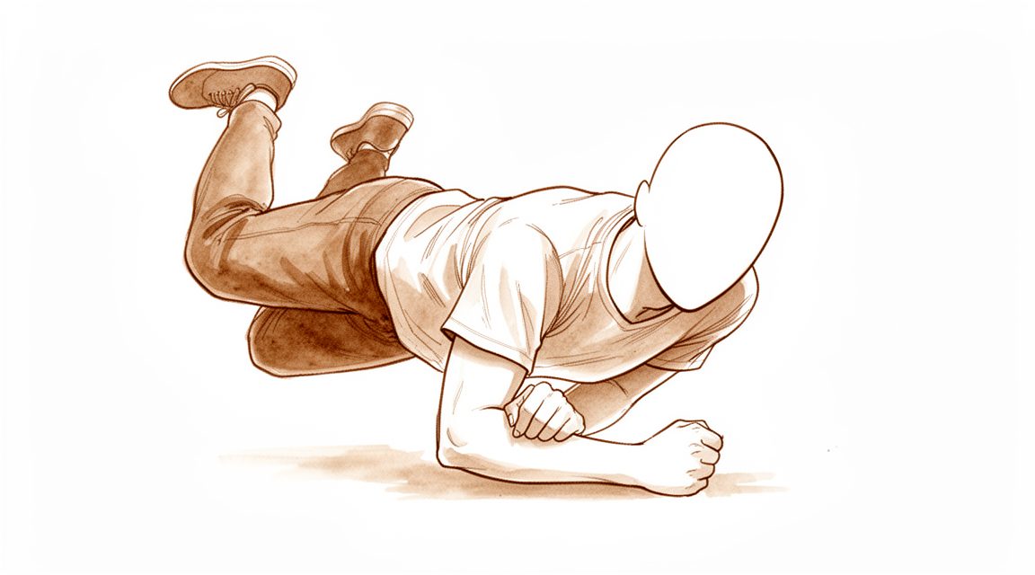
Olecranon fractures result from a direct blow, a fall on an outstretched hand with the elbow in flexion, or high-energy trauma associated with radial head fractures or elbow dislocation [36]. Sudden, violent triceps contraction can produce an avulsion fracture of varying size at the olecranon tip [36]. A bimodal distribution exists, with high-energy trauma in young patients and low-energy trauma, such as falls from standing, in elderly patients [36]. The bone typically fails from a direct blow to the elbow or as a tensile force from the pull of the triceps tendon [37]. Regardless of the mechanism, the triceps acts as the main deforming force, pulling the fractured fragment proximally to create a gap at the articular surface and an incompetent extensor mechanism [37]. A flexion-extension "arc of injury" relates fracture types to elbow position at the moment of impact [88].
In pediatric patients, olecranon fractures usually result from hyperextension injury [48]. Other mechanisms include a direct blow to the flexed elbow, hyperflexion injury, or shear force [48]. Valgus hyperextension forces may produce an associated radial neck or medial epicondyle fracture [48]. Varus hyperextension injuries may be associated with lateral dislocation of the radial head or a Bado type III Monteggia lesion [48]. Flexion injuries, usually caused by a fall on an outstretched hand with the elbow flexed resulting from strong eccentric triceps contraction, are generally transverse, displaced posteriorly rather than anteriorly, and rarely associated with other injuries [48]. Shear injuries result from a force to the proximal ulna just anterior to the humeral condyles, causing the olecranon to fracture through metaphyseal bone with anterior displacement of the distal fragment [48].
Associated Injuries
Olecranon fractures typically occur in isolation, but the subcutaneous location poses a risk to soft tissues and a risk of the fracture being open [37]. In an epidemiologic study, 22% of patients had injuries to the ipsilateral limb and 6.4% of fractures were open [37]. In pediatric patients, olecranon fractures are associated with other elbow injuries, usually the medial epicondyle, in 20% to 50% of cases [48]. Hyperextension injuries in pediatric patients are frequently associated with other elbow injuries [48].
Classification
The Mayo classification is the most common system used for olecranon fractures, based on fracture displacement, presence of comminution, and stability of the ulnohumeral joint [37]. It demonstrates good interobserver reliability [37]. The classification defines: * Type I: Nondisplaced fractures [37]. * Type II: Displaced fractures [37]. * Type III: Fractures with an unstable ulnohumeral joint [37].
Modifiers indicate comminution: A denotes simple fractures and B denotes comminuted fractures [37]. Displaced olecranon fracture lines enter along the medial side of the trochlear notch and exit at the base of the coronoid, while minimally displaced fractures enter and exit the trochlear notch at the base of the coronoid [37]. The Schatzker and Colton classification attempted to categorize fractures based on pattern morphology, but its application was limited [37]. In a pediatric context, olecranon fractures account for approximately 5% of elbow fractures [48].
Epidemiologic data from a Swedish nationwide study showed a larger share of comminuted central fractures (29%) compared to a single-center Scottish report [11]. This study also observed an almost threefold larger share of distal olecranon fractures compared to the 6.2% reported in a Scottish study of 64 olecranon fractures [11].
Classification
Classification Systems and Reliability
Mayo: The Mayo classification is the most commonly used system for olecranon fractures in clinical practice [45]. It consists of three types with a modifier to indicate comminution [45]. Type I fractures are nondisplaced [45], Type II fractures are displaced [45], and Type III fractures have accompanying injuries [45]. Specifically, Type 1 is not displaced and stable, Type 2 is displaced but stable, and Type 3 is displaced and unstable [33]. Each type can be subdivided into subtype A (non-comminuted) or B (comminuted) [33]. Although designed to simplify categorization, the system does not achieve this goal due to poor reproducibility [49].
Schatzker: The Schatzker classification divides olecranon fractures into six types based on fracture pattern: transverse, transverse impacted, oblique, comminuted, oblique distal, and fracture dislocation [120]. It groups fractures based on the quantity of fragments and fracture lines [116].
AO/OTA: The AO classification subsumes olecranon fractures under proximal forearm injuries [116]. It divides olecranon fractures into two groups: intra-articular and extra-articular [120].
Colton: The Colton classification provides treatment options based on the fracture line (avulsion, transverse, comminuted) and whether the elbow is dislocated [120]. It also divides fractures into groups based on the quantity of fragments and fracture lines [116].
Horne and Tanzer: This classification describes three types of oblique or transverse fractures located at different thirds of the olecranon [120]. It is less frequently used nowadays [120].
Other Considerations: Multiple classification systems exist, including AO, Mayo, Schatzker and Colton, but none are widely accepted or provide direct and reliable advice on operative strategies [116]. The low reproducibility rates of these systems raise questions about their use in clinical and research contexts [116]. Current systems do not inform surgical planning or reduction strategy [120]. They may be useful for simple two-part fractures but are not precise enough for complex multifragmentary fractures [120]. Quantitative analysis has further clarified the fracture morphology of Mayo type I, II, and III fractures [70].
Fracture Patterns and Epidemiology
In a Swedish study, the distribution of olecranon fractures included a larger share of comminuted central fractures (29%) compared to a single-center report from Scotland [11]. The Swedish study observed an almost threefold larger share of distal olecranon fractures than the 6.2% reported in the Scottish study [11]. Regarding mechanism, 87.4% of female patients and 77.8% of male patients sustained low-energy injuries, while 5.7% of female patients and 11.6% of male patients sustained high-energy injuries [11].
In a study of proximal ulna fractures, the most frequent injury according to the AO fracture classification was the 21-B1.1 type [126]. In the same study, a simple isolated displaced olecranon fracture (Mayo type 2A) was the most common according to the Mayo classification [126]. In a study of 44 patients treated with Kirschner wires with eyelets, Type IIA fractures were the most common pattern, accounting for 91% of cases, while Type IIB fractures accounted for 9% of cases [140].
Olecranon fractures in children are usually undisplaced and incomplete [26]. The 'longitudinal split' fracture is not uncommon in children with olecranon fractures [26]. Fracture of the olecranon in a child is often only part of a complex injury to the bone and soft tissues around the elbow [31]. Articular impaction is a common feature of geriatric olecranon fractures [40].
Clinical Presentation
Mechanisms and Epidemiology
Olecranon fractures typically result from a sudden and violent contraction of the triceps muscle, which produces an avulsion fracture of varying size at the olecranon tip [36]. The triceps acts as the primary deforming mechanism, pulling the fractured fragment proximally to create a gap at the articular surface and render the extensor mechanism incompetent [37]. Epidemiological data indicate that the incidence of olecranon fractures increased by 29% over the 20-year study period from 1999 to 2018 in Denmark [16]. Patients with these injuries possess demographic characteristics essentially similar to those with distal radius fractures [24]. In the elderly population, olecranon fractures are associated with higher than expected 1-year mortality rates [30].
Physical Examination
Pain is usually localized to the posterior part of the elbow [36]. Given the subcutaneous location of the olecranon, the fracture itself may be palpable [36]. Extensive posterior swelling is typical, and the elbow often presents with a large fluid collection subcutaneously over the olecranon [36, 37]. A careful examination of the integrity of the extensor mechanism, with gravity eliminated, can aid surgical decision making [36]. The affected extremity from shoulder to hand should be thoroughly examined for soft tissue compromise and open fractures given the subcutaneous nature of the olecranon [37].
Imaging
Plain radiographs are usually sufficient for isolated fractures of the olecranon [36]. Anteroposterior and lateral radiographs of the elbow are required [37]. A true lateral radiograph is necessary to accurately identify the plane of the fracture and the number of fracture fragments [36]. The examiner should assess for fracture comminution and impaction on radiographs [36]. If there is concern for a radial head fracture, a radiocapitellar view of the elbow can be obtained [37]. In the setting of an elbow dislocation, post-reduction radiographs should be obtained [37]. Computed tomography (CT) may help delineate comminution or impaction better in more complex cases but is not routinely required [36]. CT can be used for preoperative planning for comminuted fractures of the olecranon if there is an associated radial head or coronoid fracture, but this is not routinely utilized [37].
Classification
The Mayo classification is based on the amount of fracture displacement, the presence of comminution, and stability of the ulnohumeral joint [37]. However, the Mayo classification was designed to simplify categorization of olecranon fractures but does not achieve this goal due to poor reproducibility [49]. Displaced olecranon fracture lines entered along the medial side of the trochlear notch and exited at the base of the coronoid, while minimally displaced fractures entered and exited the trochlear notch at the base of the coronoid [37].
Pediatric Presentation
Olecranon fractures in children are usually undisplaced and incomplete, with the 'longitudinal split' fracture being not uncommon [26]. Fractures of the ipsilateral olecranon associated with the radial neck are not so rare as previously reported [44].
Investigations
History and Mechanism: The history helps distinguish a triceps avulsion from an actual direct blow to the elbow [36]. Pain is usually localized to the posterior part of the elbow [36].
Physical Examination: Extensive posterior swelling is typical in olecranon fractures [36]. The elbow often presents with a large fluid collection subcutaneously over the olecranon [37]. Open wounds are typically posterior and result from the direct impact of the posterior surface of the elbow against an unyielding structure [36]. Given the subcutaneous nature of the olecranon, one should have a low suspicion for open fractures, and the arm should be examined for any soft tissue compromise [37]. The affected extremity from shoulder to hand requires thorough examination due to the significant incidence of associated ipsilateral injuries [37]. Examination of the extensor mechanism, both with and without gravity eliminated, is performed to assess competency [37].
CT: Computed tomography (CT) is not routinely utilized but can be used for preoperative planning for comminuted fractures of the olecranon if there is an associated radial head or coronoid fracture [37]. In more complex cases, CT may help delineate comminution or impaction better, though this is not routinely required [36]. Quantitative analysis of olecranon fractures using 3-dimensional computed tomography further clarified fracture morphology of Mayo type I, II, and III fractures [70].
Other Considerations: The Mayo classification is the most common classification used for olecranon fractures [37]. The Swedish study observed an almost threefold larger share of distal olecranon fractures than the 6.2% reported in the Scottish study of 64 olecranon fractures [11].
Treatment
General Principles
The majority of olecranon fractures are treated surgically [45], with good results generally expected after surgical management [45]. Most fractures heal uneventfully with good or excellent results, though a small loss of motion is anticipated [13]. Fixation must be secure enough to permit early motion to avoid significant stiffness of the elbow joint [22]. Outcomes are good to excellent when adhering to a treatment algorithm based on displacement, comminution, and joint stability [69]. Symptomatic hardware requiring removal remains a recurring and frequent problem in olecranon fracture treatment [45].
Non-Operative
Non-operative management is supported for isolated displaced fractures of the olecranon in the elderly [15], including patients aged ≥75 years who achieve excellent functional results at 6 months without associated complications [2]. Some isolated displaced (>2 mm) olecranon fractures in elderly patients can be successfully treated nonoperatively [9]. In older, lower-demand patients, nonoperative management of isolated displaced olecranon fractures yields satisfactory short-term and long-term outcomes [18]. Displaced olecranon fractures in patients older than 70 years may be effectively managed with nonoperative measures to produce high satisfaction and functional range of motion [43]. Non-operative functional treatment of displaced olecranon fractures in the elderly provides a satisfactory and functional range of motion with a high rate of satisfaction [104]. In elderly and medically unwell patients, nonoperative treatment can result in reasonable range of motion, minimal pain, and maintenance of extension against gravity [108]. Non-operative treatment of Mayo Type II olecranon fractures may be successful, extending the age range for which such treatment of displaced olecranon fractures can be considered [35]. Patients who present with a non-union after a displaced olecranon fracture managed non-operatively have reasonable elbow function and uncommonly request operative treatment [34]. However, surgical management remains the standard of care for displaced olecranon fractures until more convincing evidence supports nonsurgical treatment [87].
Operative
Indications: Surgical intervention is the standard of care for displaced olecranon fractures [87], particularly when fixation must be secure enough to permit early motion to avoid significant stiffness [22]. Treatment algorithms based on displacement, comminution, and joint stability guide operative decision-making [69].
Implant Selection: Internal fixation by Cable Pin System (CPS) is associated with a shorter healing time, fewer complications, and better function than Tension Band Wiring (TBW) [42]. Double Tension Band Wiring (DTBW) produces good clinical and radiological outcomes and provides additional stability through a second TBW [60]. Suture tension band fixation reduces hardware complications in olecranon fractures compared to traditional techniques [3]. The Attenborough springs and Rush nail method provides a quick, safe, and effective method of fixing olecranon fractures with a high level of patient satisfaction [101]. Treatment with the Locking Compression Plate (LCP) results in good long-term outcome [1]. Low-profile double-plate osteosynthesis for treating olecranon fractures resulted in good clinical outcomes [107]. Plate fixation of complex olecranon fracture is an effective, reliable method of treatment with low risk of non-union [66]. Plate has better efficacy and safety for Mayo II olecranon fractures compared to tension band wire [52]. Both operative procedures (Kirschner wire tension band and anatomical locking plate) effectively treat Mayo type II olecranon fractures [12]. Suture anchor fixation of displaced olecranon fractures resulted in excellent midterm functional outcomes [20]. Both locking-plate osteosynthesis and intramedullary nailing could be appropriate surgical techniques for fixation of selected olecranon fractures and osteotomies [62]. The Olecranon Memory Connector (OMC) could be an effective alternative to treat olecranon fractures [85].
Other Considerations: In cases with concomitant injuries, the risk of limited elbow motion is high following open reduction and plate osteosynthesis in comminuted fracture of the olecranon [58]. The clinical role of locked intramedullary nail (IMN) fixation for the treatment of olecranon fractures and olecranon osteotomies needs to be defined by controlled, randomized prospective trials [68]. There was a considerably higher rate of separation at the fracture after screwing than after inserting a tension band in displaced olecranon fractures [47]. The technical quality of fixation was open to criticism in half of the cases in both tension band and screwing groups for displaced olecranon fractures [47].
Complications
Hardware-related complications: Symptomatic hardware requiring removal is a recurring and frequent problem following surgical management of olecranon fractures [45]. Implant migration occurs in only 3% of patients who undergo operative fixation [142]. Internal fixation by cable pin system is associated with fewer complications than tension band wiring [42].
Stiffness / Arthrofibrosis: The most common complication following plating of the olecranon is lack of full extension, observed in 39% of patients [25].
Wound complications: Suture fixation for simple olecranon fractures resulted in no re-operations or wound complications in the reported series [71].
Other Considerations: Plate fixation for displaced olecranon fractures is associated with a low incidence of complications [17]. Non-operative treatment of olecranon fracture in patients aged ≥75 years was provided without associated complications [2]. Olecranon fractures in the elderly are associated with higher than expected 1-year mortality rates [30]. Further displacement can occur after initial nonsurgical treatment of minimally displaced olecranon fractures in children [6]. Fracture of the olecranon in a child is often part of a complex injury to the bone and soft tissues around the elbow [31]. Post-traumatic osteoarthritis is a potential complication following isolated olecranon fractures, with incidence influenced by fracture stability and comminution [33].
Recovery
Light activity (weeks): The provided evidence does not specify a timeline for the resumption of desk work, driving, or light activities of daily living.
Full activity (months): The provided evidence does not specify a timeline for the return to manual work, sport, or full range of motion and strength.
Complete recovery / outcome plateau (months): The provided evidence does not specify a timeline for when pain, strength, and final functional outcomes stabilise.
Rehabilitation protocol: The provided evidence does not detail specific physiotherapy phasing, immobilisation duration, weight-bearing or range-of-motion progression, or sling and brace removal timing.
Functional milestones: Among active patients with a simple isolated, displaced fracture of the olecranon, no difference was found between tension band wiring and plate fixation in the patient-reported outcome at 1 year following surgery [148].
Other Considerations: Good functional long-term results are expected in patients with complex olecranon fractures treated with open reduction and internal fixation, despite arthritic changes in the elbow joint [91]. The treatment of olecranon fractures with the use of the Locking Compression Plate results in good long-term outcome [1]. Plating of the olecranon leads to predictable union, though the most common complication was lack of full extension in 39% of patients [25]. Internal fixation by Cable Pin System is an effective method for olecranon fracture and is associated with a shorter healing time, fewer complications and better function than tension band wiring [42]. Close radiographic follow-up for nonsurgically treated olecranon fractures is recommended [6]. Olecranon fractures in the elderly have higher than expected 1 year mortality rates [30]. The incidence of ulnohumeral osteoarthritis following isolated olecranon fractures was assessed using the Broberg and Morrey classification, where grade 1 or higher was considered to have osteoarthritis [33].
Key Evidence
- [L4] The treatment of olecranon fractures with the use of the LCP results in good long-term outcome. [1] (10.1016/s0020-1383(12)70083-9)
- [L4] Non-operative treatment of olecranon fracture in patients aged ≥75 years provided excellent functional results at 6 months, without associated complications. [2] (10.1016/j.otsr.2017.10.015)
- [L3] Future studies should focus on the long-term outcomes of this technique, as compared to traditional and more established techniques to treat olecranon fractures. [3] (10.1016/j.jseint.2026.101734)
- [L3] 96% of individuals with a closed displaced fracture of the olecranon treated by open reduction and internal fixation had excellent or good outcomes up to 25 years after the fracture. [4] (10.1067/mse.2002.124548)
- [L3] Low-profile double-plate osteosynthesis is a safe and effective alternative treatment of olecranon fractures with excellent subjective and objective clinical outcome measures. [5] (10.1016/j.otsr.2019.08.019)
- [L4] Close radiographic follow-up for nonsurgically treated olecranon fractures is recommended. [6] (10.5435/jaaos-d-25-00821)
- [L4] Some isolated displaced (>2 mm) olecranon fractures in elderly patients can be successfully treated nonoperatively. [9] (10.1016/j.ocl.2016.08.011)
- [L4] The literature on the treatment of olecranon fractures in elderly patients is limited. [10] (10.1007/s11678-018-0488-7)
- [L4] [11] (10.1007/s00068-021-01765-2)
- [L3] Both operative procedures effectively treat Mayo type II olecranon fractures. [12] (10.1186/s12891-025-08843-1)
- [L4] A majority of olecranon fractures heal uneventfully with good/excellent results with a small loss of motion to be expected. [13] (10.1016/j.hcl.2015.07.003)
- [L4] For these less than 5 per cent of all fractures of the olecranon, a bent plate is a practical and satisfactory alternative. [14] (10.1016/s0020-1383(73)80090-7)
- [L1] These data further support the role of primary non-operative management of isolated displaced fractures of the olecranon in the elderly. [15] (10.1302/0301-620x.99b7.bjj-2016-1112.r2)
- [L3] The incidence of olecranon fractures increased by 29% over the 20-year study period. [16] (10.1186/s13018-025-05970-2)
- [L4] Plate fixation is an effective treatment option for displaced olecranon fractures with a good functional outcome and a low incidence of complications. [17] (10.1097/00005131-200111000-00002)
- [L4] We found satisfactory short-term and long-term outcomes following the nonoperative management of isolated displaced olecranon fractures in older, lower-demand patients. [18] (10.2106/jbjs.l.01137)
- [L2] While they did not definitively settle the debate about whether we should manage olecranon fractures nonoperatively in patients over 75, they did offer valuable data that surgeons and patients can use to develop personalized treatment plans tailored to each patient's needs. [19] (10.2106/jbjs.24.01097)
- [L4] Suture anchor fixation of displaced olecranon fractures resulted in excellent midterm functional outcomes. [20] (10.5397/cise.2023.00528)
- [L3] The timing of fixation of displaced olecranon fractures does not significantly increase the rate of early complications or reoperation. [21] (10.1016/j.jhsg.2023.09.002)
- [L5] Fixation must be secure enough to permit early motion to avoid significant stiffness of the elbow joint. [22] (10.5435/00124635-200007000-00007)
- [Paper] No one technique is suitable for the management of all olecranon fractures. [23] (10.1016/j.injury.2008.12.013)
- [L3] Patients with olecranon fractures have essentially similar demographic characteristics compared to patients with distal radius fractures. [24] (10.1177/17585732221124301)
- [L3] Plating of the olecranon leads to predictable union, though the most common complication was lack of full extension in 39% of patients. [25] (10.1016/j.injury.2016.04.015)
- [L4] Olecranon fractures in children are usually undisplaced and incomplete, with the 'longitudinal split' fracture being not uncommon. [26] (10.1016/0020-1383(75)90056-x)
- [L3] Olecranon fractures in the elderly have higher than expected 1 year mortality rates. [30] (10.1177/1758573221994860)
- [L4] Fracture of the olecranon in a child is often only part of a complex injury to the bone and soft tissues around the elbow. [31] (10.1016/0020-1383(80)90009-1)
- [L4] [33] (10.1016/j.jse.2026.02.024)
- [L4] Patients who present with a non-union after a displaced olecranon fracture managed non-operatively have reasonable elbow function and uncommonly request operative treatment. [34] (10.1111/j.1758-5740.2012.00194.x)
- [L4] Non-operative treatment of Mayo Type II olecranon fractures may be successful, extending the age range for which such treatment of displaced olecranon fractures can be considered. [35] (10.1177/1758573217711889)
- [L4] Articular impaction is a common feature of geriatric olecranon fractures. [40] (10.5435/jaaos-d-20-01293)
- [L1] Internal fixation by CPS is an effective method for olecranon fracture and is associated with a shorter healing time, fewer complications and better function than TBW. [42] (10.1177/147323001204000324)
- [L4] Displaced olecranon fractures in patients older than 70 years may be effectively managed with nonoperative measures to produce high satisfaction and functional range of motion. [43] (10.1177/1558944720944261)
- [L3] Fractures of the ipsilateral olecranon associated with the radial neck are not so rare as previously reported. [44] (10.1186/s13018-021-02373-x)
- [L4] [45] (10.1016/j.jhsa.2012.12.036)
- [L4] [47] (10.1016/0020-1383(87)90386-x)
- [L5] The Mayo classification was designed to simplify categorization of olecranon fractures but does not achieve this goal due to poor reproducibility. [49] (10.1097/corr.0000000000000614)
- [L1] Plate has better efficacy and safety for Mayo II olecranon fractures. [52] (10.1186/s13018-022-03262-7)
- [L4] In cases with concomitant injuries, the risk of limited elbow motion is high. [58] (10.1016/j.jse.2010.11.023)
- [L4] DTBW produced good clinical and radiological outcomes and could be an effective option for the treatment of olecranon fractures by providing additional stability through a second TBW. [60] (10.1016/j.jhsa.2014.09.020)
- [L4] TBW remains an effective treatment for appropriately selected olecranon fractures and in this cohort outperformed plate osteosynthesis. [61] (10.1007/s00590-015-1724-0)
- [L3] Both implant types could be appropriate surgical techniques for fixation of selected olecranon fractures and osteotomies. [62] (10.1007/s00264-013-1854-0)
- [L4] Plate fixation of complex olecranon fracture is an effective, reliable method of treatment with low risk of non-union. [66] (10.1016/j.ijscr.2017.10.052)
- [L5] The clinical role of locked IMN fixation for the treatment of olecranon fractures and olecranon osteotomies needs to be defined, however, by controlled, randomized prospective trials. [68] (10.1097/00005131-200403000-00008)
- [L4] Olecranon fractures are commonly seen in orthopedic practice and have good to excellent outcomes with adherence to a treatment algorithm based on displacement, comminution, and joint stability. [69] (10.1016/j.ocl.2008.01.002)
- [L4] Quantitative analysis of olecranon fractures further clarified fracture morphology of Mayo type I, II, and III fractures. [70] (10.1016/j.jse.2015.10.002)
- [L4] Suture fixation is now the mainstay of treatment for all simple olecranon fractures, with no re-operations or wound complications observed in this series. [71] (10.1177/1758573216687305)
- [L2] The study showed that OMC could be an effective alternative to treat olecranon fractures. [85] (10.1007/s00264-013-1878-5)
- [Letter] The authors of the original review acknowledge that nonsurgical management was limited to nondisplaced fractures due to editorial constraints but maintain that surgical management remains the standard of care for displaced olecranon fractures until more convincing evidence supports nonsurgical treatment. [87] (10.1016/j.jhsa.2013.04.013)
- [Paper] A flexion-extension 'arc of injury' was shown which relates fracture types to elbow position at the moment of impact. [88] (10.1016/0020-1383(95)93494-3)
- [Abstract] Good functional long-term results are to be expected in patients with complex olecranon fractures treated with open reduction and internal fixation, despite arthritic changes in the elbow joint. [91] (10.1016/j.jse.2007.02.092)
- [L4] This method provides a quick, safe and effective method of fixing olecranon fractures and appears to give a high level of patient satisfaction. [101] (10.1016/s0020-1383(97)00056-9)
- [L4] Non-operative functional treatment of displaced olecranon fractures in the elderly provides a satisfactory and functional range of motion with a high rate of satisfaction. [104] (10.1302/0301-620x.96b4.33339)
- [L3] Low-profile double-plate osteosynthesis for treating olecranon fractures resulted in good clinical outcomes. [107] (10.1016/j.jse.2020.01.091)
- [L4] Displaced olecranon fractures in elderly and medically unwell patients treated nonoperatively can result in reasonable range of motion, minimal pain, and maintenance of extension against gravity. [108] (10.1016/j.jseint.2020.11.001)
- [L4] [116] (10.1016/j.jor.2019.09.017)
- [L4] Surgical treatment of olecranon fractures is associated with a high rate of complications, and patients undergoing revisions beyond implant removal had poorer functional outcomes; however, no significant differences in functional outcomes or secondary operations were found with respect to fracture type, gender, or surgical method. [119] (10.1016/j.xrrt.2025.08.004)
- [L4] [120] (10.1016/j.jse.2023.12.021)
- [L3] [126] (10.1016/j.injury.2011.10.017)
- [L3] The study supports the rationale for excision of the fragment of the olecranon with repair of the triceps mechanism as the preferred method of treatment of fractures of the olecranon due to significantly less morbidity compared with internal fixation. [131] (10.2106/00004623-198163050-00005)
- [L4] [140] (10.1016/j.jhsa.2013.05.012)
- [L3] Patients who have operative fixation of a fracture of the olecranon can be counseled that most patients keep their implants, that only 3% experience implant migration, and that technical factors such as the type or configuration of an implant seem less important than personal factors in determining who requests a second surgery for implant removal. [142] (10.1007/s11999-015-4488-2)
- [L1] Among active patients with a simple isolated, displaced fracture of the olecranon, no difference was found between TBW and plate fixation in the patient-reported outcome at 1 year following surgery. [148] (10.2106/jbjs.16.00773)
See Also
References
[1] R-S1.2 Long-term outcome of operative treatment of olecranon fractures with the use of the Locking Compression Plate: clinical results and quality of life 4 years postoperatively. Injury. 2012. DOI: 10.1016/s0020-1383(12)70083-9
[2] Results of non-operative treatment of olecranon fracture in over 75-year-olds. Orthopaedics & Traumatology: Surgery & Research. 2018. DOI: 10.1016/j.otsr.2017.10.015
[3] Suture tension band fixation reduces hardware complications in olecranon fractures: a comparative study. JSES International. 2026. DOI: 10.1016/j.jseint.2026.101734
[4] Comparison of tension-band and figure-of-eight wiring techniques for treatment of olecranon fractures. Journal of Shoulder and Elbow Surgery. 2002. DOI: 10.1067/mse.2002.124548
[5] Clinical evaluation of double-plate osteosynthesis for olecranon fractures: A retrospective case-control study. Orthopaedics & Traumatology: Surgery & Research. 2019. DOI: 10.1016/j.otsr.2019.08.019
[6] Further Displacement After Initial Nonsurgical Treatment of Minimally Displaced Olecranon Fractures in Children. Journal of the American Academy of Orthopaedic Surgeons. 2026. DOI: 10.5435/jaaos-d-25-00821
[9] Controversies in Fractures of the Proximal Ulna. Orthopedic Clinics of North America. 2017. DOI: 10.1016/j.ocl.2016.08.011
[10] Nonoperative treatment of olecranon fractures in the elderly—a systematic review. Obere Extremität. 2018. DOI: 10.1007/s11678-018-0488-7
[11] Epidemiology, classification and treatment of olecranon fractures in adults: an observational study on 2462 fractures from the Swedish Fracture Register. European Journal of Trauma and Emergency Surgery. 2021. DOI: 10.1007/s00068-021-01765-2
[12] Efficacy evaluation of Kirschner wire tension band combined with anatomical locking plate in the treatment of Mayo type II olecranon fractures. BMC Musculoskeletal Disorders. 2025. DOI: 10.1186/s12891-025-08843-1
[13] Olecranon Fractures. Hand Clinics. 2015. DOI: 10.1016/j.hcl.2015.07.003
[14] A technique of plating severe olecranon fractures. Injury. 1973. DOI: 10.1016/s0020-1383(73)80090-7
[15] Prospective randomised trial of non-operativeversusoperative management of olecranon fractures in the elderly. The Bone & Joint Journal. 2017. DOI: 10.1302/0301-620x.99b7.bjj-2016-1112.r2
[16] Epidemiology and Treatment of Olecranon Fractures: a nationwide register-based analysis of 27,880 cases in Denmark from 1999 to 2018. Journal of Orthopaedic Surgery and Research. 2025. DOI: 10.1186/s13018-025-05970-2
[17] Outcome of Plate Fixation of Olecranon Fractures. Journal of Orthopaedic Trauma. 2001. DOI: 10.1097/00005131-200111000-00002
[18] Nonoperative Management of Displaced Olecranon Fractures in Low-Demand Elderly Patients. Journal of Bone and Joint Surgery. 2014. DOI: 10.2106/jbjs.l.01137
[19] Treatment of Displaced Olecranon Fractures in the Elderly: Should the Pendulum Swing?. Journal of Bone and Joint Surgery. 2025. DOI: 10.2106/jbjs.24.01097
[20] Midterm outcomes of suture anchor fixation for displaced olecranon fractures. Clinics in Shoulder and Elbow. 2024. DOI: 10.5397/cise.2023.00528
[21] Timing of Olecranon Fracture Fixation Does Not Affect Early Complication or Reoperation Rates. Journal of Hand Surgery Global Online. 2024. DOI: 10.1016/j.jhsg.2023.09.002
[22] Olecranon Fractures: Treatment Options. Journal of the American Academy of Orthopaedic Surgeons. 2000. DOI: 10.5435/00124635-200007000-00007
[23] Olecranon fractures. Injury. 2009. DOI: 10.1016/j.injury.2008.12.013
[24] Mortality and subsequent fractures of patients with olecranon fractures compared to other upper extremity osteoporotic fractures. Shoulder & Elbow. 2022. DOI: 10.1177/17585732221124301
[25] Outcomes after plating of olecranon fractures: A multicenter evaluation. Injury. 2016. DOI: 10.1016/j.injury.2016.04.015
[26] Olecranon fractures in children. Injury. 1975. DOI: 10.1016/0020-1383(75)90056-x
[30] Complications and mortality associated with olecranon fractures in the elderly: a retrospective cohort comparison from a large level one trauma centre. Shoulder & Elbow. 2021. DOI: 10.1177/1758573221994860
[31] Fractures of the olecranon in children. Injury. 1980. DOI: 10.1016/0020-1383(80)90009-1
[33] Incidence of Post-traumatic Osteoarthritis in Olecranon Fractures and the Role of Instability and Comminution in its Development: A Systematic Review. Journal of Shoulder and Elbow Surgery. 2026. DOI: 10.1016/j.jse.2026.02.024
[34] Non-union of Non-operatively Treated Displaced Olecranon Fractures. Shoulder & Elbow. 2012. DOI: 10.1111/j.1758-5740.2012.00194.x
[35] Pilot report: non-operative treatment of Mayo Type II olecranon fractures in any-age adult patient. Shoulder & Elbow. 2017. DOI: 10.1177/1758573217711889
[36] Aaos Comprehensive Orthopaedic Review 3. Fractures of the Elbow > II. Olecranon Fractures.
[37] Rockwood And Green S Fractures In Adults. 40: Fractures of the Proximal Forearm: Olecranon, Proximal Radius, and Radial Head > Assessment of Olecranon Fractures.
[40] Incidence and Management of Articular Impaction in Geriatric Olecranon Fractures. Journal of the American Academy of Orthopaedic Surgeons. 2021. DOI: 10.5435/jaaos-d-20-01293
[42] Randomized Prospective Study of Olecranon Fracture Fixation: Cable Pin System versus Tension Band Wiring. Journal of International Medical Research. 2040. DOI: 10.1177/147323001204000324
[43] Nonoperative Management of Olecranon Fractures in Elderly Patients: A Systematic Review. HAND. 2020. DOI: 10.1177/1558944720944261
[44] Management strategies of pediatric ipsilateral olecranon with associated radial neck fractures based on multicenter experience. Journal of Orthopaedic Surgery and Research. 2021. DOI: 10.1186/s13018-021-02373-x
[45] Olecranon Fractures. The Journal of Hand Surgery. 2013. DOI: 10.1016/j.jhsa.2012.12.036
[47] The complications of surgical treatment of displaced fractures of the olecranon. Injury. 1987. DOI: 10.1016/0020-1383(87)90386-x
[48] Tachdjian S Pediatric Orthopaedics From The Texas Scottish Rite Hospital For Children E Book. Pigmented Villonodular Synovitis and Giant Cell Tumor of the Tendon Sheath > Olecranon Fractures.
[49] Classifications in Brief: Mayo Classification of Olecranon Fractures. Clinical Orthopaedics & Related Research. 2018. DOI: 10.1097/corr.0000000000000614
[52] Efficacy and safety of tension band wire versus plate for Mayo II olecranon fractures: a systematic review and meta-analysis. Journal of Orthopaedic Surgery and Research. 2022. DOI: 10.1186/s13018-022-03262-7
[58] Results of open reduction and plate osteosynthesis in comminuted fracture of the olecranon. Journal of Shoulder and Elbow Surgery. 2011. DOI: 10.1016/j.jse.2010.11.023
[60] Double Tension Band Wiring for Treatment of Olecranon Fractures. The Journal of Hand Surgery. 2014. DOI: 10.1016/j.jhsa.2014.09.020
[61] Outcome after olecranon fracture repair: Does construct type matter?. European Journal of Orthopaedic Surgery & Traumatology. 2015. DOI: 10.1007/s00590-015-1724-0
[62] Locking-plate osteosynthesis versus intramedullary nailing for fixation of olecranon fractures: a biomechanical study. International Orthopaedics. 2013. DOI: 10.1007/s00264-013-1854-0
[66] Management of type IIB and IIIB olecranon fractures. Case series. International Journal of Surgery Case Reports. 2017. DOI: 10.1016/j.ijscr.2017.10.052
[68] Biomechanical Evaluation of Intramedullary Nail Versus Tension Band Fixation for Transverse Olecranon Fractures. Journal of Orthopaedic Trauma. 2004. DOI: 10.1097/00005131-200403000-00008
[69] Olecranon Fractures. Orthopedic Clinics of North America. 2008. DOI: 10.1016/j.ocl.2008.01.002
[70] Quantitative 3-dimensional computed tomography analysis of olecranon fractures. Journal of Shoulder and Elbow Surgery. 2016. DOI: 10.1016/j.jse.2015.10.002
[71] Tension band suture fixation for olecranon fractures. Shoulder & Elbow. 2017. DOI: 10.1177/1758573216687305
[85] Design and application of Nickel-Titanium olecranon memory connector in treatment of olecranon fractures: a prospective randomized controlled trial. International Orthopaedics. 2013. DOI: 10.1007/s00264-013-1878-5
[87] Letter Regarding “Olecranon Fractures”. The Journal of Hand Surgery. 2013. DOI: 10.1016/j.jhsa.2013.04.013
[88] The mechanisms of elbow fractures: an investigation using impact tests in vitro. Injury. 1995. DOI: 10.1016/0020-1383(95)93494-3
[91] Long Term Outcome Of Surgically Treated Complex Olecranon Fractures. Journal of Shoulder and Elbow Surgery. 2007. DOI: 10.1016/j.jse.2007.02.092
[101] The Hastings experience of the Attenborough springs and Rush nail for fixation of olecranon fractures. Injury. 1997. DOI: 10.1016/s0020-1383(97)00056-9
[104] Non-surgical functional treatment for displaced olecranon fractures in the elderly. The Bone & Joint Journal. 2014. DOI: 10.1302/0301-620x.96b4.33339
[107] Can low-profile double-plate osteosynthesis for olecranon fractures reduce implant removal? A retrospective multicenter study. Journal of Shoulder and Elbow Surgery. 2020. DOI: 10.1016/j.jse.2020.01.091
[108] Outcomes of nonoperative management of displaced olecranon fractures in medically unwell patients. JSES International. 2021. DOI: 10.1016/j.jseint.2020.11.001
[116] Plate fixation and tension band wiring after isolated olecranon fracture comparison of outcome and complications. Journal of Orthopaedics. 2020. DOI: 10.1016/j.jor.2019.09.017
[119] Risk factors for complications and poor function after open reduction and fixation of olecranon fractures. JSES Reviews, Reports, and Techniques. 2025. DOI: 10.1016/j.xrrt.2025.08.004
[120] A novel fragment specific classification of complex olecranon fractures: 3-dimensional model design, radiological validation, and proposed surgical algorithm. Journal of Shoulder and Elbow Surgery. 2024. DOI: 10.1016/j.jse.2023.12.021
[126] The epidemiology of fractures of the proximal ulna. Injury. 2012. DOI: 10.1016/j.injury.2011.10.017
[131] Operative treatment of olecranon fractures. Excision or open reduction with internal fixation.. The Journal of Bone & Joint Surgery. 1981. DOI: 10.2106/00004623-198163050-00005
[140] Use of Kirschner Wires With Eyelets for Tension Band Wiring of Olecranon Fractures. The Journal of Hand Surgery. 2013. DOI: 10.1016/j.jhsa.2013.05.012
[142] Factors Associated With Reoperation After Fixation of Displaced Olecranon Fractures. Clinical Orthopaedics & Related Research. 2016. DOI: 10.1007/s11999-015-4488-2
[148] Plate Versus Tension-Band Wire Fixation for Olecranon Fractures. Journal of Bone and Joint Surgery. 2017. DOI: 10.2106/jbjs.16.00773