Clinicians › Elbow
Radial tunnel release
Surgeon-side topic for radial tunnel release. Backed by 274 articles from the corpus, retrieved via combined MeSH + title-text matching.
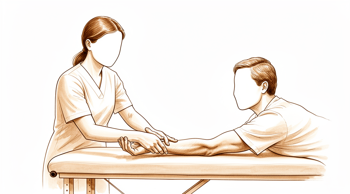
For patients: a plain-language version of this topic is available. See the patient guide.
Overview
Radial tunnel syndrome is managed conservatively as first-line therapy, with surgical decompression reserved for refractory cases despite ongoing controversy regarding diagnosis and outcomes [7]. Most publications on this uncommon upper extremity compression neuropathy are small retrospective series or case reports, meaning treatment decisions are not typically based on high levels of evidence [9]. Surgery is advocated for high radial nerve entrapment neuropathy cases resistant to conservative treatment, emphasizing the importance of dissecting the entire length of the fibrous tunnel [10]. No single surgical approach was adequate for complete visualization and release of all compression points of the radial tunnel [3].
Surgical timing and associated procedures influence outcomes. Patients who underwent surgical exploration within 3 weeks of injury had a significantly higher likelihood of regaining radial nerve function than patients who underwent nonsurgical management with or without late surgical exploration [4]. Conversely, radial nerve release performed in association with surgical treatment for lateral epicondylitis was not associated with greater improvement [1]. For a reoperation to be successful, the ulnar nerve must be examined and all potential levels of compression must be released [6]. Additionally, surgical management for elbow stiffness should evaluate the potential neurological role as root of joint contracture, and preventive nerve release and tunnel decompression must be considered when addressing surgically an elbow contracture even with normal preoperative neurological condition [12].
Advanced reconstruction options exist for severe or chronic pathology. Tendon transfers are indicated in longstanding, irreparable, isolated radial nerve lesions [18]. A vascularized ulnar nerve graft technique should be recommended for reconstruction of the median or radial nerves in selected cases [22]. Function-sparing targeted muscle reinnervation to the extensor carpi radialis brevis shows promising results of improved symptoms with unaffected wrist extension and is considered a safe and potentially impactful additional option in the treatment of refractory, symptomatic radial sensory nerve neuromas [27]. The purpose of the technical note described in the 2024 Arthroscopy Techniques article is to describe the details of endoscopic radial tunnel release [5].
Anatomy & Pathophysiology
Bony Anatomy
The elbow is a trocho-ginglymoid joint comprising medial and lateral articulations that provide bony stability [70]. The trochlea articulates with the ulna within the greater sigmoid notch to form the ulnohumeral, hinged, or trochoid portion of the joint [70]. This ulnohumeral joint exhibits highly congruent anatomy through almost 180° of articular contact, except for a bare area on the greater sigmoid notch devoid of cartilage [70]. The capitellum and radial head constitute the radiocapitellar joint [70]. The radius is held in close approximation to the ulna at the proximal radioulnar joint by the annular ligament [70]. The radial head is a concave elliptical structure covered with articular cartilage along the radiocapitellar joint and approximately 270° of the articular margin [70].
The distal humeral articulation is angled 30° from the longitudinal axis [70]. The axis of rotation is 5° to 7° angulated in the coronal plane to the epicondylar axis, with the medial side more distal than the lateral side [70]. The articular surface of the distal humerus is angled 30 degrees anterior to the humeral shaft axis [76]. In full extension, 60% of axial load is transmitted through the radiocapitellar joint [76]. The medial column diverges from the humeral shaft at a 45-degree angle, and the lateral column diverges at a 20-degree angle [78]. The trochlea has a 300-degree arc of cartilage [78].
The distal humeral shaft is triangular in cross-section with its apex directed anteriorly [81]. The medial column diverges approximately 45 degrees from the humeral shaft in the coronal plane and terminates as the medial epicondyle [81]. The lateral column diverges at approximately 20 degrees from the shaft in the coronal plane and curves anteriorly, creating a 35 to 40 degrees angle with the shaft in the sagittal plane [81]. The trochlea is covered by articular cartilage anteriorly, inferiorly, and posteriorly, creating an arc of almost 270 degrees [81]. The olecranon fossa and coronoid fossa are separated by a thin bony septum which is occasionally absent [81].
Ligamentous Anatomy
The medial epicondyle forms the attachment site for the origins of the flexor pronator mass and is larger and more posteriorly oriented than the lateral epicondyle [70]. The lateral epicondyle is the origin of the lateral extensor musculature [70]. The lateral ulnar collateral ligamentous complex originates at the geometric center of the radiocapitellar articulation, just distal to the lateral epicondyle [70].
The medial ulnar collateral ligament is the primary valgus stabilizer of the elbow [71]. The anterior bundle of the medial ulnar collateral ligament is the most important component for stability [71]. The posterior bundle of the medial ulnar collateral ligament becomes taut at flexion beyond 120 degrees [71]. The lateral ulnar collateral ligament is the posterolateral stabilizer of the elbow [71]. The medial collateral ligament originates on the posterior medial epicondyle and inserts on the sublime tubercle of the medial coronoid process [76]. The anterior bundle of the medial collateral ligament is the primary restraint to valgus stress within functional elbow range of motion [76]. The posterior bundle of the medial collateral ligament is the primary restraint to valgus stress with the elbow in maximal flexion [76].
The lateral collateral ligament complex consists of the radial collateral ligament, the lateral ulnar collateral ligament, and the annular ligament [81]. The radial collateral ligament originates from an isometric point on the lateral epicondyle and fans out to attach to the annular ligament [81]. The lateral ulnar collateral ligament arises from the isometric point on the lateral epicondyle and attaches to the crista supinatoris of the proximal ulna [81]. The lateral collateral ligament complex functions as an important restraint to varus and posterolateral rotatory instability [81].
Muscular Anatomy
The brachialis is the strongest elbow flexor and attaches to the coronoid 11 mm distal to the tip [71]. The biceps brachii inserts at the ulnar margin of the radial tuberosity and is a powerful supinator of the forearm [71]. The triceps is the primary elbow extensor and inserts on the olecranon process [71]. The mobile wad consists of the brachioradialis, extensor carpi radialis longus, and extensor carpi radialis brevis [71]. The flexor-pronator mass consists of the pronator teres, flexor carpi radialis, palmaris longus, flexor carpi ulnaris, and flexor digitorum superficialis [71].
The musculocutaneous and radial nerves contribute 42% and 27.5% respectively to elbow flexion force [29]. The biceps brachii contributes 46.7% to elbow flexion force [29]. The brachioradialis and supinator contribute 64.1% to forearm supination force [29].
Neurovascular Anatomy
The radial nerve passes through the triangular interval, which is bordered superiorly by the lower border of teres major, medially by the long head of triceps, and laterally by the shaft of humerus [71]. The radial nerve splits into the superficial sensory branch and the posterior interosseous nerve on the anterolateral side of the elbow [82]. The posterior interosseous nerve is just superficial to the anterior joint capsule at the level of the radiocapitellar joint and courses longitudinally along the medial side of the capitellum [82]. A thin layer of brachialis lies between the posterior interosseous nerve and the capsule at the level of the joint [82]. Distally at the level of the radial neck, the posterior interosseous nerve may come in direct contact with the joint capsule [82].
The ulnar nerve passes through the cubital tunnel at the medial column of the elbow [78]. The ulnar nerve enters the anterior forearm by traveling between the two heads of the flexor carpi ulnaris [78]. The ulnar nerve lies in a bony groove covered and restrained by the arcuate ligament posterior to the medial epicondyle [79]. Osborne's ligament stabilizes the ulnar nerve in the cubital tunnel [71]. The ligament of Struthers is a variant anatomy arising from a supracondylar process to attach to the medial epicondyle and is a potential site of median nerve compression [71].
Pathophysiology
Radial tunnel syndrome is classically described as a nerve compression and entrapment syndrome, but there is dispute over its etiology [35]. Signs and symptoms of radial tunnel syndrome contrast with other entrapment neuropathies by featuring prominent focal tenderness, normal neurologic function, and no confirmatory electrodiagnostic evidence of nerve dysfunction [35]. The posterior interosseous nerve carries unmyelinated group IV afferent fibers from the wrist capsule and small myelinated group IIA afferent fibers from muscles along its distribution [35]. Unmyelinated group IV fibers are associated with nociception and pain [35]. Any factor that limits radial nerve excursion at the elbow or wrist could result in repetitive traction of the nerve and play a role in the pathophysiology of a mechanical neuropathy manifesting as pain [38]. Entrapment neuropathy may produce nerve injury through ischemia or mechanical deformation of the nerve [168].
Normal ulnar nerve excursion proximal to the medial epicondyle has been reported to be as great as 10 mm and distal to the medial epicondyle as much as 6 mm [168]. The ulnar nerve may undergo as much as 4.7 mm of elongation with elbow flexion [168]. As the elbow moves from extension to flexion, the distance between the medial epicondyle and olecranon increases 5 mm for every 45 degrees of flexion [168]. Elbow flexion changes the shape of the cubital tunnel from round to a flattened oval with up to a 2.5 mm loss of height [168]. Intraneural pressures of the ulnar nerve at the cubital tunnel increase from 7 mm Hg in extension to between 11 and 24 mm Hg in flexion [168]. The humeral trochlea protrudes into the cubital tunnel during elbow flexion, causing dynamic morphologic changes in the ulnar nerve [115]. Increased elbow flexion in patients with cubital tunnel syndrome influences the intraneural blood flow of the ulnar nerve [123]. The mechanism of provocation of symptoms of cubital tunnel syndrome by the elbow flexion test cannot be explained simply by dynamic pressure in the cubital tunnel [124]. Shoulder position changes the ulnar nerve strain around the elbow in living patients with cubital tunnel syndrome [121]. Elbow flexion is the most effective way to decrease radial and median nerve tension distal to the shoulder [112].
Classification
Nomenclature: Radial tunnel syndrome and posterior interosseous nerve compression are distinct entities with different clinical presentations but share identical potential sites of nerve interference [131]. One author proposes unifying these conditions as mild and severe forms of a single disease to simplify nomenclature [131]. Conversely, radial tunnel syndrome is described as an illness construct based on a speculative pathophysiology, lacking a verifiable pathophysiology or accepted reference standard for diagnosis [34].
Other Considerations: The annual incidence rate of posterior interosseous nerve entrapment is estimated at 0.03% [26]. In a representative United States insurance database, the prevalence of radial tunnel syndrome was 0.091%, with an annual incidence of 0.0091% [154]. Approximately 5.7% of patients with radial tunnel syndrome also had a diagnosis of lateral epicondylitis on the same side within 6 months of the radial tunnel syndrome diagnosis [154].
Definitions for the degree of ulnar nerve instability at the elbow are not uniformly agreed upon, and further development of a classification system may be warranted to standardize treatment [50]. The Dellon and MacKinnon classifications categorize cubital tunnel syndrome into mild, moderate, or severe grades [165], with Dellon's Grade 3 defined as severe cubital tunnel syndrome [172]. McGowan grade I disease and grade IIB/III cubital tunnel syndrome are distinct classifications used to categorize disease severity [166]. The Bishop scoring system grades clinical outcomes as excellent, good, or fair [172].
Clinical Presentation
Radial tunnel syndrome is characterized by prominent focal tenderness, normal neurologic function, and a lack of confirmatory electrodiagnostic evidence of nerve dysfunction [35]. The diagnosis is made clinically based on activity-related pain, maximal tenderness 3–5 cm distal to the lateral epicondyle, pain exacerbation with forearm supination, radiation to the dorsoradial aspect of the forearm, and a positive Lister test [93]. A clinical diagnosis requires the presence of at least 4 of 5 diagnostic signs and symptoms, with maximal tenderness 3–5 cm distal to the lateral epicondyle being a mandatory criterion [93]. Focal tenderness at the radial tunnel differs from a positive Phalen's test in carpal tunnel syndrome because the symptoms do not occur in the distribution of the purportedly affected nerve [35].
The posterior interosseous nerve carries unmyelinated (group IV) afferent fibers from the wrist capsule and small myelinated (group IIA) afferent fibers from the muscles along its distribution [35]. Despite these anatomical details, radial tunnel syndrome is described as an illness construct based on a speculative pathophysiology with no verifiable pathophysiology or accepted reference standard for diagnosis [34].
Entrapment neuropathy of the lateral antebrachial cutaneous nerve presents as lateral elbow pain and should be considered in the differential diagnosis [100]. Electrodiagnostic evaluation can be useful in establishing and confirming the diagnosis of lateral antebrachial cutaneous nerve entrapment [100]. Patients with lateral antebrachial cutaneous nerve entrapment who present with paresthesia usually require surgical intervention because paresthesia represents a more progressive stage of the condition [100].
Investigations
Clinical Evaluation and History
The physical examination for elbow pathology is directed by the history and the specific location of the patient's pain in the anterior, posterior, medial, or lateral aspect of the elbow [32]. Pathologic entities associated with discrete compartments of the elbow aid the examiner in detecting underlying pathologic conditions [32]. The evaluation of the elbow requires an intimate understanding of anatomy, biomechanics, and diagnostic tests to understand functional biomechanics, perform physical exam maneuvers, and order appropriate imaging studies [32]. The normal elbow has a range of motion from 0° to 140° from extension to flexion and 75° and 85° in pronation and supination respectively [32]. A functional arc for the elbow is 100° for flexion and extension and forearm rotation [32]. Elbow stability is determined by primary stabilizers (ulnohumeral articulation, MUCL, LUCL complex) and secondary stabilizers (radiocapitellar articulation, common flexor tendon, common extensor tendon, joint capsule) [32].
For radial tunnel syndrome, nonsurgical management is first-line, but surgical decompression remains a viable option for refractory cases despite ongoing controversy regarding diagnosis and outcomes [7]. Most publications on uncommon compression syndromes of the radial, ulnar, and median nerves are small retrospective series or case reports, and treatment decisions are not typically based on high levels of evidence [9].
Imaging
Plain radiography: Plain radiographs remain the hallmark and the best screening test for elbow evaluation [32]. Proper selection of imaging studies can aid diagnosis and guide treatment, with each modality having specific advantages and disadvantages [32]. Radiographic evaluations are essential when diagnosing an osteochondritis dissecans (OCD) lesion of the elbow, though important aspects of the lesion may be better seen with MRI [89]. Standard AP and lateral radiographs should be obtained for the evaluation of elbow osteoarthritis [91]. Radiographs typically show osteophyte formation at the coronoid process, coronoid fossa, radial fossa, radial head, olecranon tip, and olecranon fossa in elbow osteoarthritis [91]. Joint spaces at the ulnohumeral joint are usually preserved, and those at the radiocapitellar joint are mildly narrowed in elbow osteoarthritis [91]. Radiographs typically underestimate the number of loose bodies present in elbow osteoarthritis [91]. AP, lateral, oblique, and axillary views of the elbow may reveal posteromedial olecranon osteophytes and/or loose bodies in valgus extension overload syndrome [86].
MRI: MRI is a useful diagnostic modality for confirming nerve compression, assessing space-occupying lesions, and excluding other pathologies in athletes with entrapment neuropathies of the shoulder and elbow [170]. MRI is an effective diagnostic modality for identifying primary synovial chondromatosis as a causative factor of cubital tunnel syndrome [173]. MRI can be used to evaluate ligaments and tendons in the elbow, but it is rarely indicated for stiffness [84]. MRI may be most helpful in evaluating associated injuries including partial or complete tears of the medial collateral ligament in valgus extension overload syndrome [86].
CT: CT imaging is useful to determine the paths of the radial and median nerves and their spatial relationship to heterotopic ossification at the elbow [59]. CT is helpful when assessing for malunion architecture and the location and pattern of osteophytes and/or loose bodies in the context of elbow stiffness [84]. Three-dimensional CT is used to check for heterotopic ossification in the evaluation of elbow stiffness [84]. CT with two-dimensional reconstruction and three-dimensional surface rendering best visualizes the pathology of valgus extension overload syndrome [86]. CT may be useful for surgical planning in elbow osteoarthritis, allowing a detailed assessment of osteophytes and the presence of loose bodies [91].
Electrodiagnostic Testing
Electromyography/nerve conduction velocity studies should be performed if any question about neurologic dysfunction exists during the evaluation of elbow stiffness [84]. Most clinicians order preoperative tests before offering surgery for cubital tunnel syndrome, but only 6% order both electrodiagnostic studies and ultrasound as per expert consensus [40].
Treatment
Non-Operative
Nonsurgical management is the first-line treatment for radial tunnel syndrome [7]. Treatment for radial nerve palsy is usually non-operative with satisfactory results [126]. Splinting of the wrist remains the mainstay of conservative treatment for carpal tunnel syndrome [133].
Operative
Indications: Surgical decompression is a viable option for refractory cases of radial tunnel syndrome [7]. Surgery is indicated for radial nerve transection or lack of improvement after conservative treatment [126]. Surgery is advocated for high radial nerve entrapment neuropathy cases resistant to conservative treatment [10]. The radial nerve recovered better if repaired or reconstructed within 5 months of injury [25].
Surgical Approach / Technique: No single approach was adequate for complete visualization and release of all compression points of the radial tunnel [3]. A single brachioradialis-splitting approach is adequate for complete visualization and release of all compression sites of the radial tunnel [14]. The purpose of the described technical note is to describe the details of endoscopic radial tunnel release [5]. Both methods compared in the study can quickly and safely expose the radial nerve in the posterior humeral approach [17]. An adequate preoperative evaluation of compression level may help determine the most suitable surgical approach to treat radial nerve entrapment with minimal damage to surrounding tissues [26]. Surgical exploration and decompression may be carried out along the course of the nerve for radial nerve entrapment around the elbow [26]. The authors advocate for dissecting the entire length of the fibrous tunnel in high radial nerve entrapment neuropathy [10]. Anatomic knowledge of the course of the superficial radial nerve and its branches is important during open release for avoiding nerve injury [21]. The procedure offers complete release of constricting structures while preserving blood supply to the nerve and allowing early postoperative elbow mobilization [28].
Outcomes and Efficacy: Radial nerve release, in association with surgical treatment for lateral epicondylitis, was not associated with greater improvement [1]. Improvement in pain and function after cubital tunnel release may be associated with an improvement in symptoms both within and outside the ulnar nerve distribution [2]. Current evidence suggests that different surgical methods to treat ulnar neuropathy at the elbow do not differ in their clinical outcomes [13]. Most cases of ulnar nerve compression improve with nonsurgical treatment and the majority get better with surgical decompression [57]. The endoscopic approach facilitates inspection of the ulnar nerve so that selective release of the tissue that compresses the nerve can readily be performed [16]. Endoscopic techniques to release the ulnar nerve at the elbow have been presented as safe and effective since 1992, though the procedure is not yet standardized [15]. Increasing evidence shows minimally invasive open in situ release to be both safe and effective for the relief of ulnar nerve compression symptoms [11]. The described surgical technique provides surgeons with the ability to directly decompress the ulnar nerve while decreasing postoperative complications such as instability and adhesion formation [113]. Decompression of the ulnar nerve under local anaesthetic is a reliable procedure, which is well tolerated by the majority of patients [145]. Good pain control can be achieved during all steps of the wide-awake surgery using local anaesthesia for ulnar nerve decompression and medial epicondylectomy [150]. The authors describe a 2-stage local anesthetic injection method that safely introduces local anesthetic within the cubital tunnel, achieving comfort, sufficient visualization, and the ability to decompress multiple compression sites through a minimal incision [138]. PINN can provide relief in patient's chronic wrist pain [169]. Neurolysis of the superficial radial nerve offers the opportunity for pain relief, but it does not reliably produce success [55].
Nerve Reconstruction and Transfers: The median nerve provides a reliable source of donor nerve fascicles for radial nerve reinnervation [56]. A vascularized ulnar nerve graft technique should be recommended for reconstruction of the median or radial nerves in selected cases [22]. Avoiding donor harvest from another location may help with postoperative pain management and facilitate mobilization when using the superficial radial nerve as a donor for grafting ipsilateral forearm nerve defects [155]. Transhumeral radial nerve transposition allowed less worrisome dissection during revision surgery despite multiple previous procedures [30]. The technique of radial nerve transposition with humeral fracture fixation is safe, does not cause iatrogenic injury, and protects the radial nerve during all subsequent approaches to the fracture site [51].
Complications and Safety Considerations: The radial nerve must be identified and protected in all cases to prevent palsy during treatment of distal third humeral shaft fractures with posterior minimally invasive plate osteosynthesis [20]. Return of function is less likely when power instruments or ultrasound is used for cement removal, while formal exposure and protection of the radial nerve are predictive of recovery [24]. Immediate surgical removal of the extruded cement and decompression of the radial nerve may aid in recovery of nerve function after thermal injury due to extruded cement [23]. In cadaveric analysis, both the modified proximal and direct lateral portals provide adequate distance from the radial nerve and may be safe for clinical use [117]. The data help predict the humeral course of the radial nerve and define a safe zone for pin implantation, but due to variability, a safe zone cannot fully ensure prevention of iatrogenic injury [180].
Complications
Iatrogenic Nerve Injury: Arthroscopic treatment for lateral epicondylitis carries a risk of radial nerve or posterior interosseous nerve (PIN) transection, with reported cases involving 60% radial nerve transection and complete PIN transection [49]. In humeral shaft fracture surgery, iatrogenic transient radial nerve dysfunction occurs in approximately 1 in 5 patients with lateral exposure, 1 in 9 with posterior exposure, and 1 in 25 with anterolateral exposure [204]. Radial nerve palsy could not be completely avoided when using the extended delto-pectoral approach for complex humeral shaft fractures [189]. Other causes of radial nerve injury include superficial thrombophlebitis leading to entrapment of the radial nerve branch [187] and axonamonosis caused by impingement from distal locking screws during humeral nailing [196]. Ulnar nerve dysfunction at the elbow after platelet-rich plasma treatment for partial ulnar collateral ligament injuries may be more likely in patients presenting with a subluxing ulnar nerve [192].
Surgical Failure and Recurrence: Cubital tunnel surgery is considered a failure if symptoms do not relieve or recur shortly after the procedure [37]. Revision surgery for recurrent or persistent cubital tunnel syndrome yields less predictable and satisfying results than primary surgery [66]. The risk of revision cubital tunnel release is low, with an incidence of failure requiring ipsilateral revision surgery remaining steadily low at 1.4% during the study period [43, 47]. Revision surgery was required in 3.2% of cases following in situ ulnar nerve decompression for idiopathic cubital tunnel syndrome [114]. The rate of revision surgery following cubital tunnel release with transposition is quite low, with no major differences in revision rates among different types of surgical transposition [193]. Endoscopic cubital tunnel release has a recurrence rate that is not higher than open cubital tunnel release literature controls [195]. Reoperation after primary surgery for patients who fail conservative treatment gives satisfactory results [136].
Causes of Persistent Symptoms: Persistent symptoms after cubital tunnel release are most often associated with incorrect diagnosis or incomplete release [148]. Incomplete release of all compressive sites is the most common cause of persistent cubital tunnel syndrome, occurring in 39% to 90% of cases [63]. Perineural adhesions, fascicular scar, and fibrosis are present in 79% of persistent cubital tunnel syndrome cases [63]. Recurrent symptoms are most often associated with postoperative fibrosis [148]. Mechanical neural "kinking" after transposition occurs in 1.4% of persistent cubital tunnel syndrome cases [63]. Median antebrachial cutaneous nerve (MABCN) neuroma is a cause of new symptoms after cubital tunnel release, occurring in 32% of cases [63].
Complication Rates: Short-term complication rates for cubital tunnel surgery are low at 5.6% overall [64]. The short-term complication rate for ulnar nerve transposition is 9.6%, compared to 3.6% for in situ cubital tunnel release [64]. The difference in complications between simple decompression and ulnar nerve transposition is significant, indicating greater odds of complications with ulnar nerve transposition [203]. The secondary surgery rate after cubital tunnel surgery was 5.7% overall, with higher rates for patients with prior elbow trauma and those undergoing ulnar nerve transposition [67]. In ulnar nerve anterior transmuscular transposition, minor complications occurred in 24 cases, most of which were seromas or ecchymoses [106]. Major complications requiring a second operation occurred in 4.3% of cases, and reoperation for symptom recurrence was needed in 2.5% of cases [106]. Good or excellent results were achieved in 89% of patients with ulnar nerve decompression and submuscular transposition, with a low complication rate [198]. Preoperative antibiotics do not significantly decrease the risk for postoperative infections in patients undergoing uncomplicated ulnar nerve releases at the elbow [194].
Recovery and Prognosis: Patients who underwent surgical exploration within 3 weeks of radial nerve injury had a significantly higher likelihood of regaining radial nerve function than those managed nonsurgically [4]. Spontaneous recovery occurs in the majority of patients with primary or secondary radial nerve palsy associated with closed humeral shaft fracture [52]. Late surgery for persistent radial nerve palsy in the setting of humeral shaft fracture gives a 69% chance of recovery and carries a 31% risk of no recovery at the end of treatment [162]. Operative treatment for humeral shaft fractures results in a similar permanent radial nerve palsy rate compared to nonoperative treatment [157]. Immediate surgical removal of extruded cement and decompression of the radial nerve may aid in recovery of nerve function after thermal injury during total elbow arthroplasty [23]. Return of radial nerve function is less likely when power instruments or ultrasound is used for cement removal during total elbow arthroplasty revision, whereas formal exposure and protection of the radial nerve are predictive of recovery [24]. Spontaneous recovery of radial nerve function following axonamonosis caused by impingement from distal locking screws may be more common than previously assumed [196]. Posterior interosseous nerve palsy after distal biceps repair typically resolves within 3 months, and at the latest, 5 months after surgery [202]. Nerve grafting for high radial nerve injury achieved relatively good wrist extension but poor thumb extension [68]. Outcomes are better in patients who underwent nerve transfer versus tendon transfer procedures for radial nerve paralysis reconstruction [200].
Recovery
Spontaneous Recovery and Prognosis: Spontaneous recovery occurs in 70%–88% of radial nerve injuries [125]. In primary or secondary radial nerve palsy associated with closed humeral shaft fracture, spontaneous recovery occurs in the majority of patients [52]. Patients with a congenital radial nerve palsy typically achieve full spontaneous recovery by 3 months of age [129].
Surgical Timing and Outcomes: In clinical settings in which the likelihood of spontaneous recovery of nerve function is low or when an informed patient has a strong preference for surgery, early surgery may optimize outcome [184]. Immediate surgical removal of the extruded cement and decompression of the radial nerve may aid in recovery of nerve function [23].
Reconstructive Interventions: Tendon transfers are one reconstructive option in a ladder of reconstructive alternatives that can improve function after injury to the radial, median, or ulnar nerves [182]. Early tendon transfer quickly restored efficient grip while awaiting reinnervation of wrist extensors, avoiding the need for prolonged external splintage [181]. Successful recovery of radial nerve lacerations can be achieved after nerve grafting [65]. Good to excellent motor recovery can be expected in 83% of patients who have a large gap of the radial nerve when sural nerve autografts are sutured to the areas that have motor fascicles dominantly [179].
Rehabilitation and Adjuncts: Customized dynamic splints promoted optimal function and recovery in a patient with high radial nerve injury by addressing functional goals and tissue biological needs without the need for wire bending or outriggers [128].
Other Considerations: Surgically treated humeral shaft fractures associated with radial nerve palsies are expected to show signs of neurologic recovery during the first 6 months [185]. These patients should recover completely after 12 months of follow-up in almost all cases [185].
Key Evidence
- [L1] Radial nerve release, in association with surgical treatment for lateral epicondylitis, was not associated with greater improvement. [1] (10.1016/j.jhsa.2018.06.009)
- [L3] Improvement in pain and function after cubital tunnel release may be associated with an improvement in symptoms both within and outside the ulnar nerve distribution. [2] (10.1007/s11552-014-9688-9)
- [L5] No single approach was adequate for complete visualization and release of all compression points of the radial tunnel. [3] (10.1016/j.jhsa.2015.03.009)
- [L2] Patients who underwent surgical exploration within 3 weeks of injury had a significantly higher likelihood of regaining radial nerve function than patients who underwent nonsurgical management with or without late surgical exploration. [4] (10.5435/jaaos-d-18-00142)
- [L5] The purpose of this Technical Note is to describe the details of endoscopic radial tunnel release. [5] (10.1016/j.eats.2024.103391)
- [L4] For a reoperation to be successful, the ulnar nerve must be examined and all potential levels of compression must be released. [6] (10.2106/jbjs.24.00493)
- [L4] The article reviews the anatomy, diagnosis, and treatment of radial tunnel syndrome, noting that while nonsurgical management is first-line, surgical decompression remains a viable option for refractory cases despite ongoing controversy regarding diagnosis and outcomes. [7] (10.5435/jaaos-d-23-00314)
- [L4] This article reviews uncommon compression syndromes of the radial, ulnar, and median nerves, noting that most publications are small retrospective series or case reports and treatment decisions are not typically based on high levels of evidence. [9] (10.1016/j.hcl.2013.04.014)
- [Case_report] The authors advocate for surgery in high radial nerve entrapment neuropathy cases resistant to conservative treatment, emphasizing the importance of dissecting the entire length of the fibrous tunnel. [10] (10.1016/j.jse.2025.02.060)
- [L4] Increasing evidence shows minimally invasive open in situ release to be both safe and effective for the relief of ulnar nerve compression symptoms. [11] (10.1016/j.hcl.2013.08.019)
- [L5] Surgical management for elbow stiffness should evaluate the potential neurological role as root of joint contracture, and preventive nerve release and tunnel decompression must be considered when addressing surgically an elbow contracture even with normal preoperative neurological condition. [12] (10.1016/j.jisako.2023.12.007)
- [L4] Current evidence suggests that different surgical methods to treat ulnar neuropathy at the elbow do not differ in their clinical outcomes. [13] (10.1016/j.hcl.2013.04.013)
- [L5] A single brachioradialis-splitting approach is adequate for complete visualization and release of all compression sites of the radial tunnel. [14] (10.1177/1558944717750916)
- [L5] The authors state that endoscopic techniques to release the ulnar nerve at the elbow have been presented as safe and effective since 1992, though the procedure is not yet standardized, and their study aimed to present a novel endoscopically assisted technique. [15] (10.1016/j.jhsa.2008.01.022)
- [L4] The endoscopic approach facilitates inspection of the ulnar nerve so that selective release of the tissue that compresses the nerve can readily be performed. [16] (10.1177/1753193408094443)
- [L5] Both methods can quickly and safely expose the radial nerve. [17] (10.1186/s12891-023-06291-3)
- [L3] Tendon transfers are indicated in longstanding, irreparable, isolated radial nerve lesions. [18] (10.1016/j.jhsa.2007.10.015)
- [L4] The radial nerve must be identified and protected in all cases to prevent palsy. [20] (10.1016/j.xrrt.2023.08.006)
- [L5] Anatomic knowledge of the course of the superficial radial nerve and its branches is important during open release for avoiding nerve injury. [21] (10.1016/j.jhsa.2013.12.004)
- [L4] This technique should be recommended for reconstruction of the median or radial nerves in selected cases. [22] (10.1016/j.jhsa.2005.03.017)
- [L4] Immediate surgical removal of the extruded cement and decompression of the radial nerve may aid in recovery of nerve function. [23] (10.1016/j.jse.2013.10.002)
- [L4] Return of function is less likely when power instruments or ultrasound is used for cement removal, while formal exposure and protection of the radial nerve are predictive of recovery. [24] (10.1016/j.jse.2010.08.012)
- [L4] The radial nerve recovered better if repaired or reconstructed within 5 months of injury. [25] (10.1177/1753193409360283)
- [L5] [26] (10.1016/j.jisako.2024.03.001)
- [L4] This technique shows promising results of improved symptoms with unaffected wrist extension and is considered a safe and potentially impactful additional option in the treatment of refractory, symptomatic radial sensory nerve neuromas. [27] (10.1016/j.jhsg.2022.01.006)
- [L5] The procedure offers complete release of constricting structures while preserving blood supply to the nerve and allowing early postoperative elbow mobilization. [28] (10.1016/s0749-0712(21)00325-5)
- [L4] The musculocutaneous and radial nerves contribute 42% and 27.5% respectively to elbow flexion force, while the biceps brachii contributes 46.7% and the brachioradialis and supinator contribute 64.1% to forearm supination force. [29] (10.1177/1753193408087036)
- [L4] Transhumeral radial nerve transposition allowed less worrisome dissection during revision surgery despite multiple previous procedures. [30] (10.1016/j.jhsa.2017.04.008)
- [L5] Radial tunnel syndrome is an illness construct based on a speculative pathophysiology with no verifiable pathophysiology or accepted reference standard for diagnosis. [34] (10.1016/j.jhsa.2010.03.020)
- [L5] [35] (10.1016/j.jhsa.2009.10.016)
- [L5] Cubital tunnel surgery should be considered a failure if patients have no relief of their symptoms or if the symptoms recur shortly after the surgery. [37] (10.1016/s0749-0712(21)00329-2)
- [L5] Any factor that limits excursion at these sites could result in repetitive traction of the nerve and possibly could play a role in the pathophysiology of a mechanical neuropathy, which in the case of the radial nerve most often manifests as pain. [38] (10.1016/j.jhsa.2005.06.008)
- [L4] Most clinicians order preoperative tests before offering surgery for cubital tunnel syndrome, but only 6% order both EDX and ultrasound as per expert consensus. [40] (10.1177/17531934261434155)
- [L3] The risk of revision cubital tunnel release was low. [43] (10.1097/corr.0000000000002629)
- [L4] The incidence of failure requiring ipsilateral revision surgery after cubital tunnel release remained steadily low (1.4%) during the study period. [47] (10.1016/j.jse.2016.10.028)
- [L4] [49] (10.1016/j.jhsa.2012.01.038)
- [L4] Definitions for the degree of ulnar nerve instability at the elbow are not uniformly agreed upon, and further development of a classification system may be warranted to standardize treatment. [50] (10.1055/s-0038-1665548)
- [L4] The technique is safe, does not cause iatrogenic injury, and protects the radial nerve during all subsequent approaches to the fracture site. [51] (10.1097/01.blo.0000072470.32680.60)
- [L4] In primary or secondary radial nerve palsy associated with closed humeral shaft fracture, spontaneous recovery occurs in the majority of patients, so initial nonsurgical management is recommended. [52] (10.1016/j.jhsa.2008.05.029)
- [L4] [55] (10.1177/1753193407087892)
- [L4] [56] (10.1016/j.jhsa.2010.09.034)
- [L4] Most cases of ulnar nerve compression improve with nonsurgical treatment and the majority get better with surgical decompression. [57] (10.1007/s12593-009-0020-9)
- [L4] This study demonstrates the usefulness of CT imaging to determine the paths of the radial and median nerves and their spatial relationship to HO at the elbow. [59] (10.1016/j.jse.2014.12.030)
- [L5] [63] (10.5435/jaaos-d-20-01381)
- [L3] The short-term complication rates of cubital tunnel surgery are low (5.6%), but higher for ulnar nerve transposition (9.6%) than in situ cubital tunnel release (3.6%). [64] (10.1016/j.jhsa.2016.07.033)
- [L4] Successful recovery of radial nerve lacerations can be achieved after nerve grafting. [65] (10.1016/j.jhsa.2014.05.036)
- [L4] Results of revision surgery for recurrent or persistent cubital tunnel syndrome are less predictable and satisfying than primary surgery. [66] (10.1016/j.jhsa.2011.11.024)
- [L4] The secondary surgery rate after cubital tunnel surgery was 5.7% overall, but higher for patients with prior elbow trauma and for patients undergoing ulnar nerve transposition. [67] (10.1016/j.jhsa.2017.01.020)
- [L4] Nerve grafting for high radial nerve injury achieved relatively good wrist extension but poor thumb extension and is affected by certain prognostic factors. [68] (10.1177/17531934221147651)
- [L2] [93] (10.1016/j.jhsa.2024.09.023)
- [L4] [100] (10.1016/j.jhsa.2004.06.011)
- [L4] [106] (10.1016/j.jse.2018.11.054)
- [L5] Elbow flexion was the most effective way to decrease nerve tension, while elbow extension should be avoided when implanting the humeral component. [112] (10.1016/j.jseint.2024.03.013)
- [L4] The described surgical technique provides surgeons with the ability to directly decompress the ulnar nerve while decreasing postoperative complications such as instability and adhesion formation. [113] (10.1016/j.jseint.2019.11.004)
- [L3] [114] (10.1016/j.jhsa.2015.12.012)
- [L5] The humeral trochlea protrudes into the cubital tunnel during elbow flexion, causing dynamic morphologic changes in the ulnar nerve. [115] (10.1016/j.jse.2022.05.026)
- [L5] In cadaveric analysis, both the modified proximal and direct lateral portals provide adequate distance from the radial nerve and may be safe for clinical use. [117] (10.1016/j.arthro.2017.06.012)
- [L4] To the best of our knowledge, this is the first study showing that shoulder position changes the ulnar nerve strain around the elbow in living patients with CubTS. [121] (10.1016/j.jse.2015.01.014)
- [L3] Increased elbow flexion in patients with CuTS influences the intraneural blood flow of the ulnar nerve. [123] (10.1016/j.jhsa.2021.06.024)
- [L3] The mechanism of provocation of symptoms of cubital tunnel syndrome by the elbow flexion test could not be explained simply by dynamic pressure in the cubital tunnel, and other pathophysiological factors could also be contributing. [124] (10.1016/j.jhsa.2010.11.013)
- [L4] The choice of surgical technique and timing depends on the type of trauma, site of injury, and time elapsed since injury, with spontaneous recovery occurring in 70%–88% of radial nerve injuries. [125] (10.1177/17531934241240867)
- [L5] Treatment is usually non-operative with satisfactory results, though surgery is indicated for nerve transection or lack of improvement after conservative treatment. [126] (10.1302/2058-5241.1.000028)
- [Case_report] Customized dynamic splints promoted optimal function and recovery in a patient with high radial nerve injury by addressing functional goals and tissue biological needs without the need for wire bending or outriggers. [128] (10.1197/j.jht.2006.11.013)
- [L4] Typically, patients with a congenital radial nerve palsy achieve full spontaneous recovery by 3 months of age whether formal therapy and/or splinting was utilized or not. [129] (10.1016/j.jhsa.2014.08.040)
- [L5] Radial tunnel syndrome (RTS) and posterior interosseous nerve (PIN) compression are distinct entities with different clinical presentations but share identical potential sites of nerve interference; the author proposes unifying them as mild (RTS) and severe (PIN compression) forms of one disease to simplify nomenclature. [131] (10.1177/1753193420953990)
- [L5] Splinting of the wrist remains the mainstay of conservative treatment for carpal tunnel syndrome. [133] (10.1016/s0749-0712(21)00306-1)
- [L4] Reoperation after primary surgery of cubital tunnel syndrome gave satisfactory results for patients who fail conservative treatment. [136] (10.1055/s-2001-19937)
- [L4] The authors describe a 2-stage local anesthetic injection method that safely introduces local anesthetic within the cubital tunnel, achieving comfort, sufficient visualization, and the ability to decompress multiple compression sites through a minimal incision. [138] (10.1016/j.jhsa.2022.04.004)
- [L4] Decompression of the ulnar nerve under local anaesthetic is a reliable procedure, which is well tolerated by the majority of patients. [145] (10.1054/jhsb.2000.0525)
- [L5] [148] (10.1177/17531934241238533)
- [L4] Good pain control can be achieved during all steps of the procedure. [150] (10.1177/17531934241252518)
- [L3] [154] (10.1016/j.jhsa.2023.03.007)
- [L4] Avoiding donor harvest from another location may help with postoperative pain management and facilitate mobilization. [155] (10.1177/1753193418796753)
- [L1] Furthermore, operative treatment results in a similar permanent radial nerve palsy rate, despite its inherent additional surgery-related risks. [157] (10.1016/j.jse.2020.01.072)
- [L5] [162] (10.5435/jaaos-d-17-00325)
- [L4] [165] (10.1016/j.jhsa.2014.04.030)
- [L3] [166] (10.1016/j.jse.2014.12.015)
- [L5] [168] (10.1016/s0749-0712(21)00323-1)
- [L4] PINN can provide relief in patient's chronic wrist pain. [169] (10.1177/1558944717692093)
- [Paper] MRI is a useful diagnostic modality for confirming nerve compression, assessing space-occupying lesions, and excluding other pathologies in athletes with entrapment neuropathies of the shoulder and elbow. [170] (10.1016/j.csm.2006.03.005)
- [L1] [172] (10.1227/01.neu.0000145854.38234.81)
- [Case_report] MRI is an effective diagnostic modality, and clinicians should be aware of primary synovial chondromatosis as a causative factor of cubital tunnel syndrome. [173] (10.1177/1758573216683396)
- [L4] Good to excellent motor recovery can be expected in 83% of patients who have a large gap of the radial nerve when sural nerve autografts are sutured to the areas that have motor fascicles dominantly. [179] (10.1016/j.jhsa.2007.10.004)
- [L5] The data help predict the humeral course of the radial nerve and define a safe zone for pin implantation, but due to variability, a safe zone cannot fully ensure prevention of iatrogenic injury. [180] (10.1016/j.jhsa.2014.03.019)
- [L4] Early tendon transfer quickly restored efficient grip while awaiting reinnervation of wrist extensors, avoiding the need for prolonged external splintage. [181] (10.1177/1753193410384696)
- [L5] Tendon transfers are one reconstructive option in a ladder of reconstructive alternatives that can improve function after injury to the radial, median, or ulnar nerves. [182] (10.5435/jaaos-21-11-675)
- [L2] In clinical settings in which the likelihood of spontaneous recovery of nerve function is low or when an informed patient has a strong preference for surgery, early surgery may optimize outcome. [184] (10.1016/j.jhsa.2008.12.029)
- [L3] Surgically treated humeral shaft fractures associated with radial nerve palsies are expected to show signs of neurologic recovery during the first 6 months and should recover completely after 12 months of follow-up in almost all cases. [185] (10.1016/j.jhsa.2024.11.024)
- [Case_report] Superficial thrombophlebitis resulting in entrapment of the radial nerve branch in the forearm is a rare yet noteworthy condition. [187] (10.1186/s12891-024-07545-4)
- [L4] No increased morbidity was attributed to the open surgical approach, though radial nerve palsy could not be completely avoided. [189] (10.1186/s12891-024-07915-y)
- [L4] This complication may be more likely to develop in patients who present with a subluxing ulnar nerve. [192] (10.1016/j.xrrt.2020.11.006)
- [L3] The rate of revision surgery following cubital tunnel release with transposition is quite low, and there do not appear to be major differences in the rate of revision among the different types of surgical transposition, indicating that a true subcutaneous transposition may be adequate. [193] (10.1016/j.jhsg.2025.100815)
- [L4] Preoperative antibiotics do not demonstrate benefit for patients undergoing uncomplicated ulnar nerve releases at the elbow and do not significantly decrease the risk for postoperative infections in patients, regardless of patient comorbidities. [194] (10.1177/15589447221107688)
- [L4] We are 95% confident that our true recurrence rate is between 0.02% and 5.24% and that endoscopic cubital tunnel release has a recurrence rate, which is not higher than open cubital tunnel release literature controls. [195] (10.1007/s11552-009-9227-2)
- [L4] The case demonstrates spontaneous recovery of radial nerve function following axonamonosis caused by impingement from distal locking screws, suggesting that this phenomenon may be more common than previously assumed and that the risk could be reduced by using a mini-open method during screw insertion. [196] (10.1111/sae.12018)
- [L4] Overall, good or excellent results were achieved in 89% of patients with a low complication rate. [198] (10.1016/j.jhsa.2013.09.017)
- [L4] Overall, we observed better outcomes in those who underwent nerve transfer versus tendon transfer procedures. [200] (10.1016/j.jhsa.2019.12.009)
- [L4] These injuries typically resolve within 3 months, and at the latest, 5 months after surgery. [202] (10.1016/j.jse.2012.08.001)
- [L1] However, the difference in complications between SD versus UNT was significant, indicating greater odds of complications with UNT. [203] (10.1055/s-0038-1670928)
- [L4] Iatrogenic transient dysfunction of the radial nerve occurs in approximately 1 in 5 patients treated with lateral exposure, 1 in 9 with posterior exposure, and 1 in 25 with anterolateral exposure. [204] (10.1016/j.jse.2015.07.012)
See Also
- Radial Tunnel Syndrome
- Neuropathies
- Cubital Tunnel Syndrome
- Elbow Osteoarthritis
- Cubital Tunnel Release
- Ligament Injuries
- Elbow Arthroplasty
- Distal biceps repair
References
[1] Effect of Radial Nerve Release on Lateral Epicondylitis Outcomes: A Prospective, Randomized, Double-Blinded Trial. The Journal of Hand Surgery. 2019. DOI: 10.1016/j.jhsa.2018.06.009
[2] Prospective Cohort Study of Symptom Resolution outside of the Ulnar Nerve Distribution following Cubital Tunnel Release. HAND. 2014. DOI: 10.1007/s11552-014-9688-9
[3] Anatomical Study of the Surgical Approaches to the Radial Tunnel. The Journal of Hand Surgery. 2015. DOI: 10.1016/j.jhsa.2015.03.009
[4] Radial Nerve Palsy Recovery With Fractures of the Humerus: An Updated Systematic Review. Journal of the American Academy of Orthopaedic Surgeons. 2020. DOI: 10.5435/jaaos-d-18-00142
[5] Endoscopic Radial Tunnel Release. Arthroscopy Techniques. 2024. DOI: 10.1016/j.eats.2024.103391
[6] Risk Factors for Lumbar Spinal Fusion Reoperation: An Unusual Suspect. Journal of Bone and Joint Surgery. 2024. DOI: 10.2106/jbjs.24.00493
[7] Radial Tunnel Syndrome: Review and Best Evidence. Journal of the American Academy of Orthopaedic Surgeons. 2023. DOI: 10.5435/jaaos-d-23-00314
[9] Uncommon Upper Extremity Compression Neuropathies. Hand Clinics. 2013. DOI: 10.1016/j.hcl.2013.04.014
[10] High radial nerve entrapment neuropathy: an anatomical cadaver study and case report. Journal of Shoulder and Elbow Surgery. 2025. DOI: 10.1016/j.jse.2025.02.060
[11] Minimal-Incision In Situ Ulnar Nerve Decompression at the Elbow. Hand Clinics. 2014. DOI: 10.1016/j.hcl.2013.08.019
[12] Causes, symptoms, and treatments of nerve entrapments around the elbow: Current concepts. Journal of ISAKOS. 2024. DOI: 10.1016/j.jisako.2023.12.007
[13] Ulnar Neuropathy at the Elbow. Hand Clinics. 2013. DOI: 10.1016/j.hcl.2013.04.013
[14] Transbrachioradialis Approach to the Radial Tunnel: An Anatomic Study of 5 Potential Compression Sites. HAND. 2018. DOI: 10.1177/1558944717750916
[15] Endoscopic Decompression of the Ulnar Nerve at the Elbow. The Journal of Hand Surgery. 2008. DOI: 10.1016/j.jhsa.2008.01.022
[16] Endoscopic Anatomical Nerve Observation and Minimally Invasive Management of Cubital Tunnel Syndrome. Journal of Hand Surgery (European Volume). 2008. DOI: 10.1177/1753193408094443
[17] Compare the clinical value of two minimally invasive approaches to locating radial nerve in the posterior humeral approach. BMC Musculoskeletal Disorders. 2023. DOI: 10.1186/s12891-023-06291-3
[18] An Analysis of Results After Selective Tendon Transfers Through the Interosseous Membrane to Provide Selective Finger and Thumb Extension in Chronic Irreparable Radial Nerve Lesions. The Journal of Hand Surgery. 2008. DOI: 10.1016/j.jhsa.2007.10.015
[20] Treatment of distal third humeral shaft fractures with posterior minimally invasive plate osteosynthesis (MIPO) with segmental isolation of the radial nerve: minimum one-year follow-up. JSES Reviews, Reports, and Techniques. 2024. DOI: 10.1016/j.xrrt.2023.08.006
[21] The Relationship of the Superficial Radial Nerve and Its Branch to the Thumb to the First Extensor Compartment. The Journal of Hand Surgery. 2014. DOI: 10.1016/j.jhsa.2013.12.004
[22] Vascularized Ulnar Nerve Graft for Reconstruction of a Large Defect of the Median or Radial Nerves After Severe Trauma of the Upper Extremity. The Journal of Hand Surgery. 2005. DOI: 10.1016/j.jhsa.2005.03.017
[23] Radial nerve recovery after thermal injury due to extruded cement during humeral revision in total elbow arthroplasty. Journal of Shoulder and Elbow Surgery. 2013. DOI: 10.1016/j.jse.2013.10.002
[24] Radial nerve palsy after humeral revision in total elbow arthroplasty. Journal of Shoulder and Elbow Surgery. 2011. DOI: 10.1016/j.jse.2010.08.012
[25] Outcomes of Nerve Reconstruction for Radial Nerve Injuries Based on the Level of Injury in 244 Operative Cases. Journal of Hand Surgery (European Volume). 2010. DOI: 10.1177/1753193409360283
[26] Current concepts of surgical approach for radial nerve entrapment around the elbow. Journal of ISAKOS. 2024. DOI: 10.1016/j.jisako.2024.03.001
[27] Treatment of Refractory Radial Sensory Neuroma With Function-Sparing Targeted Muscle Reinnervation to the Extensor Carpi Radialis Brevis. Journal of Hand Surgery Global Online. 2023. DOI: 10.1016/j.jhsg.2022.01.006
[28] CUBITAL TUNNEL SYNDROME. Hand Clinics. 1996. DOI: 10.1016/s0749-0712(21)00325-5
[29] The Role of the Musculocutaneous and Radial Nerves in Elbow Flexion and Forearm Supination: A Biomechanical Study. Journal of Hand Surgery (European Volume). 2008. DOI: 10.1177/1753193408087036
[30] Transhumeral Anterior Radial Nerve Transposition to Simplify Anticipated Future Humeral Reconstruction. The Journal of Hand Surgery. 2017. DOI: 10.1016/j.jhsa.2017.04.008
[32] Orthopaedic Knowledge Update 13 Ebook Without Multimedia. Anatomy, Biomechanics, Physical Examination, and Imaging of the Elbow > Summary and Conclusions.
[34] Radial Tunnel Syndrome. The Journal of Hand Surgery. 2010. DOI: 10.1016/j.jhsa.2010.03.020
[35] Unusual Compression Neuropathies of the Forearm, Part I: Radial Nerve. The Journal of Hand Surgery. 2009. DOI: 10.1016/j.jhsa.2009.10.016
[37] CUBITAL TUNNEL SURGERY. Hand Clinics. 1996. DOI: 10.1016/s0749-0712(21)00329-2
[38] Radial Nerve Excursion and Strain at the Elbow and Wrist Associated With Upper-Extremity Motion. The Journal of Hand Surgery. 2005. DOI: 10.1016/j.jhsa.2005.06.008
[40] Preoperative tests before cubital tunnel decompression in 2025. Journal of Hand Surgery (European Volume). 2026. DOI: 10.1177/17531934261434155
[43] What Factors Are Associated With Revision Cubital Tunnel Release Within 3 Years?. Clinical Orthopaedics & Related Research. 2023. DOI: 10.1097/corr.0000000000002629
[47] Risk factors for revision surgery following isolated ulnar nerve release at the cubital tunnel: a study of 25,977 cases. Journal of Shoulder and Elbow Surgery. 2017. DOI: 10.1016/j.jse.2016.10.028
[49] Nerve Injuries Resulting From Arthroscopic Treatment of Lateral Epicondylitis: Report of 2 Cases. The Journal of Hand Surgery. 2012. DOI: 10.1016/j.jhsa.2012.01.038
[50] Decision-Making Factors for Ulnar Nerve Transposition in Cubital Tunnel Surgery. Journal of Wrist Surgery. 2018. DOI: 10.1055/s-0038-1665548
[51] Radial Nerve Transposition With Humeral Fracture Fixation. Clinical Orthopaedics & Related Research. 2003. DOI: 10.1097/01.blo.0000072470.32680.60
[52] Current Treatment of Radial Nerve Palsy Following Fracture of the Humeral Shaft. The Journal of Hand Surgery. 2008. DOI: 10.1016/j.jhsa.2008.05.029
[55] Neurolysis of the Distal Superficial Radial Nerve for Dysaesthesia Due to Nerve Tethering. Journal of Hand Surgery (European Volume). 2008. DOI: 10.1177/1753193407087892
[56] Clinical Outcomes Following Median to Radial Nerve Transfers. The Journal of Hand Surgery. 2011. DOI: 10.1016/j.jhsa.2010.09.034
[57] Cubital tunnel syndrome – Review of current literature on causes, diagnosis and treatment. Journal of Hand and Microsurgery. 2009. DOI: 10.1007/s12593-009-0020-9
[59] Preoperative nerve imaging using computed tomography in patients with heterotopic ossification of the elbow. Journal of Shoulder and Elbow Surgery. 2015. DOI: 10.1016/j.jse.2014.12.030
[63] Management of Recalcitrant Cubital Tunnel Syndrome. Journal of the American Academy of Orthopaedic Surgeons. 2021. DOI: 10.5435/jaaos-d-20-01381
[64] Complication Rates of Cubital Tunnel Surgery: In situ Cubital Tunnel Release Compared with Ulnar Nerve Transposition. The Journal of Hand Surgery. 2016. DOI: 10.1016/j.jhsa.2016.07.033
[65] Posterior Interosseous Nerve Laceration Following Elbow Arthroscopy. The Journal of Hand Surgery. 2015. DOI: 10.1016/j.jhsa.2014.05.036
[66] Recurrent or Persistent Cubital Tunnel Syndrome. The Journal of Hand Surgery. 2012. DOI: 10.1016/j.jhsa.2011.11.024
[67] Rates of Complications and Secondary Surgeries After In Situ Cubital Tunnel Release Compared With Ulnar Nerve Transposition: A Retrospective Review. The Journal of Hand Surgery. 2017. DOI: 10.1016/j.jhsa.2017.01.020
[68] Outcomes and prognostic factors for nerve grafting following high radial nerve injury. Journal of Hand Surgery (European Volume). 2023. DOI: 10.1177/17531934221147651
[70] Orthopaedic Knowledge Update 13 Ebook Without Multimedia. Anatomy, Biomechanics, Physical Examination, and Imaging of the Elbow > Anatomy > Bony Anatomy.
[71] Miller S Review Of Orthopaedics. SECTION 16 PATELLAR TRACKING IN TOTAL KNEE ARTHROPLASTY > TABLE 2.3 Shoulder Spaces.
[76] Miller S Review Of Orthopaedics. ELBOW.
[78] A Lange Medical Book Current Diagnosis Treatment In Orthopedics Fifth Edition. 2Musculoskeletal Trauma Surgery > INJURIES AROUND THE ELBOW.
[79] Orthopaedic Knowledge Update Trauma. Distal Humerus Fracture > Anatomy.
[81] Rockwood And Green S Fractures In Adults. 29: Principles of Nonunion and Bone Defect Treatment > Pathoanatomy and Applied Anatomy Related to Distal Humerus Fractures.
[82] Green S Operative Hand Surgery. PERTINENT ANATOMY AND PORTALS.
[84] Aaos Comprehensive Orthopaedic Review 3. Elbow Stiffness* > IV. Evaluation.
[86] Aaos Comprehensive Orthopaedic Review 3. Elbow Injuries in the Athlete* > III. Valgus Extension Overload Syndrome and Posterior Impingement.
[89] Orthopaedic Knowledge Update. Osteochondritis Dissecans of the Knee and Elbow* > Summary.
[91] Aaos Comprehensive Orthopaedic Review 3. Arthritis and Arthroplasty of the Elbow > I. Osteoarthritis.
[93] Investigating the Effect of Triamcinolone Local Injection on Clinical Outcomes of Patients With Radial Tunnel Syndrome: A Placebo-Controlled Clinical Trial. The Journal of Hand Surgery. 2025. DOI: 10.1016/j.jhsa.2024.09.023
[100] Painful entrapment of the lateral antebrachial cutaneous nerve at the elbow. The Journal of Hand Surgery. 2004. DOI: 10.1016/j.jhsa.2004.06.011
[106] Outcomes of ulnar nerve anterior transmuscular transposition and significance of ulnar nerve instability in cubital tunnel syndrome. Journal of Shoulder and Elbow Surgery. 2019. DOI: 10.1016/j.jse.2018.11.054
[112] Radial and median nerves distal peripheral tension after reverse shoulder arthroplasty: a cadaveric study. JSES International. 2024. DOI: 10.1016/j.jseint.2024.03.013
[113] Cubital tunnel syndrome: a surgical modification to in situ decompression to improve results. JSES International. 2020. DOI: 10.1016/j.jseint.2019.11.004
[114] Predicting Revision Following In Situ Ulnar Nerve Decompression for Patients With Idiopathic Cubital Tunnel Syndrome. The Journal of Hand Surgery. 2016. DOI: 10.1016/j.jhsa.2015.12.012
[115] Dynamic analysis of the ulnar nerve and cubital tunnel morphology using ultrasonography: a cadaveric study. Journal of Shoulder and Elbow Surgery. 2022. DOI: 10.1016/j.jse.2022.05.026
[117] Modified Anterolateral Portals in Elbow Arthroscopy: A Cadaveric Study on Safety. Arthroscopy. 2017. DOI: 10.1016/j.arthro.2017.06.012
[121] Shoulder position increases ulnar nerve strain at the elbow of patients with cubital tunnel syndrome. Journal of Shoulder and Elbow Surgery. 2015. DOI: 10.1016/j.jse.2015.01.014
[123] Dynamic Evaluation of Intraneural Microvascularity of the Ulnar Nerve Using Contrast-Enhanced Ultrasonography in Patients With Cubital Tunnel Syndrome. The Journal of Hand Surgery. 2022. DOI: 10.1016/j.jhsa.2021.06.024
[124] Association Between the Elbow Flexion Test and Extraneural Pressure Inside the Cubital Tunnel. The Journal of Hand Surgery. 2011. DOI: 10.1016/j.jhsa.2010.11.013
[125] Timing of surgery in peripheral nerve injury of the upper extremity. Journal of Hand Surgery (European Volume). 2024. DOI: 10.1177/17531934241240867
[126] Radial nerve palsy. EFORT Open Reviews. 2016. DOI: 10.1302/2058-5241.1.000028
[128] Customized Dynamic Splinting: Orthoses that Promote Optimal Function and Recovery after Radial Nerve Injury: A Case Report. Journal of Hand Therapy. 2007. DOI: 10.1197/j.jht.2006.11.013
[129] Congenital Radial Nerve Palsy. The Journal of Hand Surgery. 2015. DOI: 10.1016/j.jhsa.2014.08.040
[131] Radial tunnel syndrome: definition, distinction and treatments. Journal of Hand Surgery (European Volume). 2020. DOI: 10.1177/1753193420953990
[133] THE ROLE OF SPLINTING AND REHABILITATION IN THE TREATMENT OF CARPAL AND CUBITAL TUNNEL SYNDROMES. Hand Clinics. 1996. DOI: 10.1016/s0749-0712(21)00306-1
[136] Recurrent Cubital Tunnel Syndrome. Etiology and Treatment. min - Minimally Invasive Neurosurgery. 2001. DOI: 10.1055/s-2001-19937
[138] Wide Awake Local Anesthesia No Tourniquet Endoscopic Cubital Tunnel Release. The Journal of Hand Surgery. 2022. DOI: 10.1016/j.jhsa.2022.04.004
[145] Ulnar Nerve Decompression in the Cubital Canal using Local Anaesthesia. Journal of Hand Surgery. 2001. DOI: 10.1054/jhsb.2000.0525
[148] State of the art review. Upper extremity revision nerve compression surgery. Journal of Hand Surgery (European Volume). 2024. DOI: 10.1177/17531934241238533
[150] Wide-awake surgery using local anaesthesia with adrenaline for ulnar nerve decompression and medial epicondylectomy. Journal of Hand Surgery (European Volume). 2024. DOI: 10.1177/17531934241252518
[154] The Epidemiology of Radial Tunnel Syndrome and Its Overlap With Lateral Epicondylitis. The Journal of Hand Surgery. 2023. DOI: 10.1016/j.jhsa.2023.03.007
[155] Utility of the superficial radial nerve as donor for grafting of ipsilateral forearm nerve defects. Journal of Hand Surgery (European Volume). 2018. DOI: 10.1177/1753193418796753
[157] Conservative vs. operative treatment for humeral shaft fractures: a meta-analysis and systematic review of randomized clinical trials and observational studies. Journal of Shoulder and Elbow Surgery. 2020. DOI: 10.1016/j.jse.2020.01.072
[162] Updates on and Controversies Related to Management of Radial Nerve Injuries. Journal of the American Academy of Orthopaedic Surgeons. 2019. DOI: 10.5435/jaaos-d-17-00325
[165] An Anatomical Basis for Endoscopic Cubital Tunnel Release and Associated Clinical Outcomes. The Journal of Hand Surgery. 2014. DOI: 10.1016/j.jhsa.2014.04.030
[166] Predictors of surgical revision after in situ decompression of the ulnar nerve. Journal of Shoulder and Elbow Surgery. 2015. DOI: 10.1016/j.jse.2014.12.015
[168] GENERAL PRINCIPLES OF PATIENT EVALUATION AND NONOPERATIVE MANAGEMENT OF CUBITAL SYNDROME. Hand Clinics. 1996. DOI: 10.1016/s0749-0712(21)00323-1
[169] Outcomes Following Isolated Posterior Interosseous Nerve Neurectomy: A Systematic Review. HAND. 2017. DOI: 10.1177/1558944717692093
[170] Entrapment Neuropathies of the Shoulder and Elbow in the Athlete. Clinics in Sports Medicine. 2006. DOI: 10.1016/j.csm.2006.03.005
[172] Simple Decompression versus Anterior Submuscular Transposition of the Ulnar Nerve in Severe Cubital Tunnel Syndrome: A Prospective Randomized Study. Neurosurgery. 2005. DOI: 10.1227/01.neu.0000145854.38234.81
[173] Ulnar nerve palsy caused by synovial protrusion in synovial chondromatosis of the elbow: a case report and literature review. Shoulder & Elbow. 2016. DOI: 10.1177/1758573216683396
[179] Sural Nerve Autografts for High Radial Nerve Injury With Nine Centimeter or Greater Defects. The Journal of Hand Surgery. 2008. DOI: 10.1016/j.jhsa.2007.10.004
[180] Course of the Radial Nerve in Relation to the Center of Rotation of the Elbow—The Need for a Rational Safe Zone for Lateral Pin Placement. The Journal of Hand Surgery. 2014. DOI: 10.1016/j.jhsa.2014.03.019
[181] Functional restoration after early tendon transfer in high radial nerve paralysis. Journal of Hand Surgery (European Volume). 2010. DOI: 10.1177/1753193410384696
[182] Tendon Transfers for Radial, Median, and Ulnar Nerve Palsy. Journal of the American Academy of Orthopaedic Surgeons. 2013. DOI: 10.5435/jaaos-21-11-675
[184] Management of Radial Nerve Palsy Associated With Humeral Shaft Fracture: A Decision Analysis Model. The Journal of Hand Surgery. 2009. DOI: 10.1016/j.jhsa.2008.12.029
[185] Time to Recovery of Radial Nerve Palsy After Surgically Treated Humeral Shaft Fractures. The Journal of Hand Surgery. 2025. DOI: 10.1016/j.jhsa.2024.11.024
[187] Superficial thrombophlebitis in the forearm leading to entrapment of the radial nerve branch: a first case report and literature review. BMC Musculoskeletal Disorders. 2024. DOI: 10.1186/s12891-024-07545-4
[189] Complications and morbidity of the extended delto-pectoral approach in treating complex humeral shaft fractures with proximal metaphyseal extension using a long locking plate for internal fixation. BMC Musculoskeletal Disorders. 2024. DOI: 10.1186/s12891-024-07915-y
[192] Ulnar nerve dysfunction at the elbow after platelet-rich plasma treatment for partial ulnar collateral ligament injuries. JSES Reviews, Reports, and Techniques. 2021. DOI: 10.1016/j.xrrt.2020.11.006
[193] Cubital Tunnel Revision After Transposition: A Single Center Experience. Journal of Hand Surgery Global Online. 2025. DOI: 10.1016/j.jhsg.2025.100815
[194] The Effectiveness of Preoperative Antibiotic Prophylaxis in Ulnar Nerve Release at the Cubital Tunnel. HAND. 2022. DOI: 10.1177/15589447221107688
[195] Endoscopic Cubital Tunnel Recurrence Rates. HAND. 2009. DOI: 10.1007/s11552-009-9227-2
[196] Spontaneous Recovery of Radial Nerve Function Following Axonamonosis Caused by Impingement from Distal Locking Screws during Humeral Nailing. Shoulder & Elbow. 2013. DOI: 10.1111/sae.12018
[198] Minimum 6-Year Follow-Up After Ulnar Nerve Decompression and Submuscular Transposition for Primary Entrapment. The Journal of Hand Surgery. 2013. DOI: 10.1016/j.jhsa.2013.09.017
[200] Nerve Versus Tendon Transfer for Radial Nerve Paralysis Reconstruction. The Journal of Hand Surgery. 2020. DOI: 10.1016/j.jhsa.2019.12.009
[202] Prognosis for recovery of posterior interosseous nerve palsy after distal biceps repair. Journal of Shoulder and Elbow Surgery. 2013. DOI: 10.1016/j.jse.2012.08.001
[203] Ulnar Nerve In Situ Decompression versus Transposition for Idiopathic Cubital Tunnel Syndrome: An Updated Meta-Analysis. Journal of Hand and Microsurgery. 2019. DOI: 10.1055/s-0038-1670928
[204] Factors associated with radial nerve palsy after operative treatment of diaphyseal humeral shaft fractures. Journal of Shoulder and Elbow Surgery. 2015. DOI: 10.1016/j.jse.2015.07.012