Clinicians › Hand
Thumb UCL injury
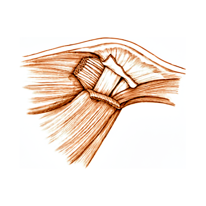
For patients: a plain-language version of this topic is available. See the patient guide.
Overview
Ulnar collateral ligament (UCL) injury of the thumb metacarpophalangeal joint is a significant clinical entity where untreated cases can result in long-term pain and functional limitations [2]. Conservative management is often insufficient for complete ruptures, with 75% of such cases failing to heal without surgical intervention [4]. While transection of the UCL complex does not significantly alter the position of the proximal phalanx relative to the metacarpal [3], the injury necessitates a structured diagnostic and therapeutic approach. Ultrasonography serves as a reliable adjunct to clinical examination [6], while MRI provides detailed visualization that enhances understanding and management of these injuries [11]. A 4-stage, treatment-oriented classification system guides decision-making by correlating the degree of UCL displacement with the likelihood of success for either immobilization or operative intervention [15].
Surgical management of thumb UCL injuries yields overall favorable results [1], with patient-reported outcomes improving significantly at three and 12 months post-repair compared to baseline [13]. Return-to-play rates are high, with players achieving preinjury levels of performance and experiencing few complications [8]. In Major League Baseball, athletes undergoing repair have demonstrated similar games per season and career lengths compared to controls [5], and injuries can be safely managed according to football position demands [9]. Both acute and chronic injuries present similarly acceptable functional outcomes, postoperative pain, and satisfaction [16], although late repair results in a slightly greater loss of thumb motion than immediate repair [47].
Operative techniques vary based on patient characteristics and surgeon preference [7], with options including pull-out sutures, bone anchors, and suture tape augmentation [23, 26, 33]. Custom-made hinged splints show promise for conservative management of compliant patients with complete ruptures [18], while mini hook plate fixation may serve as an alternative for avulsion fractures [28]. Biomechanical studies indicate that suture tape augmentation increases load to failure, facilitating early range of motion [69], and controlled active motion therapy following suture anchor repair is safe [25]. The standard surgical approach utilizes a lazy S incision on the ulnar aspect of the thumb [21], requiring careful longitudinal incision of the adductor aponeurosis [21]. Patients are positioned supine with a bump to rest the thumb [21], and motion is restricted for 4 to 6 weeks, with more chronic injuries immobilized longer [21]. A dorsal approach has been used safely for repair and reconstruction of both ulnar and radial collateral ligaments [41].
Anatomy & Pathophysiology
Ligament Anatomy and Function
The ulnar collateral ligament (UCL) of the thumb metacarpophalangeal (MCP) joint serves as the primary stabilizer against valgus stress [19]. Together with the radial collateral ligament (RCL), the UCL acts as an essential lateral stabilizer of the thumb MCP joint [44]. These ligaments play an essential role in pinch strength, grasping function, and dexterity [19], and are particularly important for precision grip and pinch in elite athletes [29]. Injuries to the thumb collateral ligaments can result in pain, instability, and diminished pinch and grip strength [44]. Collateral ligament injuries of the thumb MCP joint comprise up to 86% of all injuries to the thumb [44]. The UCL is injured more frequently than the RCL, with reported ratios ranging from five times more common [59] to ten times more common [43].
The metacarpophalangeal articulation is of a condylar type and capable of small lateral movements, especially to the radial side [51]. Flexion of the thumb MCP joint is always accompanied by radial deviation and pronation, which stretches the ulnar metacarpophalangeal ligament [51]. This stretching mechanism helps ensure stability of the MCP articulation, which is more important than movement from a functional viewpoint [51].
Injury Mechanisms and Terminology
Acute UCL injuries are commonly referred to as “Skier's thumb,” derived from the mechanism of the thumb being forcibly abducted while entrapped by a ski pole during a fall [19]. The injury mechanism for Skier's thumb is fundamentally related to a sudden valgus force applied to the thumb MCP joint [19]. More broadly, UCL injuries typically result from sudden thumb MCP joint abduction and/or extension (radially deviated force) [44], often resulting from a fall on an outstretched hand with the thumb abducted [43]. Chronic attenuation of the UCL has historically been termed “Gamekeeper's thumb” [19], a term originally referring to an attritional ulnar collateral ligament injury [45].
Changes in ski pole design have been shown to reduce the incidence of this injury [45]; however, most modifications in ski pole design, including elimination of the strap, have not been shown to substantially reduce the incidence of thumb injuries [43].
Pathoanatomy and Associated Injuries
Distal tears of the UCL at its insertion in the proximal phalangeal base are more common than proximal tears from the metacarpal [43]. Ruptures within the substance of the ligament occasionally occur [43]. Associated injuries to UCL tears include tears of the dorsal capsule and ulnar aspect of the volar plate [43], as well as a rent in the adductor aponeurosis [43]. Volar subluxation of the MCP joint may result from concomitant tears of the dorsal capsule and UCL [43].
If one collateral ligament is torn, the proximal phalanx tends to rotate volarly on the side of the tear, with the opposite intact ligament serving as the axis [43]. In an isolated UCL rupture, the proximal phalanx rotates in supination around the intact RCL [43]. If the RCL is torn, the proximal phalanx pronates relative to the metacarpal [43]. An avulsion fracture of the ulnar base of the proximal phalanx at the insertion of the ligament is the most common fracture pattern associated with UCL injuries [43]. Fractures involving more than 10% of the articular surface can occur and may require fixation if displaced 2 mm or more and associated with articular incongruity [43].
RCL ruptures are frequently associated with dorsal or dorsoradial capsular tears and with extensor pollicis brevis avulsions or tears [67]. Disruption of the RCL leads to unopposed dynamic force of the adductor pollicis and flexor pollicis longus tendons [44]. In the setting of RCL disruption, the MCP joint is prone to a sequence of ligamentous laxity, joint instability, volar and ulnar subluxation, and degenerative joint disease [44]. In elite athletes, combined UCL–RCL injuries account for up to 25% of thumb collateral ligament injuries [24].
Stener Lesion Pathophysiology
A Stener lesion occurs when the ruptured UCL becomes displaced superficial to the adductor aponeurosis [19]. In 25 of 39 patients with complete UCL ruptures, Stener found the adductor aponeurosis interposed between the ruptured ulnar collateral ligament and its site of insertion on the base of the proximal phalanx [45]. This adductor aponeurosis interposition prevents direct ligament healing without surgery [42]. Consequently, in a Stener lesion, the ligament is unable to heal anatomically without surgical intervention [19]. If left uncorrected, a Stener lesion prevents proper healing and leads to chronic instability and subsequent arthrosis [45].
On clinical examination of a Stener lesion, a prominent lump can be palpated that represents the ulnar collateral ligament being proximally and superficially displaced by the adductor aponeurosis [45]. Pathologic rotation of the thumb may be evident in the presence of a Stener lesion [45]. No equivalent of the Stener lesion exists for RCL ruptures [67], as the radial-based abductor cannot create a Stener-type lesion [68].
Chronic Instability and Degeneration
Untreated complete UCL ruptures may result in chronic instability, weakness of pinch, pain, and functional impairment [19]. Causes of chronic instability of the UCL include an untreated acute tear, failure to recognize a Stener lesion, or progressive attenuation of the ligament [158]. Patients with chronic UCL instability usually present with pain, swelling, and weakness of the involved thumb [158]. Pain in chronic UCL instability is exacerbated by forceful pinch and activities requiring torsional motions of the hand, such as unscrewing jar tops [158]. Holding large objects may be painful and cause a feeling of instability or lack of power due to the thumb’s inability to resist counterpressure [158]. Crepitus with active motion in chronic UCL instability suggests degenerative disease [158]. RCL ruptures seem susceptible to chronic instability [67].
Classification
Five-Stage Grading System: A five-stage grading system is helpful in separating thumb UCL injuries into operative and non-operative groups [61].
Four-Stage Treatment-Oriented Classification: A 4-stage, treatment-oriented classification of thumb UCL injury is based on the degree of UCL displacement [15]. This classification correlates with the likelihood of success with either immobilization or operative intervention [15].
Other Considerations: The clinical examination of thumb metacarpophalangeal collateral ligament injuries is crucial for distinguishing between partial and complete tears [29]. Incomplete tears are generally treated conservatively using a cast or splint immobilization [44]. For complete tears, the treatment course typically involves surgical intervention [44]. Grade I or II and certain grade III UCL injuries can be managed with nonoperative immobilization with good long-term results [81]. Grade III UCL injuries with a Stener lesion are best treated with surgery [81]. Grade III UCL injuries with avulsion fractures involving greater than 30% of the articular surface are best treated with surgery [81]. A Stener lesion is defined as a specific acute UCL injury in which the torn ligament becomes displaced superficial to the adductor aponeurosis, preventing spontaneous healing and typically requiring operative treatment [24]. The presence of a displaced fleck sign has implications for offering surgery to patients with thumb UCL injuries because of a high likelihood of a Stener lesion [22].
Clinical Presentation
Mechanism and Epidemiology
Acute thumb ulnar collateral ligament (UCL) injuries are estimated to account for approximately 50 in 100,000 presentations to Emergency Departments in the United Kingdom [12]. These injuries are 10 times more common than radial collateral ligament injuries [43, 59]. The mechanism of injury is sudden, forced radial deviation (abduction), often resulting from a fall on an outstretched hand with the thumb abducted [43]. In skiing, the handle abducts the thumb if a person falls while gripping the ski pole [43]. Snowboarders may also injure a thumb during falls and turns [43]. The term "Skier's thumb" refers to an acute valgus injury of the thumb UCL [19, 24], while the term "Gamekeeper's thumb" classically describes chronic attenuation of the same ligament [19, 24].
Symptoms and Signs
Patients with acute injuries commonly recount a traumatic episode with an accompanying onset of pain and swelling [31]. Patients with chronic injuries may present with less specific symptoms [31]. Tenderness is greatest over the ulnar aspect of the thumb metacarpophalangeal (MCP) joint [45]. A prominent lump can be palpated on the ulnar aspect representing the UCL being proximally and superficially displaced by the adductor aponeurosis in a Stener lesion [45]. Pathologic rotation of the thumb may be evident in cases of UCL injury [45]. In isolated UCL rupture, the proximal phalanx rotates in supination around the intact radial collateral ligament [43]. Volar subluxation of the MCP joint may result from concomitant tears of the dorsal capsule and UCL [43].
Clinical Examination
The diagnosis of UCL injury relies primarily on the clinical examination of the patient [31]. Physical examination is performed with a valgus stress applied in neutral rotation to the thumb in both extension and 30° to 35° flexion [42]. Testing in extension assesses the accessory ligament, while testing at 30° to 35° flexion assesses the proper ligament [42]. Instability to valgus stress with the lack of a firm end point is a strong indicator of complete rupture of the UCL [31]. New evidence suggests that a lack of definite end point rather than comparison with the uninjured side should be used to define complete UCL rupture [42]. More than 35° of laxity alone or more than 15° of laxity compared with the contralateral side has historically been considered a positive test result [42]. Valgus instability of more than 30°—or more than 10° compared with the contralateral thumb in both flexion and extension—indicates complete rupture of the proper and accessory UCL [98]. Instability of the MCP joint in flexion indicates rupture of the proper UCL only [98]. The ulnar side of the joint should be palpated for a Stener lesion, where both the proper and accessory ligament are retracted and lie on the adductor aponeurosis [42]. A digital block may be necessary to carry out the stress test in patients with significant guarding [27]. The clinical evaluation of the acutely unstable thumb MCP joint should include evaluation of tendon integrity so that such an injury is not missed [105].
Imaging and Diagnostic Adjuncts
Plain radiographs should be obtained prior to any stress examinations to avoid displacing an otherwise nondisplaced fracture [42, 45]. Stress radiographs are useful for identifying Stener lesions but are often uncomfortable for the patient and guarding may lead to false-negative results [42]. An injured thumb that shows more than 30 degrees of instability compared with the uninjured side on stress radiographs indicates a complete rupture [45]. Ultrasonography has evolved as a reliable adjunct to clinical examination in evaluation of the UCL of the thumb [6]. A magnetic resonance imaging scan is recommended for additional preoperative planning because it provides a detailed assessment of tear location, injury grade, and ligament quality [98]. UCL retraction more than 3 mm and interposed soft tissue on imaging are reasonable guides to surgical intervention [45]. The presence of a displaced fleck sign on radiographs has implications for offering surgery due to a high likelihood of a Stener lesion [22]. A minimally displaced (<2 mm) avulsion fracture signifies a complete avulsion without a Stener lesion [45]. In skeletally immature individuals, stress films are contraindicated if a Salter-Harris type I or type II fracture is present [45]. Diagnosis of incompetence of the UCL of the MCP joint of the thumb remains a clinical rather than a radiographic diagnosis [55]. Clinical assessment of a thumb ulnar collateral ligament injury should be supplemented with radiographs, as underlying pathology, such as an enchondroma, may be a factor [66]. A history of minor trauma resulting in ligament rupture of the thumb’s MCP joint should lead to a suspicion of an underlying pathology [35].
Associated Injuries
Associated injuries include tears of the dorsal capsule and ulnar aspect of the volar plate [43, 59]. An avulsion fracture of the ulnar base of the proximal phalanx at the insertion of the ligament is the most common associated fracture pattern [43]. Fractures involving more than 10% of the articular surface can occur and may require fixation if they are displaced 2 mm or more and associated with articular incongruity [43]. Avulsion fractures from the metacarpal head and intraarticular shearing fractures of the volar surface of the radial condyle of the metacarpal head have also been reported [43]. In elite athletes, combined UCL and RCL injuries account for up to 25% of thumb collateral ligament injuries [24].
Investigations
Clinical Examination
The thumb metacarpophalangeal joint (MCPJ) is examined by applying a radial force in extension with approximately 30° of MCPJ flexion to relax the palmar plate [12]. This maneuver is thought to isolate the UCL proper [12]. Careful stress testing of both the injured and uninjured sides in extension and flexion often elucidates significant tears of the UCL or RCL [27]. The absence of a normal “endpoint” with radial stress is a key diagnostic finding signifying a complete ligament tear [27]. For patients with significant guarding, a digital block may be necessary to carry out the stress test [27]. Surgery is recommended if a hard endpoint is not encountered and there is increased deviation (>20 degrees) at the MP joint [27]. Patients with a soft endpoint, incongruent joint, or Stener lesion require surgical intervention [27]. When evaluating pediatric abduction thumb injuries, the physician must consider complete ligamentous UCL disruption, especially when x-ray films show significant joint subluxation [17].
Radiography
Plain radiography: Plain radiographs or radiographs while applying a force to the MCPJ (stress radiographs) are included in the clinical investigations for UCL injuries [12]. Radiographs identify associated fractures in the evaluation of thumb metacarpophalangeal joint injuries [24]. In UCL injuries involving two bone fragments, plain radiographs remain insufficient for diagnosis unless there is significant displacement of the avulsed fragment [163]. Stress radiography should be performed cautiously in thumbs with a prominent radial condyle, as it may cause locking due to entrapment of the avulsed ligament or volar plate [167].
Fluoroscopy: Fluoroscopy can aid the diagnosis to see if the joint becomes incongruent and to measure the deviation compared to the contralateral limb [27].
Ultrasound
Ultrasound: High-resolution US is increasingly used due to its accuracy, availability, and ability to evaluate and compare to the contralateral side [24]. Comprehensive ultrasound assessment of thumb metacarpophalangeal collateral ligament injuries reveals frequent concomitant abnormalities [24]. If it is difficult to ascertain if there is a Stener lesion based on palpation, an ultrasound can be helpful to gather more information [27]. Ultrasound or MRI findings do not have 100% accuracy and their findings should only be one component of the decision-making algorithm [27].
Magnetic Resonance Imaging
MRI: Magnetic resonance imaging (MRI) scanning is included in the clinical investigations for UCL injuries [12]. MRI or ultrasound directly depict ligament integrity in the accurate diagnosis of MCP injuries [24]. If it is difficult to ascertain if there is a Stener lesion based on palpation, an MRI can be helpful to gather more information [27]. Magnetic resonance imaging was helpful in diagnosing a displaced ulnar collateral ligament in two cases and an intraligamentous injury in the other [145].
Treatment
Non-Operative
Incomplete acute tears and nondisplaced avulsion fractures are managed with a thumb spica cast for up to 4 weeks, followed by removable protective splinting for 3 additional weeks with active range-of-motion exercises [42]. Nonoperative treatment of partial ligament injuries with cast or splint immobilization results in a stable and painless thumb [148]. In patients exhibiting less than 30 degrees of laxity of the UCL, less than a 15-degree differential in laxity compared with the contralateral side, and a discrete endpoint to joint opening, the injury is assumed to be incomplete; nonoperative treatment consists of immobilization of the MP joint for 4 weeks in neutral alignment and slight flexion [161]. Tears of the UCL of the thumb MCP joint without a Stener lesion are believed to heal with 4 to 6 weeks of immobilization [67].
Operative
Indications: Complete tears require open repair with suture anchors or bone tunnels placed at the site of the avulsion, which most commonly is the proximal phalanx [42]. Injuries that are unstable, usually defined as 30° more opening with radial stress than the opposite uninjured side, are believed to have Stener lesions and are treated surgically [67]. Surgical repair of acute UCL ruptures is widely accepted as the gold standard of treatment in the presence of gross instability, Stener lesions, or displaced avulsion fractures [71]. Acute complete rupture of the ulnar collateral ligament should be surgically repaired [45].
Surgical Approach / Technique: Open repair of the UCL involves a lazy S approach on the ulnar aspect of the thumb, careful identification and retraction of the radial sensory nerve, incision of the adductor aponeurosis, and repair of the ligament using nonabsorbable suture with pullout button, bone anchors, or figure-of-eight imbrication [21]. Both pull-out suture and bone anchor techniques are safe and effective for treating thumb ulnar collateral ligament injuries [23]. The use of an intraosseous suture anchor is a safe and effective method for repair of complete tears of the ulnar collateral ligament of the thumb MP joint [33]. A K-wire across the joint is often employed to allow proper ligament tensioning and to maintain joint stability postoperatively [68]. In a biomechanical model, thumb UCL repair with suture tape augmentation demonstrated greater maximum and clinical failure loads compared with nonaugmented repair at time 0 [40]. Arthroscopic reduction of a Stener lesion allows healing of the ulnar collateral ligament without the need for open repair [140].
Chronic Repair and Reconstruction: Chronic ruptures of the UCL can be challenging to reconstruct, and if local tissue is not repairable, a graft is often used such as the palmaris longus tendon [68]. Graft reconstruction of the UCL can often be accomplished with either an autograft palmaris/plantaris tendon or with an allograft [27]. Newer techniques for chronic UCL reconstruction involve the use of 3.0-mm drills, the graft, fibertape, and interference screws [27]. An X or Y type configuration is often needed to appropriately reconstruct the surface area of the ligament to prevent recurrent instability in chronic UCL repairs [27]. If a Y construct is used for chronic UCL reconstruction, it is preferred to place the single limb distally and the dual limb proximally to prevent iatrogenic injury [27]. Nonbiological ligament reconstruction of the thumb ulnar collateral ligament generates short-term outcomes comparable with those of biological reconstruction, potentially allowing for expedited recovery and rehabilitation [80]. Chronic collateral ligament reconstructions with tendon graft, although less successful than acute repair, can restore stability of the thumb MCP joint [29]. Patient-reported outcomes, including pain and function, improved after thumb UCL reconstruction with a tendon autograft [38]. No static ligament reconstruction restores the normal stability characteristics of the thumb UCL [10]. Nonanatomic reconstruction of the UCL alters normal MCP joint range of motion [156].
Outcomes and Return to Activity: Successful management of acute injuries of the ulnar collateral ligament of the thumb MCP joint prevents the sequelae of instability and pain [20]. Thumb MCP joint collateral ligament injuries have a good outcome with appropriate diagnosis and treatment [34]. The methods adopted in the treatment of fresh ruptures of the ulnar collateral ligament of the metacarpophalangeal joint of the thumb have been good [49].
Complications
Consequences of Untreated or Inadequately Treated Injury
Chronic Instability and Functional Impairment: Untreated complete ruptures may result in chronic instability, weakness of pinch, pain, and functional impairment [19]. Failure to address the ensuing laxity of the metacarpophalangeal joint can lead to compromised grip and pinch, pain, and ultimately osteoarthritis [31].
Post-Surgical Complications and Outcomes
General Outcomes: Both repair of acute ruptures and reconstruction for chronic injuries yield excellent results, with complications being rare and most patients showing preservation of motion, key pinch, and grip strength [31]. Fixation-Related Complications: The complication rate of pullout suture fixation is higher than that of suture anchor repair [79]. The surgical treatment of an ulnar collateral ligament lesion of the thumb using the Fiji Anchor® can lead to an excellent clinical outcome with a minor complication rate [146]. Long-term dangers and the cost effectiveness of the procedure using the Fiji Anchor® are not known yet [146].
Long-Term Degenerative Changes
Osteoarthritis: Eighty-eight percent of patients had some degree of osteoarthritis following repair of a chronic UCL injury [76]. Increased age at the time of injury and higher DASH scores were correlated with increased grades of the thumb metacarpophalangeal osteoarthritis [76]. Delay to treatment and VAS pain scores had no correlation with radiographic findings in patients with chronic UCL injury repair [76].
Biomechanical and Functional Limitations
Range of Motion and Stiffness: Nonanatomic repairs of collateral ligament alter normal metacarpophalangeal joint range of motion [29]. Immobilization has biochemical, physiological, and biomechanical effects that may cause stiffness and weakness [79].
Adjacent Injury Risks
Adjacent Joint Stress: Thumb spica casting protects the surgically repaired thumb UCL and allows for earlier return to play, but risks placing additional stress upon adjacent joints and causing adjacent injury [39].
Recovery
Outcomes and Function: Nonbiological ligament reconstruction of the thumb ulnar collateral ligament generates short-term outcomes comparable with those of biological reconstruction [80]. For chronic UCL injuries, repair with available local tissue appears to be a reasonable alternative to ligament reconstruction, resulting in durable long-term outcomes despite the majority of patients progressing to osteoarthritis [76]. In this chronic injury population, delay to treatment and VAS pain scores had no correlation with radiographic findings [76].
Return to Sport and Work: Players who underwent thumb UCL surgery played in a similar number of games per season and had similar career lengths in the NFL as controls [172].
Rehabilitation Protocol: The first clinical application of an accelerated rehabilitation protocol in athletes following thumb MCP UCL repair has been described [65]. Early motion is the key to preventing stiffness [79]. Application of controlled low-load prolonged stress results in increased ROM and function [79].
Key Evidence
- [L1] Surgical management of thumb UCL injuries produces overall favorable results. [1] (10.5435/jaaosglobal-d-25-00082)
- [L5] Untreated ulnar collateral ligament (UCL) injury of the thumb metacarpophalangeal joint can lead to long-term pain and functional limitations. [2] (10.2106/jbjs.k.01024)
- [Paper] Transection of the UCL complex did not significantly alter the position of the proximal phalanx relative to the metacarpal of the thumb. [3] (10.1067/jhsu.2000.jhsu25a0313)
- [L5] 75% of cases with complete rupture of the ulnar collateral ligament of the thumb will fail to heal with conservative treatment. [4] (10.1007/s11552-008-9145-8)
- [L3] Players who underwent thumb UCL repair played in a similar number of games per season and had similar career lengths in the MLB as controls. [5] (10.1177/2325967117747268)
- [L4] Ultrasonography (US) has evolved as a reliable adjunct to clinical examination in evaluation of the UCL of the thumb. [6] (10.1148/rg.264055117)
- [L3] The rate of surgery for acute thumb MP UCL injury varies based on patient characteristics and the individual treating surgeon. [7] (10.1177/1558944716681974)
- [L4] Return-to-play rates after surgical treatment of thumb UCL injuries are high, with reassuring return to preinjury level of play with few complications. [8] (10.1016/j.jhsg.2023.03.005)
- [L4] Thumb ulnar collateral ligament injuries can be safely and effectively managed by football position demands. [9] (10.1177/2325967114s00092)
- [L5] No static ligament reconstruction restores the normal stability characteristics of the thumb UCL. [10] (10.1016/j.jhsa.2004.09.012)
- [L4] MRI evaluation reveals greater details enabling better understanding and management of ulnar collateral ligament injuries of the thumb MCP joint. [11] (10.1016/s0363-5023(09)60139-8)
- [L1] [12] (10.1177/17531934241274612)
- [L2] Patient-reported outcomes improve significantly at three and 12 months after open surgical repair of the thumb UCL compared to baseline. [13] (10.1016/j.jhsa.2023.05.003)
- [L4] Our 4-stage, treatment-oriented classification of thumb UCL injury is based on the degree of UCL displacement, with correlation with the likelihood of success with either immobilization or operative intervention. [15] (10.1016/j.jhsa.2014.08.033)
- [L3] Patients with both acute and chronic thumb UCL injuries have similarly acceptable functional outcomes, postoperative pain, and satisfaction. [16] (10.1016/j.jhsg.2022.02.008)
- [L5] When evaluating pediatric abduction thumb injuries, the physician must consider complete ligamentous UCL disruption, especially when x-ray films show significant joint subluxation. [17] (10.1016/0363-5023(94)90115-5)
- [L4] Treatment of a thumb UCL complete rupture (Grade III) using a custom-made hinged splint shows promise in the conservative management of compliant patients. [18] (10.1016/j.jht.2009.10.001)
- [L4] [19] (10.1016/j.injury.2026.113711)
- [L5] Successful management of acute injuries of the ulnar collateral ligament of the thumb MCP joint prevents the sequelae of instability and pain. [20] (10.5435/00124635-199707000-00006)
- [L4] Presence of a displaced fleck sign has implications for offering surgery to patients with thumb UCL injuries because of a high likelihood of a Stener lesion. [22] (10.1016/j.jhsa.2024.12.003)
- [L3] Both repair methods are safe and effective for treating thumb ulnar collateral ligament injuries. [23] (10.1097/prs.0b013e3181882163)
- [L4] [24] (10.1007/s00402-026-06430-0)
- [L5] A controlled active motion therapy protocol after suture anchor repair of a ruptured UCL of the thumb is safe from a biomechanical point of view. [25] (10.1016/j.jhsa.2004.04.017)
- [L4] Thumb UCL repair with suture tape augmentation demonstrates short-term outcomes comparable to what has been reported for other methods of repair. [26] (10.1142/s2424835520500046)
- [L4] It could be an alternative treatment technique for thumb UCL avulsion fractures. [28] (10.1016/j.otsr.2019.01.008)
- [L5] [29] (10.1016/j.hcl.2012.05.024)
- [L5] [31] (10.1016/j.ocl.2014.11.007)
- [L3] The authors concluded that this is a safe and effective method for repair of complete tears of the ulnar collateral ligament of the thumb MP joint. [33] (10.1016/s0363-5023(97)80113-x)
- [L5] Thumb MCP joint collateral ligament injuries have a good outcome with appropriate diagnosis and treatment. [34] (10.1016/j.csm.2014.09.004)
- [L5] We conclude from this case that a history of minor trauma resulting in ligament rupture of the thumb’s MCP joint should lead to a suspicion of an underlying pathology. [35] (10.1177/17531934211004437)
- [L4] Patient-reported outcomes, including pain and function, improved after thumb UCL reconstruction with a tendon autograft. [38] (10.1016/j.jhsa.2024.05.005)
- [L4] Thumb spica casting protects the surgically repaired thumb UCL and allows for earlier return to play, but risks placing additional stress upon adjacent joints and causing adjacent injury. [39] (10.1177/1558944718788644)
- [L5] In this model, thumb UCL repair with suture tape augmentation demonstrated greater maximum and clinical failure loads compared with nonaugmented repair at time 0, that is, without any biological healing. [40] (10.1016/j.jhsa.2018.02.002)
- [L5] The authors have used the described dorsal approach with safety and convenience over a several year period for repair and reconstruction of both the ulnar and radial collateral ligaments at the thumb metacarpophalangeal joint. [41] (10.1097/00130911-200403000-00003)
- [L3] [44] (10.1177/15589447251338586)
- [L4] Late repair gives equally good subjective results and causes a slightly greater loss of thumb motion than immediate repair. [47] (10.1016/0363-5023(93)90353-5)
- [L4] The methods adopted in the treatment of fresh ruptures of the ulnar collateral ligament of the metacarpophalangeal joint of the thumb have been good. [49] (10.1016/s0020-1383(70)80123-1)
- [L5] Diagnosis of incompetence of the UCL of the MCP joint of the thumb remains a clinical rather than a radiographic diagnosis. [55] (10.1016/s0363-5023(98)80158-5)
- [L5] [59] (10.1016/j.hcl.2009.05.014)
- [L4] When no fracture is present, a proper physical examination of an injured thumb is sufficient to determine the degree of instability of the ulnar collateral ligament, and a five-stage grading system is helpful in separating injuries into operative and non-operative groups. [61] (10.2106/00004623-198668090-00003)
- [L4] This is the first clinical application of an accelerated rehabilitation protocol in athletes following thumb MCP UCL repair. [65] (10.1016/j.jht.2022.07.007)
- [L5] This case highlights that clinical assessment of a thumb ulnar collateral ligament injury should be supplemented with radiographs, as underlying pathology, such as an enchondroma, may be a factor. [66] (10.1177/17531934251315313)
- [L5] SutureTape augmentation has been shown in biomechanical studies of other hand ligaments such as the thumb UCL to greatly increase the load to failure, allowing patients to initiate early range of motion after surgery. [69] (10.1016/j.jhsg.2026.101050)
- [L4] [71] (10.1016/j.injury.2009.01.107)
- [L4] [76] (10.1177/1558944716628482)
- [L3] [79] (10.1016/j.hansur.2020.03.002)
- [L4] Nonbiological ligament reconstruction of the thumb ulnar collateral ligament generates short-term outcomes comparable with those of BLR, potentially allowing for expedited recovery and rehabilitation. [80] (10.1016/j.jhsa.2024.01.008)
- [Paper] [81] (10.1016/j.csm.2019.12.003)
- [L5] [98] (10.1016/j.eats.2025.103957)
- [L5] The clinical evaluation of the acutely unstable thumb MCP joint should include evaluation of tendon integrity so that such an injury is not missed. [105] (10.1016/s0363-5023(05)80475-7)
- [L4] [140] (10.1016/s0363-5023(05)80156-x)
- [L4] Magnetic resonance imaging was helpful in diagnosing a displaced ulnar collateral ligament in two cases and an intraligamentous injury in the other. [145] (10.1016/0363-5023(89)90203-7)
- [Paper] The surgical treatment of an ulnar collateral ligament lesion of the thumb using the Fiji Anchor® can lead to an excellent clinical outcome with a minor complication rate; however, long-term dangers and the cost effectiveness of the procedure are not known yet. [146] (10.1007/s00402-020-03625-x)
- [L5] Nonoperative treatment of partial ligament injuries with cast or splint immobilization will result in a stable and painless thumb. [148] (10.5435/00124635-201105000-00006)
- [L5] This study demonstrates that nonanatomic reconstruction of the UCL alters normal MCP joint range of motion. [156] (10.1053/jhsu.1999.0283)
- [L5] In UCL injuries involving two bone fragments, plain radiographs remain insufficient for diagnosis unless there is significant displacement of the avulsed fragment. [163] (10.1016/s0020-1383(01)00206-6)
- [L4] The case suggests that stress radiography should be performed cautiously in thumbs with a prominent radial condyle, as it may cause locking due to entrapment of the avulsed ligament or volar plate. [167] (10.1007/s00402-009-0933-6)
- [L4] Players who underwent thumb UCL surgery played in a similar number of games per season and had similar career lengths in the NFL as controls. [172] (10.1177/1558944718760001)
See Also
References
[1] Surgical Management of Thumb Ulnar Collateral Ligament Injuries: A Systematic Review and Meta-analysis of 614 Patients With a Minimum 1 Year Follow-up. JAAOS: Global Research and Reviews. 2025. DOI: 10.5435/jaaosglobal-d-25-00082
[2] Management of Thumb Metacarpophalangeal Ulnar Collateral Ligament Injuries. Journal of Bone and Joint Surgery. 2012. DOI: 10.2106/jbjs.k.01024
[3] The effects of transection and reconstruction of the ulnar collateral ligament complex on the position of the proximal phalanx of the thumb during simulated tip pinch. The Journal of Hand Surgery. 2000. DOI: 10.1067/jhsu.2000.jhsu25a0313
[4] Injury to the Ulnar Collateral Ligament of the Thumb. HAND. 2008. DOI: 10.1007/s11552-008-9145-8
[5] Performance and Return to Sport After Thumb Ulnar Collateral Ligament Repair in Major League Baseball Players. Orthopaedic Journal of Sports Medicine. 2018. DOI: 10.1177/2325967117747268
[6] US Diagnosis of UCL Tears of the Thumb and Stener Lesions: Technique, Pattern-based Approach, and Differential Diagnosis. RadioGraphics. 2006. DOI: 10.1148/rg.264055117
[7] Variation in the Rate of Surgery for Ulnar Collateral Ligament Injury of the Metacarpophalangeal Joint of the Thumb. HAND. 2016. DOI: 10.1177/1558944716681974
[8] Return to Play After Thumb Ulnar Collateral Ligament Injuries Managed Surgically in Athletes—A Systematic Review. Journal of Hand Surgery Global Online. 2023. DOI: 10.1016/j.jhsg.2023.03.005
[9] Return to Play and Long-Term Clinical Outcomes after Suture Anchor Repair of Thumb Ulnar Collateral Ligament Injuries in Collegiate Football Athletes. Orthopaedic Journal of Sports Medicine. 2014. DOI: 10.1177/2325967114s00092
[10] Reconstruction of the ulnar collateral ligament of the thumb metacarpophalangeal joint: A cadaver study. The Journal of Hand Surgery. 2005. DOI: 10.1016/j.jhsa.2004.09.012
[11] MRI Classification of Ulnar Collateral Ligament Injuries of the Thumb MCP Joint. The Journal of Hand Surgery. 2009. DOI: 10.1016/s0363-5023(09)60139-8
[12] Guideline on managing thumb ulnar collateral ligament injuries: the British Society of Surgery for the Hand Evidence for Surgical Treatment (BEST) findings and recommendations. Journal of Hand Surgery (European Volume). 2024. DOI: 10.1177/17531934241274612
[13] Patient-Reported Outcomes and Function After Surgical Repair of the Ulnar Collateral Ligament of the Thumb. The Journal of Hand Surgery. 2023. DOI: 10.1016/j.jhsa.2023.05.003
[15] Gamekeeper’s Thumb—A Treatment-Oriented Magnetic Resonance Imaging Classification. The Journal of Hand Surgery. 2015. DOI: 10.1016/j.jhsa.2014.08.033
[16] A Comparison of Acute Versus Chronic Thumb Ulnar Collateral Ligament Surgery Using Primary Suture Anchor Repair and Local Soft Tissue Advancement. Journal of Hand Surgery Global Online. 2022. DOI: 10.1016/j.jhsg.2022.02.008
[17] Late presentation of a ligamentous ulnar collateral ligament injury in a child. The Journal of Hand Surgery. 1994. DOI: 10.1016/0363-5023(94)90115-5
[18] Treatment of Grade III Thumb Metacarpophalangeal Ulnar Collateral Ligament Injuries with Early Controlled Motion Using a Hinged Splint. Journal of Hand Therapy. 2010. DOI: 10.1016/j.jht.2009.10.001
[19] Return to Duty After Suture Anchor Repair of Thumb Metacarpophalangeal Ulnar Collateral Ligament Injuries in Active Duty Soldiers. Injury. 2026. DOI: 10.1016/j.injury.2026.113711
[20] Injuries to the Ulnar Collateral Ligament of the Thumb Metacarpophalangeal Joint. Journal of the American Academy of Orthopaedic Surgeons. 1997. DOI: 10.5435/00124635-199707000-00006
[21] Rockwood And Green S Fractures In Adults. 42: Fractures of the Distal Radius and Ulna > Open Repair of the Ulnar or Radial Collateral Ligament of the MCP Joint of the Thumb: Preoperative Planning Checklist.
[22] The Displaced Fleck Sign: Description of a Radiographic Finding Consistent with Grade III Thumb Ulnar Collateral Ligament Tears with Stener Lesions. The Journal of Hand Surgery. 2025. DOI: 10.1016/j.jhsa.2024.12.003
[23] Repair of Acute Ulnar Collateral Ligament Injuries of the Thumb Metacarpophalangeal Joint: A Retrospective Comparison of Pull-Out Sutures and Bone Anchor Techniques. Plastic and Reconstructive Surgery. 2008. DOI: 10.1097/prs.0b013e3181882163
[24] Comprehensive ultrasound assessment of thumb metacarpophalangeal collateral ligament injuries reveals frequent concomitant abnormalities. Archives of Orthopaedic and Trauma Surgery. 2026. DOI: 10.1007/s00402-026-06430-0
[25] A biomechanical modeling of injury, repair, and rehabilitation of ulnar collateral ligament injuries of the thumb. The Journal of Hand Surgery. 2004. DOI: 10.1016/j.jhsa.2004.04.017
[26] Thumb Ulnar Collateral Ligament Repair with Suture Tape Augmentation. The Journal of Hand Surgery (Asian-Pacific Volume). 2020. DOI: 10.1142/s2424835520500046
[27] Rockwood And Green S Fractures In Adults. 42: Fractures of the Distal Radius and Ulna > Thumb MCP Ligament Repair.
[28] Mini hook plate fixation for thumb ulnar collateral ligament avulsion fracture: A technical report. Orthopaedics & Traumatology: Surgery & Research. 2019. DOI: 10.1016/j.otsr.2019.01.008
[29] Thumb Metacarpophalangeal Joint Collateral Ligament Injury Management. Hand Clinics. 2012. DOI: 10.1016/j.hcl.2012.05.024
[31] Ulnar Collateral Ligament Injuries of the Thumb. Orthopedic Clinics of North America. 2015. DOI: 10.1016/j.ocl.2014.11.007
[33] Repair of acute ulnar collateral ligament injuries of the thumb metacarpophalangeal joint with an intraosseous suture anchor. The Journal of Hand Surgery. 1997. DOI: 10.1016/s0363-5023(97)80113-x
[34] Thumb Ulnar Collateral and Radial Collateral Ligament Injuries. Clinics in Sports Medicine. 2015. DOI: 10.1016/j.csm.2014.09.004
[35] Tenosynovial giant cell tumour mimicking ulnar collateral ligament rupture at the thumb metacarpophalangeal joint. Journal of Hand Surgery (European Volume). 2021. DOI: 10.1177/17531934211004437
[38] Patient-Reported and Clinical Outcomes After Tendon Autograft Reconstruction of the Thumb Ulnar Collateral Ligament. The Journal of Hand Surgery. 2025. DOI: 10.1016/j.jhsa.2024.05.005
[39] Early Return to Play With Thumb Spica Gauntlet Casting for Ulnar Collateral Ligament Injuries Complicated by Adjacent Joint Dislocations in Collegiate Football Linemen. HAND. 2018. DOI: 10.1177/1558944718788644
[40] Suture Tape Augmentation of the Thumb Ulnar Collateral Ligament Repair: A Biomechanical Study. The Journal of Hand Surgery. 2018. DOI: 10.1016/j.jhsa.2018.02.002
[41] Surgical Approaches to the Metacarpophalangeal Joint of the Thumb for Repair and Reconstruction of the Collateral Ligaments and Capsule. Techniques in Hand and Upper Extremity Surgery. 2004. DOI: 10.1097/00130911-200403000-00003
[42] Orthopaedic Knowledge Update Sports Medicine 6. Hand and Wrist Injuries > Hand Injuries > Skier’s Thumb.
[43] Green S Operative Hand Surgery. Dislocations and Ligament Injuries of the Digits > Acute Ulnar Collateral Ligament Injuries (Skier’s Thumb).
[44] Thumb Radial Collateral Ligament Injuries Show Greater Rates of Metacarpophalangeal Subluxation Compared With Thumb Ulnar Collateral Ligament Injuries: A Comparative Imaging Analysis. HAND. 2025. DOI: 10.1177/15589447251338586
[45] Campbell S Operative Orthopaedics 4 Volume Set. FRACTURES, DISLOCATIONS, AND LIGAMENTOUS INJURIES OF THE HAND AND WRIST > THUMB METACARPOPHALANGEAL JOINT ULNAR COLLATERAL LIGAMENT RUPTURE.
[47] Acute and late radial collateral ligament injuries of the thumb metacarpophalangeal joint. The Journal of Hand Surgery. 1993. DOI: 10.1016/0363-5023(93)90353-5
[49] Surgical treatment of ruptured ulnar collateral ligament of the metacarpo-phalangeal joint of the thumb. Injury. 1970. DOI: 10.1016/s0020-1383(70)80123-1
[51] Exam Of The Hand Wrist 2Ed. Movements of the thumb.
[55] The stener lesion revisited: A case report. The Journal of Hand Surgery. 1998. DOI: 10.1016/s0363-5023(98)80158-5
[59] Acute Ulnar Collateral Ligament Injury in the Athlete. Hand Clinics. 2009. DOI: 10.1016/j.hcl.2009.05.014
[61] Rupture and displacement of the ulnar collateral ligament of the metacarpophalangeal joint of the thumb. Preoperative diagnosis.. The Journal of Bone & Joint Surgery. 1986. DOI: 10.2106/00004623-198668090-00003
[65] Accelerated rehabilitation following thumb ulnar collateral ligament repair with suture tape augmentation: A case series of professional basketball players. Journal of Hand Therapy. 2023. DOI: 10.1016/j.jht.2022.07.007
[66] Clinical thumb ulnar collateral ligament injury owing to a pathological fracture through an enchondroma of the proximal phalanx. Journal of Hand Surgery (European Volume). 2025. DOI: 10.1177/17531934251315313
[67] Aaos Comprehensive Orthopaedic Review 3. Hand Trauma > VI. Thumb Metacarpophalangeal Ligament Injuries and Dislocations.
[68] Rockwood And Green S Fractures In Adults. 42: Fractures of the Distal Radius and Ulna > Open Repair of the Ulnar or Radial Collateral Ligament of the MCP Joint of the Thumb.
[69] Use of Internal Brace for the Surgical Management of the “Baby Stener” Lesion of the Radial Collateral Ligament to the Small Finger. Journal of Hand Surgery Global Online. 2026. DOI: 10.1016/j.jhsg.2026.101050
[71] Ulnar collateral ligament injuries of the thumb: 10 years of surgical experience. Injury. 2009. DOI: 10.1016/j.injury.2009.01.107
[76] Long-Term Outcomes of Primary Repair of Chronic Thumb Ulnar Collateral Ligament Injuries. HAND. 2016. DOI: 10.1177/1558944716628482
[79] The effects of rehabilitation on the outcomes of surgically treated acute and chronic thumb metacarpophalangeal ulnar collateral ligament ruptures. Hand Surgery and Rehabilitation. 2020. DOI: 10.1016/j.hansur.2020.03.002
[80] Biological Versus Nonbiological Reconstruction of the Ulnar Collateral Ligament of the Thumb Metacarpophalangeal Joint: A Retrospective Study. The Journal of Hand Surgery. 2024. DOI: 10.1016/j.jhsa.2024.01.008
[81] Thumb Metacarpophalangeal Ulnar and Radial Collateral Ligament Injuries. Clinics in Sports Medicine. 2020. DOI: 10.1016/j.csm.2019.12.003
[98] Thumb Ulnar Collateral Ligament Repair Using Suture Anchors and Suture Tape Augmentation. Arthroscopy Techniques. 2025. DOI: 10.1016/j.eats.2025.103957
[105] Ulnar collateral ligament tear with concommitant extensor pollicis brevis and extensor pollicis longus disruptions: A case report. The Journal of Hand Surgery. 1998. DOI: 10.1016/s0363-5023(05)80475-7
[140] Arthroscopic treatment of acute complete thumb metacarpophalangeal ulnar collateral ligament tears. The Journal of Hand Surgery. 1995. DOI: 10.1016/s0363-5023(05)80156-x
[145] Magnetic resonance imaging of the collateral ligaments of the thumb. The Journal of Hand Surgery. 1989. DOI: 10.1016/0363-5023(89)90203-7
[146] Clinical results of the BoneWelding®Fiji® anchor for the treatment of Stener lesions of the thumb. Archives of Orthopaedic and Trauma Surgery. 2020. DOI: 10.1007/s00402-020-03625-x
[148] Collateral Ligament Injuries of the Thumb Metacarpophalangeal Joint. Journal of the American Academy of Orthopaedic Surgeons. 2011. DOI: 10.5435/00124635-201105000-00006
[156] The effect of thumb metacarpophalangeal ulnar collateral ligament attachment site on joint range of motion: An in vitro study. The Journal of Hand Surgery. 1999. DOI: 10.1053/jhsu.1999.0283
[158] Green S Operative Hand Surgery. Dislocations and Ligament Injuries of the Digits > Chronic Ulnar Collateral Ligament Injuries (Gamekeeper’s Thumb) > Preoperative Evaluation.
[161] Green S Operative Hand Surgery. Dislocations and Ligament Injuries of the Digits > Chronic Ulnar Collateral Ligament Injuries (Gamekeeper’s Thumb) > Types of Operations.
[163] Two in one: a variant of the Stener lesion. Injury. 2002. DOI: 10.1016/s0020-1383(01)00206-6
[167] Locking of the metacarpophalangeal joint of the thumb with the radial collateral ligament rupture after stress radiography. Archives of Orthopaedic and Trauma Surgery. 2009. DOI: 10.1007/s00402-009-0933-6
[172] Performance and Return to Sport After Thumb Ulnar Collateral Ligament Surgery in National Football League Players. HAND. 2018. DOI: 10.1177/1558944718760001