Clinicians › Hand
Mucous Cyst
Mucous (myxoid) cysts at the DIP joint — retinacular ganglia driven by underlying OA.
For patients: a plain-language version of this topic is available. See the patient guide.
Overview
Digital mucous cysts are benign periungual lesions typically associated with underlying marginal osteophytes. Surgical management aims for complete eradication with minimal recurrence and acceptable cosmesis. Excision of the cyst combined with complete removal of the marginal osteophyte eradicates the lesion with extremely rare recurrence [2]. Osteophyte excision without cyst excision provides a less invasive method that achieves complete resolution in most cases [11]. Alternatively, total dorsal capsulectomy alone did not lead to any recurrence in the treatment of mucous cysts [9]. The recurrence rate for osteophyte-sparing treatment is less than 1.5% [4].
Surgical techniques vary, including local advancement skin flaps, which demonstrate a recurrence rate of 1.4% and are associated with high patient satisfaction regarding the scar and willingness to undergo the procedure again [1]. Mucous cystectomy and osteophytectomy using a random nonadvancement flap technique resulted in 2 cyst recurrences and only 1 secondary procedure in the study cohort [3]. The use of a Wolfe Graft provides satisfactory cosmesis with acceptable recurrence rates [5]. Excision using a proximal nail fold flap resulted in no recurrence even after one year of surgery [8]. A simple modification of the rotation flap originally described by Kleinert et al is used for coverage after excision of large mucous cysts [12]. Biquadrangular and Hueston flaps yield comparable aesthetic satisfaction and clinical outcomes after surgical excision [19].
In-office excision is a safe and effective option for treating digital mucous cysts [7]. Pathohistological analysis is useful in cases where doubts arise about the initial diagnosis of a benign tumorous lesion presenting as a mucous cyst [10]. The scientific data regarding mucous cysts consist almost entirely of retrospective studies, and much of what is done or recommended is based on expert opinion [6].
Anatomy & Pathophysiology
Clinical Presentation
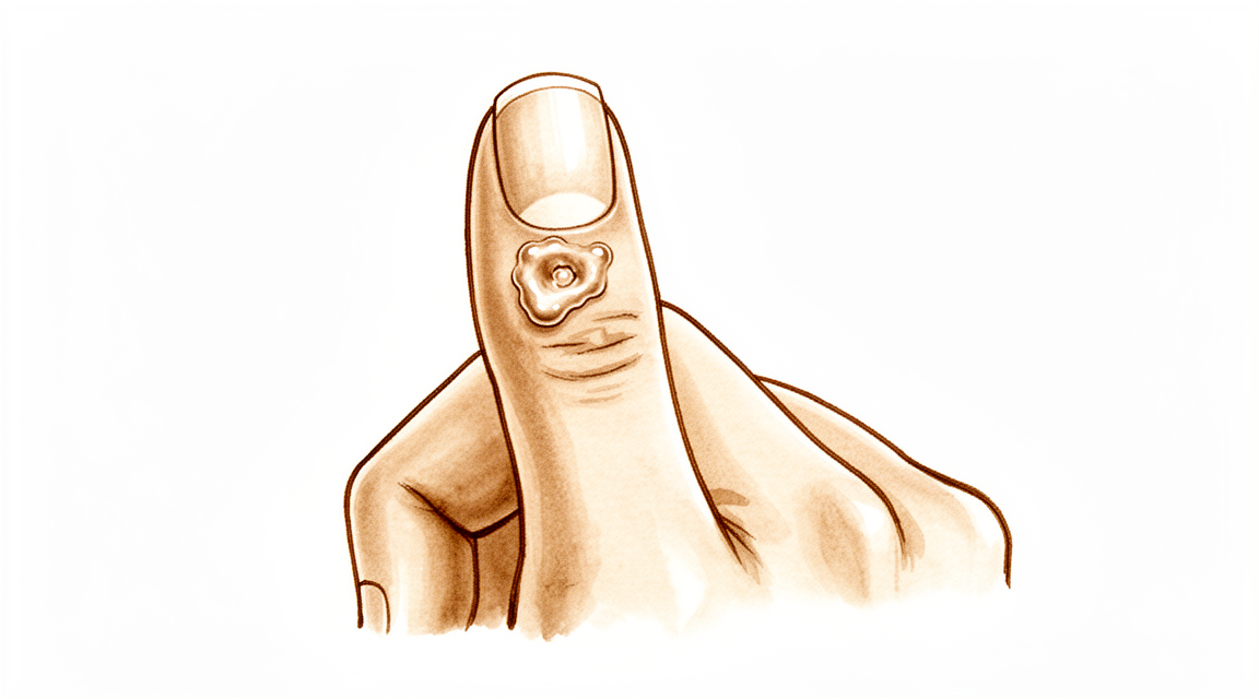
A digital mucous cyst presents as a small, firm, cystic mass located just distal to the distal interphalangeal joint [16]. The lesion is always located to one side of the mid-line [16] and is rarely greater than fifteen millimeters in diameter [16]. The overlying skin is thinned out and occasionally may be ulcerated [16]. Pressure on the lesion does not usually result in a decrease in its size [16]. Longitudinal grooving of the nail may be noted, occasionally occurring prior to the appearance of the cyst [16].
Histology & Etiology
The histological appearance of a digital mucous cyst is essentially identical to that of ganglions, synovial cysts, and similar lesions appearing on other areas of the extremities [16]. The lesion is surrounded by an undemarcated fibrous capsule [16], and its basic structure is myxomatous with interspersed fibroblasts [16]. Areas of myxomatous degeneration tend to coalesce to form a multiloculated cyst [16]. An epithelial lining has not been reported for the lesion [16]. Degenerative arthritis of the distal interphalangeal joint is a frequent associated finding with an incidence of 78 per cent [16]. The etiology of the lesion is that it arises from the joint capsule [16].
Classification
Morphological: The mucous cyst presents as a small, firm, cystic mass located just distal to the distal interphalangeal joint [16].
Histological: The histological appearance is essentially identical to that of ganglions, synovial cysts, and similar lesions appearing on other areas of the extremities [16]. The basic structure is myxomatous with interspersed fibroblasts [16]. An epithelial lining has not been reported [16].
Clinical Presentation
The mucous cyst presents as a small, firm, cystic mass located just distal to the distal interphalangeal joint [16]. On inspection, the mass is always located to one side of the mid-line [16]. Palpation confirms the firm, cystic nature of the lesion in this extra-articular position [16].
Degenerative arthritis of the distal interphalangeal joint is a frequent associated finding [16]. An incidence of 78 per cent has been reported for degenerative arthritis of the distal interphalangeal joint associated with the lesion [16].
Investigations
Other Considerations: Pathohistological analysis is useful in cases where doubts arise about the initial diagnosis of a benign tumorous lesion [10].
Treatment
Non-Operative
The provided evidence does not detail specific conservative management protocols such as weight loss, physical therapy, NSAIDs, or injections for mucous cysts.
Operative
Indications: Surgical intervention is appropriate for the management of digital mucous cysts, which present as a small, firm, cystic mass appearing just distal to the distal interphalangeal joint [16].
Surgical Approach / Technique: Operative management involves mucous cystectomy and osteophytectomy. A random nonadvancement flap technique for this procedure has resulted in only 1 secondary procedure [3]. The Wolfe graft technique is considered simple and easy to perform, providing satisfactory cosmesis with acceptable recurrence rates [5]. Regarding soft tissue closure, both biquadrangular and Hueston flaps yield comparable aesthetic satisfaction and clinical outcomes after surgical excision [19].
Other Considerations: Osteophyte excision without cyst excision is a less invasive method that provides complete resolution in most cases for mucous cyst of the finger [11]. Osteophyte-sparing treatment of mucous cysts has a final recurrence rate of less than 1.5% [4]. Osteophyte removal results in a low cyst recurrence rate and should be undertaken regardless of the surgeon's plan for the soft tissues [24].
Complications
Recurrence: Recurrence rates vary significantly by surgical technique. Following excision with a local advancement skin flap, recurrence occurred in 1.4% of cases [1]. When the cyst is excised and the marginal osteophyte is completely removed, recurrence is extremely rare [2]. In contrast, mucous cystectomy and osteophytectomy using a random nonadvancement flap technique resulted in recurrence in 3% of patients [3]. Osteophyte-sparing treatment demonstrated a final recurrence rate of less than 1.5% [4]. Recurrence rates for the Wolfe Graft technique are described as acceptable [5]. Most recurrences of focal periosteal chondroma occur early after initial surgery [14].
Associated Pathology and Malignancy: Degenerative arthritis of the distal interphalangeal joint is a frequent associated finding, with an incidence of 78% reported [16]. Longitudinal grooving of the nail may be noted in association with mucous cysts, occasionally occurring prior to the appearance of the cyst [16]. The skin over a mucous cyst is thinned out and occasionally may be ulcerated [16]. A malignant natural-killer cell neoplasm has presented as a mucous cyst on the distal interphalangeal joint of the finger [10]. In 22 patients, a solitary metastasis to the hand was the first indication of a primary malignant tumor elsewhere [15].
Functional Outcomes: The Zitelli bilobed flap allows excision of the cyst and thinned skin with no added risk to the nail matrix [18]. There is no loss of flexion at the distal interphalangeal joint following treatment with a bilobed flap [39].
Recovery
Other Considerations: The provided evidence base does not contain specific data regarding recovery timelines, such as weeks for light activity, months for full activity, or the duration of complete recovery and outcome plateau. Consequently, these phases are omitted. Available data focuses on surgical outcomes and recurrence rates rather than rehabilitation protocols or functional milestones.
Surgical excision with a local advancement skin flap for digital mucous cysts demonstrates a recurrence rate of 1.4% [1]. Patients treated with this technique report high satisfaction regarding the scar and willingness to undergo the procedure again [1]. Mucous cystectomy and osteophytectomy using a random nonadvancement flap technique resulted in 2 cyst recurrences (3%) in the study cohort [3]. This same technique required only 1 secondary procedure in the study cohort [3]. Total dorsal capsulectomy alone for mucous cysts did not lead to any recurrence [9]. Osteophyte excision without cyst excision for mucous cyst of the finger provides complete resolution in most cases [11]. Biquadrangular and Hueston flaps yield comparable aesthetic satisfaction and clinical outcomes after surgical excision of digital mucous cysts [19].
Key Evidence
- [L4] Surgical excision with a local advancement skin flap is a reliable treatment for digital mucous cysts, demonstrating a low recurrence rate of 1.4% and high patient satisfaction regarding the scar and willingness to undergo the procedure again. [1] (10.1177/1753193413508540)
- [L4] Excision of the cyst and complete removal of the marginal osteophyte eradicates mucous cysts with extremely rare recurrence. [2] (10.2106/00004623-197355030-00013)
- [L4] There were 2 cyst recurrences in the study cohort (3%) and only 1 secondary procedure. [3] (10.1177/15589447241288257)
- [L4] The final recurrence rate of less than 1.5% may be acceptable and comparable with other techniques. [4] (10.5435/jaaosglobal-d-21-00164)
- [L4] The technique is simple, easy to perform, and provides satisfactory cosmesis with acceptable recurrence rates. [5] (10.1177/1753193408103498)
- [L4] The scientific data regarding mucous cysts consist almost entirely of retrospective studies, and much of what is done or recommended is based on expert opinion. [6] (10.1016/j.jhsa.2010.01.029)
- [Paper] In-office excision is a safe and effective option for treating digital mucous cysts. [7] (10.1177/15589447251350168)
- [L4] Our patients did very well and excellent cosmetic results were seen with no recurrence even after one year of surgery. [8] (10.21276/aimdr.2017.3.5.dt1)
- [L4] A total dorsal capsulectomy alone was a simple treatment for mucous cysts and did not lead to any recurrence. [9] (10.1016/j.jhsa.2014.03.004)
- [L5] This case emphasizes the utility of a pathohistological analysis in cases where doubts arise about the initial diagnosis of a benign tumorous lesion. [10] (10.1007/s00402-008-0794-4)
- [L4] Osteophyte excision without cyst excision may be a good treatment choice for mucous cyst of the finger, providing a less invasive method with complete resolution in most cases. [11] (10.1177/1753193413478549)
- [L4] The authors present a simple modification of the rotation flap originally described by Kleinert et al after excision of large mucous cysts. [12] (10.1097/bth.0b013e31803c4e19)
- [Paper] Most recurrences occur early after initial surgery. [14] (10.1016/j.otsr.2014.05.014)
- [L4] In 22 patients, a solitary metastasis to the hand was the first indication of a primary malignant tumor elsewhere. [15] (10.2106/00004623-198365090-00016)
- [L4] [16] (10.2106/00004623-197254070-00008)
- [L4] It allows excision of the cyst and thinned skin with no added risk to the nail matrix. [18] (10.1016/j.jhsa.2017.03.013)
- [L4] Both biquadrangular and Hueston flaps yield comparable aesthetic satisfaction and clinical outcomes after surgical excision of digital mucous cysts. [19] (10.1177/17531934261433822)
- [Commentary] The article shows that osteophyte removal results in a low cyst recurrence rate, indicating that it should be undertaken regardless of the surgeon's plan for the soft tissues. [24] (10.1177/1753193413510663)
- [L4] [39] (10.1054/jhsb.1998.0191)
References
[1] A reliable surgical treatment for digital mucous cysts. Journal of Hand Surgery (European Volume). 2013. DOI: 10.1177/1753193413508540
[2] Marginal Osteophyte Excision in Treatment of Mucous Cysts. The Journal of Bone & Joint Surgery. 1973. DOI: 10.2106/00004623-197355030-00013
[3] Outcomes of Mucous Cystectomy and Osteophytectomy Using a Random Nonadvancement Flap Technique. HAND. 2024. DOI: 10.1177/15589447241288257
[4] Osteophyte-Sparing Treatment of Mucous Cysts: Case Analysis and Surgical Technique. JAAOS: Global Research and Reviews. 2021. DOI: 10.5435/jaaosglobal-d-21-00164
[5] Use of Wolfe Graft for the Treatment of Mucous Cysts. Journal of Hand Surgery (European Volume). 2009. DOI: 10.1177/1753193408103498
[6] Mucous Cysts. The Journal of Hand Surgery. 2010. DOI: 10.1016/j.jhsa.2010.01.029
[7] Safety and Cost-Effectiveness of In-Office Digital Mucous Cyst Excisions. HAND. 2025. DOI: 10.1177/15589447251350168
[8] Excision of Periungual Myxoid Cyst Using Proximal Nail Fold Flap. Annals of International medical and Dental Research. 2017. DOI: 10.21276/aimdr.2017.3.5.dt1
[9] Total Dorsal Capsulectomy for the Treatment of Mucous Cysts. The Journal of Hand Surgery. 2014. DOI: 10.1016/j.jhsa.2014.03.004
[10] Malignant Natural-Killer cell neoplasm presenting as a mucous cyst on the distal interphalangeal joint of the finger. Archives of Orthopaedic and Trauma Surgery. 2008. DOI: 10.1007/s00402-008-0794-4
[11] Osteophyte excision without cyst excision for a mucous cyst of the finger. Journal of Hand Surgery (European Volume). 2013. DOI: 10.1177/1753193413478549
[12] Flap Advancement Coverage After Excision of Large Mucous Cysts. Techniques in Hand & Upper Extremity Surgery. 2007. DOI: 10.1097/bth.0b013e31803c4e19
[14] Focal periosteal chondroma of the hand: A review of 24 cases. Orthopaedics & Traumatology: Surgery & Research. 2014. DOI: 10.1016/j.otsr.2014.05.014
[15] Metastatic tumors of the hand. A review of the literature.. The Journal of Bone & Joint Surgery. 1983. DOI: 10.2106/00004623-198365090-00016
[16] Etiology and Treatment of the So-Called Mucous Cyst of the Finger. The Journal of Bone & Joint Surgery. 1972. DOI: 10.2106/00004623-197254070-00008
[18] The Zitelli Bilobed Flap on Skin Coverage After Mucous Cyst Excision: A Retrospective Cohort of 33 Cases. The Journal of Hand Surgery. 2017. DOI: 10.1016/j.jhsa.2017.03.013
[19] Aesthetic outcome of biquadrangular and Hueston dorsal flaps in digital mucous cyst surgery. Journal of Hand Surgery (European Volume). 2026. DOI: 10.1177/17531934261433822
[24] Commentary on Lee et al. Osteophyte excision without cyst excision for a mucous cyst of the finger. Journal of Hand Surgery (European Volume). 2014. DOI: 10.1177/1753193413510663
[39] The Bilobed Flap in Treatment of Mucous Cysts of the Distal Interphalangeal Joint. Journal of Hand Surgery. 1999. DOI: 10.1054/jhsb.1998.0191