Patients › Hand
Mucous Cyst
Mucous cysts – common bumps near finger joints, often linked to arthritis, and treatment options.
What you're feeling
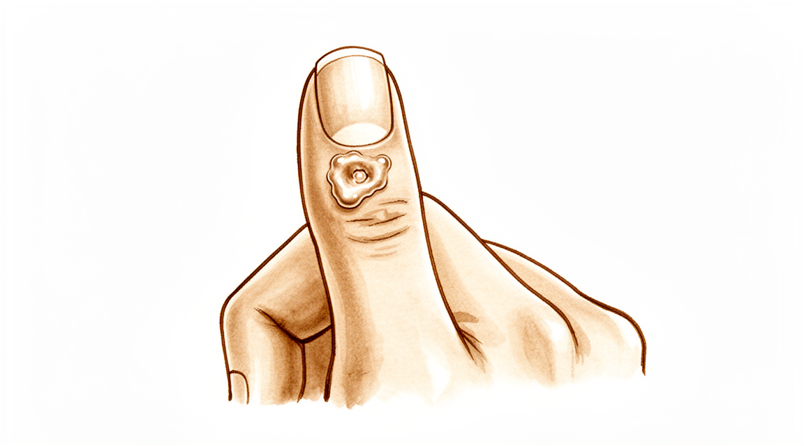
A mucous cyst shows up as a small, firm lump on your finger, just below the last joint before your fingertip. It always sits to one side of the finger, never right in the middle. The lump is filled with thick fluid, which is why it feels firm rather than soft.
The same joint often has wear-and-tear arthritis (the medical word is osteoarthritis). In fact, most people with a mucous cyst have arthritis in that joint as well. The cyst and the arthritis are connected: a small bone spur at the arthritic joint can irritate the joint lining and push fluid out towards the skin, forming the cyst.
The lump itself may ache, and the joint underneath can be stiff or sore. It tends to bother you most when you grip or pinch with that finger, things like turning a key, opening a jar, holding a pen, or buttoning a shirt. The ache often settles when you rest the hand. Some people notice the joint is worse first thing in the morning or after a busy day using the hands.
Because the cyst sits close to the nail, it can press on the nail bed and change how the nail grows. The nail above the lump may develop a groove or ridge. The skin over the cyst can also become thin and shiny.
If the lump is painful, growing, or affecting how your nail looks, that is usually the point where people seek advice. Your surgeon can confirm what it is by examining your finger, and an x-ray will show the arthritis and any bone spur at the joint.
What's actually happening
The last joint of your finger is wrapped in a sleeve of tissue called the joint capsule. It seals the joint like a gasket, keeping the joint fluid inside where it belongs. When that joint develops wear-and-tear arthritis, the lining of the capsule can weaken. Fluid then leaks out through the weak spot and pools under the skin as a small, firm lump. That lump is your mucous cyst.
The cyst is not a growth or a tumour. It is a pocket of thick, jelly-like fluid, much like the fluid that fills a ganglion cyst on the back of the wrist. It has no skin lining of its own, just a loose fibrous wall around it. The fluid inside comes from the joint itself, which is why pressing on the lump does not squeeze it flat. The fluid has nowhere to go.
The arthritis underneath is the real driver. As the joint wears, a small bone spur often forms at its edge. That spur sits right where the cyst appears, and it keeps irritating the capsule and feeding fluid into the cyst. This is also why the cyst can come back if only the lump is removed and the spur is left behind.
The symptoms you have read about above all flow from this setup. The lump aches because it is stretched full of fluid under a tight patch of skin. The skin over it becomes thin and shiny because the cyst is pushing out from underneath. The groove in your nail happens because the cyst presses on the nail bed as it grows, and that pressure changes how the nail forms. And the stiffness or soreness when you grip or pinch comes from the arthritic joint itself, not from the cyst alone.
What we can do about it
Dr Kieran Hirpara, an upper-limb surgeon at Mater Private Hospital Rockhampton, starts with the least invasive options that suit your condition. Patients are generally referred to our clinic by their GP; if a physiotherapist has suggested you see us, you will still need a referral from your GP in order to be eligible for the Medicare rebate. At your first visit we take a history, examine your finger, and arrange an x-ray if it is needed. The x-ray shows the arthritis and the bone spur at the joint, which we already know sit behind the cyst.
Because this is a long-standing problem, we usually begin with non-operative care. That means changing how you use the hand, so gripping and pinching tasks bother the joint less. Hand therapy or physiotherapy aims to settle the ache and keep the joint moving. Splinting can rest the joint and take pressure off the cyst. We give these measures a fair trial before talking about anything further.
If the lump still troubles you after that, surgery is the next step. The aim is to remove the bone spur at the joint, because that spur keeps feeding fluid into the cyst. Taking away the spur gives the cyst nothing to draw on, so it settles and does not return. In many cases removing the spur alone is enough, and the cyst itself can be left alone. This makes the operation smaller than removing both the lump and the bone. We will talk through what the operation involves and agree on a plan together.
What to expect
Left alone, a mucous cyst does not simply vanish overnight. It may stay much the same size, or it can come and go, shrinking and swelling over months. The ache tends to track the arthritis underneath rather than the lump itself, so flare-ups of joint pain often bring the cyst back to your attention. If the cyst keeps growing, the skin over it can thin out further and occasionally break down into an ulcer, and the groove in your nail will usually persist while the pressure continues.
With treatment, the outlook is steady. Removing the bone spur at the joint takes away the source of fluid, and once that supply is cut off the cyst settles. When the spur is fully removed, the cyst coming back is extremely rare. Even when treatment focuses on the spur and leaves the cyst itself alone, most people get complete resolution. Where the cyst is removed as well, recurrence happens in a small minority of cases, around 1.4% to 3% depending on the approach, and only a very small number of people ever need a second procedure.
Recovery from surgery is usually straightforward. The scar settles well, and a published series reported high satisfaction with its appearance and willingness to have the procedure again. Finger movement is preserved: there is no loss of bending at that last joint after treatment. The nail groove often improves once the pressure on the nail bed is relieved, though the nail may take some months to grow out fully.
Two honest caveats. First, much of the advice around mucous cysts rests on expert experience rather than large trials, so your surgeon will lean on clinical judgement as much as published numbers. Second, any new lump on the hand deserves a proper look. Rarely, other conditions can mimic a mucous cyst, and if there is any doubt about the diagnosis, a sample can be sent for testing to confirm it. If your lump changes quickly, becomes painful, or the skin breaks down, come back for review rather than waiting it out.
When to see someone
Most mucous cysts are not urgent. See your GP if the lump is painful, growing, or changing how your nail looks, or if the skin over it becomes thin and starts to break down. Ask for a specialist review if the ache keeps troubling you despite rest, splinting, or changing how you use your hand. Go to an emergency department or seek urgent assessment if a lump on your finger appears suddenly, grows quickly, or looks or feels different from what has been described here. Rarely, a lump that looks like a mucous cyst can turn out to be something else, so any new or changing lump deserves a proper look rather than waiting it out.
In more depth
Advanced reading: the deeper science (optional)
This section goes further than you need for your own treatment decisions. Mucous cysts are worth the extra reading because of a small, well-supported surgical insight: the cyst is not the problem, and the operation that works best does not necessarily remove it.
The bone spur is the cause, not the cyst
A mucous cyst arises from an arthritic fingertip joint. A bony spur, an osteophyte, irritates and perforates the joint capsule, and joint fluid tracks out through the defect and collects under the skin. The cyst is the visible end of that process, not its origin.
That understanding has a direct surgical consequence, demonstrated in a series where osteophyte excision without cyst excision produced complete resolution in most cases, and was described as a good treatment choice offering a less invasive method [1].
Removing the spur closes the tap. The cyst, no longer being filled, resolves. This is the same logic that governs the wrist ganglion, where the stalk rather than the sac determines recurrence, and it explains why simply draining or puncturing a mucous cyst so reliably fails.
Where the cyst is excised, results are also good
The alternative approach removes the cyst along with a local skin flap to close the defect. That is also reliable: across 69 patients, surgical excision with a local advancement flap showed a recurrence rate of 1.4% with high patient satisfaction regarding the scar and willingness to undergo the procedure again [2].
Both approaches work, and both address the underlying joint. The practical difference is how much skin is involved: a long-standing cyst thins the overlying skin, sometimes to the point of discharging, and in that situation the thinned skin needs excising and replacing regardless of what is done to the bone.
Why the nail becomes deformed, and whether it recovers
A groove or ridge running the length of the nail is a common accompaniment, and it worries people more than the lump. The cause is mechanical: the cyst sits immediately over the germinal matrix, the part of the nail bed that generates the nail, and presses on it, so the nail is produced with a defect.
The useful part is that this is pressure rather than destruction. Once the cyst is decompressed, the nail typically grows out normally, though it takes several months for the deformed portion to grow off the end. A nail deformity is therefore a reason to treat the cyst rather than a permanent consequence of it.
The reason to be careful about puncturing one
A mucous cyst communicates directly with the joint. Puncturing it, deliberately, or by the skin breaking down over a large one, creates a channel from the outside world into a finger joint, and septic arthritis of a small joint is a considerably more serious problem than the cyst.
This is the practical argument against home drainage of a cyst that appears to be simply a blister of fluid, and the reason a spontaneously discharging cyst is treated with some urgency rather than watched.
References for the advanced reading
- Lee H, Kim P, Jeon I, Kyung H, Ra I, Kim T. Osteophyte excision without cyst excision for a mucous cyst of the finger. J Hand Surg Eur Vol. 2013;39(3):258-61.
- Johnson SM, Treon K, Thomas S, Cox QGN. A reliable surgical treatment for digital mucous cysts. J Hand Surg Eur Vol. 2013;39(8):856-60.
Evidence & references
This is the clinical evidence summary written for health professionals. It is technical, and it lists the research this page was built from. You do not need to read it to understand your treatment or to make a decision about it.
Overview
- The scientific data regarding mucous cysts consist almost entirely of retrospective studies [1].
- Much of what is done or recommended for mucous cysts is based on expert opinion [1].
- Excision of the cyst and complete removal of the marginal osteophyte eradicates mucous cysts with extremely rare recurrence [2].
- In-office excision is a safe and effective option for treating digital mucous cysts [3].
- A total dorsal capsulectomy alone was a simple treatment for mucous cysts and did not lead to any recurrence [4].
- Eccrine porocarcinomas have a substantial risk of metastasis, high risk of local recurrence, and are potentially fatal [5].
- Prompt recognition and appropriate treatment are critical for eccrine porocarcinomas presenting as a hand cyst [5].
- The final recurrence rate of less than 1.5% for osteophyte-sparing treatment may be acceptable and comparable with other techniques [6].
- The Wolfe Graft technique is simple, easy to perform, and provides satisfactory cosmesis with acceptable recurrence rates [7].
- Surgical excision with a local advancement skin flap is a reliable treatment for digital mucous cysts [8].
- Surgical excision with a local advancement skin flap demonstrates a low recurrence rate of 1.4% [8].
- Surgical excision with a local advancement skin flap demonstrates high patient satisfaction regarding the scar and willingness to undergo the procedure again [8].
- Osteophyte excision without cyst excision may be a good treatment choice for mucous cyst of the finger [12].
- Osteophyte excision without cyst excision provides a less invasive method with complete resolution in most cases [12].
- A successful result was obtained by surgical treatment for an intraneural mucoid cyst in the digital nerve [14].
Anatomy & Pathophysiology
- Mucous cysts present as small, firm, cystic masses located just distal to the distal interphalangeal joint [11].
- The lesion is always located to one side of the mid-line [11].
- Mucous cysts are rarely greater than fifteen millimeters in diameter [11].
- The skin overlying a mucous cyst is thinned out and occasionally may be ulcerated [11].
- Pressure on a mucous cyst does not usually result in a decrease in its size [11].
- Longitudinal grooving of the nail may be noted, occasionally occurring prior to the appearance of the cyst [11].
- Degenerative arthritis of the distal interphalangeal joint is a frequent associated finding with mucous cysts [11].
- An incidence of 78 percent for degenerative arthritis of the distal interphalangeal joint has been reported in association with mucous cysts [11].
- The histological appearance of a mucous cyst is essentially identical to that of ganglions, synovial cysts, and similar lesions appearing on other areas of the extremities [11].
- A mucous cyst is surrounded by an undemarcated fibrous capsule [11].
- The basic structure of a mucous cyst is myxomatous with interspersed fibroblasts [11].
- Areas of myxomatous degeneration tend to coalesce to form a multiloculated cyst [11].
- An epithelial lining has not been reported for mucous cysts [11].
- The etiology of the mucous cyst is indicated by evidence that the lesion arises from the joint capsule [11].
- Fibro-osseous pseudotumor of the digit should be considered in the differential diagnosis of rapidly enlarging digital lesions [10].
Classification
- The lesion is a small, firm, cystic mass which appears just distal to the distal interphalangeal joint [11].
- The lesion is rarely greater than fifteen millimeters in diameter [11].
- The skin over the lesion is thinned out and occasionally may be ulcerated [11].
- Pressure on the lesion does not usually result in a decrease in its size [11].
- Degenerative arthritis of the distal interphalangeal joint is a frequent associated finding [11].
- An incidence of 78 per cent has been reported for degenerative arthritis of the distal interphalangeal joint as an associated finding [11].
- The histological appearance is essentially identical to that of ganglions, synovial cysts, and similar lesions appearing on other areas of the extremities [11].
- The lesion is surrounded by an undemarcated fibrous capsule [11].
- The basic structure is myxomatous with interspersed fibroblasts [11].
- An epithelial lining has not been reported [11].
Clinical Presentation
- Mucous cysts present as a small, firm, cystic mass located just distal to the distal interphalangeal joint [11].
- An incidence of 78 per cent has been reported for degenerative arthritis of the distal interphalangeal joint associated with mucous cysts [11].
Investigations
- Ultrasound is a powerful modality for evaluation of pathologic conditions in the hand and wrist [18].
- Ultrasound provides a cost-effective and expedient alternative and/or adjunct to MRI [18].
- Ultrasound is best used when there is a specific clinical question regarding a well-localized abnormality [18].
- A careful physical examination is essential to direct care and future testing if indicated [19].
- Diagnostic tests such as imaging and serum laboratory studies are useful in the determination of pathology but can be expensive, time consuming, and often nonspecific [19].
- Pathohistological analysis is useful in cases where doubts arise about the initial diagnosis of a benign tumorous lesion [9].
Treatment
Operative Techniques
- The use of a Wolfe Graft for the treatment of mucous cysts is a simple technique that provides satisfactory cosmesis with acceptable recurrence rates [7].
- Surgical excision with a local advancement skin flap is a reliable treatment for digital mucous cysts, demonstrating a low recurrence rate of 1.4% and high patient satisfaction regarding the scar and willingness to undergo the procedure again [8].
- Osteophyte excision without cyst excision may be a good treatment choice for mucous cyst of the finger, providing a less invasive method with complete resolution in most cases [12].
- Osteophyte removal results in a low cyst recurrence rate, indicating that it should be undertaken regardless of the surgeon's plan for the soft tissues [37].
- Surgical treatment of an intraneural mucoid cyst in the digital nerve resulted in a successful outcome [14].
Diagnostic Considerations
- Pathohistological analysis is useful in cases where doubts arise about the initial diagnosis of a benign tumorous lesion, such as when a malignant natural-Killer cell neoplasm presents as a mucous cyst [9].
- Prompt recognition and appropriate treatment are critical for eccrine porocarcinomas presenting as hand cysts because they have a substantial risk of metastasis, high risk of local recurrence, and are potentially fatal [5].
Evidence Quality and Decision Factors
- The scientific data regarding mucous cysts consist almost entirely of retrospective studies, and much of what is done or recommended is based on expert opinion [1].
- Tumor location, preoperative diagnosis, prior upper extremity tumor, and surgeon affect the likelihood of surgery for an upper extremity tumor [16].
Recurrence
- Surgical excision with a local advancement skin flap is a reliable treatment for digital mucous cysts, demonstrating a low recurrence rate of 1.4% [8].
- The Wolfe graft technique provides acceptable recurrence rates for the treatment of mucous cysts [7].
Diagnostic Errors and Malignancy
- Eccrine porocarcinomas presenting as hand cysts have a substantial risk of metastasis, high risk of local recurrence, and are potentially fatal [5].
- Pathohistological analysis is useful in cases where doubts arise about the initial diagnosis of a benign tumorous lesion, such as a malignant natural-killer cell neoplasm presenting as a mucous cyst [9].
Surgical Complications and Outcomes
- The Zitelli bilobed flap allows excision of the cyst and thinned skin with no added risk to the nail matrix [15].
- The bilobed flap allows radical excision with primary skin healing, no loss of flexion at the distal interphalangeal joint, and excellent cosmesis [21].
Evidence Quality
Differential Diagnosis and Pathology
- Eccrine porocarcinomas presenting as hand cysts carry a substantial risk of metastasis, high risk of local recurrence, and are potentially fatal [5].
Surgical Outcomes and Recurrence
- A total dorsal capsulectomy alone did not lead to any recurrence for mucous cysts [4].
- The final recurrence rate for osteophyte-sparing treatment of mucous cysts was less than 1.5% [6].
- Surgical excision with a local advancement skin flap for digital mucous cysts demonstrated a low recurrence rate of 1.4% [8].
- The use of a Wolfe graft for the treatment of mucous cysts provides acceptable recurrence rates [7].
Patient Satisfaction and Safety
- Patients undergoing surgical excision with a local advancement skin flap for digital mucous cysts reported high satisfaction regarding the scar and willingness to undergo the procedure again [8].
- The Wolfe graft technique for mucous cysts provides satisfactory cosmesis [7].
Key Evidence
- [L4] The scientific data regarding mucous cysts consist almost entirely of retrospective studies, and much of what is done or recommended is based on expert opinion. [1] (10.1016/j.jhsa.2010.01.029)
- [L4] Excision of the cyst and complete removal of the marginal osteophyte eradicates mucous cysts with extremely rare recurrence. [2] (10.2106/00004623-197355030-00013)
- [Paper] In-office excision is a safe and effective option for treating digital mucous cysts. [3] (10.1177/15589447251350168)
- [L4] A total dorsal capsulectomy alone was a simple treatment for mucous cysts and did not lead to any recurrence. [4] (10.1016/j.jhsa.2014.03.004)
- [L4] Prompt recognition and appropriate treatment are critical because eccrine porocarcinomas have a substantial risk of metastasis, high risk of local recurrence, and are potentially fatal. [5] (10.1016/j.jhsa.2016.07.112)
- [L4] The final recurrence rate of less than 1.5% may be acceptable and comparable with other techniques. [6] (10.5435/jaaosglobal-d-21-00164)
- [L4] The technique is simple, easy to perform, and provides satisfactory cosmesis with acceptable recurrence rates. [7] (10.1177/1753193408103498)
- [L4] Surgical excision with a local advancement skin flap is a reliable treatment for digital mucous cysts, demonstrating a low recurrence rate of 1.4% and high patient satisfaction regarding the scar and willingness to undergo the procedure again. [8] (10.1177/1753193413508540)
- [L5] This case emphasizes the utility of a pathohistological analysis in cases where doubts arise about the initial diagnosis of a benign tumorous lesion. [9] (10.1007/s00402-008-0794-4)
- [Paper] Fibro-osseous pseudotumor of the digit should be considered in the differential diagnosis of rapidly enlarging digital lesions. [10] (10.1177/15589447261481209)
- [L4] [11] (10.2106/00004623-197254070-00008)
- [L4] Osteophyte excision without cyst excision may be a good treatment choice for mucous cyst of the finger, providing a less invasive method with complete resolution in most cases. [12] (10.1177/1753193413478549)
- [L4] A successful result was obtained by surgical treatment in our case. [14] (10.1177/1753193408099819)
- [L4] It allows excision of the cyst and thinned skin with no added risk to the nail matrix. [15] (10.1016/j.jhsa.2017.03.013)
- [L2] Tumor location, preoperative diagnosis, prior upper extremity tumor, and surgeon affect the likelihood of surgery for an upper extremity tumor. [16] (10.1007/s11552-013-9518-5)
- [L5] Ultrasound is a powerful modality for evaluation of pathologic conditions in the hand and wrist, providing a cost-effective and expedient alternative and/or adjunct to MRI, best used when there is a specific clinical question regarding a well-localized abnormality. [18] (10.1016/j.jhsa.2009.02.010)
- [L4] The bilobed flap allows radical excision with primary skin healing, no loss of flexion at the distal interphalangeal joint, and excellent cosmesis. [21] (10.1054/jhsb.1998.0191)
- [Commentary] The article shows that osteophyte removal results in a low cyst recurrence rate, indicating that it should be undertaken regardless of the surgeon's plan for the soft tissues. [37] (10.1177/1753193413510663)
References
[1] Mucous Cysts. The Journal of Hand Surgery. 2010. DOI: 10.1016/j.jhsa.2010.01.029
[2] Marginal Osteophyte Excision in Treatment of Mucous Cysts. The Journal of Bone & Joint Surgery. 1973. DOI: 10.2106/00004623-197355030-00013
[3] Safety and Cost-Effectiveness of In-Office Digital Mucous Cyst Excisions. HAND. 2025. DOI: 10.1177/15589447251350168
[4] Total Dorsal Capsulectomy for the Treatment of Mucous Cysts. The Journal of Hand Surgery. 2014. DOI: 10.1016/j.jhsa.2014.03.004
[5] Eccrine Porocarcinoma Presenting as a Hand Cyst. The Journal of Hand Surgery. 2016. DOI: 10.1016/j.jhsa.2016.07.112
[6] Osteophyte-Sparing Treatment of Mucous Cysts: Case Analysis and Surgical Technique. JAAOS: Global Research and Reviews. 2021. DOI: 10.5435/jaaosglobal-d-21-00164
[7] Use of Wolfe Graft for the Treatment of Mucous Cysts. Journal of Hand Surgery (European Volume). 2009. DOI: 10.1177/1753193408103498
[8] A reliable surgical treatment for digital mucous cysts. Journal of Hand Surgery (European Volume). 2013. DOI: 10.1177/1753193413508540
[9] Malignant Natural-Killer cell neoplasm presenting as a mucous cyst on the distal interphalangeal joint of the finger. Archives of Orthopaedic and Trauma Surgery. 2008. DOI: 10.1007/s00402-008-0794-4
[10] Recurrent Fibro-Osseous Pseudotumour of the Digit Mimicking Infection: A Narrative Review and Case Report. HAND. 2026. DOI: 10.1177/15589447261481209
[11] Etiology and Treatment of the So-Called Mucous Cyst of the Finger. The Journal of Bone & Joint Surgery. 1972. DOI: 10.2106/00004623-197254070-00008
[12] Osteophyte excision without cyst excision for a mucous cyst of the finger. Journal of Hand Surgery (European Volume). 2013. DOI: 10.1177/1753193413478549
[14] Intraneural mucoid cyst in the digital nerve. Journal of Hand Surgery (European Volume). 2009. DOI: 10.1177/1753193408099819
[15] The Zitelli Bilobed Flap on Skin Coverage After Mucous Cyst Excision: A Retrospective Cohort of 33 Cases. The Journal of Hand Surgery. 2017. DOI: 10.1016/j.jhsa.2017.03.013
[16] Benign Upper Extremity Tumors: Factors Associated with Operative Treatment. HAND. 2013. DOI: 10.1007/s11552-013-9518-5
[18] Ultrasound of the Hand and Wrist. The Journal of Hand Surgery. 2009. DOI: 10.1016/j.jhsa.2009.02.010
[19] Orthopaedic Knowledge Update 13 Ebook Without Multimedia. Anatomy, Evaluation, Clinical Examination, and Imaging > Evaluation and Clinical Examination: Current Concepts.
[21] The Bilobed Flap in Treatment of Mucous Cysts of the Distal Interphalangeal Joint. Journal of Hand Surgery. 1999. DOI: 10.1054/jhsb.1998.0191
[37] Commentary on Lee et al. Osteophyte excision without cyst excision for a mucous cyst of the finger. Journal of Hand Surgery (European Volume). 2014. DOI: 10.1177/1753193413510663