Patients › Hand
PIP Joint Fusion
PIP joint fusion stabilizes a severely damaged middle finger joint, relieving pain when other treatments fail.
Why this operation has been suggested
Dr Kieran Hirpara, an upper-limb surgeon at Mater Private Hospital Rockhampton, starts with the least invasive options that suit your condition. Patients are generally referred to our clinic by their GP; if a physiotherapist has suggested you see us, you will still need a referral from your GP in order to be eligible for the Medicare rebate. At your appointment we take a history, examine your hand, and arrange imaging if it is needed to work out what is wrong.
This operation is a fusion of the middle joint of your finger. The two bones forming that joint are joined together so they heal as one, and the joint no longer moves. We usually suggest it when the joint is painful, stiff or deformed, and when simpler care such as activity change, physiotherapy or hand therapy, and splinting has not given you enough improvement. For some injuries or structural problems, surgery may be recommended straight away. The aim is to restore hand function by taking away the pain and holding the joint steady.
Before the operation
In the weeks before surgery we confirm the plan with fresh images of your finger, such as an X-ray, and sometimes an MRI or ultrasound scan. These show the surgeon what the joint looks like and help plan the fusion angle. Most people need nothing more than that. If you have other medical conditions, you may also need blood tests or a review with the anaesthetist, but this is not routine. In the days before your operation we will tell you which of your usual medicines to stop and when. Do not eat for seven hours before surgery; we ask for a little extra time so your slot can be brought forward if the theatre list runs early. Arrange a lift home, as you will not be able to drive yourself. Bring a written list of your current medicines and wear loose, comfortable clothing.
On the day
You arrive at the hospital's surgical admissions unit, where you are checked in and prepared for theatre. You will meet the anaesthetist there. This operation is done under general anaesthetic. You will be fully asleep for the operation. Some patients may also have a regional nerve block for post-operative pain relief; the anaesthetist decides on the day based on your individual circumstances. You are then taken into the operating theatre, where the operation is performed.
You wake up in the recovery area, where nurses monitor you while the anaesthetic wears off. Once you are stable, you either go to the ward or go home, depending on the procedure and your recovery.
What the operation involves
Your surgeon makes a single cut over the middle joint of your finger. Through this cut, the joint is opened up and the worn, damaged surfaces of the two bones are prepared so they can grow together. The bone ends are shaped to fit against each other, and the finger is set at a bent position that lets your hand work well for gripping and pinching.
The bones are then held together while they heal. Your surgeon uses small metal implants, such as wires or screws, to keep the two bones still and in the right position. This holding is what allows the joint to fuse into one solid bone. Once the bones are lined up and fixed, the wound is closed with stitches and covered with a dressing.
The aim of all this is simple: the painful, moving joint is taken away and replaced with a steady, pain-free connection between the two bones. Because the joint no longer moves, the finger will be stiffer than before, but it should no longer catch or hurt with use.
After the operation
Most patients stay one night in hospital after this operation, though some are able to go home the same day. You wake up in the recovery ward, where nurses keep an eye on you while the anaesthetic wears off. Your hand will be dressed and may be supported with a splint, and we will give you pain relief to keep you comfortable. You can get up and move around as soon as you feel steady. Please arrange for someone to stay with you for the first 24 hours. We leave the dressing on for about 10 days; please do not take it off before then unless we tell you to. We change or remove it when we see you.
Recovery
Your finger will be sore and swollen for the first days and weeks. This is normal and settles gradually. Keeping your hand raised on a pillow, even while you sleep, helps bring the swelling down. The pain relief we give you keeps you comfortable while this happens.
You will wear a splint on your finger to protect it while the bones heal. Hand therapy after surgery is with Ruby Doolan at Extend Rehabilitation. Ruby is a hand therapist: she directs your exercises and makes any splint you need. Your therapy will focus on keeping the rest of your hand and fingers moving, so your grip and pinch stay strong while the fused joint settles.
You can do most light daily activities at home straight away, using your other hand to help. Avoid heavy lifting, gripping hard, or anything that puts strain on the healing finger until we clear you. Once the dressing comes off and the wound has healed, you can use your hand more freely for everyday tasks. Driving is not safe while the splint stops you gripping the wheel properly. Once the splint comes off and your surgeon clears you, see our page on Driving after upper-limb surgery.
Recovery varies from person to person. Your timeline may differ, and your surgeon and therapist will guide you at each review.
What can go wrong
Most patients do well, but problems can occasionally happen. Your surgeon and the team monitor you closely to spot any issue early.
The main risk with a fusion is that the two bones fail to join. If that happens, the finger may stay painful, and you might feel a click or movement where there should be none. Tell us at your next review if the pain does not settle as expected.
The metal implants holding the bones can also cause trouble. A wire or screw can work loose, break, or press on the skin. You might notice a sharp edge under the skin, or the finger looking out of line. Bring this up at your review so we can check it.
Infection is uncommon but needs fast attention. Watch for a deep, throbbing pain that does not ease with simple painkillers, redness spreading out from the wound, or fluid leaking from it. You may feel hot and generally unwell. If you see any of these signs, call the clinic straight away. If it is after hours or the redness is spreading quickly, go to the emergency department.
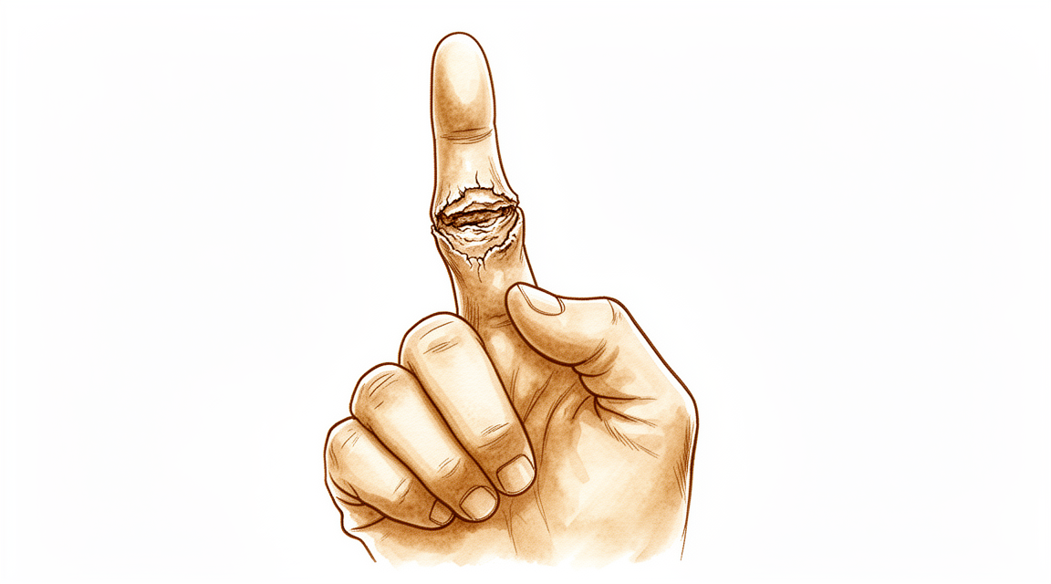
Infection can also reach a finger joint from a cut or puncture wound to the finger, from a nearby skin infection, or through the bloodstream from elsewhere in the body. Signs of infection in a small hand joint often start within days of the injury. See your GP promptly if a finger becomes hot, swollen and painful after a knock or wound, so it can be treated early.
Stiffness and ongoing pain can happen even when the fusion heals well. If your finger stays very stiff or painful after the expected recovery period, we will assess it and discuss what can be done.
The complications table on this page lists typical rates if you want the specifics.
When to call us
Call us straight away if you have a fever, if the skin around your wound becomes more red or starts leaking fluid, or if you have sudden severe pain that simple painkillers do not settle. Go to the emergency department if you have calf swelling or pain, or shortness of breath, as these can signal a blood clot. Also go to emergency if your fingers go numb, lose colour, or you cannot move them at all. If it is after hours and you are worried, go to emergency rather than waiting for the clinic to open.
Where to read more about the condition
This page is about the operation itself. The condition it treats, including what the evidence shows about when surgery helps and when it does not, is covered in more detail on the PIP Joint Arthritis page.
Evidence & references
This is the clinical evidence summary written for health professionals. It is technical, and it lists the research this page was built from. You do not need to read it to understand your treatment or to make a decision about it.
Anatomy & Pathophysiology
General Hand Architecture
- The hand is composed of 19 bones, 17 articulations, and 19 muscles situated entirely within the hand [2].
- The hand contains approximately the same number of tendons activated by forearm muscles as it has intrinsic muscles [2].
- The hand functions as an organ designed to obtain information and an organ of execution [2].
- The dorsal aspect of the hand is convex, while the palmar or volar aspect is concave [2].
- The dorsal surface of the hand is usually visible and aesthetically important, whereas the palmar surface is usually hidden and functional [2].
- The hand moves within a large volume of space with the shoulder as the apex, allowing it to reach any part of the body fairly easily due to the mobility of the shoulder, elbow, and wrist [2].
- Distal to the elbow, the wrist and forearm function as a single physiological unit that places the hand in a position for grasping [2].
- The hand is capable of conforming to the shape of objects to be grasped or studied [2].
Digits and Phalanges
- The digits are divided into the thumb and four fingers [2].
- The thumb has a more proximal and lateral position, allowing movement inward and outward from the palm [2].
- The four fingers are the distal extension of the carpometacarpal part of the hand [2].
- Each finger ray is a polyarticular chain composed of a metacarpal and three phalanges, except the thumb which has only two phalanges [2].
- When fingers are extended and separated, their tips lie on the circumference of a circle whose center is the head of the third metacarpal [2].
- The web space of the thumb is the largest and deepest among the digital web spaces [2].
Cutaneous Units
- The dorsal covering of the interphalangeal articulations of the digits forms a unique cutaneous unit characterized by a considerable excess of skin when the digits are in extension [3].
- The fine tight skin of the dorsal aspect of the middle phalanx forms a distinct cutaneous unit [3].
- The dorsal integument of the distal phalanx is characterized by the nail bed with its matrix [3].
- The palm forms a cutaneous unit extending from the distal transverse crease of the wrist up to the transverse crease at the base of the digits [3].
- The skin of the radial portion of the palm covers the thenar eminence and is relatively well vascularized and mobile [3].
- The skin of the ulnar and distal portion of the palm covers the hypothenar eminence and has poor mobility [3].
- The central triangular part of the palm has fixed, poorly vascularized skin that covers almost directly the superficial palmar aponeurosis [3].
- The integument of the palmar face of the digits is subdivided into phalangeal units separated by digital flexion folds [3].
- When a digit is completely flexed, the integument of adjacent phalanges comes into contact in the zones of the flexion creases, establishing areas of cutaneous contact in the form of a diamond [3].
- The sides of the diamond-shaped cutaneous contact zones do not undergo variations in length during movements of flexion and extension [3].
- Incisions made along the lines of the diamond-shaped cutaneous contact zones present a minimal chance of retraction [3].
- The dorsal slope of the web spaces has a gradual incline and its supple skin is not adherent to the subjacent region [3].
- The palmar surface of the web spaces is flat and precipitously interrupted, with skin densely adherent to the commissural skeleton [3].
Intrinsic Muscles
- There are seven interosseous muscles in the hand, consisting of four dorsal and three volar muscles [4].
- The dorsal interossei are abductors [4].
- The volar interossei are adductors [4].
- The middle finger has two dorsal interossei and no volar interossei because the central axis of the hand lies within it [4].
- Each dorsal interosseous muscle, with the exception of the third, has two muscle heads [4].
- The superficial head of the dorsal interosseous muscles abducts and weakly flexes the proximal phalanx [4].
- The deep head of the dorsal interosseous muscles flexes and weakly abducts the proximal phalanx while extending the middle and distal phalanges [4].
- Transverse fibers arch dorsally from each lateral band to join over the dorsum of the finger, flexing the proximal phalanx [4].
- Oblique fibers from the lateral bands insert onto the lateral tubercles at the base of the middle phalanx to extend the middle phalanx at the PIP joint [4].
- The lateral bands are joined by the lateral slips of the extensor tendon to form the conjoined lateral band [4].
- The two conjoined lateral bands to each finger unite at the distal third of the middle phalanx to form the terminal tendon [4].
- The terminal tendon inserts at the base of the distal phalanx to extend it [4].
- Each volar interosseous muscle has only one muscle head and none insert onto the proximal phalanx [4].
- The volar interossei form the ulnar lateral band of the index finger and the radial lateral band of the ring and little fingers [4].
- The abductor digiti quinti and flexor digiti quinti brevis are similar in structure and function to the superficial and deep heads of the dorsal interossei, respectively [4].
- The opponens digiti quinti arises from the pisohamate ligament and the hook of the hamate and inserts onto the ulnar side of the diaphysis of the fifth metacarpal [4].
Metacarpal Arch and Stability
- The metacarpal arch is endowed with a great deal of adaptability because of the mobility of the peripheral metacarpals [7].
- The index metacarpal is the most firmly fixed [7].
- The ring metacarpal has about 10 degrees of mobility in flexion and extension [7].
- The fifth metacarpal has a range of flexion–extension of approximately 20 degrees [7].
- The second to fifth metacarpals are bound together by various fibrous structures, the most distal of which is the deep transverse intermetacarpal ligament [7].
- The deep transverse intermetacarpal ligament is also known as the interglenoid ligament because it ties together the anterior glenoid ligaments of the metacarpophalangeal articulations [7].
- The metacarpophalangeal articulations are the keystones of the longitudinal arches of the hand [7].
- The thick anterior glenoid capsules, known as volar plates, prevent hyperextension at the metacarpophalangeal joints [7].
- The stability of the metacarpophalangeal joints is essential to the support of the longitudinal arch as well as of the transverse metacarpal arch [7].
Vascular Anatomy
- The arteries of the thumb vary in both size and number, making surgical reconstruction delicate [8].
- The layout of the thumb arteries is the result of innumerable variations regarding origin, transit, connections, and size [8].
- In anatomical studies, only 15% of dissections of the palmar arteries of the thumb fall into the classical "typical" category [8].
- The princeps pollicis artery is the terminal branch of the radial artery that crosses the first intermetacarpal space [8].
- The princeps pollicis artery runs along the ulnar side of the first metacarpal bone and along the volar surface of the adductor muscle [8].
- The princeps pollicis artery emerges onto the subcutaneous palmar tissue at the level of the cutaneous flexion crease of the metacarpophalangeal joint [8].
- At the metacarpophalangeal joint level, the princeps pollicis divides into two terminal rami known as the collateral palmar arteries of the thumb [8].
- The collateral palmar arteries run along the digital tunnel symmetrically and are of equal caliber [8].
- The collateral palmar arteries head distally to finally unite in the pulp arcade [8].
- An arcade located deep in the flexor tendon joins together the two arteries at the level of the distal metaphysis of the first phalanx [8].
- Vessels originating from the subtendinous arcade enter the vincula and irrigate the flexor tendon [8].
- In the second segment of the thumb, the main artery is the ulnar collateral artery [8].
- The subtendinous anastomosis situated at the level of the neck of the first phalanx acts as a moderator between the two collateral arteries [8].
- In cases where the palmar ulnar collateral artery is absent, the dorsal artery takes its place by means of a branch through the subtendinous arcade [8].
- In the pulp segment, the two arteries are of similar size and run through the thick fatty subcutaneous padding [8].
- The dorsal arteries of the thumb originate from the palmar arteries at the level of the first metacarpal [8].
- The dorsal arteries run laterally along the metacarpophalangeal joint and continue obliquely from volar to dorsal [8].
- At the level of the neck of the first phalanx, an anastomosis can be found which originates from the palmar arteries [8].
- The dorsal arteries are joined by three arcades: one inconstant arcade under the extensor tendon at the neck of the first phalanx, the arcade of the nail matrix, and the arcade of the nailbed [8].
Surgical Approaches and Incisions
- Distal palmar incisions are transverse, while proximal palmar incisions tend to be more longitudinal with the distal end curving radially [9].
- Proximal palmar incisions should parallel the thenar crease [9].
- Incisions extended proximal to the wrist should not cross the flexor wrist creases at a right angle [9].
- The most important structure in the thenar area is the recurrent branch (motor) of the median nerve [9].
- Anatomic studies have shown that there is no single longitudinal incision in the proximal palm that completely avoids the palmar cutaneous branches of the median and ulnar nerves [9].
- Midlateral incisions described for the fingers are also suitable for the thumb [9].
- The radial side of the thumb is more accessible for midlateral incisions [9].
- Care should be taken to avoid the dorsal branch of the superficial radial nerve to the radial side of the thumb during midlateral incisions [9].
- The volar midoblique incision crosses the flexion creases obliquely in the midline of the finger between the neurovascular bundles [9].
Investigations
- Clinical evaluation of the hand and wrist requires combining patient history with a careful physical examination to pinpoint or narrow the scope of possible pathologic processes [1].
- Diagnostic tests such as imaging and serum laboratory studies are useful in determining pathology but can be expensive, time consuming, and often nonspecific [1].
- A careful physical examination is essential to direct care and future testing if indicated [1].
- A systematic method for approaching the physical examination of the hand is essential due to the number of structures in a small space [1].
- Clinicians may organize the hand examination by anatomic location or region, or by organ system or pathology [1].
- The skeleton of the hand and wrist consists of 27 bones, of which 19 are long bones [12].
- The skeleton is divided into five rays, each comprising a polyarticulated chain of metacarpals and phalanges [12].
- The base of each metacarpal articulates with the distal row of the carpus [12].
- The wrist has three axes of movement, permitting the hand to be positioned in any spatial configuration for grasping [12].
- The radial ray (first ray) is the shortest, made up of a metacarpal and two phalanges [12].
- The other four digital rays are formed by four skeletal segments: a metacarpal and three phalanges [12].
- The thumb metacarpal is the shortest, while the index metacarpal is the longest [12].
- The proximal and middle phalanges of the middle and ring fingers are longer than those of the index finger [12].
- The transverse axis of the palm forms an acute angle of approximately 75 degrees with the longitudinal axis [12].
- Epiphyseal plates are located at the proximal ends of the phalanges and the first metacarpal [12].
- Epiphyseal plates are located at the distal ends of the other metacarpals [12].
References
[1] Orthopaedic Knowledge Update 13 Ebook Without Multimedia. Anatomy, Evaluation, Clinical Examination, and Imaging > Evaluation and Clinical Examination: Current Concepts.
[2] Exam Of The Hand Wrist 2Ed. INTRODUCTION.
[3] Exam Of The Hand Wrist 2Ed. Functional cutaneous units.
[4] Green S Operative Hand Surgery. Interosseous and Hypothenar Muscles.
[7] Exam Of The Hand Wrist 2Ed. The arches of the hand > The metacarpal arch.
[8] Exam Of The Hand Wrist 2Ed. Techniques of investigation of the arterial supply by J P Melki > Vascularization of the thumb > Palmar aspect.
[9] Campbell S Operative Orthopaedics 4 Volume Set. RESULTS OF SUTURE OF THE SCIATIC NERVE > PALMAR INCISIONS.
[12] Exam Of The Hand Wrist 2Ed. 1.1 SKELETON OF THE HAND > The osseous skeleton.