Clinicians › Wrist
Wrist ganglion excision
Surgeon-side topic for wrist ganglion excision. Backed by 123 articles from the corpus, retrieved via combined MeSH + title-text matching.
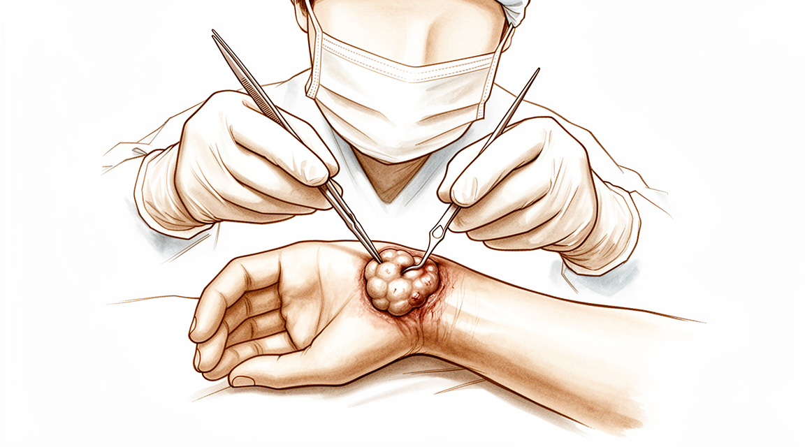
For patients: a plain-language version of this topic is available. See the patient guide.
Overview
Wrist ganglions are common soft-tissue lesions that frequently affect the dorsal wrist, particularly in pediatric populations where a female predilection is noted [28]. The natural history of these lesions varies by age; in children under 10 years, 69% to 79% of ganglions display spontaneous regression within 12-18 months [13], and pediatric hand ganglions resolve at a greater rate than wrist ganglions [4]. In the broader population, approximately 40% of wrist ganglion lesions decrease over the first 6 years after evaluation by a hand surgeon [11]. While there is no consensus on the best management of pediatric wrist ganglia, with no single treatment modality conferring a particular advantage or disadvantage [17], surgical intervention remains a standard option for persistent symptoms.
Surgical excision of primary wrist ganglia significantly reduces patient symptoms with low recurrence rates and high patient satisfaction [16]. Open surgical excision offers a significantly lower chance of recurrence compared with aspiration, which is associated with most ganglions recurring [11]. The overall recurrence rate for surgical intervention is about 10% [11], though a retrospective review reports a low recurrence rate of 5.3% for pediatric wrist ganglion cysts [18]. Arthroscopic techniques have advanced to allow excision with recurrence rates similar to open management [5], although open excision of dorsal wrist ganglia leads to a lower recurrence rate than arthroscopic excision [12]. At 12 months follow-up, arthroscopic dorsal ganglion excision recurrence rates are comparable with and not superior to open excision [33]. Routine midcarpal joint exploration during arthroscopic excision appeared to reduce recurrence at 1 year without negatively impacting patient outcomes [3].
Complications include scarring and adverse events [11]. Female patients with preoperative pain around dorsal wrist ganglia are the most likely to have residual pain after surgery [2]. Patients whose occupation or activities require forceful wrist extension should be counseled on the considerable risk of residual pain and functional limitations after open dorsal wrist ganglion excision [6]. Operation-related complications after arthroscopic volar wrist ganglionectomy are associated with anatomical location distal to the bifurcation of the radial artery and concurrently penetrated up to the superficial fascia layer [7]. Management considerations include performing at least 1 aspiration before surgical excision to improve cost-effectiveness [8]. Hand surgeons are divided regarding the need to immobilize the wrist after dorsal wrist ganglion excision [10]. In patients with a clinical diagnosis of wrist ganglion cyst, quality of care would not be compromised by abandoning the practice of routinely submitting surgical specimens for pathological examination after excision [14, 15]. Surgery for dorsal, volar, or wrist ganglia is often unrewarding for both the surgeon and the patient in young, less educated patients [20].
Anatomy & Pathophysiology
Anatomical Location and Distribution
The dorsal wrist ganglion is the prototype of all hand and wrist ganglions, accounting for 60% to 70% of all hand and wrist ganglions [34]. Dorsal wrist ganglions arising from within the scapholunate joint are the most common type of ganglions of the hand and wrist [27]. The main cyst of a dorsal wrist ganglion is usually located directly over the scapholunate ligament [34]. Volar wrist ganglions are the second most common ganglion of the hand and wrist, accounting for 18% to 20% of cases [35]. The majority of volar ganglions occur either directly over the distal edge of the radius or slightly more distally over the scaphoid tubercle [35]. In pediatric populations, ganglions most commonly affect the dorsal wrist [28]. In children aged <10 years, ganglions mainly occur on the volar wrist [13]. The ulnar nerve is the most commonly involved nerve in the upper extremity for intraneural ganglions [31].
Pathogenesis and Associated Pathology
The articular (synovial) theory has gained worldwide acceptance as a scientifically proven explanation for the formation of intraneural ganglions [31]. A small, mucin-filled duct is invariably seen piercing the transverse fibers of the scapholunate ligament, connecting the underlying scapholunate joint with the main cyst [34]. Failure to identify and excise the pedicle attachment to the scapholunate ligament increases the likelihood of recurrence [34]. Associated abnormalities of the intracarpal ligaments have been described in patients undergoing arthroscopic resection of wrist ganglions [22]. Central perforations of the triangular fibrocartilage complex (TFCC) have been found in patients with prior unsuccessful treatment of dorsal wrist ganglions [22]. Patients with wrist hyperlaxity have a predisposition to developing ganglions [45]. An association between wrist ganglions and ligamentous hyperlaxity does not prove causation, but the possibility of the same underlying pathological entity causing both can be envisioned [37]. Wrist arthroscopy is associated with a 1.24% incidence of ipsilateral wrist ganglion cyst formation [29]. Female gender is a significant predictor of the development of ganglion cysts after wrist arthroscopy [29].
Natural History and Demographics
Approximately 40% of wrist ganglion lesions decrease over the first 6 years after evaluation by a hand surgeon [11]. In children aged <10 years, 69% to 79% of volar wrist ganglions display spontaneous regression within a span of 12-18 months [13].
Wrist Anatomy and Biomechanics
The wrist is an anatomic region between the forearm and the hand, including the distal radioulnar, radiocarpal, and ulnocarpal joints and the eight carpal bones [46]. The eight carpal bones are divided into a proximal row (scaphoid, lunate, triquetrum, pisiform) and a distal row (trapezium, trapezoid, capitate, hamate) [46]. The wrist can be considered a two-joint system linking the hand to the forearm around the highly mobile bones of the proximal carpal row [55]. The two principal articulations of the wrist are the radiocarpal and midcarpal joints, situated proximal and distal to the mobile proximal carpal row [55]. With axial loading through the neutral wrist, approximately 80% of forces are transmitted through the distal radius and 20% through the distal ulna [54].
The scapholunate interosseous ligament is C-shaped in the sagittal plane, with the dorsal third being the thickest and strongest portion [54]. The volar portion of the lunotriquetral ligament is the thickest [54]. The triangular fibrocartilage complex (TFCC) is formed by the central meniscus homolog, dorsal and volar radioulnar ligaments, the floor of the extensor carpi ulnaris tendon sheath, and volar ulnocarpal ligaments [54]. The distal radius articular surface has two concave facets (scaphoid and lunate) separated by the scapholunate ridge [54]. The sigmoid notch along the ulnar border of the distal radius is a shallow concavity for the articulating ulnar head at the distal radioulnar joint [54].
The terminal branches of the radial, ulnar, and anterior interosseous arteries provide extraosseous blood supply to the carpus through three dorsal and three palmar transverse arterial arches [57]. The dorsal intercarpal arch is the largest of the dorsal arches and supplies the distal carpal row [57]. The deep palmar arch at the level of the metacarpal bases is consistent and communicates with the dorsal basal metacarpal arch and the palmar metacarpal arteries [57]. The radial artery is frequently intimately attached to the wall of a volar ganglion and may be completely encircled by it [35]. The main cyst of a volar ganglion may be intertwined with bifurcating branches of the radial artery [35].
The extensor pollicis longus and extensor digitorum communis tendons are retracted radially and ulnarly, respectively, to expose a typical dorsal ganglion [34]. The dorsal ganglion typically appears between the extensor pollicis longus and extensor digitorum communis tendons [34].
Classification
Histological Subtypes: Cystic soft tissue tumours of the dorsal aspect of the wrist have two distinct histological subtypes [26].
Other Considerations: Intraneural ganglions most commonly affect patients aged 30 to 50 years, though occurrence in children has also been reported [31]. Most patients with intraneural ganglions present with a painless mass, while some present with symptoms of nerve irritation or entrapment neuropathy [31]. In pediatric populations, ganglions most commonly affect the dorsal wrist and demonstrate a female predilection [28]. Female gender is a significant predictor of ipsilateral wrist ganglion cyst formation after wrist arthroscopy [29].
Clinical Presentation
Surgical intervention for wrist ganglions results in scarring and carries a risk of adverse events [11]. Female patients presenting with preoperative pain around dorsal wrist ganglia are the most likely to experience residual pain following surgery [2]. An association between wrist ganglions and ligamentous hyperlaxity has been identified, although this does not prove causation [37]. Additionally, wrist arthroscopy is associated with a 1.24% incidence of ipsilateral wrist ganglion cyst formation, with female gender serving as a significant predictor of development [29].
Volar Wrist Ganglions
Volar ganglions arising over the distal edge of the radius originate from the capsular and ligamentous fibers of the radiocarpal joint [35]. These lesions occur under the volar wrist crease between the flexor carpi radialis and abductor pollicis longus tendons [35]. The main cyst may be intertwined with bifurcating branches of the radial artery [35]. Another type of volar ganglion arises from the capsule of the scaphotrapezial joint [35]. Although volar ganglions may appear small clinically, they can be surprisingly extensive at surgery [35]. Multiloculated volar ganglion cysts can extend under the thenar muscles, along the flexor carpi radialis tendon, into the carpal canal, and under the first extensor compartment adjacent to the dorsal branch of the radial artery as far dorsally as the first web space [35]. Extensions of volar ganglions can often be appreciated preoperatively by careful palpation and digital compression of the ganglion [35]. The Allen test should be performed routinely to assess the patency of the radial and ulnar arteries and exclude ulnar artery occlusion in patients with volar wrist ganglions [35]. Operation-related complications after arthroscopic volar wrist ganglionectomy are associated with anatomical locations distal to the bifurcation of the radial artery and concurrently penetrated up to the superficial fascia layer [7].
Pediatric Wrist Ganglions
In a child with a wrist ganglion, if the cyst ultimately resolved, it usually did so within 18 months [23].
Intraneural Ganglions
Involvement of the peripheral nerves of the upper extremity by intraneural ganglion is rare [31]. Most patients with intraneural ganglions present with a painless mass [31]. Some patients present with symptoms of nerve irritation or entrapment neuropathy [31]. In many patients, definitive diagnosis is made only at the time of surgery [31].
Investigations
Clinical Examination: Careful preoperative palpation of the cyst with digital compression often reveals its extent and the direction of the pedicle [34]. Transillumination or aspiration confirms the diagnosis preoperatively [34]. Ultrasonography and electromyographic studies are helpful in confirming the diagnosis [75].
Plain Radiography: Review of the patient's preoperative radiographs to rule out an interosseous component is wise [34].
MRI: Recent advances in MRI technology have provided better visualization of intraneural ganglions and articular connections to the cyst [31]. The 3-dimensional FSE extended echo train MRI sequence, called cube, has revolutionized the visualization of intraneural ganglions and the articular branches that connect them to the joint [31]. High-resolution MRI techniques have the potential to improve patient outcomes by allowing better preoperative planning and more accurate surgical intervention [31]. MRI with contrast enhancement is most commonly used to determine whether soft-tissue lesions are solid or cystic [52].
The anatomical location of a volar wrist ganglion on preoperative MRI is a risk factor for operation-related complications after arthroscopic ganglionectomy [7]. Operation-related complications after arthroscopic volar wrist ganglionectomy are associated with the cyst being distal to the bifurcation of the radial artery and concurrently penetrated up to the superficial fascia layer [7].
A properly performed, high-resolution MRI aids in the evaluation of ligament injuries of the wrist [62]. A static magnetic field strength of at least 1.5 T using a dedicated wrist coil is recommended for analyzing the interosseous, intrinsic, and extrinsic ligament insertions [62]. The volar extrinsic, SL interosseous, dorsal intercarpal, and LT ligaments are best visualized using 1 mm slices (with no interslice gap) in the coronal plane [62]. The DRC and intercarpal ligaments are best viewed on both the coronal and sagittal images [62]. Oblique axial views along the longitudinal axes of these ligaments allow further analysis, especially when an injury is suspected [62]. Concomitant cartilage-sensitive imaging is integrative to influence the assessment and surgical management [62]. Real-time MRI has been used to investigate dynamic instabilities, although its routine use in clinical practice is yet to be further determined [62].
MRI has an expanding role in the evaluation of pathologic conditions of the elbow and wrist [64]. Successful study of both articulations requires high-resolution images that are best obtained with surface coil technique and high field system [64]. With proper technique, injuries to the triangular fibrocartilage complex (TFCC) can be demonstrated with MRI [64]. Perforations in the TFCC appear as linear defects or gaps filled with hyperintense fluid on coronal gradient-echo or T2-weighted pulse sequences [64]. The addition of arthrographic contrast improves the visualization of carpal ligaments on MR images [64]. MRI is useful in detecting additional marrow abnormalities in osteonecrosis, as seen in the lunate in Kienböck disease or in the scaphoid after fracture [64]. MRI currently has a limited role in the evaluation of carpal tunnel syndrome [64]. Axial imaging with T2 weighting can clearly display masses within the confines of the carpal tunnel, as well as edema and swelling of the median nerve [64]. Tenosynovitis and tendon injuries in the wrist and hand can be assessed with MRI [64]. MRI provides earlier detection of synovitis and erosive bone changes associated with rheumatoid arthritis than do radiographs [64].
Pathology: In patients with the clinical diagnosis of wrist ganglion cyst, quality of care would not be compromised by abandoning the practice of routinely submitting surgical specimens for pathological examination after excision of the ganglion cyst [14, 15].
Treatment
Non-Operative
The provided evidence does not detail specific conservative management protocols such as weight loss, physical therapy, NSAIDs, or injections. Surgical intervention is discussed in the context of managing cystic soft tumours where open surgery has been suggested to be ineffective due to high recurrence rates [26].
Operative
Indications: Surgical excision is indicated for dorsal and volar wrist ganglions, particularly when non-operative management is insufficient or when recurrence occurs. Arthroscopic resection is a viable option for dorsal ganglions, though it is described as a difficult procedure requiring adequate experience [21].
Surgical Approach / Technique: For dorsal wrist ganglions, arthroscopic resection has demonstrated low recurrence rates; one prospective study of 55 patients reported no recurrences at 2 years [25], while another study of 41 patients noted only 2 recurrences that required two attempts at open resection for successful eradication [27]. During arthroscopic dorsal resection, electrocoagulation should be avoided to prevent cartilage and extensor tendon lesions [43]. Open surgery is considered less effective for dorsal cysts due to high recurrence rates [26].
Volar wrist ganglions typically occur directly over the distal edge of the radius or slightly more distally over the scaphoid tubercle [35]. The main cyst may be intertwined with bifurcating branches of the radial artery, necessitating delicate dissection [35]. The radial artery is frequently intimately attached to the cyst wall and may be completely encircled by the ganglion [35]. To avoid arterial injury, some authors recommend leaving a portion of the cyst wall attached to the radial artery [35]. Failure to identify the pedicle and excise its attachment to the scapholunate ligament increases the likelihood of recurrence [34].
Anesthesia: Wide-awake local anesthesia no tourniquet technique offers an alternative for ganglion excision, providing a painless experience with no pain rebound after surgery [72].
Complications and Risks: The reported complication rate for arthroscopic resection of wrist ganglions is comparable to open resection at 2% [24]. However, recent reviews suggest this incidence may be an underestimate, citing studies with complication rates up to 20% [24]. Specific complications include lacerations of multiple digital extensor tendons during arthroscopic dorsal resection [24] and a rare synovial fistula following recurrent dorsal excision, potentially caused by extensive capsular excision and steroid injection [30].
In volar ganglion excision, operation-related complications are associated with the ganglion’s location distal to the bifurcation of the radial artery and penetration up to the superficial fascia layer [7]. Unexpected branches of the radial sensory or lateral antebrachial cutaneous nerves may be injured, leading to troublesome neuromas [35]. Extensions of the routine incision into the carpal canal must avoid injury to the palmar cutaneous branch of the median nerve [35]. Injuries to the radial artery can be repaired microscopically [35].
Postoperative Care: Motion of the wrist should begin within the first 2 weeks after volar wrist ganglion surgery [35]. Stiffness is less common than with dorsal ganglions but can occur if early motion is not encouraged [35].
Complications
Recurrence
Recurrence rates: Open surgical excision offers a significantly lower chance of recurrence compared with aspiration [1]. While surgical intervention for wrist ganglions carries an approximate 10% recurrence rate [11], reported ranges vary by modality: aspiration ranges from 60% to 95% [53], arthroscopic resection ranges from 8.5% to 30% [53], and surgical excision ranges from 1% to 50% [53]. Open excision of dorsal wrist ganglia leads to a lower recurrence rate than arthroscopic excision [12].
Arthroscopic outcomes: In a prospective study of 55 patients undergoing arthroscopic resection, there were no recurrences at 2 years [25]. Conversely, in a series of 41 patients undergoing arthroscopic resection, 2 ganglions recurred and required open resection for successful eradication [27]. In a series of 114 patients undergoing arthroscopic resection, the recurrence rate was 17% after 1 year [22].
Other populations and controversies: In a retrospective review of pediatric wrist ganglion cysts, the recurrence rate was 5.3% [18]. Open surgery is suggested to be an ineffective way of managing cystic soft tumours of the dorsal aspect of the wrist due to high recurrence rates [26].
Pain and Functional Limitations
Risk factors: Female patients with preoperative pain around dorsal wrist ganglia are the most likely to have residual pain after surgery [2]. Patients whose occupation or activities require forceful wrist extension should be counseled on the considerable risk of residual pain and functional limitations after open dorsal wrist ganglion excision [6].
Functional outcomes: Worse hand function following dorsal wrist ganglion excision is associated with recurrence following prior surgery, worse baseline hand function, and lower treatment credibility [40].
Intraoperative and Postoperative Injury
Tendon injury: Lacerations of multiple digital extensor tendons are a reported complication of arthroscopic resection of a dorsal ganglion [24]. A cadaveric study examining the risk to extensor tendons in inexperienced arthroscopists' hands revealed 2 injuries to extensor digiti minimi out of 35 portals for 3 different case-types [24].
Vascular and fascial injury: Operation-related complications after arthroscopic volar wrist ganglionectomy are associated with anatomical location distal to the bifurcation of the radial artery and concurrent penetration up to the superficial fascia layer [7].
Recovery
Light activity (weeks): The evidence provided does not specify a typical week range for the resumption of desk work, driving, or light activities of daily living following wrist ganglion excision.
Full activity (months): The evidence provided does not specify a month range for the return to manual work, sport, or full range of motion and strength.
Complete recovery / outcome plateau (months): The evidence provided does not specify a month range for the stabilization of pain, strength, and final functional outcomes.
Rehabilitation protocol: The evidence provided does not detail specific physiotherapy phasing, immobilisation duration, weight-bearing or range-of-motion progression schedules, or sling and brace removal timing.
Functional milestones: At 6 weeks post-arthroscopic resection, average grip strengths increased by 5.9 kg and wrist flexion decreased 13° [25]. Preoperative Disabilities of the Arm, Shoulder, and Hand scores improved from 14.2 to 1.7 at 6 weeks post-arthroscopic resection and remained stable at 2 years [25]. At 1 year after arthroscopic surgery for wrist ganglions, range of motion was normal in all wrists apart from 2 patients in the dorsal ganglion group who had a 50% decrease [22]. At 1 year after arthroscopic surgery for wrist ganglions with associated TFCC lesions, the recurrence rate was 17% [22]. At 1 year after arthroscopic surgery for wrist ganglions, there were no significant differences between recurrent and de novo groups in VAS and DASH scores, but both groups showed significant improvement compared with preoperative scores [22].
Other Considerations: Current best evidence suggests that surgical intervention for wrist ganglions has about a 10% recurrence rate, leaves scars, and has some risk for adverse events [11]. At 2 years post-arthroscopic resection, all patients demonstrated motion to within 5° of preoperative measurements, and there were no recurrences [25]. In children aged <10 years, ganglions mainly occur on the volar wrist and can be treated expectantly, with 69% to 79% displaying spontaneous regression within a span of 12-18 months [13].
Key Evidence
- [L1] Open surgical excision offers significantly lower chance of recurrence compared with aspiration in the treatment of wrist ganglions. [1] (10.1016/j.jhsa.2014.12.014)
- [L4] Female patients who have preoperative pain around dorsal wrist ganglia were the most likely to have residual pain after surgery. [2] (10.1016/j.arthro.2013.04.002)
- [L3] Routine midcarpal joint exploration during arthroscopic excision of dorsal wrist ganglions appeared to reduce recurrence at 1 year without negatively impacting patient outcomes. [3] (10.1177/17531934251405730)
- [L4] Pediatric ganglions of the hand have a greater rate of resolution than ganglions of the wrist. [4] (10.1016/j.jhsa.2023.07.002)
- [L4] Advances in surgical techniques have allowed surgeons to conduct arthroscopic ganglion excision with recurrence rates similar to those of open management. [5] (10.5435/jaaos-d-22-00105)
- [L4] Patients whose occupation or activities require forceful wrist extension should be counseled on the considerable risk of residual pain and functional limitations that may occur after open dorsal wrist ganglion excision. [6] (10.1016/j.jhsa.2015.05.030)
- [L3] The operation-related complications after arthroscopic volar wrist ganglionectomy are associated with its anatomical location: distal to the bifurcation of the radial artery and concurrently penetrated up to the superficial fascia layer. [7] (10.1186/s12891-025-08766-x)
- [L2] As patient preferences may preclude routinely performing 2 aspirations, performing at least 1 aspiration before surgical excision improves the cost-effectiveness of dorsal wrist ganglions treatment. [8] (10.1016/j.jhsa.2022.09.002)
- [L4] The outcomes, recurrence, and complications rates after 4 years of follow-up presented in this study support the use of arthroscopy as a treatment for dorsal wrist ganglion. [9] (10.1177/1558944717743601)
- [L2] The systematic review and survey of Canadian hand surgeons reveal that hand surgeons are divided regarding the need to immobilize the wrist after dorsal wrist ganglion excision. [10] (10.1177/15589447211014631)
- [L5] Current best evidence suggests that about 40% of lesions decrease over the first 6 years after evaluation by a hand surgeon, that most ganglions recur after aspiration, and that surgical intervention has about a 10% recurrence rate, leaves scars, and has some risk for adverse events. [11] (10.1016/j.jhsa.2010.11.048)
- [L3] This study suggests that open excision of dorsal wrist ganglia leads to a lower recurrence rate than does arthroscopic excision. [12] (10.1177/15589447211003184)
- [L4] In children aged <10 years, ganglions mainly occur on the volar wrist and can be treated expectantly, with 69% to 79% displaying spontaneous regression within a span of 12-18 months. [13] (10.1016/j.jhsa.2021.12.015)
- [L3] This study suggests that, in patients with the clinical diagnosis of wrist ganglion cyst, quality of care would not be compromised by abandoning the practice of routinely submitting surgical specimens for pathological examination after excision of the ganglion cyst. [14] (10.1016/j.jhsa.2010.03.021)
- [L4] In patients with the clinical diagnosis of wrist ganglion cyst, the quality of care would not be compromised by abandoning the practice of routine submission of surgical specimens for pathological examination after excision of the ganglion cyst. [15] (10.1016/s0363-5023(10)60107-4)
- [L4] Surgical excision of primary wrist ganglia significantly reduces patient symptoms with low recurrence rates and high patient satisfaction. [16] (10.1177/1753193411434376)
- [L4] There is no consensus within the literature regarding the best management of pediatric wrist ganglia, and no single treatment modality confers a particular advantage or disadvantage over another. [17] (10.1177/1558944720966716)
- [L4] This retrospective review reports a low recurrence rate of 5.3% for pediatric wrist ganglion cysts, indicating potential merit in the surgeon's operative and postoperative techniques. [18] (10.1177/1558944717751195)
- [Commentary] Be wary of offering surgery to young, less educated patients with dorsal volar or wrist ganglia as the surgery is often unrewarding for both the surgeon and the patient. [20] (10.1177/1753193412466985)
- [L4] Arthroscopic treatment of a dorsal wrist ganglion is a good alternative to open surgery, though it is a difficult procedure requiring adequate experience. [21] (10.1054/jhsb.1999.0290)
- [L4] [22] (10.1016/j.jhsa.2012.04.042)
- [L4] In a child with a wrist ganglion, if the cyst ultimately resolved, it usually did so within 18 months. [23] (10.1016/j.jhsa.2019.10.032)
- [Case_report] [24] (10.1016/j.jhsa.2013.07.005)
- [L4] [25] (10.1016/j.jhsa.2008.11.025)
- [L4] The authors suggest that open surgery continues to be an ineffective way of managing cystic soft tumours of the dorsal aspect of the wrist due to high recurrence rates. [26] (10.1177/17531934241251721)
- [L4] [27] (10.1016/j.jhsa.2003.10.018)
- [L2] Ganglions in pediatric populations, which most commonly affect the dorsal wrist, demonstrate a female predilection. [28] (10.1016/j.jhsa.2021.02.026)
- [L3] Wrist arthroscopy is associated with a 1.24% incidence of ipsilateral wrist ganglion cyst formation, with female gender being a significant predictor of development. [29] (10.1177/1558944720939203)
- [Case_report] A synovial fistula is a rare complication of recurrent dorsal wrist ganglion excision, potentially caused by extensive capsular excision and steroid injection. [30] (10.1016/j.jhsa.2012.02.015)
- [L4] [31] (10.1016/j.jhsa.2015.05.025)
- [L1] At 12 months follow-up, the rates of recurrence with arthroscopic dorsal ganglion excision are comparable with and not superior to those of open excision. [33] (10.1016/j.jhsa.2008.01.009)
- [L3] Although an association between wrist ganglions and ligamentous hyperlaxity does not prove causation, the possibility of the same underlying pathological entity causing both can be envisioned. [37] (10.1016/j.jhsa.2013.08.109)
- [L2] Worse hand function was associated with recurrence following prior surgery, worse baseline hand function and lower treatment credibility. [40] (10.1177/17531934231153029)
- [Paper] [43] (10.1016/j.hcl.2017.07.012)
- [L3] Patients with wrist hyperlaxity have a predisposition to developing ganglions, a finding corroborated by independent investigations using similar prospective cohort designs. [45] (10.1016/j.jhsa.2013.11.025)
- [L3] [53] (10.1177/1558944720921477)
- [L2] Wide-awake local anesthesia no tourniquet technique in the excision of ganglions provides another option of anesthesia with painless experiences, and no pain rebound after surgery. [72] (10.1016/j.jhsg.2022.07.002)
See Also
References
[1] Wrist Ganglion Treatment: Systematic Review and Meta-Analysis. The Journal of Hand Surgery. 2015. DOI: 10.1016/j.jhsa.2014.12.014
[2] Arthroscopic Excision of Dorsal Wrist Ganglion: Factors Related to Recurrence and Postoperative Residual Pain. Arthroscopy. 2013. DOI: 10.1016/j.arthro.2013.04.002
[3] Arthroscopic resection of dorsal wrist ganglions with or without midcarpal exploration. Journal of Hand Surgery (European Volume). 2025. DOI: 10.1177/17531934251405730
[4] Natural History of Pediatric Hand and Wrist Ganglion Cysts: Longitudinal Follow-Up of a Prospective, Dual-Center Cohort. The Journal of Hand Surgery. 2023. DOI: 10.1016/j.jhsa.2023.07.002
[5] Ganglions in the Hand and Wrist: Advances in 2 Decades. Journal of the American Academy of Orthopaedic Surgeons. 2023. DOI: 10.5435/jaaos-d-22-00105
[6] Outcomes of Open Dorsal Wrist Ganglion Excision in Active-Duty Military Personnel. The Journal of Hand Surgery. 2015. DOI: 10.1016/j.jhsa.2015.05.030
[7] Anatomical location of volar wrist ganglion in preoperative MRI is a risk factor for operation-related complications after arthroscopic ganglionectomy. BMC Musculoskeletal Disorders. 2025. DOI: 10.1186/s12891-025-08766-x
[8] Minimizing Costs for Dorsal Wrist Ganglion Treatment: A Cost-Minimization Analysis. The Journal of Hand Surgery. 2023. DOI: 10.1016/j.jhsa.2022.09.002
[9] Arthroscopic Resection of Dorsal Wrist Ganglion: Results and Rate of Recurrence Over a Minimum Follow-up of 4 Years. HAND. 2017. DOI: 10.1177/1558944717743601
[10] Immobilization of the Wrist After Dorsal Wrist Ganglion Excision: A Systematic Review and Survey of Current Practice. HAND. 2021. DOI: 10.1177/15589447211014631
[11] Wrist Ganglions. The Journal of Hand Surgery. 2011. DOI: 10.1016/j.jhsa.2010.11.048
[12] Recurrence Rates of Dorsal Wrist Ganglion Cysts After Arthroscopic Versus Open Surgical Excision: A Retrospective Comparison. HAND. 2021. DOI: 10.1177/15589447211003184
[13] Pediatric Ganglions of the Hand and Wrist: A Review of Current Literature. The Journal of Hand Surgery. 2022. DOI: 10.1016/j.jhsa.2021.12.015
[14] Necessity of Routine Pathological Examination After Surgical Excision of Wrist Ganglions. The Journal of Hand Surgery. 2010. DOI: 10.1016/j.jhsa.2010.03.021
[15] Necessity of Routine Pathological Examination following Surgical Excision of Wrist Ganglions. The Journal of Hand Surgery. 2010. DOI: 10.1016/s0363-5023(10)60107-4
[16] Patient outcomes following wrist ganglion excision surgery. Journal of Hand Surgery (European Volume). 2012. DOI: 10.1177/1753193411434376
[17] Wrist Ganglion Cysts in Children: An Update and Review of the Literature. HAND. 2020. DOI: 10.1177/1558944720966716
[18] Pediatric Ganglion Cysts: A Retrospective Review. HAND. 2018. DOI: 10.1177/1558944717751195
[20] Commentary on Peters et al. Ganglions of the hand and wrist: determinants of treatment choice. Journal of Hand Surgery (European Volume). 2013. DOI: 10.1177/1753193412466985
[21] Arthroscopic Resection of Dorsal Wrist Ganglia and Treatment of Recurrences. Journal of Hand Surgery. 2000. DOI: 10.1054/jhsb.1999.0290
[22] Ganglions of the Wrist and Associated Triangular Fibrocartilage Lesions: A Prospective Study in Arthroscopically-treated Patients. The Journal of Hand Surgery. 2012. DOI: 10.1016/j.jhsa.2012.04.042
[23] Wrist Ganglia in Children: Nonsurgical Versus Surgical Treatment. The Journal of Hand Surgery. 2020. DOI: 10.1016/j.jhsa.2019.10.032
[24] Extensor Tendon Lacerations From Arthroscopic Excision of Dorsal Wrist Ganglion: Case Report. The Journal of Hand Surgery. 2013. DOI: 10.1016/j.jhsa.2013.07.005
[25] Prospective Outcomes and Associations of Wrist Ganglion Cysts Resected Arthroscopically. The Journal of Hand Surgery. 2009. DOI: 10.1016/j.jhsa.2008.11.025
[26] Cystic soft tissue tumours of the dorsal aspect of the wrist have two distinct histological subtypes. Journal of Hand Surgery (European Volume). 2024. DOI: 10.1177/17531934241251721
[27] Arthroscopic resection in the management of dorsal wrist ganglions: results with a minimum 2-year follow-up period. The Journal of Hand Surgery. 2004. DOI: 10.1016/j.jhsa.2003.10.018
[28] Clinical Presentation and Characteristics of Hand and Wrist Ganglion Cysts in Children. The Journal of Hand Surgery. 2021. DOI: 10.1016/j.jhsa.2021.02.026
[29] Prevalence of Ganglion Cyst Formation After Wrist Arthroscopy: A Retrospective Longitudinal Analysis of 2420 Patients. HAND. 2020. DOI: 10.1177/1558944720939203
[30] Synovial Fistula as a Complication of Recurrent Dorsal Wrist Ganglion Excision: Case Report. The Journal of Hand Surgery. 2012. DOI: 10.1016/j.jhsa.2012.02.015
[31] Intraneural Ganglions of the Hand and Wrist. The Journal of Hand Surgery. 2015. DOI: 10.1016/j.jhsa.2015.05.025
[33] Arthroscopic Versus Open Dorsal Ganglion Excision: A Prospective, Randomized Comparison of Rates of Recurrence and of Residual Pain. The Journal of Hand Surgery. 2008. DOI: 10.1016/j.jhsa.2008.01.009
[34] Green S Operative Hand Surgery. BOX 59.1 Ganglions of the Hand and Wrist > Operative Treatment > Dorsal Wrist Ganglion.
[35] Green S Operative Hand Surgery. BOX 59.1 Ganglions of the Hand and Wrist > Operative Treatment > Volar Wrist Ganglion.
[37] Ligamentous Hyperlaxity and Dorsal Wrist Ganglions. The Journal of Hand Surgery. 2013. DOI: 10.1016/j.jhsa.2013.08.109
[40] Factors associated with self-reported pain and hand function following dorsal wrist ganglion excision. Journal of Hand Surgery (European Volume). 2023. DOI: 10.1177/17531934231153029
[43] Arthroscopic Management of Dorsal and Volar Wrist Ganglion. Hand Clinics. 2017. DOI: 10.1016/j.hcl.2017.07.012
[45] Increased Prevalence of Ganglion Formation Among Patients With Wrist Hyperlaxity. The Journal of Hand Surgery. 2014. DOI: 10.1016/j.jhsa.2013.11.025
[46] Campbell S Operative Orthopaedics 4 Volume Set. NERVE INJURIES AT THE LEVEL OF THE HAND AND WRIST > ANATOMY.
[52] Orthopaedic Knowledge Update 13 Ebook Without Multimedia. Anatomy, Evaluation, Clinical Examination, and Imaging > Imaging: Advances in Imaging of the Hand and Upper Extremity > Magnetic Resonance Imaging.
[53] Factors Impacting Recurrence Rate After Open Ganglion Cyst Excision. HAND. 2020. DOI: 10.1177/1558944720921477
[54] Aaos Comprehensive Orthopaedic Review 3. Anatomy of the Hand and Wrist > VII. The Wrist.
[55] Green S Operative Hand Surgery. WRIST BIOMECHANICS > Carpal Kinematics.
[57] Campbell S Operative Orthopaedics 4 Volume Set. NERVE INJURIES AT THE LEVEL OF THE HAND AND WRIST > CIRCULATION.
[62] Green S Operative Hand Surgery. Advanced Imaging.
[64] Campbell S Operative Orthopaedics 4 Volume Set. WRIST AND ELBOW.
[72] Prospective Evaluation of Pain and Anxiety Levels Between Wide-Awake Local Anesthesia No Tourniquet and General Anesthesia With Tourniquet in Excision of Wrist Ganglions. Journal of Hand Surgery Global Online. 2022. DOI: 10.1016/j.jhsg.2022.07.002
[75] A REPORT OF THREE CASES*. 1991.