Patients › Shoulder
தோள்பட்டையின் முன்புற நிலைப்படுத்தல் (Anterior Shoulder Stabilisation)
Arthroscopic Bankart repair for anterior shoulder instability — distinct from open Latarjet.
இந்த அறுவை சிகிச்சை ஏன் பரிந்துரைக்கப்பட்டுள்ளது
Mater Private Hospital Rockhampton-இல் பணியாற்றும் மேல் அவயவ (upper-limb) அறுவை சிகிச்சை நிபுணரான டாக்டர் கியரன் ஹிர்பரா, உங்கள் நிலைக்குப் பொருந்தும், மிகக் குறைவாக ஊடுருவும் சிகிச்சை வழிகளிலிருந்தே தொடங்குகிறார். நோயாளிகள் பொதுவாக அவர்களுடைய GP (பொது மருத்துவர்) மூலம் எங்கள் கிளினிக்கிற்குப் பரிந்துரைக்கப்படுகின்றனர்; ஒரு இயன்முறை சிகிச்சையாளர் (physiotherapist) எங்களைப் பார்க்கும்படி பரிந்துரைத்திருந்தாலும், Medicare தள்ளுபடியைப் (rebate) பெறத் தகுதி பெற உங்கள் GP-யிடமிருந்து பரிந்துரைக் கடிதம் (referral) இன்னும் தேவைப்படும். உங்கள் சந்திப்பின்போது, உங்கள் நோய் வரலாற்றைக் கவனமாகக் கேட்டறிந்து, உங்கள் தோள்பட்டையைப் பரிசோதித்து, தேவைப்பட்டால் உருவப் பரிசோதனைகளுக்கு (imaging) ஏற்பாடு செய்கிறோம். தோள்பட்டை நிலையற்ற தன்மை (shoulder instability) என்றால், மூட்டு திரும்பத் திரும்ப நழுவுகிறது அல்லது அதன் இடத்திலிருந்து விலகுகிறது என்று பொருள்; பொதுவாக இது முன்புறமாக நிகழ்கிறது. நாங்கள் பொதுவாக முதலில், உங்கள் செயல்களை மாற்றிக்கொள்ளுதல், இயன்முறை சிகிச்சை (physiotherapy) போன்ற அறுவை சிகிச்சை அல்லாத கவனிப்பை முயல்கிறோம். அது போதுமான முன்னேற்றத்தைத் தராதபோது, அல்லது அதையும் மீறி உங்கள் தோள்பட்டை தொடர்ந்து மூட்டு விலகும்போது, அறுவை சிகிச்சை பரிசீலிக்கப்படுகிறது.
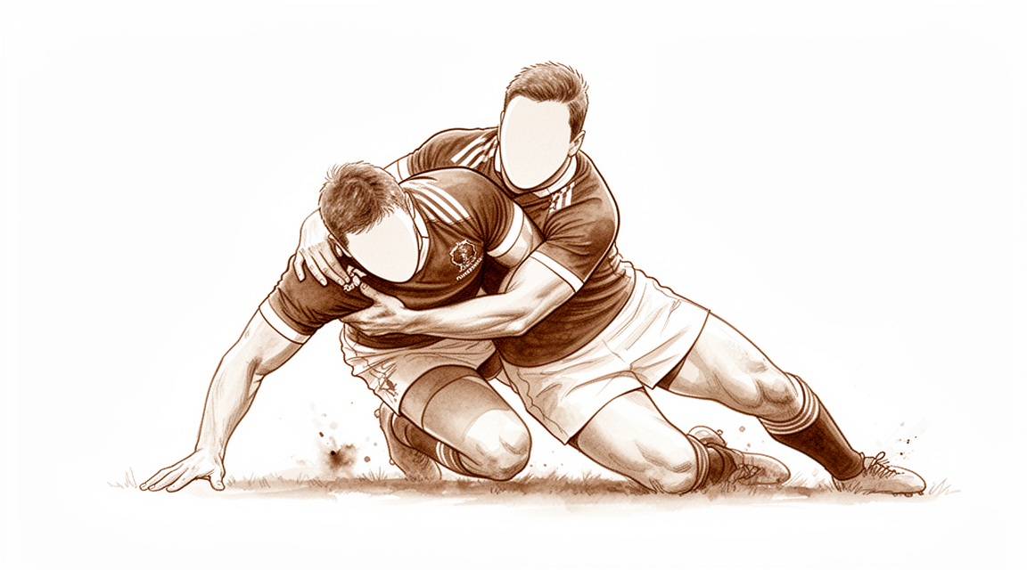
தோள்பட்டையின் முன்புற நிலைப்படுத்தல் (anterior shoulder stabilisation) என்பது, தோள்பட்டை மூட்டை உறுதியாக அதன் இடத்தில் பிடித்து வைத்து, அது முன்புறமாக நழுவி வெளியேறுவதை நிறுத்தும் ஒரு அறுவை சிகிச்சை. திரும்பத் திரும்ப மூட்டு விலகல்கள் ஏற்பட்டவர்களுக்கு இது பொதுவாக வழங்கப்படுகிறது; இவை பெரும்பாலும், கை பக்கவாட்டில் நீட்டப்பட்டு வெளிப்புறமாகத் திரும்பியிருக்கும் நிலையில் ஏற்படும் விழுதல் அல்லது காயத்துக்குப் பிறகு நிகழ்கின்றன. மற்ற சிகிச்சைகளால் உங்கள் தோள்பட்டை சீராகாததாலோ, அல்லது காயத்தின் தன்மை மேலும் மூட்டு விலகல்களை ஏற்படுத்த வாய்ப்புள்ளதாலோ, உங்கள் அறுவை சிகிச்சை நிபுணர் இதைப் பரிந்துரைத்திருக்கலாம். மீண்டும் மூட்டு விலகிவிடுமோ என்ற பயம் இல்லாமல் நீங்கள் கையை அசைக்கவும், வேலை செய்யவும், விளையாடவும் உதவும் உறுதியான தோள்பட்டையே நோக்கம்.
அறுவை சிகிச்சைக்கு முன்
அறுவை சிகிச்சைக்கு முந்தைய வாரங்களில், அறுவை சிகிச்சையைத் திட்டமிடத் தேவையான எக்ஸ்-ரேக்கள், MRI அல்லது அல்ட்ராசவுண்ட் ஸ்கேன் போன்ற உருவப் பரிசோதனைகளுக்கு ஏற்பாடு செய்கிறோம். அறுவை சிகிச்சை நாளில், உங்கள் அறுவை சிகிச்சைக்கு ஏழு மணி நேரத்துக்கு முன்பிருந்து நீங்கள் உண்பதையும் குடிப்பதையும் நிறுத்த வேண்டும். அறுவை சிகிச்சைப் பட்டியல் முன்கூட்டியே நகர்ந்தால் உங்கள் நேரத்தை முன்னதாக மாற்ற முடியும் என்பதற்காக, ஆறு மணி நேரத்துக்குப் பதிலாக ஏழு மணி நேரம் கேட்கிறோம். உங்கள் வழக்கமான மருந்துகளில் எவற்றை, எப்போது நிறுத்த வேண்டும் என்று உங்கள் அறுவை சிகிச்சை நிபுணர் சொல்வார்; எனவே நீங்கள் எடுத்துக்கொள்ளும் அனைத்தின் எழுதப்பட்ட பட்டியலையும் கொண்டு வாருங்கள். நீங்களே வாகனம் ஓட்ட முடியாது என்பதால், அதன் பிறகு உங்களை வீட்டுக்கு வாகனத்தில் அழைத்துச் செல்ல ஒருவரை ஏற்பாடு செய்யுங்கள். தோள்பட்டையின் மேல் எளிதாக அணியக்கூடிய, தளர்வான, வசதியான ஆடைகளை அணியுங்கள். உங்களுக்கு வேறு உடல்நலப் பிரச்சினைகள் இருந்தால், இரத்தப் பரிசோதனைகள் அல்லது மயக்க மருந்து நிபுணரின் (anaesthetist) பரிசோதனை தேவைப்படலாம்.
அறுவை சிகிச்சை நாளில்
நீங்கள் மருத்துவமனையின் அறுவை சிகிச்சை அனுமதிப் பிரிவுக்கு (surgical admissions unit) வருவீர்கள்; அங்கே உங்கள் வருகை பதிவு செய்யப்பட்டு, நீங்கள் அறுவை சிகிச்சை அரங்குக்குத் தயார்படுத்தப்படுவீர்கள். இந்த அறுவை சிகிச்சை, முழு மயக்க மருந்துடன் (general anaesthetic) பகுதி நரம்புத் தடுப்பு ஊசியையும் (regional nerve block) சேர்த்துச் செய்யப்படுகிறது. அறுவை சிகிச்சைக்கு முன் மயக்க மருந்து நிபுணர் உங்களைச் சந்தித்து, இரண்டு பகுதிகளையும் பற்றி விளக்குவார்.
பின்னர் நீங்கள் அறுவை சிகிச்சை அரங்குக்கு அழைத்துச் செல்லப்படுவீர்கள்; அங்கே அறுவை சிகிச்சை செய்யப்படும். அது முடிந்ததும், மீட்புப் பகுதியில் (recovery area) நீங்கள் கண் விழிப்பீர்கள்; மயக்க மருந்தின் விளைவு குறையும் வரை அங்கே செவிலியர்கள் உங்களைக் கண்காணிப்பார்கள். உங்கள் நிலை சீரானதும், அறுவை சிகிச்சையையும் உங்கள் மீட்சியையும் பொறுத்து, நீங்கள் வார்டுக்குச் செல்வீர்கள் அல்லது வீட்டுக்குச் செல்வீர்கள்.
அறுவை சிகிச்சையில் என்ன செய்யப்படுகிறது
இது ஒரு துளை வழி (keyhole) அறுவை சிகிச்சை. உங்கள் அறுவை சிகிச்சை நிபுணர் உங்கள் தோள்பட்டையைச் சுற்றிப் பல சிறிய வெட்டுகளைப் போட்டு — அவற்றில் ஒன்று பின்புறத்தில் — மூட்டுக்குள் வைக்கப்படும் ஒரு சிறிய கேமராவின் உதவியுடன் செயல்படுகிறார். சேதமடைந்த திசுவை அந்தக் கேமரா ஒரு திரையில் காட்டுகிறது.
முக்கியப் பிரச்சினை, குழியின் முன்புறத்தில் உள்ள குருத்தெலும்பு வளையம் எலும்பிலிருந்து கிழிந்து விலகியிருப்பதுதான். குழிக்குள் பொருத்தப்படும் சிறிய தையல் நங்கூரங்களைப் (suture anchors) பயன்படுத்தி, திசுவைக் கீழே பிடித்து வைக்கும் தையல்களுடன், உங்கள் அறுவை சிகிச்சை நிபுணர் இந்த வளையத்தை எலும்புடன் மீண்டும் இணைக்கிறார். திசுவுடன் ஒரு எலும்புத் துண்டும் பிரிந்து வந்திருந்தால், அதையும் அதே வெட்டுகளின் வழியாகப் பழுதுபார்க்கலாம். மூட்டின் பந்தில் உள்ள ஒரு பள்ளம் சிக்கிக்கொண்டால், அருகிலுள்ள திசுவின் ஒரு சிறு பகுதியை மூட்டுக்குள் நகர்த்தி, உங்கள் அறுவை சிகிச்சை நிபுணர் அந்தப் பள்ளத்தையும் நிரப்பலாம்.
சிறிய வெட்டுகள் தையல்களால் மூடப்பட்டு, அவற்றின் மேல் கட்டு (dressing) போடப்படுகிறது.
அறுவை சிகிச்சைக்குப் பிறகு
இந்த அறுவை சிகிச்சைக்குப் பிறகு பெரும்பாலான நோயாளிகள் மருத்துவமனையில் ஒரு இரவு தங்குகிறார்கள்; இருப்பினும் சிலர் அதே நாளில் வீட்டுக்குச் செல்ல முடியும். மீட்புப் பகுதியில் நீங்கள் கண் விழிப்பீர்கள்; பின்னர் வார்டுக்குச் செல்வீர்கள். செவிலியர்கள் மாத்திரைகள் மூலம் உங்கள் வலியைக் கட்டுக்குள் வைத்திருப்பார்கள்; தேவைப்பட்டால் அவற்றை மாற்றியமைப்பார்கள். வசதிக்காக உங்கள் கை ஒரு சாதாரண கைத்தாங்கியில் (sling) ஓய்வெடுக்கும்; பயிற்சிகளுக்கும் குளிப்பதற்கும் அது கழற்றப்படும். தொடங்குவதற்கான மென்மையான அசைவுகளை ஒரு செவிலியர் காட்டித் தருவார்; அதே நாளிலேயே நீங்கள் எழுந்து நடமாடுவீர்கள். முதல் 24 மணி நேரம் யாராவது உங்களுடன் தங்கும்படி ஏற்பாடு செய்யுங்கள். கட்டைச் சுமார் 10 நாட்கள் அப்படியே வைத்திருக்கிறோம்; நாங்கள் சொன்னால் தவிர, அதற்கு முன் அதை அகற்ற வேண்டாம். உங்களைப் பார்க்கும்போது நாங்கள் அதை மாற்றுவோம் அல்லது அகற்றுவோம்.
மீட்சி
முதல் சில நாட்களுக்கு உங்கள் தோள்பட்டையில் வலியும் வீக்கமும் இருக்கும்; அதைச் சுற்றியுள்ள தோல் கன்றிப்போனது போலத் தோன்றலாம். இது படிப்படியாகத் தணியும். உங்கள் கைத்தாங்கி அனுமதிக்கும் அளவுக்குள் உங்கள் கையையும் (hand) முழங்கையையும் அசைத்துக்கொண்டிருப்பது உதவும்; இரவில் உங்கள் கையைத் தலையணைகளின் மீது வைத்து ஓய்வெடுப்பது அசௌகரியத்தைக் குறைக்கும். அறிவுறுத்தப்பட்டபடி எடுத்துக்கொள்ளும் எளிய வலி நிவாரண மாத்திரைகள் பொதுவாக உங்களை வசதியாக வைத்திருக்கும்.
வசதிக்காக நீங்கள் ஒரு சாதாரண கைத்தாங்கியை அணிவீர்கள். உங்கள் பயிற்சிகளுக்கும் குளிப்பதற்கும் அது கழற்றப்படும். ஓர் இயன்முறை சிகிச்சையாளர், தொடக்கத்திலேயே மென்மையான அசைவுகளுக்கு உங்களுக்கு வழிகாட்டுவார்; சிறிய ஊசல் (pendulum) அசைவுகளிலிருந்து தொடங்கி, வலி அனுமதிக்கும் அளவுக்கு மெதுவாக அதிகரிப்பீர்கள். காலப்போக்கில், பயிற்சிகள் நீங்களே தசைகளைப் பயன்படுத்திச் செய்யும் அசைவுகளையும், பின்னர் எதிர்ப்பு விசையுடன் (resistance) செய்யும் வலிமைப் பயிற்சிகளையும் உள்ளடக்கும் அளவுக்கு விரிவடையும். பெரும்பாலான நாட்களில் இவற்றை வீட்டிலேயே செய்வீர்கள்; உங்கள் இயன்முறை சிகிச்சையாளர் உங்கள் முன்னேற்றத்தைச் சரிபார்த்து, போகப்போகத் திட்டத்தை மாற்றியமைப்பார்.
தொடக்கத்தில் அன்றாட வேலைகளுக்குச் சிறிது திட்டமிடல் தேவை. உங்கள் அறுவை சிகிச்சை நிபுணர் அனுமதிக்கும் வரை — பொதுவாக ஆறு வார மறுபரிசோதனையில் — நீங்கள் வாகனம் ஓட்டக் கூடாது; எங்கள் வாகனம் ஓட்டுதல் வழிகாட்டி இதை மேலும் விரிவாக விளக்குகிறது. கனமான வேலைகளுக்கு முன்பே மேசைப் பணியும் இலகுவான அன்றாட வேலைகளும் திரும்பும். தோள்பட்டை வலுவாகி, உங்கள் அசைவிலும் நிலைத்தன்மையிலும் உங்கள் அறுவை சிகிச்சை நிபுணர் திருப்தியடையும் வரை, விளையாட்டு, உடற்பயிற்சிக்கூடப் (gym) பயிற்சி, உடலுழைப்பு வேலை ஆகியவை காத்திருக்க வேண்டும். தொடக்கத்தில், கையைத் தலையணைகள் தாங்கும் வகையில் நிமிர்ந்த நிலையிலோ மல்லாந்தோ தூங்குவது பெரும்பாலும் அதிக வசதியாக இருக்கும்; வலி தணியும்போது பக்கவாட்டில் தூங்குவது எளிதாவதைப் பெரும்பாலானோர் காண்கிறார்கள்.
மீட்சி ஒவ்வொருவருக்கும் மாறுபடுகிறது. உங்கள் கால அளவு வேறுபடலாம்; ஒவ்வொரு கட்டத்திலும் உங்கள் அறுவை சிகிச்சை நிபுணரும் இயன்முறை சிகிச்சையாளரும் உங்களுக்கு வழிகாட்டுவார்கள்.
என்ன தவறு நேரலாம்
பெரும்பாலான நோயாளிகள் நன்றாகவே குணமடைகிறார்கள்; ஆனால் அவ்வப்போது சிக்கல்கள் ஏற்படலாம். எந்தப் பிரச்சினையையும் ஆரம்பத்திலேயே கண்டறிய, உங்கள் அறுவை சிகிச்சை நிபுணரும் மருத்துவக் குழுவும் உங்களை நெருக்கமாகக் கண்காணிக்கிறார்கள்.
நன்றாக நடந்த பழுதுபார்ப்புக்குப் பிறகும், சில நேரங்களில் தோள்பட்டை மீண்டும் நழுவலாம் அல்லது அதன் இடத்திலிருந்து விலகலாம். இது பல ஆண்டுகள் கழித்து, பெரும்பாலும் கீழே விழுந்த பிறகோ விளையாட்டின்போதோ நிகழலாம். உங்கள் தோள்பட்டை மூட்டு விலகி, மீண்டும் உள்ளே செல்லாவிட்டால், அவசர சிகிச்சைப் பிரிவுக்குச் செல்லுங்கள். அது நழுவி வெளியேறி, தானாகவே மீண்டும் உள்ளே சென்றுவிட்டால், நாங்கள் உங்களைப் பார்க்கும்படி கிளினிக்கை அழையுங்கள். அது சீராகிவிட்டாலும், உங்கள் அடுத்த மறுபரிசோதனையில் அதைக் குறிப்பிடுங்கள்.
தொற்று (infection) ஏற்படுவது அரிது. காயத்திலிருந்து வெளிப்புறமாகப் பரவும் சிவப்பு, அதிலிருந்து கசியும் திரவம் அல்லது சீழ், அல்லது காய்ச்சல் ஆகியவற்றைக் கவனியுங்கள். இவற்றில் எதையாவது கவனித்தால், அதே நாளில் கிளினிக்கை அழையுங்கள். உங்கள் வலி மருந்துகளை எடுத்தும் தொடர்ந்து மோசமாகி வரும் வலிக்கும் அதே நாளில் அழைக்க வேண்டும்.
அறுவை சிகிச்சைக்குப் பிறகு தோள்பட்டை விறைப்பாகலாம். கையை உயர்த்துவதையோ, முதுகுக்குப் பின்னால் கையைக் கொண்டு செல்வதையோ, ஆடை அணிவதையோ கட்டுப்படுத்தும் இறுக்கத்தை நீங்கள் கவனிக்கலாம். ஆரம்பத்திலேயே சிகிச்சை அளிப்பது அசைவு திரும்ப உதவுவதால், உங்கள் மறுபரிசோதனையில் இதைக் குறிப்பிடுங்கள்.
நரம்புகள் தோள்பட்டைக்கு அருகில் செல்கின்றன; எனவே அறுவை சிகிச்சையின்போது அவை எரிச்சலடையலாம். நரம்புத் தடுப்பு ஊசிக்குப் பிறகு முதல் 24 மணி நேரத்தில் மரத்துப்போதலும் பலவீனமும் எதிர்பார்க்கப்படுபவையே. அறுவை சிகிச்சைக்குச் சுமார் 24 மணி நேரத்துக்குப் பிறகு, தடுப்பு ஊசியின் விளைவு நீங்கியதும், உங்கள் கை (arm), கை (hand) அல்லது விரல்கள் தொடர்ந்து மரத்துப்போயிருந்தால், அல்லது அவற்றை அசைக்க முடியாவிட்டால், கிளினிக்கை அழையுங்கள். பெரும்பாலான நரம்பு எரிச்சல் காலப்போக்கில் தானாகவே தணிந்துவிடும்.
இரத்த உறைவு (blood clot) அரிது; ஆனால் தீவிரமானது. உங்கள் கெண்டைக்காலில் (calf) வீக்கமோ வலியோ, அல்லது மூச்சுத் திணறலோ நெஞ்சு வலியோ இருந்தால், அவசர சிகிச்சைப் பிரிவுக்குச் செல்லுங்கள். இவை இரத்த உறைவின் அறிகுறிகளாக இருக்கலாம்.
பழுதுபார்ப்பை அதன் இடத்தில் பிடித்து வைக்கப் பயன்படுத்தப்படும் சிறிய நங்கூரங்களும் தையல்களும், எப்போதாவது சொடுக்கும் அல்லது அரைபடும் உணர்வு, அல்லது ஒரு உலோகத் துண்டு உரசுவதால் ஏற்படும் வலி போன்ற பிரச்சினைகளை ஏற்படுத்தலாம். இதை நீங்கள் கவனித்தால், மறுபரிசோதனையில் உங்கள் அறுவை சிகிச்சை நிபுணரிடம் தெரிவியுங்கள்.
குறிப்பிட்ட விவரங்களை நீங்கள் அறிய விரும்பினால், இந்தப் பக்கத்தில் உள்ள சிக்கல்கள் அட்டவணை வழக்கமான விகிதங்களைப் பட்டியலிடுகிறது.
எப்போது எங்களை அழைக்க வேண்டும்
பெரும்பாலான பிரச்சினைகள் ஆரம்பத்திலேயே வெளிப்படுகின்றன; நீங்கள் வீட்டில் கவலைப்பட்டுக்கொண்டிருப்பதைவிட, அவற்றைப் பற்றி எங்களிடம் சொல்வதையே நாங்கள் விரும்புகிறோம். உங்களுக்குக் காய்ச்சல் இருந்தால், காயத்தைச் சுற்றிச் சிவப்பு பரவினால், அல்லது அதிலிருந்து திரவமோ சீழோ கசிந்தால், அதே நாளில் கிளினிக்கை அழையுங்கள். உங்கள் வலி மருந்துகளை எடுத்தும் வலி தொடர்ந்து மோசமாகி வந்தாலும், அதே நாளில் எங்களை அழையுங்கள். உங்கள் கெண்டைக்காலில் (calf) வீக்கமோ வலியோ, அல்லது மூச்சுத் திணறலோ நெஞ்சு வலியோ இருந்தால், அவசர சிகிச்சைப் பிரிவுக்குச் செல்லுங்கள்; ஏனெனில் இவை இரத்த உறைவின் அறிகுறிகளாக இருக்கலாம். அறுவை சிகிச்சைக்குச் சுமார் 24 மணி நேரத்துக்குப் பிறகு, உங்கள் நரம்புத் தடுப்பு ஊசியின் விளைவு நீங்கியதும், உங்கள் கை (arm), கை (hand) அல்லது விரல்கள் தொடர்ந்து மரத்துப்போயிருந்தால், அல்லது அவற்றை அசைக்க முடியாவிட்டால், எங்களை அழையுங்கள். முதல் 24 மணி நேரத்தில் மரத்துப்போதலும் பலவீனமும் எதிர்பார்க்கப்படுபவையே. உங்கள் தோள்பட்டை மூட்டு விலகி, மீண்டும் உள்ளே செல்லாவிட்டால், அவசர சிகிச்சைப் பிரிவுக்குச் செல்லுங்கள். அது நழுவி வெளியேறி, தானாகவே மீண்டும் உள்ளே சென்றுவிட்டால், நாங்கள் உங்களைப் பார்க்கும்படி கிளினிக்கை அழையுங்கள். அலுவல் நேரத்துக்குப் பிறகோ வார இறுதியிலோ எங்களைத் தொடர்புகொள்ள முடியாவிட்டால், உங்களுக்கு அருகிலுள்ள அவசர சிகிச்சைப் பிரிவுக்குச் செல்லுங்கள்.
இந்த நிலை பற்றி மேலும் எங்கே படிக்கலாம்
இந்தப் பக்கம் அறுவை சிகிச்சையைப் பற்றியது மட்டுமே. இது சிகிச்சையளிக்கும் நிலை, அறுவை சிகிச்சை எப்போது உதவுகிறது எப்போது உதவுவதில்லை என்பது பற்றி ஆய்வுச் சான்றுகள் என்ன காட்டுகின்றன என்பது உட்பட, தோள்பட்டை நிலையற்ற தன்மை (Shoulder Instability) பக்கத்தில் விரிவாக விளக்கப்பட்டுள்ளது.
Evidence & references
This is the clinical evidence summary written for health professionals. It is technical, and it lists the research this page was built from. You do not need to read it to understand your treatment or to make a decision about it.
Overview
- As knowledge of the basic science behind the pathophysiology of shoulder instability improves and more clinical reports emerge, the exact indications for arthroscopic stabilization are gradually being refined [1].
- The benefits of arthroscopic stabilization include decreased morbidity, faster recovery and rehabilitation, improved cosmesis, and diminished patient pain [11].
- Arthroscopic shoulder stabilization using a bioabsorbable tac is a labral repair procedure with little capability of shifting the anterior capsule superiorly greater than 1 cm [3].
- This combined procedure of Bankart repair and anterior capsular shift provides good long term stability for shoulders with high rate of recurrency and which seem to be not a successful case for an arthroscopic procedure [6].
- Arthroscopic management of a failed instability repair provides similar success to open reconstruction if one selects proper indications [7].
- Concomitant arthroscopic rotator cuff repair and anterior shoulder stabilization is associated with low rates of recurrent instability but a meaningful risk of structural rotator cuff failure [5].
- This study defines thresholds for MCID and PASS achievement at a minimum 2-year follow-up for patients undergoing arthroscopic anterior shoulder stabilization [8].
- Despite a higher complication rate than in the younger population, shoulder stabilization using the Latarjet procedure is effective in patients over 50 without associated cuff damage [9].
- Glenohumeral capsule can be preserved, and this should be attempted wherever possible to optimize stability [10].
- This simplified arthroscopic Latarjet technique is safe and reproducible in the treatment of recurrent anterior shoulder dislocations [12].
- The authors express caution regarding the consensus that the WOSI should be used as the primary outcome measure for all studies on the treatment of shoulder instability before further prospective, high-level, evidence-based studies and multi-center studies are performed [14].
- Patients should be counseled pre-operatively on the expected outcomes over time following ABR of anterior shoulder instability [16].
- At a mean follow-up of 128.1 months, 25.0% of patients experienced recurrent instability and 19.6% underwent subsequent surgery [18].
- This technique provides a safe and efficient way to augment anterior stabilization [20].
- One-third of adolescents in our series presenting with anterior shoulder instability had bipolar bone loss at the initial evaluation, with male sex and ≥5 prior dislocations as independent predictors [21].
- Sixty eight patients with traumatic anterior shoulder instability treated with arthroscopic Bankart repair were reviewed retrospectively [22].
- The combined procedure of arthroscopic Latarjet and Hill-Sachs remplissage deserves consideration in a high-risk population including combined bone loss, recurrent anterior instability after failed previous stabilization procedures and/or seizure [23].
- The aim of this study was to evaluate the long-term clinical outcomes of arthroscopic Bankart repair using a standardized, modern technique with a minimum of three suture anchors in patients with traumatic anterior instability and to assess possible risk factors for recurrent instability [25].
- The use of the Panalok suture anchor with Panacryl suture appears to be a safe and effective fixation alternative to metal devices for arthroscopic shoulder stabilization [26].
- This arthroscopic Bankart repair technique is reliable for the recurrent anterior dislocations and subluxations under the considerations of the indications [42].
Anatomy & Pathophysiology
Bony Anatomy
- The glenoid is a convex structure of shallow depth shaped like an inverted pear [63].
- The glenoid cavity is a shallow socket, approximately one third the size of the humeral head [64].
- The subchondral bone of the glenoid is relatively flat, and the articular concavity is augmented by cartilage and a circumferential labrum [66].
- The glenoid averages 5° of retroversion in relation to the axis of the scapular body [66].
- The humeral head is spherical with a diameter of 37 to 57 mm [63].
- The humeral head averages 19° of retroversion and 41° of inclination (neck-shaft angle) [66].
- The articular surface of the humeral head is essentially spherical, with an arc of approximately 160 degrees covered by articular cartilage [75].
- The radius of curvature of the humeral head is approximately 25 mm and is slightly larger in men than in women [75].
- The glenoid articular surface radius of curvature is 2 to 3 mm larger than that of the humeral head [75].
- The average neck-shaft angle is 45 degrees (±5 degrees), with a range of 30 to 50 degrees [75].
- The superior margin of the humeral head articular surface is normally superior to the top of the greater tuberosity by 8 to 10 mm [75].
- The distance from the lateral base of the coracoid process to the lateral margin of the greater tuberosity is called the lateral humeral offset [75].
- The glenoid is connected with the flat body of the scapula by the scapular neck [65].
- The hook-shaped coracoid process curves forwards from the superior surface of the scapular neck [65].
- The acromion, the coracoacromial ligament, and the coracoid process form the coracoacromial arch, a rigid bony-ligamentous structure that imparts stability to the shoulder girdle [63].
- The proximal humerus receives its blood supply from the anterior and posterior humeral circumflex branches from the third division of the axillary artery [63].
- The anterior humeral circumflex artery (AHCA) arises from the axillary artery at the inferior border of the subscapularis and provides vascular inflow to the humeral head by way of its terminal anterolateral branch known as the artery of Laing (also known as the arcuate artery) [63].
- The major blood supply to the humeral head is through the ascending branch of the anterior humeral circumflex artery, which penetrates the head at the bicipital groove and becomes the arcuate artery [64].
- The anterolateral ascending branch of the anterior humeral circumflex artery provides the primary blood supply to the humeral head [66].
- The terminal intraosseous portion of the anterior humeral circumflex artery enters at the proximal aspect of the intertubercular groove as the arcuate artery [66].
- The bicipital groove lies between the greater and lesser tuberosities and serves as a pathway for the long head of the biceps as it traverses from its intraarticular origin into the proximal arm [63].
- The distal aspect of the bicipital groove is internally rotated with respect to the proximal portion [63].
- The greater tuberosity serves as the attachment site for the supraspinatus, infraspinatus, and teres minor tendons of the rotator cuff [63].
- The lesser tuberosity serves as the attachment site for the subscapularis tendon [63].
- The anatomic neck of the proximal humerus is located at the junction of the articular surface and the tuberosities [63].
- The surgical neck represents an indistinct region (metadiaphyseal junction) below the tuberosities but above the humeral shaft [63].
- The scapula is attached to the axial skeleton by the clavicle, specifically by the acromioclavicular (AC) and sternoclavicular (SC) joints [65].
- The scapula is separated from the chest wall by thin gliding fibro-fatty tissue, allowing its smooth excursion over the chest wall [65].
- The distribution of bony mass in the scapula is highly uneven, with the highest concentration in the glenoid, the scapular neck (including the base of the coracoid process), and the lateral border of the scapular body [65].
- Two bony pillars transmit compressive forces from the glenoid fossa: the lateral pillar connects the inferior border of the glenoid with the inferior angle, and the spinal pillar arises from the central part of the glenoid and continues medially to become part of the base of the scapular spine [65].
- The weakest bone in the scapula is located primarily in the central part of the biomechanical body, i.e., in the infraspinous fossa [65].
- The weakest area of the circumference of the biomechanical body of the scapula is the connection of the scapular spine and the medial border of the scapula, known as the spinomedial angle [65].
- The clavicle is the first bone to ossify (fifth week of gestation) and is the only long bone to ossify by intramembranous ossification [66].
- The medial (sternal) epiphysis of the clavicle is the last ossification center to fuse, at age 20 to 25 years [66].
- The primary blood supply to the clavicle is periosteal, and no nutrient artery is present [66].
- The scapula has only one true diarthrodial articulation, the acromioclavicular (AC) joint [66].
- Normal shoulder motion is approximately two-thirds glenohumeral and one-third scapulothoracic [66].
- Ossification of the scapular body begins at the eighth week of gestation [66].
- The acromion has three ossification centers: the metacromion (base), the mesoacromion (middle), and the preacromion (tip) [66].
- Failure of fusion of acromial ossification centers results in os acromiale [66].
- The coracobrachialis muscle and the short head of the biceps tendon originate from the coracoid process [66].
- The pectoralis minor muscle inserts onto the medial coracoid process [66].
- The proximal humerus has three centers of ossification: the humeral head (4 to 6 months), the greater tuberosity (1 to 3 years), and the lesser tuberosity (3 to 5 years) [66].
- The proximal humeral ossification centers fuse to the shaft at age 17 to 20 years [66].
- The glenoid diameter ranges from 18-30 mm superior anteroposterior, 21-35 mm inferior anteroposterior, and 30-48 mm superoinferior [75].
- The glenoid inclination averages 4.2 degrees (range –7 to 20 degrees) [75].
- The glenoid version is 1.5 degrees retroversion (range 10.5-9.5 degrees anteversion) [75].
- The glenoid surface area is 4-6 mm [75].
- The humeral head surface area is 11-19 mm [75].
- The glenoid cartilage thickness is 2.16 mm [75].
- The humeral head cartilage thickness is 1.44 mm [75].
- The glenoid radius of curvature is 22-28 mm [75].
- The humeral head radius of curvature is 23-28 mm, smaller in women than men [75].
- The medial (coronal) humeral offset is 4-14 mm [75].
- The posterior (transverse) humeral offset is –2 to 10 mm [75].
- The head-shaft angle ranges from 30-55 degrees [75].
- The scapula spans the second through seventh ribs and serves as an attachment for 17 muscles [77].
- The scapula is anteverted on the chest wall approximately 30 degrees relative to the body [77].
- The glenoid is retroverted approximately 5 degrees relative to the scapular body [77].
- Os acromiale is incomplete fusion of secondary ossification centers, most commonly between the mesoacromion and meta-acromion [77].
- The coracoid process has attachments to the coracoacromial ligament, coracoclavicular ligaments (conoid [medial] and trapezoid [lateral]), conjoined tendon (coracobrachialis and short head of biceps), and pectoralis minor [77].
- The suprascapular artery passes superior to the superior transverse scapular ligament and the suprascapular nerve passes inferior to the ligament through the suprascapular notch [77].
- In the spinoglenoid notch, both the artery and nerve are inferior to the inferior transverse scapular ligament [77].
- The coracoacromial ligament contributes to anterosuperior stability in rotator cuff deficiency and should be preserved with irreparable cuff tears to prevent anterosuperior escape [77].
- The acromial branch of the thoracoacromial artery runs on the medial aspect of the coracoacromial ligament [77].
- The coracoacromial ligament is the arthroscopic landmark for a complete release of the rotator interval for adhesive capsulitis [77].
- The humeral head is retroverted 30 degrees relative to the transepicondylar axis of the humerus [77].
- The head height is approximately 5.6 cm above the superior border of the pectoralis major tendon [77].
- The anatomic neck is directly below the humeral head and serves as an attachment for the shoulder capsule [77].
- The surgical neck is more distal and more often involved in fractures [77].
- The transverse humeral ligament is an important stabilizer of the biceps tendon [77].
- The formation of the humerus begins with the appearance of the cartilage anlage, which is present by the fifth week of gestation [70].
- The primary ossification center for the humerus appears at about the sixth week [70].
- By the time of birth, the entire humeral diaphysis is completely ossified [70].
- The proximal humerus is primarily cartilaginous at birth, but ossification centers can be detected with ultrasonography as early as the 38th week of gestation and are generally present between the 38th and 42nd week of gestation [70].
- The ossification center for the humeral head is usually present at birth [70].
- The greater tuberosity ossification center appears by 1 to 3 years of age [70].
- The lesser tuberosity ossification center appears by 5 years of age [70].
- These ossification centers fuse by 5 to 7 years of age to form the humeral head [70].
- The proximal humeral ossification center remains separated from the humeral shaft by the proximal humeral physis, which closes by 14 to 17 years of age in girls and by 16 to 18 years in boys [70].
- Humeral retroversion averages 65 degrees in infants and young children and gradually decreases, approaching adult values by 11 years of age [70].
- At birth, the body or shaft of the humerus is completely ossified [70].
- Eighty percent of subsequent growth comes from the proximal humeral physis, accounting for approximately 40% of the growth of the entire upper extremity [70].
- Less than 75% of growth from the proximal humerus occurs before 2 years of age [70].
- More than 85% of growth from the proximal humerus occurs by 8 years of age [70].
- The capsule of the glenohumeral joint extends from the glenoid rim, progressing laterally toward the surgical neck of the humerus and blending with the tendons of the rotator cuff musculature [70].
- The posteromedial metaphysis, a portion of the physis, and the epiphysis are intracapsular [70].
- A large part of the physis is extracapsular, making it susceptible to traumatic injury [70].
- The proximal humeral physis is irregularly shaped, with its apex located on the posteromedial portion of the proximal humerus [70].
- The periosteum is thicker and stronger in the posteromedial portion of the proximal humerus compared to the anterolateral portion, which is often quite thin [70].
- The subscapularis originates from the anterior scapula and inserts anteriorly onto the lesser tuberosity [70].
- The greater tuberosity provides attachment superiorly and posteriorly for the supraspinatus, infraspinatus, and teres minor, all of which originate from the posterior scapula [70].
- The deltoid forward flexes and abducts the shoulder and courses from the clavicle and acromion superiorly, coalescing into a common tendinous insertion onto the lateral upper third of the humeral shaft [70].
- The pectoralis major powers adduction and internal rotation due to its tendinous insertion anteriorly onto the lateral wall of the bicipital groove [70].
- The pectoralis major forms the roof of the distal continuation of the bicipital tunnel, which is a closed space that extends proximally to the glenohumeral joint [70].
- The anterior and posterior humeral circumflex arteries provide a rich blood supply to the proximal humerus [70].
- 64% of the humeral head blood supply arises from the posterior humeral circumflex artery [70].
- The brachial plexus is prone to injury when the proximal humerus is injured in fractures or dislocations, or during traction [70].
- The axillary nerve circles the humeral neck just inferior to the glenohumeral joint as it courses posteriorly [70].
- The humeral shaft extends from the level of the insertion of the pectoralis major muscle proximally to the supracondylar ridge distally [64].
- The upper portion of the humeral shaft is cylindrical and becomes more flattened in an anteroposterior direction as it proceeds distally [64].
- Medial and lateral intermuscular septae divide the arm into anterior and posterior compartments [64].
- In the anterior compartment reside the biceps brachii, coracobrachialis, and brachialis muscles, along with the neurovascular bundle coursing along the medial border of the biceps with the brachial artery and vein and the median, musculocutaneous, and ulnar nerves [64].
- In the posterior compartment reside the triceps brachii muscle and the radial nerve [64].
- The neck-shaft angle measures an average of 135 degrees [64].
- The humeral head is retroverted an average of 30 degrees [64].
- The rotator cuff consists of four muscles: the subscapularis, supraspinatus, infraspinatus, and teres minor muscles [64].
- The teres major is not a rotator cuff muscle [64].
- The cuff muscles serve as depressors of the humeral head to allow the deltoid to efficiently abduct the humerus [64].
- The infraspinatus and teres minor are external rotators, while the subscapularis is an internal rotator of the humerus [64].
- The deltoid and pectoralis major muscles, along with the rotator cuff, cause predictable displacement of fractures around the proximal humerus [64].
- The brachial plexus and axillary artery lie anterior to the coracoid process of the scapula and humeral head [64].
- Nerves innervating muscles around the shoulder include the axillary, suprascapular, subscapular, and musculocutaneous nerves [64].
- Fractures of the anatomic neck have a poor prognosis because of complete disruption of the blood supply to the head [64].
- Surgical neck fractures are common, and with these, the blood supply to the head is preserved [64].
- Within the bicipital groove lies the biceps tendon, which is covered by the transverse humeral ligament [64].
- The greater tuberosity provides attachment for the supraspinatus, infraspinatus, and teres minor muscles [64].
- The lesser tuberosity contains the attachment of the subscapularis muscle [64].
- The scapula in humans is suspended by muscles alone and has shifted caudally from the cervical position in lower animals [73].
- The scapular index, defined as the relationship between the length (measured along the base of the spine) and the breadth (measured from the superior to the inferior angle) of the scapula, is extremely high in pronograde animals with a long, narrow scapula [73].
- The scapula is broader in humans and other primates, with the most pronounced differences observed in the infraspinatus fossa [73].
- Broadening of the infraspinatus foss
Classification
Bony Anatomy and Defects
- The superior glenohumeral ligament was absent in 5.8% of cadaver shoulders, the middle glenohumeral ligament in 15.4%, and the inferior glenohumeral ligament in 6.8% [29].
- In a cadaver study, four shoulders with secondary signs of instability (Hill-Sachs fracture and bony Bankart lesion) lacked the middle glenohumeral ligament and possessed a large anterior type IV recess [29].
- One-third of adolescents presenting with anterior shoulder instability had bipolar bone loss at initial evaluation [21].
- Male sex and having five or more prior dislocations were identified as independent predictors of bipolar bone loss in adolescents with anterior shoulder instability [21].
- The open Latarjet procedure is recognized as a reliable option to address complex glenohumeral instability, particularly in cases with glenoid bone loss [40].
- Arthroscopic autologous iliac crest bone grafting combined with the remplissage procedure shows excellent clinical outcomes for glenoid reconstruction in recurrent anterior shoulder dislocation with significant bone loss [4].
- The inlay dynamic anterior stabilization using the long head of the biceps tendon and remplissage procedure is aimed at improving stability in patients with subcritical glenoid bone loss and on-track Hill-Sachs lesions [2].
Soft Tissue and Ligamentous Anatomy
- The glenohumeral ligaments, joint capsule, and glenoid labrum are the structures of concern for reconstruction in recurrent anterior dislocation [29].
- The glenohumeral ligaments have been proven to be valuable passive stabilizers of the glenohumeral joint [29].
- The glenohumeral capsule can be preserved during arthroscopic Latarjet and capsular shift procedures, and preservation should be attempted wherever possible to optimize stability [10].
- Preserving the anteroinferior capsule and repairing it to place the bone block in an extra-articular position reinforces the anterior stabilization of the glenohumeral joint [15].
- Arthroscopic shoulder stabilization using a bioabsorbable tac is a labral repair procedure with limited capability of shifting the anterior capsule superiorly by more than 1 cm [3].
Lesion Classification and Pathology
- Posterior shoulder instability accounts for 2% to 10% of all shoulder instability, with recurrent posterior subluxation being the most common type [56].
- The most common lesion in posterior shoulder instability is a posterior labral lesion [56].
- The presence of an anterior humeral notch, also known as a reverse Hill-Sachs lesion, is one of the most important risk factors for posterior shoulder instability [56].
- In a study of 104 cadaver shoulders, most synovial recesses belonged to group I (38.5%) and group III (46.2%) based on the DePalma classification system [29].
Clinical Presentation
History
- The history should define the mechanism of injury, including the position of the arm, the amount of force applied, and the point of force application [48].
- Injury with the arm in extension, abduction, and external rotation favors anterior dislocation [48].
- If instability is recurrent, the history defines the initial injury, the position or action that results in instability, how long the shoulder stays out, whether radiographs are available with the shoulder out of joint, and what means have been necessary to reduce the shoulder [48].
- The history solicits evidence of neurologic or rotator cuff problems after previous episodes of shoulder instability [48].
- Previous treatment of the recurrent instability, as well as the effectiveness of this treatment, should be documented [48].
- In traumatic anterior shoulder dislocation, patients report a shoulder in abduction and external rotation and receiving a hit to the arm, with the arm in full outstretched motion [88].
- Patients may complain of anterolateral and anterior shoulder pain with overhead activities and motion [88].
- Patients with anterior shoulder instability will experience symptoms of apprehension with shoulder abduction and external rotation, and also can experience symptoms of pain and instability with placement of the arm in an overhead position [91].
- The most common complaint of shoulder instability is pain coupled with restricted shoulder motion [91].
- A failed arthroscopic stabilization procedure often starts not in the operating room but with the failure to identify red flags in the patient’s history and physical examination that might preclude a successful arthroscopic repair [97].
- Risk factors associated with treatment failure include age, gender, presence of osseous Bankart, and/or large Hill-Sachs lesions, participation in competitive collision or forced overhead sports, hypermobility, time lapse between dislocation and reduction, and the number of instability episodes prior to operation [97].
Physical Examination
- An acutely dislocated shoulder is usually very painful, and muscles are in spasm in an attempt to stabilize the joint [48].
- The humeral head may be palpable anteriorly in an acutely dislocated shoulder [48].
- The posterior and lateral aspect of the shoulder shows a hollow beneath the acromion in an acutely dislocated shoulder [48].
- The arm is held in slight abduction in an acutely dislocated shoulder [48].
- Passive and active motions are limited by pain in an acutely dislocated shoulder [48].
- Assessment of the neurovascular status of the upper extremity and charting of the findings before reduction is an essential part of the physical examination of an anteriorly dislocated shoulder [48].
- A thorough neurovascular exam, including assessment of the axillary nerve, should be performed [88].
- The axillary nerve is the most commonly injured nerve in up to 42% of traumatic anterior shoulder dislocations [88].
- Initial examination should include a complete neurovascular examination to document any neurologic or vascular deficits [91].
- Documentation of active and passive ROM of the shoulder for internal and external rotation as well as forward flexion and abduction is important [91].
- Marked loss of motion is seen with persistent dislocations and rotator cuff lesions [91].
- Rotator cuff testing is an essential part of the shoulder instability examination particularly in patients over the age of 40 years [91].
- The belly press or bear hug test is the most effective test to evaluate the function of the subscapularis in the acutely injured patient [91].
- Testing of resisted shoulder abduction in the first 30 degrees of shoulder flexion with the arm internally rotated is effective for evaluating the supraspinatus [91].
- Evaluation of the infraspinatus is performed by applying resisted external rotation with the elbow flexed to 90 degrees [91].
- The anterior apprehension sign is performed by placing the arm into an abducted (90 degrees) and maximally externally rotated position with the patient in the supine position, resulting in a feeling of pain, discomfort, and potential instability [91].
- The relocation test is performed from the ABER position by applying a posteriorly directed force to the proximal humerus, which elicits a feeling of reduced apprehension or pain from the patient [91].
- An anterior release test (surprise test) is performed by removing the posteriorly directed force abruptly when the patient's arm is in 90 degrees of abduction, 90 degrees of elbow flexion, and maximal external rotation position [91].
- A feeling of pain or apprehension is a positive result for the anterior release test [91].
- Apprehension and relocation tests assess anterior GH instability [88].
- The patient should be lying in supine position on the examination table with their arm abducted to 90 degrees and externally rotated for apprehension and relocation tests [88].
- A positive relocation test occurs if the patient feels a slipping sensation or fear of an impending dislocation with motion that is improved with applying a posterior force to the GH joint [88].
- The sulcus sign is performed at 0 degrees of abduction by applying downward traction on the humerus [88].
- Dimpling or a “gap” formed in the GH joint is a positive sulcus sign, indicating laxity of the superior GH ligament [88].
- The load and shift test is used to evaluate anterior and posterior GH laxity and is performed while the patient is in a seated or supine position with the humeral head centered in the glenoid fossa and translated [88].
- Grade 0 on the load and shift test means normal translation [88].
- Grade 1 on the load and shift test means translation to rim and back, less than 1 cm [88].
- Grade 2 on the load and shift test means translation over the rim followed by spontaneous reduction, 1 to 2 cm [88].
- Grade 3 on the load and shift test means translation over the rim without spontaneous reduction, greater than 2 cm [88].
- Generalized joint laxity should be assessed using the Beighton score (0–9 point scale) [88].
- Inspection for shoulder deformity or muscle atrophy is part of the physical examination [88].
- Constitutional ligamentous laxity, such as the Marshall test or Beighton score, is part of the physical examination for shoulder instability [97].
- Neck range of motion and Spurling’s maneuver are part of the physical examination for shoulder instability [97].
- Shoulder range of motion and scapular symmetry are part of the physical examination for shoulder instability [97].
- Strength testing of shoulder girdle muscles is part of the physical examination for shoulder instability [97].
- Lift-off and belly press tests are part of the physical examination for shoulder instability [97].
- The Jerk test is part of the physical examination for shoulder instability [97].
- O’Brien’s active compression test is part of the physical examination for shoulder instability [97].
- Hawkins and Neer’s impingement tests are part of the physical examination for shoulder instability [97].
- Cross-body adduction is part of the physical examination for shoulder instability [97].
- An examination under anesthesia is critical to the success of arthroscopic stabilization and is more sensitive for determining both the degree and direction of instability [97].
- The pattern of instability can be determined during examination under anesthesia without being affected by patient apprehension or guarding [97].
- The axial load test or load-and-shift test is conducted during examination under anesthesia, and the translation is noted in the anterior, inferior, and posterior directions [97].
- Grading of translation during examination under anesthesia reflects the degree of humeral head translation anterior and posterior to the glenoid rim [97].
- Grade 1+ corresponds to the translation of the humeral head to the edge of the glenoid [97].
- Grade 2+ corresponds to the humeral head being subluxated over the glenoid rim but reducing spontaneously [97].
- Grade 3+ corresponds to a frank dislocation of the humeral head over the glenoid rim that does not reduce spontaneously [97].
Investigations
Plain Radiography and CT
- At least two X-ray views should be obtained for shoulder imaging: an anteroposterior view in the plane of the glenoid and an axillary projection with the arm in abduction [80].
- The axillary view is a necessary view in the evaluation of glenohumeral joint instability and enables determination of the humeral head position in the glenoid fossa [92].
- CT with three-dimensional reconstructions is the advanced imaging study of choice for determining the extent of glenoid bone loss in the setting of shoulder instability [92].
- CT imaging is frequently used to evaluate fractures of the shoulder, to assess for bony lesions in recurrent instability cases, or for preoperative templating for shoulder arthritis [90].
- The West Point view is a special radiographic view indicated for anterior glenoid bone loss [92].
- The Stryker notch view is a special radiographic view indicated to evaluate Hill-Sachs lesions after dislocation [92].
- The apical oblique view is a special radiographic view indicated to evaluate for glenoid rim fracture in instability [92].
- Standardized plain films are almost always sufficient to garner the information needed for shoulder evaluation, and there is information that can be gathered from properly taken plain films that cannot be obtained from CT scans [47].
- Although CT scans may offer a few degrees of increased precision in the measurement of glenoid version, this precision does not necessarily improve the quality of the surgery or the clinical outcome [47].
- The temptation to "overimage" should be resisted, obtaining only the scans or reconstructions that are necessary for the care of the patient [47].
Magnetic Resonance Imaging
- MRI is useful to identify osteonecrosis of the humeral head, bone tumours, labral tears, and rotator cuff tears [80].
- The accuracy of MRI for identifying labral tears and rotator cuff tears is enhanced by combining the scan with arthrography [80].
- MRI is the modality of choice for evaluating the rotator cuff, biceps, and subacromial/subdeltoid bursa [90].
- T1-weighted MRI can reveal Hill-Sachs lesions and is often used with magnetic resonance arthrograms to provide a more detailed picture of the joint surfaces [90].
- T2-weighted MRI provides better visualization of full-thickness rotator cuff tears [90].
- Magnetic resonance accuracy in identifying labral and rotator cuff tears in the literature ranges from 70% to 100% [86].
- MR arthrography (MRA) refers to MRI of a joint that has been injected with an intra-articular contrast agent such as diluted gadolinium or saline solution [86].
- MRA has proven utility by increasing both sensitivity and specificity in detecting injuries to the capsulolabral–ligamentous complex as compared to traditional MRI [86].
- In a meta-analysis of 6 studies including 4,667 shoulders, MRA had greater diagnostic test accuracy for glenoid labral lesions than MRI, with MRA sensitivity of 88% and specificity of 93% versus MRI sensitivity of 76% and specificity of 87% [86].
- Abduction and external rotation (ABER) of the arm is an alternative MRI position utilized to increase the sensitivity and specificity for detecting anteroinferior labroligamentous injury [86].
- Limited range of motion or pain may prohibit patients from performing the ABER provocative maneuver [86].
- MR arthrography is considered the benchmark for evaluation of labral tears and is rarely indicated for evaluation of rotator cuff pathology [90].
- When MRI or MR arthrography is contraindicated, such as in patients with pacemakers or vascular clips, CT arthrography is indicated [90].
Ultrasonography
- Ultrasonography is a simple and accurate test for identifying rotator cuff tears and calcific tendinitis [80].
- Ultrasonography can be useful in guiding injections or barbotage [80].
- Ultrasonography is a low-cost alternative to MRI and arthrography for evaluating both skeletal and soft-tissue structures of the shoulder [90].
- Ultrasonography can provide immediate, real-time visualization of the rotator cuff, biceps tendon, and calcific deposits [90].
- Ultrasonography can be used to measure the subacromial space and detect atrophy of rotator cuff muscles [90].
- Ultrasonography can evaluate impingement in various positions and motions due to real-time imaging [90].
- Ultrasonography is highly operator dependent and is not as useful for evaluating labral tears or rotator cuff tears that are very small or larger than 3 cm [90].
- For the detection of full-thickness rotator cuff tears, ultrasonography has a sensitivity of 98%, specificity of 80%, positive predictive value of 90%, negative predictive value of 95%, and accuracy of 94% [92].
- For the detection of full-thickness rotator cuff tears, MRI has a sensitivity of 100%, specificity of 68%, positive predictive value of 85%, negative predictive value of 100%, and accuracy of 89% [92].
Arthroscopy
- Arthroscopy is useful for diagnosing and treating subacromial impingement, intra-articular lesions, detachment of the glenoid labrum, and rotator cuff tears [80].
- Among patients undergoing arthroscopy at the time of open Latarjet, 89.1% required extensive debridement that would not have otherwise been performed with a Latarjet alone [35].
- Excluding extensive debridements, an additional arthroscopic procedure was required in 9.2% of cases among patients undergoing arthroscopy at the time of open Latarjet [35].
- Among patients requiring additional arthroscopic procedures who had a preoperative MRI, the MRI identified pathology in only 1 case (0.5%) [35].
Treatment
Arthroscopic Bankart Repair
- Arthroscopic stabilization offers decreased morbidity, faster recovery and rehabilitation, improved cosmesis, and diminished patient pain [11].
- Arthroscopic Bankart repair using a bioabsorbable tac is a labral repair procedure with limited capability of shifting the anterior capsule superiorly greater than 1 cm [3].
- The use of the Panalok suture anchor with Panacryl suture is a safe and effective fixation alternative to metal devices for arthroscopic shoulder stabilization [26].
- Arthroscopic Bankart repair using a grasping stitcher system is reliable for recurrent anterior dislocations and subluxations when appropriate indications are considered [42].
- Arthroscopic stabilization results were superior to open techniques regarding length of inpatient stay and time to return to normal activities, but were associated with a higher rate of redislocation [44].
- A shorter rehabilitation regimen following Bankart repair does not prejudice ultimate shoulder stability outcomes but is generally more successful in patients with an intense desire to return to top-level sport [27].
- Adequate mobilization of the inferior glenohumeral ligament-labrum complex during arthroscopic Bankart repair can be verified using the "suction-reduction" sign without requiring additional expensive equipment [28].
- Three treatment failures in a cohort of Bankart repairs utilizing intra-articular suturing required an open stabilization procedure [30].
- Patient-reported outcomes following arthroscopic Bankart repair for anterior shoulder instability decline over time, necessitating pre-operative counseling on expected long-term outcomes [16].
- Arthroscopic anterior shoulder stabilization achieves defined thresholds for Minimal Clinically Important Difference (MCID) and Patient Acceptable Symptom State (PASS) at a minimum 2-year follow-up [8].
Open Bankart Repair
- The transosseous suture technique is the treatment of choice for stabilization of the shoulder with the open Bankart repair [52].
- A combined procedure of Bankart repair and anterior capsular shift provides good long-term stability for shoulders with a high rate of recurrence that are not successful candidates for arthroscopic procedures [6].
- Ambulatory open Bankart repair under a single general anesthesia is a reliable technique for major shoulder surgery in an ambulatory care unit [54].
Revision and Combined Procedures
- Arthroscopic management of a failed instability repair provides similar success to open reconstruction when proper indications are selected [7].
- Concomitant arthroscopic rotator cuff repair and anterior shoulder stabilization is associated with low rates of recurrent instability but carries a meaningful risk of structural rotator cuff failure [5].
Bony Augmentation (Latarjet)
- The open Latarjet procedure via a deltopectoral approach is a reliable option to address complex glenohumeral instability [40].
- Shoulder stabilization using the Latarjet procedure is effective in patients over 50 years old without associated cuff damage, despite a higher complication rate than in younger populations [9].
- The arthroscopic Latarjet technique using a 3 anterior portal approach is safe and reproducible for treating recurrent anterior shoulder dislocations [12].
- In arthroscopic Latarjet procedures, the glenohumeral capsule can be preserved, which should be attempted wherever possible to optimize stability [10].
- Preserving and repairing the anteroinferior capsule allows the bone block to be placed in an extra-articular position, further reinforcing anterior stabilization of the glenohumeral joint [15].
- The subscapularis split approach in modified arthroscopic Latarjet permits orthogonal pin and screw positioning while offering greater purchase on the glenoid vault [55].
- Arthroscopic Latarjet using double suture-button fixation involves a 5-step process including coracoid osteotomy, glenoid preparation, subscapularis splitting with axillary nerve protection, transfer and fixation, and capsulolabral repair when tissue quality permits [19].
- In a two-center study of arthroscopic Latarjet using double suture-button fixation, capsulolabral repair was performed in 72 of 80 patients (90%), with repair not possible in the remaining 8 cases (10%) due to insufficient tissue quality [19].
- The metal-free arthroscopic Latarjet technique involves detaching the coracoacromial ligament, resecting the pectoralis minor from the coracoid, and using a parallel drill guide for fixation [58].
- Open Latarjet with metal-free cerclage fixation involves splitting the subscapularis slightly below the mid-level and decorticating the anterior surface of the glenoid neck to enhance bony contact and healing potential [59].
- Arthroscopic autologous iliac crest bone grafting for augmentation of glenoid bone loss using suture anchor fixation combined with the remplissage procedure shows excellent clinical outcomes for glenoid reconstruction in recurrent anterior shoulder dislocation with significant bone loss [4].
Remplissage and Soft Tissue Augmentation
- The remplissage technique involves preparing the Hill-Sachs lesion by removing the pseudomembrane and placing anchors in the middle of the lesion, typically 1 cm from the insertion of the rotator cuff [113].
- The triple-double technique of arthroscopic Hill-Sachs remplissage involves preparing the defect with a ringed curette and shaver to achieve a healthy, bleeding bone bed before placing two medial anchors through the posterior cannulated portal [117].
- Knotless suture staple remplissage for Hill-Sachs lesions in the beach chair position provides a safe and efficient way to augment anterior stabilization [20].
- The arthroscopic all-inside remplissage technique with a knotless tape bridge allows a complete glenohumeral view through a single superolateral portal and simplifies the procedure [34].
- Ultrasound-assisted arthroscopic remplissage offers a safer and more effective alternative in treating recurrent shoulder instability with engaging Hill-Sachs lesions [37].
- Arthroscopic remplissage with knotless all-suture anchors and concomitant Bankart repair is a reproducible technique that restores stability with minimal morbidity [45].
- The inlay dynamic anterior stabilization with the long head of the biceps tendon and remplissage procedure aims to improve stability and outcomes in patients with subcritical glenoid bone loss and on-track Hill-Sachs lesions [2].
- Combined arthroscopic and mini-open subpectoral dynamic anterior shoulder stabilization with the biceps tendon benefits from an onlay effect that produces a bumper acting as a dynamic soft-tissue block on the anterior glenoid [36].
- The combined procedure of arthroscopic Latarjet and Hill-Sachs remplissage is an efficient solution for patients with significant bipolar glenohumeral bone loss [23].
- The combined procedure of arthroscopic Latarjet and Hill-Sachs remplissage deserves consideration in high-risk populations including those with combined bone loss, recurrent anterior instability after failed previous stabilization procedures, and/or seizure [23].
General Indications and Outcomes
- Authors express caution regarding the consensus that the Western Ontario Shoulder Instability Index (WOSI) should be used as the primary outcome measure for all studies on the treatment of shoulder instability before further prospective, high-level, evidence-based studies and multi-center studies are performed [14].
Complications
Recurrence and Instability
- Re-dislocation occurred in 8 of 86 shoulders (10.5%) following arthroscopic Bankart repair for recurrent anterior dislocation and subluxation [24].
- At a mean follow-up of 128.1 months, 25.0% of patients experienced recurrent instability and 19.6% underwent subsequent surgery after arthroscopic Bankart repair for on-track lesions [18].
- Arthroscopic stabilisation using a suture anchor was associated with a higher rate of redislocation compared to open technique, despite superior inpatient stay and return to activities [44].
- Incomplete anchoring of the STATAK soft-tissue attachment device seemed to cause subluxation after the operation in two of three patients with unsatisfactory results [124].
- Three treatment failures following Bankart repair utilizing intra-articular suturing required an open stabilization procedure [30].
- The combined procedure of Bankart repair and anterior capsular shift provides good long-term stability for shoulders with a high rate of recurrency that are not successful candidates for an arthroscopic procedure [6].
- Arthroscopic management of a failed instability repair provides similar success to open reconstruction if proper indications are selected [7].
- Concomitant arthroscopic rotator cuff repair and anterior shoulder stabilization is associated with low rates of recurrent instability [5].
- Anterior anatomic glenoid reconstruction demonstrates lower redislocation rates than arthroscopic Bankart repair in patients with anterior shoulder instability [41].
- The open Latarjet procedure has a reported recurrence rate as low as 1% to 3% [123].
- There were 8 total clinical failures (4/31 arthroscopic and 4/29 open) by defined criteria, resulting in long-term failure rates of 12.9% for arthroscopic and 13.8% for open anterior shoulder stabilization [108].
- Subjective shoulder instability following anterior stabilization was not associated with an increased risk of dislocation [50].
Structural and Bony Complications
- Complications following the Latarjet procedure have been reported at a rate of 15% to 30%, including nerve injury, graft mispositioning, osteolysis, nonunion, screw breakage, and prominence [123].
- Reoperation rates as high as 10% have been reported for the Latarjet procedure, with the commonest reason being related to screw malposition, screw prominence, and screw breakage [123].
- The presence of 2 screws within the glenoid adds to the complexity of a subsequent total shoulder arthroplasty in patients with symptomatic anterior instability treated with a Latarjet procedure [123].
- Concomitant arthroscopic rotator cuff repair and anterior shoulder stabilization carries a meaningful risk of structural rotator cuff failure [5].
- Arthroscopic autologous iliac crest bone grafting for augmentation of glenoid bone loss shows excellent clinical outcomes for glenoid reconstruction in recurrent anterior shoulder dislocation with significant bone loss [4].
- Arthroscopic shoulder stabilization using a bioabsorbable tac has little capability of shifting the anterior capsule superiorly greater than 1 cm [3].
- One-third of adolescents presenting with anterior shoulder instability had bipolar bone loss at initial evaluation, with male sex and ≥5 prior dislocations as independent predictors [21].
- The combined arthroscopic Latarjet and Hill-Sachs remplissage procedure is considered for high-risk populations including those with combined bone loss, recurrent anterior instability after failed previous stabilization procedures, and/or seizure [23].
Functional and Patient-Reported Outcomes
- Patient-reported outcomes decline over time following arthroscopic Bankart repair for anterior shoulder instability [16].
- The duration of subjective shoulder instability following anterior stabilization was similar between Latarjet and Bankart repair techniques [50].
- A shorter rehabilitation regimen after Bankart repair does not prejudice the ultimate stability of the shoulder but is generally more successful in patients with an intense desire to return to top-level sport [27].
- Modified Bankart repair in capsular reconstruction resulted in a loss of up to 15% of external rotation [43].
- The no-sling group had a significantly greater SANE instability score at 2 years postoperatively than the sling group following open Latarjet procedures [104].
- The transosseous suture technique is the treatment of choice for stabilization of a shoulder with the open Bankart repair, particularly regarding potential risks with suture anchors [52].
- Open repair could predict a good result and rare postoperative instability compared to arthroscopic Bankart repair [130].
- The arthroscopic Bankart repair technique using the grasping stitcher system is reliable for recurrent anterior dislocations and subluxations under the considerations of indications [42].
- The inlay dynamic anterior stabilization with the long head of the biceps tendon and remplissage procedure aims to improve stability and outcomes in patients with complex shoulder instability issues [2].
- The knotless suture staple remplissage technique provides a safe and efficient way to augment anterior stabilization [20].
- The reproducible technique of arthroscopic remplissage with knotless all-suture anchors and concomitant Bankart repair restores stability with minimal morbidity [45].
- The simplified arthroscopic Latarjet technique using a 3 anterior portal technique is safe and reproducible in the treatment of recurrent anterior shoulder dislocations [12].
- Shoulder stabilization using the Latarjet procedure is effective in patients over 50 without associated cuff damage, despite a higher complication rate than in the younger population [9].
- The arthroscopic Latarjet technique using double suture-button fixation involves a learning curve and specific graft positioning considerations [19].
- The metal-free arthroscopic Latarjet surgical technique involves specific patient positioning and portal creation steps [58].
- The open Latarjet procedure using double suture-button fixation aims to decrease complications and issues associated with screws [123].
- The open Latarjet procedure with metal-free cerclage fixation involves specific steps for subscapularis split and glenoid neck exposure [59].
- The arthroscopic Latarjet stabilization of the shoulder with capsulolabral repair involves preserving the anteroinferior capsule to place the bone block in an extra-articular position [15].
- The arthroscopic Latarjet procedure using a 70° arthroscope through a standard posterior portal plus 5 anterior portals follows 5 consecutive steps including coracoid preparation, glenoid preparation, subscapularis splitting, transfer and fixation, and capsulolabral repair [19].
- Capsulolabral repair was performed in 72 patients (90%) during arthroscopic Latarjet using double suture-button fixation, while it was not possible in 8 cases (10%) due to insufficient tissue quality [19].
- The study evaluated clinical and radiological outcomes following arthroscopic Latarjet stabilization with a capsule-labral reconstruction in a large single surgeon cohort [62].
- The aim of the study on long-term outcomes of standardized arthroscopic Bankart repair was to evaluate clinical outcomes and assess possible risk factors for recurrent instability [25].
- The study on defining the minimal clinically important difference and patient acceptable symptom state defines thresholds for MCID and PASS achievement at a minimum 2-year follow-up for patients undergoing arthroscopic anterior shoulder stabilization [8].
- The authors express caution regarding the consensus that the WOSI should be used as the primary outcome measure for all studies on the treatment of shoulder instability before further prospective, high-level, evidence-based studies are performed [14].
- The use of Mitek anchoring for Bankart repair was compared with traditional bone sutures in a prospective study [51].
- The utility of shoulder arthroscopy at the time of open Latarjet demonstrated that most patients required an additional procedure, including extensive debridement (89.1%) [35].
- The association between craniocaudal position of the Hill-Sachs lesion, anchor type, and postoperative stiffness after remplissage was no longer statistically significant when controlling for age, primary vs revision, and number of anchors [127].
- The single-portal arthroscopic treatment of posterior shoulder instability with labral tear and reverse Hill-Sachs remplissage using MGHL uses the accessory posterior approach to optimize debridement and placement of anchors [129].
- Posterior dislocation of the humeral head represents only 10% of all instability [32].
- Recurrent instability in posterior shoulder dislocation typically occurs within the first 8 months after the initial dislocation and occurs in 17.7% of shoulders [32].
- Risk factors for recurrent posterior instability include patients younger than 40 years old and a seizure as the mechanism of primary dislocation [32].
- The Hill-Sachs lesion is not a significant prognostic factor for recurrence of shoulder redislocation after arthroscopic Bankart repair [33].
- The arthroscopic Bankart repair with a 6 o'clock anchor reduces recurrence and improves clinical outcomes in recurrent anterior instability [103].
- The immediate self-rehabilitation with versus without sling immobilization after open Latarjet procedures showed comparable SANE instability scores at 6 months [104].
- The immediate self-rehabilitation with versus without sling immobilization after open Latarjet procedures showed comparable VAS pain scores at 6 months and 24 months postoperatively [104].
- The minimum 5-year follow-up after arthroscopic Latarjet evaluated outcomes and complications in a cohort where 42% underwent the procedure as a revision and 58% as primary surgery [106].
- The clinical and radiologic outcomes of the Bankart repair using Mason-Allen (BRUMA) technique for small bony Bankart lesions included assessment of recurrent instability, bony union, change in anterior glenoid defect size, ASES score improvement, Rowe score improvement, other complications, and revision surgery [122].
- Other complications in the BRUMA technique study included any intraoperative or postoperative adverse events directly related to the surgical procedure, including infection, nerve injury, anchor pull-out, implant failure, or stiff shoulder requiring intervention [122].
- Revision surgery in the BRUMA technique study included any secondary surgical intervention performed on the ipsilateral shoulder due to failure of the index procedure [122].
- The arthroscopic versus open Bankart repair for traumatic anterior shoulder instability indicates that exact indications for arthroscopic stabilization are gradually being refined as knowledge of pathophysiology improves [1].
- The 68 patients with traumatic anterior shoulder instability treated with arthroscopic Bankart repair (Caspari's technique) were reviewed retrospectively [22].
- The early results of Bankart repair with a patient-controlled rehabilitation program indicated that the shorter rehabilitation regimen is generally more successful in patients who have an intense desire to return to top-level sport [27].
- The arthroscopic Bankart repair using long-term absorbable anchors and sutures appears to be a safe and effective fixation alternative to metal devices [26].
- The Bankart repair utilizing intra-articular suturing had a minimum three-year follow-up [30].
- The arthroscopic Bankart versus open Latarjet as a primary operative treatment for traumatic anteroinferior instability in young males was a randomised controlled trial with 2-year follow-up [31].
- The posterior stabilization and reverse Hill-Sachs remplissage using linked knotless anchors addresses posterior instability where the humeral head internally rotates and translates posteriorly on the glenoid [32].
- The Hill-Sachs lesion eligibility criteria for studies on recurrence after arthroscopic Bankart repair included availability of data regarding preoperative or intraoperative diagnosis of HS and average follow-up duration longer than 2 years [33].
- The utility of shoulder arthroscopy at the time of open Latarjet showed that excluding extensive debridements, an additional arthroscopic procedure was required in 9.2% of cases [35].
- The evaluating return to sport outcomes study found that anterior AAGR demonstrates superior patient-reported RTS rates than ABR with comparable recovery timelines despite the AAGR group having higher GBL [41].
- The arthroscopic Bankart repair (grasping stitcher system) is reliable for recurrent anterior dislocations and subluxations under the considerations of the indications [42].
- The modified Bankart repair in capsular reconstruction for shoulder instability had excellent results in 59 patients (91%) and good results in 5 patients (7%) [43].
- The Bankart repair using a suture anchor comparison between arthroscopic and open technique found that arthroscopic stabilisation was superior in terms of length of inpatient stay and time taken to return to normal activities [44].
- The arthroscopic remplissage with knotless all-suture anchors and concomitant Bankart repair is a reproducible technique that restores stability with minimal morbidity [45].
- The posterior translation of the humeral head after arthroscopic Bankart repair was analyzed quantitatively using preoperative and postoperative axial MRI [49].
- The subjective shoulder instability following anterior stabilization incidence and time to resolution were compared between Latarjet and Bankart repair [50].
- The use of Mitek anchoring for Bankart repair involved 40 patients with anterior posttraumatic instability, none of whom had signs of generalized joint laxity [51].
- The recurrences after the open Bankart repair study identified a potential risk with the use of suture anchors [52].
- The ambulatory open Bankart repair under a single general anesthesia was a prospective study of the immediate outcome [54].
- The metal-free arthroscopic Latarjet surgical technique uses general anesthesia and interscalene block with the patient in the beach-chair position [58].
- The open Latarjet procedure using double suture-button fixation aims to improve graft fixation and decrease complications associated with screws [123].
- The open Latarjet procedure with metal-free cerclage fixation involves releasing the coracoacromial ligament laterally and isolating the conjoined tendon [59].
- The arthroscopic Latarjet: clinical outcomes from a large case series evaluated clinical and radiological outcomes following arthroscopic Latarjet stabilization with a capsule-labral reconstruction [62].
- The immediate self-rehabilitation with versus without sling immobilization after open Latarjet procedures for recurrent anterior shoulder instability was a study published in the Journal of Bone and Joint Surgery [104].
- The minimum 5-year follow-up after arthroscopic Latarjet: outcomes and complications was published in The American Journal of Sports Medicine [106].
- The corrigendum to "Arthroscopic Versus Open Anterior Shoulder Stabilization: A Prospective Randomized Clinical Trial With 15-Year Follow-up" reported 8 total clinical failures [108].
- The clinical and radiologic outcomes of the Bankart repair using Mason-Allen (BRUMA) technique for small bony Bankart lesions was an objective, performance criteria-referenced, single-surgeon case series [122].
- The open Latarjet procedure using double suture-button fixation was published in Arthroscopy Techniques [123].
- The efficacy and pitfalls of the STATAK soft-tissue attachment device for the Bankart repair was published in the Journal of Shoulder and Elbow Surgery [124].
- The poster 228 on the association between craniocaudal position of the Hill-Sachs lesion, anchor type, and postoperative stiffness after remplissage was published in the Orthopaedic Journal of Sports Medicine [127].
- The single-portal arthroscopic treatment of posterior shoulder instability with labral tear with reverse Hill-Sachs remplissage using MGHL was published in Arthroscopy Techniques [129].
- The open versus arthroscopic Bankart repair was published in the Journal of Shoulder and Elbow Surgery [130].
Recovery
Rehabilitation Protocols
- A shorter rehabilitation regimen following Bankart repair does not prejudice the ultimate stability of the shoulder [27].
- Shorter rehabilitation regimens are generally more successful in patients with an intense desire to return to top-level sport [27].
- Arthroscopic stabilization is associated with faster recovery and rehabilitation compared to open techniques [11].
Return to Sport and Activity
- In a cohort of 24 baseball players undergoing a modified Bankart repair, all participants returned to play [125].
- Of the 24 baseball players who underwent modified Bankart repair, 83.3% (20/24) resumed the same or a higher level of competition [125].
- Arthroscopic anatomic glenoid reconstruction demonstrates comparable recovery timelines to arthroscopic Bankart repair in patients with anterior shoulder instability [41].
- Arthroscopic stabilization is associated with diminished patient pain [11].
Long-Term Outcomes and Recurrence
- At a mean follow-up of 128.1 months, 25.0% of patients undergoing arthroscopic Bankart repair for on-track lesions experienced recurrent instability [18].
- At a mean follow-up of 128.1 months, 19.6% of patients undergoing arthroscopic Bankart repair for on-track lesions underwent subsequent surgery [18].
- Patient-reported outcomes may decline over time following arthroscopic Bankart repair for patients who experience recurrent anterior shoulder instability and underwent isolated arthroscopic Bankart repair compared to those who underwent arthroscopic Bankart repair with remplissage [61].
- Patients should be counseled pre-operatively on the expected outcomes over time following arthroscopic Bankart repair for anterior shoulder instability [16].
- Concomitant arthroscopic rotator cuff repair and anterior shoulder stabilization is associated with a meaningful risk of structural rotator cuff failure [5].
- Open Latarjet procedure is effective in patients over 50 years old without associated cuff damage, despite a higher complication rate than in the younger population [9].
- Patients who underwent open Latarjet experienced significantly better long-term survivorship without any recurrence compared to isolated Bankart repairs, despite patient selection without preoperative bone loss [128].
- Arthroscopic anatomic glenoid reconstruction demonstrates lower redislocation rates than arthroscopic Bankart repair in patients with anterior shoulder instability [41].
- Arthroscopic anatomic glenoid reconstruction demonstrates superior patient-reported return-to-sport rates than arthroscopic Bankart repair in patients with anterior shoulder instability [41].
Functional Deficits and Complications
- Modified Bankart repair in capsular reconstruction resulted in excellent outcomes in 59 patients (91%) and good outcomes in 5 patients (7%), associated with a loss of up to 15% of external rotation [43].
- Arthroscopic stabilization results were superior to open techniques in terms of length of inpatient stay and time taken to return to normal activities, but were associated with a higher rate of redislocation [44].
Key Evidence
- [Paper] As the knowledge of the basic science behind the pathophysiology of shoulder instability improves and as more clinical reports emerge, the exact indications for arthroscopic stabilization are gradually being refined. [1] (10.1016/s0278-5919(05)70294-5)
- [L5] The method aims to improve stability and outcomes in patients with complex shoulder instability issues. [2] (10.1016/j.eats.2024.103256)
- [L4] Arthroscopic shoulder stabilization using a bioabsorbable tac is a labral repair procedure with little capability of shifting the anterior capsule superiorly greater than 1 cm. [3] (10.1016/s1058-2746(95)80032-8)
- [L5] The technique shows excellent clinical outcomes for glenoid reconstruction in recurrent anterior shoulder dislocation with significant bone loss. [4] (10.1016/j.eats.2025.103952)
- [L4] Concomitant arthroscopic rotator cuff repair and anterior shoulder stabilization is associated with low rates of recurrent instability but a meaningful risk of structural rotator cuff failure. [5] (10.1016/j.xrrt.2026.100868)
- [L4] This combined procedure provides good long term stability for shoulders with high rate of recurrency and which seem to be not a successful case for an arthroscopic procedure. [6] (10.1016/s1058-2746(95)80098-0)
- [L4] Arthroscopic management of a failed instability repair provides similar success to open reconstruction if one selects proper indications. [7] (10.1097/00132589-200212000-00008)
- [L4] This study defines thresholds for MCID and PASS achievement at a minimum 2-year follow-up for patients undergoing arthroscopic anterior shoulder stabilization. [8] (10.1177/23259671261442972)
- [L4] Despite a higher complication rate than in the younger population, shoulder stabilization using the Latarjet procedure is effective in patients over 50 without associated cuff damage. [9] (10.1016/j.jseint.2025.101518)
- [L5] Glenohumeral capsule can be preserved, and this should be attempted wherever possible to optimize stability. [10] (10.1097/bth.0000000000000068)
- [L5] The benefits of arthroscopic stabilization include decreased morbidity, faster recovery and rehabilitation, improved cosmesis, and diminished patient pain. [11] (10.1016/s0278-5919(05)70295-7)
- [L5] This simplified arthroscopic Latarjet technique is safe and reproducible in the treatment of recurrent anterior shoulder dislocations. [12] (10.1002/atn2.70147)
- [L5] The authors express caution regarding the consensus that the WOSI should be used as the primary outcome measure for all studies on the treatment of shoulder instability before further prospective, high-level, evidence-based studies and multi-center studies are performed. [14] (10.1177/0363546518765992)
- [L4] They feel it is worth preserving the anteroinferior capsule and repairing it to place the bone block in an extra-articular position, and further reinforce the anterior stabilization of the glenohumeral joint. [15] (10.1097/bte.0000000000000056)
- [L3] Patients should be counseled pre-operatively on the expected outcomes over time following ABR of anterior shoulder instability. [16] (10.1177/2325967126s00552)
- [L4] At a mean follow-up of 128.1 months, 25.0% of patients experienced recurrent instability and 19.6% underwent subsequent surgery. [18] (10.1177/2325967126s00277)
- [L4] [19] (10.1016/j.xrrt.2026.100792)
- [L5] This technique provides a safe and efficient way to augment anterior stabilization. [20] (10.1002/atn2.70058)
- [L3] One-third of adolescents in our series presenting with anterior shoulder instability had bipolar bone loss at the initial evaluation, with male sex and ≥5 prior dislocations as independent predictors. [21] (10.1177/03635465261483327)
- [L4] Sixty eight patients with traumatic anterior shoulder instability treated with arthroscopic Bankart repair were reviewed retrospectively. [22] (10.1016/s1058-2746(95)80225-8)
- [L3] The combined procedure deserves consideration in a high-risk population including combined bone loss, recurrent anterior instability after failed previous stabilization procedures and/or seizure. [23] (10.1016/j.jseint.2024.08.148)
- [L4] Re-dislocation occurred in 8 of total 86 shoulders (10.5%). [24] (10.1016/s1058-2746(98)90172-0)
- [L4] The aim of this study was to evaluate the long-term clinical outcomes of arthroscopic Bankart repair using a standardized, modern technique with a minimum of three suture anchors in patients with traumatic anterior instability and to assess possible risk factors for recurrent instability. [25] (10.1016/j.jseint.2025.101490)
- [L3] The use of the Panalok suture anchor with Panacryl suture appears to be a safe and effective fixation alternative to metal devices for arthroscopic shoulder stabilization. [26] (10.1097/00132589-200001040-00009)
- [L4] The shorter rehabilitation regimen does not prejudice outcome in terms of the ultimate stability of the shoulder but is generally more successful in patients who have an intense desire to return to top-level sport. [27] (10.1016/s1058-2746(99)90008-3)
- [L4] The technique does not require any additional expensive equipment beyond a normal shoulder arthroscopy setup and provides another way to verify proper anatomic positioning when placing suture anchors. [28] (10.1097/bte.0b013e3181b76c56)
- [L5] [29] (10.1016/s1058-2746(98)90221-x)
- [L4] Three treatment failures required an open stabilization procedure. [30] (10.1016/s1058-2746(96)80381-8)
- [L1] [31] (10.1136/bjsports-2021-104028)
- [L5] [32] (10.1016/j.eats.2024.103285)
- [L1] [33] (10.1136/jisakos-2015-000017)
- [L5] The technique allows a complete glenohumeral view through a single superolateral portal and simplifies the procedure. [34] (10.1016/j.eats.2023.04.021)
- [L4] [35] (10.1177/23259671261415839)
- [L5] It benefits from the onlay effect by producing a bumper that acts as a dynamic soft-tissue block on the anterior glenoid, which aids anterior shoulder stability. [36] (10.1016/j.eats.2025.103863)
- [L5] This technique offers a safer and more effective alternative in treating recurrent shoulder instability with engaging Hill–Sachs lesions. [37] (10.1002/atn2.70181)
- [L5] Despite various modifications to the technique over the years, the open Latarjet via a deltopectoral approach has been recognized as a reliable option to address complex glenohumeral instability. [40] (10.1016/j.eats.2025.103757)
- [L3] In patients with anterior shoulder instability, anterior AAGR demonstrates superior patient-reported RTS rates and lower redislocation rates than ABR, with comparable recovery timelines despite the AAGR group having higher GBL. [41] (10.1177/23259671261440934)
- [L4] This arthroscopic Bankart repair technique is reliable for the recurrent anterior dislocations and subluxations under the considerations of the indications. [42] (10.1016/s1058-2746(95)80236-3)
- [L3] Results were excellent in 59 patients (91%) and good in 5 patients (7%), this was due to a loss of up to 15% of external rotation. [43] (10.1016/s1058-2746(96)80117-0)
- [L3] The results of arthroscopic stabilisation were superior in terms of length of inpatient stay and time taken to return to normal activities, however with a higher rate of redislocation. [44] (10.1016/s1058-2746(96)80394-6)
- [L5] This reproducible technique restores stability with minimal morbidity. [45] (10.1002/atn2.70152)
- [L4] [49] (10.1016/j.jse.2026.07.031)
- [L3] Its duration was similar between techniques, and its presence was not associated with an increased risk of dislocation. [50] (10.1177/23259671261470584)
- [L1] [51] (10.1016/s1058-2746(09)80023-2)
- [L3] The transosseous suture technique is the treatment of choice for stabilization of a shoulder with the open Bankart repair. [52] (10.1016/s1058-2746(99)90052-6)
- [L3] It indicates that a single anesthesic with proper management of analgesia is a reliable technique for major shoulder surgery in an ambulatory care unit. [54] (10.1016/j.jse.2005.07.011)
- [L5] The subscapularis split approach permits an orthogonal pin and screw positioning while offering greater purchase on the glenoid vault. [55] (10.1016/j.eats.2024.103293)
- [L5] [56] (10.1016/j.eats.2023.03.009)
- [L5] [58] (10.1016/j.eats.2025.103727)
- [Paper] [59] (10.1016/j.eats.2022.11.030)
- [L4] PROs may decline over time following ABR for anterior shoulder instability for patients who experience recurrent anterior shoulder instability and undergo isolated ABR compared to ABR with remplissage. [61] (10.1016/j.xrrt.2026.100719)
- [L4] The study evaluated clinical and radiological outcomes following arthroscopic Latarjet stabilization with a capsule-labral reconstruction in a large single surgeon cohort. [62] (10.1016/j.jseint.2025.101503)
- [L3] [103] (10.1002/arj.70560)
- [L1] [104] (10.2106/jbjs.25.00560)
- [L3] [106] (10.1177/03635465261475810)
- [L1] There were 8 total clinical failures (4/31 arthroscopic and 4/29 open) by the defined criteria, resulting in arthroscopic and open long-term failure rates of 12.9% and 13.8%, respectively. [108] (10.1177/03635465211067445)
- [Paper] [113] (10.1016/j.eats.2024.103292)
- [L5] [117] (10.1016/j.eats.2023.04.024)
- [L4] [122] (10.1177/23259671261442219)
- [L5] [123] (10.1002/atn2.70182)
- [L4] In two of the three patients with unsatisfactory results, incomplete anchoring of the device seemed to have caused subluxation after the operation. [124] (10.1016/1058-2746(93)90065-o)
- [L4] All 24 participants returned to play, with 83.3% (20/24) resuming the same or a higher level of competition. [125] (10.1177/23259671261449195)
- [L4] However, when controlling for age, primary vs revision, and number of anchors this association was no longer statistically significant. [127] (10.1177/2325967126s00524)
- [L3] Patients who underwent open Latarjet experienced significantly better long-term patients without any recurrence survivorship compared to isolated Bankart repairs despite patient selection without bone loss. [128] (10.1016/j.jseint.2024.08.143)
- [L5] Using the accessory posterior approach only helps to optimize the debridement and placement of anchors. [129] (10.1016/j.eats.2024.103271)
- [L4] Open repair could predict a good result and rare postoperative instability. [130] (10.1016/s1058-2746(96)80443-5)
References
[1] ARTHROSCOPIC VERSUS OPEN BANKART REPAIR FOR TRAUMATIC ANTERIOR SHOULDER INSTABILITY. Clinics in Sports Medicine. 2000. DOI: 10.1016/s0278-5919(05)70294-5
[2] Inlay Dynamic Anterior Stabilization With the Long Head of the Biceps Tendon and Remplissage Procedure for Patients With Subcritical Glenoid Bone Loss and On‐Track Hill‐Sachs Lesion. Arthroscopy Techniques. 2024. DOI: 10.1016/j.eats.2024.103256
[3] Arthroscopic anterior shoulder stabilization: 2–5 year follow-up using a bioabsorbable tac. Journal of Shoulder and Elbow Surgery. 1995. DOI: 10.1016/s1058-2746(95)80032-8
[4] Arthroscopic Autologous Iliac Crest Bone Grafting for Augmentation of Glenoid Bone Loss Using Suture Anchor Fixation Combined With the Remplissage Procedure. Arthroscopy Techniques. 2025. DOI: 10.1016/j.eats.2025.103952
[5] Clinical Outcomes Following Concomitant Arthroscopic Rotator Cuff Repair and Anterior Shoulder Stabilization: A Systematic Review and Meta-Analysis. JSES Reviews, Reports, and Techniques. 2026. DOI: 10.1016/j.xrrt.2026.100868
[6] Bankart repair and anterior capsularshift as a combined procedure for traumatic anterior glenohumeral instability. Journal of Shoulder and Elbow Surgery. 1995. DOI: 10.1016/s1058-2746(95)80098-0
[7] Arthroscopic Revision Bankart Repair for Failed Anterior Shoulder Instability. Techniques in Shoulder and Elbow Surgery. 2002. DOI: 10.1097/00132589-200212000-00008
[8] Defining the Minimal Clinically Important Difference and Patient Acceptable Symptom State for Arthroscopic Anterior Shoulder Stabilization at 2-Year Follow-up. Orthopaedic Journal of Sports Medicine. 2026. DOI: 10.1177/23259671261442972
[9] Long-Term Outcomes of the Open Latarjet Procedure for Recurrent Anterior Shoulder Instability in Patients Over 50 Years Old. JSES International. 2026. DOI: 10.1016/j.jseint.2025.101518
[10] Arthroscopic Latarjet and Capsular Shift (ALCS) Procedure. Techniques in Hand & Upper Extremity Surgery. 2015. DOI: 10.1097/bth.0000000000000068
[11] ARTHROSCOPIC BANKART REPAIR. Clinics in Sports Medicine. 2000. DOI: 10.1016/s0278-5919(05)70295-7
[12] Arthroscopic Latarjet Using a 3 Anterior Portal Technique. Arthroscopy Techniques. 2026. DOI: 10.1002/atn2.70147
[14] Arthroscopic Versus Open Latarjet in the Treatment of Recurrent Anterior Shoulder Dislocation With Marked Glenoid Bone Loss: A Prospective Comparative Study: Response. The American Journal of Sports Medicine. 2018. DOI: 10.1177/0363546518765992
[15] Arthroscopic Latarjet Stabilization of the Shoulder With Capsulolabral Repair. Techniques in Shoulder & Elbow Surgery. 2015. DOI: 10.1097/bte.0000000000000056
[16] Poster 260. Patient-Reported Outcomes Decline Over Time following Arthroscopic Bankart Repair for Anterior Shoulder Instability. Orthopaedic Journal of Sports Medicine. 2026. DOI: 10.1177/2325967126s00552
[18] Paper 17. Long-Term Outcomes after Arthroscopic Bankart Repair for On-Track Lesions at Mean 10 Year Follow-Up. Orthopaedic Journal of Sports Medicine. 2026. DOI: 10.1177/2325967126s00277
[19] Arthroscopic Latarjet using double suture-button fixation: learning curve, graft positioning, and complications – A two-center study of 80 patients. JSES Reviews, Reports, and Techniques. 2026. DOI: 10.1016/j.xrrt.2026.100792
[20] Knotless Suture Staple Remplissage for Hill‐Sachs Lesions in the Beach Chair Position. Arthroscopy Techniques. 2026. DOI: 10.1002/atn2.70058
[21] Indications for Isolated Arthroscopic Bankart Repair in the Adolescent Shoulder May Be Limited: Bipolar Bone Loss and Low Distance to Dislocation Found Commonly in an Adolescent Population With Glenohumeral Instability. The American Journal of Sports Medicine. 2026. DOI: 10.1177/03635465261483327
[22] The clinical results of arthroscopic bankart repair (Caspari's technique) for patients of traumatic anterior shoulder instability. Journal of Shoulder and Elbow Surgery. 1995. DOI: 10.1016/s1058-2746(95)80225-8
[23] Arthroscopic Latarjet Combined With Hill-Sachs Remplissage Is An Efficient Solution For Patients With Significant Bipolar Glenohumeral Bone Loss. JSES International. 2024. DOI: 10.1016/j.jseint.2024.08.148
[24] 73 Arthroscopic bankart repair for recurrent anterior dislocation and subluxation of the shoulder: Intraarticular pathology versus clinical results. Journal of Shoulder and Elbow Surgery. 1998. DOI: 10.1016/s1058-2746(98)90172-0
[25] Long-term outcomes of a standardized arthroscopic Bankart repair in patients with traumatic anterior shoulder instability: Minimum 10-Year Follow-up. JSES International. 2026. DOI: 10.1016/j.jseint.2025.101490
[26] Arthroscopic Bankart Repair Using Long-Term Absorbable Anchors and Sutures. Techniques in Shoulder and Elbow Surgery. 2000. DOI: 10.1097/00132589-200001040-00009
[27] Early results of Bankart repair with a patient-controlled rehabilitation program. Journal of Shoulder and Elbow Surgery. 1999. DOI: 10.1016/s1058-2746(99)90008-3
[28] Adequate Mobilization of the Inferior Glenohumeral Ligament-labrum Complex During Arthroscopic Bankart Repair: the “Suction-reduction” Sign. Techniques in Shoulder & Elbow Surgery. 2009. DOI: 10.1097/bte.0b013e3181b76c56
[29] The anatomy of the glenohumeral ligamentous complex and its contribution to anterior shoulder stability. Journal of Shoulder and Elbow Surgery. 1998. DOI: 10.1016/s1058-2746(98)90221-x
[30] Bankart repair utilizing intraaticular suturing (minimum three year follow-up). Journal of Shoulder and Elbow Surgery. 1996. DOI: 10.1016/s1058-2746(96)80381-8
[31] Arthroscopic Bankart versus open Latarjet as a primary operative treatment for traumatic anteroinferior instability in young males: a randomised controlled trial with 2-year follow-up. British Journal of Sports Medicine. 2021. DOI: 10.1136/bjsports-2021-104028
[32] Posterior Stabilization and Reverse Hill‐Sachs Remplissage Using Linked Knotless Anchors. Arthroscopy Techniques. 2024. DOI: 10.1016/j.eats.2024.103285
[33] Hill-Sachs lesion is not a significant prognostic factor for recurrence of shoulder redislocation after arthroscopic Bankart repair. Journal of ISAKOS. 2016. DOI: 10.1136/jisakos-2015-000017
[34] Arthroscopic All‐Inside Remplissage Technique With Knotless Tape Bridge for Hill‐Sachs Lesions. Arthroscopy Techniques. 2023. DOI: 10.1016/j.eats.2023.04.021
[35] The Utility of Shoulder Arthroscopy at the Time of Open Latarjet. Orthopaedic Journal of Sports Medicine. 2026. DOI: 10.1177/23259671261415839
[36] Combined Arthroscopic and Mini‐Open Subpectoral Dynamic Anterior Shoulder Stabilization With Biceps Tendon for Anterior Shoulder Instability. Arthroscopy Techniques. 2025. DOI: 10.1016/j.eats.2025.103863
[37] Ultrasound‐Assisted Arthroscopic Remplissage. Arthroscopy Techniques. 2026. DOI: 10.1002/atn2.70181
[40] Open Latarjet Procedure for Recurrent Anterior Shoulder Instability with Glenoid Bone Loss: Technical Considerations. Arthroscopy Techniques. 2025. DOI: 10.1016/j.eats.2025.103757
[41] Evaluating Return to Sport Outcomes: Arthroscopic Anatomic Glenoid Reconstruction vs. Bankart Repair. Orthopaedic Journal of Sports Medicine. 2026. DOI: 10.1177/23259671261440934
[42] Arthroscopic bankart repair (grasping stitcher system). Journal of Shoulder and Elbow Surgery. 1995. DOI: 10.1016/s1058-2746(95)80236-3
[43] Modified bankart repair in capsular reconstruction for shoulder instability. Journal of Shoulder and Elbow Surgery. 1996. DOI: 10.1016/s1058-2746(96)80117-0
[44] Bankart repair using a suture anchor: A comparison between an arthroscopic and open technique. Journal of Shoulder and Elbow Surgery. 1996. DOI: 10.1016/s1058-2746(96)80394-6
[45] Arthroscopic Remplissage With Knotless All‐Suture Anchors and Concomitant Bankart Repair: Technical Considerations. Arthroscopy Techniques. 2026. DOI: 10.1002/atn2.70152
[47] Rockwood And Matsen S The Shoulder. Arthroscopic Management of Prearthritic and Arthritic Conditions of the Shoulder and the Postarthroplasty Shoulder > Radiographic Evaluation.
[48] Rockwood And Matsen S The Shoulder. Fractures, Dislocations, and Acquired Problems of the Shoulder in Children > Clinical Findings.
[49] Posterior Translation of the Humeral Head after Arthroscopic Bankart Repair: A Quantitative Analysis of Preoperative and Postoperative Axial MRI. Journal of Shoulder and Elbow Surgery. 2026. DOI: 10.1016/j.jse.2026.07.031
[50] Subjective Shoulder Instability Following Anterior Stabilization: Incidence and Time to Resolution After Latarjet Versus Bankart Repair. Orthopaedic Journal of Sports Medicine. 2026. DOI: 10.1177/23259671261470584
[51] Use of Mitek anchoring for Bankart repair: A comparative, randomized, prospective study with traditional bone sutures. Journal of Shoulder and Elbow Surgery. 1994. DOI: 10.1016/s1058-2746(09)80023-2
[52] Recurrences after the open Bankart repair: A potential risk with use of suture anchors. Journal of Shoulder and Elbow Surgery. 1999. DOI: 10.1016/s1058-2746(99)90052-6
[54] Ambulatory open Bankart repair under a single general anesthesia: A prospective study of the immediate outcome. Journal of Shoulder and Elbow Surgery. 2006. DOI: 10.1016/j.jse.2005.07.011
[55] Modified Arthroscopic Latarjet for Glenoid Rim Fracture Fixation. Arthroscopy Techniques. 2024. DOI: 10.1016/j.eats.2024.103293
[56] Arthroscopic Posterior Bankart Repair Using the Wilmington Portal to Facilitate Suture Anchor Implantation. Arthroscopy Techniques. 2023. DOI: 10.1016/j.eats.2023.03.009
[58] Metal‐Free Arthroscopic Latarjet Surgical Technique. Arthroscopy Techniques. 2025. DOI: 10.1016/j.eats.2025.103727
[59] Open Latarjet with Metal‐Free Cerclage Fixation. Arthroscopy Techniques. 2023. DOI: 10.1016/j.eats.2022.11.030
[61] Patient-reported outcomes decline over time following arthroscopic Bankart repair for anterior shoulder instability. JSES Reviews, Reports, and Techniques. 2026. DOI: 10.1016/j.xrrt.2026.100719
[62] Arthroscopic Latarjet: Clinical Outcomes from a Large Case Series. JSES International. 2026. DOI: 10.1016/j.jseint.2025.101503
[63] Rockwood And Matsen S The Shoulder. Shoulder and Elbow Specialty Clinic Workers’ Survey > ANATOMY.
[64] A Lange Medical Book Current Diagnosis Treatment In Orthopedics Fifth Edition. 2Musculoskeletal Trauma Surgery > SHOULDER AND ARM INJURIES.
[65] Rockwood And Green S Fractures In Adults. 29: Principles of Nonunion and Bone Defect Treatment > Applied Anatomy Related to Scapular Fractures.
[66] Aaos Comprehensive Orthopaedic Review 3. Anatomy of the Shoulder, Arm, and Elbow > I. Shoulder.
[70] Rockwood And Matsen S The Shoulder. Fractures, Dislocations, and Acquired Problems of the Shoulder in Children > FRACTURES OF THE PROXIMAL HUMERUS.
[73] Rockwood And Matsen S The Shoulder. Developmental Anatomy of the Shoulder and Anatomy of the Glenohumeral Joint > Development of Individual Regions.
[75] Campbell S Operative Orthopaedics 4 Volume Set. RECONSTRUCTIVE PROCEDURES OF THE SHOULDER AND ELBOW IN ADULTS > ANATOMY AND BIOMECHANICS.
[77] Miller S Review Of Orthopaedics. Genetics of musculoskeletal conditions and abnormalities are summarized in Table 1.27 > UPPER EXTREMITY > SHOULDER.
[80] Apley And Solomon S Concise System Of Orthopaedics And Trauma. INVESTIGATION.
[86] Rockwood And Green S Fractures In Adults. 29: Principles of Nonunion and Bone Defect Treatment > Magnetic Resonance Imaging and Arthrography.
[88] Tachdjian S Pediatric Orthopaedics From The Texas Scottish Rite Hospital For Children E Book. Plate 35.2 Scapulocostal Stabilization for Scapular Winging (Ketenjian Technique) > History and Physical Exam.
[90] Orthopaedic Knowledge Update 13 Ebook Without Multimedia. Shoulder Anatomy and Biomechanics, Clinical Evaluation, Imaging > Clinical Evaluation > Imaging.
[91] Rockwood And Green S Fractures In Adults. 29: Principles of Nonunion and Bone Defect Treatment > Physical Examination for Glenohumeral Instability.
[92] Aaos Comprehensive Orthopaedic Review 3. Imaging of the Shoulder and Elbow > I. Shoulder.
[97] Rockwood And Matsen S The Shoulder. Fractures, Dislocations, and Acquired Problems of the Shoulder in Children > DIAGNOSIS AND DIAGNOSTIC ARTHROSCOPY IN SHOULDER INSTABILITY.
[103] Arthroscopic Bankart Repair With a 6 o’Clock Anchor Reduces Recurrence and Improves Clinical Outcomes in Recurrent Anterior Instability. Arthroscopy. 2026. DOI: 10.1002/arj.70560
[104] Immediate Self-Rehabilitation With Versus Without Sling Immobilization After Open Latarjet Procedures for Recurrent Anterior Shoulder Instability. Journal of Bone and Joint Surgery. 2026. DOI: 10.2106/jbjs.25.00560
[106] Minimum 5-Year Follow-up After Arthroscopic Latarjet: Outcomes and Complications. The American Journal of Sports Medicine. 2026. DOI: 10.1177/03635465261475810
[108] Corrigendum to “Arthroscopic Versus Open Anterior Shoulder Stabilization: A Prospective Randomized Clinical Trial With 15-Year Follow-up With an Assessment of the Glenoid Being “On-Track” and “Off-Track” as a Predictor of Failure”. The American Journal of Sports Medicine. 2021. DOI: 10.1177/03635465211067445
[113] The Remplissage Technique as a Treatment for Shoulder Instability. Arthroscopy Techniques. 2024. DOI: 10.1016/j.eats.2024.103292
[117] The Triple‐Double Technique of Arthroscopic Hill‐Sachs Remplissage. Arthroscopy Techniques. 2023. DOI: 10.1016/j.eats.2023.04.024
[122] Clinical and Radiologic Outcomes of the Bankart Repair Using Mason-Allen (BRUMA) Technique for Small Bony Bankart Lesions: An Objective, Performance Criteria-Referenced, Single-Surgeon Case Series. Orthopaedic Journal of Sports Medicine. 2026. DOI: 10.1177/23259671261442219
[123] Open Latarjet Procedure Using Double Suture‐Button Fixation. Arthroscopy Techniques. 2026. DOI: 10.1002/atn2.70182
[124] Efficacy and pitfalls of the STATAK soft-tissue attachment device for the Bankart repair. Journal of Shoulder and Elbow Surgery. 1993. DOI: 10.1016/1058-2746(93)90065-o
[125] A Modified Bankart Repair Technique for the Throwing Shoulder to Prevent External Rotation Loss and Facilitate Return to Play in Baseball Players. Orthopaedic Journal of Sports Medicine. 2026. DOI: 10.1177/23259671261449195
[127] Poster 228. Association Between Craniocaudal Position of the Hill-Sachs Lesion, Anchor Type, and Postoperative Stiffness After Remplissage. Orthopaedic Journal of Sports Medicine. 2026. DOI: 10.1177/2325967126s00524
[128] Long-Term Recurrence Rates After Isolated Arthroscopic Bankart In Selected Patients Without Preoperative Bone Loss Versus Open Latarjet A Matched-Pair Analysis. JSES International. 2024. DOI: 10.1016/j.jseint.2024.08.143
[129] Single‐Portal Arthroscopic Treatment of Posterior Shoulder Instability With Labral Tear With Reverse Hill‐Sachs Remplissage Using MGHL. Arthroscopy Techniques. 2024. DOI: 10.1016/j.eats.2024.103271
[130] Open versus arthroscopic bankart repair. Journal of Shoulder and Elbow Surgery. 1996. DOI: 10.1016/s1058-2746(96)80443-5