Patients › Hand
பூட்டோனியர் சிதைவு (Boutonnière Deformity)
Extensor central-slip injury at the middle finger joint; early splinting prevents the zigzag deformity, established cases need surgery.
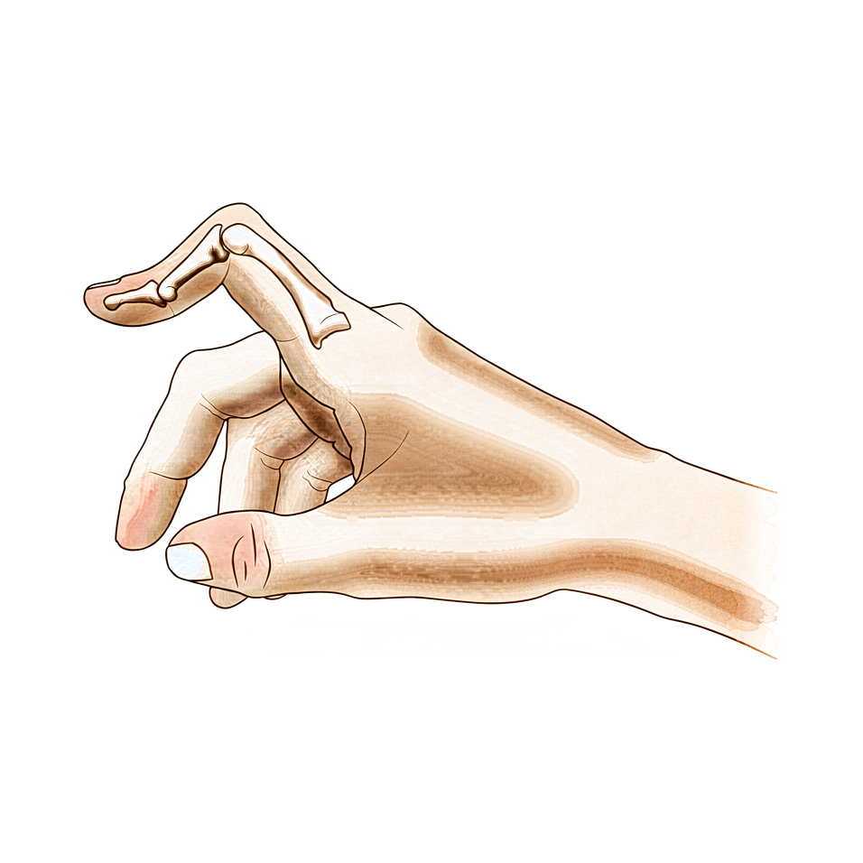
நீங்கள் உணர்வது
பூட்டோனியர் சிதைவில் (boutonnière deformity), உங்கள் விரலின் நடு மூட்டு உள்ளங்கையை நோக்கிக் கீழ்நோக்கி வளைகிறது; விரல் நுனி மறுபக்கமாகப் பின்னோக்கி வளைகிறது. நடு மூட்டின் மேல் விரல் வீங்கியிருப்பது போலத் தோன்றலாம்; அந்த இடத்தில் வலியும் இருக்கலாம். நடு மூட்டை நேராக்குவது கடினமாகிறது; நீங்கள் எவ்வளவு அதிகமாக முயல்கிறீர்களோ, விரல் அவ்வளவு அதிகமாக எதிர்ப்பது போலத் தோன்றும்.
விரல் நேராகி, பிறகு மீண்டும் வளைய வேண்டிய வேலைகளில்தான் பிரச்சினை அதிகம் வெளிப்படுகிறது. நாணயத்தை எடுப்பது, கோப்பையை அதன் கைப்பிடியால் பிடிப்பது, தட்டச்சு செய்வது, சட்டைப் பொத்தான்களைப் போடுவது, அல்லது சாவியைத் திருப்புவது ஆகிய அனைத்தும் தடுமாற்றமாகலாம். பிடி பெரும்பாலும் இன்னும் வேலை செய்கிறது; அதனால்தான் பலர் உதவியை நாடுவதற்கு முன் வாரக்கணக்கில் அந்த விரலைச் சகித்துக்கொண்டிருக்கிறார்கள்.
சிதைவு ஒரு காயத்தால் ஏற்பட்டிருந்தால், கையைப் பயன்படுத்திய பிறகு நடு மூட்டு வலிக்கலாம்; விரல் தளர்வதற்கு முன் காலையில் விறைப்பு அதிகமாக இருக்கலாம். ருமட்டாய்டு மூட்டழற்சியில் (rheumatoid arthritis), மூட்டின் உள்ளுறை பல மாதங்களாக அழற்சியடைந்தும் வீங்கியும் இருப்பதால், இதே வளைந்த நிலை மெதுவாக உருவாகிறது.
சில அறிகுறிகளுக்கு விரைவான கவனம் தேவை. உங்கள் விரலோ கையோ (hand) சூடாகவும், சிவந்தும், வீங்கியும், வலியுடனும் ஆனால் — குறிப்பாகக் காய்ச்சலுடன் — அதே நாளில் அவசர சிகிச்சைப் பிரிவுக்குச் செல்லுங்கள். ஒரு விரல் வெளிறியோ, குளிர்ந்தோ, வெள்ளையாகவோ, நீலமாகவோ மாறினால், அல்லது காயத்துக்குப் பிறகு அதில் திடீரென உணர்வையோ அசைவையோ இழந்தால், இதுவே பொருந்தும். மாறாக, அறிகுறிகள் தணியவில்லை என்றால், வாரக்கணக்கில் மோசமாகிக்கொண்டே போனால், இரவில் உங்களைத் தூக்கத்திலிருந்து எழுப்பினால், அல்லது உங்களை வேலை செய்யவோ கையைப் பயன்படுத்தவோ விடாமல் தடுத்தால், உங்கள் GP-யைப் பாருங்கள் அல்லது சிறப்பு நிபுணர் மதிப்பாய்வைக் கேளுங்கள்.
உண்மையில் என்ன நடக்கிறது
ஒவ்வொரு விரலின் மேற்புறம் நெடுகிலும், மூட்டுகளை நேராக்கும் தசைநாண்களின் (tendons) ஒரு தொகுதி ஓடுகிறது. விரலின் மேற்புறத்தில் உள்ள ஒரு மூடுதிரையின் (hood) கீழ் ஓடும் கயிறுகளாக அவற்றை நினைத்துக்கொள்ளுங்கள். நடு மூட்டை நேராக்கும் கயிறு மையத் தசைநாண் (central slip) என்று அழைக்கப்படுகிறது; அது நடு எலும்பின் அடிப்பகுதியில் இணைந்துள்ளது. பக்கவாட்டு இழைகள் (lateral bands) எனப்படும் மேலும் இரண்டு நாண்கள் அதன் இருபுறமும் ஓடி, நடு மூட்டையும் விரல் நுனியையும் நேராக்க உதவுகின்றன.
பூட்டோனியர் சிதைவில், மையத் தசைநாண் கிழிந்து அல்லது நீண்டு நடு எலும்பிலிருந்து விலகிவிடுகிறது. நடு மூட்டை இனி நேராக இழுக்க முடியாது; எனவே அது வளைந்த நிலைக்குச் சரிகிறது. அதன் பிறகு, இரண்டு பக்கவாட்டு இழைகளும் மூட்டின் மேற்புறத்திலிருந்து நழுவி, உள்ளங்கையை நோக்கிக் கீழே சரிகின்றன. அவை மூட்டின் சுழல் அச்சுக்குக் (pivot point) கீழே வந்துவிட்டால், அதே நாண்கள் நடு மூட்டை நேராக்குவதற்குப் பதிலாக வளைக்கத் தொடங்குகின்றன; விரல் நுனியை மறுபக்கமாகப் பின்னோக்கி இழுக்கின்றன. அதனால்தான் மேலே விவரிக்கப்பட்டபடி விரல், வளைந்த நடு மூட்டுடனும் பின்னோக்கி வளைந்த நுனியுடனும் அமர்கிறது; அதை நேராக்க நீங்கள் எடுக்கும் ஒவ்வொரு முயற்சியும், நாண்கள் உங்களுக்கு எதிராக இன்னும் பலமாக இழுப்பது போலத் தோன்றுவதற்கும் இதுவே காரணம்.
இந்த நிலையை உருவாக்க ஒன்றுக்கு மேற்பட்ட பகுதிகள் சேதமடைய வேண்டும். சிதைவு உருவாவதற்கு முன், மையத் தசைநாண், விரலின் மேற்புறத்தில் இரண்டு நாண்களையும் ஒன்றாகக் கட்டி வைக்கும் தசைநார் (ligament), எல்லாவற்றையும் அதனதன் இடத்தில் பிடித்து வைக்கும் மூடுதிரையின் இழைகள் ஆகிய அனைத்தும் காயமடைய வேண்டும். மையத் தசைநாண் மட்டும் கிழிந்தால், முழுச் சிதைவு இல்லாமல் நடு மூட்டு பலவீனமாக மட்டும் இருக்கலாம்; ஆரம்பச் சிகிச்சை முக்கியம் என்பதற்கு இது ஒரு காரணம்: அது சிதைவு உருவாகாமலேயே தடுக்க முடியும்.
ஒரே இறுதி விளைவு வெவ்வேறு தொடக்கங்களிலிருந்து வரலாம். விரலில் ஏற்படும் அடி அல்லது மூட்டின் மேற்புறத்தில் ஏற்படும் வெட்டுக்காயம் போன்ற காயம் இதை ஏற்படுத்தலாம்; அதே அமைப்புகளை மெல்ல மெல்லத் தேய்த்து அறுக்கும், ருமட்டாய்டு மூட்டழற்சியின் தொடர்ந்த அழற்சியும் இதை ஏற்படுத்தலாம். பிரச்சினை மூட்டின் உள்ளங்கைப் பக்கத்தில் இருக்கும்போது, தசைநாண் காயமே இல்லாமலும் ஒரு விரல் இப்படித் தோன்றலாம்; அந்த வகைக்கு வேறு விதமாகச் சிகிச்சையளிக்கப்படுகிறது; அதனால்தான் உங்களுக்கு எந்த வகை உள்ளது என்பதைக் கண்டறிவதே முதல் படி.
இதற்கு நாங்கள் என்ன செய்ய முடியும்
Mater Private Hospital Rockhampton-இல் மேல் அவயவ (upper-limb) அறுவை சிகிச்சை நிபுணரான டாக்டர் கியரன் ஹிர்பரா, உங்கள் நிலைக்குப் பொருத்தமான, உடலுக்குள் மிகக் குறைவாகத் தலையிடும் (least invasive) தேர்வுகளுடன் தொடங்குகிறார். நோயாளிகள் பொதுவாக அவர்களுடைய GP (பொது மருத்துவர்) மூலம் எங்கள் கிளினிக்கிற்குப் பரிந்துரைக்கப்படுகின்றனர்; ஒரு இயன்முறை சிகிச்சையாளர் (physiotherapist) எங்களைப் பார்க்கும்படி பரிந்துரைத்திருந்தாலும், Medicare தள்ளுபடியைப் (rebate) பெறத் தகுதி பெற உங்கள் GP-யிடமிருந்து பரிந்துரைக் கடிதம் (referral) இன்னும் தேவைப்படும். கிளினிக்கில், என்ன நடக்கிறது என்பதைக் கண்டறிய, நாங்கள் உங்கள் மருத்துவ வரலாற்றைக் கேட்டறிந்து, உங்கள் கையைப் (hand) பரிசோதித்து, தேவைப்பட்டால் ஸ்கேன்களுக்கு ஏற்பாடு செய்கிறோம். இந்தச் சிதைவு பல்வேறு தொடக்கங்களிலிருந்து வரக்கூடும் என்பதால், எந்தச் சிகிச்சைக்கும் முன், உங்களுக்கு எந்த வகை உள்ளது என்பதைத் தெளிவுபடுத்துவதே முதலில் வருகிறது.
முதல் படி பொதுவாக ஸ்பிளிண்ட் (splint) கட்டுவதும் கை சிகிச்சையும் (hand therapy). சமீபத்திய காயத்துக்கு, நடு மூட்டை நேராக வைத்து ஸ்பிளிண்ட் கட்டுவது சிதைவு உருவாகாமலேயே தடுக்க முடியும்; அது எவ்வளவு விரைவில் தொடங்குகிறதோ அவ்வளவு நல்லது: காயம் ஏற்பட்ட ஆறு வாரங்களுக்குள் சிகிச்சை பெறுபவர்கள், அதற்குப் பிறகு சிகிச்சை பெறுபவர்களை விடச் சிறப்பாகக் குணமடைகிறார்கள். ஸ்பிளிண்ட் நடு மூட்டை மட்டுமே நேராகப் பிடித்து வைக்கிறது. உங்கள் விரல் நுனியும், உங்கள் கணுவும் (knuckle), உங்கள் மற்ற விரல்களும் தொடர்ந்து அசைகின்றன; ஏனெனில் முழு விரலையும் அசையாமல் பிடித்து வைப்பது விரல் நுனியை விறைப்படையச் செய்கிறது. சிறிது காலமாக இருந்துவரும் சிதைவுக்கு, நடு மூட்டை முடிந்த அளவு நேராக்கக் கட்டுகள் (casts) போடலாம்; அதைத் தொடர்ந்து, விரல் வேலை செய்யும்போது அதை வளைக்க அனுமதிக்கும் ஒரு ஸ்பிளிண்டை மூன்று மாதங்கள் பயன்படுத்துவீர்கள். அறுவை சிகிச்சையைப் பரிசீலிப்பதற்கு முன் இந்த அணுகுமுறையை முயற்சிப்பது பயனுள்ளது.
ஸ்பிளிண்ட் கட்டுவதும் சிகிச்சையும் போதுமான முன்னேற்றத்தைத் தராதபோது, அறுவை சிகிச்சை பரிசீலிக்கப்படலாம். அறுவை சிகிச்சை, நடு மூட்டை நேராக்கும் தசைநாணை மீண்டும் கட்டியமைக்கிறது; சில சமயங்களில் உங்கள் சொந்த முன்கையிலிருந்து எடுக்கப்படும் ஒரு சிறிய தசைநாண் துண்டை ஒட்டாக (graft) பயன்படுத்துகிறது. சில நிலைகளில், உள்ளங்கையை நோக்கி நழுவிய தசைநாண்கள், அவை இருக்க வேண்டிய இடமான மூட்டின் மேற்புறத்துக்கு மீண்டும் நகர்த்தி வைக்கப்படுகின்றன. ஒவ்வொரு நிலையிலும் நோக்கம் ஒன்றே: நடு மூட்டை நீங்கள் மீண்டும் நேராக்கவும், விரல் சிக்கிக்கொள்ளாமலோ கீழ்நோக்கி வளையாமலோ அதைப் பயன்படுத்தவும் உதவுவது. இந்த அறுவை சிகிச்சைக்குத் தனிப் பக்கம் உள்ளது; எந்த முடிவும் எடுக்கப்படுவதற்கு முன், உங்கள் குறிப்பிட்ட விரலுக்கு அது எதை உள்ளடக்கும் என்பதை நாங்கள் விரிவாகப் பேசுவோம்.
என்ன எதிர்பார்க்கலாம்
எதிர்கால நிலை பெருமளவு நேரத்தைப் பொறுத்தது. ஆரம்பத்திலேயே கண்டறியப்பட்டால், பூட்டோனியர் சிதைவு நிலைபெறுவதற்கு முன்பே பெரும்பாலும் அதைத் தடுத்து நிறுத்த முடியும். காயம் ஏற்பட்ட சிறிது காலத்திலேயே நடு மூட்டை நேராக வைத்து ஸ்பிளிண்ட் கட்டுவது, சிதைவு உருவாகாமலேயே தடுக்க முடியும்; ஆறு வாரங்களுக்குள் சிகிச்சை பெறுபவர்கள், அதற்குப் பிறகு சிகிச்சை பெறுபவர்களை விடச் சிறப்பாகக் குணமடைகிறார்கள். கவனிக்காமல் விடப்பட்டால், சிதைவு அப்படியே இருப்பதற்குப் பதிலாக மோசமடையும் போக்கு உள்ளது; ஏனெனில் இடம் விலகி நழுவிய தசைநாண்கள் விரலை அதன் வளைந்த நிலைக்குள் மேலும் மேலும் இழுத்துக்கொண்டே இருக்கின்றன. சிறியதாகத் தோன்றிய ஒரு காயம், வாரங்கள் கழித்து விறைப்பான, நிலைத்துவிட்ட விரலை ஏற்படுத்தலாம்.
சிறிது காலமாக இருந்துவரும் சிதைவுக்கும், அறுவை சிகிச்சை அல்லாத சிகிச்சைக்கு உண்மையான பங்கு உள்ளது. நடு மூட்டை முடிந்த அளவு நேராக்கக் கட்டுகள் (casts) போட்டு, அதைத் தொடர்ந்து, நீங்கள் விரலைப் பயன்படுத்தும்போது அதை வளைக்க அனுமதிக்கும் ஒரு ஸ்பிளிண்டை மூன்று மாதங்கள் பயன்படுத்துவது நன்றாகப் பலனளிக்கலாம். கட்டுகளால் நடு மூட்டை ஏறக்குறைய நேராகக் கொண்டுவர முடிந்து, அந்த மூன்று மாதங்களிலும் ஸ்பிளிண்டுடன் நீங்கள் உங்கள் கையைச் சுறுசுறுப்பாகப் பயன்படுத்தும்போது, விரல் இந்த அணுகுமுறைக்குப் பலனளிக்கிறது; வழியில் வளையும் திறனும் (flexion) திரும்புகிறது. கட்டைவிரல் காயங்களுக்கு, சிதைவு உருவாவதற்கு முன்பே தொடங்கினால், கணுவை (knuckle) நேராக வைத்து ஸ்பிளிண்ட் கட்டுவது உதவலாம்.
நடு மூட்டை நீங்கள் மீண்டும் நேராக்க உதவுவதே அறுவை சிகிச்சையின் நோக்கம். நீண்ட காலச் சிதைவைத் திருத்தத் தசைநாண் ஒட்டு (tendon graft) அறுவை சிகிச்சை செய்துகொண்ட 18 பேர் கொண்ட, அறிக்கையிடப்பட்ட ஒரு குழுவில், 16 பேருக்கு நல்ல அல்லது மிகச் சிறந்த முடிவுகள் கிடைத்தன. விரலை ஆரம்பத்திலேயே அசைக்கச் செய்யும் ஒரு புதிய நுட்பம் 11 நிகழ்வுகளில் பலனளித்ததாக அறிவிக்கப்பட்டுள்ளது. பிறவியிலிருந்தே இருக்கும் இந்தச் சிதைவின் பிறவி வகைக்கும் (congenital form) அறுவை சிகிச்சை உதவலாம்.
ஒவ்வொரு திருத்தமும் நீடிப்பதில்லை என்று நேர்மையாகச் சொல்ல வேண்டும். ருமட்டாய்டு மூட்டழற்சியால் பாதிக்கப்பட்ட விரல்களில், பிற்பட்ட நிலைகளில் மென்திசு அறுவை சிகிச்சை மட்டும் திருத்தத்தைத் தக்கவைக்காமல் போகலாம்; நீண்ட கால முடிவுகளையும் அவ்வளவு உறுதியாகக் கணிக்க முடியாது. சிதைவு திரும்பி வந்தாலோ நீடித்தாலோ, வேறொரு அறுவை சிகிச்சை முறை சிறந்த தீர்வாக இருக்கலாம். தொடக்கம் எதுவாக இருந்தாலும், யதார்த்தமான இலக்கு, நீங்கள் நேராக்கிப் பயன்படுத்தக்கூடிய ஒரு விரலே — முற்றிலும் இயல்பான ஒரு விரலுக்கான வாக்குறுதி அல்ல.
எப்போது மருத்துவரைப் பார்க்க வேண்டும்
பூட்டோனியர் சிதைவில் ஆரம்பத்திலேயே கவனம் செலுத்துவது நல்ல பலனைத் தருகிறது; எனவே எப்போது நடவடிக்கை எடுக்க வேண்டும் என்று தெரிந்துகொள்வது பயனுள்ளது. உங்கள் விரலில் அடிபட்டு, நடு மூட்டு நேராக வரவில்லை என்றால், அல்லது காயம் ஏற்பட்டு நாட்கள் கழித்தும் விரல் வளைந்தும் வீங்கியும் இருந்தால், உங்கள் GP-யைப் பார்த்து, சிறப்பு நிபுணர் மதிப்பாய்வைக் கேளுங்கள். ஸ்பிளிண்ட் கட்டுவது எவ்வளவு விரைவில் தொடங்குகிறதோ, சிதைவு நிலைபெறுவதற்கு முன் அதைத் தடுக்கும் வாய்ப்பு அவ்வளவு அதிகம்.
சில அறிகுறிகளுக்கு இன்னும் விரைவான கவனிப்பு தேவை. உங்கள் விரலோ கையோ (hand) சூடாகவும், சிவந்தும், வீங்கியும், வலியுடனும் ஆனால் — குறிப்பாகக் காய்ச்சலுடன் — அதே நாளில் அவசர சிகிச்சைப் பிரிவுக்குச் செல்லுங்கள். ஒரு விரல் வெளிறியோ, குளிர்ந்தோ, வெள்ளையாகவோ, நீலமாகவோ மாறினால், அல்லது காயத்துக்குப் பிறகு அதில் திடீரென உணர்வையோ அசைவையோ இழந்தால், இதுவே பொருந்தும். காயமடைந்த மூட்டின் மேல் தோல் கிழிந்திருந்தால், அல்லது விரல் வெளிப்படையாக உருக்குலைந்து தோன்றினால், அப்போதும் அவசர சிகிச்சைப் பிரிவுக்குச் செல்லுங்கள்.
மாறாக, அறிகுறிகள் தணியவில்லை என்றால், வாரக்கணக்கில் மோசமாகிக்கொண்டே போனால், இரவில் உங்களைத் தூக்கத்திலிருந்து எழுப்பினால், அல்லது உங்களை வேலை செய்யவோ கையைப் பயன்படுத்தவோ விடாமல் தடுத்தால், உங்கள் GP-யைப் பாருங்கள் அல்லது சிறப்பு நிபுணர் மதிப்பாய்வைக் கேளுங்கள். வேலை நேரத்துக்குப் பிறகோ வார இறுதியிலோ கிளினிக்கைத் தொடர்புகொள்ள முடியாவிட்டால், உங்களுக்கு அருகிலுள்ள அவசர சிகிச்சைப் பிரிவுக்குச் செல்லுங்கள்.
ஆழமான விளக்கம்
Advanced reading: the deeper science (optional)
உங்கள் சொந்த சிகிச்சை முடிவுகளுக்குத் தேவையானதைவிட இந்தப் பிரிவு ஆழமாகச் செல்கிறது. பூட்டோனியர் சிதைவைப் பற்றி மேலும் படிப்பது பயனுள்ளது; ஏனெனில் வலியுடனும் வீக்கத்துடனும் வளைந்திருக்கும் ஒரு விரலுக்கு உள்ளுணர்வால் தோன்றும் சிகிச்சையான, அதற்கு ஓய்வு கொடுத்து அசைக்காமல் வைத்திருப்பதுதான், ஆதாரங்கள் குறிப்பாக ஆதரிக்காத ஒரே விஷயம்.
நீண்ட கால அசைவின்மை அல்ல, ஆரம்ப அசைவே
மையத் தசைநாண் காயத்திற்கான சிகிச்சைகள் குறித்த ஒரு முறையான மதிப்பாய்வு, ஆதாரத் தளம் குறைவாகவே உள்ளது எனக் கண்டறிந்தது; திறந்த மற்றும் மூடிய காயங்கள் இரண்டிற்கும் வெவ்வேறு உத்திகளின் பங்கு நன்கு ஆதரிக்கப்படவில்லை [1]. அந்த வரம்புக்குள், தனித்தனி ஆய்வுகளிலிருந்து கிடைத்த ஆதாரம் ஆரம்ப அசைவை (early mobilisation) தயக்கத்துடன் ஆதரிக்கிறது; நீண்ட கால அசைவின்மையை ஆதரிக்கவில்லை [1].
இதை வெளிப்படையாகச் சொல்வது அவசியம்; ஏனெனில் இது உள்ளுணர்வுக்கு எதிரானது. வேறுபாடு இதுதான்: குணமடைந்து வரும் மையத் தசைநாணைப் பாதுகாப்பது — இதற்கு நடு மூட்டை நேராகப் பிடித்து வைக்க வேண்டும் — என்பது ஒன்று; விரல் முழுவதையும் அசைவின்றி வைப்பது என்பது வேறு. பிந்தையது விரல் நுனி மூட்டு நீட்டிய நிலையில் விறைத்துப் போகவும், விரல் முழுவதும் இறுகிப் போகவும் வழிவகுக்கிறது. மற்ற மூட்டுகள் தொடர்ந்து அசைந்துகொண்டிருக்கும்போது ஒரு மூட்டை மட்டும் பிடித்து வைப்பதே ஸ்பிளிண்டின் நோக்கம்.
சிதைவு ஏன் அப்படியே நிலைக்காமல் மோசமடைகிறது
பூட்டோனியர் ஒரு முன்னேறும் சிதைவு; இது அசாதாரணமானது, புரிந்துகொள்ளத் தக்கது. மையத் தசைநாண் நடு எலும்பின் அடிப்பகுதியில் ஒட்டிக்கொண்டு, நடு மூட்டை நேராக்குகிறது. அது செயலிழக்கும்போது, அந்த மூட்டு வளைந்த நிலைக்கு (flexion) இறங்குகிறது; வழக்கமாக மூட்டின் அச்சுக்கு மேலே ஓடும் இரண்டு பக்கவாட்டுப் பட்டைகளும் (lateral bands) அதற்குக் கீழே நழுவிவிடுகின்றன.
அவை அச்சுக்குக் கீழே வந்துவிட்டால், முன்பு நடு மூட்டை நேராக்க உதவிய அதே தசைநாண்களே அதை வளைக்கத் தொடங்குகின்றன; அதே நேரத்தில் விரல் நுனியை அளவுக்கு மீறிய நீட்டல் நிலைக்கு (hyperextension) இழுக்கின்றன. எனவே இந்தச் சிதைவு தன்னைத்தானே வலுப்படுத்திக்கொள்கிறது: விரலை நேராக்கும் ஒவ்வொரு முயற்சியும், இப்போது அதை வளைந்த நிலையில் பிடித்து வைத்திருக்கும் அமைப்புகளை மேலும் இறுக்குகிறது. சிறியதாகத் தோன்றிய ஒரு காயம் சில வாரங்களுக்குப் பிறகு நிலைத்த சிதைவை உருவாக்க முடிவதற்கும், எளிய ஸ்பிளிண்ட் சிகிச்சைக்கான கால அவகாசம் ஆரம்பத்திலேயே இருப்பதற்கும் இதுவே காரணம்.
பூட்டோனியர் அல்லாத சிதைவு
மையத் தசைநாண் காயமே இல்லாமலும் ஒரு விரல் அதே நிலையை எடுக்கக்கூடும்; அவற்றுக்கான சிகிச்சைகள் முற்றிலும் வேறுபடுகின்றன. பூட்டோனியர் மற்றும் சூடோபூட்டோனியர் (pseudoboutonnière) சிதைவுகள் நோய்-உடற்கூற்றியலிலும், நோயறிதலிலும், நிர்வாகத்திலும் வேறுபடுகின்றன [2]; சூடோ வகை, நடு மூட்டில் ஏற்படும் வோலார் தட்டு (volar plate) காயத்தால் உருவாகிறது — அங்கே விரல் நுனி மூட்டு அளவுக்கு மீறிய நீட்டல் நிலைக்கு இழுக்கப்படாமல், இயல்பான செயலற்ற வளைவுத் திறனைத் (passive flexion) தக்கவைத்துக்கொள்கிறது.
நோயாளியின் அருகிலேயே இவற்றை வேறுபடுத்துவது விரல் நுனியைப் பொறுத்தது. உண்மையான பூட்டோனியரில், நுனி அளவுக்கு மீறி நீட்டிய நிலையில் பிடிக்கப்பட்டிருக்கும், வளைக்கப்படுவதை எதிர்க்கும் — குறிப்பாக நடு மூட்டை நேராக்கி வைத்திருக்கும்போது; சூடோபூட்டோனியரில் அது தடையின்றி வளையும். ஒரு சூடோபூட்டோனியரை உண்மையான பூட்டோனியர் போலக் கருதி ஸ்பிளிண்ட் கட்டுவது தவறான மூட்டைக் கவனிப்பதாகும்.
தசைநாண் எங்கே செயலிழக்கிறது என்பதால் வரையறுக்கப்படும் ஒரு காயக் குடும்பத்தைச் சேர்ந்தது இது
மூடிய நீட்டுத் தசைநாண் அமைப்புக் (extensor mechanism) காயங்கள் மூன்றில் ஒன்றாக, மேலட் விரல் (mallet finger) மற்றும் சாஜிட்டல் பேண்ட் (sagittal band) காயத்துடன் சேர்ந்தே பூட்டோனியர் இடம்பெறுகிறது; அந்த அமைப்பு எந்த மண்டலத்தில் செயலிழக்கிறது என்பதைக் கொண்டு இவை வகைப்படுத்தப்படுகின்றன [3]. மூன்றுமே, காயம் ஏற்பட்ட சில நாட்களுக்குள் வீக்கத்துடன் இயல்புக்கு மாறான நிலையில் பிடிக்கப்பட்டிருக்கும் ஒரு விரலாகவே தோன்றக்கூடும்; ஒவ்வொன்றுக்கும் வெவ்வேறு ஸ்பிளிண்ட் நிலை தேவைப்படுகிறது — பல வாரங்கள் ஸ்பிளிண்ட் கட்டுவதற்கு உறுதியளிப்பதற்கு முன் ஒரு திட்டவட்டமான நோயறிதலை எட்டுவது ஏன் மதிப்புடையது என்பதற்கு இதுவே நடைமுறைக் காரணம்.
மேற்கோள்கள்
[1] Geoghegan L, Wormald JCR, Adami RZ, Rodrigues JN. Central slip extensor tendon injuries: a systematic review of treatments. J Hand Surg Eur Vol. 2019;44(8):825-32. https://doi.org/10.1177/1753193419845311
[2] Hanson ZC, Thompson RG, Andrews JR, Lourie GM. Boutonniere versus pseudoboutonniere deformities: pathoanatomy, diagnosis, and treatment. J Hand Surg Am. 2023;48(5):489-97. https://doi.org/10.1016/j.jhsa.2022.10.019
[3] Lin JD, Strauch RJ. Closed soft tissue extensor mechanism injuries (mallet, boutonniere, and sagittal band). J Hand Surg Am. 2014;39(5):1005-11. https://doi.org/10.1016/j.jhsa.2013.11.018
Evidence & references
This is the clinical evidence summary written for health professionals. It is technical, and it lists the research this page was built from. You do not need to read it to understand your treatment or to make a decision about it.
Overview
- A staged technique for the repair of traumatic boutonniere deformity allows the surgeon to divide the deformity into various categories and use the operative procedure indicated for each particular problem, often avoiding unnecessary surgery [1].
- Differentiating a true boutonniere deformity from a pseudoboutonniere injury is critical in determining clinical management [2].
- A retrospective study of fifty-four longstanding post-burn boutonnière deformities was carried out to evaluate the problems associated with the surgical procedures [3].
- An understanding of the anatomy, clinical presentation, treatment options, and expected outcomes is crucial for optimal treatment of posttraumatic boutonnière and swan neck deformities [4].
- Early treatment of central slip injuries can prevent the deformity [5].
- Transection of the terminal extensor tendon is a useful procedure for salvage of the boutonniere deformity [6].
- No boutonniere deformities were produced in a series of 20 patients who had chronic mallet finger deformity treated with central slip tenotomy [7].
- Reconstruction of the central slip by the transverse retinacular ligament is not indicated for boutonnière deformities with severe contracture of the lateral band [8].
- The best indication for reconstruction of the central slip by the transverse retinacular ligament is in cases in which the central slip is defective and conservative treatment is not effective [8].
- Similar results occurred for chronic boutonniere deformity using serial casting for adequate extension followed by 3 months of relative motion flexion (RMF) orthotic use, which should be attempted prior to surgical intervention [9].
- A new surgical technique with early mobilization is an effective and simplified treatment of Boutonniere deformity [10].
- The natural history of the boutonnière deformity in rheumatoid arthritis is outlined, and a simple method of repair is described [11].
- Chronic boutonniere deformity will respond to relative motion flexion splinting if serial casting can place the proximal interphalangeal joint in less than -20° extension, and the patient actively uses the hand in a relative motion flexion orthosis for 3 months, recovering flexion [12].
- The Y-shaped tendon graft can be a useful procedure for the correction of chronic boutonniere deformity [13].
- In a patient series of 18 patients, the Y-shaped tendon graft provided good or excellent results in 16 patients [13].
- Extension splinting of the metacarpophalangeal (MP) joint may be effective if traumatic thumb injuries are treated before development of a boutonnière deformity [14].
- Congenital boutonniere deformity is understood as various embryonic developmental failures [15].
- A reliable surgical option for congenital boutonniere deformity involves central slip reconstruction [15].
- Extensor tenotomy to correct a boutonnière deformity of long standing markedly lessened disability with restoration of flexion of the distal joint [16].
- In chronic boutonniere deformity, when the lateral bands are deficient or damaged, the cross-lateral band reconstruction technique using palmaris longus autograft is a treatment option with satisfactory results [17].
- Extension splinting may be effective if traumatic thumb injuries are treated before deformity development [18].
- Surgical treatment with advancement of the extensor pollicis brevis tendon and imbrication of the dorsoradial capsule is necessary once a traumatic thumb boutonnière deformity is established [18].
- The prevalence of boutonnière deformity without rheumatoid arthritis or trauma is approximately 13% [19].
- Current concepts relating to the presentation and treatment of closed soft tissue injuries of the extensor mechanism in zones I, III, and V, commonly known as mallet finger, boutonniere deformities, and sagittal band injuries, are summarized [20].
- Long-term results following soft tissue reconstruction for boutonniere finger deformity in rheumatoid arthritis are unreliable [24].
- Recurrent or persistent deformity in rheumatoid arthritis is best treated with a salvage procedure [24].
Anatomy & Pathophysiology
Extensor Mechanism Anatomy
- The extensor mechanism of the finger at the level of the proximal interphalangeal (PIP) joint consists of both intrinsic and extrinsic contributions [28].
- The extensor tendon entering the digit is the continuation of the extensor digitorum communis (EDC), with contributions from the extensor indicis proprius in the index finger and the extensor digiti quinti in the small finger [28].
- In 30% of cases, the EDC has a separate insertion into the base of the proximal phalanx, but extension of the metacarpophalangeal (MP) joint typically occurs through the pull of the sagittal bands [28].
- The principal bony insertion of the extrinsic digital extensors is on the dorsal proximal aspect of the middle phalanx [32].
- Metacarpophalangeal joint extension is provided by extrinsic extensor force transmitted through the sagittal bands [32].
- Distal interphalangeal joint extension is achieved through conjoined lateral bands composed of tendinous slips from both extrinsic and intrinsic tendons [32].
- The deep head of each dorsal interosseous muscle forms a lateral tendon, or lateral band, at the level of the MP joint [41].
- At the level of the middle of the proximal phalanx, transverse fibers arch dorsally from each lateral band to join over the dorsum of the finger, flexing the proximal phalanx [41].
- Oblique fibers (spiral fibers) from the lateral bands sweep over the distal third of the proximal phalanx to insert onto the lateral tubercles at the base of the middle phalanx, extending the middle phalanx [41].
- The lateral bands are joined by the lateral slips of the extensor tendon to form the conjoined lateral band [41].
- The two conjoined lateral bands to each finger unite at the distal third of the middle phalanx to form the terminal tendon, which inserts at the base of the distal phalanx to extend it [41].
- The volar interossei form the ulnar lateral band of the index finger and the radial lateral band of the ring and little fingers [41].
- The flexor digiti quinti brevis is structurally and functionally similar to the deep head of the dorsal interossei, forming the ulnar lateral band of the little finger [41].
Pathophysiology & Biomechanics
- A boutonniere deformity requires subluxation of the lateral bands volar to the axis of rotation of the PIP joint [22].
- Damage to the central slip alone does not cause a boutonniere deformity [22].
- A boutonniere deformity occurs only when the central slip, triangular ligament, and transverse and oblique fibers of the interosseous hood are all damaged [22].
- Detachment of the central slip from the middle phalanx produces a decrease in extension of the PIP joint [22].
- When the transverse and oblique fibers of the interosseous hood are divided in addition to the central slip, extension at the PIP joint is further decreased [22].
- Early treatment of central slip injuries can prevent the development of the deformity [5].
- The extensor mechanism is a complex structure that demands a delicate balance from intrinsic and extrinsic tendons of the hand [28].
- Injury to the extensor mechanism can disrupt this delicate balance, leading to significant sequelae [28].
- Understanding the anatomy, clinical presentation, treatment options, and expected outcomes is crucial for optimal treatment of posttraumatic boutonnière and swan neck deformities [4].
Classification
- A staged management plan allows the surgeon to divide traumatic boutonniere deformity into various categories and use the operative procedure indicated for each particular problem [1].
- The choice of surgical procedure for rheumatoid boutonniere deformity is dependent on accurate staging of the deformity, which is based on the flexibility of the proximal interphalangeal joint and the state of the articular cartilage [23].
- In a cadaveric model, damage to the central slip alone does not cause a boutonniere deformity [22].
- The boutonniere deformity requires subluxation of the lateral bands volar to the axis of rotation of the PIP joint [22].
- In a cadaveric model, the central slip, triangular ligament, and oblique fibers need to all be damaged to cause subluxation of the lateral bands which leads to an acute boutonniere deformity [29].
- Anterior dislocation of the proximal interphalangeal joint is associated with ruptures of the central slip of the extensor mechanism and results in boutonnière deformity unless repaired [27].
- The incidence of uncorrectable swan-neck and boutonniere deformities is estimated to be between 8% and 16% during the first 2 years after the onset of systemic disease in rheumatoid arthritis [23].
- The prevalence of boutonniere deformities in patients with established rheumatoid arthritis is approximately 36% [23].
Clinical Presentation
Diagnostic Differentiation and Classification
- The surgeon can divide the boutonniere deformity into various categories to use the operative procedure indicated for each particular problem, often avoiding unnecessary surgery [1].
Etiology and Pathomechanics
- The cause of boutonniere deformity in rheumatoid arthritis is chronic synovitis of the proximal interphalangeal joint [23].
- Combined injury of the central slip, triangular ligament, and transverse and oblique fibers of the interosseous hood causes a boutonniere deformity [22].
Prevalence and Natural History
- A swan-neck or boutonniere deformity occurs in approximately half of patients with rheumatoid arthritis [23].
- The incidence of uncorrectable swan-neck and boutonniere deformities is estimated to be between 8% and 16% during the first 2 years after the onset of systemic disease [23].
- The prevalence of finger deformities in patients with established rheumatoid arthritis is approximately 36% for boutonniere deformities [23].
- The natural history of the boutonnière deformity in rheumatoid arthritis is outlined in the literature [11].
Clinical Assessment and Testing
- The central slip tenodesis test detects disruption of the central slip before the boutonniere deformity becomes established [34].
- The central slip tenodesis test is applicable in uncooperative children and unconscious patients [34].
- Treating swan neck and boutonniere deformities of the PIP joint requires understanding the cause, biomechanical changes, and articular status [26].
Investigations
Clinical Examination and Diagnostic Tests
- A careful physical examination is essential to direct care and future testing if indicated [31].
- Diagnostic tests such as imaging and serum laboratory studies are useful in the determination of pathology but can be expensive, time consuming, and often nonspecific [31].
Biomechanical and Pathomechanical Assessment
Imaging and Advanced Modalities
- A computational 3D model validation reassures the reliability of research in understanding the pathomechanics of boutonniere deformity [21].
Treatment
Non-Operative
- Early treatment of central slip injuries can prevent the development of boutonniere deformity [5].
- Extension splinting of the metacarpophalangeal joint may be effective for traumatic thumb boutonniere deformity if treated before the deformity develops [14].
- Extension splinting may be effective for traumatic thumb boutonniere deformity if treated before deformity development [18].
- Chronic boutonniere deformity responds to relative motion flexion splinting if serial casting places the proximal interphalangeal joint in less than -20° extension and the patient actively uses a relative motion flexion orthosis for 3 months [12].
- Serial casting for adequate extension followed by 3 months of relative motion flexion orthotic use should be attempted prior to surgical intervention for chronic boutonniere deformity [9].
- The use of relative motion flexion orthoses is effective in increasing active distal interphalangeal joint flexion and improving proximal interphalangeal joint extension in patients with Burton stage 1 chronic boutonniere deformity [30].
- Conservative treatment of closed central slip injuries involves immobilization of the proximal interphalangeal, distal interphalangeal, and metacarpophalangeal joints in full extension for four weeks followed by a Capener spring splint for two further weeks [92].
- In a review of 115 central slip injuries, 17 of 24 patients treated conservatively achieved an outcome of less than a 20° extension deficit at the proximal interphalangeal joint and more than 80% return of proximal interphalangeal joint flexion [92].
- Patients treated for central slip injuries within six weeks of injury had better outcomes than those treated later [92].
Operative
- A staged technique allows the surgeon to divide traumatic boutonniere deformity into various categories and use the operative procedure indicated for each particular problem, often avoiding unnecessary surgery [1].
- Reconstruction of the central slip by the transverse retinacular ligament is best indicated in cases in which the central slip is defective and conservative treatment is not effective [8].
- A simple method of repair for the rheumatoid boutonnière deformity is described in the literature [11].
- In a patient series, the Y-shaped tendon graft provided good or excellent results in 16 of 18 patients with chronic boutonniere deformity [13].
- Surgical treatment with advancement of the extensor pollicis brevis tendon and imbrication of the dorsoradial capsule is necessary once a traumatic boutonnière deformity of the thumb is established [18].
- The cross-lateral band reconstruction technique using palmaris longus autograft is a treatment option with satisfactory results for chronic boutonniere deformity when the lateral bands are deficient or damaged [17].
- A reliable surgical option involving central slip reconstruction is suggested for the congenital form of boutonniere deformity [15].
- A simple staged procedure using a central slip facilitation device is a valid alternative in the management of severe Dupuytren's proximal interphalangeal joint contracture, demonstrating reliable, reproducible correction of the deformity and acceptable patient outcomes [38].
Complications
- In a patient series of 18 patients, the Y-shaped tendon graft provided good or excellent results in 16 of 18 patients for the correction of chronic boutonniere deformity [13].
- Extension splinting of the MP joint may be effective if traumatic thumb injuries are treated before development of a boutonnière deformity [14].
- The case extends understanding of congenital boutonniere deformity as various embryonic developmental failures and suggests a reliable surgical option involving central slip reconstruction [15].
- Surgical treatment with advancement of the extensor pollicis brevis tendon and imbrication of the dorsoradial capsule is necessary once a boutonnière deformity is established in the thumb [18].
- This article summarizes current concepts relating to the presentation and treatment of closed soft tissue injuries of the extensor mechanism in zones I, III, and V, commonly known as mallet finger, boutonniere deformities, and sagittal band injuries [20].
- This validation reassures the reliability of the research in understanding the pathomechanics of boutonniere deformity [21].
- In a cadaver model, detachment of the central slip from the middle phalanx produced a decrease in extension of the PIP joint [22].
- When the transverse and oblique fibers of the interosseous hood were also divided after central slip detachment, extension at the PIP joint was further decreased [22].
- A boutonniere deformity occurred only when all 3 structures (central slip, transverse and oblique fibers of the interosseous hood, and triangular ligament) were damaged [22].
- Division of the central slip leads to loss of extension at the PIP joint [22].
- The prevalence of finger deformities in patients with established rheumatoid arthritis is approximately 14% for swan-neck deformities and 36% for boutonniere deformities [23].
- In the later stages of both swan-neck and boutonniere deformities, soft-tissue procedures alone may not result in lasting operative correction [23].
- Long-term results following soft tissue reconstruction are unreliable [24].
- Recurrent or persistent deformity is best treated with a salvage procedure [24].
- Treating swan neck and boutonniere deformities of the PIP joint is a difficult challenge that requires understanding the cause, biomechanical changes, and articular status [26].
- Anterior dislocation of the proximal interphalangeal joint is associated with ruptures of the central slip of the extensor mechanism [27].
- Anterior dislocation of the proximal interphalangeal joint results in boutonnière deformity unless repaired [27].
Recovery
Non-Operative Management
- Serial casting for adequate extension followed by 3 months of relative motion flexion (RMF) orthotic use should be attempted prior to surgical intervention for chronic boutonniere deformity [9].
- Chronic boutonniere deformity responds to relative motion flexion splinting if serial casting places the proximal interphalangeal joint in less than -20° extension and the patient actively uses the hand in a relative motion flexion orthosis for 3 months, recovering flexion [12].
- Extension splinting of the metacarpophalangeal joint may be effective for traumatic thumb boutonniere injuries if treated before the development of a boutonnière deformity [14].
Operative Management
- A staged technique allows the surgeon to divide the traumatic boutonniere deformity into various categories and use the operative procedure indicated for each particular problem, often avoiding unnecessary surgery [1].
- The Y-shaped tendon graft provided good or excellent results in 16 of 18 patients for the correction of chronic boutonniere deformity [13].
- Reconstruction of the central slip by the transverse retinacular ligament is indicated in cases in which the central slip is defective and conservative treatment is not effective, but is not indicated for boutonnière deformities with severe contracture of the lateral band [8].
- A simple method of repair for the correction of the rheumatoid boutonnière deformity has been described [11].
- A new surgical technique with early mobilization for chronic boutonniere deformity was reported to be an effective and simplified treatment in 11 cases [10].
- Central slip reconstruction is a reliable surgical option for the congenital form of boutonniere deformity [15].
Outcomes and Prognosis
- Early treatment of central slip injuries can prevent the boutonniere deformity [5].
- Long-term results following soft tissue reconstruction for boutonniere finger deformity in rheumatoid arthritis are unreliable, and recurrent or persistent deformity is best treated with a salvage procedure [24].
- In the later stages of rheumatoid boutonniere deformities, soft-tissue procedures alone may not result in lasting operative correction [23].
- A patient with boutonniere deformity of the second toe after planter dislocation of the proximal interphalangeal joint reported neither deformity nor difficulty when last seen 22 months after the operation [95].
Key Evidence
- [L4] With this plan of management, the surgeon is able to divide the boutonniere deformity into various categories and then use the operative procedure indicated for each particular problem, often avoiding unnecessary surgery. [1] (10.1016/s0363-5023(83)80009-4)
- [L5] Differentiating a true boutonniere deformity from a pseudoboutonniere injury is critical in determining clinical management. [2] (10.1016/j.jhsa.2022.10.019)
- [L4] A retrospective study of fifty-four longstanding post-burn boutonnière deformities was carried out to evaluate the problems associated with the surgical procedures. [3] (10.1016/0266-7681(86)90006-9)
- [L5] An understanding of the anatomy, clinical presentation, treatment options, and expected outcomes is crucial for optimal treatment of posttraumatic boutonnière and swan neck deformities. [4] (10.5435/jaaos-d-14-00272)
- [L5] The paper outlines the anatomy, pathoanatomy, and treatment concepts for boutonniere deformity, emphasizing that early treatment of central slip injuries can prevent the deformity. [5] (10.1016/s0749-0712(21)00060-3)
- [L4] Transection of the terminal extensor tendon is a useful procedure for salvage of the boutonniere deformity. [6] (10.1016/s0749-0712(21)00065-2)
- [L4] No boutonniere deformities were produced in this series of 20 patients who had chronic mallet finger deformity. [7] (10.1016/s0363-5023(87)80205-8)
- [L4] This procedure is not indicated for boutonnière deformities with severe contracture of the lateral band; its best indication is in cases in which the central slip is defective and conservative treatment is not effective. [8] (10.1016/0266-7681(90)90080-n)
- [L4] Similar results occurred for chronic boutonniere deformity using serial casting for adequate extension followed by 3 months of RMF orthotic use, which should be attempted prior to surgical intervention. [9] (10.1016/j.jht.2023.02.005)
- [L4] Our technique is an effective and simplified treatment of Boutonniere deformity. [10] (10.1097/bth.0000000000000152)
- [L4] The natural history of the boutonnière deformity in rheumatoid arthritis is outlined, and a simple method of repair is described. [11] (10.2106/00004623-196951070-00009)
- [L4] Chronic boutonniere deformity will respond to relative motion flexion splinting if serial casting can place the proximal interphalangeal joint in less than -20° extension, and the patient actively uses the hand in a relative motion flexion orthosis for 3 months, recovering flexion. [12] (10.1097/sap.0000000000002307)
- [L4] The Y-shaped tendon graft can be a useful procedure for the correction of chronic boutonniere deformity; in our patient series, this provided good or excellent results in 16 of 18 patients. [13] (10.1016/j.jhsa.2021.01.003)
- [L4] Extension splinting of the MP joint may be effective if these injuries are treated before development of a boutonnière deformity. [14] (10.1016/s0266-7681(00)80024-8)
- [Case_report] The case extends understanding of congenital boutonniere deformity as various embryonic developmental failures and suggests a reliable surgical option involving central slip reconstruction. [15] (10.1016/j.jhsa.2014.05.030)
- [L4] In chronic boutonniere deformity, when the lateral bands are deficient or damaged, the cross-lateral band reconstruction technique using palmaris longus autograft is a treatment option with satisfactory results. [17] (10.1016/j.jhsa.2017.04.010)
- [L4] Extension splinting may be effective if treated before deformity development, but surgical treatment with advancement of the extensor pollicis brevis tendon and imbrication of the dorsoradial capsule is necessary once a boutonnière deformity is established. [18] (10.1054/jhsb.1999.0303)
- [L3] The prevalence of boutonnière deformity without rheumatoid arthritis or trauma is approximately 13%. [19] (10.1177/1753193417704610)
- [L5] This article summarizes current concepts relating to the presentation and treatment of closed soft tissue injuries of the extensor mechanism in zones I, III, and V, commonly known as mallet finger, boutonniere deformities, and sagittal band injuries. [20] (10.1016/j.jhsa.2013.11.018)
- [L5] This validation reassures the reliability of the research in understanding the pathomechanics of boutonniere deformity. [21] (10.1186/s13018-025-06329-3)
- [L5] [22] (10.1016/j.jhsa.2017.07.011)
- [L5] [23] (10.5435/00124635-199903000-00002)
- [L5] Long-term results following soft tissue reconstruction are unreliable, and recurrent or persistent deformity is best treated with a salvage procedure. [24] (10.1016/j.jhsa.2011.05.029)
- [L4] Treating swan neck and boutonniere deformities of the PIP joint is a difficult challenge that requires understanding the cause, biomechanical changes, and articular status. [26] (10.1016/j.hcl.2017.12.006)
- [Paper] [28] (10.1016/j.hcl.2012.05.044)
- [Paper] In a cadaveric model, the central slip, triangular ligament, and oblique fibers need to all be damaged to cause subluxation of the lateral bands which leads to an acute boutonniere deformity. [29] (10.1016/s0363-5023(12)60014-8)
- [L4] The use of RMFO is effective in increasing active distal interphalangeal joint flexion and improving PIP extension in patients with Burton stage 1 chronic boutonniere deformity. [30] (10.1016/j.jhsa.2022.08.007)
- [Paper] The test detects disruption of the central slip before the boutonniere deformity becomes established and is applicable in uncooperative children and unconscious patients. [34] (10.1016/0266-7681(94)90057-4)
- [L4] The simple staged procedure is a valid alternative in the management of severe Dupuytren's PIPJ contracture, demonstrating reliable, reproducible correction of the deformity and acceptable patient outcomes. [38] (10.1177/1753193412439673)
- [L4] [92] (10.1177/1758998318822663)
- [Case_report] The patient reported neither deformity nor difficulty when last seen 22 months after the operation. [95] (10.1007/s00402-009-0816-x)
References
[1] A staged technique for the repair of the traumatic boutonniere deformity. The Journal of Hand Surgery. 1983. DOI: 10.1016/s0363-5023(83)80009-4
[2] Boutonniere Versus Pseudoboutonniere Deformities: Pathoanatomy, Diagnosis, and Treatment. The Journal of Hand Surgery. 2023. DOI: 10.1016/j.jhsa.2022.10.019
[3] Reconstructive surgery of the post-burn boutonnière deformity. The Journal of Hand Surgery: Journal of the British Society for Surgery of the Hand. 1986. DOI: 10.1016/0266-7681(86)90006-9
[4] Posttraumatic Boutonnière and Swan Neck Deformities. Journal of the American Academy of Orthopaedic Surgeons. 2015. DOI: 10.5435/jaaos-d-14-00272
[5] BOUTONNIERE DEFORMITY. Hand Clinics. 1995. DOI: 10.1016/s0749-0712(21)00060-3
[6] TREATMENT OF THE CHRONIC BOUTONNIERE DEFORMITY BY EXTENSOR TENOTOMY. Hand Clinics. 1995. DOI: 10.1016/s0749-0712(21)00065-2
[7] Central slip tenotomy for chronic mallet finger deformity. The Journal of Hand Surgery. 1987. DOI: 10.1016/s0363-5023(87)80205-8
[8] Reconstruction of the Central Slip by the Transverse Retinacular Ligament for Boutonnière Deformity. Journal of Hand Surgery. 1990. DOI: 10.1016/0266-7681(90)90080-n
[9] The relative motion concept in acute and chronic boutonniere deformity: Invited commentary. Journal of Hand Therapy. 2023. DOI: 10.1016/j.jht.2023.02.005
[10] Chronic Boutonniere Deformities, Supple, or Stiff: A New Surgical Technique With Early Mobilization in 11 Cases. Techniques in Hand & Upper Extremity Surgery. 2017. DOI: 10.1097/bth.0000000000000152
[11] Correction of the Rheumatoid Boutonnière Deformity. The Journal of Bone & Joint Surgery. 1969. DOI: 10.2106/00004623-196951070-00009
[12] A Paradigm Shift in Managing Acute and Chronic Boutonniere Deformity. Annals of Plastic Surgery. 2020. DOI: 10.1097/sap.0000000000002307
[13] Y-Shaped Tendon Graft—A Technique in the Reconstruction of Posttraumatic Chronic Boutonniere Deformity. The Journal of Hand Surgery. 2021. DOI: 10.1016/j.jhsa.2021.01.003
[14] Traumatic Boutonnière Deformity of the Thumb. Journal of Hand Surgery. 2000. DOI: 10.1016/s0266-7681(00)80024-8
[15] Restoration of the Central Slip in Congenital Form of Boutonniere Deformity: Case Report. The Journal of Hand Surgery. 2014. DOI: 10.1016/j.jhsa.2014.05.030
[16] Extensor Tenotomy for Chronic Boutonniere Deformity of the Finger: REPORT OF TWO CASES.. The Journal of Bone and Joint Surgery. American Volume. 1965.
[17] Chronic Boutonniere Deformity: Cross-Lateral Band Technique Using Palmaris Longus Autograft. The Journal of Hand Surgery. 2017. DOI: 10.1016/j.jhsa.2017.04.010
[18] TRAUMATIC BOUTONNIÈRE DEFORMITY OF THE THUMB. The Journal of Hand Surgery: Journal of the British Society for Surgery of the Hand. 2000. DOI: 10.1054/jhsb.1999.0303
[19] Thumb boutonnière deformity without rheumatoid arthritis or trauma. Journal of Hand Surgery (European Volume). 2017. DOI: 10.1177/1753193417704610
[20] Closed Soft Tissue Extensor Mechanism Injuries (Mallet, Boutonniere, and Sagittal Band). The Journal of Hand Surgery. 2014. DOI: 10.1016/j.jhsa.2013.11.018
[21] Assessment of the ligamentous stress distribution in the pathomechanics of the boutonniere deformity through a computational 3D model. Journal of Orthopaedic Surgery and Research. 2025. DOI: 10.1186/s13018-025-06329-3
[22] Biomechanics of the Acute Boutonniere Deformity. The Journal of Hand Surgery. 2018. DOI: 10.1016/j.jhsa.2017.07.011
[23] Operative Correction of Swan-Neck and Boutonniere Deformities in the Rheumatoid Hand. Journal of the American Academy of Orthopaedic Surgeons. 1999. DOI: 10.5435/00124635-199903000-00002
[24] Treatment of Boutonniere Finger Deformity in Rheumatoid Arthritis. The Journal of Hand Surgery. 2011. DOI: 10.1016/j.jhsa.2011.05.029
[26] Treating the Proximal Interphalangeal Joint in Swan Neck and Boutonniere Deformities. Hand Clinics. 2018. DOI: 10.1016/j.hcl.2017.12.006
[27] Anterior dislocation of the proximal interphalangeal joint. A cause of rupture of the central slip of the extensor mechanism.. The Journal of bone and joint surgery. American volume. 1970.
[28] Boutonnière and Pulley Rupture in Elite Athletes. Hand Clinics. 2012. DOI: 10.1016/j.hcl.2012.05.044
[29] Biomechanics of the Acute Boutonniere Deformity. The Journal of Hand Surgery. 2012. DOI: 10.1016/s0363-5023(12)60014-8
[30] The Use of Relative Motion Flexion Orthoses for Chronic Boutonniere Deformity. The Journal of Hand Surgery. 2024. DOI: 10.1016/j.jhsa.2022.08.007
[31] Orthopaedic Knowledge Update 13 Ebook Without Multimedia. Anatomy, Evaluation, Clinical Examination, and Imaging > Evaluation and Clinical Examination: Current Concepts.
[32] A Lange Medical Book Current Diagnosis Treatment In Orthopedics Fifth Edition. 9Hand Surgery > Image DISORDERS OF THE MUSCULATURE OF THE HAND.
[34] The Central Slip Tenodesis Test for Early Diagnosis of Potential Boutonnière Deformities. Journal of Hand Surgery. 1994. DOI: 10.1016/0266-7681(94)90057-4
[38] Management of severe Dupuytren’s contracture of the proximal interphalangeal joint with use of a central slip facilitation device. Journal of Hand Surgery (European Volume). 2012. DOI: 10.1177/1753193412439673
[41] Green S Operative Hand Surgery. Interosseous and Hypothenar Muscles.
[92] Therapeutic management of closed central slip injuries: Outcome of a service evaluation. Hand Therapy. 2019. DOI: 10.1177/1758998318822663
[95] Boutonniere deformity of the second toe after planter dislocation of proximal interphalangeal joint: a case report. Archives of Orthopaedic and Trauma Surgery. 2009. DOI: 10.1007/s00402-009-0816-x