Patients › Shoulder
ஆஸ் அக்ரோமியேல்
Patient-facing topic on os acromiale — failed fusion of an acromial ossification centre that may cause shoulder pain and contribute to subacromial impingement.
நீங்கள் உணர்வது
ஆஸ் அக்ரோமியேல் (os acromiale) இருக்கும்போது, வலி பொதுவாக உங்கள் தோள்பட்டையின் முன்புறத்திலும் பக்கவாட்டிலும், மூட்டின் எலும்புக் கூரைக்கு மேல் உணரப்படுகிறது. தலைக்கு மேல் கையை உயர்த்தும் செயல்கள் அதை மோசமாக்குகின்றன. உயரமான அலமாரியை எட்டிப் பிடிப்பது, துவைத்த துணிகளைக் காயப்போடுவது, அல்லது தோள்பட்டை உயரத்துக்கு மேல் எதையாவது தூக்குவது — இவை அனைத்தும் வலியைத் தூண்டலாம். பலர் இரவிலும் வலியைக் கவனிக்கிறார்கள்; அந்தப் பக்கமாகப் படுக்கும்போது அது உங்களைத் தூக்கத்திலிருந்து எழுப்பலாம்.
வளர்ச்சியின்போது எலும்பின் மற்ற பகுதியோடு ஒருபோதும் இணையாத, தோள்பட்டையின் உச்சியில் உள்ள ஒரு சிறிய எலும்புத் துண்டிலிருந்து இந்த வலி வருகிறது. நீங்கள் கையை உயர்த்தும்போது, ஒரு தசை இந்தத் தளர்வான துண்டை இழுக்கிறது; அது சற்று அசைந்து, அதற்கு அடியில் உள்ள தசைநாணின் மீது அழுத்துகிறது. இரண்டு எலும்புத் துண்டுகளும் சந்திக்கும் இடத்தைத் தொட்டாலும் வலிக்கலாம்; அதன் மீது நேரடியாக அழுத்துவது வலியை வரவழைக்கலாம்.
அன்றாட வாழ்க்கையில், கையைப் பக்கவாட்டில் வெளிப்புறமாக உயர்த்தும்போதோ அதை வெளிப்புறமாகத் திருப்பும்போதோ உங்கள் கை பலவீனமாக இருப்பதை நீங்கள் உணரலாம். வலிமிகு வளைவு (painful arc) பொதுவானது: கையை உயர்த்தும்போது பாதி வழியில் தோள்பட்டை வலிக்கிறது; பிறகு இன்னும் உயரமாகச் செல்லும்போது வலி தணிகிறது. தோள்பட்டைக் கூரைக்கு அடியில் உள்ள தசைநாணின் தேய்மானத்தை இந்த அறிகுறிகள் மிகவும் ஒத்திருப்பதால், இந்த நிலையைச் சுழற்சி தசைநாண் பட்டை (rotator cuff) பிரச்சினை என்று எளிதில் தவறாக எண்ணிவிடலாம்; அதற்குச் சிகிச்சை பெற்ற பிறகும் இது தொடர்ந்து வலியை ஏற்படுத்தலாம்.
இந்த எலும்பு அமைப்பு உள்ள பெரும்பாலானவர்கள் எதையுமே ஒருபோதும் உணர்வதில்லை. வேறு எதற்காகவோ எடுக்கப்பட்ட ஸ்கேனில் இது பெரும்பாலும் தற்செயலாகக் கண்டறியப்படுகிறது. இது வலியை ஏற்படுத்தும்போது, இளவயது வயதுவந்தவர்களிடம், பெரும்பாலும் எந்தக் காயமும் இல்லாமலே, தோன்றும் போக்கு உள்ளது; இயன்முறை சிகிச்சை (physiotherapy), விளையாட்டிலிருந்து ஓய்வு, அல்லது ஊசி ஆகியவற்றால் உங்கள் தோள்பட்டை வலி தணியவில்லை என்றால், இதைச் சந்தேகிப்பது பயனுள்ளது.
உங்கள் அறிகுறிகள் தணியவில்லை என்றால், வாரக்கணக்கில் மோசமாகிக்கொண்டே வந்தால், இரவில் உங்களைத் தூக்கத்திலிருந்து எழுப்பினால், அல்லது வேலை செய்யவோ கையைப் பயன்படுத்தவோ முடியாமல் தடுத்தால், உங்கள் GP-யைப் பாருங்கள் அல்லது நிபுணர் பரிசோதனையைக் கேளுங்கள்.
உண்மையில் என்ன நடக்கிறது
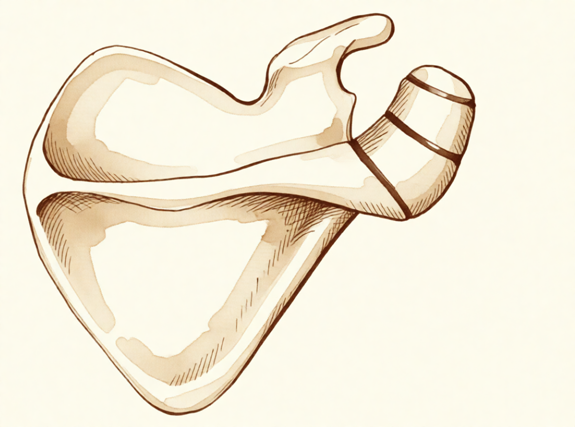
உங்கள் தோள்பட்டையின் எலும்புக் கூரை ஒரே ஒரு எலும்புத் துண்டாக உருவாவதில்லை. அது மூன்று தனித்தனி மையங்களாகத் தொடங்கி, மெதுவாக ஒன்றுசேர்கிறது; இது சுமார் 15 வயதில் தொடங்கி, பொதுவாக 25 வயதுக்குள் முடிந்துவிடுகிறது. சுமார் 8% பேருக்கு, இந்த இணைப்புகளில் ஒன்று ஒருபோதும் மூடுவதில்லை. அந்த இடைவெளி எலும்புக்குப் பதிலாக உறுதியான, குருத்தெலும்பு போன்ற திசுவால் (gristle) நிரம்புகிறது; இதனால் கூரையின் ஒரு சிறிய துண்டு திடமான எலும்பால் அல்லாமல், வளையக்கூடிய ஓர் இணைப்பால் இணைக்கப்பட்டிருக்கிறது.
ஒரு திருகாணி மட்டும் தளர்வாக உள்ள நிலையில் சுவரில் பொருத்தப்பட்ட ஒரு பலகையைப் போல இதை நினைத்துக்கொள்ளுங்கள். அது தாங்கி நிற்கிறது; ஆனால் அதன் மீது சாயும்போது சற்று அசைகிறது. கையைப் பக்கவாட்டில் வெளிப்புறமாக உயர்த்தும் தசை இந்தக் கூரையோடு இணைந்துள்ளது; எனவே நீங்கள் கையை உயர்த்தும் ஒவ்வொரு முறையும், அந்தத் தசை தளர்வான துண்டை இழுக்கிறது, அது சற்று வளைகிறது. அந்த அசைவு கூரைக்கு அடியில் செல்லும் தசைநாணின் மீது கீழ்நோக்கி அழுத்துகிறது; அந்த உரசல் அதை எரிச்சலடையச் செய்கிறது. அதனால்தான் தலைக்கு மேல் கையை உயர்த்தும் செயல்கள் வலிக்கின்றன; அதனால்தான் வலி உங்கள் தோள்பட்டையின் முன்புறத்திலும் பக்கவாட்டிலும் உணரப்படுகிறது.
வளையக்கூடிய அந்த இணைப்பே பிரச்சினையாகவும் இருக்கலாம். தேய்ந்த ஒரு மூட்டைப் போலவே, அதுவே தனியாகத் தொட்டால் வலிப்பதாக மாறலாம்; அதன் மீது அழுத்துவது வலியை வரவழைக்கிறது. இந்த எலும்பு அமைப்பு உள்ள பெரும்பாலானவர்கள் எதையும் ஒருபோதும் உணர்வதில்லை; இது தானாகவே தோள்பட்டை வலியை ஏற்படுத்துவதாகவோ, தசைநாண் கிழிவுக்கான வாய்ப்பை அதிகரிப்பதாகவோ தெரியவில்லை. ஆனால் அது உருவான விதத்தினாலோ, அந்த இடத்தில் அடிபட்ட பிறகோ, தளர்வான துண்டு வழக்கத்தை விட அதிகமாக அசையத் தொடங்கும்போது, நீங்கள் கையை உயர்த்தும் ஒவ்வொரு முறையும் அது தசைநாணின் மீது அழுத்தலாம். மேலே விவரிக்கப்பட்ட வலிமிகு வளைவையும் இரவு நேர வலியையும் உண்டாக்குவது அதுவே.
தளர்வான துண்டு தசைநாண் இருக்கும் இடத்துக்கு நேர் மேலேயே அமைந்திருப்பதால், இந்த இரண்டு பிரச்சினைகளும் பெரும்பாலும் சேர்ந்தே தோன்றுகின்றன. உங்கள் தோள்பட்டைக்கு ஏற்கெனவே ஒரு தசைநாண் பிரச்சினைக்காகச் சிகிச்சை அளிக்கப்பட்டு, வலி தணியவில்லை என்றால், இந்த இணையாத துண்டு உள்ளதா என்று பரிசோதிப்பது பயனுள்ளது; ஏனெனில் தசைநாணில் மட்டும் அறுவை சிகிச்சை செய்வது, அதற்கு மேலே அசைந்துகொண்டிருக்கும் எலும்புத் துண்டை அமைதிப்படுத்தாது.
இதற்கு நாங்கள் என்ன செய்ய முடியும்
Mater Private Hospital Rockhampton-இல் பணியாற்றும் மேல் அவயவ (upper-limb) அறுவை சிகிச்சை நிபுணரான டாக்டர் கியரன் ஹிர்பரா, உங்கள் நிலைக்குப் பொருத்தமான, உடலுக்குள் மிகக் குறைவாகத் தலையிடும் (least invasive) தேர்வுகளுடன் தொடங்குகிறார். நோயாளிகள் பொதுவாக அவர்களுடைய GP (குடும்ப மருத்துவர்) மூலம் எங்கள் கிளினிக்கிற்குப் பரிந்துரைக்கப்படுகிறார்கள்; ஒரு இயன்முறை சிகிச்சையாளர் (physiotherapist) எங்களைச் சந்திக்கும்படி பரிந்துரைத்திருந்தாலும், Medicare தள்ளுபடியைப் (rebate) பெறத் தகுதி பெற உங்கள் GP-யிடமிருந்து பரிந்துரைக் கடிதம் (referral) இன்னும் தேவைப்படும். உங்கள் முதல் சந்திப்பில் நாங்கள் உங்கள் மருத்துவ வரலாற்றைக் கேட்டறிந்து, உங்கள் தோள்பட்டையைப் பரிசோதித்து, தேவைப்படும் இடங்களில் படமெடுப்புக்கு (imaging) ஏற்பாடு செய்கிறோம்.
முதல் படி பொதுவாக, நீங்கள் தோள்பட்டையைப் பயன்படுத்தும் விதத்தை மாற்றுவதும், இயன்முறை சிகிச்சையை முறையாக முயற்சித்துப் பார்ப்பதும்தான். எலும்புக் கூரைக்கு அடியில் உள்ள எரிச்சலைத் தணிப்பதும், தலைக்கு மேல் கையை உயர்த்திச் செய்யும் வேலையை நன்றாகச் சமாளிக்கும்படி உங்கள் தோள்பட்டையைச் சுற்றியுள்ள தசைகளை வலுப்படுத்துவதுமே இயன்முறை சிகிச்சையின் நோக்கம். இதற்கு மேல் எதையும் யோசிப்பதற்கு முன், இதை நியாயமான அளவு காலம் முயற்சித்துப் பாருங்கள்.
அது நடக்கும் வரை தொடர்ந்து அசைந்து செயல்பட வலி நிவாரண மருந்துகள் உதவலாம். உங்கள் GP அறிவுறுத்தியபடி எடுத்துக்கொள்ளப்படும் எளிய வீக்கமடக்கி மருந்துகள் (anti-inflammatories) வலியைத் தணிக்கின்றன; இதனால் உங்கள் பயிற்சிகளை நீங்கள் வசதியாகச் செய்ய முடியும். வலி தணியவில்லை என்றால், வாரக்கணக்கில் மோசமாகிக்கொண்டே வந்தால், இரவில் உங்களைத் தூக்கத்திலிருந்து எழுப்பினால், அல்லது வேலை செய்யவோ கையைப் பயன்படுத்தவோ முடியாமல் தடுத்தால், உங்கள் GP-யைப் பாருங்கள் அல்லது நிபுணர் பரிசோதனையைக் கேளுங்கள்.
அறுவை சிகிச்சை அல்லாத கவனிப்பு போதுமான முன்னேற்றத்தைத் தரவில்லை என்றால், நாங்கள் அறுவை சிகிச்சையைப் பரிசீலிக்கிறோம். அதற்கு முன், சாதாரண எக்ஸ்-ரே (X-ray) படங்கள் மூலம் நோயறிதலை உறுதிப்படுத்துகிறோம்; இணையாத துண்டைக் காட்ட அவை கிட்டத்தட்ட எப்போதும் போதுமானவை. அக்குளுக்குக் கீழிருந்து எடுக்கப்படும் ஒரு படம், மூட்டின் வடிவத்தையும் கூரையின் முன்பகுதியையும் தெளிவாகக் காட்டுகிறது; ஒரு காயம் இரண்டு எலும்புத் துண்டுகளும் சந்திக்கும் இடத்தைச் சேதப்படுத்தக்கூடும் என்பதால் இது முக்கியமானது. கூரைக்கு அடியில் செல்லும் தசைநாணைச் சோதிக்க அல்ட்ராசவுண்ட் (ultrasound) ஓர் எளிய, துல்லியமான வழி; MRI ஸ்கேன் அந்தத் தசைநாணையும் சுற்றியுள்ள மென்திசுக்களையும் பற்றிய முழுமையான படத்தைத் தருகிறது.
நாங்கள் பெரும்பாலும் வழங்கும் அறுவை சிகிச்சையில், முதலில் சாவித்துளை (arthroscopic) முறையில் மூட்டை மதிப்பிட்டு, பர்செக்டமி (bursectomy) செய்கிறோம் — அதாவது கூரைக்கு அடியில் உள்ள இடத்திலிருந்து வீக்கமடைந்த திசுவை அகற்றுகிறோம்; பிறகு எலும்பு ஒட்டுதலுக்கும் (bone grafting) பொருத்தி நிலைநிறுத்துதலுக்கும் (fixation) ஒரு சிறிய திறந்த வெட்டு செய்கிறோம் — பொருத்தி நிலைநிறுத்துதல் என்பது, தளர்வான துண்டு எலும்பின் மற்ற பகுதியோடு இணையும்படி அதை அசையாமல் பிடித்து வைப்பதாகும். அதை அசையாமல் வைத்திருப்பது இரண்டு துண்டுகளும் ஒன்றாக வளர்ந்து இணைய உதவுகிறது. தளர்வான துண்டு சிறியதாக இருந்தால், அதைப் பொருத்துவதற்குப் பதிலாக நாங்கள் அதை அகற்றக்கூடும். பெரிய துண்டைப் பொருத்துவதற்குக் கவனம் தேவை; ஏனெனில் கையை உயர்த்தும் தசை அதைத் தொடர்ந்து இழுக்கிறது, மேலும் அந்தத் துண்டு மெல்லியது. சில நேரங்களில், பொருத்திய பிறகும் துண்டு இணைவதில்லை; இது ஒட்டாமை (non-union) என்று அழைக்கப்படுகிறது. உங்கள் தோள்பட்டைக்கு எந்தத் தேர்வு பொருத்தமானது என்பதை நாங்கள் உங்களுடன் விரிவாகப் பேசுவோம்; நாம் சேர்ந்தே முடிவெடுப்போம்.
என்ன எதிர்பார்க்கலாம்
இந்த எலும்பு அமைப்பு உள்ள பெரும்பாலானவர்களுக்கு ஒருபோதும் எந்தப் பிரச்சினையும் ஏற்படுவதில்லை; இதற்கு எந்தச் சிகிச்சையும் தேவையில்லை. இது வலியை ஏற்படுத்தும்போது, எதிர்காலப் போக்கு தளர்வான துண்டு உறுதியாக இருக்கிறதா என்பதையும், உண்மையில் அதுதான் உங்கள் வலிக்கு மூலமா என்பதையும் பொறுத்தது.
வலிக்கும் துண்டுக்கு, கவனிப்பு பொதுவாக அறுவை சிகிச்சை இல்லாமலே தொடங்குகிறது. நீங்கள் தோள்பட்டையைப் பயன்படுத்தும் விதத்தை மாற்றுவது, இயன்முறை சிகிச்சை, எளிய வலி நிவாரணம் ஆகியவை பலருக்கு நிலைமையைத் தணிக்கின்றன. நியாயமான அளவு காலம் முயற்சித்த பிறகும் அது பலனளிக்கவில்லை என்றால், அறுவை சிகிச்சை வலியை உண்மையாகவே குறைத்து, தோள்பட்டைச் செயல்பாட்டை மேம்படுத்தலாம். தளர்வான துண்டு அசையாமல் பிடித்து வைக்கப்பட்டு எலும்பின் மற்ற பகுதியோடு இணையும்போது, இரண்டு துண்டுகளும் தனித்தனியாகவே இருக்கும் நிலையை விட, தோள்பட்டைச் செயல்பாட்டு அளவீடுகளில் மக்கள் சிறப்பாக இருக்கிறார்கள். இருப்பினும், அறுவை சிகிச்சை எப்போதும் தீர்வாக இருப்பதில்லை. இந்த நிலையுடன் தசைநாண் கிழிவும் சேர்ந்துள்ள சிலருக்கு, அறுவை சிகிச்சைக்குப் பிந்தைய முடிவுகள் கலவையாக இருந்துள்ளன; ஒட்டுமொத்தமாக 53% பேர் திருப்திகரமான முடிவுகளை அடைந்தனர்.
இதைக் கவனிக்காமல் விட்டுவிட்டு, அது உங்களைத் தொடர்ந்து தொந்தரவு செய்தால், வலி பொதுவாகத் தானாக மறைந்துவிடுவதில்லை. இதைத் தவறவிடுவதும் எளிது: சிகிச்சைக்குப் பிறகும் — ஒரு தசைநாண் பிரச்சினைக்கான அறுவை சிகிச்சைக்குப் பிறகும் கூட — தோள்பட்டை வலி தணியவில்லை என்றால், இந்த நிலையைச் சந்தேகிப்பது பயனுள்ளது; ஏனெனில் தசைநாணில் மட்டும் அறுவை சிகிச்சை செய்வது, அதற்கு மேலே அசைந்துகொண்டிருக்கும் எலும்புத் துண்டை அமைதிப்படுத்தாது.
அறுவை சிகிச்சைக்குப் பிந்தைய மீட்சி படிப்படியானது. நீங்கள் உங்கள் இயன்முறை சிகிச்சையாளருடன் சேர்ந்து, முதலில் அசைவையும் பிறகு வலிமையையும் வளர்த்துக்கொள்வீர்கள்; தலைக்கு மேல் தூக்கும் திறன் வாரங்கள் முதல் மாதங்கள் வரையிலான காலத்தில் மெதுவாகத் திரும்பி வரும். நீங்கள் வந்தபோது இருந்த, கையை உயர்த்தும்போது ஏற்படும் கூர்மையான வலி இல்லாமல், அன்றாட வாழ்க்கையைச் சமாளிக்கக்கூடிய ஒரு தோள்பட்டையே இதன் நோக்கம்.
ஒரு விரல், கை (hand) அல்லது முழுக் கை (arm) சூடாகி, சிவந்து, வீங்கி, வலிப்பதை நீங்கள் கவனித்தால் — குறிப்பாகக் காய்ச்சலுடன் — அன்றே அவசர சிகிச்சைப் பிரிவுக்குச் செல்லுங்கள். GP பரிந்துரைக் கடிதம் தேவையில்லை.
எப்போது மருத்துவரைப் பார்க்க வேண்டும்
இந்த எலும்பு அமைப்பு உள்ள பெரும்பாலானவர்களுக்கு ஒருபோதும் சிகிச்சை தேவைப்படுவதில்லை; ஆனால் உங்கள் தோள்பட்டை வலி தணியவில்லை என்றால், வாரக்கணக்கில் மோசமாகிக்கொண்டே வந்தால், இரவில் உங்களைத் தூக்கத்திலிருந்து எழுப்பினால், அல்லது வேலை செய்யவோ கையைப் பயன்படுத்தவோ முடியாமல் தடுத்தால், உங்கள் GP-யைப் பாருங்கள் அல்லது நிபுணர் பரிசோதனையைக் கேளுங்கள். வலி தோள்பட்டையின் முன்புறத்திலும் பக்கவாட்டிலும் இருந்தால், தலைக்கு மேல் கையை உயர்த்தும் செயல்களின்போது மோசமானால், அல்லது அந்தப் பக்கமாகப் படுக்கும்போது உங்களைத் தொடர்ந்து எழுப்பினால், இன்னும் விரைவில் பரிசோதனையைக் கேளுங்கள். தோள்பட்டையின் உச்சியில் உள்ள வலிக்கும் இடத்தை அழுத்தினால் வலித்தால், கையைப் பக்கவாட்டில் வெளிப்புறமாக உயர்த்தும்போதோ வெளிப்புறமாகத் திருப்பும்போதோ கை பலவீனமாக உணர்ந்தால், அல்லது கையை உயர்த்தும்போது பாதி வழியில் தோள்பட்டை வலித்தால், பரிசோதனை செய்துகொள்வதும் பயனுள்ளது. இந்த அறிகுறிகள் உள்ள, எந்தக் காயமும் இல்லாத இளவயது வயதுவந்தவர்கள் — குறிப்பாக இயன்முறை சிகிச்சை, விளையாட்டிலிருந்து ஓய்வு, அல்லது ஊசி உதவவில்லை என்றால் — இந்த நிலைக்காகப் பரிசோதிக்கப்பட வேண்டும். ஒரு விரல், கை (hand) அல்லது முழுக் கை (arm) சூடாகி, சிவந்து, வீங்கி, வலித்தால் — குறிப்பாகக் காய்ச்சலுடன் — அன்றே அவசர சிகிச்சைப் பிரிவுக்குச் செல்லுங்கள்; GP பரிந்துரைக் கடிதம் தேவையில்லை.
ஆழமான விளக்கம்
Advanced reading: the deeper science (optional)
உங்கள் சொந்தச் சிகிச்சை முடிவுகளுக்குத் தேவையானதை விட இந்தப் பகுதி மேலும் ஆழமாகச் செல்கிறது. ஆஸ் அக்ரோமியேல் பற்றிக் கூடுதலாகப் படிப்பது பயனுள்ளது; ஏனெனில் இது கணிசமான சிறுபான்மையினரிடம் காணப்படும் ஓர் இயல்பான உடற்கூற்று வேறுபாடு, பொதுவாக எதையும் ஏற்படுத்துவதில்லை, ஆனால் அது காரணமல்லாத தோள்பட்டை வலிக்கும் அடிக்கடி இதன் மீதே பழி சுமத்தப்படுகிறது.
இது என்ன, எவ்வளவு பொதுவானது
தோள்பட்டைக்கு மேலுள்ள எலும்புக் கூரையான அக்ரோமியன், தனித்தனியான பல எலும்பு உருவாக்க மையங்களிலிருந்து உருவாகிறது; அவை பொதுவாகப் பதின்ம வயதின் பிற்பகுதியிலோ இருபதுகளின் தொடக்கத்திலோ ஒன்றுசேர்ந்துவிடும். சிலருக்கு அவற்றில் ஒன்று இணையத் தவறி, ஒரு நிரந்தர நார்த்திசு இணைப்பை விட்டுச் செல்கிறது. அதுவே ஆஸ் அக்ரோமியேல்; இது பின்னாளில் வந்து சேர்வது அல்ல, பதின்ம வயதிலிருந்தே இருப்பது.
இது காணப்படும் அளவு மக்கள்தொகைக்கு மக்கள்தொகை மாறுபடுகிறது. 6,842 நோயாளிகளை உள்ளடக்கிய ஒரு பன்முக மைய ஆய்வு, ஆஸ் அக்ரோமியேல் சுழற்சி தசைநாண் பட்டைக் காயங்களுடன் தொடர்புடையது எனவும், ஆய்வு செய்யப்பட்ட ஜப்பானிய மக்கள்தொகையில் இது காணப்படும் அளவு ஆசியரல்லாத மக்கள்தொகைகளை விடக் குறைவு எனவும், எலும்புத் துண்டின் அளவு சிறியதாக இருக்கும் போக்கு உள்ளது எனவும் கண்டறிந்தது [1].
தசைநாண் பட்டைக் காயத்துடனான அந்தத் தொடர்பைக் கவனமாகப் படிக்க வேண்டும். இவ்விரண்டும் தற்செயலாக நிகழ்வதை விட அடிக்கடி சேர்ந்து நிகழ்கின்றன என்பதை அது நிறுவுகிறது; ஆஸ் அக்ரோமியேல்தான் கிழிவை ஏற்படுத்துகிறது என்பதை அல்ல. ஏற்கத்தக்க வழிமுறை இரு திசைகளிலும் செல்கிறது: இணையாத துண்டு டெல்டாய்டு (deltoid) தசையின் இழுவையால் சற்று அசைகிறது, அது அடியில் உள்ள தசைநாணுக்கு எரிச்சலை ஏற்படுத்தலாம் — அல்லது தசைநாண் பட்டை நோய்க்கு வழிவகுக்கும் அதே தோள்பட்டை இயக்கவியலே, நிலையற்ற ஒரு துண்டை அறிகுறிகளைத் தரச் செய்கிறது.
வலிக்கு இதுவே மூலமா என்பதே முழுக் கேள்வி
படமெடுப்பில் இது தெரிகிறது, நிரந்தரமானது, இயல்புக்கு மாறானதாகத் தோன்றுகிறது என்பதால், தோள்பட்டை வலிக்கான விளக்கமாக ஆஸ் அக்ரோமியேல் எளிதில் பதிவாகிவிடுகிறது. பெரும்பாலானவை அமைதியானவை; இது உள்ளவர்களில் பெரும்பாலானவர்களுக்கு அது இருப்பதே ஒருபோதும் தெரிவதில்லை.
அறிகுறிகளைத் தரும் ஒன்றை வேறுபடுத்திக் காட்டுவது: அந்தத் துண்டுக்கு நேர் மேலே குறிப்பிட்ட இடத்தில் மட்டும் தொட்டால் வலிப்பது, அதை அழுத்தும்போது அதே வலி மீண்டும் வருவது, மேலும் நோயறிதல் ஊசி பயன்படுத்தப்படும்போது, அதற்கு அடியில் உள்ள அக்ரோமியனுக்குக் கீழான இடைவெளியில் அல்லாமல் அந்த இணைப்பில் ஊசி போடுவதால் குறிப்பாக நிவாரணம் கிடைப்பது. இவை இல்லாதபோது, அந்தக் கண்டுபிடிப்பைத் தற்செயலானதாகவே கருதுவது சிறந்தது.
எந்த இணைப்பு இடம் சம்பந்தப்பட்டுள்ளது என்பதன் அடிப்படையிலேயே இதன் உட்பிரிவுகள் வழக்கமாக வகைப்படுத்தப்படுகின்றன [2]; அசையும் துண்டின் அளவே அதை அகற்ற முடியுமா அல்லது பொருத்தி நிலைநிறுத்த வேண்டுமா என்பதைத் தீர்மானிப்பதால், இது அறுவை சிகிச்சை ரீதியாக முக்கியம்.
வேறு தோள்பட்டை அறுவை சிகிச்சைகளை இது ஏன் சிக்கலாக்குகிறது
ஆஸ் அக்ரோமியேலின் நடைமுறை முக்கியத்துவம், அதன் சொந்த அறிகுறிகளைப் பற்றியதை விட, வேறு எதற்காகவோ திட்டமிடப்பட்ட அறுவை சிகிச்சைகளுக்கு அது என்ன செய்கிறது என்பதைப் பற்றியதாகவே பெரும்பாலும் இருக்கிறது.
டெல்டாய்டு தசை அக்ரோமியனோடு இணைந்துள்ளது; எனவே இணையாத ஒரு துண்டு, அந்த இழுவையை அசையக்கூடிய ஓர் இணைப்பின் குறுக்கே சுமக்கிறது. அக்ரோமியோபிளாஸ்டியில் (acromioplasty) செய்வது போல் கீழ்ப் பரப்பிலிருந்து எலும்பை அகற்றுவது, ஏற்கெனவே நிலையற்று இருக்கும் ஒரு துண்டை மேலும் மெலிதாக்குகிறது; அறிகுறிகளே இல்லாத ஒரு வேறுபாட்டை வலிக்கும் ஒன்றாக மாற்றலாம், அல்லது ஒட்டாமையை (nonunion) ஏற்படுத்தலாம். அறுவை சிகிச்சைக்கு முந்தைய படமெடுப்பில் இதைக் கண்டறிவதற்கான வலுவான காரணம் இதுவே: அதற்குச் சிகிச்சை அளிப்பதற்காக அல்ல, அதை நிலைகுலையச் செய்வதைத் தவிர்ப்பதற்காக.
அதற்குச் சிகிச்சை தேவைப்படும்போது, சிறிய துண்டை அகற்றுவதற்கும் பெரிய துண்டைப் பொருத்தி நிலைநிறுத்துவதற்கும் இடையிலேயே தேர்வு உள்ளது; இந்த இணைப்பின் குறுக்கே பொருத்தி நிலைநிறுத்துவது கடினம் என்பது அறியப்பட்டது — துண்டு மெல்லியது, டெல்டாய்டு தசை அதைத் தொடர்ந்து இழுக்கிறது, ஒட்டாமை விகிதங்களும் கணிசமானவை. அந்தச் சிரமமே, அதற்கு அறுவை சிகிச்சை செய்வதற்கு முன், ஆஸ் அக்ரோமியேல்தான் உண்மையில் வலிக்கு மூலம் என்பதில் உறுதியாக இருக்க வேண்டியதற்கான ஒரு காரணம்.
மேற்கோள்கள்
[1] Kozono N, Nishii A, Ishitani E, Mizuki Y, Kimura T, Yamamoto S, et al. Prevalence and factors associated with os acromiale: a multicenter study. JSES Int. 2025;9(5):1541-5. https://doi.org/10.1016/j.jseint.2025.05.015
[2] Boehm TD, Matzer M, Brazda D, Gohlke FE. Os acromiale associated with tear of the rotator cuff treated operatively. Review of 33 patients. J Bone Joint Surg Br. 2003;85(4):545-9. https://doi.org/10.1302/0301-620X.85B4.13634
Evidence & references
This is the clinical evidence summary written for health professionals. It is technical, and it lists the research this page was built from. You do not need to read it to understand your treatment or to make a decision about it.
Overview
- A symptomatic, unstable os acromiale is relatively rare despite the radiographic appearance of an os acromiale being not uncommon in patients presenting with shoulder pain [1].
- An os acromiale should always be suspected in cases of failed rotator cuff surgery or ongoing shoulder pain after failed treatment [2].
- The prevalence of os acromiale was 2.13% in Thai patients with shoulder problems who required MRI evaluation [3].
- Os acromiale is associated with rotator cuff injuries [7].
- The greater the distance of the acromioclavicular joint from the anterior edge of the acromion, the higher the likelihood of an os acromiale [15].
- The authors recommend that the acromion be carefully examined roentgenographically prior to operation in any patient with a rotator cuff tear [10].
- Surgical treatment is usually not indicated for os acromiale in the professional tennis player [9].
- Operative management of a symptomatic os acromiale that has failed initial nonoperative treatment leads to decreased symptoms and improvement in clinical outcomes [4].
- Surgical options for symptomatic os acromiale include arthroscopic sub-total excision, arthroscopic subacromial decompression of stable fragments, and open reduction and internal fixation of unstable fragments [13].
- Patients with a united os acromiale had a significantly better functional outcome as measured by the Constant score (P = .0169) [12].
- The advantages of arthroscopic internal fixation include improved visualization of the undersurface of the acromion, allowing excellent reduction, the possibility of treating concomitant shoulder pathologies, and to avoid increased morbidity of the open approach [14].
- Postoperative local tenderness at the os acromiale can be expected in 1 out of 4 patients but resolves spontaneously over time in the majority of patients [5].
- The presence of os acromiale does not appear to have a negative impact on the clinical outcomes after surgery and rTSA remains a safe and effective treatment option [6].
- The outcome of RTSA does not seem to be negatively affected by the presence of an os acromiale [11].
- Ipsilateral os acromiale may be a relative contraindication to the clavicle hook plate [16].
Anatomy & Pathophysiology
Bony Anatomy
- The acromion has three ossification centers: the metacromion (base), the mesoacromion (middle), and the preacromion (tip) [38].
- Failure of fusion of the acromial ossification centers results in os acromiale [38].
- Os acromiale is defined as incomplete fusion of secondary ossification centers, most commonly between the mesoacromion and meta-acromion [49].
- The unfused segment in os acromiale is most commonly the meso-acromion, a configuration often termed a meso-type os acromiale [22].
- The acromion, coracoacromial ligament, and coracoid process form the coracoacromial arch, a rigid bony-ligamentous structure that imparts stability to the shoulder girdle [35].
- The rotator cuff, subacromial bursa, and subdeltoid bursa pass underneath the coracoacromial arch [35].
- The scapula is attached to the axial skeleton by the acromioclavicular and sternoclavicular joints [37].
- The acromion is a flattened bony process that curves forwards from the scapular spine [37].
Prevalence and Associations
- In Thai patients with shoulder problems who required MRI evaluation, the prevalence of os acromiale was 2.13% [3].
- The radiographic appearance of an os acromiale is not uncommon in patients presenting with shoulder pain [1].
- A symptomatic, unstable os acromiale is relatively rare [1].
Pathophysiology
- Most diagnoses of os acromiale are made incidentally on axillary view radiographs of the shoulder [22].
- Primary shoulder pain is usually unrelated to the unfused os acromiale [22].
- When an os acromiale drives symptoms, the two principal causes are motion at the non-union site or an impingement-type syndrome resulting from flexion of the os fragment during deltoid contraction and arm elevation [22].
- Pain from an os acromiale due to these mechanisms has been reported in a variety of sports, particularly among throwing athletes and swimmers [22].
- Previously stable non-unions can become unstable following an episode of blunt trauma to the region [22].
- The synchondrosis of an os acromiale can be injured following trauma [24].
- Extrinsic impingement occurs when the space available for the rotator cuff is diminished, with examples including acromial fracture or pathologic os acromiale [62].
Epidemiology and Prevalence
- Liberson reviewed 1800 shoulder girdles and identified an incidence of os acromiale of 1.4% [21].
- The lesion was bilateral in 62% of patients in Liberson's review of 1800 shoulder girdles [21].
Associated Pathology and Risk Factors
- The greater the distance of the AC joint from the anterior edge of the acromion, the higher the likelihood of an os acromiale [15].
Diagnostic Considerations
- Awareness of the os acromiale in the young athlete, appropriate clinical examination, and image studies are crucial to confirm diagnosis [19].
- Appropriate radiographic investigation including axillary views is emphasized for the diagnosis of os acromiale injuries [24].
Clinical Presentation
- An os acromiale should always be suspected in cases of failed rotator cuff surgery [2].
- An os acromiale should always be suspected in cases of ongoing shoulder pain after failed treatment [2].
- Postoperative local tenderness at the os acromiale can be expected in 1 out of 4 patients [5].
- Postoperative local tenderness at the os acromiale resolves spontaneously over time in the majority of patients [5].
- The lesion was bilateral in 62% of patients in Liberson's review [21].
- The unfused segment is most commonly the meso-acromion [22].
- Any primary shoulder pain is usually unrelated to the unfused os [22].
- If the os acromiale is driving symptoms, one principle cause is motion at the non-union site [22].
- If the os acromiale is driving symptoms, another principle cause is an impingement-type syndrome resulting from flexion of the os fragment during deltoid contraction and arm elevation [22].
- Pain from an os acromiale by the mechanism of impingement has been reported in a variety of sports [22].
- Pain from an os acromiale by the mechanism of impingement has been reported in particular among throwing athletes and swimmers [22].
- Meta–os acromiale is the rarest subtype of os acromiale [23].
Investigations
Prevalence and Demographics
- Liberson reviewed 1800 shoulder girdles and identified an incidence of os acromiale of 1.4%, with the lesion being bilateral in 62% of patients [21].
- A multicenter study aimed to determine the prevalence of and factors associated with os acromiale in the Japanese population [8].
- The radiographic appearance of an os acromiale is not uncommon in patients presenting with shoulder pain, but a symptomatic, unstable os acromiale is relatively rare [1].
Radiographic Evaluation
- The acromion should be carefully examined roentgenographically prior to operation in any patient with a rotator cuff tear [10].
- Appropriate radiographic investigation for os acromiale includes axillary views [24].
- Awareness of the os acromiale in the young athlete requires appropriate clinical examination and image studies to confirm diagnosis [19].
- The standard shoulder series should include orthogonal views of the shoulder, including a true AP view in the scapular plane, an AP view, an axillary view, and a scapular Y view [60].
- The scapular Y view provides visualization of the coracoacromial arch and can reveal coracoacromial spurs, which have been closely associated with the presence of rotator cuff pathology [60].
- The scapular Y view can also show acromial shape [60].
- At least two X-ray views should be obtained for shoulder imaging: an anteroposterior in the plane of the glenoid and an axillary projection with the arm in abduction to show the relationship of the humeral head to the glenoid [52].
- The purpose of imaging of the shoulder is to help establish the diagnosis, determine the severity of the pathoanatomy, assist in surgical planning, and enable the surgeon to illustrate the condition of the shoulder to the patient [30].
- Standardized plain films are almost always sufficient to garner the information needed for shoulder evaluation [30].
- The first key view for shoulder imaging is the anteroposterior (AP) in the plane of the scapula taken so that the x-ray beam passes through the glenohumeral joint [30].
- The second key view for shoulder imaging is the axillary view taken with the arm in the functional position of elevation in the plane of the scapula and oriented so that both the spinoglenoid notch and the scapular neck are visible [30].
- The axillary view is referred to as the "truth view" because it demonstrates the glenohumeral relationships in the functional position of elevation [30].
- CT scans have the disadvantage of being taken with the arm in the adducted position, unlike the axillary truth view [30].
- The temptation to "overimage" should be resisted, obtaining only the scans or reconstructions that are necessary for the care of the patient [30].
Magnetic Resonance Imaging
- In Thai patients with shoulder problems, os acromiale was identified via Magnetic Resonance Imaging evaluation [3].
- MRI is useful to identify labral tears and rotator cuff tears, although the accuracy for these is enhanced by combining the scan with arthrography [52].
- MRI is the modality of choice for evaluating the rotator cuff, biceps, and subacromial/subdeltoid bursa [59].
- T2-weighted MRI provides better visualization of full thickness rotator cuff tears [59].
- MR arthrography is considered the benchmark for evaluation for labral tears and rarely is indicated for evaluation of rotator cuff pathology [59].
- When MRI or MR arthrography is contraindicated, CT arthrography is indicated [59].
Ultrasonography
- Ultrasonography is a simple and accurate test for identifying rotator cuff tears and calcific tendinitis [52].
- Ultrasonography is a low-cost alternative to MRI and arthrography for evaluating both skeletal and soft-tissue structures of the shoulder [59].
- Ultrasonography can provide immediate, real-time visualization of the rotator cuff, biceps tendon, and calcific deposits [59].
- Ultrasonography can be used to measure the subacromial space and detect atrophy of rotator cuff muscles [59].
- Ultrasonography is highly operator dependent and is not as useful for evaluating labral tears or rotator cuff tears that are very small or larger than 3 cm [59].
- The most commonly performed joint examination using ultrasonography is the shoulder examination, and accuracy depends on the skill of the scanner operator and an awareness of pitfalls that are encountered [50].
Clinical Associations and Diagnostic Implications
- The synchondrosis of an os acromiale can be injured following trauma, though rarely [24].
Treatment
Non-Operative
- A symptomatic os acromiale in a competitive female fastball pitcher was treated successfully with open reduction and internal fixation [27].
Operative
- Symptomatic unstable os acromiale can be effectively treated with autogenous bone-grafting and internal fixation using a rigid tension-band construct and cannulated screws, which results in a high rate of union [65].
- This new arthroscopic technique of fixation of os acromiale with absorbable screws provides promising clinical, cosmetic, and radiologic results with high patient satisfaction [28].
- After 3 to 6 years of follow-up, all 6 shoulders were rated as excellent according to criteria similar to that of the American Shoulder and Elbow Surgeons [18].
- Modified arthroscopic subacromial decompression can provide results similar to standard arthroscopic subacromial decompression for routine impingement [25].
- Meta–os acromiale is the rarest subtype of os acromiale, and special consideration must be given to the type of tension-band construct used to achieve adequate compression and fixation [23].
Complications
Post-operative and Surgical Complications
- Postoperative local tenderness at the os acromiale can be expected in 1 out of 4 patients following reverse total shoulder arthroplasty [5].
Diagnostic and Clinical Implications
Pathophysiology and Mechanisms of Symptom
- Pain from an os acromiale by the mechanism of impingement has been reported in a variety of sports, in particular among throwing athletes and swimmers [22].
Recovery
- After 3 to 6 years of follow-up, all 6 shoulders treated with osteosynthesis of an unstable mesoacromion were rated as excellent according to criteria similar to that of the American Shoulder and Elbow Surgeons [18].
- At a two to five year follow-up, all patients treated for fusion of the unstable mesoacromion had fused [69].
Key Evidence
- [L4] While the radiographic appearance of an os acromiale is not uncommon in patients presenting with shoulder pain, a symptomatic, unstable os acromiale is relatively rare. [1] (10.1097/00132589-200412000-00006)
- [L4] An os acromiale should always be suspected in cases of failed rotator cuff surgery or ongoing shoulder pain after failed treatment. [2] (10.1097/00132589-200609000-00006)
- [L3] In Thai patients with shoulder problems who required MRI evaluation, the prevalence of os acromiale was 2.13%. [3] (10.1177/23259671221078806)
- [L4] Operative management of a symptomatic os acromiale that has failed initial nonoperative treatment leads to decreased symptoms and improvement in clinical outcomes. [4] (10.1016/j.jse.2019.05.047)
- [L3] Postoperative local tenderness at the os acromiale can be expected in 1 out of 4 patients but resolves spontaneously over time in the majority of patients. [5] (10.1177/2325967120965131)
- [L4] The presence of os acromiale does not appear to have a negative impact on the clinical outcomes after surgery and rTSA remains a safe and effective treatment option. [6] (10.1016/j.xrrt.2025.01.002)
- [L3] The study supports previous findings that os acromiale is associated with rotator cuff injuries. [7] (10.1016/j.jseint.2025.05.015)
- [L3] This multicenter study aimed to determine the prevalence of and factors associated with os acromiale in the Japanese population. [8] (10.1016/j.jse.2025.01.008)
- [L4] Surgical treatment is usually not indicated for os acromiale in the professional tennis player. [9] (10.1177/2325967118773723)
- [L4] The authors recommend that the acromion be carefully examined roentgenographically prior to operation in any patient with a rotator cuff tear. [10] (10.2106/00004623-198466080-00029)
- [L4] The outcome of RTSA does not seem to be negatively affected by the presence of an os acromiale. [11] (10.1016/j.jse.2017.02.012)
- [L4] Patients with a united os acromiale had a significantly better functional outcome as measured by the Constant score (P = .0169). [12] (10.1016/s1058-2746(98)90008-8)
- [L5] Surgical options for symptomatic os acromiale include arthroscopic sub-total excision, arthroscopic subacromial decompression of stable fragments, and open reduction and internal fixation of unstable fragments. [13] (10.5435/jaaos-d-17-00011)
- [L5] The advantages of this technique are improved visualization of the undersurface of the acromion, allowing excellent reduction, the possibility of treating concomitant shoulder pathologies, and to avoid increased morbidity of the open approach. [14] (10.1097/bte.0000000000000191)
- [L4] Our data suggest that the greater the distance of the AC joint from the anterior edge of the acromion, the higher the likelihood of an os acromiale. [15] (10.1067/mse.2003.128136)
- [L4] Ipsilateral os acromiale may be a relative contraindication to the clavicle hook plate. [16] (10.1186/s12891-021-04841-1)
- [L4] After 3 to 6 years of follow-up, all 6 shoulders were rated as excellent according to criteria similar to that of the American Shoulder and Elbow Surgeons. [18] (10.1016/s1058-2746(99)90004-6)
- [L4] Awareness of the os acromiale in the young athlete, appropriate clinical examination, and image studies are crucial to confirm diagnosis. [19] (10.1016/j.jseint.2020.02.008)
- [L4] [22] (10.1302/2058-5241.4.180100)
- [L4] Meta–os acromiale is the rarest subtype of os acromiale, and special consideration must be given to the type of tension-band construct used to achieve adequate compression and fixation. [23] (10.1177/03635465211028238)
- [L4] This case highlights that the synchondrosis of an os acromiale can be injured following trauma, though rarely, and emphasizes the need for appropriate radiographic investigation including axillary views and a flexible surgical approach. [24] (10.1016/j.jse.2008.02.012)
- [L5] Increased awareness of the diagnosis and improved techniques have shown that modified arthroscopic subacromial decompression can provide results similar to standard arthroscopic subacromial decompression for routine impingement. [25] (10.1097/00132589-200206000-00003)
- [L4] A symptomatic os acromiale in a competitive female fastball pitcher was treated successfully with open reduction and internal fixation. [27] (10.1177/0363546506288305)
- [L4] This new arthroscopic technique of fixation of os acromiale with absorbable screws provides promising clinical, cosmetic, and radiologic results with high patient satisfaction. [28] (10.1016/j.jse.2011.12.011)
- [L4] Symptomatic unstable os acromiale can be effectively treated with autogenous bone-grafting and internal fixation using a rigid tension-band construct and cannulated screws, which results in a high rate of union. [65] (10.2106/00004623-199809000-00011)
- [L4] At a two to five year followup all had fused. [69] (10.1016/s1058-2746(95)80192-8)
References
[1] Treatment of the Symptomatic Os Acromiale. Techniques in Shoulder and Elbow Surgery. 2004. DOI: 10.1097/00132589-200412000-00006
[2] Management of Os Acromiale. Techniques in Shoulder and Elbow Surgery. 2006. DOI: 10.1097/00132589-200609000-00006
[3] Prevalence of Os Acromiale in Thai Patients With Shoulder Problems: A Magnetic Resonance Imaging Study. Orthopaedic Journal of Sports Medicine. 2022. DOI: 10.1177/23259671221078806
[4] Os acromiale: systematic review of surgical outcomes. Journal of Shoulder and Elbow Surgery. 2020. DOI: 10.1016/j.jse.2019.05.047
[5] Os Acromiale in Reverse Total Shoulder Arthroplasty: A Cohort Study. Orthopaedic Journal of Sports Medicine. 2020. DOI: 10.1177/2325967120965131
[6] Clinical implications of reverse total shoulder arthroplasty with an os acromiale: a systematic review. JSES Reviews, Reports, and Techniques. 2025. DOI: 10.1016/j.xrrt.2025.01.002
[7] Prevalence and factors associated with os acromiale: a multicenter study. JSES International. 2025. DOI: 10.1016/j.jseint.2025.05.015
[8] The prevalence and associated factors of os acromiale: a multicenter study. Journal of Shoulder and Elbow Surgery. 2025. DOI: 10.1016/j.jse.2025.01.008
[9] Os Acromiale in Professional Tennis Players. Orthopaedic Journal of Sports Medicine. 2018. DOI: 10.1177/2325967118773723
[10] Rotator cuff tears associated with os acromiale.. The Journal of Bone & Joint Surgery. 1984. DOI: 10.2106/00004623-198466080-00029
[11] Reverse shoulder arthroplasty in patients with os acromiale. Journal of Shoulder and Elbow Surgery. 2017. DOI: 10.1016/j.jse.2017.02.012
[12] Transacromial approach to obtain fusion of unstable os acromiale. Journal of Shoulder and Elbow Surgery. 1998. DOI: 10.1016/s1058-2746(98)90008-8
[13] Symptomatic, Unstable Os Acromiale. Journal of the American Academy of Orthopaedic Surgeons. 2018. DOI: 10.5435/jaaos-d-17-00011
[14] Arthroscopic Internal Fixation of Symptomatic Os Acromiale. Techniques in Shoulder & Elbow Surgery. 2020. DOI: 10.1097/bte.0000000000000191
[15] Relationship between os acromiale and acromioclavicular joint anatomic position. Journal of Shoulder and Elbow Surgery. 2003. DOI: 10.1067/mse.2003.128136
[16] Os acromiale may be a contraindication of the clavicle hook plate: case reports and literature review. BMC Musculoskeletal Disorders. 2021. DOI: 10.1186/s12891-021-04841-1
[18] Successful osteosynthesis of an unstable mesoacromion in 6 shoulders: A new technique. Journal of Shoulder and Elbow Surgery. 1999. DOI: 10.1016/s1058-2746(99)90004-6
[19] The unstable os acromiale: a cause of pain in the young athlete. JSES International. 2020. DOI: 10.1016/j.jseint.2020.02.008
[21] Types of os acromiale according to Liberson. 2006.
[22] Os acromiale: a review of its incidence, pathophysiology, and clinical management. EFORT Open Reviews. 2019. DOI: 10.1302/2058-5241.4.180100
[23] Rare Symptomatic Meta–Os Acromiale in an Athlete. The American Journal of Sports Medicine. 2021. DOI: 10.1177/03635465211028238
[24] Fracture of an os acromiale with associated rupture of the coracoclavicular ligaments. Journal of Shoulder and Elbow Surgery. 2008. DOI: 10.1016/j.jse.2008.02.012
[25] Arthroscopic Management of the Mesoacromion. Techniques in Shoulder and Elbow Surgery. 2002. DOI: 10.1097/00132589-200206000-00003
[27] Surgical Stabilization of Os Acromiale in a Fast-Pitch Softball Pitcher. The American Journal of Sports Medicine. 2006. DOI: 10.1177/0363546506288305
[28] Arthroscopically assisted internal fixation of the symptomatic unstable os acromiale with absorbable screws. Journal of Shoulder and Elbow Surgery. 2012. DOI: 10.1016/j.jse.2011.12.011
[30] Rockwood And Matsen S The Shoulder. Arthroscopic Management of Prearthritic and Arthritic Conditions of the Shoulder and the Postarthroplasty Shoulder > Radiographic Evaluation.
[35] Rockwood And Matsen S The Shoulder. Shoulder and Elbow Specialty Clinic Workers’ Survey > ANATOMY.
[37] Rockwood And Green S Fractures In Adults. 29: Principles of Nonunion and Bone Defect Treatment > Applied Anatomy Related to Scapular Fractures.
[38] Aaos Comprehensive Orthopaedic Review 3. Anatomy of the Shoulder, Arm, and Elbow > I. Shoulder.
[49] Miller S Review Of Orthopaedics. Genetics of musculoskeletal conditions and abnormalities are summarized in Table 1.27 > UPPER EXTREMITY > SHOULDER.
[50] Orthopaedic Knowledge Update Sports Medicine 6. Diagnostic Ultrasonography and Ultrasonography-Guided Procedures > Annotated References.
[52] Apley And Solomon S Concise System Of Orthopaedics And Trauma. INVESTIGATION.
[59] Orthopaedic Knowledge Update 13 Ebook Without Multimedia. Shoulder Anatomy and Biomechanics, Clinical Evaluation, Imaging > Clinical Evaluation > Imaging.
[60] Aaos Comprehensive Orthopaedic Review 3. Imaging of the Shoulder and Elbow > I. Shoulder.
[62] Campbell S Operative Orthopaedics 4 Volume Set. ANTERIOR CRUCIATE LIGAMENT RECONSTRUCTION WITH BONE-PATELLAR TENDON-BONE GRAFT > Developmental Stages of Impingement Syndrome.
[65] The Treatment of Symptomatic Os Acromiale. The Journal of Bone & Joint Surgery*. 1998. DOI: 10.2106/00004623-199809000-00011
[69] Successful fusion of the unstable mesoacromion: Technique and report of five cases. Journal of Shoulder and Elbow Surgery. 1995. DOI: 10.1016/s1058-2746(95)80192-8