Patients › Shoulder
சுழற்சி தசைநாண் பட்டைக் கோளாறுகள்
Rotator cuff disorders: common causes of shoulder pain, ranging from mild ache to debilitating injury.
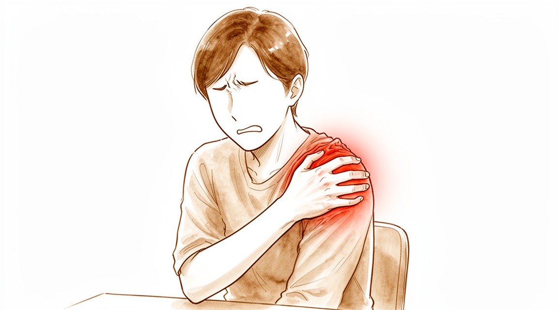
நீங்கள் உணர்வது
சுழற்சி தசைநாண் பட்டை (rotator cuff) வலி பொதுவாக உங்கள் தோள்பட்டையின் முன்பகுதியிலோ வெளிப்பக்கத்திலோ இருக்கும். அது உங்கள் கையின் நடுப்பகுதியை நோக்கிக் கீழே பரவலாம். உங்கள் கையைத் தலைக்கு மேல் உயர்த்துவது, ஓர் அலமாரியை எட்டுவது, அல்லது கையை உடலிலிருந்து விலக்கி நீட்டியபடி எதையாவது தூக்குவது வலியை அதிகரிக்கும் போக்கு உள்ளது. பலர் இரவில்தான் வலியை அதிகம் உணர்கிறார்கள்; அது உங்களைத் தூக்கத்திலிருந்து எழுப்பலாம், அல்லது அந்தப் பக்கமாகப் படுத்துத் தூங்குவதைக் கடினமாக்கலாம். சிலர் காலையில் எழுந்தவுடனும் அதை உணர்கிறார்கள்.
வலி பெரும்பாலும், நீங்கள் சுட்டிக்காட்டக்கூடிய எந்தக் காயமும் இல்லாமல், மெதுவாகத் தொடங்குகிறது — குறிப்பாக, தலைக்கு மேல் கையை உயர்த்தும் வேலையையோ விளையாட்டையோ நீங்கள் அதிகம் செய்பவராக இருந்தால். வேறு சிலருக்கு, கீழே விழுந்த பிறகோ தோள்பட்டையில் திடீர் அதிகப் பளு (strain) ஏற்பட்ட பிறகோ, உடனடி வலியுடனும் பலவீனத்துடனும் அது திடீரென வருகிறது. பலவீனம் இந்தப் பிரச்சினையின் ஒரு முக்கியப் பகுதி. உங்கள் கையைப் பக்கவாட்டில் தூக்குவதோ, அதை உயர்த்தியபடி வைத்திருப்பதோ, அல்லது கையை வெளிப்புறமாகத் திருப்புவதோ உங்களுக்குச் சிரமமாக இருக்கலாம். துவைத்த துணிகளைக் காயப்போடுவது, உங்களுக்குப் பின்னால் கையை நீட்டுவது, அல்லது கெட்டிலைத் தூக்குவது போன்ற அன்றாட வேலைகள் கடினமாகவோ வலி தருவனவாகவோ ஆகலாம்.
உங்கள் அறிகுறிகளின் தன்மை ஆளுக்கு ஆள் மாறுபடலாம். சிலருக்குத் தலைக்கு மேல் கையை உயர்த்தும் அசைவின்போது லேசானது முதல் மிதமானது வரையிலான வலியும், எப்போதாவது மட்டும் இரவு வலியும் இருக்கும். வேறு சிலருக்குத் தொடர்ச்சியான வலி, இரவு வலி, பலவீனம், விறைப்பு ஆகிய அனைத்தும் ஒன்றாக இருக்கும். பிரச்சினை நீண்ட காலமாக இருந்தால், தோள்பட்டை எலும்பைச் சுற்றியுள்ள தசைகள் சற்று மெலிந்துபோகலாம்; மூட்டு விறைப்பாக உணரலாம், அல்லது அசையும்போது நறநறக்கலாம்.
உங்கள் அறிகுறிகள் தணியாவிட்டால், வாரக்கணக்கில் மோசமாகிக்கொண்டே போனால், இரவில் உங்களைத் தூக்கத்திலிருந்து எழுப்பினால், அல்லது நீங்கள் வேலை செய்வதையோ உங்கள் கையைப் பயன்படுத்துவதையோ தடுத்தால், உங்கள் குடும்ப மருத்துவரைப் (GP) பாருங்கள், அல்லது நிபுணரின் பரிசோதனையைக் கேளுங்கள்.
உண்மையில் என்ன நடக்கிறது
உங்கள் சுழற்சி தசைநாண் பட்டை என்பது நான்கு தசைநாண்களால் (tendons) ஆன ஒரு படலம்; அவை தோள்பட்டை மூட்டைச் சுற்றி வளைந்து, உங்கள் மேற்கை எலும்பின் மேல்முனையுடன் இணைகின்றன. ஒரு கூடாரத்தை நிமிர்த்திப் பிடித்திருக்கும் இழுகயிறுகளைப் (guy ropes) போல அவை ஒன்றாகச் செயல்படுகின்றன. ஒவ்வொரு முறை நீங்கள் கையைத் தூக்கும்போதும், இந்தத் தசைநாண்கள் மூட்டின் பந்தை அதன் குழிக்குள் உறுதியாக இழுத்துப் பிடிக்கின்றன; அதனால் கை தள்ளாடாமல் சீராக அசைகிறது.
பிரச்சினை பொதுவாகத் தசைநாண்களுக்கு மேலே உள்ள இடைவெளியில் தொடங்குகிறது. ஒரு சிறிய எலும்புத் திட்டும் ஒரு தசைநாரும் (ligament) தோள்பட்டையின் மேல் ஒரு வளைவை உருவாக்குகின்றன; அந்த வளைவுக்கும் தசைநாண்களுக்கும் இடையே, அவை சறுக்கி நகர உதவும் மெல்லிய திரவப் பை ஒன்று அமைந்துள்ளது. தலைக்கு மேல் கையைத் திரும்பத் திரும்ப உயர்த்துவதாலோ, தசைநாண் வயதாவதாலோ, இந்த வளைவுக்குக் கீழே தசைநாண்கள் நெருக்கப்பட்டு உரசப்படலாம். கரடுமுரடான விளிம்பில் உரசித் தேயும் கயிற்றைப் போல, தசைநாண் இழைகள் நைந்துபோகின்றன. காலப்போக்கில் இந்த நைவு ஒரு பகுதிக் கிழிவாகவும், பின்னர் தசைநாணை முழுவதுமாக ஊடுருவிச் செல்லும் கிழிவாகவும் மாறலாம்.
தசைநாண்கள் மேற்கை எலும்புடன் இணையும் இடத்துக்கு அருகே, இரத்த ஓட்டம் குறைவாக உள்ள ஒரு பகுதியும் அவற்றில் உள்ளது. குறைந்த இரத்த ஓட்டம் கொண்ட திசு மெதுவாகவே குணமடைகிறது; இந்தக் கிழிவுகள் பெரும்பாலும் தாமாகவே சரியாகாமல் இருப்பதற்கு இதுவே காரணம். ஒரு கிழிவு பெரிதாகும்போது, அதனுடன் இணைந்த தசை தேய்ந்து கொழுப்பால் நிரம்பலாம்; ஒரு கிழிவு எவ்வளவு காலம் அப்படியே விடப்படுகிறதோ, அதைச் சரிசெய்வது அவ்வளவு கடினமாகிறது.
இந்தப் பிரச்சினை ஒரு தொடர்வரிசையில் அமைந்துள்ளது. அது வளைவுக்குக் கீழே ஏற்படும் எரிச்சலுடனும் உரசலுடனும் தொடங்கி, பகுதிக் கிழிவுகள், பின்னர் முழுக் கிழிவுகள் வழியாக முன்னேறுகிறது; நீண்ட காலமாக இருக்கும் நிலைகளில், பந்தைக் கீழே பிடித்து வைக்க எதுவும் இல்லாததால் அது மேல்நோக்கி நகர்ந்து, மூட்டைத் தேய்த்துவிடுவதில் முடிகிறது. கிழிவுகள் அவற்றின் அளவைப் பொறுத்து வகைப்படுத்தப்படுகின்றன: சிறியது (1 செ.மீ.-க்குக் குறைவு) முதல் மிகப் பெரியது (5 செ.மீ.-ஐ விடப் பெரியது, அல்லது இரண்டு தசைநாண்களைப் பாதிப்பது) வரை. பெரிய கிழிவுகள் அதிக பலவீனத்தை ஏற்படுத்தும் போக்கு உடையவை; அவற்றுக்கு அறுவை சிகிச்சை தேவைப்படும் வாய்ப்பும் அதிகம்.
சில சமயங்களில், இதற்குப் பதிலாகத் தசைநாணுக்குள் சுண்ணாம்புப் படிகங்கள் (calcium crystals) உருவாகின்றன. இது சிறிது காலத்துக்குக் கடுமையான வலியைத் தரலாம்; ஆனால் பொதுவாகத் தானாகவே தணிந்துவிடும்.
இதற்கு நாங்கள் என்ன செய்ய முடியும்
Mater Private Hospital Rockhampton-இல் பணியாற்றும் மேல் அவயவ (upper-limb) அறுவை சிகிச்சை நிபுணரான டாக்டர் கியரன் ஹிர்பரா, உங்கள் நிலைக்குப் பொருத்தமான, உடலில் மிகக் குறைவாகத் தலையிடும் சிகிச்சைகளிலிருந்தே தொடங்குகிறார். நோயாளிகள் பொதுவாக அவர்களின் குடும்ப மருத்துவரால் (GP) எங்கள் கிளினிக்குக்கு அனுப்பப்படுகிறார்கள்; ஓர் இயன்முறை சிகிச்சையாளர் எங்களைப் பார்க்கச் சொல்லியிருந்தாலும், Medicare திருப்பிச் செலுத்தலுக்குத் தகுதி பெற உங்கள் குடும்ப மருத்துவரிடமிருந்து பரிந்துரைக் கடிதம் உங்களுக்குத் தேவைப்படும். உங்கள் சந்திப்பின்போது, நாங்கள் உங்கள் நோய் வரலாற்றைக் கேட்டறிந்து, உங்கள் தோள்பட்டையைப் பரிசோதித்து, தேவைப்படும் இடத்தில் ஸ்கேன்களுக்கு ஏற்பாடு செய்கிறோம். பின்னர் உங்களுடன் இணைந்து ஒவ்வொரு தேர்வையும் பரிசீலிக்கிறோம்.
முதல் படி பெரும்பாலும், உங்கள் தோள்பட்டையை நீங்கள் பயன்படுத்தும் விதத்தை மாற்றி, தசைநாண் தணிய அவகாசம் தருவது. அதாவது, வலியை அதிகரிக்கும் அசைவுகளிலிருந்து ஓய்வு எடுத்து, சிறிது காலத்துக்கு வேலையையோ விளையாட்டையோ மாற்றியமைப்பது. அறுவை சிகிச்சை அல்லாத கவனிப்பின் முதன்மை அம்சம் இயன்முறை சிகிச்சையே. தோள்பட்டையில் அசைவை மீட்டெடுப்பது, உங்கள் தோள்பட்டை எலும்பை நிலைப்படுத்தும் தசைகளை வலுப்படுத்துவது, சுழற்சி தசைநாண் பட்டையையே வலுப்படுத்துவது ஆகியவை அதன் நோக்கம். 6 முதல் 12 வாரச் சிகிச்சை ஒரு நியாயமான முயற்சிக் காலம். முழு-தடிமன் கிழிவு (full-thickness tear) உள்ள பலருக்கு இந்த அணுகுமுறையே போதுமானது: அறுவை சிகிச்சை அல்லாத சிகிச்சை 60% நோயாளிகளில் வெற்றி பெறுகிறது; 2 ஆண்டுகளுக்குப் பிறகு இது 75%. பெரும்பாலான பகுதி-தடிமன் கிழிவுகளும் அறுவை சிகிச்சை இல்லாமலே தணிந்துவிடுகின்றன.
வலி நிவாரணிகளும் அழற்சி எதிர்ப்பு மாத்திரைகளும் (NSAIDs), தசைநாண் தணியும்போது நீங்கள் சுறுசுறுப்பாக இருக்க உதவலாம். தசைநாணுக்கு மேலே உள்ள இடைவெளியில் போடப்படும் கார்டிசோன் (cortisone) ஊசி வலியைத் தணிக்கலாம்; இருப்பினும் சுழற்சி தசைநாண் பட்டைப் பிரச்சினைகளில் அதற்கான சான்றுகள் குறைவாகவே உள்ளன; பின்னர் அறுவை சிகிச்சை தேவைப்படக்கூடும் என்றால், அதை நாங்கள் கவனமாக எடைபோடுகிறோம். PRP (உங்கள் சொந்த இரத்தத்திலிருந்து தயாரிக்கப்படும் ஒரு கலவை) ஊசிகள், நீண்ட காலமாக இருக்கும் சுழற்சி தசைநாண் பட்டை நோய்க்குக் குறுகிய காலத்தில் தெளிவான பயனைக் காட்டவில்லை. தசைநாணுக்குள் சுண்ணாம்புப் படிகங்கள் உருவாகும் சுண்ணாம்புத் தசைநாண் அழற்சிக்கு (calcific tendinitis) இன்னும் அதிகமான தேர்வுகள் உள்ளன: இயன்முறை சிகிச்சையுடன் ஸ்டீராய்டு ஊசி, அதிர்வலை சிகிச்சை (shockwave therapy — சுண்ணாம்பை நோக்கி ஒலி அலைகளைச் செலுத்துவது), அல்ட்ராசவுண்ட் வழிகாட்டலுடன் படிவை ஊசியால் கழுவி வெளியேற்றுவது, அல்லது துளை அறுவை சிகிச்சை மூலம் தசைநாணைக் கழுவிச் சுத்தப்படுத்துவது.
அறுவை சிகிச்சை அல்லாத கவனிப்பு போதிய முன்னேற்றத்தைத் தராதபோது, அல்லது கிழிவு திடீரென ஏற்பட்ட முழு-தடிமன் கிழிவாக இருக்கும்போது, அறுவை சிகிச்சை பரிசீலனைக்கு வருகிறது. வழக்கமான அறுவை சிகிச்சை, துளை வழி (arthroscopic) சரிசெய்தல்; இதில், சிறிய கீறல்கள் வழியாக உள்ளே செலுத்தப்படும் கேமராவின் உதவியுடன், தசைநாண் மீண்டும் எலும்புடன் தைத்து இணைக்கப்படுகிறது. சில கிழிவுகளுக்குச் சரிசெய்தலே சிறந்தது; வேறு சிலவற்றுக்குத் தசைநாணைச் சீர்செய்து சுத்தப்படுத்துவதே (debridement) சிறந்தது; பகுதிக் கிழிவுகளில், தசைநாணின் அகலத்தில் 50%-க்கு மேல் பாதிக்கும் கிழிவுகள் பொதுவாகச் சரிசெய்யப்படுகின்றன, அதைவிடச் சிறியவை சுத்தப்படுத்தப்படுகின்றன. உங்கள் ஸ்கேனும் உடல் பரிசோதனையும் என்ன காட்டுகின்றன என்பதைப் பற்றி உங்களுடன் பேசி, அறுவை சிகிச்சையே உங்களுக்குச் சரியான அடுத்த படியா என்பதை நாம் இணைந்து முடிவு செய்வோம்.
என்ன எதிர்பார்க்கலாம்
சுழற்சி தசைநாண் பட்டை வலி வந்து வந்து போகும் போக்கு உடையது. அது பெரும்பாலும் சிறிது காலம் தணிந்து, பின்னர் மீண்டும் அதிகரிக்கிறது; அடியில் உள்ள தேய்மானம் காலப்போக்கில் மெதுவாகத் தொடர்ந்து முன்னேறுகிறது. நீண்ட காலமாக இருந்து, சிகிச்சையின்றி விடப்படும் கிழிவில், இறுதியில் மூட்டே தேய்ந்துபோகலாம். பெரும்பாலான சுழற்சி தசைநாண் பட்டைப் பிரச்சினைகள் அறுவை சிகிச்சை இல்லாத சிகிச்சைக்கு நன்றாகப் பலன் தருகின்றன; முழு-தடிமன் கிழிவுகளுக்கு அது எவ்வளவு அடிக்கடி பலனளிக்கிறது என்பதை நீங்கள் ஏற்கெனவே படித்திருக்கிறீர்கள்.
அறுவை சிகிச்சை அல்லாத வழியில் நீங்கள் சென்றால், முன்னேற்றம் பொதுவாக முதல் ஆண்டில் படிப்படியாக வளர்ந்து, பின்னர் ஒரே நிலையில் நிற்கிறது. தலைக்கு மேல் கையை நீட்டுவது, அல்லது மீண்டும் அந்தப் பக்கமாகப் படுத்துத் தூங்குவது போன்றவற்றைக் குறைந்த வலியுடன் அதிகம் செய்ய முடிவதாகவே பலர் இந்த மாற்றத்தை உணர்கிறார்கள். இயன்முறை சிகிச்சை உதவும் என்ற உங்கள் சொந்த நம்பிக்கை, அது உண்மையில் உதவுமா என்பதைக் கணிக்கும் வலுவான காரணிகளில் ஒன்று. வலியோ, எவ்வளவு காலமாக அறிகுறிகள் இருக்கின்றன என்பதோ, கிழிவு எவ்வளவு கடுமையானது என்பதை எங்களுக்குக் காட்டுவதில்லை; எனவே வலிக்கும் தோள்பட்டை எப்போதும் பெரிய கிழிவைக் குறிப்பதில்லை, வலிக்காத தோள்பட்டை எப்போதும் சிறிய கிழிவைக் குறிப்பதுமில்லை.
அறிகுறிகள் இல்லாமலும் கிழிவுகள் பெரிதாகலாம். அறிகுறிகளை ஏற்படுத்தும் கிழிவுகளில், 53% குறைந்தது 6 மாதக் காலத்தில் பெரிதாகின்றன; குறைந்தது 18 மாதங்கள் பின்தொடரப்பட்டவற்றில் 48% பெரிதாகின்றன. 60 வயதுக்குப் பிறகு கிழிவு பெரிதாகும் வாய்ப்பு அதிகம்: அது 54% கிழிவுகளில் நிகழ்கிறது; இளையவர்களில் இது 17%. வலியே தராத கிழிவுகள்கூடச் சுமார் பாதி நிலைகளில் பெரிதாகின்றன; இது பொதுவாகச் சுமார் 2.8 ஆண்டுகளுக்குள் நிகழ்கிறது. பெரிதாகும் கிழிவைப் பின்னர் சரிசெய்வது கடினமாகலாம்; வெறுமனே காத்திருப்பதற்குப் பதிலாக நாங்கள் தொடர்ந்து கண்காணிப்பதற்கு இதுவும் ஒரு காரணம்.
அறுவை சிகிச்சையே சரியான வழி என்றால், சரிசெய்தலுக்குப் பிறகு முதல் 6 மாதங்களில் பெரும்பாலானோர் சீராக மேம்படுகிறார்கள்; அதற்குப் பிறகும் மேலும் முன்னேற்றம் தொடர்கிறது. சரிசெய்யப்பட்ட தசைநாண் அதன் குணமடைதலில் பெரும்பகுதியை அந்த முதல் 6 மாதங்களிலேயே முடிக்கிறது; சரிசெய்தல் மிக அதிக அபாயத்தில் இருக்கும் காலமும் அதுவே. சுமார் 20% சரிசெய்தல்கள் தோல்வியடைந்து, தசைநாண் மீண்டும் கிழிகிறது; பெரிய கிழிவுகளில் இதற்கான வாய்ப்பு அதிகம். வயது அதிகமாக இருந்தால், புகைப்பழக்கம், நீரிழிவு நோய், எலும்புப்புரை (osteoporosis — மெலிந்த எலும்புகள்), அதிக கொலஸ்ட்ரால் இருந்தால், அல்லது கிழிவு பெரியதாகவும் அதிக தூரம் பின்வாங்கியதாகவும் இருந்தால், குணமடையும் வாய்ப்பு குறைவு.
மூட்டில் ஏற்கெனவே மூட்டழற்சி (arthritis) உள்ள, மிகப் பெரிய, நீண்ட காலக் கிழிவுகளுக்கு, சிகிச்சை முக்கியமாக வலி நிவாரணத்தையே நோக்கமாகக் கொண்டுள்ளது; சிறிய கிழிவுகளுடன் ஒப்பிடும்போது முடிவுகள் பொதுவாக அவ்வளவு சிறப்பாக இருப்பதில்லை.
எப்போது மருத்துவரைப் பார்க்க வேண்டும்
இங்கு விவரிக்கப்படும் பெரும்பாலான தோள்பட்டை வலி ஓர் இயந்திரவியல் (mechanical) பிரச்சினை; எனவே எச்சரிக்கை அறிகுறிகள், வலி எப்படி நடந்துகொள்கிறது, உங்கள் கையால் இன்னும் என்ன செய்ய முடிகிறது என்பவற்றைப் பற்றியவை. உங்கள் அறிகுறிகள் தணியாவிட்டால், வாரக்கணக்கில் மோசமாகிக்கொண்டே போனால், இரவில் உங்களைத் தூக்கத்திலிருந்து எழுப்பினால், அல்லது நீங்கள் வேலை செய்வதையோ உங்கள் கையைப் பயன்படுத்துவதையோ தடுத்தால், உங்கள் குடும்ப மருத்துவரைப் பாருங்கள். உண்மையான பலவீனத்தை நீங்கள் கவனித்தால் — எடுத்துக்காட்டாக, உங்கள் கையைப் பக்கவாட்டில் தூக்கவோ உயர்த்தியபடி வைத்திருக்கவோ முடியாவிட்டால், குறிப்பாகக் கீழே விழுந்த பிறகோ திடீர் அதிகப் பளு ஏற்பட்ட பிறகோ — நிபுணரின் பரிசோதனையை விரைவில் கேளுங்கள். 65 வயதுக்குப் பிறகு இரவு வலியும் பலவீனமும் சேர்ந்து இருப்பது, சுழற்சி தசைநாண் பட்டையில் கிழிவு இருப்பதை வலுவாகச் சுட்டுகிறது; எனவே இரண்டையும் சேர்த்துக் குறிப்பிடுங்கள். தோள்பட்டையின் முன்பகுதியில் உள்ள வலியையும், மாதக்கணக்கில் தலைக்கு மேல் கையை உயர்த்தும் வேலை செய்த பிறகு எந்தக் காயமும் இல்லாமல் தொடங்கிய வலியையும் பரிசோதித்துக்கொள்வது நல்லது. கிழிவு இருப்பதாகச் சந்தேகிக்கப்பட்டு, ஆனால் நிலைமை தெளிவாக இல்லாவிட்டால், தசைநாண் பகுதியாகக் கிழிந்துள்ளதா முழுமையாகக் கிழிந்துள்ளதா என்பதைக் கண்டறிய, உங்கள் குடும்ப மருத்துவரோ நிபுணரோ மருத்துவ அறையிலேயே செய்யக்கூடிய எளிய சோதனைகளையும், சில சமயங்களில் தசைநாணுக்கு மேலே உள்ள இடைவெளியில் போடப்படும் மரத்துப்போகச் செய்யும் ஊசியையும் பயன்படுத்தலாம்.
ஆழமான விளக்கம்
Advanced reading: the deeper science (optional)
உங்கள் சொந்த சிகிச்சை முடிவுகளுக்குத் தேவையானதை விட இந்தப் பகுதி மேலும் ஆழமாகச் செல்கிறது. சுழற்சி தசைநாண் பட்டைக் கோளாறுகளைப் பற்றிக் கூடுதலாகப் படிப்பது பயனுள்ளது; ஏனெனில் ஒவ்வொரு ஸ்கேன் அறிக்கையையும் புதிய கண்ணோட்டத்தில் பார்க்க வைக்கும் ஒரு தொற்றுநோயியல் உண்மை உள்ளது: அறிகுறிகளே இல்லாதவர்களிடையே சுழற்சி தசைநாண் பட்டைக் கிழிவுகள் மிகவும் பொதுவானவை; எனவே ஒரு கிழிவைக் கண்டறிவது, அதுவே உங்கள் வலிக்குக் காரணம் என்பதை நிறுவிவிடாது.
வயதாகும்போது கிழிந்த சுழற்சி தசைநாண் பட்டை ஓர் இயல்பான கண்டுபிடிப்பு
6,112 தோள்பட்டைகளை ஒன்றிணைத்துப் பார்த்தபோது, அறிகுறிகளே இல்லாதவர்களிடையே சுழற்சி தசைநாண் பட்டைக் கோளாறுகள் இருக்கும் விகிதம், சுழற்சி தசைநாண் பட்டையின் சிதைவு இயல்பான மனித வயதாகுதலின் ஒரு பொதுவான அம்சமாகவே கருதப்பட வேண்டும் என்னும் அளவுக்கு அதிகமாக உள்ளது; மேலும், ஒரு கோளாறு எப்போது புதிதாக ஏற்பட்டது, அல்லது அதுவே அறிகுறிகளுக்குக் காரணமா என்பதைத் தீர்மானிப்பது கடினம் என்னும் அளவுக்கும் அது அதிகம் [1].
ஒரு முறையான மதிப்பாய்விலிருந்து வரும், வழக்கத்துக்கு மாறான நேரடியான கூற்று இது; ஸ்கேன் அறிக்கையை எப்படிப் படிக்க வேண்டும் என்பதை இது மாற்ற வேண்டும். உங்கள் MRI-யில் கண்டறியப்பட்ட ஒரு கிழிவு, தானாகவே உங்கள் வலிக்கான விளக்கம் ஆகிவிடாது; தோள்பட்டைப் பிரச்சினையே இல்லாத, உங்கள் வயதையொத்த பெரும்பாலானவர்களிடம் இருப்பதைப் போலவே, அது பல ஆண்டுகளாக அறிகுறியின்றி அங்கேயே இருந்திருக்கலாம்.
இதன் மருத்துவ விளைவு: நோய் கண்டறிதல் ஸ்கேனை மட்டுமே சார்ந்தது அல்ல; உடல் பரிசோதனையும் நோய் வரலாறும் ஸ்கேனுடன் பொருந்துகின்றனவா என்பதையே சார்ந்தது. நோயாளிக்குப் பதிலாக ஸ்கேனுக்குச் சிகிச்சை அளிப்பது இங்கே ஓர் அறியப்பட்ட ஆபத்தான பொறியாகக் கருதப்படுவதற்கும் இதுவே காரணம்.
அறுவை சிகிச்சை உதவுகிறது; அந்தப் பலனின் அளவே நேர்மையான பகுதி
சிதைவால் ஏற்படும் முழு-தடிமன் கிழிவுகளில், மாற்று வழிகளை விட அறுவை சிகிச்சையே சிறந்த பலனைத் தருகிறது. 677 நோயாளிகளைக் கொண்ட ஆய்வில், வயதானவர்களிடையே பழமைவாதச் சிகிச்சையுடனோ, சப்அக்ரோமியல் டிகம்ப்ரஷன் (subacromial decompression) மட்டும் செய்வதுடனோ ஒப்பிடும்போது, அறுவை சிகிச்சை மூலம் சரிசெய்வது கணிசமாக மேம்பட்ட விளைவுகளைத் தந்தது; அறுவை சிகிச்சைக்கும் பழமைவாதச் சிகிச்சைக்கும் இடையிலான வேறுபாட்டின் அளவு கவனத்துக்கு உரியது என்றும் ஆசிரியர்கள் சேர்த்துக் குறிப்பிட்டனர் [2]. 269 நோயாளிகளைக் கொண்ட தனி ஒப்பீடு ஒன்று, ஓராண்டில் Constant மதிப்பெண்ணிலும் வலி மதிப்பெண்ணிலும் அறுவை சிகிச்சைக்குச் சாதகமான, புள்ளியியல் ரீதியாக முக்கியத்துவம் வாய்ந்த வேறுபாடுகளைக் கண்டறிந்தது [3].
எனவே சரிசெய்யும் அறுவை சிகிச்சையே வெல்கிறது; ஆனால் பெரிய ஆய்வின் ஆசிரியர்கள் வெறும் திசையை மட்டுமல்ல, விளைவின் அளவையும் கவனமாகச் சுட்டிக்காட்டுகிறார்கள். ஒரு வேறுபாடு புள்ளியியல் ரீதியாக உண்மையானதாக இருந்தாலும், நோயாளி உணரக்கூடிய அளவை விடச் சிறியதாக இருக்கலாம். அதனால்தான் இந்தத் தேர்வு வெளிப்படையானது அல்ல, உண்மையான ஒரு தேர்வாகவே இருக்கிறது — குறிப்பாக, கையிலிருந்து குறைவான தேவைகளையே எதிர்பார்ப்பவருக்கு, அல்லது கிழிவைச் சரிசெய்ய முடியாமல் போகக்கூடியவருக்கு.
பயிற்சி என்பது ஒரே சிகிச்சை அல்ல; அதன் வகையும் முக்கியம் போலத் தெரிகிறது
அறுவை சிகிச்சை அல்லாத கவனிப்பு பொதுவாக வெறுமனே "இயன்முறை சிகிச்சை" என்றே விவரிக்கப்படுகிறது; இது ஓர் உண்மையான கண்டுபிடிப்பை மறைத்துவிடுகிறது. 947 நோயாளிகளிடையே ஏழு வகைப் பயிற்சிகளை ஒப்பிட்டபோது, தோள்பட்டை வலிக்கும் செயலிழப்புக்கும் சுருங்கும் தசை வலிமைப் பயிற்சியே (concentric strengthening) சிறந்த பலனைத் தந்தது; சுருங்கும் வகைப் பயிற்சி பொருத்தமில்லாத இடத்தில் நீளும் தசை வலிமைப் பயிற்சியும் (eccentric strengthening) இயக்கக் கட்டுப்பாட்டுப் பயிற்சியும் (motor control exercise) மாற்று வழிகளாகும் [4].
இதைத் தெரிந்துகொள்வது பயனுள்ளது; ஏனெனில் "நான் இயன்முறை சிகிச்சையை முயற்சித்தேன், அது உதவவில்லை" என்பதை இது ஒரு முடிவாக அல்லாமல் ஒரு கேள்வியாக மாற்றுகிறது — உண்மையில் என்ன செய்யப்பட்டது, எவ்வளவு காலத்துக்கு, அது படிப்படியாக அதிகரிக்கும் வலிமைப் பயிற்சியா, அல்லது நீட்டிப் பயிற்சிகள் அச்சிடப்பட்ட ஒரு தாளா.
சேர்த்துச் செய்யப்படும், ஆனால் சேர்க்க வேண்டியதில்லாத அறுவை சிகிச்சை
அக்ரோமியோபிளாஸ்டி (acromioplasty) — அதாவது அக்ரோமியன் எலும்பின் கீழ்ப்பரப்பிலிருந்து எலும்பைச் சீவி எடுப்பது — சரிசெய்யப்பட்ட தசைநாணின் மீது அழுத்தம் ஏற்படுத்தும் ஒரு காரணியை அது நீக்குகிறது என்னும் காரணத்தால், சுழற்சி தசைநாண் பட்டையைச் சரிசெய்யும் அறுவை சிகிச்சையுடன் சேர்த்து அடிக்கடி செய்யப்படுகிறது.
இந்தச் சேர்க்கைக்குச் சான்றுகள் ஆதரவு தரவில்லை. 373 நோயாளிகளைக் கொண்ட ஆய்வில், நடுத்தரக் கால பின்தொடர் பரிசோதனையில், அக்ரோமியோபிளாஸ்டியுடன் செய்யப்பட்ட, அல்லது அது இல்லாமல் செய்யப்பட்ட, துளை வழிச் சுழற்சி தசைநாண் பட்டை சரிசெய்தலுக்குப் பிறகு, நோயாளி உணரும் விளைவில் புள்ளியியல் ரீதியாக முக்கியத்துவம் வாய்ந்த வேறுபாடு எதுவும் இல்லை [5].
உங்கள் சரிசெய்தலுடன் இதைச் சேர்த்துச் செய்ய முன்மொழியப்பட்டால், அதைப் பற்றிக் கேட்பது நியாயமானது.
மேற்கோள்கள்
[1] Teunis T, Lubberts B, Reilly BT, Ring D. A systematic review and pooled analysis of the prevalence of rotator cuff disease with increasing age. J Shoulder Elbow Surg. 2014;23(12):1913-21. https://doi.org/10.1016/j.jse.2014.08.001
[2] Schemitsch C, Chahal J, Vicente M, Nowak L, Flurin P, Lambers Heerspink F, et al. Surgical repair versus conservative treatment and subacromial decompression for the treatment of rotator cuff tears: a meta-analysis of randomized trials. Bone Joint J. 2019;101-B(9):1100-6. https://doi.org/10.1302/0301-620X.101B9.BJJ-2018-1591.R1
[3] Piper CC, Hughes AJ, Ma Y, Wang H, Neviaser AS. Operative versus nonoperative treatment for the management of full-thickness rotator cuff tears: a systematic review and meta-analysis. J Shoulder Elbow Surg. 2018;27(3):572-6. https://doi.org/10.1016/j.jse.2017.09.032
[4] Zhang W, Du M, Xia L, Hao F, Tian M. Effects of seven types of exercise in the treatment of rotator cuff-related shoulder pain: a network meta-analysis. J Orthop Surg Res. 2025;20(1). https://doi.org/10.1186/s13018-025-06514-4
[5] Chahal J, Mall N, MacDonald PB, Van Thiel G, Cole BJ, Romeo AA, et al. The role of subacromial decompression in patients undergoing arthroscopic repair of full-thickness tears of the rotator cuff: a systematic review and meta-analysis. Arthroscopy. 2012;28(5):720-7. https://doi.org/10.1016/j.arthro.2011.11.022
Evidence & references
This is the clinical evidence summary written for health professionals. It is technical, and it lists the research this page was built from. You do not need to read it to understand your treatment or to make a decision about it.
Overview
- The majority of rotator cuff conditions are amenable to conservative treatment, although rotator cuff dysfunction may necessitate surgical treatment [1].
- Clinical decision-making for the management of rotator cuff tears is complex and lacks consensus among orthopedic surgeons [2].
- Neither the American Academy of Orthopaedic Surgeons clinical practice guidelines nor Cochrane systematic reviews provide guidance on the management of rotator cuff tears [2].
- Patients with rotator cuff tears can be divided into three main categories based on the potential risk of nonoperative treatments and the proposed benefits of surgical intervention: those needing urgent or early operative repair, those that can benefit from a trial of conservative treatment, and those best suited for nonoperative treatment [2].
- Current consensus suggests rotator cuff injuries are most accurately diagnosed with a combination of cuff- and impingement-specific clinical tests [3].
- Increasing knowledge about rotator cuff syndrome, including better imaging, has facilitated patient treatment for a stable spectrum of rotator cuff pathology [4].
- The application of endoscopic surgery has facilitated patient treatment for a stable spectrum of rotator cuff pathology [4].
- The major indication for revision rotator cuff repair is the persistence of clinical symptoms despite nonsurgical management in the absence of substantial risk factors for failure [5].
- Deltoid complications combined with rotator cuff pathology represent a rare but devastating complication with no well-described surgical option [11].
- Early operative treatment appears to be better for rotator cuff tears with a sudden onset of symptoms and poor function to achieve maximal return of shoulder function [15].
- Open approaches for rotator cuff repairs continue to have indications in certain circumstances, such as after high-velocity trauma [16].
- Arthroscopic surgeons who treat rotator cuff disorders can use information regarding surgical techniques irrespective of their preferred surgical technique [17].
- Severely impaired deltoid function is a contraindication to reverse total shoulder arthroplasty (RTSA) in rotator cuff disease [30].
- An isolated supraspinatus tear is a contraindication to reverse total shoulder arthroplasty (RTSA) in rotator cuff disease [30].
- The presence of full active shoulder elevation with a massive rotator cuff tear and arthritis is a contraindication to reverse total shoulder arthroplasty (RTSA) [30].
- There is no statistically significant difference in subjective outcome after arthroscopic rotator cuff repair with or without acromioplasty at intermediate follow-up [31].
- There is little reproducible evidence to support the efficacy of subacromial corticosteroid injection in managing rotator cuff disease [38].
- Treatment of patients with chronic massive rotator cuff tears is challenging, and results are comparatively inferior to those of treating patients with smaller rotator cuff tears [44].
- Patients undergoing operative treatment for rotator cuff tears had significantly better pain and functional outcomes as compared with patients undergoing nonoperative treatment [48].
- Shoulder arthroscopy literature remains controversial, with conclusions often unsupported due to bias and limitations [51].
- No clinical guidelines for shoulder arthroscopy are definitive pending higher levels of evidence [51].
- No recommendations regarding suprascapular nerve release in conjunction with rotator cuff repair can be made at this time [54].
- Further research is necessary to better delineate the indications for suprascapular nerve release in conjunction with rotator cuff repair [54].
- Following nonoperative treatment for at least 6 weeks, subacromial decompression (SAD) is a viable and good surgical option for the treatment of shoulder impingement with an intact rotator cuff [75].
- Critical Shoulder Angle (CSA) and Acromial Index (AI) do not appear to influence 24-month functional outcomes postoperatively after arthroscopic rotator cuff repair [81].
- Critical Shoulder Angle (CSA) and Acromial Index (AI) are not contraindications to arthroscopic rotator cuff repair [81].
- Arthroscopy is a safe and effective treatment for symptomatic calcific tendonitis of the shoulder, excluding or including patients with rotator cuff tears [82].
- Patients with intact rotator cuffs and symptomatic calcific tendonitis could benefit from needling or extracorporeal shockwave therapy (ESWT) in some cases [82].
- Routine arthroscopic suprascapular nerve release (SSNR) is not recommended when treating patients with rotator cuff tear [83].
- Data on predictors of pain and functional outcomes can be used to select optimal candidates for operative treatment of rotator cuff tears [88].
- Data on predictors of pain and functional outcomes can assist with patient education and expectations before treatment for rotator cuff tears [88].
- There were no differences of clinically relevant size between arthroscopic and open rotator cuff surgery in postoperative pain [160].
- Augmentation strategies may improve outcomes in rotator cuff repairs, particularly in high-risk cases [172].
- There is a lack of consensus among surgeons on the most effective augmentation strategies for each scenario in rotator cuff repair [172].
Anatomy & Pathophysiology
Anatomy
- The rotator cuff is a sheet of conjoined tendons closely applied over the shoulder capsule, inserting mainly into the greater tuberosity of the humerus, with the subscapularis inserting into the lesser tuberosity [19].
- The rotator cuff consists of the subscapularis anteriorly, supraspinatus superiorly, and infraspinatus and teres minor posteriorly [19].
- The rotator cuff muscles stabilize the humeral head by pulling it firmly into the glenoid whenever the deltoid lifts the arm forwards or sideways [19].
- The coracoacromial arch is a fibro-osseous canopy formed by the acromion process posterosuperiorly, the coracoid process anteriorly, and the coracoacromial ligament joining them [19].
- The subacromial bursa separates the rotator cuff tendons from the coracoacromial arch, allowing them to glide [19].
- The subscapularis arises from the anterior aspect of the scapula and attaches over much of the lesser tuberosity, innervated by the upper and lower subscapular nerves [100].
- The supraspinatus arises from the fossa superior to the scapular spine, passes beneath the acromion and acromioclavicular joint, and attaches to the superior aspect of the greater tuberosity, innervated by the suprascapular nerve after it passes through the suprascapular notch [100].
- The infraspinatus arises from the fossa below the scapular spine and attaches to the posterolateral aspect of the greater tuberosity, innervated by the suprascapular nerve after it passes through the spinoglenoid notch [100].
- The teres minor arises from the lower lateral aspect of the scapula and attaches to the lower portion of the greater tuberosity, innervated by a branch of the axillary nerve [100].
- Histologic studies describe the rotator cuff tendons as having five distinct layers, with the most superficial layer being coracohumeral ligament fibers and the fifth layer being the continued sheet of collagen fibrils composing the superior joint capsule [100].
- The supraspinatus footprint measures 13 mm width medial-lateral and 20 mm anteroposterior, with 7 mm medial-lateral tears corresponding to 50% partial thickness [59].
- The infraspinatus footprint measures 14 mm wide and 20 mm superoinferior [59].
- The rotator cable is a thick bundle of fibers running perpendicular to the supraspinatus tendon fibers, connecting the supraspinatus and infraspinatus tendons [59].
- The rotator cable is a thickened horseshoe-shaped region within the tendinous tissue that attaches on the anterior 8 to 12 mm of the supraspinatus and posteriorly in the infraspinatus insertion [139].
- The rotator crescent is the thinner, stress-shielded area of the supraspinatus/infraspinatus attachment lateral to the cable between its insertion points [139].
- The hypovascular critical zone is located on the articular side of the rotator cuff close to the insertion on the greater tuberosity [59].
- The long head of the biceps tendon attaches to the supraglenoid tubercle, runs between the subscapularis and supraspinatus, exits through the bicipital groove under the transverse humeral ligament, and attaches to its muscle in the proximal arm [100].
- The coracohumeral ligament and transverse humeral ligament keep the long head of the biceps tendon aligned in the bicipital groove [100].
- The human scapula is characterized by a lateral orientation of the glenoid cavity and a narrow coraco-acromial arch [56].
Pathophysiology
- Rotator cuff disease is a continuum beginning with mild impingement and progressing through partial tear, full-thickness tear, and massive tear to rotator cuff tear arthropathy [85].
- The prevalence of rotator cuff tears increases with age, ranging from 13% in patients in their fifties to 50% in patients aged 80 years or older [18].
- The overall prevalence of full-thickness rotator cuff tears is approximately 25% in patients over 60 years of age and increases to 50% in patients over 80 years of age [7].
- If a patient has a symptomatic full-thickness rotator cuff tear in one shoulder, they have a 50% chance of an asymptomatic full-thickness rotator cuff tear in the contralateral shoulder [7].
- Rotator cuff abnormalities are prevalent in both symptomatic and asymptomatic patients, and bilateral tears are common despite often unilateral symptoms [18].
- Known risk factors for rotator cuff tear development include male sex, manual labor, and history of trauma [18].
- Risk factors for rotator cuff disease include age, smoking, female sex, family history, diabetes, and high cholesterol [59].
- Intrinsic degeneration involves age-related changes in collagen, proteoglycan, water content, and vascularity (tendinosis), usually involving the supraspinatus and infraspinatus starting on the articular side [59].
- Extrinsic factors include chronic impingement on the coracoacromial arch, with tears usually starting on the bursal side of the tendon, potentially associated with a hook-shaped acromion [59].
- Acute traumatic tears typically occur after a fall and/or dislocation of the shoulder in patients under 40 years of age [59].
- Degenerative rotator cuff tears are suggested to initiate approximately 13 to 17 mm posterior to the long head of the biceps tendon, followed by propagation anteriorly and posteriorly [84].
- The area around 15 mm posterior to the biceps tendon is most frequently involved in degenerative rotator cuff tear initiation, correlating with the posterior supraspinatus and anterior infraspinatus [84].
- Tears involving the anterior cable of the rotator cuff have a significant impact on the rate of irreversible muscle degeneration associated with rotator cuff tears [84].
- Loss of integrity of the anterior supraspinatus tissue may lead to accelerated retraction and degeneration of muscle tissue associated with lower rates of tendon healing [84].
- Biomechanical testing shows that tears involving the rotator cable result in increased tear gap distance and strain compared with those in the crescent area [139].
- The critical shoulder angle is formed from lines drawn from the inferior glenoid to the superior glenoid and from the inferior glenoid to the inferolateral acromion [139].
- A critical shoulder angle above 35° to 38° has been shown to correlate with rotator cuff tears and postoperative retears [139].
- A critical shoulder angle less than 30° has been shown to correlate with osteoarthritis [139].
- The acromial index is a ratio of the medial/lateral length from the glenoid to the lateral acromion divided by the length from the glenoid to the lateral aspect of the greater tuberosity [139].
- An acromial index above 0.7 has been associated with full-thickness rotator cuff tears and higher retear rates [139].
- The critical shoulder angle is a static measure that does not change over time [6].
- Patients with a critical shoulder angle greater than 38° and an acromial index greater than 0.7 had higher retear rates but similar functional rates compared to control patients [6].
- A study of 1,552 radiographs determined that the critical shoulder angle was higher in patients with rotator cuff tears but that these differences are small and depend on excellent radiographic technique [6].
- A study of 147 patients up to 2-year follow-up did not see a difference in functional scores between patients with higher critical shoulder angle/acromial index and patients with lower values [6].
- The critical shoulder angle may not be responsible for rotator cuff tears; rather, patient activities throughout several decades could induce both cuff lesions and bone remodeling at the acromial level [90].
- Primary glenohumeral osteoarthritis is associated with significantly smaller critical shoulder angles, while degenerative rotator cuff tears are associated with significantly larger critical shoulder angles compared to asymptomatic shoulders [180].
- The critical shoulder angle, posterior acromial height, and posterior acromial tilt do not change significantly over a long-term follow-up of at least 10 years [152].
- Rotator cuff disease may involve a genetic predisposition to intrinsic degenerative tendinopathic changes in molecular composition and vascularity [139].
- Studies have shown familial hereditary patterns of rotator cuff disease and genes associated with significant pathology [139].
- A genome-wide association study identified two significant single-nucleotide polymorphisms associated with full-thickness rotator cuff tears, lending evidence to a genetic component [14].
- The etiology of anterior shoulder pain with macroscopic changes in the biceps tendon in the context of rotator cuff disease is related to the complex interaction of the tendon and surrounding soft tissues, rather than a single entity [22].
- The rotator cuff functions as a dynamic stabilizer for the glenohumeral joint, contributing between one-third and one-half of the power of the shoulder in abduction and at least 80% of the power in external rotation [24].
- Shoulders with rotator cuff tears require considerable compensatory deltoid function to prevent abduction motion loss [148].
- Simulated isolated supraspinatus cord and strap tears significantly reduced shoulder abduction force, with cord tears causing a larger decline than strap tears [153].
- In massive rotator cuff tear, the pectoralis major and latissimus dorsi muscles are effective in improving glenohumeral kinematics and reducing acromiohumeral pressures [121].
- Biomechanical studies indicate that the long head of the biceps contributes to stability of the glenohumeral joint in all directions, though in vivo studies have yet to establish this stabilizing effect [128].
- Tears of the subscapularis have greater biomechanical consequences than do tears of the infraspinatus [104].
- Increasing supraspinatus tendon loading causes a mechanical interaction between the supraspinatus and infraspinatus tendons, paralleling the increase in supraspinatus tendon strain [142].
- Additional repair of a partial subscapularis tear with a supraspinatus tear did not affect external rotation or glenohumeral kinematics [145].
- This study confirms dynamic superior migration of the humeral head during abduction in patients with rotator cuff tears using in vivo 3D kinematic analysis [115].
- Pain reduction caused by subacromial injection resulted in shifts in scapulohumeral rhythm, increasing glenohumeral motion and reducing reliance on scapular rotation [150].
- Supraspinatus muscle passive tension increases in a tendon tear size-dependent manner after tendon injury, with the increase in muscle stiffness originating outside the muscle cell in the extracellular matrix [196].
- Surgical repair of an isolated supraspinatus tear may be sufficient to keep the torn rotator cuff intact and achieve satisfactory patient-reported outcomes, but glenohumeral joint mechanics and shoulder strength are not fully restored with current repair techniques [178].
- Cuff tear arthropathy is the final stage of the shoulder impingement syndrome spectrum, affecting patients with long-term insufficient massive rotator cuff tears, superior migration of the humeral head, subchondral osteoporosis, humeral head collapse, and painful debilitating shoulder arthritis [27].
- Cuff tear arthropathy affects women at a 3:1 female to male ratio, more commonly in patients over 70 years old and on the dominant shoulder [27].
- Risk factors for cuff tear arthropathy include chronic rotator cuff tears, hemorrhagic shoulder (oral anticoagulants and hematologic diseases), rheumatic disease, and crystal-induced arthropathy [27].
- Mechanical factors in cuff tear arthropathy include insufficient cuff, superior migration of the humeral head, instability, eccentric wear of the glenoid, humeral head deformity, and decreased shoulder function [27].
- Nutritional factors in cuff tear arthropathy include hypomobility-induced cartilage atrophy, poor nutrition (decrease in glycosaminoglycans), dehydration, and subchondral osteoporosis [27].
- Crystalline-induced arthropathy involves synovial-based matrix proteins degradation destroying rotator cuff tendons and cartilage, with end-stage calcium-phosphate crystal deposition [27].
- The prevalence of calcific tendinitis in a working population is 2.7%, with 35% of these shoulders being symptomatic [68].
- Calcific tendinitis typically affects patients aged 30 to 60 years and women more commonly than men [68].
- The supraspinatus tendon is most often involved in calcific tendinitis [68].
- The pathogenesis of calcific tendinitis involves an active, cell-mediated process with three main stages: precalcific, calcific, and postcalcific [68].
- The precalcific stage of calcific tendinitis consists of predominantly fibrocartilaginous metaplasia presumably within less vascular areas of the tendon [68].
- In the formative phase of the calcific stage, matrix vesicles unite to form calcium hydroxyapatite deposits separated by fibrocollagenous tissue [68].
- The resorption phase of calcific tendinitis involves an inflammatory response and is exquisitely painful [68].
- Calcific tendinitis is generally self-limited, with most cases resolving spontaneously [68].
- Calcific tendinitis most commonly affects the "critical zone" of relative hypovascularity of the supraspinatus tendon (70% to 83%) [143].
- Recent studies indicate calcific tendinitis is the result of a metaplastic cell-mediated transformation of tenocytes into chondrocytes, which then induce calcification [143].
- Foci of metaplasia in calcific tendinitis are phagocytized by giant cells and ultimately the tendon remodels to reform normal tendon [143].
- The reported prevalence of calcific tendinitis varies between 2% and 20%, often identified as an incidental finding on chest radiographs [143].
- In a study of 925 patients presenting with a painful shoulder, the overall prevalence of calcific tendinitis was 6.8%, with the highest prevalence amongst patients aged 31 to 60 years [143].
- Rotator cuff impingement is thought to arise from repetitive compression or rubbing of the tendons, mainly supraspinatus, under the coracoacromial arch [110].
- The "impingement position" is defined as abduction, slight flexion, and internal rotation of the arm [110].
- Intrinsic factors for impingement include degeneration of the tendon, changes in highly sulphated glycosaminoglycans, changes in collagen composition with loading, age-related degeneration, cell-mediated response, and changes in vascularity [110].
- Extrinsic factors for impingement include spurs growing down the coracoacromial ligament and osteoarthritic thickening of the acromioclavicular joint [110].
- The cause of partial-thickness rotator cuff tears is likely multifactorial, with degeneration, impingement, and overload as contributors [194].
- Intrinsic causes of partial-thickness rotator cuff tears refer to injuries arising within the tendon from degeneration, tendon overload, or other insults [194].
- Tensile overload during eccentric contraction with overhead activities is a common mechanism of injury for specific athletes and vocations [194].
- An avascular "critical zone" develops at the site of injury due to an intrinsically underdeveloped microvascular system, reducing the potential for recovery [194].
- Extrinsic causes of partial-thickness rotator cuff tears refer to injuries caused by external impingement from compressive forces exerted by surrounding structures such as the acromion, coracoacromial ligaments, coracoid process, and acromioclavicular joint with osteoarthritic changes [194].
- Glenohumeral instability can lead to secondary compressive forces such as impingement of the rotator cuff during subluxation of the glenohumeral joint [194].
- Tears associated with chronic impingement syndrome typically begin on the bursal surface or within the tendon substance [85].
- Tears occurring on the articular surface are due to tension failure in younger athletes participating in overhead activities or owing to intrinsic degeneration [85].
- Bursa-side tears are considered more ominous than articular-side tears [85].
- The majority of rotator cuff tears involve the supraspinatus and infraspinatus [85].
- As rotator cuff tears increase in size or in chronicity, the muscle atrophies and fatty infiltration occurs [85].
- The DeOrio and Cofield classification is based on tear size: small (less than 1 cm), medium (1 to 3 cm), large (3 to 5 cm), and massive (larger than 5 cm or two tendons) [85].
- The DeOrio and Cofield classification does not predict prognosis [85].
- Full-thickness rotator cuff tears may have a crescent shape, U shape, L shape, or massive contracted pattern [85].
- A crescent tear involves the supraspinatus tendon, presenting a crescent-shaped defect beginning near the long head of the biceps tendon and arching medially and posteriorly for 2 to 3 cm [147
Classification
Tear Size and Extent
- The DeOrio and Cofield classification categorizes rotator cuff tears by size: small (<1 cm), medium (1–3 cm), large (3–5 cm), and massive (>5 cm or involving two tendons) [85].
- Ellman’s classification defines full-thickness tear grades as: Grade 1 (small, <2 cm), Grade 2 (large, 2–4 cm), Grade 3 (massive, >5 cm), and Grade 4 (cuff arthropathy) [162].
- The area of a rotator cuff defect is estimated by multiplying the length of the base of the tear by the distance of maximum retraction [162].
Partial-Thickness Tear Classification
- Ellman’s classification categorizes partial-thickness tears by location (articular, bursal, or interstitial) and depth: Grade 1 (<3 mm), Grade 2 (3–6 mm), and Grade 3 (>6 mm) [162].
- In Ellman’s classification, a Grade 1 partial tear involves less than 3 mm of depth and represents definite disruption of tendinous fibers [162].
- In Ellman’s classification, a Grade 2 partial tear involves 3–6 mm of depth and extends into the tendon substance without exceeding one-half of the thickness [162].
- In Ellman’s classification, a Grade 3 partial tear involves more than 6 mm of depth and represents a significant disruption of more than one-half the substance of the cuff [162].
- The normal rotator cuff is considered to be 10–12 mm thick in the context of Ellman’s partial-thickness classification [162].
- Neer’s classification describes three stages of rotator cuff disease: Stage I (hemorrhage and edema), Stage II (fibrosis), and Stage III (tear) [76].
- In Neer’s classification, partial tears were not categorized separately and have been considered advanced Stage II or early Stage III lesions by various authors [76].
- Ellman proposed a classification scheme that includes specific consideration of the site and extent of partial cuff tears to address difficulties in Neer’s scheme [76].
- Classification of partial-thickness rotator cuff tears should be descriptive regarding location (tendon and surface), size (depth), and cause [76].
- The presence of a Grade III partial-thickness rotator cuff tear is often considered a relative indication for surgical repair in the symptomatic patient [76].
Sagittal Topography (Patte)
- Patte’s classification assesses rotator cuff tears based on extent, topography in the sagittal plane, topography in the frontal plane, trophic quality of the muscle, and state of the long head of the biceps [66].
- In Patte’s sagittal classification, Segment 1 is a subscapularis tear [66].
- In Patte’s sagittal classification, Segment 2 is a coracohumeral ligament tear [66].
- In Patte’s sagittal classification, Segment 3 is an isolated supraspinatus tear [66].
- In Patte’s sagittal classification, Segment 4 is a tear of the entire supraspinatus and one-half of the infraspinatus [66].
- In Patte’s sagittal classification, Segment 5 is a tear of the supraspinatus and infraspinatus [66].
- In Patte’s sagittal classification, Segment 6 is a tear of the subscapularis, supraspinatus, and infraspinatus [66].
- Anteriorly situated rotator cuff defects are more painful, whereas posterior lesions interfere more with function [66].
- Isolated subscapularis tears (Segment 1) are seldom exclusively involved in degenerative tears and are generally due to traumatic avulsions often associated with medial dislocation of the long head of the biceps [66].
- Isolated coracohumeral ligament tears (Segment 2) are traumatic in nature and do not contribute to the pathology of the cuff [66].
- Secondary osteoarthritis was most common among patients with total-cuff tears (Segment 6) [66].
Muscle Fatty Infiltration
- The Goutallier classification grades fatty infiltration of rotator cuff musculature from 0 to 4, where Grade 0 is normal, Grade 1 has some fatty streaks, Grade 2 has more muscle than fat, Grade 3 has equal amounts of muscle and fat, and Grade 4 has more fat than muscle [7].
- Goutallier grades 3 and 4 indicate a long-term chronic rotator cuff tear with a higher potential for surgical failure and are likely deemed irreparable [7].
- The Goutallier classification was originally based on CT evaluation and modified by Fuchs et al. for MRI evaluation [7].
- MRI is key for evaluating fatty infiltration, although the Goutallier classification was originally based on CT [77].
MRI Signal Grading
- A modified grading system for rotator cuff tendons on MRI includes Grade 0 (normal homogeneous low signal), Grade 1A (diffuse intermediate signal within substance), Grade 1B (intermediate signal extending to articular or bursal surface), Grade 1C (focal intermediate signal), Grade 2 (partial tear with high signal less than full thickness), and Grade 3 (full tear with high signal through full thickness) [93].
- MRI interobserver agreement is good for tear type, size, and number of tendons involved, but only fair for muscle volume, fat content, and grade of partial-thickness cuff tears [170].
Acromial Morphology
- The acromial morphology classification system is an unreliable method to assess the acromion [20].
- The acromial index shows no association with the presence of rotator cuff disease [20].
- Given low interobserver agreement, the diagnosis of impingement and rotator cuff tears should be based on clinical findings supplemented by imaging, with less diagnostic reliance placed on the assessment of acromial morphology [41].
Rotator Cuff Tear Arthropathy
- The Hamada et al. classification is a five-grade system based on radiographic features including narrowing of the subacromial space and degenerative changes of the glenohumeral joint [179].
- In the Hamada et al. classification, Grade 4 or 5 corresponds to cuff tear arthropathy (CTA) [179].
- In the Hamada et al. classification, Grade 1, 2, or 3 corresponds to massive cuff tears (MCT) [179].
Clinical Presentation
History and Symptoms
- Patients with symptomatic full-thickness rotator cuff tears typically complain of anterolateral shoulder pain that radiates into the subdeltoid location [7].
- Complaints of pain with overhead activity and lifting objects with an outstretched arm are common in patients with symptomatic full-thickness rotator cuff tears [7].
- Frequent night pain and sleep disturbance are reported in patients with symptomatic full-thickness rotator cuff tears [7].
- In cases of chronic rotator cuff disease, patients often describe an insidious onset of lateral and/or anterior shoulder pain associated with overhead activities [113].
- Night pain is a common presenting symptom in chronic rotator cuff disease [113].
- A patient may present with a clear history of trauma resulting in acute pain and weakness, strongly suggesting acute rotator cuff tear [113].
- A family or personal history of rotator cuff disease makes the diagnosis more likely [113].
- The combination of age >65 years, night pain, and weakness has 95% specificity for rotator cuff tears [59].
- Patients with rotator cuff syndrome complain of pain over the front and lateral aspect of the shoulder [19].
- Weakness on abduction is present for supraspinatus involvement, tears of the cuff, and tendinitis [19].
- Pain over the front of the shoulder is associated with biceps pathology [19].
- If the cuff or biceps has ruptured, there will also be weakness [19].
- Patients with subacromial bursitis and rotator cuff tendinosis present with mild or moderate pain with overhead shoulder motion [39].
- Occasional night pain is a feature of subacromial bursitis and rotator cuff tendinosis [39].
- A history of repetitive overhead activity is associated with subacromial bursitis and rotator cuff tendinosis [39].
- Pain associated with rotator cuff disease occurs in the anterior and lateral shoulder [34].
- Pain associated with other shoulder conditions, such as stiff shoulder, arthritis, and biceps pathologies, can also radiate into the anterior and lateral deltoid areas [34].
- Patients with cuff tear arthropathy present with chronic shoulder pain, night pain, weakness, and stiffness [27].
- Clinicians need to appreciate the intensity and shocking nature of pain experienced by patients with known rotator cuff tears [71].
Physical Examination
- The most useful physical examination findings for symptomatic rotator cuff disease are weakness to manual muscle testing with resisted abduction or weakness to resisted external rotation with the arm at the side [34].
- Inspection of the shoulder in the upright position at rest and in motion may reveal rotator cuff muscle atrophy, signs of rotator cuff dysfunction, and anterosuperior escape [130].
- Passive range of motion assessment can rule out adhesive capsulitis [130].
- The Jobe or empty can test isolates the supraspinatus with resistance testing of the arm at 90° of abduction, 30° of flexion in the scapular plane, and thumb pointed down [130].
- External rotation strength tests assess the infraspinatus with the arm in adduction and the elbow at 90° of flexion [130].
- The lift-off and belly press tests assess the subscapularis [130].
- The external rotation lag sign and hornblower’s sign both assess for massive failure of the infraspinatus and teres minor [130].
- The drop arm test examines failure of the superior rotator cuff [130].
- No one test for rotator cuff disease has reliably high diagnostic value, and a combination of tests increases diagnosis specificity [130].
- The empty can test has a sensitivity of 71.7% and a specificity of 64.6% for full-thickness supraspinatus tears [113].
- The lift-off and belly-press tests have high specificity but low sensitivity for full-thickness subscapularis tears [113].
- Patients with an external rotation lag sign at the side likely have a large posterosuperior tear involving the infraspinatus [113].
- A positive hornblower sign suggests a massive posterosuperior cuff tear that prohibits the active positioning of the hand in space [113].
- The painful arc test has a sensitivity of 71% and specificity of 81% for rotator cuff disease [113].
- The Hawkins test has a sensitivity of 76% and specificity of 48% for rotator cuff disease [113].
- The Neer test has a sensitivity of 64–68% and specificity of 30–61% for rotator cuff disease [113].
- The Yocum test has a sensitivity of 79% and specificity of 40% for rotator cuff disease [113].
- The drop arm test has a sensitivity of 24% and specificity of 93% for rotator cuff disease [113].
- The dropping sign has a sensitivity of 73% and specificity of 77% for full-thickness rotator cuff tears [113].
- The Gerber (lift-off) test has a sensitivity of 34–68% and specificity of 50–77% for rotator cuff disease [113].
- The external rotation resistance test has a sensitivity of 63% and specificity of 75% for rotator cuff disease [113].
- The full can test has a sensitivity of 75% and specificity of 68% for rotator cuff disease [113].
- The Patte test has a sensitivity of 58% and specificity of 60% for rotator cuff disease [113].
- The empty can (Jobe) test has a sensitivity of 71% and specificity of 49% for rotator cuff disease [113].
- Resisted abduction has a sensitivity of 58% and specificity of 20% for rotator cuff disease [113].
- The combination of Hawkins and Neer tests both positive has a sensitivity of 78% and specificity of 50% for rotator cuff disease [113].
- The crepitus test has a sensitivity of 67%, specificity of 80%, positive predictive value of 91%, and negative predictive value of 43% for all types of rotator cuff tears [99].
- For full-thickness or high-grade partial tears, the crepitus test has a sensitivity of 82% and specificity of 73% [99].
- In patients older than 55 years, the presence of crepitus has a sensitivity of 76%, specificity of 100%, positive predictive value of 100%, and negative predictive value of 38% [99].
- Inspection for muscular atrophy of the deltoid and supraspinatus and infraspinatus fossa is part of the physical examination for chronic rotator cuff tears [59].
- Palpation of the greater tuberosity, acromioclavicular joint, bicipital groove, and coracoid process is part of the physical examination for rotator cuff tears [59].
- Cervical spine palpation, range of motion, and Spurling test are used to rule out cervical spine pathologies as a cause of shoulder pain [59].
- Provocative tests such as Neer, Hawkins-Kennedy, Yocum, and painful arc are impingement signs with low specificity for rotator cuff tears [59].
- Resisted elevation tests including Jobe, empty can, and drop arm sign assess supraspinatus function [59].
- External rotation tests including lag sign, Patte, and hornblower sign assess infraspinatus/teres minor function [59].
- A positive lag sign indicates a massive rotator cuff tear [59].
- Internal rotation tests including belly press, lift-off (Gerber), modified lift-off, and bear hug assess subscapularis function [59].
- Deltoid strength and axillary nerve function are assessed during the physical examination [59].
- In patients with cuff tear arthropathy, inspection reveals supraspinatus and infraspinatus atrophy, anterior prominence of the humeral head with arm elevation (anterosuperior escape), and subcutaneous effusion [27].
- Subacromial/glenohumeral crepitus with movement is observed in cuff tear arthropathy [27].
- Very limited range of motion for elevation, external, and internal rotation is observed in cuff tear arthropathy [27].
- Pseudoparalysis in cuff tear arthropathy is defined as less than 60º elevation, lack of active external rotation, and incompetent subscapularis [27].
- Chronic long head of biceps rupture is usually present in cuff tear arthropathy [27].
- The external rotation lag sign and hornblower sign indicate teres minor insufficiency in cuff tear arthropathy [27].
- If the diagnosis of a full-thickness rotator cuff tear is in doubt, pain can be eliminated by injecting a local anaesthetic into the subacromial space [129].
- If active abduction is possible after subacromial local anaesthetic injection, the tear must be only partial [129].
- If active abduction remains impossible after subacromial local anaesthetic injection, a complete tear is likely [129].
- There is usually wasting of the supraspinatus and infraspinatus in long-standing cases of partial or complete rotator cuff rupture [129].
- On testing the biceps, there may be an old tear of the long head of the biceps tendon in long-standing cases of rotator cuff rupture [129].
- There is often tenderness of the acromioclavicular joint in long-standing cases of rotator cuff rupture [129].
- Active range of shoulder motion may be limited by pain in subacromial bursitis and rotator cuff tendinosis [39].
- No atrophy of the shoulder muscles is present in subacromial bursitis and rotator cuff tendinosis [39].
- Manual muscle testing demonstrates mild weakness in subacromial bursitis and rotator cuff tendinosis [39].
- When the internally rotated shoulder is moved into forward flexion, the patient will experience discomfort, known as the Neer impingement sign [39].
- Pain resolves and there is a dramatic increase in strength and range of motion with the Neer impingement test after injection of 10 mL of lidocaine into the subacromial space [39].
- Clinical tests including the O’Brien, Yergason, Speed, and direct palpation tests have limited specificity for long head of biceps tendon pathology [156].
- A history of radiating anterior shoulder pain may inform the examiner of pain generation from the long head of the biceps tendon [156].
Imaging and Diagnostic Studies
- MRI is the benchmark for diagnosing rotator cuff tears with 94% sensitivity and 93% specificity [59].
- T2-weighted images best visualize rotator cuff tears [59].
- T1 sagittal oblique cuts reveal muscle/tendon retraction and muscle atrophy to determine chronicity, reparability, and outcome of surgical rotator cuff repairs [59].
- Intra-articular contrast-enhanced magnetic resonance arthrography (MRA) is best for detecting partial-thickness rotator cuff tears with 95% sensitivity and 95% specificity [59].
- Ultrasonography has good accuracy with 92% sensitivity and 93% specificity for rotator cuff tears [59].
- Ultrasonography allows dynamic assessment of the cuff insertion [59].
- Ultrasonography is operator dependent and has limited assessment of chondral lesions [59].
- Ultrasonography has poor sensitivity to diagnose partial-thickness rotator cuff tears [59].
- CT arthrography is useful in postoperative assessment, retear evaluation in patients with retained metallic anchors causing artifact on MRI, and when MRI is contraindicated [59].
- Radiographs are used to rule out glenohumeral and acromioclavicular arthritis [59].
- The true AP view evaluates the acromiohumeral interval, critical shoulder angle, and acromion index [59].
- The normal acromiohumeral interval is 7 to 14 mm [59].
- Greater tuberosity excrescences on AP views in external and internal rotation are pathognomonic for cuff disease [59].
- The supraspinatus outlet view evaluates acromial morphology according to Bigliani classification [59].
- The axillary view assesses glenohumeral joint morphology and joint space and rules out dislocations [59].
- MRI allows the surgeon to characterize the location, size, and amount of retraction of the rotator cuff tear, as well as the degree of atrophy and fatty infiltration of the rotator cuff musculature [7].
- An MRI is indicated in younger, active patients with acute rotator cuff tears and in patients with chronic rotator cuff tears in whom a trial of nonoperative treatment has failed [7].
- The quality of the rotator cuff musculature is classified according to the degree of fatty infiltration originally described by Goutallier et al. for CT evaluation and modified by Fuchs et al. for MRI evaluation [7].
- In the Goutallier-Fuchs classification, grade 0 is normal muscle, grade 1 has some fatty streaks, grade 2 has more muscle than fat, grade 3 has equal amounts of muscle and fat, and grade 4 has more fat than muscle [7].
- Goutallier grades 3 and 4 are indications of a long-term chronic rotator cuff tear, which has a higher potential for failure when surgery is undertaken and likely is deemed irreparable [7].
- Whether any tendon stump remains attached to the greater tuberosity is an important consideration on MRI evaluation, as it decreases the length of tendon for repair [7].
- The goal of diagnostic imaging is to determine the presence, size, and orientation of the rotator cuff tear; evaluate the healing capacity of the tendon; and assess associated pathology such as long head biceps tendinitis, acromioclavicular joint pathology, and arthrosis [113].
- For full-thickness rotator cuff tears, ultrasonography approaches the sensitivity and specificity of MRI for detecting the presence of a tear with an experienced practitioner [113].
- Ultrasonography is relatively inexpensive, allowing for dynamic testing, guided injections, and immediate feedback [113].
- MRI accurately assesses muscle, bone, and cartilage, which has advantages for surgical planning [113].
- MRI continues to be the imaging modality of choice for most providers, with ultrasonography becoming common in certain centers [113].
- Ultrasonography is more accurate for full-thickness rotator cuff tears, comparable to MRI [22].
- The use of MRI before a trial of conservative management in patients with atraumatic shoulder pain, minimal to no strength deficits on physical examination, and suspected cuff tendinopathy other than full-thickness tears provides negative value in the management of these patients [49].
- Imaging is an essential tool for evaluation of patients with shoulder pain; understanding the extent of an injury with imaging is key to successful management [50].
- In the setting of an intact rotator cuff, the sensitivity, specificity, and accuracy of postoperative ultrasound were all 100% [60].
- If the index procedure included a rotator cuff repair, the sensitivity, specificity, and accuracy of the postoperative ultrasound were 90%, 79%, and 85%, respectively [60].
- The diagnostic role of magnetic resonance imaging of a shoulder that has undergone surgical treatment is controversial, with reported accuracy rates ranging from 70% to 90% [60].
- In patients who have undergone shoulder surgery, sutures, suture anchors, and/or osseous changes may alter signal intensities within the acromion, humeral head, and rotator cuff tissue, and these altered signals may be indistinguishable from those of an acute rotator cuff tear [60].
- Radiographs for cuff tear arthropathy include AP, axillary, and true AP views [27].
- Acetabularization of the acromion and femoralization of the humeral head are radiographic findings in cuff tear arthropathy [27].
- Eccentric superior glenoid wear is a radiographic finding in cuff tear arthropathy [27].
- Absence of typical peripheral osteophytes around the humeral head is a radiographic finding in cuff tear arthropathy [27].
- Osteopenia and subarticular sclerosis (snowcap sign) are radiographic findings in cuff tear arthropathy [27].
- Loss of the coracoacromial arch indicating anterosuperior escape is a radiographic finding in cuff tear arthropathy [27].
- MRI/CT scan is not routinely necessary for cuff tear arthropathy, especially if radiographs show anterosuperior escape [27].
- MRI/CT scan establishes the extent of rotator cuff tear, retraction, and fatty infiltration in cuff tear arthropathy [27].
- CT is helpful to quantify glenoid bone stock when reverse shoulder replacement is considered in cases of advanced arthropathy [27].
- Radiographic views of the subacromial space such as the supraspinatus outlet view may show a spur on the undersurface of the acromion, causing narrowing of the subacromial space [39].
- Advances in imaging methods such as ultrasonography and MRI have aided in the diagnosis of subacromial bursitis, rotator cuff tendinosis, and rotator cuff tendon tear [39].
- Ultrasound is a useful tool for
Investigations
Clinical Examination
- A positive tangent sign predicts the repairability of rotator cuff tears [21].
- The tangent sign is defined as failure of the supraspinatus muscle belly to cross a line from the superior border of the coracoid to the superior border of the scapular spine [77, 78].
- The tangent sign correlates with muscle atrophy and fatty infiltration of the supraspinatus [77, 78].
- Patients with the presence of the tangent sign are more likely to have an irreparable rotator cuff tear [77, 78].
- The Neer impingement sign is characterized by discomfort when the internally rotated shoulder is moved into forward flexion, which resolves with a dramatic increase in strength and range of motion following a subacromial lidocaine injection [39].
- Pain from rotator cuff pathology is typically located over the front and lateral aspect of the shoulder, with weakness on abduction for supraspinatus involvement, tears, and tendinitis [19].
- Biceps pathology presents with pain over the front of the shoulder [19].
- Rupture of the cuff or biceps tendon results in weakness [19].
- In cuff tear arthropathy, inspection reveals supraspinatus and infraspinatus atrophy, anterior prominence of the humeral head with arm elevation (anterosuperior escape), and subcutaneous effusion [27].
- Cuff tear arthropathy is associated with subacromial/glenohumeral crepitus with movement [27].
- The hornblower sign indicates teres minor insufficiency and is defined as the inability to keep external rotation when the shoulder is 90º flexed and 90º abducted [27].
- Subacromial bursitis and rotator cuff tendinosis present with pain during overhead motion, occasional night pain, and no muscle atrophy [39].
- Symptoms of pain do not correlate with rotator cuff tear severity in patients with symptomatic atraumatic full-thickness rotator cuff tears [14].
- Patient-reported outcomes and pain are not correlated with rotator cuff healing on ultrasonography or MRI [69].
Plain Radiography
- The shoulder plain radiograph series includes a Grashey AP view, lateral outlet view/scapular Y, and an axillary lateral view [190].
- Plain radiographs should be the first diagnostic study obtained for rotator cuff disease [190].
- Radiographic findings in rotator cuff disease include superior migration of the humeral head with a diminished acromiohumeral interval [190].
- Glenohumeral joint space narrowing suggestive of osteoarthritis is a radiographic finding in rotator cuff disease [190].
- AC joint arthrosis is a radiographic finding in rotator cuff disease [190].
- Cuff tear arthropathy features include morphologic changes of the humeral head, glenoid, and acromial arch on radiographs [190].
- Radiographs in cuff tear arthropathy show acetabularization of the acromion and femoralization of the humeral head [27].
- Radiographs may demonstrate classic changes within the acromion or coracoacromial ligament, including spurring and calcification [77, 78].
- Cystic changes within the greater tuberosity may be demonstrated on radiographs [77, 78].
- With chronic rotator cuff disease, superior migration of the humeral head with extensive degenerative change may be present on radiographs [77, 78].
- Irreparable rotator cuff tears are more likely to occur when the acromiohumeral distance appears shorter than 7 mm on AP radiograph [77, 78].
- The critical shoulder angle (CSA) was higher in patients with rotator cuff tears compared to controls in a study of 1,552 radiographs [6].
- The critical shoulder angle is a static measure that did not change over time in the evaluation of 1,552 radiographs [6].
- Differences in critical shoulder angle between groups are small and depend on excellent radiographic technique [6].
- Given the low level of agreement in acromial morphology classification, diagnosis of impingement and rotator cuff tears should be based on clinical findings supplemented by rotator cuff imaging [41].
Magnetic Resonance Imaging (MRI)
- MRI is obtained if the patient has no contraindications to evaluate a potential rotator cuff tear [7].
- MRI allows characterization of the location, size, and amount of retraction of the rotator cuff tear [7].
- MRI evaluates the degree of atrophy and fatty infiltration of the rotator cuff musculature [7].
- MRI is indicated in younger, active patients with acute rotator cuff tears [7].
- MRI is indicated in patients with chronic rotator cuff tears in whom a trial of nonoperative treatment has failed [7].
- MRI is used to define the extent of tear, degree of tear retraction, and presence of muscular atrophy [77, 78].
- MRI and MRA are both sensitive and specific for full-thickness rotator cuff tears [131].
- The sensitivity of MRI for full-thickness rotator cuff tears is 92.1% [131].
- The specificity of MRI for full-thickness rotator cuff tears is 92.9% [131].
- The sensitivity of MRA for full-thickness rotator cuff tears is 95.4% [131].
- The specificity of MRA for full-thickness rotator cuff tears is 98.9% [131].
- The sensitivity of MRI for partial-thickness rotator cuff tears is 63.6% [131].
- The specificity of MRI for partial-thickness rotator cuff tears is 91.7% [131].
- The sensitivity of MRA for partial-thickness rotator cuff tears is 85.9% [131].
- The specificity of MRA for partial-thickness rotator cuff tears is 96.0% [131].
- A full-thickness rotator cuff tear is identified on MRI if fiber discontinuity spans the entire thickness of the tendon, with the defect filled with fluid signal intensity on T2-weighted sequences [131].
- If a tear is chronic and retracted, the humeral head migrates superiorly, contacting the undersurface of the acromion and resulting in osseous remodeling (acetabularization) [131].
- The description of a full-thickness tear on MRI should include the tendon(s) involved, location within the tendon, AP dimension, and extent of retraction [131].
- The AP dimension of a rotator cuff tear is best measured on the sagittal view [131].
- The extent of retraction for supraspinatus and infraspinatus is best measured on the coronal sequence [131].
- The extent of retraction for the subscapularis is best measured on the axial sequence [131].
- A partial-thickness rotator cuff tear spans only a portion of the tendon thickness [131].
- Partial-thickness articular-surface tears are more common than bursal-surface tears [131].
- Articular-side thickening of the supraspinatus and infraspinatus on images obtained in the neutral position suggests the presence of a partial-thickness articular-side rotator cuff tear [131].
- MRA is more sensitive for the detection of partial-thickness, particularly articular-sided tears, which can be filled with contrast when the cuff relaxes with the arm in ABER position [131].
- The description of a partial-thickness tear on MRI should include the tendon(s) involved, location within the tendon, percentage of tendon thickness affected, and the side of the tendon torn [131].
- MRI provides a qualitative diagnosis and shows quantitative information on muscle condition, tear size/location, tendon retraction degree, and concomitant pathology [190].
- The use of MRI before a trial of conservative management in patients with atraumatic shoulder pain, minimal to no strength deficits, and suspected cuff tendinopathy other than full-thickness tears provides negative value in management [49].
- Most abnormal MRI findings were not different in frequency between symptomatic and asymptomatic shoulders [73].
- Supraspinatus pathology as defined by MRI is not associated with clinical signs of shoulder impingement, indicating these are nonspecific findings related to age [74].
- Preoperative MRI scans interpreted by orthopaedic surgeons using a systematic approach resulted in improved accuracy in diagnosing subscapularis tendon tears [154].
- Radial-slice magnetic resonance images have high sensitivity for subscapularis tendon tears [198].
- A non-contrast shoulder MRI obtained after non-dislocating shoulder trauma has a moderate sensitivity for most intraarticular pathologies when interpreted by musculoskeletal radiologists [158].
- Tendinosis severity assessed by preoperative MRI was the only factor associated with failure to heal in patients with partial-thickness and small full-thickness rotator cuff tears [165].
- The MRI tendinosis grade is associated with stiffness assessed using sonoelastography in patients with rotator cuff tendinopathy [161].
- Biceps tendon pain in the absence of tears is associated with microscopic changes consistent with tendinopathy, which are often missed by MRI [197].
- Proton density–weighted sequences with fat suppression have the greatest sensitivity for detecting biceps tendon degeneration [138].
- Tendon caliber change is more specific for detecting biceps tendon degeneration than signal intensity [138].
- MRA has a sensitivity of 82% to 89% and specificity of 87% to 98% in the evaluation of the biceps pulley [138].
- Diagnostic criteria for biceps pulley lesions on MRA include nonvisualization or discontinuity of the superior glenohumeral ligament, medial subluxation of the biceps tendon on axial images, biceps tendinopathy, and inferior displacement on oblique sagittal images [138].
- MRI findings in adhesive capsulitis include thickening of at least 4 mm in the coracohumeral ligament, thickening of the joint capsule in the rotator cuff interval, and obliteration of the subcoracoid fat [138].
- A capacious rotator cuff interval capsule may be seen in the setting of instability [138].
- The rotator cuff interval was found to be significantly larger in patients with chronic anterior instability [138].
- MRI is not routinely necessary for cuff tear arthropathy if radiographs show anterosuperior escape [27].
- MRI establishes the extent of rotator cuff tear, retraction, and fatty infiltration in cuff tear arthropathy [27].
Ultrasonography
- Ultrasonography is increasing in popularity as a tool for diagnosis of rotator cuff disease and for confirmation of intraarticular or subacromial location of injections [77, 78].
- Studies show that the accuracy of ultrasonography is comparable to that of MRI in diagnosing full-thickness rotator cuff tears [190].
- Ultrasonography may not be as sensitive as MRI in detecting partial-thickness tears [190].
- Ultrasonography accurately assesses the muscle degeneration degree in chronic rotator cuff tears [190].
- Ultrasonography is less expensive than MRI [190].
- Ultrasonography may be used to monitor repair integrity in postoperative patients, especially for those with metallic implants as it is not subject to artifact [190].
- Ultrasound is a useful tool for discovering subjects in pre-symptomatic stages who may undergo shoulder symptomatic pathologies [23].
- If the index procedure involved treatment of a shoulder with an intact rotator cuff, the sensitivity, specificity, and accuracy of postoperative ultrasound were all 100% [60].
- If the index procedure included a rotator cuff repair, the sensitivity of postoperative ultrasound was 90% [60].
- If the index procedure included a rotator cuff repair, the specificity of postoperative ultrasound was 79% [60].
- If the index procedure included a rotator cuff repair, the accuracy of postoperative ultrasound was 85% [60].
- MRI and US provide similar assessments of postoperative rotator cuff healing, although US is less sensitive [149].
Other Imaging and Diagnostic Modalities
- Bone scintigraphy shows higher radioisotope uptake in shoulders with a symptomatic rotator cuff tear compared to those with an asymptomatic tear [182].
- CT scan was originally used for pre- and postoperative evaluation of fatty muscle degeneration in cuff ruptures [6].
- The Goutallier classification for fatty infiltration was originally described for CT evaluation and modified for MRI evaluation [7].
- In the Goutallier classification, grade 0 is normal muscle, grade 1 has some fatty streaks, grade 2 has more muscle than fat, grade 3 has equal amounts of muscle and fat, and grade 4 has more fat than muscle [7].
- Intratendinous rotator cuff tears are difficult to diagnose preoperatively [65].
- Intratendinous tears identified on MRI must be localized through preoperative planning and use of a spinal needle to identify the tear site [28].
- PVNS may present with sub-acromial erosion of the shoulder and should be considered in the differential diagnosis of patients presenting with shoulder pain [70].
- Rotator cuff injuries in adolescents may be overlooked as a cause of disability, leading to significant delays in diagnosis [64].
Treatment
Non-Operative Management
- Conservative management of rotator cuff pathology typically consists of 6 to 12 weeks of rest or activity modification, symptom management often with NSAIDs, and physical therapy [123].
- Physical therapy for rotator cuff pathology focuses on passive and active range of motion plus strengthening of scapular stabilizing muscles and the rotator cuff itself [123].
- Nonoperative treatment leads to a successful outcome in 60% of patients with symptomatic full-thickness rotator cuff tears [7].
- Nonoperative treatment of full-thickness rotator cuff tears is effective in 75% of patients after 2 years [53].
- In a randomized controlled trial for small to medium-sized tears (<3 cm), the surgery group had better Constant and American Shoulder and Elbow Surgeons scores that were statistically significant but likely not clinically significant at 5-year follow-up [123].
- At a 2-year follow-up, shoulder function evaluated in terms of CMS was not significantly improved when comparing conservative versus surgical management for rotator cuff tears [10].
- In a prospective cohort study, patients undergoing operative treatment had significantly better pain and functional outcomes as compared with patients undergoing nonoperative treatment for rotator cuff tears [48].
- A systematic review indicates that there is little reproducible evidence to support the efficacy of subacromial corticosteroid injection in managing rotator cuff disease [38].
- The use of MRI before a trial of conservative management in patients with atraumatic shoulder pain, minimal to no strength deficits, and suspected cuff tendinopathy other than full-thickness tears provides negative value in the management of these patients [49].
- Current physiotherapy practice in relation to rotator cuff disorders is variable, which might reflect the lack of high-quality evidence available [42].
- The currently limited available evidence on PRP for nonoperative treatment of chronic rotator cuff disease suggests that in the short term, PRP injections may not be beneficial [55].
- Orthobiologics offer a relatively safe management option with inconclusive evidence for or against its use for rotator cuff pathology [127].
- In patients with isolated subacromial pain syndrome, increasing acromial curvature (more hook-shaped acromion) was associated with poorer non-surgical treatment outcome [195].
- Nonoperative treatment with nonsteroidal anti-inflammatory medications, targeted ultrasound-guided corticosteroid injections, and supervised physical therapy is indicated as the first-line option in all cases of proximal biceps pathology in overhead athletes [192].
Operative Indications and Decision-Making
- Clinical decision-making for the management of rotator cuff tears can be complex and lacks consensus among orthopedic surgeons [2].
- Neither the clinical practice guidelines set out by the American Academy of Orthopaedic Surgeons nor the Cochrane systematic reviews provide guidance on the management of rotator cuff tears [2].
- Patients with rotator cuff tears can generally be divided into three main categories: those needing urgent or early operative repair, those that can benefit from a trial of conservative treatment, and those that may be best suited for nonoperative treatment [2].
- In patients with acute, full-thickness rotator cuff tears or chronic, full-thickness rotator cuff tears after failure of nonoperative treatment, rotator cuff repair is indicated [7].
- Patients with intact cuffs could benefit from needling or ESWT in some cases of calcific tendonitis [82].
- Steroid injection with physical therapy, extracorporeal shockwave therapy, ultrasound-guided needle lavage, and arthroscopic débridement are all valid options for treatment of rotator cuff calcific tendinitis [69].
- The rate of satisfactory results was similar in patients with and without associated rotator cuff tears undergoing surgical treatment of os acromiale [136].
- Outcomes were significantly worse in patients involved in workers' compensation claims undergoing surgical treatment of os acromiale with or without associated rotator cuff tears [136].
Surgical Techniques and Adjuncts
- The current standard of care for rotator cuff repair is arthroscopic repair [36].
- Arthroscopic repair has been shown to have similar outcomes and failure rates compared to open or mini-open repair, with decreased short-term pain and more rapid return to activity [36].
- Advances in arthroscopic equipment and increased surgeon familiarity have made arthroscopic surgery the preferred method of rotator cuff repair for many shoulder surgeons [137].
- Arthroscopy allows a more comprehensive assessment of intra-articular pathology and rotator cuff tear configuration by viewing from multiple angles [137].
- Injury to the deltoid muscle is minimized in arthroscopic repair because the acromial deltoid origin is preserved, eliminating the risk of deltoid dehiscence [137].
- A potential disadvantage of arthroscopic repair historically was the inability to completely mirror the open procedure with regard to fixation options, a concern that has eased with improved implants and techniques [137].
- Controlled laboratory studies have generally shown superiority of double-row techniques over single-row in terms of initial and ultimate failure strength, decreased gap formation, decreased strain and suture cut-through, and improved vascularity in transosseous-equivalent double-row repair [36].
- While biomechanical studies show double-row repair outperforms single-row repair in failure strength, superior clinical results with double-row fixation over single-row fixation is still controversial [137].
- On the basis of the currently available literature, there is no statistically significant difference in subjective outcome after arthroscopic rotator cuff repair with or without acromioplasty at intermediate follow-up [31].
- PRP does not have an effect on overall retear rates or shoulder-specific outcomes after arthroscopic rotator cuff repair [52].
- No recommendations regarding suprascapular nerve release in conjunction with rotator cuff repair can be made at this time, and further research is necessary to better delineate the indications [54].
- Outcomes after repair of partial- and full-thickness rotator cuff tears using a bioinductive implant show safety and efficacy at 1-year follow-up [47].
- Preoperative acromial lesions are not a contraindication to reverse shoulder arthroplasty [159].
- Severely impaired deltoid function, an isolated supraspinatus tear, and the presence of full active shoulder elevation with a massive rotator cuff tear and arthritis are contraindications to reverse total shoulder arthroplasty (RTSA) [30].
- Open approaches for rotator cuff repairs continue to have indications in certain circumstances, such as complete rotator cuff tendon avulsion and glenohumeral joint incarceration [16].
Partial-Thickness Tear Management
- Most partial-thickness rotator cuff tears are best initially managed with nonoperative treatment [28].
- Surgical treatment with either rotator cuff repair or debridement is indicated for patients with partial-thickness tears in whom nonoperative treatment fails [28].
- Most surgeons agree that partial-thickness tears involving more than 50% of tendon width are best treated with repair and those involving less than 50% with debridement and potential decompression [28].
- A higher failure rate of debridement has been suggested for partial-thickness bursal-sided rotator cuff tears compared to articular-sided rotator cuff tears [28].
- In a randomized clinical trial of PASTA tears, there was no difference between treatment groups at a minimum of 2-year follow-up when comparing transtendon repair or completion and repair [14].
- ASD without cuff repair appears to be a safe, efficacious, and sustainable procedure for patients with partial rotator cuff tears [122].
- Improvement in symptoms and functional outcomes was significantly worse in patients who had a partial-thickness rotator cuff tear compared with patients who had an isolated tendinopathy following subacromial platelet-rich plasma injections [80].
- For partial-thickness rotator cuff tears, risk factors for conservative management failure include bursal-sided tears, tears in the dominant upper extremity, and tears involving greater than 50% of the tendon thickness [123].
- The presence of a grade III partial-thickness rotator cuff tear (involving more than half of the cuff thickness) is often considered a relative indication for surgical repair in the symptomatic patient [76].
Rehabilitation and Postoperative Care
- Two important and surgeon-controlled factors that can affect the outcomes of rotator cuff repair are surgical technique and the postoperative rehabilitation protocol [87].
- Significant variation exists in postoperative rehabilitation protocols, with some surgeons preferring immobilization to protect the repair and others preferring early mobilization to improve function [87].
- Postoperative rotator cuff rehabilitation begins with early passive range of motion to protect the repair while preventing the development of postoperative stiffness [126].
- Postoperative rotator cuff rehabilitation is followed by scapular stabilization exercise and finally rotator cuff strengthening to restore full active motion, muscular strength, and endurance [126].
- It is imperative that both nonsurgical and postoperative rotator cuff rehabilitation begin with a key foundation of scapular stabilization exercises [126].
- Weakness or dyskinesis of the scapula is a common clinical finding in patients with rotator cuff disorders, and early rehabilitation and emphasis on the serratus anterior and lower trapezius force couple is recommended [126].
- Surgeons should consider the effects on tissue quality and infection risk attributed to steroid injections associated with rotator cuff repair, especially when given preoperatively within 1 month of surgery and up to 3 months postoperatively [123].
- The timing of elective shoulder surgery after shoulder injection affects postoperative infection risk in Medicare patients [6].
- Shoulder stiffness following surgical intervention for rotator cuff tears is common and is typically preceded by a course of prolonged immobilization [141].
- Postoperative stiffness following rotator cuff surgery has significantly lower pain scores, patient satisfaction, and functional restoration compared with primary frozen shoulder or postfracture stiffness [141].
- Postoperative strength is better in individuals with healed rotator cuffs versus those with defects after repair [69].
Complications
Postoperative Failure and Retear
- Re-tear rates following rotator cuff repair are reported to be about 20%, with rates being even greater for larger tears [87].
- The "critical period" for healing following rotator cuff repair, during which risks of retears are high, extends to the first 6 months [94].
- Increased age and longer duration of follow-up were associated with lower healing rates after double-row rotator cuff repair [79].
- Patients with a critical shoulder angle (CSA) greater than 38° and an acromial index (AI) greater than 0.7 had higher retear rates after arthroscopic rotator cuff repair [11].
- In patients with a large critical shoulder angle, tendon integrity was poor after rotator cuff repair, although postoperative functional outcomes were not influenced by the CSA [95].
- Factors associated with failure after rotator cuff repair include larger tears, greater retraction, advanced Goutallier grade, older age, smoking status, osteoporosis, diabetes mellitus, hypercholesterolemia, and more aggressive rehabilitation protocols [7].
- Goutallier stages III and IV tears accompanied by a tendinous stump of less than 15 mm and a positive tangent sign have a 90% failure rate [7].
- The rate of recurrent rotator cuff tears has not changed significantly over time despite advances in technology and techniques [87].
Infection and Timing
- The timing of elective shoulder surgery after a shoulder injection affects postoperative infection risk in Medicare patients [24].
Adverse Outcomes and Functional Limitations
- Treatment of chronic massive rotator cuff tears yields results that are comparatively inferior to those of treating patients with smaller rotator cuff tears [44].
- In the context of rotator cuff disease, the etiology of anterior shoulder pain with macroscopic changes in the biceps tendon is related to the complex interaction of the tendon and surrounding soft tissues, rather than a single entity [22].
- Failure of the rotator cuff in weight-bearing shoulders occurs primarily anterosuperiorly [32].
- Pseudoparalytic shoulders associated with cuff tear arthropathy usually do not improve with conservative measures [27].
- External or internal rotation may not improve after reverse shoulder arthroplasty due to infraspinatus/teres minor and subscapularis deficiency [27].
- Hemiarthroplasty for cuff tear arthropathy provides pain relief only, and no function improvement over 90º elevation can be promised to patients [27].
- Arthroscopic débridement for cuff tear arthropathy has unpredictable outcomes [27].
Rehabilitation and Stiffness
- Immobilizing the shoulder after surgery to protect the repair site risks shoulder stiffness and decreased shoulder function [87].
- Early mobilization after rotator cuff repair may put the repair construct at risk [87].
- Significant variation exists in postoperative rehabilitation protocols, with some surgeons preferring immobilization and others preferring early mobilization [87].
Diagnostic and Prognostic Uncertainties
- The acromial morphology classification system is an unreliable method to assess the acromion, and the acromial index shows no association with the presence of rotator cuff disease [20].
- The critical shoulder angle is a static measure that does not change over time, and differences in CSA between patients with and without tears are small and depend on excellent radiographic technique [12].
- The exact cause of pain in rotator cuff syndromes is unknown, making the interpretation of an injection that relieves pain conjectural [34].
Recovery
Natural History and Prognosis
- Symptoms of rotator cuff tears usually wax and wane periodically while a physiological decline occurs over time, resulting in eventual cuff tear arthropathy [18].
- In patients with full-thickness rotator cuff tears, symptoms improve over the first year after treatment but then plateau, representing a regression to the mean [6].
- Nonoperative treatment of atraumatic full-thickness rotator cuff tears is effective in 75% of patients after 2 years [53].
- A patient’s perception of whether physical therapy would be beneficial is a strong predictor of whether conservative treatment will be successful for full-thickness rotator cuff tears [53].
- Pain and duration of symptoms are not associated with the severity of rotator cuff tears in atraumatic full-thickness tears [53].
- Tear progression correlates with presenting tear size, with 53% of symptomatic full or partial-thickness rotator cuff tears increasing in size over a minimum of 6 months [14].
- In symptomatic rotator cuff tears followed for at least 18 months, 48% were found to have progression of tear size [53].
- Tear progression is more likely in patients over the age of 60 years, occurring in 54% of tears compared with 17% in patients under 60 years [53].
- Approximately 50% of asymptomatic rotator cuff tears have a significant increase in size at a median time of 2.8 years [53].
- Only the baseline tear size of the asymptomatic shoulder was predictive of pain development in contralateral asymptomatic degenerative rotator cuff tears [12].
- Younger age, lower BMI, more functional capacity, a shorter symptomatic period, reversible changes on MRI, and higher Constant and ASES scores at the first evaluation were good prognostic factors for the natural course of subacromial impingement syndrome [202].
Nonoperative Management
- The majority of rotator cuff conditions are amenable to conservative treatment [1].
- At a 2-year follow-up, shoulder function evaluated in terms of Constant and Murley Score (CMS) was not significantly improved between conservative and surgical management groups [10].
- Patients with rotator cuff disease treated without surgery experience a clinically important change in self-assessed outcome with a 2-point change in the Simple Shoulder Test (SST) score or a 12 to 17-point change in the American Shoulder and Elbow Surgeons (ASES) score [201].
- The natural history of rotator cuff tendinopathy probably plays a significant role in the results in the long-term [37].
- In the short term, platelet-rich plasma (PRP) injections for nonoperative treatment of chronic rotator cuff disease may not be beneficial [55].
- Improvement in symptoms and functional outcomes is significantly worse in patients with partial-thickness rotator cuff tears compared with patients who have isolated tendinopathy following subacromial PRP injections [80].
Operative Indications and Timing
- Clinical decision-making for the management of rotator cuff tears lacks consensus among orthopedic surgeons [2].
- Patients are generally divided into three categories based on the risk of nonoperative treatments and benefits of surgical intervention: those needing urgent or early operative repair, those that can benefit from a trial of conservative treatment, and those best suited for nonoperative treatment [2].
- Tears involving the anterior portion of the supraspinatus may need earlier surgical intervention due to associations with significantly greater rates of fatty infiltration of both the supraspinatus and infraspinatus [53].
- A critical tear area of 175 mm2, correlating to tear dimensions of approximately 15 mm × 12 mm, is associated with proximal humeral migration [53].
Postoperative Outcomes and Healing
- Although functional status improved with time after 6 months, the structural status of repaired cuffs remained unchanged between 6 and 19 months postoperatively [146].
- Patients with a critical shoulder angle (CSA) greater than 38° and acromial index (AI) greater than 0.7 had higher retear rates but similar functional rates compared to control patients [6].
- CSA did not appear to influence postoperative functional outcomes, while those in the large CSA group had poor tendon integrity after rotator cuff repair [95].
- On the basis of currently available literature, there is no statistically significant difference in subjective outcome after arthroscopic rotator cuff repair with or without acromioplasty at intermediate follow-up [31].
- Arthroscopic acromioplasty significantly improves long-term clinical outcomes up to 2 years in patients with chronic rotator cuff tendinopathy [91].
- Platelet-rich plasma (PRP) does not have an effect on overall retear rates or shoulder-specific outcomes after arthroscopic rotator cuff repair [52].
- Use of an interpositional scaffold anchor for rotator cuff footprint tear repair was associated with excellent early clinical outcomes regardless of shoulder function impairment duration [57].
- Improvement in functional outcome after arthroscopic repair of a subscapularis tendon tear is maintained long-term [89].
- Arthroscopic tenotomy of the long head of the biceps does not appear to alter the progressive radiographic changes that occur with long-standing rotator cuff tears [204].
- In weight-bearing shoulders, failure of the rotator cuff occurs primarily anterosuperiorly [32].
- In weight-bearing shoulders, analytic results and healing rates are comparable to those in the general population if the suture is protected by postponing any transfer beyond 4 months and the shoulder is protected on a daily basis [203].
Key Evidence
- [L4] The majority of conditions are amenable to conservative treatment, although rotator cuff dysfunction may necessitate surgical treatment. [1] (10.1002/art.20668)
- [L5] Current consensus suggests rotator cuff injuries are most accurately diagnosed with a combination of cuff- and impingement-specific clinical tests. [3] (10.1016/j.arthro.2013.07.265)
- [L3] Increasing knowledge about this syndrome, including better imaging, has facilitated patient treatment for a stable spectrum of rotator cuff pathology, as has the application of endoscopic surgery. [4] (10.1016/j.arthro.2010.02.029)
- [L5] The major indication for revision rotator cuff repair is the persistence of clinical symptoms despite nonsurgical management in the absence of substantial risk factors for failure. [5] (10.5435/00124635-201111000-00002)
- [L1] At a 2-year follow-up, shoulder function evaluated in terms of CMS was not significantly improved. [10] (10.1186/s12891-020-03872-4)
- [Case_report] Deltoid complications combined with rotator cuff pathology represent a rare but devastating complication with no well-described surgical option. [11] (10.1016/j.jse.2011.09.023)
- [L2] Our analysis showed that only the baseline tear size of the asymptomatic shoulder was predictive of pain development. [12] (10.1016/j.jse.2023.09.008)
- [L3] Early operative treatment appears to be better for rotator cuff tears with a sudden onset of symptoms and poor function to achieve maximal return of shoulder function. [15] (10.1016/j.jse.2005.07.006)
- [Case_report] This case highlights the importance of the initial workup after high-velocity trauma and that open approaches for rotator cuff repairs continue to have indications in certain circumstances. [16] (10.1016/j.jse.2009.07.014)
- [L5] Arthroscopic surgeons who treat rotator cuff disorders will be able to use this information in treating their patients irrespective of their preferred surgical technique. [17] (10.1016/j.arthro.2016.05.016)
- [L3] The acromial morphology classification system is an unreliable method to assess the acromion, and the acromial index shows no association with the presence of rotator cuff disease. [20] (10.1016/j.jse.2011.09.028)
- [L3] [21] (10.1097/corr.0000000000003342)
- [L4] In the context of rotator cuff disease, the etiology of anterior shoulder pain with macroscopic changes in the biceps tendon is related to the complex interaction of the tendon and surrounding soft tissues, rather than a single entity. [22] (10.1016/j.jse.2008.05.044)
- [L3] Ultrasound is an useful tool for discovering in pre-symptomatic stages the subjects that may undergo shoulder symptomatic pathologies. [23] (10.1186/1471-2474-11-278)
- [L5] The rotator cuff functions as a dynamic stabilizer for the glenohumeral joint, contributing between one-third and one-half of the power of the shoulder in abduction and at least 80% of the power in external rotation. [24] (10.1016/s0894-1130(12)80077-9)
- [L5] Severely impaired deltoid function, an isolated supraspinatus tear, and the presence of full active shoulder elevation with a massive rotator cuff tear and arthritis are contraindications to RTSA. [30] (10.1007/s11999-009-1188-9)
- [L1] On the basis of the currently available literature, there is no statistically significant difference in subjective outcome after arthroscopic rotator cuff repair with or without acromioplasty at intermediate follow-up. [31] (10.1016/j.arthro.2011.11.022)
- [L4] Failure of the rotator cuff in weight-bearing shoulders occurs primarily anterosuperiorly. [32] (10.1016/j.jse.2015.05.051)
- [L4] [34] (10.5435/jaaos-d-16-00076)
- [L1] The natural history of rotator cuff tendinopathy probably plays a significant role in the results in the long-term. [37] (10.1302/0301-620x.99b6.bjj-2016-0569.r1)
- [L1] This systematic review of the available literature indicates that there is little reproducible evidence to support the efficacy of subacromial corticosteroid injection in managing rotator cuff disease. [38] (10.5435/00124635-200701000-00002)
- [Paper] Given this relatively low level of agreement, the diagnosis of impingement and rotator cuff tears should be based on clinical findings supplemented, when indicated, by rotatory cuff imaging with less diagnostic reliance placed on the assessment of acromial morphology. [41] (10.1016/s1058-2746(97)90017-3)
- [L4] Current physiotherapy practice in relation to rotator cuff disorders is variable, which might reflect the lack of high-quality evidence available. [42] (10.1177/1758573217717103)
- [L5] However, treatment of these patients is challenging, and results are comparatively inferior to those of treating patients with smaller rotator cuff tears. [44] (10.5435/00124635-200309000-00005)
- [L4] Outcomes after repair of partial- and full-thickness rotator cuff tears using a bioinductive implant show safety and efficacy at 1-year follow-up. [47] (10.1016/j.arthro.2019.02.019)
- [L3] In this prospective cohort study, patients undergoing operative treatment had significantly better pain and functional outcomes as compared with patients undergoing nonoperative treatment for rotator cuff tears. [48] (10.1177/0363546519873840)
- [L4] The use of MRI before a trial of conservative management in patients with atraumatic shoulder pain, minimal to no strength deficits on physical examination, and suspected cuff tendinopathy other than full-thickness tears provides negative value in the management of these patients, at both the individual and population level. [49] (10.1016/j.jse.2019.04.003)
- [L4] Imaging is an essential tool for evaluation of patients with shoulder pain; understanding the extent of an injury with imaging is key to successful management. [50] (10.1016/j.csm.2013.03.009)
- [L5] The editorial states that shoulder arthroscopy literature remains controversial, conclusions are often unsupported due to bias and limitations, and no clinical guidelines are definitive pending higher levels of evidence. [51] (10.1016/j.arthro.2012.07.001)
- [L1] PRP does not have an effect on overall retear rates or shoulder-specific outcomes after arthroscopic rotator cuff repair. [52] (10.1016/j.arthro.2012.03.007)
- [L4] No recommendations regarding suprascapular nerve release in conjunction with rotator cuff repair can be made at this time, and further research is necessary to better delineate the indications in the future. [54] (10.1016/j.jse.2011.11.033)
- [L2] The currently limited available evidence on PRP for nonoperative treatment of chronic rotator cuff disease suggests that in the short term, PRP injections may not be beneficial. [55] (10.1016/j.arthro.2018.10.115)
- [L5] The study identified two distinctive characteristics of the human scapula: a lateral orientation of the glenoid cavity and a narrow coraco-acromial arch. [56] (10.1016/j.otsr.2014.09.011)
- [L2] Use of this scaffold anchor was associated with excellent early clinical outcomes regardless of shoulder function impairment duration. [57] (10.1007/s00167-023-07383-2)
- [L2] [60] (10.2106/00004623-200306000-00016)
- [L4] Rotator cuff injuries in adolescents may be overlooked as a cause of disability, leading to significant delays in diagnosis. [64] (10.1177/0363546504269033)
- [L4] Intratendinous rotator cuff tears are difficult to diagnose preoperatively. [65] (10.1016/j.jse.2010.01.013)
- [L4] PVNS may present with sub-acromial erosion of the shoulder and should be considered in the differential diagnosis of patients presenting with shoulder pain. [70] (10.1007/s00167-009-0752-x)
- [L4] Clinicians need to appreciate and understand the intensity and shocking nature of pain that may be experienced by participants with known rotator cuff tears and understand the detrimental impact tears can have upon all areas of patient's lives. [71] (10.1186/1471-2474-15-228)
- [L3] Most abnormal MRI findings were not different in frequency between symptomatic and asymptomatic shoulders. [73] (10.1016/j.jse.2019.04.001)
- [L4] This finding indicates that these pathologic conditions are nonspecific findings related to age and not to shoulder impingement. [74] (10.1016/s1058-2746(99)90090-3)
- [L5] Following nonoperative treatment for at least 6 weeks, SAD is a viable and good surgical option for the treatment of shoulder impingement with an intact rotator cuff. [75] (10.1016/j.arthro.2019.06.012)
- [L5] [76] (10.5435/00124635-199901000-00004)
- [L4] Increased age and longer duration of follow-up were associated with lower healing rates after double-row rotator cuff repair. [79] (10.1177/0363546510382835)
- [L2] However, improvement in symptoms and functional outcomes was significantly worse in patients who had a partial-thickness rotator cuff tear compared with patients who had an isolated tendinopathy. [80] (10.1016/j.arthro.2023.03.019)
- [L3] CSA and AI do not appear to influence 24-month functional outcomes postoperatively and hence are not contraindications to arthroscopic rotator cuff repair. [81] (10.1177/0363546517717947)
- [L5] Arthroscopy is a safe and effective treatment for symptomatic calcific tendonitis of the shoulder, excluding or including patients with rotator cuff tears, but patients with intact cuffs could benefit from needling or ESWT in some cases. [82] (10.1016/j.arthro.2015.11.003)
- [L1] Routine arthroscopic SSNR is not recommended when treating patients with rotator cuff tear. [83] (10.1007/s00167-022-07066-4)
- [L2] These data can be used to select optimal candidates for operative treatment of rotator cuff tears and assist with patient education and expectations before treatment. [88] (10.1016/j.jse.2018.04.016)
- [L4] This study shows that improvement in functional outcome after arthroscopic repair of a subscapularis tendon tear is maintained long-term. [89] (10.1016/j.arthro.2012.02.031)
- [L5] The critical shoulder angle may not be responsible for rotator cuff tears; rather, patient activities throughout several decades could induce both cuff lesions and bone remodeling at the acromial level. [90] (10.1016/j.arthro.2020.04.030)
- [L1] Arthroscopic acromioplasty significantly improves long-term clinical outcomes up to 2 years. [91] (10.1177/0363546515608485)
- [L4] [93] (10.1177/03635465020300012501)
- [L3] The 'critical period' for healing following rotator cuff repair, during which risks of retears are high, extends to the first 6 months. [94] (10.1007/s00167-016-4276-x)
- [L3] CSA did not appear to influence postoperative functional outcomes, while those in the large CSA group had poor tendon integrity after rotator cuff repair. [95] (10.1177/0363546518767634)
- [L2] [99] (10.1016/j.jse.2013.12.037)
- [L5] Tears of the subscapularis have greater biomechanical consequences than do tears of the infraspinatus. [104] (10.1016/j.arthro.2009.09.007)
- [L3] This study confirms dynamic superior migration of the humeral head during abduction in patients with rotator cuff tears using in vivo 3D kinematic analysis. [115] (10.1016/j.arthro.2015.08.031)
- [L5] In massive rotator cuff tear, the pectoralis major and latissimus dorsi muscles are effective in improving glenohumeral kinematics and reducing acromiohumeral pressures. [121] (10.1016/j.jse.2013.11.030)
- [L4] ASD without cuff repair appears to be a safe, efficacious, and sustainable procedure for patients with partial rotator cuff tears. [122] (10.1016/j.arthro.2015.08.026)
- [L2] Orthobiologics offer a relatively safe management option with inconclusive evidence for or against its use for rotator cuff pathology. [127] (10.3233/bmr-201844)
- [L5] Biomechanical studies indicate that the long head of the biceps contributes to stability of the glenohumeral joint in all directions, though in vivo studies have yet to establish this stabilizing effect and the physiologic load required remains unknown. [128] (10.1016/j.arthro.2010.10.014)
- [L4] The rate of satisfactory results was similar in patients with and without associated rotator cuff tears, but outcomes were significantly worse in patients involved in workers' compensation claims. [136] (10.1016/j.jse.2005.08.024)
- [L5] Increasing supraspinatus tendon loading causes a mechanical interaction between the two tendons, paralleling the increase in supraspinatus tendon strain. [142] (10.1016/j.jse.2009.10.003)
- [L5] Additional repair of the partial subscapularis tear with supraspinatus tear did not affect external rotation or glenohumeral kinematics. [145] (10.1016/j.jse.2013.09.015)
- [L4] Although functional status improved with time after 6 months, the structural status of repaired cuffs remained unchanged between 6 and 19 months. [146] (10.1016/j.jse.2011.05.027)
- [L5] Shoulders with rotator cuff tears require considerable compensatory deltoid function to prevent abduction motion loss. [148] (10.1177/0363546518768276)
- [L3] MRI and US provide similar assessments of postoperative rotator cuff healing, although US is less sensitive. [149] (10.1016/j.otsr.2015.06.006)
- [L3] Pain reduction caused shifts in scapulohumeral rhythm resulting in an increase in glenohumeral motion and a reduced reliance on scapular rotation. [150] (10.1016/j.jse.2007.05.010)
- [L3] The critical shoulder angle, posterior acromial height, and posterior acromial tilt do not change significantly over a long-term follow-up of at least 10 years, supporting the hypothesis that these scapular morphologic parameters are stable anthropometric characteristics. [152] (10.1016/j.jse.2020.09.042)
- [L5] Simulated isolated supraspinatus cord and strap tears significantly reduced shoulder abduction force, with cord tears causing a larger decline than strap tears. [153] (10.1016/j.jse.2023.07.003)
- [L3] Preoperative MRI scans of the shoulder interpreted by orthopaedic surgeons with the described systematic approach resulted in improved accuracy in diagnosing subscapularis tendon tears compared with previous studies. [154] (10.1016/j.arthro.2012.04.142)
- [L4] A non-contrast shoulder MRI obtained in the community setting after non-dislocating shoulder trauma has a moderate sensitivity for most intraarticular pathologies when interpreted by musculoskeletal radiologists. [158] (10.1007/s00167-014-3102-6)
- [L3] Preoperative acromial lesions are not a contraindication to reverse shoulder arthroplasty. [159] (10.1016/j.jse.2008.12.002)
- [L2] There were no differences of clinically relevant size between arthroscopic and open rotator cuff surgery in this comparative series. [160] (10.1007/s11999-014-3715-6)
- [L3] The MRI tendinosis grade is associated with stiffness assessed using sonoelastography in patients with rotator cuff tendinopathy. [161] (10.1016/j.jse.2015.10.019)
- [L3] Tendinosis severity assessed by preoperative MRI was the only factor associated with failure to heal in patients with partial-thickness and small full-thickness rotator cuff tears. [165] (10.1177/0363546514561004)
- [L3] [170] (10.1177/0363546507307504)
- [L4] Augmentation strategies may improve outcomes in rotator cuff repairs, particularly in high-risk cases; however, there is a lack of consensus among surgeons on the most effective strategies for each scenario. [172] (10.2106/jbjs.rvw.25.00007)
- [L4] Surgical repair of an isolated supraspinatus tear may be sufficient to keep the torn rotator cuff intact and achieve satisfactory patient-reported outcomes, but glenohumeral joint mechanics and shoulder strength are not fully restored with current repair techniques. [178] (10.1177/0363546511412164)
- [L4] [179] (10.1007/s11999-011-1833-y)
- [L3] Primary glenohumeral osteoarthritis is associated with significantly smaller critical shoulder angles, while degenerative rotator cuff tears are associated with significantly larger critical shoulder angles compared to asymptomatic shoulders. [180] (10.1302/0301-620x.95b7.31028)
- [L3] Shoulders with a symptomatic rotator cuff tear showed higher radioisotope uptake on bone scintigraphy than those with an asymptomatic tear. [182] (10.1177/0363546513494741)
- [L5] Nonoperative treatment with nonsteroidal anti-inflammatory medications, targeted ultrasound-guided corticosteroid injections, and supervised physical therapy is indicated as the first-line option in all cases. [192] (10.1016/j.csm.2015.08.009)
- [L4] [194] (10.5397/cise.2022.01417)
- [L2] In patients with isolated subacromial pain syndrome, increasing acromial curvature (more hook-shaped acromion) was associated with poorer non-surgical treatment outcome, while ultrasonographic impingement and scapular control were not. [195] (10.1002/ksa.70424)
- [L3] [196] (10.2106/jbjs.m.01315)
- [L5] Biceps tendon pain in the absence of tears is associated with microscopic changes consistent with tendinopathy, which are often missed by MRI. [197] (10.1016/j.csm.2015.08.002)
- [L3] Radial-slice magnetic resonance images have high sensitivity for subscapularis tendon tears and are useful for diagnosing these lesions. [198] (10.1016/j.jse.2014.03.011)
- [L2] Patients with rotator cuff disease who are treated without surgery and have a 2-point change in the SST score or a 12 to 17-point change in the ASES score experience a clinically important change in self-assessed outcome. [201] (10.2106/jbjs.h.01296)
- [L2] Younger age, lower BMI, more functional capacity, a shorter symptomatic period, reversible changes on MRI, and higher Constant and ASES scores at the first evaluation were good prognostic factors for the natural course of subacromial impingement syndrome. [202] (10.1016/j.jse.2015.06.007)
- [L4] On condition that the suture is protected by postponing any transfer beyond 4 months and that the shoulder is protected on a daily basis, analytic results and healing rates are comparable to those in the general population. [203] (10.1016/j.otsr.2021.103170)
- [L4] Despite these improvements, arthroscopic tenotomy does not appear to alter the progressive radiographic changes that occur with long-standing rotator cuff tears. [204] (10.1016/j.jse.2004.07.008)
References
[1] Rotator cuff disorders: Recognition and management among patients with shoulder pain. Arthritis & Rheumatism. 2004. DOI: 10.1002/art.20668
[2] Rockwood And Matsen S The Shoulder. Fractures, Dislocations, and Acquired Problems of the Shoulder in Children > Indications for Rotator Cuff Repair.
[3] Management of Disorders of the Rotator Cuff: Proceedings of the ISAKOS Upper Extremity Committee Consensus Meeting. Arthroscopy. 2013. DOI: 10.1016/j.arthro.2013.07.265
[4] Arthroscopy and the Dramatic Increase in Frequency of Anterior Acromioplasty From 1980 to 2005: An Epidemiologic Study. Arthroscopy. 2010. DOI: 10.1016/j.arthro.2010.02.029
[5] Arthroscopic Revision Rotator Cuff Repair. American Academy of Orthopaedic Surgeon. 2011. DOI: 10.5435/00124635-201111000-00002
[6] Orthopaedic Knowledge Update Sports Medicine 6. Rotator Cuff Disease > Annotated References.
[7] Campbell S Operative Orthopaedics 4 Volume Set. ARTHROSCOPIC REPAIR OF POSTERIOR HUMERAL AVULSION OF THE GLENOHUMERAL LIGAMENT > FULL-THICKNESS ROTATOR CUFF TEARS.
[10] Conservative versus surgical management for patients with rotator cuff tears: a systematic review and META-analysis. BMC Musculoskeletal Disorders. 2021. DOI: 10.1186/s12891-020-03872-4
[11] Rotator cuff tear arthropathy and deltoid avulsion treated with reverse total shoulder arthroplasty and latissimus dorsi transfer: case report and review of the literature. Journal of Shoulder and Elbow Surgery. 2012. DOI: 10.1016/j.jse.2011.09.023
[12] Predictors of pain development for contralateral asymptomatic degenerative rotator cuff tears based on features of an ipsilateral painful cuff tear: a prospective longitudinal cohort study. Journal of Shoulder and Elbow Surgery. 2024. DOI: 10.1016/j.jse.2023.09.008
[14] Orthopaedic Knowledge Update 13 Ebook Without Multimedia. Shoulder Instability, Rotator Cuff Disorders, Muscular Ruptures, Adhesive Capsulitis, Calcific Tendinitis > Annotated References.
[15] Results of early operative treatment of rotator cuff tears with acute symptoms. Journal of Shoulder and Elbow Surgery. 2006. DOI: 10.1016/j.jse.2005.07.006
[16] Complete rotator cuff tendon avulsion and glenohumeral joint incarceration in a young patient: A case report. Journal of Shoulder and Elbow Surgery. 2010. DOI: 10.1016/j.jse.2009.07.014
[17] Editorial Commentary: Your Surgical “Tool Box” Just Got a Little Bit Bigger. Arthroscopy. 2016. DOI: 10.1016/j.arthro.2016.05.016
[18] Orthopaedic Knowledge Update Sports Medicine 6. Rotator Cuff Disease > Natural History and Societal Impact.
[19] Apley And Solomon S Concise System Of Orthopaedics And Trauma. DISORDERS OF THE ROTATOR CUFF.
[20] Relationship of radiographic acromial characteristics and rotator cuff disease: a prospective investigation of clinical, radiographic, and sonographic findings. Journal of Shoulder and Elbow Surgery. 2012. DOI: 10.1016/j.jse.2011.09.028
[21] Long Head of Biceps Tendinopathy Is Associated With Age and Cuff Tendinopathy on MRI Obtained for Evaluation of Shoulder Pain. Clinical Orthopaedics & Related Research. 2024. DOI: 10.1097/corr.0000000000003342
[22] Biceps tendinitis in chronic rotator cuff tears: A histologic perspective. Journal of Shoulder and Elbow Surgery. 2008. DOI: 10.1016/j.jse.2008.05.044
[23] Sonographic evaluation of the shoulder in asymptomatic elderly subjects with diabetes. BMC Musculoskeletal Disorders. 2010. DOI: 10.1186/1471-2474-11-278
[24] Rotator Cuff Disorders of the Shoulder. Journal of Hand Therapy. 1994. DOI: 10.1016/s0894-1130(12)80077-9
[27] Aaos Comprehensive Orthopaedic Review 3. Rotator Cuff Tears and Cuff Tear Arthropathy > II. Cuff Tear Arthropathy.
[28] Campbell S Operative Orthopaedics 4 Volume Set. ARTHROSCOPIC REPAIR OF POSTERIOR HUMERAL AVULSION OF THE GLENOHUMERAL LIGAMENT > PARTIAL-THICKNESS ROTATOR CUFF TEARS.
[30] Indications for Reverse Total Shoulder Arthroplasty in Rotator Cuff Disease. Clinical Orthopaedics & Related Research. 2010. DOI: 10.1007/s11999-009-1188-9
[31] The Role of Subacromial Decompression in Patients Undergoing Arthroscopic Repair of Full‐Thickness Tears of the Rotator Cuff: A Systematic Review and Meta‐analysis. Arthroscopy. 2012. DOI: 10.1016/j.arthro.2011.11.022
[32] Arthroscopic rotator cuff repair in the weight-bearing shoulder. Journal of Shoulder and Elbow Surgery. 2015. DOI: 10.1016/j.jse.2015.05.051
[34] Diagnostic Injections About the Shoulder. Journal of the American Academy of Orthopaedic Surgeons. 2017. DOI: 10.5435/jaaos-d-16-00076
[36] Orthopaedic Basic Science Fifth Edition Print Ebook. Lumbar Spondylosis, Degenerative Disk Disease, and Radiculopathy > Clinical Example: Rotator Cuff Repair.
[37] Arthroscopic decompression not recommended in the treatment of rotator cuff tendinopathy. The Bone & Joint Journal. 2017. DOI: 10.1302/0301-620x.99b6.bjj-2016-0569.r1
[38] The Efficacy of Subacromial Corticosteroid Injection in the Treatment of Rotator Cuff Disease: A Systematic Review. Journal of the American Academy of Orthopaedic Surgeons. 2007. DOI: 10.5435/00124635-200701000-00002
[39] A Lange Medical Book Current Diagnosis Treatment In Orthopedics Fifth Edition. 3Sports Medicine > 1. Subacromial Bursitis and Rotator Cuff Tendinosis.
[41] Interobserver reliability of acromial morphology classification: An anatomic study. Journal of Shoulder and Elbow Surgery. 1997. DOI: 10.1016/s1058-2746(97)90017-3
[42] Rotator cuff disorders: a survey of current (2016) UK physiotherapy practice. Shoulder & Elbow. 2017. DOI: 10.1177/1758573217717103
[44] Chronic Massive Rotator Cuff Tears: Evaluation and Management. Journal of the American Academy of Orthopaedic Surgeons. 2003. DOI: 10.5435/00124635-200309000-00005
[47] Patient‐Reported Outcomes After Use of a Bioabsorbable Collagen Implant to Treat Partial and Full‐Thickness Rotator Cuff Tears. Arthroscopy. 2019. DOI: 10.1016/j.arthro.2019.02.019
[48] Comparative Effectiveness of Operative Versus Nonoperative Treatment for Rotator Cuff Tears: A Propensity Score Analysis From the ROW Cohort. The American Journal of Sports Medicine. 2019. DOI: 10.1177/0363546519873840
[49] A value-based care analysis of magnetic resonance imaging in patients with suspected rotator cuff tendinopathy and the implicated role of conservative management. Journal of Shoulder and Elbow Surgery. 2019. DOI: 10.1016/j.jse.2019.04.003
[50] Imaging of the Shoulder with Arthroscopic Correlation. Clinics in Sports Medicine. 2013. DOI: 10.1016/j.csm.2013.03.009
[51] Shoulder Arthroscopy Literature Remains Controversial. Arthroscopy. 2012. DOI: 10.1016/j.arthro.2012.07.001
[52] The Role of Platelet‐Rich Plasma in Arthroscopic Rotator Cuff Repair: A Systematic Review With Quantitative Synthesis. Arthroscopy. 2012. DOI: 10.1016/j.arthro.2012.03.007
[53] Rockwood And Matsen S The Shoulder. Fractures, Dislocations, and Acquired Problems of the Shoulder in Children > Natural History of Symptomatic Rotator Cuff Tears.
[54] Suprascapular neuropathy: what does the literature show?. Journal of Shoulder and Elbow Surgery. 2012. DOI: 10.1016/j.jse.2011.11.033
[55] Nonoperative Treatment of Rotator Cuff Disease With Platelet‐Rich Plasma: A Systematic Review of Randomized Controlled Trials. Arthroscopy. 2019. DOI: 10.1016/j.arthro.2018.10.115
[56] The human acromion viewed from an evolutionary perspective. Orthopaedics & Traumatology: Surgery & Research. 2014. DOI: 10.1016/j.otsr.2014.09.011
[57] Interpositional scaffold anchor rotator cuff footprint tear repair: excellent survival, healing, and early outcomes. Knee Surgery, Sports Traumatology, Arthroscopy. 2023. DOI: 10.1007/s00167-023-07383-2
[59] Aaos Comprehensive Orthopaedic Review 3. Rotator Cuff Tears and Cuff Tear Arthropathy > I. Rotator Cuff Tears.
[60] ACCURACY OF ULTRASOUND IMAGING OF THE ROTATOR CUFF IN SHOULDERS THAT ARE PAINFUL POSTOPERATIVELY. The Journal of Bone and Joint Surgery-American Volume. 2003. DOI: 10.2106/00004623-200306000-00016
[64] Rotator Cuff Tears in Adolescent Athletes. The American Journal of Sports Medicine. 2005. DOI: 10.1177/0363546504269033
[65] Surgical treatment of confirmed intratendinous rotator cuff tears: Retrospective analysis after an average of eight years of follow-up. Journal of Shoulder and Elbow Surgery. 2010. DOI: 10.1016/j.jse.2010.01.013
[66] Classifications And Scores Of The Shoulder. Classifications of rotator cuff.
[68] Orthopaedic Knowledge Update 13 Ebook Without Multimedia. Shoulder Instability, Rotator Cuff Disorders, Muscular Ruptures, Adhesive Capsulitis, Calcific Tendinitis > Calcific Tendinitis > Pathophysiology.
[69] Orthopaedic Knowledge Update 13 Ebook Without Multimedia. Shoulder Instability, Rotator Cuff Disorders, Muscular Ruptures, Adhesive Capsulitis, Calcific Tendinitis > Summary.
[70] Subacromial bony erosion: a rare presentation of pigmented villonodular synovitis of the shoulder. Knee Surgery, Sports Traumatology, Arthroscopy. 2009. DOI: 10.1007/s00167-009-0752-x
[71] Living with a symptomatic rotator cuff tear ‘bad days, bad nights’: a qualitative study. BMC Musculoskeletal Disorders. 2014. DOI: 10.1186/1471-2474-15-228
[73] Bilateral magnetic resonance imaging findings in individuals with unilateral shoulder pain. Journal of Shoulder and Elbow Surgery. 2019. DOI: 10.1016/j.jse.2019.04.001
[74] Is supraspinatus pathology as defined by magnetic resonance imaging associated with clinical sign of shoulder impingement?. Journal of Shoulder and Elbow Surgery. 1999. DOI: 10.1016/s1058-2746(99)90090-3
[75] Indications for Arthroscopic Subacromial Decompression. A Level V Evidence Clinical Guideline. Arthroscopy. 2019. DOI: 10.1016/j.arthro.2019.06.012
[76] Partial-Thickness Tears of the Rotator Cuff: Evaluation and Management. Journal of the American Academy of Orthopaedic Surgeons. 1999. DOI: 10.5435/00124635-199901000-00004
[77] Miller S Review Of Orthopaedics. ROTATOR CUFF DISEASE > 4. Imaging.
[78] Miller S Review Of Orthopaedics. SECTION 16 PATELLAR TRACKING IN TOTAL KNEE ARTHROPLASTY > ROTATOR CUFF DISEASE > 4. Imaging.
[79] Factors Affecting Healing Rates after Arthroscopic Double-Row Rotator Cuff Repair. The American Journal of Sports Medicine. 2010. DOI: 10.1177/0363546510382835
[80] Subacromial Platelet‐Rich Plasma Injections Produce Significantly Worse Improvement in Functional Outcomes in Patients With Partial Supraspinatus Tears Than in Patients With Isolated Tendinopathy. Arthroscopy. 2023. DOI: 10.1016/j.arthro.2023.03.019
[81] Critical Shoulder Angle and Acromial Index Do Not Influence 24-Month Functional Outcome After Arthroscopic Rotator Cuff Repair. The American Journal of Sports Medicine. 2017. DOI: 10.1177/0363546517717947
[82] Editorial Commentary: Options Abound for Calcific Tendonitis of the Shoulder Without a Rotator Cuff Tear. Arthroscopy. 2016. DOI: 10.1016/j.arthro.2015.11.003
[83] Suprascapular nerve release does not provide additional benefits in arthroscopic rotator cuff repair surgery: a systematic review and meta‐analysis. Knee Surgery, Sports Traumatology, Arthroscopy. 2022. DOI: 10.1007/s00167-022-07066-4
[84] Rockwood And Matsen S The Shoulder. Fractures, Dislocations, and Acquired Problems of the Shoulder in Children > NATURAL HISTORY OF ROTATOR CUFF PATHOLOGY AND IMPLICATIONS ON SURGICAL INDICATIONS > Natural History of Asymptomatic Rotator Cuff Tears.
[85] Miller S Review Of Orthopaedics. ROTATOR CUFF DISEASE > 1. Overview.
[87] Rockwood And Matsen S The Shoulder. Fractures, Dislocations, and Acquired Problems of the Shoulder in Children > Rehabilitation After Rotator Cuff Repair.
[88] Predictors of pain and functional outcomes after operative treatment for rotator cuff tears. Journal of Shoulder and Elbow Surgery. 2018. DOI: 10.1016/j.jse.2018.04.016
[89] Long‐Term Outcome of a Consecutive Series of Subscapularis Tendon Tears Repaired Arthroscopically. Arthroscopy. 2012. DOI: 10.1016/j.arthro.2012.02.031
[90] The Law of Use and Disuse: Critical Shoulder Angle and Rotator Cuff Tears—Association Does Not Imply Causation. Arthroscopy. 2020. DOI: 10.1016/j.arthro.2020.04.030
[91] Platelet-Rich Plasma Injection With Arthroscopic Acromioplasty for Chronic Rotator Cuff Tendinopathy. The American Journal of Sports Medicine. 2015. DOI: 10.1177/0363546515608485
[93] Magnetic Resonance Imaging of the Shoulder in Asymptomatic Professional Baseball Pitchers. The American Journal of Sports Medicine. 2002. DOI: 10.1177/03635465020300012501
[94] Critical period and risk factors for retear following arthroscopic repair of the rotator cuff. Knee Surgery, Sports Traumatology, Arthroscopy. 2016. DOI: 10.1007/s00167-016-4276-x
[95] Large Critical Shoulder Angle Has Higher Risk of Tendon Retear After Arthroscopic Rotator Cuff Repair. The American Journal of Sports Medicine. 2018. DOI: 10.1177/0363546518767634
[99] Rotator cuff crepitus: could Codman really feel a cuff tear?. Journal of Shoulder and Elbow Surgery. 2014. DOI: 10.1016/j.jse.2013.12.037
[100] Rockwood And Matsen S The Shoulder. Fractures, Dislocations, and Acquired Problems of the Shoulder in Children > RELEVANT SHOULDER ANATOMY > Rotator Cuff.
[104] The Effect of Posterosuperior Rotator Cuff Tears and Biceps Loading on Glenohumeral Translation. Arthroscopy. 2010. DOI: 10.1016/j.arthro.2009.09.007
[110] Apley And Solomon S Concise System Of Orthopaedics And Trauma. SUPRASPINATUS IMPINGEMENT, SUPRASPINATUS TENDINITIS AND TEARS OF THE ROTATOR CUFF.
[113] Orthopaedic Knowledge Update 13 Ebook Without Multimedia. Shoulder Instability, Rotator Cuff Disorders, Muscular Ruptures, Adhesive Capsulitis, Calcific Tendinitis > Rotator Cuff Tears > Evaluation.
[115] Alterations in Glenohumeral Kinematics in Patients With Rotator Cuff Tears Measured With Biplane Fluoroscopy. Arthroscopy. 2015. DOI: 10.1016/j.arthro.2015.08.031
[121] The role of pectoralis major and latissimus dorsi muscles in a biomechanical model of massive rotator cuff tear. Journal of Shoulder and Elbow Surgery. 2014. DOI: 10.1016/j.jse.2013.11.030
[122] Patients With Impingement Syndrome With and Without Rotator Cuff Tears Do Well 20 Years After Arthroscopic Subacromial Decompression. Arthroscopy. 2015. DOI: 10.1016/j.arthro.2015.08.026
[123] Orthopaedic Knowledge Update Sports Medicine 6. Rotator Cuff Disease > Nonsurgical Management.
[126] Orthopaedic Knowledge Update Sports Medicine 6. Current Concepts in Rehabilitation of Rotator Cuff Pathology: Nonsurgical and Postoperative Considerations > Summary.
[127] Non-operative orthobiologic use for rotator cuff disorders and glenohumeral osteoarthritis: A systematic review. Journal of Back and Musculoskeletal Rehabilitation. 2021. DOI: 10.3233/bmr-201844
[128] Anatomy, Function, Injuries, and Treatment of the Long Head of the Biceps Brachii Tendon. Arthroscopy. 2011. DOI: 10.1016/j.arthro.2010.10.014
[129] Apley And Solomon S Concise System Of Orthopaedics And Trauma. SECONDARY ARTHROPATHY > TEARS OF THE ROTATOR CUFF.
[130] Orthopaedic Knowledge Update Sports Medicine 6. Rotator Cuff Disease > Physical Examination.
[131] Orthopaedic Knowledge Update Sports Medicine 6. Magnetic Resonance Imaging of the Glenohumeral Joint > The Rotator Cuff.
[136] Surgical treatment of os acromiale with and without associated rotator cuff tears. Journal of Shoulder and Elbow Surgery. 2006. DOI: 10.1016/j.jse.2005.08.024
[137] Rockwood And Matsen S The Shoulder. Fractures, Dislocations, and Acquired Problems of the Shoulder in Children > ARTHROSCOPIC ROTATOR CUFF REPAIR.
[138] Orthopaedic Knowledge Update Sports Medicine 6. Magnetic Resonance Imaging of the Glenohumeral Joint > The Biceps Tendon and Rotator Cuff Interval.
[139] Orthopaedic Knowledge Update Sports Medicine 6. Rotator Cuff Disease > Biomechanics, Anatomy, and Genetics.
[141] Rockwood And Matsen S The Shoulder. Arthroscopic Treatment of Shoulder Stiffness and Rotator Cuff Calcific Tendinitis > SECONDARY SHOULDER STIFFNESS.
[142] Effect of anterior supraspinatus tendon partial-thickness tears on infraspinatus tendon strain through a range of joint rotation angles. Journal of Shoulder and Elbow Surgery. 2010. DOI: 10.1016/j.jse.2009.10.003
[143] Rockwood And Matsen S The Shoulder. Arthroscopic Treatment of Shoulder Stiffness and Rotator Cuff Calcific Tendinitis > CALCIFIC TENDINITIS.
[145] The influence of partial subscapularis tendon tears combined with supraspinatus tendon tears. Journal of Shoulder and Elbow Surgery. 2014. DOI: 10.1016/j.jse.2013.09.015
[146] Serial structural and functional assessments of rotator cuff repairs: do they differ at 6 and 19 months postoperatively?. Journal of Shoulder and Elbow Surgery. 2012. DOI: 10.1016/j.jse.2011.05.027
[148] Relationship Between Deltoid and Rotator Cuff Muscles During Dynamic Shoulder Abduction: A Biomechanical Study of Rotator Cuff Tear Progression. The American Journal of Sports Medicine. 2018. DOI: 10.1177/0363546518768276
[149] Evaluating postoperative rotator cuff healing: Prospective comparison of MRI and ultrasound. Orthopaedics & Traumatology: Surgery & Research. 2015. DOI: 10.1016/j.otsr.2015.06.006
[150] Shoulder kinematics in patients with full-thickness rotator cuff tears after a subacromial injection. Journal of Shoulder and Elbow Surgery. 2008. DOI: 10.1016/j.jse.2007.05.010
[152] The critical shoulder angle does not change over time: a radiographic study. Journal of Shoulder and Elbow Surgery. 2021. DOI: 10.1016/j.jse.2020.09.042
[153] Relative contributions of the supraspinatus cord and strap tendons to shoulder abduction and translation. Journal of Shoulder and Elbow Surgery. 2024. DOI: 10.1016/j.jse.2023.07.003
[154] A Systematic Approach for Diagnosing Subscapularis Tendon Tears With Preoperative Magnetic Resonance Imaging Scans. Arthroscopy. 2012. DOI: 10.1016/j.arthro.2012.04.142
[156] Orthopaedic Knowledge Update 13 Ebook Without Multimedia. Shoulder Instability, Rotator Cuff Disorders, Muscular Ruptures, Adhesive Capsulitis, Calcific Tendinitis > Rotator Cuff Tears > Role of the Biceps Tendon.
[158] Moderate value of non‐contrast magnetic resonance imaging after non‐dislocating shoulder trauma. Knee Surgery, Sports Traumatology, Arthroscopy. 2014. DOI: 10.1007/s00167-014-3102-6
[159] Acromial insufficiency in reverse shoulder arthroplasties. Journal of Shoulder and Elbow Surgery. 2009. DOI: 10.1016/j.jse.2008.12.002
[160] No Difference in Postoperative Pain After Arthroscopic versus Open Rotator Cuff Repair. Clinical Orthopaedics & Related Research. 2014. DOI: 10.1007/s11999-014-3715-6
[161] Real-time sonoelastography in the diagnosis of rotator cuff tendinopathy. Journal of Shoulder and Elbow Surgery. 2016. DOI: 10.1016/j.jse.2015.10.019
[162] Classifications And Scores Of The Shoulder. 5.3 Arthroscopic classification of partial-thickness rotator cuff tears according to Ellman [32].
[165] Arthroscopic Repair of Partial-Thickness and Small Full-Thickness Rotator Cuff Tears. The American Journal of Sports Medicine. 2014. DOI: 10.1177/0363546514561004
[170] Interobserver Agreement in the Classification of Rotator Cuff Tears Using Magnetic Resonance Imaging. The American Journal of Sports Medicine. 2007. DOI: 10.1177/0363546507307504
[172] Augmentation Techniques for Rotator Cuff Repairs. JBJS Reviews. 2025. DOI: 10.2106/jbjs.rvw.25.00007
[178] In Vivo Shoulder Function After Surgical Repair of a Torn Rotator Cuff. The American Journal of Sports Medicine. 2011. DOI: 10.1177/0363546511412164
[179] Reverse Prostheses in Arthropathies With Cuff Tear: Are Survivorship and Function Maintained Over Time?. Clinical Orthopaedics & Related Research. 2011. DOI: 10.1007/s11999-011-1833-y
[180] Is there an association between the individual anatomy of the scapula and the development of rotator cuff tears or osteoarthritis of the glenohumeral joint?. The Bone & Joint Journal. 2013. DOI: 10.1302/0301-620x.95b7.31028
[182] Symptomatic Rotator Cuff Tears Show Higher Radioisotope Uptake on Bone Scintigraphy Compared With Asymptomatic Tears. The American Journal of Sports Medicine. 2013. DOI: 10.1177/0363546513494741
[190] Orthopaedic Knowledge Update Sports Medicine 6. Rotator Cuff Disease > Diagnostic Imaging.
[192] Proximal Biceps in Overhead Athletes. Clinics in Sports Medicine. 2016. DOI: 10.1016/j.csm.2015.08.009
[194] Partial-thickness rotator cuff tears: a review of current literature on evaluation and management. Clinics in Shoulder and Elbow. 2024. DOI: 10.5397/cise.2022.01417
[195] Association between acromial morphology, scapular control, ultrasonographic impingement and non‐surgical treatment outcome in patients with isolated subacromial pain syndrome. Knee Surgery, Sports Traumatology, Arthroscopy. 2026. DOI: 10.1002/ksa.70424
[196] Effect of Supraspinatus Tendon Injury on Supraspinatus and Infraspinatus Muscle Passive Tension and Associated Biochemistry. Journal of Bone and Joint Surgery. 2014. DOI: 10.2106/jbjs.m.01315
[197] How Accurate Are We in Detecting Biceps Tendinopathy?. Clinics in Sports Medicine. 2016. DOI: 10.1016/j.csm.2015.08.002
[198] Diagnostic accuracy of magnetic resonance imaging for subscapularis tendon tears using radial-slice magnetic resonance images. Journal of Shoulder and Elbow Surgery. 2014. DOI: 10.1016/j.jse.2014.03.011
[201] Minimal Clinically Important Differences in ASES and Simple Shoulder Test Scores After Nonoperative Treatment of Rotator Cuff Disease. The Journal of Bone & Joint Surgery. 2010. DOI: 10.2106/jbjs.h.01296
[202] Medium-term natural history of subacromial impingement syndrome. Journal of Shoulder and Elbow Surgery. 2015. DOI: 10.1016/j.jse.2015.06.007
[203] Weight-bearing shoulder and rotator cuff tear. Orthopaedics & Traumatology: Surgery & Research. 2022. DOI: 10.1016/j.otsr.2021.103170
[204] Arthroscopic tenotomy of the long head of the biceps in the treatment of rotator cuff tears: Clinical and radiographic results of 307 cases. Journal of Shoulder and Elbow Surgery. 2005. DOI: 10.1016/j.jse.2004.07.008