Patients › Hand
கையின் தசைநாண் மற்றும் நரம்புக் காயங்கள் (Hand Tendon and Nerve Injuries)
Hand tendon and nerve injuries – understanding symptoms, diagnosis, and treatment options.
நீங்கள் உணர்வது
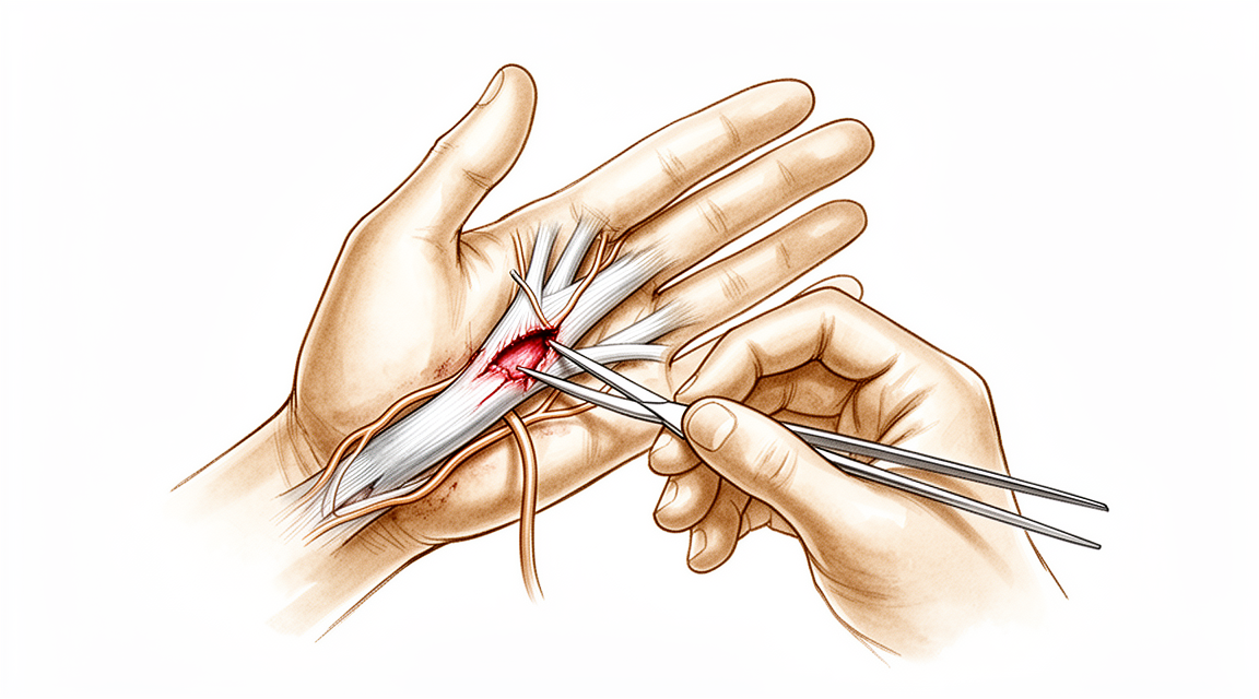
உங்கள் கையில் (hand) ஒரு தசைநாண் அல்லது நரம்பு வெட்டுப்படும்போது, அதன் விளைவுகள் எது காயமடைந்தது, எங்கே என்பதைப் பொறுத்தது. வெட்டுப்பட்ட தசைநாண் என்றால், பொதுவாக ஒன்று அல்லது அதற்கு மேற்பட்ட விரல்களை நீங்கள் வழக்கமாக மடக்குவது போலவோ நேராக்குவது போலவோ செய்ய முடியாது. வெட்டுப்பட்ட நரம்பு, அது உணர்வு தரும் பகுதியில் மரத்துப்போதலையோ ஊசி குத்துவது போன்ற கூச்ச உணர்வையோ (pins and needles) ஏற்படுத்துகிறது; காயத்தின் மேல் தட்டும்போது ஒரு கூச்ச உணர்வையோ மின்னதிர்ச்சி போன்ற உணர்வையோ நீங்கள் கவனிக்கலாம். தட்டும்போது ஏற்படும் அந்த உணர்வு, நரம்பு மீளத் தொடங்குகிறது என்பதன் அறிகுறியாக இருக்கலாம்.
வலி பொதுவாகக் காயம் உள்ள இடத்திலேயே உணரப்படுகிறது; கையைப் பயன்படுத்த முயலும்போது அது அதிகரிக்கும் போக்கு உண்டு: பற்றிப் பிடிப்பது, கிள்ளிப் பிடிப்பது, கடைப் பைகளைச் சுமப்பது, சாவியைத் திருப்புவது, அல்லது பொத்தான்களைப் போடுவது. இரவிலோ விழித்தெழும்போதோ கை அதிகம் வலிப்பதைப் பலர் காண்கிறார்கள். கைக்கு ஓய்வு கொடுத்து அசையாமல் வைத்திருப்பது பெரும்பாலும் வலியைத் தணிக்கிறது; அசைவும் சுமையும் அதை மோசமாக்குகின்றன. ஒரு தசைநாண் பகுதியளவு மட்டுமே வெட்டுப்பட்டிருந்தால், விரல் முதலில் வேலை செய்யலாம்; ஆனால் பின்னர் சிக்கிக்கொள்ளலாம், 'டிக்' என்று ஒலிக்கலாம், அல்லது விட்டுக்கொடுக்கலாம் (give way).
விரல் நுனிகளில் உணர்வு தேவைப்படும் அன்றாட வேலைகள் கடினமாகின்றன: நாணயங்கள் போன்ற சிறிய பொருட்களை எடுப்பது, பொத்தான்களையோ ஜிப்புகளையோ போடுவது, பேனாவைப் பிடிப்பது, கரண்டி, முள்கரண்டி போன்றவற்றைக் கையாள்வது, அல்லது தண்ணீர் சூடாக இருக்கிறதா என்று உணர்வது. எவ்வளவு இறுக்கமாகப் பிடிக்கிறீர்கள் என்பதைக் கையால் உங்களுக்குச் சொல்ல முடியாததால், கவனிக்காமலேயே பொருட்களைக் கீழே போட்டுவிடலாம். காயத்தில் தசைநாண் அல்லது நரம்புடன் சேர்த்து ஓர் எலும்பும் முறிந்திருந்தால், பலவீனமும் மரத்துப்போதலும் பெரும்பாலும் மேலும் கடுமையாக இருக்கும்.
சில எச்சரிக்கை அறிகுறிகளுக்கு அவசர சிகிச்சை தேவை. உங்கள் விரல், கை (hand) அல்லது முழுக் கை (arm) சூடாகி, சிவந்து, வீங்கி, வலித்தால் — குறிப்பாகக் காய்ச்சலுடன் — அல்லது உங்கள் விரல்களோ கையோ வெளிறியோ, குளிர்ந்தோ, வெண்மையாகவோ, நீலமாகவோ மாறினால், அல்லது ஒரு காயத்துக்குப் பிறகு திடீரென உணர்வையோ அசைவையோ இழந்தால், அன்றே அவசர சிகிச்சைப் பிரிவுக்குச் செல்லுங்கள். அறிகுறிகள் தணியவில்லை என்றால், வாரக்கணக்கில் மோசமாகிக்கொண்டே வந்தால், இரவில் உங்களைத் தூக்கத்திலிருந்து எழுப்பினால், அல்லது வேலை செய்யவோ கையைப் பயன்படுத்தவோ முடியாமல் தடுத்தால், உங்கள் GP-யைப் பாருங்கள் அல்லது சிறப்பு மருத்துவரின் மதிப்பீட்டைக் கேளுங்கள்.
உண்மையில் என்ன நடக்கிறது
உங்கள் கை 27 சிறிய எலும்புகளைச் சுற்றி அமைந்துள்ளது; அவை ஐந்து விரல் வரிசைகளாக (rays) அமைக்கப்பட்டுள்ளன. ஒவ்வொரு வரிசையும் ஒன்றாக மடங்கி நேராகும் எலும்புகளின் ஒரு சங்கிலி. உங்கள் விரல்களை அசைக்கும் தசைகள் ஓரளவு முன்கையிலும் ஓரளவு கைக்குள்ளேயும் அமைந்துள்ளன; அவை தசைநாண்கள் எனப்படும் கயிறுகள் மூலம் விரல்களை இழுக்கின்றன. ஒரு தசைநாணை, வழுவழுப்பான சுரங்கத்துக்குள் செல்லும் ஒரு கயிறாக நினைத்துக்கொள்ளுங்கள். ஒவ்வொரு விரலின் உள்ளங்கைப் பக்கத்திலும் இதுபோன்ற இரண்டு கயிறுகள் உள்ளன: ஒன்று நடு மூட்டை மடக்குகிறது; மற்றொன்று விரல் நுனி வரை சென்று கடைசி மூட்டை மடக்குகிறது. புறங்கையில் (back of the hand), வேறு தசைநாண்கள் விரல்களை நேராக்குகின்றன.
நீங்கள் கையை வெட்டிக்கொள்ளும்போது, இந்தக் கயிறுகளில் ஒன்று முழுமையாகவோ பகுதியளவோ அறுபடலாம். முழுமையாக வெட்டுப்பட்ட தசைநாண் என்றால், அதை இழுக்கும் தசை விரலுடனான இணைப்பை இழந்துவிட்டது; எனவே அந்த விரல் தானாக மடங்கவோ நேராகவோ செய்யாது. பகுதியளவு வெட்டுப்பட்ட தசைநாண் முதலில் இன்னும் வேலை செய்யலாம்; அதனால்தான் விரல் சிக்கிக்கொள்ளவோ விட்டுக்கொடுக்கவோ தொடங்குவதற்கு முன்பு அது நன்றாக இருப்பதுபோல் தோன்றலாம். தசைநாண்கள் அந்த வழுவழுப்பான சுரங்கங்களுக்குள் நழுவுகின்றன; எனவே பழுதுபார்ப்பு, தாங்கி நிற்பதோடு சீராக நழுவவும் போதுமான வலிமையுடன் குணமாக வேண்டும்.
நரம்புகள் மின்கம்பிகள் போன்றவை. அவை உங்கள் விரல் நுனிகளிலிருந்து மூளைக்கு உணர்வைக் கொண்டுசெல்கின்றன; கட்டளைகளைத் தசைகளுக்குக் கொண்டுசெல்கின்றன. வெட்டுப்பட்ட நரம்பு அந்தச் சுற்றை (circuit) உடைக்கிறது; அதனால்தான் அது உணர்வு தரும் பகுதியில் மரத்துப்போதல், கூச்ச உணர்வு, அல்லது மின்னதிர்ச்சி போன்ற உணர்வு ஏற்படுகிறது. தசைநாணைப் போல் அல்லாமல், ஒரு நரம்பை வெறுமனே மீண்டும் தைத்து உடனடியாக வேலை செய்ய வைக்க முடியாது. நரம்பு அதன் வெளிப்புற உறையின் வழியே நாளொன்றுக்கு ஏறக்குறைய ஒரு மில்லிமீட்டர் வீதம் மீண்டும் வளர வேண்டும்; எனவே உணர்வு மாதக்கணக்கில் மெதுவாகத் திரும்புகிறது; பயனுள்ளதாக ஆவதற்கு முன்பு, அது பெரும்பாலும் அசௌகரியமான மிகை உணர்திறனாக (oversensitivity) முதலில் திரும்புகிறது.
காயத்தில் எலும்பு முறிவும் சேர்ந்திருந்தால், சேதம் ஒரே நேரத்தில் ஒன்றுக்கு மேற்பட்ட அமைப்புகளைப் பாதிக்கிறது; பலவீனமும் மரத்துப்போதலும் மேலும் மோசமாக இருக்கும் போக்கு உண்டு.
இதற்கு நாங்கள் என்ன செய்ய முடியும்
Mater Private Hospital Rockhampton-இல் மேல் அவயவ (upper-limb) அறுவை சிகிச்சை நிபுணரான டாக்டர் கியரன் ஹிர்பரா, சிகிச்சையை உங்கள் குறிப்பிட்ட காயத்திற்கு ஏற்பப் பொருத்துகிறார். கையின் தசைநாண் மற்றும் நரம்புக் காயங்களில் சில அறுவை சிகிச்சையின்றிக் குணமாகின்றன; மற்றவற்றுக்குக் காயம் ஏற்பட்ட சிறிது காலத்திலேயே அறுவை சிகிச்சை தேவைப்படுகிறது; எனவே உடனடி மதிப்பீடு முக்கியம். நோயாளிகள் பொதுவாக அவர்களுடைய GP (பொது மருத்துவர்) மூலம் எங்கள் கிளினிக்கிற்குப் பரிந்துரைக்கப்படுகின்றனர்; ஓர் இயன்முறை சிகிச்சையாளர் (physiotherapist) எங்களைப் பார்க்கும்படி பரிந்துரைத்திருந்தாலும், Medicare தள்ளுபடியைப் (rebate) பெறத் தகுதி பெற உங்கள் GP-யிடமிருந்து பரிந்துரைக் கடிதம் (referral) இன்னும் தேவைப்படும். கிளினிக்கில், நாங்கள் உங்கள் மருத்துவ வரலாற்றைக் கவனமாகக் கேட்டறிந்து, உங்கள் கையைப் பரிசோதித்து, தேவைப்படும் இடங்களில் படமெடுப்புக்கோ (imaging) நரம்புப் பரிசோதனைகளுக்கோ ஏற்பாடு செய்கிறோம். நரம்பு அல்லது தசைநாண் எவ்வளவு மோசமாகக் காயமடைந்துள்ளது என்பதை அந்தப் பரிசோதனைகள் காட்டி, சிகிச்சையைத் தீர்மானிக்க எங்களுக்கு உதவுகின்றன.
எல்லாக் காயங்களுக்கும் அறுவை சிகிச்சை தேவையில்லை. நரம்பு வெட்டுப்படாமல் நசுங்கி அடிபட்டிருந்தால் (bruised), அது தானாக மீளும் வரை நாங்கள் அதை வெறுமனே கண்காணிக்கலாம். 6 வாரக் கண்காணிப்புக்குப் பிறகும் தசைகள் மீண்டும் வேலை செய்வதற்கான எந்த அறிகுறியும் இல்லையென்றால், அல்லது பலவீனம் தொடர்ந்து மோசமாகிக்கொண்டே இருந்தால், நரம்பை நேரடியாகப் பார்த்து ஆராய்வதைப் பரிந்துரைப்போம். ஒரு நரம்போ தசைநாணோ குணமாகும்போது, ஸ்பிளிண்ட் (splint) அல்லது மாவுக்கட்டு (cast) போன்று கையை அசையாமல் பிடித்து வைக்கும் ஒன்று பழுதுபார்ப்பைப் பாதுகாக்கிறது. அறுவை சிகிச்சைக்குப் பிறகு, உங்கள் மறுவாழ்வு கை சிகிச்சை (hand therapy) ஆகும், இயன்முறை சிகிச்சை (physiotherapy) அல்ல: Extend Rehabilitation-இல் உள்ள Ruby Doolan உங்கள் பயிற்சிகளுக்கு வழிகாட்டி, உங்களுக்குத் தேவையான எந்த ஸ்பிளிண்டையும் செய்து தருவார்.
அறுவை சிகிச்சை தேவைப்பட்டால், வெட்டுப்பட்ட அமைப்பு செய்துவந்த வேலையை மீட்டெடுப்பதே அதன் நோக்கம்: விரல் மீண்டும் மடங்கவோ நேராகவோ முடியும்படி தசைநாண் மீண்டும் ஒன்றாகத் தைக்கப்படுகிறது; உணர்வு மீண்டும் வளரும்படி வெட்டுப்பட்ட நரம்பு இணைக்கப்படுகிறது. இழுக்காமல் இரண்டு நரம்பு முனைகளையும் ஒன்றுசேர்க்க முடியாவிட்டால், ஒரு ஒட்டு (graft) அல்லது ஒரு சிறிய குழாய் மூலம் இடைவெளியைப் பாலம் போட்டு இணைக்கலாம்; அல்லது சில சமயங்களில், வேலை செய்யும் ஒரு நரம்பை மேலும் பயனுள்ள ஒரு வேலைக்கு வேறு வழியில் திருப்பலாம். நரம்புக் காயம் ஒரு தசையை நிரந்தரமாகப் பலவீனமாக்கியிருந்தால், விட்டுக்கொடுக்கக்கூடிய ஒரு வேலையிலிருந்து ஒரு தசைநாணை நகர்த்தி, அந்தத் தசையின் வேலையை ஏற்கச் செய்யலாம். சில சமயங்களில் ஒரே அறுவை சிகிச்சையில் நரம்பைப் பழுதுபார்த்து, தசைநாணையும் நகர்த்துகிறோம். நரம்பு மோசமாகக் குணமடைந்த இடத்தில் வலிமிகுந்த ஒரு கட்டி இருந்தால், அறுவை சிகிச்சையின் முதன்மை நோக்கம் வலியைத் தணிப்பது; இரண்டாம் நோக்கம் உணர்வை மீட்டுத் தருவது.
என்ன எதிர்பார்க்கலாம்
மீட்சி, எந்த அமைப்பு காயமடைந்தது, எவ்வளவு மோசமாக என்பதைப் பொறுத்தது. இன்னும் ஒரே துண்டாக இருக்கும், நசுங்கி அடிபட்ட நரம்பு, வெட்டுப்பட்டுப் பழுதுபார்க்கப்பட்ட நரம்பைவிடப் பொதுவாக நன்றாக மீள்கிறது. பழுதுபார்க்கப்பட்ட தசைநாண் பொதுவாக விரல் மீண்டும் அசையும்படி குணமாகிறது; ஆனால் சில வாரங்கள் கழித்து, விரல் நன்றாக உணரத் தொடங்கும் நேரத்தில்தான், பழுதுபார்ப்பு மிகவும் பலவீனமாக இருக்கிறது; எனவே குணமாகும் காலத்தில் கையைப் பாதுகாக்க வேண்டும்.
பழுதுபார்ப்பு எவ்வளவு நன்றாகச் செய்யப்பட்டாலும் நரம்பு மீட்சி மெதுவானதே. உணர்வு நாளொன்றுக்கு ஏறக்குறைய ஒரு மில்லிமீட்டர் வீதம் திரும்புகிறது; எனவே மணிக்கட்டில் வெட்டுப்பட்ட ஒரு நரம்புக்குப் பல மாதங்கள் ஆகலாம்; பயனுள்ளதாக ஆவதற்கு முன்பு, உணர்வு பெரும்பாலும் அசௌகரியமான மிகை உணர்திறனாக முதலில் திரும்புகிறது. சில காரணிகள் அந்த மீட்சிக்கான வாய்ப்பை அதிகரிக்கின்றன: நரம்பைத் தாமதிக்காமல் விரைவில் பழுதுபார்ப்பது, நரம்பு முனைகளுக்கு இடையே குறுகிய இடைவெளி, இளவயது. 6 மாதங்களுக்குக் குறைவான பழுதுபார்ப்புத் தாமதமும், 5 cm-க்குக் குறைவான இடைவெளியும் சிறந்த மீட்சியுடன் தொடர்புடையவை. அப்படியிருந்தும், பழுதுபார்க்கப்பட்ட ஒவ்வொரு நரம்பும் இயல்பு நிலைக்குத் திரும்புவதில்லை. பழுதுபார்க்கப்பட்ட விரல் நரம்புகளில் (digital nerves) 24% மட்டுமே காயத்துக்கு முந்தைய நிலைக்கு நெருக்கமான அல்லது சமமான உணர்வைத் திரும்பப் பெறுகின்றன; எனவே உணர்வில் ஓரளவு மாற்றம் நிரந்தரமாக இருக்கலாம்.
தசைநாண் பழுதுபார்ப்புகளுக்கும் எடைபோட வேண்டிய அவற்றுக்கே உரிய அபாயங்கள் உண்டு. விரலை மடக்கும் தசைநாண் (flexor tendon) பழுதுபார்ப்புகளில் சுமார் 6%-க்கு மற்றொரு அறுவை சிகிச்சை தேவைப்படுகிறது; பெரும்பாலும் வடுத் திசு தசைநாணை ஒட்டவைத்து அது நழுவுவதைத் தடுப்பதாலோ, பழுதுபார்ப்பு பிரிந்துவிடுவதாலோ. பழுதுபார்ப்பு பிரிந்துவிடுவது சுமார் 4% நிகழ்வுகளில் நடக்கிறது. நரம்புக் காயம் ஒரு தசையை நிரந்தரமாகப் பலவீனமாக்கியிருந்தால், தசைநாண் மாற்றம் (tendon transfer) பயனுள்ள அசைவை மீட்டுத் தரலாம்; நரம்புப் பழுதுபார்ப்பு மட்டுமே செயல்பாட்டைத் திரும்பக் கொண்டுவராதபோது, இது நன்கு நிறுவப்பட்ட ஒரு தேர்வு.
காயத்தைக் கவனிக்காமல் விட்டால் பொதுவாக என்ன நடக்கும் என்பது, எந்த அமைப்பு என்பதைப் பொறுத்தது. முழுமையாக வெட்டுப்பட்ட தசைநாண் தானாகக் குணமாகாது; எனவே அந்த விரலால் மடங்கவோ நேராகவோ முடியாமலே இருக்கும். வெட்டுப்பட்ட நரம்பும் ஓர் இடைவெளியைக் கடந்து மீண்டும் வளராது; மரத்துப்போதல் நீடிக்கும். காயம் ஏற்பட்ட நேரத்துக்கு அருகில் செய்யப்படும் பழுதுபார்ப்பைவிட, தாமதமான சிகிச்சை பொதுவாகக் குறைவான நல்ல முடிவுகளுக்கே வழிவகுக்கிறது; இருப்பினும் நீண்ட தாமதத்துக்குப் பிறகும் நல்ல முடிவுகள் சாத்தியமே.
அறிகுறிகள் தணியவில்லை என்றால், அல்லது வாரக்கணக்கில் மோசமாகிக்கொண்டே வந்தால், உங்கள் GP-யைப் பாருங்கள் அல்லது சிறப்பு மருத்துவரின் மதிப்பீட்டைக் கேளுங்கள்.
எப்போது மருத்துவரைப் பார்க்க வேண்டும்
தோலைக் கிழிக்கும் கைக் காயத்தை உடனடியாகப் பரிசோதிக்க வேண்டும்; ஏனெனில் வெட்டுப்பட்ட தசைநாணோ நரம்போ தானாகக் குணமாகாது. உங்கள் விரல், கை (hand) அல்லது முழுக் கை (arm) சூடாகி, சிவந்து, வீங்கி, வலித்தால் — குறிப்பாகக் காய்ச்சலுடன் — உங்கள் விரல்களோ கையோ வெளிறியோ, குளிர்ந்தோ, வெண்மையாகவோ, நீலமாகவோ மாறினால், ஒரு காயத்துக்குப் பிறகு திடீரென உணர்வையோ அசைவையோ இழந்தால், அல்லது ஓர் எலும்பு முறிவின் அல்லது மூட்டின் மேல் தோல் கிழிந்திருந்தால், எலும்பு வெளியே தெரிந்தால், அல்லது விரல் வெளிப்படையாக உருக்குலைந்து தோன்றினால், அன்றே அவசர சிகிச்சைப் பிரிவுக்குச் செல்லுங்கள். முதலில் நன்றாக இருப்பதுபோல் தோன்றிய ஒரு விரல் சிக்கிக்கொள்ளவோ, 'டிக்' என்று ஒலிக்கவோ, விட்டுக்கொடுக்கவோ தொடங்கினால், சிறப்பு மருத்துவரின் மதிப்பீட்டைக் கேளுங்கள்; ஒரு தசைநாண் பகுதியளவு மட்டுமே வெட்டுப்பட்டிருக்கும்போது இது நிகழலாம். வலி கடுமையாக இருந்து, பரிசோதனைக்காகக் கையைப் போதுமான அளவு அசையாமல் வைத்திருக்க முடியாவிட்டால், உங்கள் GP-யைப் பாருங்கள்; தசைநாண் முழுமையாக இருக்கிறதா என்பதை ஒரு ஸ்கேன் தீர்மானிக்க முடியும்.
ஆழமான விளக்கம்
Advanced reading: the deeper science (optional)
உங்கள் சொந்த சிகிச்சை முடிவுகளுக்குத் தேவையானதைவிட இந்தப் பிரிவு ஆழமாகச் செல்கிறது. கையில் செய்யப்படும் தசைநாண் மற்றும் நரம்புப் பழுதுபார்ப்பு பற்றி மேலும் படிப்பது பயனுள்ளது; ஏனெனில் அறுவை சிகிச்சை என்பது சிகிச்சையின் பாதி மட்டுமே — அதைத் தொடர்ந்து வரும் வாரங்களில் நடப்பது, பழுதுபார்ப்பைப் போலவே முடிவைத் தீர்மானிக்கிறது; அந்த விஷயத்தில் ஆதாரம் இப்போது மிகவும் குறிப்பிட்டதாக மாறியுள்ளது.
ஆரம்ப அசைவு, பாதுகாத்து வைப்பதைவிட மேலானது
ஒரு வளைப்புத் தசைநாண் பழுதுபார்ப்புக்குப் பிறகு இரண்டு விஷயங்கள் ஒன்றோடொன்று போட்டியிடுகின்றன. விரலை அசைத்தால் பழுதுபார்ப்பு கிழிந்துவிடக்கூடும்; அசைக்காமல் வைத்திருந்தால் தசைநாண் தன் உறையுடன் வடுத் திசுவால் ஒட்டிக்கொள்கிறது — அதனால் அது சேதமின்றிக் குணமடைகிறது, ஆனால் நழுவி இயங்க முடிவதில்லை; அதாவது, குணமடைந்த ஒரு தசைநாணை உள்ளே கொண்ட ஒரு விறைப்பான விரல்.
569 நோயாளிகளை உள்ளடக்கிய ஆய்வில், ஆரம்பச் செயலற்ற அசைவுக் குழுவைவிட ஆரம்பச் செயலூக்க அசைவுக் குழு அதிக மொத்தச் செயலூக்க அசைவைப் பெற்றது. கண்டுபிடிப்பைப் போலவே அதன் நிபந்தனையும் முக்கியம்: பழுதுபார்ப்பில் 2-இழை மையத் தையல் (2-strand core suture) மட்டுமே பயன்படுத்தப்பட்ட, செயலூக்க வளைத்தல்-மற்றும்-நீட்டல் உட்குழுவில் கிழிவு ஏற்படும் அபாயம் அதிகமாக இருந்தது குறிப்பிடப்பட்டது [1].
அதுவே இந்தப் பரிமாற்றத்தைத் துல்லியமாகச் சொல்கிறது. ஆரம்பச் செயலூக்க அசைவு சிறந்த இறுதி அசைவை உருவாக்குகிறது; ஆனால் அதைத் தாங்கும் அளவுக்கு வலிமையான ஒரு பழுதுபார்ப்பை அது கோருகிறது. பழுதுபார்ப்பைக் கடந்து செல்லும் இழைகளின் எண்ணிக்கை ஏன் விவாதிக்கப்படுகிறது என்பதற்கு இதுவே காரணம் — அது தனக்காகவே பேசப்படும் தொழில்நுட்ப விவரம் அல்ல; பாதுகாப்பான, சிறந்த மறுவாழ்வுப் பயிற்சியைப் பயன்படுத்த முடியுமா என்பதைத் தீர்மானிப்பது அதுவே.
சார்பு-அசைவு அணுகுமுறை, அதன் ஆதாரம் உண்மையில் எங்கே பொருந்துகிறது
சார்பு-அசைவு ஆர்த்தோசிஸ்கள் (relative motion orthoses), காயமடைந்த விரலை அதன் அண்டை விரல்களிலிருந்து சற்றே வேறுபட்ட ஒரு சீரமைப்பில் பிடித்து வைக்கின்றன; இது கையைப் பயன்படுத்த அனுமதிக்கும் அதே வேளையில் பழுதுபார்ப்பின் மேல் உள்ள சுமையை இறக்குகிறது. கையை ஆறு வாரங்கள் அசைவின்றி வைத்திருப்பதோடு ஒப்பிடும்போது, பொறுத்துக்கொள்ளும் தன்மையில் இது கணிசமான ஒரு முன்னேற்றம்.
ஆதாரத் தளம் சீரற்றது; அதைத் துல்லியமாக அறிந்துகொள்வது மதிப்புடையது. மண்டலம் V-VI நீட்டுத் தசைநாண் பழுதுபார்ப்புகளில் சார்பு-அசைவு அணுகுமுறை பாதுகாப்பானது என்பதற்கு இப்போது நல்ல ஆதாரம் உள்ளது; ஆனால் மண்டலம் IV மற்றும் VII நீட்டுத் தசைநாண் பழுதுபார்ப்புகளுக்கும், வளைப்புத் தசைநாண் பழுதுபார்ப்புகளுக்கும் ஆதாரம் குறைவாகவே உள்ளது [2].
எனவே, புறங்கையில் செய்யப்படும் நீட்டுத் தசைநாண் பழுதுபார்ப்புகளுக்கு நன்கு ஆதரிக்கப்படும் ஓர் அணுகுமுறை, தற்போதைய ஆதாரத்தின்படி, விரலில் செய்யப்படும் வளைப்புத் தசைநாண் பழுதுபார்ப்புகளுக்கு அதே அளவுக்கு நிலைநாட்டப்பட்டதாக இல்லை. அங்கே அது பயன்படுத்தப்படும்போது, அது நிரூபிக்கப்பட்ட நடைமுறை என்பதைவிட நியாயமான ஒரு நீட்டிப்பு ஊகமே.
நரம்புகளுக்கு, நுட்பத்தைவிட இடைவெளியே முக்கியம்
விரல் நரம்புப் பழுதுபார்ப்பில் பல்வேறு நுட்பங்கள் பயன்பாட்டுக்கு வந்துள்ளன — நேரடித் தையல், உடலின் வேறு இடத்திலிருந்து எடுக்கப்பட்ட சொந்த ஒட்டு (autograft), செயலாக்கப்பட்ட பிறர் ஒட்டு (allograft), மற்றும் செயற்கைக் குழாய்கள் (conduits).
625 பழுதுபார்ப்புகளை உள்ளடக்கிய ஆய்வில், கிடைக்கக்கூடிய அனைத்து நுட்பங்களுமே நியாயமான முடிவுகளைத் தருகின்றன; மேலும், நேரடிப் பழுதுபார்ப்பைத் தவிர்த்து, ஓர் இடைவெளி இருக்கும் இடங்களில் சொந்த ஒட்டும் பிறர் ஒட்டும் ஒப்பிடத்தக்க அளவில் செயல்பட்டன [3].
எனவே தீர்மானிக்கும் காரணி, நரம்பின் முனைகளை இழுவிசை இல்லாமல் ஒன்றாகக் கொண்டுவர முடியுமா என்பதே. முடியுமானால், நேரடிப் பழுதுபார்ப்பு செய்யப்படுகிறது. முடியாவிட்டால், இடைவெளியைப் பாலம் போட்டு இணைக்க வேண்டும்; அதற்காக நோயாளியின் உடலில் வேறு இடத்திலிருந்து ஒரு நரம்பை எடுக்க வேண்டும் என்று தற்போதைய ஆதாரம் கட்டாயப்படுத்தவில்லை — இது ஒரு தானம் அளிக்கும் இடத்தையும், அதனுடன் சேர்ந்து வரும் மரத்துப்போன பகுதியையும் தவிர்க்கிறது.
மீட்சி ஏன் மாதங்களில் அளக்கப்படுகிறது
இரு திசுக்களும், எந்த நுட்பத்தாலும் சுருக்க முடியாத ஓர் உயிரியல் கால அட்டவணையின்படியே குணமடைகின்றன. பழுதுபார்க்கப்பட்ட ஒரு தசைநாண் சுமார் மூன்று வாரங்களில் தன் மிகப் பலவீனமான நிலையில் இருக்கிறது — அப்போது தொடக்கத்தில் இருந்த ஃபைப்ரின் (fibrin) உறிஞ்சப்பட்டுவிட்டது, புதிய கொலாஜன் இன்னும் முதிர்ச்சியடையவில்லை; சரியாக அப்போதுதான் விரல் நன்றாக இருப்பது போல உணரப்படுகிறது, அதைப் பயன்படுத்தும் ஆசையும் மக்களுக்கு மிக அதிகமாக இருக்கிறது.
நரம்புகள், எஞ்சியிருக்கும் உறைக்குள் நாளொன்றுக்கு ஏறத்தாழ ஒரு மில்லிமீட்டர் என்ற வேகத்தில் அச்சிழைகளை (axons) வளர்த்துக்கொண்டே மீண்டும் உருவாகின்றன. எனவே விரலின் அடிப்பகுதியில் ஏற்பட்ட ஒரு வெட்டிலிருந்து விரல் நுனியைச் சென்றடைய பல மாதங்கள் ஆகின்றன; உணர்வு படிப்படியாகவும் முழுமையற்ற வகையிலும் திரும்புகிறது — பயனுள்ள உணர்வாக மாறுவதற்கு முன் பெரும்பாலும் ஓர் அசௌகரியமான அதீத உணர்திறனாகவே (hypersensitivity) வருகிறது. அந்த வரிசை எதிர்பார்க்கப்படும் மீட்சியே, ஒரு சிக்கல் அல்ல.
மேற்கோள்கள்
[1] Xu H, Huang X, Guo Z, Zhou H, Jin H, Huang X. Outcome of surgical repair and rehabilitation of flexor tendon injuries in zone II of the hand: systematic review and meta-analysis. J Hand Surg Am. 2023;48(4):407.e1-407.e11. https://doi.org/10.1016/j.jhsa.2021.11.013
[2] Shaw AV, Verma Y, Tucker S, Jain A, Furniss D. Relative motion orthoses for early active motion after finger extensor and flexor tendon repairs: a systematic review. J Hand Ther. 2023;36(2):332-46. https://doi.org/10.1016/j.jht.2023.02.011
[3] Herman ZJ, Ilyas AM. Sensory outcomes in digital nerve repair techniques: an updated meta-analysis and systematic review. Hand (N Y). 2019;15(2):157-64. https://doi.org/10.1177/1558944719844346
Evidence & references
This is the clinical evidence summary written for health professionals. It is technical, and it lists the research this page was built from. You do not need to read it to understand your treatment or to make a decision about it.
Overview
Preoperative Assessment and Diagnosis
- Factors that interfere with the examination of nerves in the hand include other injuries that may be life threatening or limb threatening, patient intoxication, anxiety or lack of cooperation of the patient, and an extensive injury to the hand [9].
- If conditions are not satisfactory for a thorough examination during the initial evaluation, the hand should be reexamined within a reasonable period to determine the extent of nerve and other injuries sustained [9].
- If a flexor tendon function deficit is present after a finger laceration, at least one digital nerve probably has been injured as well [9].
- At least four areas of consideration are important when evaluating a patient with an injury to a nerve in the hand: (1) type of injury, (2) sensibility evaluation, (3) motor function, and (4) sudomotor function (sweating) [9].
- The Seddon classification of nerve injury includes Neurapraxia, Axonotmesis, and Neurotmesis [9].
- The Sunderland classification of nerve injury includes degrees I through VI, with degree VI being a combination of any of Sunderland I–V [9].
- Magnetic resonance neurography (MRA) is reported to be able to provide details regarding nerve anatomic relationships, fascicular pattern, intraneural swelling, and evaluation of downstream muscle injury [9].
- Customary methods used to evaluate damaged sensory nerves include the use of a sharp pin to assess pain, a cotton-tipped applicator or a finger eraser to assess light touch, and the tips of a paper clip or commercially prepared tool to assess two-point discrimination [9].
- Normal two-point discrimination usually is 6 mm or less [9].
- If the nerve is transected, a patient would not feel light touch, would not appreciate the pin as a sharp stimulus, and would be unable to discriminate between one and two points [9].
- Patients with closed injuries or partial injuries to nerves may show spotty appreciation of light touch and pain and have markedly widened two-point discrimination [9].
Anatomy and Surgical Considerations
- It is of paramount importance to respect and preserve the sensibility of the skin of the fingers and to avoid placing incisions in regions where pressure is commonly applied [67].
- In the palm, the nerves are protected by the overlying aponeurosis, but on the dorsum and palmar aspects of the wrist, and on the dorsum of the hand and fingers, the nerves are superficial and very much at risk if an incision is made without due care [67].
- The palmar cutaneous branch of the median nerve is the sensory nerve most susceptible to iatrogenic injury [67].
- The diameter of the palmar cutaneous branch of the median nerve is about 0.8 mm [67].
- The palmar cutaneous branch of the median nerve originates on the radial border of the median nerve 5 to 6 cm above the distal transverse flexion crease of the wrist [67].
- The palmar cutaneous branch of the median nerve courses alongside the median nerve for 2–3 cm [67].
- The palmar cutaneous branch of the median nerve follows the course of the flexor carpi radialis tendon and passes between the two layers of the forearm fascia, which provide it with its own short tunnel just ulnar to the flexor carpi radialis tendon [67].
- The palmar cutaneous branch of the median nerve passes through the superficial layer of the forearm fascia at the bifurcation of the palmaris longus into the root of the palmar aponeurosis and the tendon giving origin to the superficial portion of the abductor pollicis brevis [67].
- After only 5–10 mm, the palmar cutaneous branch of the median nerve divides into three terminal branches, which cross the mid-palmar aponeurosis to supply the deep layers of the dermis [67].
- To avoid the palmar cutaneous branch of the median nerve and its branches, which constitute the most frequent source of palmar neuromas, the forearm incision must be ulnar to the palmaris longus tendon [67].
- The main branch of the palmar cutaneous branches of the ulnar nerve arises from the cutaneous branch of the ulnar nerve, originating in the forearm a few centimeters above the origin of the dorsal branch [67].
- The cutaneous branch of the ulnar nerve runs against the palmar aspect of the nerve up to the level of the wrist, where it becomes superficial, crossing into the hypothenar eminence palmar to the palmaris brevis muscle [67].
- In 16 of 21 cases, the palmar cutaneous branch of the ulnar nerve was replaced by branches from the superficial division of the ulnar nerve or from its dorsal division [67].
- The dorsal branch of the ulnar nerve arises from the medial aspect of the ulnar nerve at an average distance of 6.4 cm from the distal aspect of the head of the ulna and 8.3 cm from the proximal border of the pisiform [67].
- The dorsal branch of the ulnar nerve passes dorsal to the flexor carpi ulnaris and pierces the deep fascia [67].
- The dorsal branch of the ulnar nerve becomes subcutaneous on the medial aspect of the forearm at a mean distance of 5 cm from the proximal edge of the pisiform [67].
- Proximal to the wrist, the dorsal branch of the ulnar nerve provides two to three branches [67].
- With the forearm supinated, the dorsal branches of the ulnar nerve pass along the medial aspect of the head of the ulna, then on the dorsal aspect of the triquetrum [67].
- With the forearm pronated, the dorsal branches of the ulnar nerve displace slightly ulnarly [67].
- The terminal sensory branch of the radial nerve passes deep to the flattened tendon of the brachioradialis and becomes superficial about 4 cm proximal to the radiocarpal joint line [67].
- The terminal sensory branch of the radial nerve crosses superficially, from radial to ulnar, the tendons of the abductor pollicis longus and extensor pollicis brevis [67].
- The terminal sensory branch of the radial nerve divides into a lateral branch, the dorsal collateral lateral of the thumb, and a medial branch that supplies the index and middle fingers [67].
- The course of the terminal sensory branch of the radial nerve is along a line drawn from the dorsolateral border of the radius toward the angle formed by the proximal junction of the first and second metacarpals [67].
- The medial branch of the terminal sensory branch of the radial nerve follows this line and divides just distal to the angle of the first and second metacarpals [67].
- The dorsal collateral nerve of the thumb follows the radial border of the extensor pollicis brevis tendon [67].
- The dorsal collateral nerve of the thumb gives the palmar cutaneous branch of the radial nerve, which follows the tendon of the abductor pollicis longus and anastomoses with or substitutes for the external branch of the palmar cutaneous branch of the median nerve [67].
- An injury to the median nerve of a hand with unusual anatomic variation of palmar sensory branches of the ulnar nerve would not experience sensory impairment of the ulnar halves of the index and middle and the radial half of the ring fingers [1].
Treatment and Outcomes
- The one-stage procedure of combined nerve repair and two-tendon transfer permits early and useful function of the hand, due to the action of the transferred tendons [2, 3].
- Tendon or tissue transfers in combined nerve palsies require longer follow-up than isolated nerve palsies to make a valid assessment of the outcome [4].
- Nerve transfers are an option for restoring hand and forearm function in patients with peripheral nerve injuries adversely affecting their ability to function [5].
- End to end nerve repair was performed in 10% of cases and 35% had excellent results in brachial plexus injury in war victims [6].
- If not corrected, secondary defects of the hand with intrinsic paralysis may compromise normal function of the hand when either nerve function recovers or tendon transfers have been performed to correct the primary functional defects [7].
- Despite the use of microsurgical techniques, outcome after median and ulnar nerve repair is suboptimal [10].
- The conservation of amputated finger-tips provides the hand surgeon with new possibilities for late reconstruction of an injured digit [13].
- The biomechanical principles, indications and limitations of tendon transfers, nerve transfers and combined approaches are compared, with particular attention to timing, patient selection, and functional goals [17].
- In lower-type injuries of the brachial plexus, transfer of median nerve branches that innervate the palm of the hand to the ulnar proper digital nerve of the little finger predictably restored protective sensation on the ulnar side of the hand [21].
- Hand therapists are going to be needed for a long time-certainly as long as tendon and nerve repairs are immobilized 3 weeks and fractures are immobilized 6 weeks [22].
- Nerve transfer is favored over nerve grafting in managing high ulnar nerve injuries because of better improvement of motor power and better restoration of grip functions of the hand [26].
- Experimental studies and positive reports from large clinical series suggest that new techniques using foreign nerves for reinnervation are worthy of integration into the management of upper brachial plexus injuries, though many questions regarding timing, donor morbidity, and comparative efficacy remain unanswered [27].
- More homogeneous follow-up protocols are needed for strength measurement and patient-reported outcome measures for both nerve and tendon transfers [30].
- The authors prefer specific nerve transfers for motor and sensory restoration, noting that median nerve repair is mandatory for patients with concomitant pain in high median nerve injury [33].
- End-to-side distal anterior interosseous nerve transfer in treatment of proximal ulnar nerve injuries provides significantly better results than a standard more proximal nerve repair [41].
- Nerve transfers provide significantly better treatment options in many cases of nerve injury where previous outcomes were expected to be poor, such as with proximal injuries, long nerve gaps, or unavailability of the proximal injured segment [43].
- Vascularized ulnar nerve graft technique should be recommended for reconstruction of the median or radial nerves in selected cases after severe trauma of the upper extremity [44].
- Nerve transfer to the median nerve using parts of the ulnar and radial nerves may offer an alternative option for proximal nerve injuries or for free functioning muscle transplantations [66].
- Tendon transfers remain the primary reconstructive procedure for paralytic injuries of the upper limb until sufficiently powered studies of nerve transfer outcomes are published [141].
Anatomy & Pathophysiology
Skeletal Architecture and Kinematics
- The skeleton of the hand and wrist consists of 27 bones, of which 19 are long bones [48].
- The hand skeleton is divided into five rays, each forming a polyarticulated chain comprising metacarpals and phalanges [48].
- The thumb ray is the shortest, consisting of a metacarpal and two phalanges, and is more mobile than the other rays [48].
- The index metacarpal is the longest, while the thumb metacarpal is the shortest [48].
- The proximal and middle phalanges of the middle and ring fingers are longer than those of the index finger [48].
- The transverse palmar axis is oblique, forming an acute angle of approximately 75 degrees with the longitudinal axis [48].
- The wrist has three axes of movement, permitting the hand to be positioned in any spatial configuration for grasping [48].
- The metacarpophalangeal joints serve as the keystones of the longitudinal arches of the hand [85].
- The volar plates prevent hyperextension at the metacarpophalangeal joints [85].
- The fourth metacarpal has approximately 10 degrees of mobility in flexion and extension [85].
- The fifth metacarpal has a range of flexion–extension of approximately 20 degrees [85].
- The index ray possesses a degree of independence at the phalangeal level due to the arrangement of its flexor and extensor muscles [85].
- The axes of flexion are arranged such that flexion of all metacarpophalangeal and proximal interphalangeal joints causes the fingers to converge toward the scaphoid [91].
- The hand presents a longitudinal and transverse concavity, giving it the shape of a cup with a palmar concavity when the thumb is placed next to the index finger [48].
Soft Tissue and Gliding Mechanisms
- The dorsal skin is thin, possesses a normal pilosebaceous system, and has loose connections with deeper planes allowing free gliding [89].
- Flexion of the fingers produces a significant lengthening of the dorsal skin, with an average increase of 3 cm in the middle finger from extension to full flexion [89].
- The dorsal and palmar areas of skin are independent due to a system of adhesions anchoring their common boundary to the underlying plane [89].
- In unrestricted areas, tendons are surrounded by paratenon and areolar connective tissue arranged in layers [88].
- In narrow crowded areas, the gliding mechanism is assured by synovial sheaths surrounded by fibrous sheaths that act as pulleys [88].
- The synovial sheaths for flexor tendons are much more developed on the palmar aspect than on the dorsal aspect [88].
- The digital neurovascular structures are surrounded by a diffuse network of thin transverse oblique fibers, with dorsal fibers called Cleland ligament and palmar fibers called Grayson ligament [90].
- The superficial palmar fascia covers a triangular area of the central palm, with the palmaris longus tendon terminating in continuity with its proximal corner [90].
- The tenosynovium lining the fibroosseous tunnel supplies nutrition and lubrication to the poorly vascularized flexor tendons [87].
- Within the flexor tendon sheath, tendon vascularity is supplied via the vincula system, specifically the vinculum longus and brevis [87].
Nerve Anatomy and Sensory Distribution
- The palmar cutaneous branch of the median nerve has a diameter of about 0.8 mm [67].
- The dorsal branch of the ulnar nerve arises from the medial aspect of the ulnar nerve at an average distance of 6.4 cm from the distal aspect of the head of the ulna [67].
- The dorsal cutaneous branch of the radial nerve becomes superficial about 4 cm proximal to the radiocarpal joint line [67].
- The palmar cutaneous branch of the ulnar nerve originates at a variable level and was replaced in 16 of 21 cases by branches from the superficial division of the ulnar nerve or its dorsal division [67].
- The ramus communicans between the ulnar and median nerves could cross over anywhere in the middle three fifths of the palm [109].
- The hand contains "functional cutaneous units" similar to those described in the face [81].
- The dorsal covering of the interphalangeal articulations forms a unique cutaneous unit characterized by a considerable excess of skin when the digits are in extension [81].
- The palmar integument of the digits is subdivided into phalangeal units separated by digital flexion folds [81].
- When a digit is completely flexed, the integument of adjacent phalanges comes into contact in the zones of the flexion creases, establishing areas of cutaneous contact in the form of a diamond [81].
Vascular Anatomy
- The "princeps pollicis" artery is the terminal branch of the radial artery that crosses the first intermetacarpal space and runs along the ulnar side of the first metacarpal bone [86].
- In anatomical studies, only 15% of dissections of the palmar arteries of the thumb fall into the classical "typical" category [86].
- The two palmar collateral arteries of the thumb run alongside the flexor tendon and behind the collateral nerves in the second segment of the thumb [86].
- The dorsal arteries of the thumb originate from the palmar arteries at the level of the first metacarpal and head distally on the side of the two distal phalanges [86].
Muscle and Tendon Anatomy
- There are seven interosseous muscles in the hand: four dorsal and three volar [82].
- The dorsal interossei are abductors, while the volar interossei are adductors [82].
- The middle finger has two dorsal interossei and no volar interossei because the central axis of the hand lies within it [82].
- The extrinsic extensors run through six different fibroosseous retinacular compartments at the wrist level [47].
- The first extensor compartment contains the abductor pollicis longus and the extensor pollicis brevis [47].
- The second extensor compartment contains the extensor carpi radialis longus and the extensor carpi radialis brevis [47].
- The third extensor compartment contains the extensor pollicis longus, which turns abruptly radialward about Lister tubercle [47].
- The fourth extensor compartment contains the extensor indicis proprius lying deep to the four tendons of the extensor digitorum communis [47].
- The fifth extensor compartment contains the extensor digiti quinti [47].
- The sixth extensor compartment contains the extensor carpi ulnaris [47].
- The flexor digitorum profundus originates from the proximal ulna and the interosseous membrane [87].
- The flexor digitorum superficialis has two heads: a radial head from the proximal shaft of the radius and a humeral ulnar head from the medial humeral epicondyle and coronoid process of the ulna [87].
- The flexor pollicis longus originates from two heads: a radial head from the proximal radius and interosseous membrane, and an accessory head from the coronoid process of the ulna and medial epicondyle of the humerus [87].
- The A2 and A4 pulleys are the most essential in maintaining the mechanical advantage of the flexor tendons [87].
- The lumbricals originate from the radial side of the index, middle, ring, and little fingers in the palm [87].
Pathophysiology and Injury Classification
- The Sunderland classification of nerve injury includes degrees I through VI, with degree VI being a combination of any of degrees I–V [9].
- Normal two-point discrimination in the hand is usually 6 mm or less [9].
- If a nerve is transected, a patient would not feel light touch, would not appreciate a pin as a sharp stimulus, and would be unable to discriminate between one and two points [9].
- Patients with closed or partial nerve injuries may show spotty appreciation of light touch and pain and have markedly widened two-point discrimination [9].
- The mechanism of ulnar nerve injury, excluding compression neuropathy, in order of frequency includes laceration, stretch, and contusion [19].
- Electrodiagnostic testing is critical in determining the level of nerve injury, but nerve conduction velocity studies and electromyography can be limited in cases of severe axonal loss or early after injury [19].
- High-resolution ultrasound imaging can show individual nerve fascicules and identify peripheral nerve disruption [19].
- Tendon injuries are present in some aspect in all cases of mutilating hand trauma [35].
- An injury to one digital nerve did not affect the result of a tendon graft in a finger, but when both nerves were injured, the result was compromised [34].
- In the thumb, there was slight but definite impairment of function when either one or both digital nerves were injured [34].
- Double crush syndrome involves nerve symptomatology from two distinct nerve lesions, with no clear consensus on what qualifies the diagnosis [56].
- Double crush syndrome patients commonly present with peripheral sensorimotor neuropathy, which may include sensory loss, burning or tingling sensation, decreased limb dexterity, limb weakness, or lack of coordination [56].
- Section of the median nerve causes paralysis of the lateral thenar muscles, resulting in loss of the anteposition of the thumb [104].
- Section of the median nerve above the elbow produces paralysis of the flexor pollicis longus, flexor digitorum superficialis, and the lateral half of the flexor profundus [104].
- The second interosseous does not provide sufficient stability to prevent ulnar drift of the middle finger, whereas the first interosseous does this for the index finger [137].
Classification
- Nerve injuries are classified based on severity and extent of damage, with recovery and outcome dependent on the original classification [20].
- The Seddon classification categorizes nerve injuries into neurapraxia, axonotmesis, and neurotmesis [9].
- The Sunderland classification categorizes nerve injuries into degrees I through VI, where degree VI is a combination of any of degrees I–V [9].
- In the Seddon-Sunderland mapping, neurapraxia corresponds to Sunderland degree I [9].
- In the Seddon-Sunderland mapping, axonotmesis corresponds to Sunderland degrees II, III, and IV [9].
- In the Seddon-Sunderland mapping, neurotmesis corresponds to Sunderland degree V [9].
- Rapid intervention generally improves outcome in peripheral nerve injuries [20].
- Prolonged denervation of nerve segments can lead to low recovery rates and other disabilities [20].
Clinical Presentation
History and Initial Assessment
- A careful history is essential to differentiate between neurapraxia and axonotmesis, which can be treated without surgery, and neurotmesis, which requires surgical intervention [19].
- Factors that interfere with the examination of nerves in the hand include other life-threatening or limb-threatening injuries, patient intoxication, anxiety, lack of cooperation, and extensive hand injury [9].
- If conditions are not satisfactory for a thorough initial evaluation, the hand should be reexamined within a reasonable period to determine the extent of nerve and other injuries [9].
- A high index of suspicion is necessary in the evaluation of patients with hand injuries [9].
- The mechanism of ulnar nerve injury, excluding compression neuropathy, occurs in order of frequency as laceration, stretch, and contusion [19].
- The presence of fractures is associated with a higher incidence of nerve and tendon injuries [18].
- Involvement of nerve and tendon structures in hand injuries is linked to an increased risk of long-term disability [18].
- Persistent neurological deficits after distal radius fractures should prompt early investigation and consideration for structural nerve injury [51].
- Such injuries are likely to be missed in casualty because of extensive soft tissue swelling, the apparent normal appearance of anteroposterior X-rays, and the technical difficulty in testing the motor branch of the ulnar nerve in the presence of pain [63].
- The case illustrates the diagnostic difficulty of ulnar nerve motor branch lesions in emergency settings [60].
Sensory Evaluation
- At least four areas of consideration are important when evaluating a patient with a nerve injury in the hand: type of injury, sensibility evaluation, motor function, and sudomotor function [9].
- Customary methods to evaluate damaged sensory nerves include using a sharp pin to assess pain, a cotton-tipped applicator or finger eraser to assess light touch, and the tips of a paper clip or commercially prepared tool to assess two-point discrimination [9].
- In simple nerve injuries, a plastic pen test can detect immediate cessation of sweating in the denervated area without any discomfort to the patient [95].
- In complicated injuries where the diagnosis is in doubt, measurement of skin resistance alone will not establish the nature of the injury [95].
- The combination of sensory threshold testing with skin conductance gives rise to great accuracy in the diagnosis of difficult nerve problems [95].
- Although electrical resistance will alter immediately following injury, it is the last thing to return to normal following repair and is not of value in assessing recovery [95].
- Additional central nervous system symptoms in double crush syndrome may include radiating nerve pain, paresthesia, loss of sensation, hand and upper extremity weakness, hyperreflexia, balance impairment, Lhermitte’s sign, and loss of fine motor control [56].
Motor and Functional Evaluation
- An injury to the digital nerves frequently is overlooked; however, if a flexor tendon function deficit is present after a finger laceration, at least one digital nerve probably has been injured as well [9].
- Proximal radial nerve injuries result in lack of wrist and digit extension with diminished grip strength and hand function [59].
- A migrating Tinel's phenomenon is a good prognostic indicator for radial nerve injuries [64].
- If not corrected, secondary defects of the hand with intrinsic paralysis may compromise normal function when either nerve function recovers or tendon transfers have been performed to correct the primary functional defects [7].
- The final status of the median nerve was at least no worse on the average in cases where the carpal tunnel was used as a pathway for a graft [12].
- Rates of median nerve symptoms were high and resolved in most cases (92%) after reduction of perilunate dislocation [40].
- At a mean final follow-up of 7 months, full recovery of median nerve function was seen in all patients with the triad of multiple metacarpal fractures/dislocations, severe hand swelling, and acute median nerve dysfunction [69].
- Grip and tip-pinch strength improve over a mean period of three years following median and/or ulnar nerve lesion [29].
- For median nerve injuries, signs of reinnervation appeared between the fourth and sixth postoperative month, but the two-point discrimination test never became normal [24].
- The sequence of early sensory recovery following repair of peripheral nerve lesions has varied significantly from that reported in the literature [38].
Diagnostic Imaging and Electrodiagnostics
- Electrodiagnostic testing is critical in determining the level of injury; however, nerve conduction velocity studies and the results of electromyography (EMG) can be limited in cases of severe axonal loss or early after injury when neurapraxia cannot be discerned from neurotmesis [19].
- EMG can also be limited by pain and an inability to identify anatomical variability [19].
- High-resolution ultrasound imaging can show individual nerve fascicules [19].
- Objective measures are limited because MRI and ultrasound have been of limited use for radial nerve injuries [64].
- Electromyography and nerve conduction velocity studies are rarely helpful acutely except when continuity is unknown because slowed but intact conduction indicates some continuity [64].
- Study is recommended around 3 to 4 months after radial nerve injury [64].
- Larger polyphasic motor action potentials of longer duration may be seen before clinical recovery, although the degree of recovery is unclear [64].
- If the amplitude of motor nerve conduction velocity is low, less than 0.3 mV, exploration can be undertaken, with repair remaining a viable option even 5 to 6 months after injury [64].
- Nerve conduction studies should be performed in patients presenting with possible carpal tunnel syndrome to assist diagnosis, and may need to be repeated at intervals in those managed conservatively [52].
- While nerve conduction studies may potentially help to find multiple lesions on the same nerve, there may still be overlap in determining lesion type which makes determining whether there are 2 distinct crush injuries challenging [56].
Investigations
Diagnostic Modalities and Limitations
- Electrodiagnostic testing is critical in determining the level of ulnar nerve injury [19].
- Nerve conduction velocity studies and electromyography (EMG) results can be limited in cases of severe axonal loss or early after injury when neurapraxia cannot be discerned from neurotmesis [19].
- EMG can be limited by pain and an inability to identify anatomical variability [19].
- Ultrasound imaging can identify a change in the hypoechoic signal within the nerve to indicate injury [19].
- Ultrasound can visualize the epineurium to suggest whether the nerve is in continuity [19].
- Ultra-sonography provides a new ability to study the complex anatomical structures of the hand, with the technique being quick and simple to perform [49].
- Nerve conduction studies should be performed in patients presenting with possible carpal tunnel syndrome to assist diagnosis [52].
- Nerve conduction studies may need to be repeated at intervals in patients managed conservatively for carpal tunnel syndrome [52].
- No predictor in clinical and MR neurography variables was found that correlates with recovery of thumb opposition in severe carpal tunnel syndrome [70].
Clinical Assessment and History
- A careful history is essential, particularly in cases of blunt trauma, to differentiate between neurapraxia and axonotmesis (which can be treated without surgery) and neurotmesis (which requires surgical intervention) [19].
- Recovery of sensitivity in reinnervated cutaneous areas is evaluated using the two-point discrimination test [14].
- Signs of reinnervation for median nerve injuries appeared between the fourth and sixth postoperative month [24].
- The two-point discrimination test never became normal for median nerve injuries in the reported series [24].
- Ulnar nerve motor branch lesions present diagnostic difficulty in emergency settings [60].
- Injuries causing transient motor neurapraxia of the ulnar nerve are likely to be missed in casualty due to extensive soft tissue swelling, apparent normal anteroposterior X-rays, and technical difficulty in testing the motor branch in the presence of pain [63].
Anatomic Variations and Innervation Patterns
- Communicating branches between the median and ulnar nerves in the palm were found in 20 of 30 (67%) specimens [80].
- Ulnar-based cutaneous nerves to the palm were found within the zone of incision in 16 of 25 specimens [45].
- Ulnar-based cutaneous nerves to the palm are not necessarily preserved by longitudinal palmar incisions in the axis of the ring finger [45].
- An injury to the median nerve in a hand with unusual anatomic variation would not experience sensory impairment of the ulnar halves of the index and middle and the radial half of the ring fingers [1].
Treatment
Nerve Repair and Grafting
- End-to-end nerve repair was performed in 10% of cases in a series of brachial plexus injuries, with 35% of those cases having excellent results [6].
- Tubular repair is an attractive alternative to primary, delayed primary, or secondary repair of severed major nerve trunks in the human forearm [74].
- Tubular repair may not be superior to conventional techniques in terms of functional recovery but represents a simple biological approach to nerve repair [74].
- When direct suture is not possible because of tension, tubular repair leaving a short distance between the nerve ends may be the key to avoiding nerve grafting [74].
- Experimental work proved that tension at the suture line has a detrimental role on nerve repair outcomes [124].
- Experimental work proved that postoperative stretching has a deleterious effect on successful functional recovery after nerve repair [124].
- Regenerating axons advanced more easily through nerve grafts of 2 cm with two tension-free anastomoses compared with a single suture under tension [124].
- The epineurium was the primary source of connective tissue proliferation in experimental nerve repair studies [124].
- Motor recovery for median nerve repair was excellent in 40%, good in 40%, and fair in 20% of cases [124].
- Motor recovery for ulnar nerve repair was excellent in 38%, good in 40%, and fair in 22% of cases [124].
- Motor recovery for radial nerve repair was excellent in 42%, good in 38%, and fair in 20% of cases [124].
- Vascularized ulnar nerve grafts should be recommended for reconstruction of the median or radial nerves in selected cases [44].
- Nerve grafting of a large defect in a high median nerve injury involves a relatively long duration of recovery with unpredictable recovery [68].
- The final status of the median nerve was at least no worse on the average in cases where the carpal tunnel was used as a pathway for the graft [12].
Nerve Transfers
- Distal nerve transfers for the treatment of high ulnar nerve injuries allow for a shorter reinnervation period and improved ulnar intrinsic recovery [39].
- End-to-side distal anterior interosseous nerve transfer provides significantly better results than a standard more proximal nerve repair for proximal ulnar nerve injuries [41].
- Transfer of the superficial radial or dorsal cutaneous branch of the ulnar nerve, or both, produced successful restoration of innervation of the thumb and index and long fingers in experimental monkey models [28].
- A dual nerve transfer from the median nerve to the distal nerve branches of the radial nerve is a preferred procedure to restore radial nerve function [59].
- In a dual median-to-radial nerve transfer, coaptation of the flexor digitorum superficialis (FDS) nerve to the extensor carpi radialis brevis (ECRB) nerve is performed to restore wrist extension [59].
- In a dual median-to-radial nerve transfer, the flexor carpi radialis (FCR) nerve is transferred to the posterior interosseous nerve (PIN) to restore finger and thumb extension [59].
- Nerve transfers for restoration of radial nerve function optimize recovery by providing pure motor donors from a near-target axon source allowing for earlier reinnervation [59].
- Nerve transfers are particularly useful for patients with hand stiffness and complex regional pain syndrome who may otherwise not be ideal candidates for tendon transfers [59].
- The authors prefer specific nerve transfers for motor and sensory restoration in high median nerve injury, noting that median nerve repair is mandatory for patients with concomitant pain [33].
- Experimental studies and positive reports from large clinical series suggest that new techniques using foreign nerves for reinnervation are worthy of integration into the management of upper brachial plexus injuries [27].
- Many questions regarding timing, donor morbidity, and comparative efficacy remain unanswered for nerve transfer techniques in upper brachial plexus injuries [27].
- A supercharged end-to-side anterior interosseous nerve transfer to the deep motor branch of the ulnar nerve was performed in 40 patients [31].
- The supercharged end-to-side anterior interosseous nerve transfer procedure involves transecting the anterior interosseous nerve at the midportion of the pronator quadratus muscle and anastomosing it to the side of the motor branch of the ulnar nerve [132].
- The supercharged end-to-side anterior interosseous nerve transfer coaptation can be augmented with fibrin glue [132].
- An above-elbow slab is applied for 3 weeks following a supercharged end-to-side anterior interosseous nerve transfer [132].
- In patients with complete pan-plexal injuries, intact extraplexal nerves (spinal accessory, intercostal, C3 and C4 nerves) can be transferred and coapted to the distal peripheral nerve of the brachial plexus as a method for reinnervation of critical sensory or motor nerves [100].
- Sensation in the median nerve distribution of the hand can be provided by the transfer of sensory branches of the intercostal nerves to the lateral cord contribution of the median nerve in pan-plexus injury [100].
Tendon Transfers
- In a high median nerve injury, if the nerve has been repaired, extrinsic transfers are seldom required because sufficient extrinsic muscle function is usually regained [55].
- If a patient needs a nerve graft for high median nerve injury, especially if done late or under unfavorable conditions, useful extrinsic functional recovery is unlikely [55].
- In high median nerve palsy where nerve grafting is required, one should consider performing early extrinsic end-to-side transfers and/or a side-to-side transfer of the index and conjoined profundus tendons [55].
- All intrinsic and extrinsic transfers for high median nerve palsy can be performed at the same time [55].
- The author does not favor early extrinsic tendon transfers for high median nerve palsy and only restores opposition if, after maximal recovery has occurred, both the patient and the surgeon believe that this will improve hand function [55].
- Most patients with combined median sensory and motor deficit will adapt and increasingly use their normal arm for precision function, making restoration of opposition rarely attempted [55].
- If an opposition transfer is indicated for high median nerve palsy, the author favors EIP transfer [55].
- The BR transfer is used for restoration of thumb flexion in high median nerve palsy, especially for conditions in which there is relative sensory sparing [55].
- An end-to-side attachment is used for the BR transfer unless there is no prospect of later reinnervation [55].
- The author does not think an ECRL transfer to restore index finger flexion is needed often in high median nerve palsy and prefers to join the index and the common profundi tendons in a side-to-side fashion [55].
- A side-to-side join of the index and common profundi tendons does not restore power to the index finger but is preferred when the hand condition is too poor to benefit from an ECRL transfer, especially if there is a marked sensory deficit [55].
- Because worthwhile sensory recovery is so unlikely in high median nerve palsy for adults, opponensplasty is typically not beneficial [55].
- The goal of any repair in high median nerve palsy is to create the best possible “helper” hand, with precision function assumed by the other hand [55].
- A reliable tendon prosthesis inserted as one stage in tendon reconstruction is the additional step needed to improve the results of flexor-tendon reconstructive surgery in hands with severe damage [50].
- The anatomic, two-tendon, three-joint finger with a perfect tendon sheath cannot always be restored to normal after severe injury to the flexor tendon system [77].
- The muscle and tendon to the middle finger arise separately and are therefore more suitable for non-synergistic transfers [149].
- None of the procedures is superior to the other statistically in terms of improvement in grip strength, improvement in active ROM of MCPJs, claw correction, or improvement in overall patient hand functions, in the intermediate follow-up duration, when comparing Zancolli Lasso versus Modified Stiles-Bunnell techniques for post-leprosy ulnar claw hand correction [32].
Nonoperative Management and Rehabilitation
- Nonoperative management of the hand is important while waiting for recovery of radial nerve function from either a presumed axonotmetic nerve injury or after nerve repair [99].
- Nonoperative management is also important while waiting for the hand and arm to recover from associated soft tissue injuries after an irreparable radial nerve injury [99].
- The most important aspects of nonoperative management for radial nerve palsy are maintenance of full passive range of motion in all joints of the wrist and hand and prevention of contractures, including contracture of the thumb–index web [99].
- For most patients with radial nerve palsy, the constant supervision of a therapist is not required, but the patient must be taught very soon after the original nerve injury how to perform an appropriate exercise program to keep the joints supple [99].
- It is the patient’s responsibility to do the exercises, and the role of the therapist at this point is to teach the patient and to monitor progress to ensure that the exercise program is performed correctly [99].
- Few, if any, patients need or want elaborate splinting with dynamic outriggers that can interfere with, rather than improve, function in radial nerve palsy [99].
- Patients’ individual needs should dictate the type of splint for radial nerve palsy, but most find an “off-the-shelf” inconspicuous $10 cock-up wrist splint, which stabilizes the wrist and restores adequate, and often remarkably good, function without interfering with clothing [99].
- A cock-up wrist splint often substantially increases grip strength in patients with radial nerve palsy [99].
- If a wrist splint is worn only during the day for radial nerve palsy, a splint to hold the wrist and fingers in extension is worn at night to prevent loss of fiber length of the flexor muscles [99].
- Clinical outcomes indicate that remote and in-person hand therapy provide similar results for patients with flexor tendon repairs in zones 1 and 2 [102].
- The study confirms that the management and follow-up of closed hand traumas are primarily the responsibility of physiotherapists [150].
- Early mobilisation after flexor tendon repair does not offer an easy solution to what remains a difficult problem and a high degree of expertise will continue to be demanded of the surgeon and hand therapist if consistently good results are to be achieved [151].
Revision and Secondary Procedures
- Revision nerve reconstruction is typically only considered if a “red flag” has been identified, or if definite clinical failure has been confirmed (via EMG and sensory mapping) before the development of irreversible denervation atrophy [36].
- Revision nerve reconstruction usually involves a repair close to the end targets, such as a median or ulnar nerve repair at the wrist [36].
- Rarely is a reexploration performed before 3 to 4 months post-repair for nerve injuries [36].
- If axons are regenerating across the repair, a delay of 3 to 4 months will allow enough axonal growth to be detectable using operative nerve-to-nerve conduction studies [36].
- Nerve transfers have become the author’s preferred reconstructive strategy in many primary situations, and the inevitable time delay associated with revision nerve surgery makes this option (if available) even more appealing [36].
- A supercharging nerve transfer may be the perfect application if successful axonal regeneration is suspected or still anticipated during revision surgery [36].
- Neurolysis and wrapping of the repair can be helpful when functional recovery is hindered by neuropathic pain, though this strategy is unpredictable at best, and the potential for iatrogenic deterioration must be considered [36].
- Prolonged expectant observation and medical management is preferred in most situations for revision nerve surgery [36].
- Unrealistic expectations and secondary gain are practically predictive of failure in revision nerve surgery [36].
- The best indication of the reversed digital artery island flap is not the treatment of fingertip injuries, but rather the coverage of large defects of the dorsal aspects of the middle and third phalanx [144].
- This protective distal finger splint allowed the patient to return to work successfully, without contraindications [142].
Complications
Diagnostic and Assessment Challenges
- Nerve injuries may be overlooked during the initial or preliminary examination if conditions are not satisfactory for a thorough evaluation [9].
- An injury to the digital nerves is frequently overlooked during the evaluation of hand injuries [9].
- Electrodiagnostic testing can be limited in cases of severe axonal loss or early after injury when neurapraxia cannot be discerned from neurotmesis [19].
- Electromyography (EMG) can be limited by pain and an inability to identify anatomical variability [19].
- The outcome of digital nerve repair is difficult to assess, particularly if only one nerve to a finger has been injured [72].
- Some improvement in sensation following digital nerve repair may be a result of crossover from the adjacent nerve territory [72].
Sensory Deficits and Neuromas
- Digital nerve injury causes loss of precise sensation on the pulps of fingers, which is important for normal hand function [72].
- Injury to digital nerves may be complicated by formation of painful neuromas, which can be more disabling than the impairment of sensation [72].
- Sensation does not recover to normal at least in adult patients following digital nerve repair [72].
- Mean two-point discrimination was 10.6 mm compared with 4.4 mm on the contralateral side in a long-term outcome study of 93 digital nerves repaired by direct epineural suture [72].
- Only 2% of patients developed painful neuromas in a long-term outcome study of 93 digital nerves repaired by direct epineural suture [72].
- Useful recovery with MRC grade S3 or S4 was achieved in 79.5% of nerves in a long-term study of secondary repair of 254 completely divided digital nerves [72].
- Age is an important factor in digital nerve recovery, with 100% of patients under age 15 years regaining useful sensation compared with 26% of patients over 40 years [72].
- Results of digital nerve repair deteriorate with increasing length of nerve grafts, with useful recovery in only 15% of nerves that required a graft over 50 mm in length [72].
- An unusual anatomic variation of palmar sensory branches of the ulnar nerve means an injury to the median nerve would not cause sensory impairment of the ulnar halves of the index and middle and the radial half of the ring fingers [1].
- Ulnar-based cutaneous nerves to the palm were found within the zone of incision in 16 of 25 specimens, indicating these nerves are not necessarily preserved by longitudinal palmar incisions in the axis of the ring finger [45].
Motor Deficits and Functional Limitations
- Recovery of the thenar muscles is likely after median nerve repair, but overall control of muscle function is compromised by the sensory impairment [72].
- For the ulnar nerve, finger abduction and thumb adduction are more likely to show recovery than finger adduction [72].
- Return of two-point discrimination is rare following median and ulnar nerve repair, although correct localization of sensation to each finger may be regained [72].
- Outcomes for median and ulnar nerve repairs are better for primary compared to delayed repair [72].
- Overall 33% of median and ulnar nerve repairs gained a good result, 50% fair, 10% poor, and 7% bad according to Seddon's report of 584 repairs [72].
- Outcomes in children were notably better than adults for median and ulnar nerve repairs, with 71% good results in those below age 10 years and 58% in those aged 11 to 15 years [72].
- The presence of fractures is associated with a higher incidence of nerve and tendon injuries, and involvement of these structures is linked to an increased risk of long-term disability [18].
- Long-term follow-up in transient patient populations with pay phone receiver cord injuries was impossible, and anticipated hand function results are less than optimal [11].
Iatrogenic and Secondary Complications
- Secondary defects of the hand with intrinsic paralysis may compromise normal function when either nerve function recovers or tendon transfers have been performed to correct primary functional defects if not corrected [7].
- Nerve compression following opponensplasty can result in almost complete sensory loss of the hand and clawing of all fingers, as reported in a case where numbness and tingling progressed to anesthetic hand [25].
- Electromyogram studies in a case of nerve compression following opponensplasty showed complete conduction loss of both the ulnar and median nerves at the wrist [25].
- In a case of nerve compression following opponensplasty, the hand became much more anesthetic as the function improved in the opponensplasty [25].
- Revision nerve reconstruction is typically considered only if a "red flag" has been identified or if definite clinical failure has been confirmed via EMG and sensory mapping before the development of irreversible denervation atrophy [36].
- Neurolysis and wrapping of the repair can be helpful when functional recovery is hindered by neuropathic pain, though this strategy is unpredictable and carries a potential for iatrogenic deterioration [36].
- Even brief delays or misguided treatment initially may doom the patient to lifelong disability in hand surgery [78].
Graft and Transfer Complications
- In neurovascular island grafts, sensibility is never normal after transfer, and more than half of patients have persistently hyperesthetic skin [76].
- All patients lack precise sensory reorientation following neurovascular island graft transfer, although reorientation seems to improve with time and use [76].
- Temporary impairment of the circulation in a neurovascular island graft can cause permanent sensory deficit in the graft and a partial failure of the operation [76].
Recovery
Nerve Repair Outcomes and Sensory Recovery
- In median nerve injuries, signs of reinnervation appeared between the fourth and sixth postoperative month [24].
- Following median nerve repair, the two-point discrimination test never became normal [24].
- Mean two-point discrimination was 10.6 mm compared with 4.4 mm on the contralateral side in a series of 93 digital nerves repaired by direct epineural suture [72].
- Only 2% of patients developed painful neuromas in a series of 93 digital nerves repaired by direct epineural suture [72].
- Useful recovery, with MRC grade S3 or S4, was achieved in 79.5% of nerves in a series of 254 completely divided digital nerves [72].
- 100% of patients under age 15 years regained useful sensation following secondary repair of digital nerves [72].
- 26% of patients over 40 years regained useful sensation following secondary repair of digital nerves [72].
- Better results were found in those who had digital nerve repair within 3 months [72].
- Useful recovery was achieved in only 15% of nerves that required a graft over 50 mm in length [72].
- Overall, 33% of median and ulnar nerve repairs gained a good result, 50% fair, 10% poor, and 7% bad [72].
- Outcomes in children were notably better than adults with 71% good results in those below age 10 years and 58% in those aged 11 to 15 years following median and ulnar nerve repair [72].
- Correct localization of sensation to each finger may be regained, but return of two-point discrimination is rare following median and ulnar nerve repair [72].
- Outcomes have been shown to be better for primary compared to delayed repair of median and ulnar nerves [72].
- An injury to the median nerve of a hand with unusual anatomic variation of palmar sensory branches would not experience sensory impairment of the ulnar halves of the index and middle and the radial half of the ring fingers [1].
Motor Recovery and Functional Strength
- Transfer of the superficial radial or dorsal cutaneous branch of the ulnar nerve, or both, produced successful restoration of innervation of the thumb and index and long fingers in experimental sensory reinnervation of the median nerve [28].
- New techniques using foreign nerves for reinnervation are worthy of integration into the management of upper brachial plexus injuries, though many questions regarding timing, donor morbidity, and comparative efficacy remain unanswered [27].
- The choice between nerve transfer and tendon transfer should consider individual patient factors, balancing early functional recovery with long-term outcomes [75].
Specific Injury Mechanisms and Complications
- The final status of the median nerve was at least no worse on the average in cases where the carpal tunnel was used as a pathway for the graft during tendon transfer for ulnar paralysis [12].
- The presence of fractures is associated with a higher incidence of nerve and tendon injuries, and involvement of these structures is linked to an increased risk of long-term disability in ballistic injuries to the hand [18].
- Numbness and tingling occurred in the areas of distribution of the ulnar and median nerves following opponensplasty, leading to almost complete sensory loss of the hand and clawing of all fingers in a reported case [25].
- An electromyogram study showed complete conduction loss of both the ulnar and median nerves at the wrist following complications from an opponensplasty in a reported case [25].
Rehabilitation and Timing
- Rapid intervention generally improves outcome in peripheral nerve injury, while prolonged denervation of the nerve segments can lead to low recovery rates and to other disabilities [20].
- Hand therapists are needed for a long time, certainly as long as tendon and nerve repairs are immobilized 3 weeks and fractures are immobilized 6 weeks [22].
- Flexor tendon repair was followed by good function in a case of simultaneous dislocation of both interphalangeal joints and flexor tendon tear [140].
- At 6 months, very significant progress was observed with complete and total recovery of active flexion amplitudes of the proximal and distal interphalangeal joints of 4 long fingers following secondary flexor tendon surgery using silicone perfusion tubing [155].
Key Evidence
- [L5] An injury to the median nerve of such a hand would not experience sensory impairment of the ulnar halves of the index and middle and the radial half of the ring fingers. [1] (10.1053/jhsu.2002.30368)
- [L4] The one-stage procedure of combined nerve repair and two-tendon transfer permits early and useful function of the hand, due to the action of the transferred tendons. [2] (10.1016/0266-7681_89_90090-9)
- [L4] The one-stage procedure of combined nerve repair and two-tendon transfer permits early and useful function of the hand, due to the action of the transferred tendons. [3] (10.1016/0266-7681(89)90090-9)
- [L5] Tendon or tissue transfers in combined nerve palsies require longer follow-up than isolated nerve palsies to make a valid assessment of the outcome. [4] (10.1016/0266-7681(92)90183-3)
- [L4] Nerve transfers are an option for restoring hand and forearm function in patients with peripheral nerve injuries adversely affecting their ability to function. [5] (10.2106/jbjs.rvw.24.00150)
- [L4] End to end nerve repair was performed in 10% of cases and 35% had excellent results. [6] (10.1016/s0363-5023(03)80320-9)
- [Paper] If not corrected, these defects may compromise normal function of the hand when either nerve function recovers or tendon transfers have been performed to correct the primary functional defects. [7] (10.1016/s0894-1130(12)80364-4)
- [L1] Despite the use of microsurgical techniques, outcome after median and ulnar nerve repair is suboptimal. [10] (10.1016/s0363-5023(03)80328-3)
- [L4] Long-term follow-up in this particular transient patient population was impossible, and anticipated hand function results are less than optimal. [11] (10.1016/s0363-5023(84)80078-7)
- [L4] The final status of the median nerve was at least no worse on the average in the cases where the carpal tunnel was used as a pathway for the graft. [12] (10.1016/s0266-7681(85)80010-3)
- [L4] The conservation of these finger-tips provides the hand surgeon with new possibilities for late reconstruction of an injured digit. [13] (10.1016/s0020-1383(73)80022-1)
- [L4] [14] (10.1016/0266-7681(88)90062-9)
- [L5] The biomechanical principles, indications and limitations of tendon transfers, nerve transfers and combined approaches are compared, with particular attention to timing, patient selection, and functional goals. [17] (10.1177/17531934261416300)
- [L4] The presence of fractures is associated with a higher incidence of nerve and tendon injuries, and involvement of these structures is linked to an increased risk of long-term disability. [18] (10.1177/15589447221092111)
- [L5] [19] (10.1016/j.jhsa.2014.04.038)
- [L4] [20] (10.1177/17531934241240867)
- [L4] In lower-type injuries of the brachial plexus, transfer of median nerve branches that innervate the palm of the hand to the ulnar proper digital nerve of the little finger predictably restored protective sensation on the ulnar side of the hand. [21] (10.1016/j.jhsa.2012.02.047)
- [Paper] Hand therapists are going to be needed for a long time-certainly as long as tendon and nerve repairs are immobilized 3 weeks and fractures are immobilized 6 weeks. [22] (10.1016/s0894-1130(89)80035-3)
- [Paper] For median nerve injuries, signs of reinnervation appeared between the fourth and sixth postoperative month, but the two-point discrimination test never became normal. [24] (10.1016/s0363-5023(82)80187-1)
- [L5] [25] (10.1016/s0363-5023(80)80014-1)
- [L4] Nerve transfer is favored over nerve grafting in managing high ulnar nerve injuries because of better improvement of motor power and better restoration of grip functions of the hand. [26] (10.1016/j.jhsa.2017.01.027)
- [L5] Experimental studies and positive reports from large clinical series suggest that new techniques using foreign nerves for reinnervation are worthy of integration into the management of upper brachial plexus injuries, though many questions regarding timing, donor morbidity, and comparative efficacy remain unanswered. [27] (10.1054/jhsb.2000.0460)
- [L5] Transfer of the superficial radial or dorsal cutaneous branch of the ulnar nerve, or both, produced successful restoration of innervation of the thumb and index and long fingers. [28] (10.2106/00004623-197759030-00016)
- [L4] Grip and tip-pinch strength improve over a mean period of three years, following median and/or ulnar nerve lesion. [29] (10.1016/s0363-5023(03)80327-1)
- [L4] More homogeneous follow-up protocols are needed for strength measurement and patient-reported outcome measures for both nerve and tendon transfers. [30] (10.1016/j.jhsg.2026.101116)
- [L2] None of the procedures is superior to the other statistically in terms of improvement in grip strength, improvement in active ROM of MCPJs, claw correction, or improvement in overall patient hand functions, in the intermediate follow-up duration. [32] (10.1177/15589447251364568)
- [L4] The authors prefer specific nerve transfers for motor and sensory restoration, noting that median nerve repair is mandatory for patients with concomitant pain. [33] (10.1016/j.hcl.2015.12.008)
- [L5] [35] (10.1016/s0749-0712(02)00130-0)
- [L4] In our clinical experience, the sequence of early sensory recovery following repair of peripheral nerve lesions has varied significantly from that reported in the literature. [38] (10.1016/s0894-1130(89)80038-9)
- [L5] Distal nerve transfers for the treatment of high ulnar nerve injuries allow for a shorter reinnervation period and improved ulnar intrinsic recovery, which is critical to function of the hand. [39] (10.1016/j.hcl.2015.12.009)
- [L4] Rates of median nerve symptoms were high and resolved in most cases (92%) after reduction. [40] (10.1177/15589447251317236)
- [L4] It provides significantly better results than a standard more proximal nerve repair. [41] (10.1016/s0363-5023(11)60008-7)
- [Paper] Such nerve transfers provide significantly better treatment options in many cases of nerve injury where previous outcomes were expected to be poor, such as with proximal injuries, long nerve gaps, or unavailability of the proximal injured segment. [43] (10.1097/bth.0b013e31812714f3)
- [L4] This technique should be recommended for reconstruction of the median or radial nerves in selected cases. [44] (10.1016/j.jhsa.2005.03.017)
- [L5] Our finding that the ulnar-based cutaneous nerves to the palm were found within the zone of incision in 16 of 25 specimens indicates that these nerves are also not necessarily preserved by longitudinal palmar incisions in the axis of the ring finger. [45] (10.1016/s0363-5023(96)80017-7)
- [L4] Ultra-sonography provides a new ability to study the complex anatomical structures of the hand, with the technique being quick and simple to perform. [49] (10.1016/0266-7681(90)90133-o)
- [L4] The authors conclude that a reliable tendon prosthesis inserted as one stage in tendon reconstruction is the additional step needed to improve the results of flexor-tendon reconstructive surgery in hands with severe damage. [50] (10.2106/00004623-197153050-00001)
- [L5] Persistent neurological deficits after distal radius fractures should prompt early investigation and consideration for structural nerve injury. [51] (10.1016/j.jhsg.2026.101074)
- [L5] Nerve conduction studies should be performed in patients presenting with possible carpal tunnel syndrome to assist diagnosis, and may need to be repeated at intervals in those managed conservatively. [52] (10.1016/j.cnp.2018.02.005)
- [Paper] [56] (10.1177/15589447251352122)
- [L5] [59] (10.1016/j.jht.2013.12.007)
- [L5] The case illustrates the diagnostic difficulty of ulnar nerve motor branch lesions in emergency settings. [60] (10.1016/j.main.2015.10.040)
- [L4] Such injuries are likely to be missed in casualty because of the extensive soft tissue swelling, the apparent normal appearance of anteroposterior X-rays and the technical difficulty in testing the motor branch of the ulnar nerve in the presence of pain. [63] (10.1016/s0020-1383(96)00207-0)
- [L5] [64] (10.5435/jaaos-d-17-00325)
- [L5] Clinically, this technique may offer an alternative option for proximal nerve injuries or for free functioning muscle transplantations. [66] (10.1054/jhsb.2000.0389)
- [L5] [68] (10.1142/s2424835520720121)
- [L4] At a mean final follow-up of 7 months, full recovery of median nerve function was seen in all patients, and all patients were able to return to work. [69] (10.1177/1753193408087105)
- [L3] In practical terms, we did not find any predictor in clinical and MR neurography variables that correlate with recovery of thumb opposition. [70] (10.1177/1558944716660555fw)
- [L1] [74] (10.1097/00130911-199706000-00008)
- [L4] The anatomic, two-tendon, three-joint finger with a perfect tendon sheath cannot always be restored to normal after severe injury to the flexor tendon system. [77] (10.1016/s0894-1130(89)80048-1)
- [L5] He states that surgery of the hand is a demanding and very difficult art, and that even brief delays or misguided treatment initially may doom the patient to lifelong disability. [78] (10.1016/s0363-5023(80)80070-0)
- [L5] The communicating branches between the median and ulnar nerves in the palm were found in 20 of the 30 (67%) specimens. [80] (10.1053/jhsu.1999.1171)
- [L5] [95] (10.1016/0266-7681(86)90198-1)
- [L3] Clinical outcomes indicate that remote and in-person hand therapy provide similar results for patients with flexor tendon repairs in zones 1 and 2. [102] (10.1177/15589447251339498)
- [L5] As a cautious measure, hand surgeons should take into account that this structure could cross over anywhere in the middle three fifths of the palm. [109] (10.1053/jhsu.2000.16352)
- [L1] [124] (10.1016/s0363-5023(03)80333-7)
- [L4] [132] (10.1186/s12891-024-07650-4)
- [Paper] The second interosseous apparently does not provide sufficient stability to prevent ulnar drift of the middle finger, whereas the first interosseous does do this for the index finger. [137] (10.1016/s0363-5023(77)80081-6)
- [L5] Flexor tendon repair was followed by good function. [140] (10.1016/s0020-1383(97)00202-7)
- [L5] Tendon transfers remain the primary reconstructive procedure for paralytic injuries of the upper limb until sufficiently powered studies of nerve transfer outcomes are published. [141] (10.1177/1753193419864838)
- [L5] This protective distal finger splint allowed the patient to return to work successfully, without contraindications. [142] (10.1016/s0894-1130(99)80032-5)
- [L4] The best indication of the reversed digital artery island flap is not the treatment of fingertip injuries, but rather the coverage of large defects of the dorsal aspects of the middle and third phalanx. [144] (10.1016/0363-5023(94)90032-9)
- [L5] The muscle and tendon to the middle finger arise separately and are therefore more suitable for non-synergistic transfers. [149] (10.1016/0266-7681(91)90131-7)
- [L4] The study confirms that the management and follow-up of these closed hand traumas are primarily the responsibility of physiotherapists. [150] (10.1016/j.hansur.2017.10.231)
- [Paper] They do not offer an easy solution to what remains a difficult problem and a high degree of expertise will continue to be demanded of the surgeon and hand therapist if consistently good results are to be achieved. [151] (10.1016/0266-7681(89)90150-2)
- [L5] At 6 months, very significant progress was observed with complete and total recovery of active flexion amplitudes of the proximal and distal interphalangeal joints of 4 long fingers. [155] (10.1016/j.hansur.2018.10.074)
References
[1] Unusual anatomic variation of palmar sensory branches of the ulnar nerve: A case report. The Journal of Hand Surgery. 2002. DOI: 10.1053/jhsu.2002.30368
[2] Early Restoration of Pinch Grip after Ulnar Nerve Repair and Tendon Transfer. Journal of Hand Surgery. 1989. DOI: 10.1016/0266-7681_89_90090-9
[3] Early restoration of pinch grip after ulnar nerve repair and tendon transfer. The Journal of Hand Surgery: Journal of the British Society for Surgery of the Hand. 1989. DOI: 10.1016/0266-7681(89)90090-9
[4] Tendon Transfers for Combined Traumatic Nerve Palsies of the Forearm and Hand. Journal of Hand Surgery. 1992. DOI: 10.1016/0266-7681(92)90183-3
[5] Review of Outcomes After Peripheral Nerve Transfers for Motor Nerve Injury in the Upper Extremity. JBJS Reviews. 2024. DOI: 10.2106/jbjs.rvw.24.00150
[6] Brachial plexus injury in war victims. The Journal of Hand Surgery. 2003. DOI: 10.1016/s0363-5023(03)80320-9
[7] Secondary defects of the hand with intrinsic paralysis. Journal of Hand Therapy. 1990. DOI: 10.1016/s0894-1130(12)80364-4
[9] Campbell S Operative Orthopaedics 4 Volume Set. NERVE INJURIES AT THE LEVEL OF THE HAND AND WRIST > EVALUATION.
[10] Meta-analysis: Repair of median and ulnar nerve transection injuries. The Journal of Hand Surgery. 2003. DOI: 10.1016/s0363-5023(03)80328-3
[11] Pay phone receiver cord injuries to the hand. The Journal of Hand Surgery. 1984. DOI: 10.1016/s0363-5023(84)80078-7
[12] Median Nerve Function After Tendon Transfer For Ulnar Paralysis. Journal of Hand Surgery. 1985. DOI: 10.1016/s0266-7681(85)80010-3
[13] Preservation of amputated finger-tips. Injury. 1973. DOI: 10.1016/s0020-1383(73)80022-1
[14] Transfer of sensory branches of radial nerve in hand surgery. The Journal of Hand Surgery: Journal of the British Society for Surgery of the Hand. 1988. DOI: 10.1016/0266-7681(88)90062-9
[17] Tendon versus nerve transfers – balancing hand function in upper extremity high nerve injuries. Journal of Hand Surgery (European Volume). 2026. DOI: 10.1177/17531934261416300
[18] Outcomes in Ballistic Injuries to the Hand: Fractures and Nerve/Tendon Damage as Predictors of Poor Outcomes. HAND. 2022. DOI: 10.1177/15589447221092111
[19] Management of Ulnar Nerve Injuries. The Journal of Hand Surgery. 2015. DOI: 10.1016/j.jhsa.2014.04.038
[20] Timing of surgery in peripheral nerve injury of the upper extremity. Journal of Hand Surgery (European Volume). 2024. DOI: 10.1177/17531934241240867
[21] Distal Sensory Nerve Transfers in Lower-Type Injuries of the Brachial Plexus. The Journal of Hand Surgery. 2012. DOI: 10.1016/j.jhsa.2012.02.047
[22] Frontiers in wound healing research that may affect the work of hand therapists. Journal of Hand Therapy. 1989. DOI: 10.1016/s0894-1130(89)80035-3
[24] Belgian Hand Group. The Journal of Hand Surgery. 1982. DOI: 10.1016/s0363-5023(82)80187-1
[25] Nerve compression following opponensplasty as a result of wrist anomalies: Report of a case. The Journal of Hand Surgery. 1980. DOI: 10.1016/s0363-5023(80)80014-1
[26] Nerve Transfer Versus Nerve Graft for Reconstruction of High Ulnar Nerve Injuries. The Journal of Hand Surgery. 2017. DOI: 10.1016/j.jhsa.2017.01.027
[27] Avulsion Injuries to the Brachial Plexus and the Value of Motor Reinnervation by Ipsilateral Nerve Transfer. Journal of Hand Surgery. 2000. DOI: 10.1054/jhsb.2000.0460
[28] Experimental sensory reinnervation of the median nerve by nerve transfer in monkeys. The Journal of Bone & Joint Surgery. 1977. DOI: 10.2106/00004623-197759030-00016
[29] Motor recovery following median and ulnar nerve injuries. The Journal of Hand Surgery. 2003. DOI: 10.1016/s0363-5023(03)80327-1
[30] Nerve and Tendon Transfers After Cervical Spinal Cord Injury: A Systematic Review of Strength Outcomes in 959 Patients. Journal of Hand Surgery Global Online. 2027. DOI: 10.1016/j.jhsg.2026.101116
[31] Supercharged_End-to-Side_Nerve_Transfer_Too_Soon_for_“Prime_Time”_S036350231300138X. n.d..
[32] Functional Outcomes of Zancolli Lasso Versus Modified Stiles-Bunnell Techniques for Post-Leprosy Ulnar Claw Hand Correction: A Prospective Randomized Comparative Study. HAND. 2025. DOI: 10.1177/15589447251364568
[33] High Median Nerve Injury. Hand Clinics. 2016. DOI: 10.1016/j.hcl.2015.12.008
[34] Flexor-Tendon Grafts in the Fingers and Thumb: A STUDY OF FACTORS INFLUENCING RESULTS IN 1000 CASES.. The Journal of Bone and Joint Surgery. American Volume. 1971.
[35] Biomechanics and hand trauma: what you need. Hand Clinics. 2003. DOI: 10.1016/s0749-0712(02)00130-0
[36] Green S Operative Hand Surgery. Technique: Target Muscle Reinnervation > AUTHOR'S PREFERRED METHOD OF TREATMENT: REVISION SURGERY.
[38] Sequence of sensory recovery: A retrospective study. Journal of Hand Therapy. 1989. DOI: 10.1016/s0894-1130(89)80038-9
[39] High Ulnar Nerve Injuries. Hand Clinics. 2016. DOI: 10.1016/j.hcl.2015.12.009
[40] Perilunate Dislocation Reduction Technique and Results. HAND. 2025. DOI: 10.1177/15589447251317236
[41] End-to-side Distal Anterior Interosseous Nerve Transfer in Treatment of Proximal Ulnar Nerve Injuries. The Journal of Hand Surgery. 2011. DOI: 10.1016/s0363-5023(11)60008-7
[43] Nerve Transfers in the Hand and Upper Extremity Surgery. Techniques in Hand & Upper Extremity Surgery. 2008. DOI: 10.1097/bth.0b013e31812714f3
[44] Vascularized Ulnar Nerve Graft for Reconstruction of a Large Defect of the Median or Radial Nerves After Severe Trauma of the Upper Extremity. The Journal of Hand Surgery. 2005. DOI: 10.1016/j.jhsa.2005.03.017
[45] The cutaneous innervation of the palm: An anatomic study of the ulnar and median nerves. The Journal of Hand Surgery. 1996. DOI: 10.1016/s0363-5023(96)80017-7
[47] A Lange Medical Book Current Diagnosis Treatment In Orthopedics Fifth Edition. 9Hand Surgery > Image DISORDERS OF THE MUSCULATURE OF THE HAND.
[48] Exam Of The Hand Wrist 2Ed. 1.1 SKELETON OF THE HAND > The osseous skeleton.
[49] Diagnostic medical ultrasound in the management of hand injuries. The Journal of Hand Surgery: Journal of the British Society for Surgery of the Hand. 1990. DOI: 10.1016/0266-7681(90)90133-o
[50] Flexor-Tendon Reconstruction in Severely Damaged Hands. The Journal of Bone & Joint Surgery. 1971. DOI: 10.2106/00004623-197153050-00001
[51] Ulnar Neuroma-in-Continuity Following Distal Radius Fracture: A Rare Complication Requiring Reconstruction with Nerve Allograft and Dynamic Tendon Transfer. Journal of Hand Surgery Global Online. 2026. DOI: 10.1016/j.jhsg.2026.101074
[52] Nerve conduction studies and EMG in carpal tunnel syndrome: Do they add value?. Clinical Neurophysiology Practice. 2018. DOI: 10.1016/j.cnp.2018.02.005
[55] Green S Operative Hand Surgery. Tendon Transfers for Median, Radial, and Ulnar Nerve Palsy > ECRL-to-Index Profundus Transfer > High Median Nerve Palsy Summary.
[56] Double Crush Syndrome: A Review of the Literature. HAND. 2025. DOI: 10.1177/15589447251352122
[59] Advances in nerve transfer surgery. Journal of Hand Therapy. 2014. DOI: 10.1016/j.jht.2013.12.007
[60] Lésion de la branche motrice du nerf ulnaire après le canal de Guyon – diagnostic, options thérapeutiques et revue de la littérature. Chirurgie de la Main. 2015. DOI: 10.1016/j.main.2015.10.040
[63] Carpometacarpal dislocation producing transient motor neurapraxia of the ulnar nerve. Injury. 1997. DOI: 10.1016/s0020-1383(96)00207-0
[64] Updates on and Controversies Related to Management of Radial Nerve Injuries. Journal of the American Academy of Orthopaedic Surgeons. 2019. DOI: 10.5435/jaaos-d-17-00325
[66] Nerve Transfer to the Median Nerve Using Parts of the Ulnar and Radial Nerves in the Rabbit – Effects on Motor Recovery of the Median Nerve and Donor Nerve Morbidity. Journal of Hand Surgery. 2000. DOI: 10.1054/jhsb.2000.0389
[67] Exam Of The Hand Wrist 2Ed. Skin incisions > Sensation.
[68] Functional Restoration Following Resection of Malignant Peripheral Nerve Sheath Tumour of the Median Nerve: A Case Report. The Journal of Hand Surgery (Asian-Pacific Volume). 2020. DOI: 10.1142/s2424835520720121
[69] The Triad of Multiple Metacarpal Fractures and/or Dislocations of the Fingers, Severe Hand Swelling and Clinical Evidence of Acute Median Nerve Dysfunction. Journal of Hand Surgery (European Volume). 2008. DOI: 10.1177/1753193408087105
[70] Does MR Neurography Parameters Predict or Correlate With Clinical Recovery in Severe Carpal Tunnel Syndrome. HAND. 2016. DOI: 10.1177/1558944716660555fw
[72] Rockwood And Green S Fractures In Adults. 19: Principles of Nerve Injuries and Their Management > Open Injuries to the Hand and Wrist.
[74] The Tube Concept in Nerve Repair. Techniques in Hand & Upper Extremity Surgery. 1997. DOI: 10.1097/00130911-199706000-00008
[75] 10.1177-15589447251406719. n.d..
[76] Campbell S Operative Orthopaedics 4 Volume Set. NERVE INJURIES AT THE LEVEL OF THE HAND AND WRIST > NEUROVASCULAR ISLAND GRAFTS.
[77] Flexor tendon salvage and functional redemption using the hunter tendon implant and the superficialis finger operation. Journal of Hand Therapy. 1989. DOI: 10.1016/s0894-1130(89)80048-1
[78] The hand and the psyche. The Journal of Hand Surgery. 1980. DOI: 10.1016/s0363-5023(80)80070-0
[80] Anatomic variations in sensory innervation of the hand and digits. The Journal of Hand Surgery. 1999. DOI: 10.1053/jhsu.1999.1171
[81] Exam Of The Hand Wrist 2Ed. Functional cutaneous units.
[82] Green S Operative Hand Surgery. Interosseous and Hypothenar Muscles.
[85] Exam Of The Hand Wrist 2Ed. The arches of the hand > The metacarpal arch.
[86] Exam Of The Hand Wrist 2Ed. Techniques of investigation of the arterial supply by J P Melki > Vascularization of the thumb > Palmar aspect.
[87] A Lange Medical Book Current Diagnosis Treatment In Orthopedics Fifth Edition. 9Hand Surgery > FLEXOR TENDON INJURY.
[88] Exam Of The Hand Wrist 2Ed. 1.3 MOVEMENTS OF THE HAND AND WRIST > Gliding mechanisms.
[89] Exam Of The Hand Wrist 2Ed. The dorsal skin.
[90] Green S Operative Hand Surgery. PERTINENT ANATOMY.
[91] Exam Of The Hand Wrist 2Ed. The digital rays.
[95] A simple device for the objective evaluation of peripheral nerve injuries. The Journal of Hand Surgery: Journal of the British Society for Surgery of the Hand. 1986. DOI: 10.1016/0266-7681(86)90198-1
[99] Green S Operative Hand Surgery. Tendon Transfers for Median, Radial, and Ulnar Nerve Palsy > Nonoperative Treatment.
[100] Green S Operative Hand Surgery. Intraoperative Decisions and Priorities of Repair > Pan-Plexal Injury.
[102] Telerehabilitation After Zone 1 and 2 Flexor Tendon Repairs: Comparison With In-Person Therapy. HAND. 2025. DOI: 10.1177/15589447251339498
[104] Exam Of The Hand Wrist 2Ed. Techniques of investigation of the arterial supply by J P Melki > Radial nerve > Median nerve palsy.
[109] Anatomic study of the ramus communicans between the ulnar and median nerves. The Journal of Hand Surgery. 2000. DOI: 10.1053/jhsu.2000.16352
[124] Introduction neurolac® trial and the preliminary results. The Journal of Hand Surgery. 2003. DOI: 10.1016/s0363-5023(03)80333-7
[132] Supercharged end-to-side anterior interosseous nerve transfer to restore intrinsic function in high ulnar nerve injury: a prospective cohort study. BMC Musculoskeletal Disorders. 2024. DOI: 10.1186/s12891-024-07650-4
[137] Ulnar drift. The Journal of Hand Surgery. 1977. DOI: 10.1016/s0363-5023(77)80081-6
[140] Simultaneous dislocation of both interphalangeal joints and flexor tendon tear in a finger. Injury. 1998. DOI: 10.1016/s0020-1383(97)00202-7
[141] Tendon transfers after peripheral nerve injuries: my preferred techniques. Journal of Hand Surgery (European Volume). 2019. DOI: 10.1177/1753193419864838
[142] Protective distal finger splint. Journal of Hand Therapy. 1999. DOI: 10.1016/s0894-1130(99)80032-5
[144] A Reversed Digital Artery Island Flap for the Treatment of Fingertip Injuries. The Journal of Hand Surgery. 1994. DOI: 10.1016/0363-5023(94)90032-9
[149] The Anatomy of the Flexor Digitorum Superficialis Relevant to Tendon Transfers. Journal of Hand Surgery. 1991. DOI: 10.1016/0266-7681(91)90131-7
[150] Fast-track rehabilitation dans les SOS mains. Étude prospective sur la traumatologie fermée prise en charge par les rééducateurs. Hand Surgery and Rehabilitation. 2017. DOI: 10.1016/j.hansur.2017.10.231
[151] Early mobilisation after flexor tendon repair. The Journal of Hand Surgery: Journal of the British Society for Surgery of the Hand. 1989. DOI: 10.1016/0266-7681(89)90150-2
[155] Tubulure de perfusion siliconée en lieu et place de tiges de Hunter dans la chirurgie secondaire des tendons fléchisseurs en zone 2 dans un environnement chirurgical à ressources limitées. Hand Surgery and Rehabilitation. 2018. DOI: 10.1016/j.hansur.2018.10.074