Patients › General-Health
எலும்பு ஆரோக்கியமும் எலும்புச் சிதைவு நோயும் (osteoporosis)
How bone strength affects fractures, fixation and recovery — bone density, fragility fractures, and what helps keep bones strong (lifestyle, supplements and medications).
அது என்ன
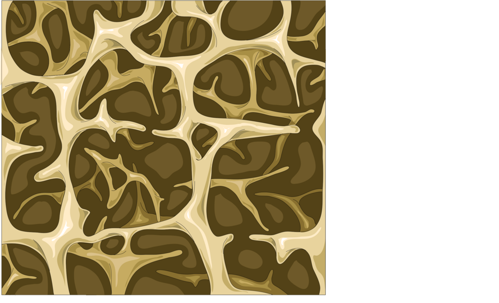
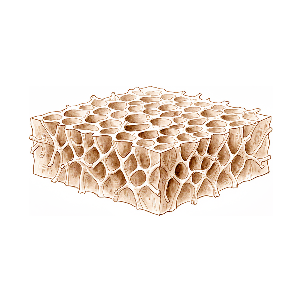
எலும்புச் சிதைவு நோய் (osteoporosis) என்பது உங்கள் எலும்புகள் வலிமையை இழக்கும் ஒரு நிலை. காலப்போக்கில் அவை மெலிந்து எளிதில் உடையக்கூடியவையாக ஆகின்றன; அதனால் ஒரு சிறிய விழுதலாலோ ஒரு சாதாரண மோதலாலோ கூட அவை முறியலாம். மிகப் பொதுவான முறிவுகள் இடுப்பு, மணிக்கட்டு, முதுகெலும்பு ஆகியவற்றில் நிகழ்கின்றன.
"பலவீன எலும்பு முறிவு" (fragility fracture) என்ற சொல்லை நீங்கள் கேட்கலாம். இருக்க வேண்டியதைவிட எளிதாக முறிந்த ஓர் எலும்பு என்று இதற்குப் பொருள். இப்படிப்பட்ட முறிவு உங்களுக்கு ஏற்பட்டிருந்தால், உங்கள் எலும்புகள் இயல்பைவிடப் பலவீனமாக உள்ளன என்பதற்கான எச்சரிக்கை அறிகுறியாக அது இருக்கலாம்.
இங்கே சிகிச்சையில் அறியப்பட்ட ஓர் இடைவெளி இருக்கிறது. இப்படி எலும்பு முறியும் மக்கள் பெரும்பாலும் எலும்புச் சிதைவு நோய்க்காகப் பரிசோதிக்கப்படுவதில்லை, அவர்களுக்குச் சிகிச்சையும் தொடங்கப்படுவதில்லை என்று ஆய்வுகள் கண்டறிந்துள்ளன [1]. வழிகாட்டு நெறிமுறைகள் இருந்தும் இது நடக்கிறது [2]. அடிப்படை எலும்பு பலவீனத்துக்குச் சிகிச்சை அளிப்பது இன்னொரு முறிவுக்கான வாய்ப்பைக் குறைக்கக்கூடும் என்பதால் இது முக்கியமானது. பலவீன எலும்பு முறிவுக்குப் பிறகு எலும்புச் சிதைவு நோய்க்குச் சிகிச்சை அளிப்பது, மேலும் முறிவுகள் ஏற்படும் அபாயத்தை 50% வரை குறைக்கவும், இறப்பு விகிதங்களை 30% வரை குறைக்கவும் முடியும் என்று ஆய்வுகள் காட்டியுள்ளன [3].
எலும்புகள் மாறாமல் இருப்பவை அல்ல. அவை தொடர்ந்து சிதைக்கப்பட்டு மீண்டும் கட்டமைக்கப்படும் உயிருள்ள திசு. எலும்பு சிதைவது, மீண்டும் கட்டமைக்கப்படுவதைவிட வேகமாக நிகழும்போது எலும்புச் சிதைவு நோய் உருவாகிறது; அதனால் எலும்பு அடர்த்தி குறைகிறது. முடிந்தவரை வலிமையான எலும்புகளைக் கட்டியெழுப்பி, பின்னர் அந்த வலிமையைத் தக்கவைத்துக்கொள்வதே தடுப்பின் நோக்கம் [4]. முதல் முறிவோ இன்னொரு முறிவோ ஒருபோதும் நிகழாதபடி, மேலும் எலும்பு இழப்பைத் தடுப்பதே சிகிச்சையின் நோக்கம் [4].
அறிந்துகொள்ள வேண்டிய ஒரு கண்டுபிடிப்பு: ஒன்பது ஆய்வுகளின் பகுப்பாய்வு ஒன்றில், உடல் நிறைக் குறியீட்டில் (body mass index) ஏற்படும் ஒவ்வொரு 1-அலகு அதிகரிப்பும், எலும்புச் சிதைவு நோய் அபாயத்தில் 9% குறைவுடன் தொடர்புடையதாகக் கண்டறியப்பட்டது [5]. வயது, ஹார்மோன்கள், ஒட்டுமொத்த உடல்நலம் போன்றவற்றுடன் சேர்த்து, உடல் எடை முழுப் படத்தின் ஒரு பகுதி மட்டுமே.
உங்களுக்கு எளிதாக எலும்பு முறிந்திருந்தால், அல்லது உங்கள் எலும்பு வலிமை பற்றி நீங்கள் கவலைப்பட்டால், எலும்பு அடர்த்திப் பரிசோதனை பற்றிக் கேளுங்கள். எலும்புச் சிதைவு நோயை முன்கூட்டியே கண்டறிவது, முறிவு நிகழும் முன் செயல்படுவதற்கான மிகச் சிறந்த வாய்ப்பை உங்களுக்குத் தருகிறது.
இது பலனளிக்கிறதா?
நேர்மையான பதில் இதுதான்: சிகிச்சை முன்கூட்டியே தொடங்கும்போதுதான் மிகச் சிறப்பாகப் பலனளிக்கிறது; அது நடைபெறும் விதத்தில் இன்னும் மேம்படுத்த இடம் இருக்கிறது. மணிக்கட்டு முறியும் மக்கள், வழக்கமான கவனிப்பைப் பெறும்போதைவிட, இதற்கென அர்ப்பணிக்கப்பட்ட ஒரு திட்டத்தின் பகுதியாக இருக்கும்போது எலும்புச் சிதைவு நோய்க்காக மிக அதிக அளவில் பரிசோதிக்கப்படுகிறார்கள் என்று ஆய்வுகள் கண்டறிந்துள்ளன: 23% உடன் ஒப்பிடும்போது 92% [6]. இடுப்பு எலும்போ முதுகெலும்பின் ஓர் எலும்போ முறியும் மக்களுக்கு, வழிகாட்டு நெறிமுறைகள் பரிந்துரைத்தாலும், எலும்பை வலுப்படுத்தும் சிகிச்சை பெரும்பாலும் தொடங்கப்படுவதே இல்லை என்று மற்ற ஆய்வுகள் கண்டறிந்துள்ளன [7] [2]. அதனால்தான், எந்த ஓர் எளிதான முறிவுக்குப் பிறகும் எலும்பு அடர்த்திப் பரிசோதனை பற்றிக் கேட்பது மிகவும் முக்கியம்.
எலும்புச் சிதைவு நோய் கண்டறியப்பட்ட பிறகு, சிகிச்சை எலும்புகள் குணமடைய உதவி, இன்னொரு முறிவுக்கான வாய்ப்பைக் குறைக்கக்கூடும் என்பதற்குச் சான்றுகள் உள்ளன. ஓர் ஆய்வில், எலும்பைக் கட்டமைக்கும் ஹார்மோன் ஊசி பெற்ற, மணிக்கட்டு முறிந்தவர்கள் 7.8 வாரங்களில் குணமடைந்தனர்; எந்தச் சிகிச்சையும் பெறாதவர்களுக்கு இது 12.6 வாரங்களாக இருந்தது [8]. உடற்பயிற்சிக்கும் ஒரு பங்கு உண்டு. எலும்புச் சிதைவு நோய் உள்ளவர்கள், எலும்பு வலிமையை அதிகப்படுத்த எதிர்ப்புப் பயிற்சியும் (resistance exercise) அதிர்வுப் பயிற்சியும் (impact exercise) செய்ய வேண்டும்; அத்துடன், விழுதல்களைக் குறைக்க வலிமையையும் சமநிலையையும் மேம்படுத்தும் செயல்பாடுகளையும், உடல் தோரணையை மேம்படுத்த முதுகெலும்பை நிமிர்த்தும் பயிற்சியையும் (spinal extension exercise) செய்ய வேண்டும் [9].
புதிய ஆய்வுகளில் சில இன்னும் உறுதியாகவில்லை. ஸ்கேன்கள் அல்லது இரத்தப் பரிசோதனைகளிலிருந்து எலும்புச் சிதைவு நோயைக் கணிக்கும் இயந்திரக் கற்றல் (machine learning) கருவிகள் நம்பிக்கை அளிக்கின்றன; ஆனால் அவற்றில் சார்புநிலைக்கான (bias) அபாயம் அதிகம் என்றும், அவை இன்னும் மருத்துவமனைகளில் முறையாகச் சோதிக்கப்படவில்லை என்றும் மீளாய்வுகள் கண்டறிந்துள்ளன [10]. மேலும், எலும்புகளைப் பாதுகாக்கும் என்று கருதப்பட்ட ஒரு நீரிழிவு மருந்து, பிற உடல்நலக் காரணிகளைக் கணக்கில் எடுத்த பிறகு, மணிக்கட்டு மற்றும் கை முறிவுகளைக் குறைக்கவில்லை என்று ஒரு பகுப்பாய்வு கண்டறிந்தது [11]. உங்களுக்கு எலும்புச் சிதைவு நோய் இருந்து, ஓர் எலும்பு முறிந்தால், அடிப்படை எலும்பு பலவீனத்துக்கும் அதே நேரத்தில் சிகிச்சை அளிக்கப்படும்போது, அந்த எலும்புக்கான சிகிச்சை சிறப்பாகப் பலனளிக்கிறது [12].
அபாயங்கள் என்ன?
எலும்புச் சிதைவு நோய்க்குச் சிகிச்சை அளிக்காமல் விடுவதன் முக்கிய அபாயம் எலும்பு முறிவுதான். உங்களுக்கு ஏற்கனவே ஒரு பலவீன எலும்பு முறிவு ஏற்பட்டிருந்தால், உங்கள் எலும்புகள் மீண்டும் முறிவதற்கான வாய்ப்பு அதிகம், குறிப்பாக அடிப்படை எலும்பு பலவீனத்துக்குச் சிகிச்சை அளிக்கப்படாவிட்டால் [7] [2]. முதல் முறிவுக்குப் பிறகு தொடங்கப்படும் சிகிச்சை இன்னொரு முறிவுக்கான வாய்ப்பைக் குறைக்கிறது என்று ஆய்வுகள் கண்டறிந்துள்ளன; முன்கூட்டியே செயல்படுவதற்கு இதுவே மிக வலுவான காரணம் [3].
முறிவு நிகழும் நேரத்தைச் சுற்றியும் அபாயங்கள் உள்ளன. எலும்புச் சிதைவு நோய் உள்ளவர்களுக்கு, அது இல்லாதவர்களைவிட, அறுவை சிகிச்சைக்குப் பிறகு முதல் 90 நாட்களில் அதிக மருத்துவச் சிக்கல்கள் ஏற்பட்டன என்று ஓர் ஆய்வு கண்டறிந்தது [13]. எலும்பு வலிமையைப் பொறுத்து, முறிந்த எலும்புகள் வெவ்வேறு விதமாகவும் குணமடையலாம். இடம் பெயர்ந்த மணிக்கட்டு முறிவுகள் பற்றிய ஓர் ஆய்வில், எலும்புச் சிதைவு நோய் உள்ளவர்களில் உருக்குலைவின் (deformity) முன்னேற்றம் 12.5 ஆக இருந்தது; இது நல்ல எலும்புத் தரம் உள்ளவர்களில் காணப்பட்ட 25.6-இல் பாதிக்கும் குறைவு [14]. வயதானவர்களின் பலவீனமான எலும்புகள், அவை மட்டுமே காரணமாக, ஒரு முறிவு ஒன்றுசேர்வதைத் தடுக்கவில்லை என்று மற்றோர் ஆய்வு கண்டறிந்தது [15].
உங்களுக்கு எலும்புச் சிதைவு நோய் இருந்து, ஒரு மூட்டிலோ தசைநாணிலோ அறுவை சிகிச்சை தேவைப்பட்டால், நிலைமை இன்னும் தெளிவாக உள்ளது. சாவித்துவார (keyhole) சுழற்சி தசைநாண் பட்டை (rotator cuff) சீரமைப்பு பற்றிய மீளாய்வுகள், குறைந்த எலும்பு அடர்த்திக்கும் 2 ஆண்டுகளில் கிடைக்கும் விளைவுகளுக்கும் இடையே எந்தத் தொடர்பையும் காணவில்லை; சிக்கல் விகிதங்களும் குறைவாக இருந்தன [16] [17]. இந்த அறுவை சிகிச்சையைத் தவிர்ப்பதற்கான காரணமாக எலும்புச் சிதைவு நோயைக் கருதக் கூடாது [17].
சில சிகிச்சைகளுக்கு அவற்றுக்கே உரிய நிச்சயமின்மைகள் உள்ளன. எலும்பை வலுப்படுத்துவதிலும் முறிவு அபாயத்தைக் குறைப்பதிலும், மூக்கு வழியாகத் தெளிக்கும் (nasal spray) வடிவிலான கால்சிடோனின் (calcitonin), மற்ற எலும்பு மருந்துகளைவிடக் குறைவாகவே பலனளிக்கிறது [18]. குத்தூசி மருத்துவம் (acupuncture) மற்றும் பாரம்பரிய சீன மருத்துவப் பொருட்களுக்கான சான்றுகள் ஆரம்ப நிலையில் உள்ளன; மேலும் சோதனைகள் தேவை [19] [20]. பாராதைராய்டு ஹார்மோன் (parathyroid hormone) ஊசிகளுக்கு, மருந்தளவுகளைக் கொடுப்பதற்கான சிறந்த நேரம் இன்னும் ஆய்வுகளில் (trials) ஆராயப்பட்டு வருகிறது [21].
நேர்மையான இடைவெளி, சிகிச்சையைத் தொடர்ந்து மேற்கொள்வதில்தான் உள்ளது. இடுப்பு, முதுகெலும்பு அல்லது மணிக்கட்டு முறியும் பலருக்கு எலும்பை வலுப்படுத்தும் சிகிச்சை ஒருபோதும் தொடங்கப்படுவதே இல்லை என்று ஆய்வுகள் காட்டுகின்றன [7] [2] [22]. எந்த ஓர் எளிதான முறிவுக்குப் பிறகும் சிகிச்சை பற்றிக் கேட்பதே நீங்கள் எடுக்கக்கூடிய மிகப் பாதுகாப்பான படி.
இது உங்களுக்குப் பொருத்தமானதா?
உங்கள் எலும்புகள் பலவீனமாக உள்ளன என்று உங்களுக்குச் சொல்லப்பட்டிருந்தால், அல்லது இருக்க வேண்டியதைவிட எளிதாக உங்களுக்கு ஓர் எலும்பு முறிந்திருந்தால், எலும்பை வலுப்படுத்தும் சிகிச்சையைக் கருத்தில் கொள்வது பயனுள்ளது. முதல் முறிவோ இன்னொரு முறிவோ நிகழும் முன் மேலும் எலும்பு இழப்பைத் தடுப்பதே நோக்கம் [4]. பலவீன எலும்பு முறிவுக்குப் பிறகான சிகிச்சையும் முக்கியமானது. அது இன்னொரு முறிவுக்கான வாய்ப்பைக் குறைத்து, இறப்பு விகிதங்களையும் குறைக்கக்கூடும் [3]. ஆனால் இடுப்பு, முதுகெலும்பு அல்லது மணிக்கட்டு முறியும் பலருக்குச் சிகிச்சை ஒருபோதும் தொடங்கப்படுவதே இல்லை என்று ஆய்வுகள் காட்டுகின்றன [7] [2]. எனவே, உங்களுக்கு எளிதான முறிவு ஏற்பட்டிருந்தால், எலும்பு அடர்த்திப் பரிசோதனை பற்றியும், சிகிச்சை உங்களுக்கு உதவுமா என்பது பற்றியும் உங்கள் மருத்துவரிடம் கேட்பது நியாயமானதே.
சிலருக்குச் சிகிச்சை தேவைப்படுவதற்கான வாய்ப்பு குறைவு. உங்கள் எலும்புகள் இன்னும் வலிமையாக இருந்தால், அவற்றை அப்படியே வைத்திருப்பதில் கவனம் செலுத்தப்படுகிறது: முடிந்தவரை வலிமையான எலும்புகளைக் கட்டியெழுப்பி, பின்னர் அந்த வலிமையைத் தக்கவைத்துக்கொள்வது [4]. இதில் உடற்பயிற்சிக்கு ஒரு பங்கு உண்டு. எலும்புச் சிதைவு நோய் உள்ளவர்கள், எலும்பு வலிமையை அதிகப்படுத்த எதிர்ப்புப் பயிற்சியும் அதிர்வுப் பயிற்சியும் செய்ய வேண்டும் [9]. உங்களுக்கு எது பாதுகாப்பானது என்பதைக் கண்டறிய உங்கள் மருத்துவர் உதவ முடியும்.
சிகிச்சை எல்லோருக்கும் ஒரே மாதிரியானது அல்ல. எலும்பு மருந்துகளில் பல வகைகள் உள்ளன; உங்களுக்குச் சரியானது உங்கள் உடல்நலம், உங்கள் முறிவு வரலாறு, உங்கள் சொந்த முன்னுரிமைகள் ஆகியவற்றைப் பொறுத்தது. இது நீங்களும் உங்கள் மருத்துவரும் சேர்ந்து எடுக்கும் முடிவாக இருக்க வேண்டும். ஒவ்வொரு வழியிலும் என்ன அடங்கியுள்ளது, சாத்தியமான பக்க விளைவுகள் என்ன (மேலே உள்ள அபாயங்கள் பகுதியில் விளக்கப்பட்டுள்ளன), எவ்வளவு காலம் நீங்கள் சிகிச்சையைத் தொடர வேண்டியிருக்கும் என்று கேளுங்கள். ஏதாவது தெளிவாக இல்லையென்றால், திட்டத்தில் நம்பிக்கை வரும் வரை கேட்டுக்கொண்டே இருங்கள்.
சுருக்கமாக
உங்கள் எலும்புகள் பலவீனமாக இருந்தால், அல்லது உங்களுக்கு எளிதாக ஓர் எலும்பு முறிந்திருந்தால், எலும்புச் சிதைவு நோய்க்கான சிகிச்சையைக் கருத்தில் கொள்வது பயனுள்ளது. ஒரு முறிவோ இன்னொரு முறிவோ நிகழும் முன் மேலும் எலும்பு இழப்பைத் தடுப்பதே நோக்கம் [4]. முன்கூட்டியே செயல்படுவது முக்கியம்; ஏனெனில், பலவீன எலும்பு முறிவுக்குப் பிறகான சிகிச்சை இன்னொரு முறிவுக்கான வாய்ப்பைக் குறைத்து, இறப்பு விகிதங்களையும் குறைக்கக்கூடும் [3]. மிக முக்கியமான ஒரே எச்சரிக்கை, சிகிச்சையைத் தொடர்ந்து மேற்கொள்வது பற்றியது: இடுப்பு, முதுகெலும்பு அல்லது மணிக்கட்டு முறியும் பலர் ஒருபோதும் பரிசோதிக்கப்படுவதோ சிகிச்சை பெறுவதோ இல்லை [7] [2]. எனவே, உங்களுக்கு எளிதான முறிவு ஏற்பட்டிருந்தால், எலும்பு அடர்த்திப் பரிசோதனையைக் கேளுங்கள்; ஒரு திட்டம் கிடைக்கும் வரை கேட்டுக்கொண்டே இருங்கள்.
மேற்கோள்கள்
[1] The osteoporosis care gap in Canada. BMC Musculoskeletal Disorders. 2004. DOI: 10.1186/1471-2474-5-11
[2] A Missed Opportunity? Osteoporosis Treatment Following Femoral Neck Fractures: Reducing the Risk of Secondary Hip Fracture. The Journal of Arthroplasty. 2026. DOI: 10.1016/j.arth.2025.07.028
[3] Medical management of osteoporosis and the doctor's' role. Injury. 2016. DOI: 10.1016/s0020-1383(16)30014-6
[4] BONE HEALTH AND OSTEOPOROSIS. Clinics in Sports Medicine. 2000. DOI: 10.1016/s0278-5919(05)70201-5
[5] The association between body mass index and osteoporosis, with consideration of sex differences: a systematic review and dose-response meta-analysis. BMC Musculoskeletal Disorders. 2026. DOI: 10.1186/s12891-026-09675-3
[6] Wristwatch—distal radial fracture as a marker for osteoporosis investigation:. Journal of Hand Therapy. 2004. DOI: 10.1197/j.jht.2004.04.001
[7] The Management of Pelvic and Vertebral Fragility Fractures: An Unmet Care Need. Journal of Bone and Joint Surgery. 2026. DOI: 10.2106/jbjs.26.00966
[8] Use of teriparatide in osteoporotic fracture patients. Injury. 2016. DOI: 10.1016/s0020-1383(16)30009-2
[9] Strong, steady and straight: UK consensus statement on physical activity and exercise for osteoporosis. British Journal of Sports Medicine. 2022. DOI: 10.1136/bjsports-2021-104634
[10] Risk prediction models for postmenopausal osteoporosis: a systematic review and meta-analysis study. BMC Musculoskeletal Disorders. 2026. DOI: 10.1186/s12891-025-09385-2
[11] Glucagon-like peptide-1 receptor agonist use for type 2 diabetes and risk of upper extremity fragility fracture. Injury. 2026. DOI: 10.1016/j.injury.2026.113456
[12] Continuity and volume of bone cement and anti osteoporosis treatment were guarantee of good clinical outcomes for percutaneous vertebroplasty: a multicenter study. BMC Musculoskeletal Disorders. 2025. DOI: 10.1186/s12891-024-08153-y
[13] Impact of osteoporosis on post-operative outcomes following rotator cuff repair. JSES Reviews, Reports, and Techniques. 2026. DOI: 10.1016/j.xrrt.2026.100723
[14] Osteoporosis and colles' fractures in the elderly. The Journal of Hand Surgery: Journal of the British Society for Surgery of the Hand. 1987. DOI: 10.1016/0266-7681(87)90058-1
[15] A89 Osteoporosis is not a risk factor for the development of nonunion: a cohort nested case-control study. Injury. 2011. DOI: 10.1016/s0020-1383(11)70106-1
[16] No difference in 2-year outcomes of arthroscopic rotator cuff repair in patients with osteoporosis. Journal of Shoulder and Elbow Surgery. 2025. DOI: 10.1016/j.jse.2025.02.011
[17] Osteoporosis is a risk factor for complications and reoperations at 1 and 3 years after arthroscopic rotator cuff repair. JSES International. 2026. DOI: 10.1016/j.jseint.2026.101678
[18] Revisiting Intranasal Salmon Calcitonin: Historical Osteoporosis Evidence and a Potential Role in Acute Orthopaedic Pain Management. JBJS Reviews. 2026. DOI: 10.2106/jbjs.rvw.26.00021
[19] Efficacy of acupuncture for primary osteoporosis: a systematic review and meta-analysis of randomized controlled trials. Journal of Orthopaedic Surgery and Research. 2025. DOI: 10.1186/s13018-025-05513-9
[20] Natural traditional Chinese medicine products: emerging therapeutic targets for the treatment of osteoporosis. Journal of Orthopaedic Surgery and Research. 2025. DOI: 10.1186/s13018-025-05879-w
[21] Timing optimization of teriparatide dosing for postmenopausal osteoporosis: a randomized controlled trial. Journal of Orthopaedic Surgery and Research. 2025. DOI: 10.1186/s13018-025-06083-6
[22] Incidence of Anti-osteoporosis Diagnosis and Treatment After Distal Radius Fractures. HAND. 2016. DOI: 10.1177/1558944716660555dj
Evidence & references
This is the clinical evidence summary written for health professionals. It is technical, and it lists the research this page was built from. You do not need to read it to understand your treatment or to make a decision about it.
Overview
- Prevention of osteoporosis is accomplished by maximizing peak bone density and maintaining that bone density [4].
- The goal of osteoporosis treatment is to stop further bone loss to prevent a fracture or any subsequent fragility fractures [4].
- Each 1-unit increase in body mass index is associated with a 9% reduction in osteoporosis risk [6].
- Current online health information on osteoporosis often fails to meet basic standards of evidence-based health information [3].
- There is evidence of a care gap between the occurrence of a fragility fracture and the diagnosis and treatment of osteoporosis in Canada [14].
- Treatment of the underlying osteoporosis to prevent secondary fractures is rarely initiated following pelvic and vertebral fragility fractures [5].
- Low rates of osteoporotic pharmacotherapy are observed in patients who have sustained femoral neck fractures despite established guidelines [33].
- Patients with osteoporosis may experience a higher incidence of medical complications within the 90-day global period following rotator cuff repair than nonosteoporotic patients [21].
- Osteoporosis should not be considered a contraindication to arthroscopic rotator cuff repair [31].
- Patients with distal radial fractures in elderly women should be screened for osteoporosis [11].
- Intranasal salmon calcitonin is less effective than bisphosphonates and modern anabolic agents for increasing bone mineral density and reducing fracture risk [12].
- The use of intranasal salmon calcitonin is best reserved for patients who cannot take first-line therapies due to contraindications, intolerance, or personal preference [12].
- Sequential anabolic-to-anti-resorptive therapy may inform treatment guidelines for high-risk postmenopausal populations [7].
- The ability of monocytes to fuse declines significantly in patients with osteoporosis [52].
How It Works
Pathophysiology and Mechanisms
- Hypothalamic-pituitary disconnection leads to osteoporosis with a reduction in both trabecular and cortical bone caused by a low bone turnover situation, with reduced osteoblast and osteoclast activity [26].
- MiR-137 promotes TLR4/NF-κB pathway activity through targeting KDM4A, which inhibits osteogenic differentiation of human bone marrow mesenchymal stem cells and aggravates osteoporosis [36].
- The gut microbiota–metabolite–bone network may be involved in the skeletal effects of leptin, providing mechanistic insights for osteoporosis management [35].
- Macrophage polarization-related genes have potential causal roles in osteoporosis, though these findings are exploratory and require validation in bone marrow-specific and functional studies [34].
- Evidence of early changes in bone geometry and increased bone turnover could be used to detect osteoporosis before estrogen deficiency has adversely affected bone strength [24].
Fracture Healing and Bone Repair
- In humans, de novo use of bisphosphonate therapy after fracture does not appear to have a significant effect on fracture healing [30].
- Prior bisphosphonate treatment affects the mineralization pattern of tissue repair consistently with a reduction in bone turnover, but does not abolish the repair process [23].
- The effect of ovariectomy on fracture repair is to delay healing, with the deleterious effect potentially related more to change of structure than to loss of bone mineral density [39].
- Fracture healing in SAMP6 mice is not detrimentally affected by impairment of bone marrow stem cell osteogenesis, suggesting that bone marrow-mediated repair processes are dispensable for normal bone healing in this senile osteoporotic fracture model [42].
- There is some promising experimental and clinical evidence for possible enhancement of the bone repair process via administration of systemic agents [25].
- Bisphosphonate treatment results in cracks that are shorter than in control or ovariectomized groups, which may explain why increased damage levels following bisphosphonate treatment have not led to increased fractures [38].
- Osteoporosis is not a risk factor for the development of nonunion, although bone quality may be diminished in the elderly [44].
Risk Factors and Screening
- Each 1-unit increase in body mass index was associated with a 9% reduction in osteoporosis risk based on a dose-response analysis of nine studies [6].
- Postmenopausal women with distal radius fractures have diminished bone mass, which might make a bone more likely to break [43].
- Patients with distal radial fractures should be screened for osteoporosis [11].
- A simple inexpensive tool has potential to increase the rate of osteoporosis investigation in an at-risk population [19].
- Machine learning application to hand radiographs represents a possible step toward more accessible, cost-effective, automated diagnosis and therefore earlier treatment of osteoporosis or osteopenia [10].
- A nomogram with an online dynamic calculator could facilitate the early prediction, diagnosis, and treatment of osteoporosis, contributing to the bone health of the elderly population [13].
- A bone density based aging model may facilitate and support the development of precision medicine strategies in osteoporosis prevention and management [1].
Treatment and Management
- Prevention of osteoporosis can be accomplished by maximizing peak bone density and maintaining that bone density, although the goal of treatment is to stop further bone loss to prevent a fracture or any subsequent fragility fractures [4].
- Randomized controlled trials have demonstrated that treatment of osteoporosis in patients with fragility fractures can reduce the risk of subsequent fractures by up to 50% and mortality rates by up to 30% [18].
- Intranasal salmon calcitonin is less effective than bisphosphonates and modern anabolic agents for increasing bone mineral density and reducing fracture risk, and its use is best reserved for patients who cannot take first-line therapies due to contraindications, intolerance, or personal preference [12].
- The current evidence suggests that the efficacy of acupuncture in improving the symptoms of primary osteoporosis is encouraging for its use in clinical practice as a physical intervention [17].
- Treatment of the underlying osteoporosis to prevent secondary fractures is rarely initiated, and treatments vary widely and are not always evidence-based [5].
- A program for secondary osteoporotic fracture prevention is needed following distal radius fractures [27].
- Natural traditional Chinese medicine products provide a theoretical and experimental basis for the development of new drugs and the improvement of osteoporosis management [22].
- A calcitonin release system confirms good in vivo behavior and a therapeutic effect of the released calcitonin in the osteopenic condition [28].
- Timing optimization of teriparatide dosing may guide personalized dosing strategies to enhance bone formation and reduce fracture risk in osteoporosis [15].
Clinical Outcomes and Surgical Considerations
- There is no relationship between decreased bone mineral density and 2-year clinical outcomes following arthroscopic rotator cuff repair in patients with osteoporosis [20].
- Findings from studies on long-term hospitalized patients with muscular dystrophy may help inform strategies for osteoporotic treatment and nutritional care in this patient population [8].
Research Models and Assessment
- Investigators must select an osteoporotic fracture model that best reflects the clinical problem being studied and the underlying pathophysiology of the osteoporosis in the target patient group [2].
- There is no perfect model for osteoporosis, but a variety of models are appropriate for answering specific questions [9].
- Multiscale biomechanical assessment approaches have potential applications in fracture risk assessment, the design of patient-specific implants, and research into the mechanisms of bone metabolic diseases [37].
- Current online health information on osteoporosis often fails to meet basic standards of evidence-based health information, highlighting the urgent need to improve quality to support informed decision-making [3].
What the Evidence Shows
Prevention and Risk Factors
- Prevention of osteoporosis involves maximizing peak bone density and maintaining that bone density [4].
- A bone density-based aging model may facilitate the development of precision medicine strategies in osteoporosis prevention and management [1].
- A nomogram with an online dynamic calculator could facilitate the early prediction, diagnosis, and treatment of osteoporosis [13].
- Machine learning-based prediction models for postmenopausal osteoporosis without fractures demonstrate good discriminative ability but are characterized by a high risk of bias, a lack of calibration performance evaluation, and insufficient validation of clinical utility [56].
- A fracture risk prediction model based on routine blood test markers was successfully developed and validated for patients newly diagnosed with osteoporosis [49].
- Diabetes shows varying associations with bone mineral density at different sites, with significant associations found for type 2 diabetes on femoral neck and total body BMD, and HbA1c on heel BMD [54].
- After adjusting for metabolic confounds, the skeletal benefit of glucagon-like peptide-1 receptor agonists did not translate to meaningful risk reductions in upper extremity fragility fracture [48].
Diagnosis and Screening
- Application of machine learning to hand radiographs represents a possible step toward more accessible, cost-effective, automated diagnosis and earlier treatment of osteoporosis or osteopenia [10].
- A simple inexpensive tool has the potential to increase the rate of osteoporosis investigation in an at-risk population [19].
- In a study of distal radial fractures, 92% of intervention subjects were investigated for osteoporosis compared with 23% in the usual-care group [29].
- The treatment of underlying osteoporosis to prevent secondary fractures is rarely initiated after pelvic and vertebral fragility fractures [5].
Pharmacological Treatment
- A randomized controlled trial is expected to provide insights into optimizing teriparatide administration timing to enhance bone formation and reduce fracture risk [15].
- Acupuncture has encouraging efficacy in improving the symptoms of primary osteoporosis for use in clinical practice as a physical intervention [17].
- Jintiange capsules are a good choice for patients with osteoporosis in terms of relieving pain, improving bone mineral density, improving activity function, improving gait, and preventing fracture [51].
Surgical and Orthopaedic Considerations
- There is no relationship between decreased bone mineral density and 2-year clinical outcomes following arthroscopic rotator cuff repair [20].
- Patients with osteoporosis may experience a higher incidence of medical complications within the 90-day global period than nonosteoporotic patients following rotator cuff repair [21].
- Continuous bone cement and standardized treatment for osteoporosis were guarantees of good clinical outcomes for percutaneous vertebroplasty [46].
- Injected bone cement greater than 5.5 ml might be a guarantee of good clinical outcomes for percutaneous vertebroplasty [46].
- Key challenges in enrollment in geriatric fracture trials comparing non-surgical management to surgical management were identified for pelvic fragility fractures [47].
- A phosphocalcic cement technique could be employed in the future to prevent the occurrence of fractures in osteoporotic patients [50].
Fracture Healing and Bone Repair
- There is promising experimental and clinical evidence for possible enhancement of the bone repair process via administration of systemic agents [25].
- Patients who received parathyroid hormone (1–84) injections accelerated radiographic and clinical fracture healing to 7.8 weeks compared to 12.6 weeks in patients who received no treatment [45].
Physical Activity and Lifestyle
- People with osteoporosis should undertake resistance and impact exercise to maximize bone strength [32].
- People with osteoporosis should undertake activities to improve strength and balance to reduce falls [32].
- People with osteoporosis should undertake spinal extension exercise to improve posture and potentially reduce risk of falls and vertebral fractures [32].
Research Models
- Hypothalamic-pituitary disconnection leads to osteoporosis with a reduction in both trabecular and cortical bone caused by a low bone turnover situation with reduced osteoblast and osteoclast activity after 12 months [26].
Practical Considerations
Prevention and Risk Assessment
- A bone density based aging model approach may facilitate the development of precision medicine strategies in osteoporosis prevention and management [1].
- Machine learning application to hand radiographs represents a possible step toward more accessible, cost-effective, automated diagnosis and earlier treatment of osteoporosis or osteopenia [10].
Screening and Diagnosis
- A simple inexpensive tool for distal radius fractures has potential to increase the rate of osteoporosis investigation in an at-risk population [19].
- Intervention subjects with distal radial fractures were investigated for osteoporosis at a rate of 92%, compared with 23% in the usual-care group [29].
- Patients with hand fragility fractures represent a high-risk cohort that is currently underserved by osteoporosis screening pathways [40].
Pharmacological Management
- Treatment of osteoporosis in patients with fragility fractures can reduce the risk of subsequent fractures by up to 50% and mortality rates by up to 30% [18].
- Randomized controlled trials are expected to provide insights into optimizing teriparatide administration timing to enhance bone formation and reduce fracture risk [15].
- The efficacy of acupuncture in improving the symptoms of primary osteoporosis is encouraging for its use in clinical practice as a physical intervention [17].
Non-Pharmacological Management
Clinical Gaps and Care Delivery
- Despite established guidelines, low rates of osteoporotic pharmacotherapy were seen in patients who had femoral neck fractures [33].
- Osteoporosis continues to be grossly underdiagnosed in the context of sacral fragility fractures [41].
- Surgical treatment remains underutilized for sacral fragility fractures [41].
Surgical Considerations
Key Evidence
- [L3] This approach may facilitate and support the development of precision medicine strategies in osteoporosis prevention and management. [1] (10.1186/s12891-025-09298-0)
- [L5] Investigators must select a model that best reflects the clinical problem being studied, and the underlying pathophysiology of the osteoporosis in the target patient group. [2] (10.1016/s0020-1383(16)30004-3)
- [Paper] Current OHI on osteoporosis often fails to meet basic standards of EBHI, highlighting the urgent need to improve the quality to support informed decision-making. [3] (10.1186/s12891-026-09711-2)
- [Paper] Prevention of osteoporosis can be accomplished by maximizing peak bone density and maintaining that bone density, although the goal of treatment is to stop further bone loss to prevent a fracture or any subsequent fragility fractures. [4] (10.1016/s0278-5919(05)70201-5)
- [L5] The treatments vary widely and are not always evidence-based and, unfortunately, treatment of the underlying osteoporosis to prevent secondary fractures is rarely initiated. [5] (10.2106/jbjs.26.00966)
- [L1] Based on our dose–response analysis of nine studies, each 1-unit increase in BMI was associated with a 9% reduction in osteoporosis risk. [6] (10.1186/s12891-026-09675-3)
- [L1] This sequential anabolic-to-anti-resorptive therapy may inform treatment guidelines for high-risk postmenopausal populations. [7] (10.1186/s13018-025-06040-3)
- [L4] These findings may help inform strategies for osteoporotic treatment and nutritional care in this patient population. [8] (10.1186/s12891-026-10045-2)
- [Paper] However, there is no perfect model for osteoporosis, but a variety of models appropriate for answering specific questions. [9] (10.1016/s0020-1383(16)30002-x)
- [L2] The findings represent a possible step toward more accessible, cost-effective, automated diagnosis and therefore earlier treatment of osteoporosis/osteopenia. [10] (10.1016/j.jhsa.2024.09.008)
- [L4] It is the authors' opinion that such patients should be screened for osteoporosis. [11] (10.1016/j.jhsb.2004.05.002)
- [Paper] IN-CAL is less effective than bisphosphonates and modern anabolic agents for increasing BMD and reducing fracture risk; accordingly, its use is best reserved for patients who cannot take first-line therapies due to contraindications, intolerance, or personal preference. [12] (10.2106/jbjs.rvw.26.00021)
- [L3] Ultimately, this tool could facilitate the early prediction, diagnosis, and treatment of osteoporosis, thus contributing to the bone health of the elderly population and promoting the development of public health. [13] (10.1186/s12891-026-09920-9)
- [L4] There is evidence of a care gap between the occurrence of a fragility fracture and the diagnosis and treatment of osteoporosis in Canada. [14] (10.1186/1471-2474-5-11)
- [L2] This trial is expected to provide crucial insights into optimizing teriparatide administration timing, potentially guiding personalized dosing strategies to enhance bone formation and reduce fracture risk in osteoporosis. [15] (10.1186/s13018-025-06083-6)
- [L1] The current evidence suggests that the efficacy of acupuncture in improving the symptoms of primary osteoporosis is encouraging for its use in clinical practice as a physical intervention. [17] (10.1186/s13018-025-05513-9)
- [Paper] Randomized controlled trials have demonstrated that treatment of osteoporosis in patients with fragility fractures can reduce the risk of subsequent fractures by up to 50% and mortality rates by up to 30%. [18] (10.1016/s0020-1383(16)30014-6)
- [L1] This simple inexpensive tool has potential to increase the rate of osteoporosis investigation in an at-risk population. [19] (10.1197/j.jht.2003.10.030)
- [L3] Despite previous literature showing the negative effect of osteoporosis on rotator cuff healing, our data showed no relationship between decreased bone mineral density and 2-year clinical outcomes following RCR. [20] (10.1016/j.jse.2025.02.011)
- [L3] In addition, patients with osteoporosis may experience a higher incidence of medical complications within the 90-day global period than nonosteoporotic patient. [21] (10.1016/j.xrrt.2026.100723)
- [L4] This paper summarises recent research progress on natural TCM products in preventing and treating osteoporosis and provides a theoretical and experimental basis for the development of new drugs and the improvement of osteoporosis management. [22] (10.1186/s13018-025-05879-w)
- [L4] Prior bisphosphonate treatment affects the mineralization pattern of the tissue repair consistently with a reduction in bone turnover as found in post-menopausal osteoporotic women, but does not abolish the repair process. [23] (10.1002/jor.22026)
- [Paper] Therefore, evidence of early changes in bone geometry and increased bone turnover could be used to detect osteoporosis before estrogen deficiency has adversely affected bone strength. [24] (10.1002/jor.20961)
- [Paper] There is some promising experimental and clinical evidence for possible enhancement of the bone repair process via administration of systemic agents. [25] (10.1016/s0020-1383(16)30003-1)
- [Paper] HPD leads after 12 month to osteoporosis with a reduction in both trabecular and cortical bone caused by a low bone turnover situation, with reduced osteoblast and osteoclast activity, as compared to controls (OVX). [26] (10.1002/jor.22066)
- [L4] The results of our study evidenced the need of a program for secondary osteoporotic fracture prevention; this program has now been developed and is active at our institution. [27] (10.1177/1558944716660555dj)
- [Paper] These data confirm a good in vivo behavior of the system, as well as a therapeutic effect of the released calcitonin in the osteopenic condition. [28] (10.1016/s0736-0266(01)00074-7)
- [L2] Results indicate that 92% of the intervention subjects were investigated for osteoporosis, compared with the usual-care group, in which only 23% were investigated. [29] (10.1197/j.jht.2004.04.001)
- [L4] In humans, de novo use of bisphosphonate therapy after fracture does not appear to have a significant effect on fracture healing. [30] (10.1016/s0020-1383(16)30015-8)
- [L3] Overall rates of complication were low and osteoporosis should not be considered a contraindication to arthroscopic RCR. [31] (10.1016/j.jseint.2026.101678)
- [L5] Key recommendations are that people with osteoporosis should undertake (1) resistance and impact exercise to maximise bone strength; (2) activities to improve strength and balance to reduce falls; (3) spinal extension exercise to improve posture and potentially reduce risk of falls and vertebral fractures. [32] (10.1136/bjsports-2021-104634)
- [L3] Despite established guidelines, low rates of osteoporotic pharmacotherapy were seen in patients who had femoral neck fractures. [33] (10.1016/j.arth.2025.07.028)
- [Paper] These findings are exploratory and hypothesis-generating and require validation in bone marrow-specific and functional studies. [34] (10.1186/s13018-026-06905-1)
- [Paper] These findings suggest that the gut microbiota–metabolite–bone network may be involved in the skeletal effects of leptin, providing novel mechanistic insights and potential therapeutic strategies for osteoporosis management. [35] (10.1186/s12891-026-09950-3)
- [Paper] This mechanism inhibits osteogenic differentiation of human bone marrow mesenchymal stem cells and aggravates osteoporosis. [36] (10.1186/s13018-023-03918-y)
- [L5] It explores the potential applications of these multiscale approaches in fracture risk assessment, the design of patient-specific implants, and research into the mechanisms of bone metabolic diseases. [37] (10.1016/j.injury.2026.113659)
- [Paper] However, these cracks were shorter than in control or OVX groups which may provide one explanation as to why increased damage levels following bisphosphonate treatment have not lead to increased fractures. [38] (10.1002/jor.21229)
- [Paper] The effect of OVX on fracture repair was to delay healing as evidenced by histological and radiological analysis, and the evidence suggests that the deleterious effect might be more related to change of structure than to loss of BMD. [39] (10.1002/jor.20505)
- [L3] Patients with hand fragility fractures represent a high-risk cohort that is currently underserved by osteoporosis screening pathways. [40] (10.1016/j.injury.2026.113261)
- [L4] While cross-sectional imaging use has expanded, surgical treatment remains underutilized, and osteoporosis continues to be grossly underdiagnosed. [41] (10.1016/j.injury.2025.112850)
- [Paper] Fracture healing in SAMP6 mice is not detrimentally affected by impairment of BMSC osteogenesis, suggesting that bone marrow-mediated repair processes are dispensable for normal bone healing in this senile osteoporotic fracture model. [42] (10.1002/jor.21041)
- [L4] They also have diminished bone mass, which might make a bone more likely to break. [43] (10.1016/0363-5023(88)90227-4)
- [L3] Although bone quality may be diminished in the elderly, this does not influence the occurrence of nonunion. [44] (10.1016/s0020-1383(11)70106-1)
- [Paper] Patients who received the PTH (1–84) injections accelerated radiographic and clinical fracture healing (7.8 weeks) when compared to patients who received no treatment (12.6 weeks). [45] (10.1016/s0020-1383(16)30009-2)
- [L3] Continuous bone cement and standardized treatment for osteoporosis were guarantees of good clinical outcomes for PVP, and injected bone cement >5.5 ml might be a guarantee. [46] (10.1186/s12891-024-08153-y)
- [L2] Key challenges in enrollment in geriatric fracture trials comparing NSM to SM were identified. [47] (10.1016/j.injury.2025.112462)
- [L3] After adjusting for metabolic confounds, we found that the postulated skeletal benefit of GLP 1RAs did not translate to meaningful risk reductions in this population. [48] (10.1016/j.injury.2026.113456)
- [L3] We successfully developed and validated a fracture risk prediction model for patients newly diagnosed with osteoporosis. [49] (10.1186/s12891-026-09768-z)
- [L5] This technique could be employed in the future to prevent the occurrence of fractures in osteoporotic patients. [50] (10.1016/j.main.2003.12.003)
- [L1] In terms of relieving pain, improving BMD, improving activity function, and improving gait and preventing fracture, JTG is a good choice for patients with osteoporosis (OP). [51] (10.1186/s12891-025-08694-w)
- [L4] It was clearly demonstrated that the ability of monocytes to fuse declines significantly in patients with osteoporosis. [52] (10.1002/jor.1100180215)
- [Paper] The results show varying associations across different BMD sites (femoral neck, lumbar spine, heel, total body) and methods (IVW, MR-Egger, Weighted median), with some significant associations found for T2D on femoral neck and total body BMD, and HbA1c on heel BMD. [54] (10.1186/s12891-024-07430-0)
- [L1] Current machine learning-based prediction models for postmenopausal osteoporosis without fractures demonstrate good discriminative ability but are generally characterized by a high risk of bias, a notable lack of calibration performance evaluation, and insufficient validation of clinical utility. [56] (10.1186/s12891-025-09385-2)
References
[1] Unveiling risk factors and predicting osteoporosis through bone density based aging model: a community-based cohort in Guangdong, China. BMC Musculoskeletal Disorders. 2025. DOI: 10.1186/s12891-025-09298-0
[2] Main differences in osteoporotic fracture models: which should I use?. Injury. 2016. DOI: 10.1016/s0020-1383(16)30004-3
[3] Mapping the quality of information on osteoporosis: a cross-sectional analysis of online health information. BMC Musculoskeletal Disorders. 2026. DOI: 10.1186/s12891-026-09711-2
[4] BONE HEALTH AND OSTEOPOROSIS. Clinics in Sports Medicine. 2000. DOI: 10.1016/s0278-5919(05)70201-5
[5] The Management of Pelvic and Vertebral Fragility Fractures: An Unmet Care Need. Journal of Bone and Joint Surgery. 2026. DOI: 10.2106/jbjs.26.00966
[6] The association between body mass index and osteoporosis, with consideration of sex differences: a systematic review and dose-response meta-analysis. BMC Musculoskeletal Disorders. 2026. DOI: 10.1186/s12891-026-09675-3
[7] Effectiveness of anabolic and anti-resorptive agents for preventing postmenopausal osteoporosis fractures: a systematic review and network meta-analysis. Journal of Orthopaedic Surgery and Research. 2025. DOI: 10.1186/s13018-025-06040-3
[8] Osteoporosis in long-term hospitalized patients with muscular dystrophy: a retrospective cross-sectional study. BMC Musculoskeletal Disorders. 2026. DOI: 10.1186/s12891-026-10045-2
[9] Can we induce osteoporosis in animals comparable to the human situation?. Injury. 2016. DOI: 10.1016/s0020-1383(16)30002-x
[10] Application of Machine Learning to Osteoporosis and Osteopenia Screening Using Hand Radiographs. The Journal of Hand Surgery. 2025. DOI: 10.1016/j.jhsa.2024.09.008
[11] The Distal Radial Fracture in Elderly Women and the Bone Mineral Density of the Lumbar Spine and Hip. Journal of Hand Surgery. 2004. DOI: 10.1016/j.jhsb.2004.05.002
[12] Revisiting Intranasal Salmon Calcitonin: Historical Osteoporosis Evidence and a Potential Role in Acute Orthopaedic Pain Management. JBJS Reviews. 2026. DOI: 10.2106/jbjs.rvw.26.00021
[13] A nomogram with online dynamic calculator for predicting osteoporosis: development and validation based on NHANES. BMC Musculoskeletal Disorders. 2026. DOI: 10.1186/s12891-026-09920-9
[14] The osteoporosis care gap in Canada. BMC Musculoskeletal Disorders. 2004. DOI: 10.1186/1471-2474-5-11
[15] Timing optimization of teriparatide dosing for postmenopausal osteoporosis: a randomized controlled trial. Journal of Orthopaedic Surgery and Research. 2025. DOI: 10.1186/s13018-025-06083-6
[17] Efficacy of acupuncture for primary osteoporosis: a systematic review and meta-analysis of randomized controlled trials. Journal of Orthopaedic Surgery and Research. 2025. DOI: 10.1186/s13018-025-05513-9
[18] Medical management of osteoporosis and the surgeons' role. Injury. 2016. DOI: 10.1016/s0020-1383(16)30014-6
[19] Wristwatch: distal radius fractures as a marker for osteoporosis investigation. Journal of Hand Therapy. 2004. DOI: 10.1197/j.jht.2003.10.030
[20] No difference in 2-year outcomes of arthroscopic rotator cuff repair in patients with osteoporosis. Journal of Shoulder and Elbow Surgery. 2025. DOI: 10.1016/j.jse.2025.02.011
[21] Impact of osteoporosis on post-operative outcomes following rotator cuff repair. JSES Reviews, Reports, and Techniques. 2026. DOI: 10.1016/j.xrrt.2026.100723
[22] Natural traditional Chinese medicine products: emerging therapeutic targets for the treatment of osteoporosis. Journal of Orthopaedic Surgery and Research. 2025. DOI: 10.1186/s13018-025-05879-w
[23] Mineralization pattern of vertebral bone material following fragility fracture of the spine. Journal of Orthopaedic Research. 2012. DOI: 10.1002/jor.22026
[24] Structural adaptation and intracortical bone turnover in an ovine model of osteoporosis. Journal of Orthopaedic Research. 2009. DOI: 10.1002/jor.20961
[25] Fracture repair: general aspects and influence of osteoporosis and anti-osteoporosis treatment. Injury. 2016. DOI: 10.1016/s0020-1383(16)30003-1
[26] Low turnover osteoporosis in sheep induced by hypothalamic‐pituitary disconnection. Journal of Orthopaedic Research. 2012. DOI: 10.1002/jor.22066
[27] Incidence of Anti-osteoporosis Diagnosis and Treatment After Distal Radius Fractures. HAND. 2016. DOI: 10.1177/1558944716660555dj
[28] Calcitonin release system in the treatment of experimental osteoporosis. Histomorphometric evaluation. Journal of Orthopaedic Research. 2001. DOI: 10.1016/s0736-0266(01)00074-7
[29] Wristwatch—distal radial fracture as a marker for osteoporosis investigation:. Journal of Hand Therapy. 2004. DOI: 10.1197/j.jht.2004.04.001
[30] How do bisphosphonates affect fracture healing?. Injury. 2016. DOI: 10.1016/s0020-1383(16)30015-8
[31] Osteoporosis is a risk factor for complications and reoperations at 1 and 3 years after arthroscopic rotator cuff repair. JSES International. 2026. DOI: 10.1016/j.jseint.2026.101678
[32] Strong, steady and straight: UK consensus statement on physical activity and exercise for osteoporosis. British Journal of Sports Medicine. 2022. DOI: 10.1136/bjsports-2021-104634
[33] A Missed Opportunity? Osteoporosis Treatment Following Femoral Neck Fractures: Reducing the Risk of Secondary Hip Fracture. The Journal of Arthroplasty. 2026. DOI: 10.1016/j.arth.2025.07.028
[34] Macrophage polarization-related genes with potential causal roles in osteoporosis: a multi-omics Mendelian randomization study. Journal of Orthopaedic Surgery and Research. 2026. DOI: 10.1186/s13018-026-06905-1
[35] Role and mechanism of leptin in improving osteoporosis via the “gut–bone axis”. BMC Musculoskeletal Disorders. 2026. DOI: 10.1186/s12891-026-09950-3
[36] MiR-137 promotes TLR4/NF-κB pathway activity through targeting KDM4A, inhibits osteogenic differentiation of human bone marrow mesenchymal stem cells and aggravates osteoporosis. Journal of Orthopaedic Surgery and Research. 2023. DOI: 10.1186/s13018-023-03918-y
[37] Progress in Multiscale Biomechanical Assessment of Cancellous Bone: From Bone Density to Multiscale Mechanical Phenotypes. Injury. 2026. DOI: 10.1016/j.injury.2026.113659
[38] Effects of estrogen deficiency and bisphosphonate therapy on osteocyte viability and microdamage accumulation in an ovine model of osteoporosis. Journal of Orthopaedic Research. 2010. DOI: 10.1002/jor.21229
[39] Effect of osteoporosis on bone mineral density and fracture repair in a rat femoral fracture model. Journal of Orthopaedic Research. 2007. DOI: 10.1002/jor.20505
[40] Hand fragility fractures as a missed opportunity for secondary prevention: A retrospective cohort study and assessment of the Second Metacarpal Cortical Index. Injury. 2026. DOI: 10.1016/j.injury.2026.113261
[41] Epidemiology, imaging, and management trends in sacral fragility fractures: A 19-year nationwide analysis in Germany. Injury. 2025. DOI: 10.1016/j.injury.2025.112850
[42] Influence of defective bone marrow osteogenesis on fracture repair in an experimental model of senile osteoporosis. Journal of Orthopaedic Research. 2010. DOI: 10.1002/jor.21041
[43] Postural stability and bone density in postmenopausal women with distal radius fractures. The Journal of Hand Surgery. 1988. DOI: 10.1016/0363-5023(88)90227-4
[44] A89 Osteoporosis is not a risk factor for the development of nonunion: a cohort nested case-control study. Injury. 2011. DOI: 10.1016/s0020-1383(11)70106-1
[45] Use of teriparatide in osteoporotic fracture patients. Injury. 2016. DOI: 10.1016/s0020-1383(16)30009-2
[46] Continuity and volume of bone cement and anti osteoporosis treatment were guarantee of good clinical outcomes for percutaneous vertebroplasty: a multicenter study. BMC Musculoskeletal Disorders. 2025. DOI: 10.1186/s12891-024-08153-y
[47] Randomized trial of surgery vs. non-surgical management for pelvic fragility fractures. Injury. 2025. DOI: 10.1016/j.injury.2025.112462
[48] Glucagon-like peptide-1 receptor agonist use for type 2 diabetes and risk of upper extremity fragility fracture. Injury. 2026. DOI: 10.1016/j.injury.2026.113456
[49] SuperLearner approach for predicting imminent risk of fracture in older Chinese patients with newly diagnosed osteoporosis based on their routine blood test markers. BMC Musculoskeletal Disorders. 2026. DOI: 10.1186/s12891-026-09768-z
[50] Augmentation expérimentale de la résistance du radius distal ostéoporotique par un ciment phosphocalcique. Chirurgie de la Main. 2004. DOI: 10.1016/j.main.2003.12.003
[51] The effect of Jintiange capsules on pain in patients with primary osteoporosis: a systematic review and meta-analysis. BMC Musculoskeletal Disorders. 2025. DOI: 10.1186/s12891-025-08694-w
[52] Ability of osteoclast formation from peripheral monocytes using anti‐fusion regulatory protein‐1/cd98/4f2 monoclonal antibodies in patients with osteoporosis. Journal of Orthopaedic Research. 2000. DOI: 10.1002/jor.1100180215
[54] Diabetes and osteoporosis: a two-sample mendelian randomization study. BMC Musculoskeletal Disorders. 2024. DOI: 10.1186/s12891-024-07430-0
[56] Risk prediction models for postmenopausal osteoporosis: a systematic review and meta-analysis study. BMC Musculoskeletal Disorders. 2026. DOI: 10.1186/s12891-025-09385-2