Patients › Wrist
TFCC பழுதுபார்ப்பு அறுவை சிகிச்சை (TFCC Repair)
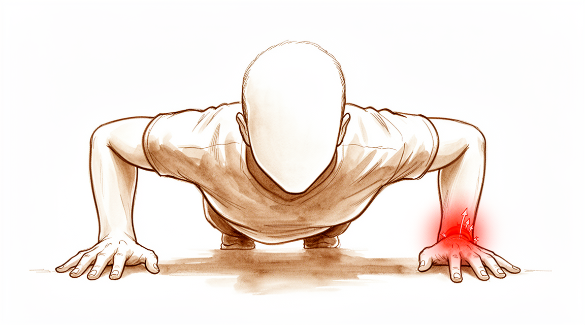
இந்த அறுவை சிகிச்சை ஏன் பரிந்துரைக்கப்பட்டுள்ளது
Mater Private Hospital Rockhampton-இல் மேல் அவயவ (upper-limb) அறுவை சிகிச்சை நிபுணரான டாக்டர் கியரன் ஹிர்பரா, உங்கள் நிலைக்குப் பொருத்தமான, உடலுக்குள் மிகக் குறைவாகத் தலையிடும் (least invasive) தேர்வுகளுடன் தொடங்குகிறார். TFCC என்பது உங்கள் மணிக்கட்டின் சுண்டுவிரல் பக்கத்தில் உள்ள, குருத்தெலும்பும் தசைநாரும் (ligament) சேர்ந்த ஓர் அமைப்பு. பழுதுபார்ப்பு (repair) என்றால், அது கிழிந்த இடத்தில் அதை மீண்டும் தைத்து இணைப்பது. நாங்கள் பொதுவாக முதலில் கை சிகிச்சை (hand therapy), சிம்பு (splint) அணிவது, செயல்களை மாற்றிக்கொள்வது ஆகியவற்றை முயற்சிக்கிறோம்; அதன் பிறகும் வலியோ நிலையின்மையோ தொடர்ந்தால் அறுவை சிகிச்சையைப் பரிசீலிக்கிறோம். நோயாளிகள் பொதுவாக அவர்களுடைய GP (பொது மருத்துவர்) மூலம் எங்கள் கிளினிக்கிற்குப் பரிந்துரைக்கப்படுகின்றனர்; ஒரு இயன்முறை சிகிச்சையாளர் (physiotherapist) எங்களைப் பார்க்கும்படி பரிந்துரைத்திருந்தாலும், Medicare தள்ளுபடியைப் (rebate) பெறத் தகுதி பெற உங்கள் GP-யிடமிருந்து பரிந்துரைக் கடிதம் (referral) இன்னும் தேவைப்படும். உங்கள் மருத்துவ வரலாறு, பரிசோதனை, தேவைப்படும் இடங்களில் ஸ்கேன் படங்கள் ஆகியவற்றுடன் கூடிய கிளினிக் மதிப்பீடு, நோயைக் கண்டறிகிறது.
பழுதுபார்க்கக்கூடிய கிழிவுகளுக்கு, மூட்டில் மூட்டழற்சி (arthritis) மாற்றங்கள் இல்லாதபோது, இந்த அறுவை சிகிச்சை வழங்கப்படுகிறது. வலியைத் தணிப்பது, இரண்டு முன்கை எலும்புகளுக்கு இடையிலான மூட்டுக்கு நிலைத்தன்மையை மீட்டுத் தருவது, உங்கள் மணிக்கட்டையும் முன்கையையும் தொடர்ந்து அசையும்படி வைத்திருப்பது ஆகியவையே இதன் நோக்கம். பழுதுபார்ப்பு துளை வழி அறுவை சிகிச்சையாக (keyhole surgery) செய்யப்படுகிறது; பொதுவாக முழு மயக்க மருந்தின் கீழ், பகல்நேர அறுவை சிகிச்சையாக (day surgery). நான்கில் சுமார் மூன்று பேர் முழுச் செயல்பாட்டுக்குத் திரும்புகிறார்கள்; சிலர் திரும்புவதில்லை. இந்த அறுவை சிகிச்சை உங்களுக்குப் பொருத்தமானதா என்பதைப் பற்றிப் பேசி, நாம் சேர்ந்து முடிவு செய்வோம்.
அறுவை சிகிச்சைக்கு முன்
அறுவை சிகிச்சை முன்பதிவு செய்யப்பட்டதும், உங்கள் அனுமதி விவரங்களையும் உண்ணாமல் இருப்பது (fasting) பற்றிய அறிவுறுத்தல்களையும் எங்கள் அலுவலகம் உறுதிப்படுத்தும். அறுவை சிகிச்சை அரங்கின் பட்டியல் முன்கூட்டியே நகர்ந்தால் உங்களை முன்னதாக அழைக்க முடியும் என்பதற்காக, உங்கள் அறுவை சிகிச்சைக்கு ஏழு மணி நேரத்துக்கு முன்பிருந்து நீங்கள் உண்பதையும் பருகுவதையும் நிறுத்த வேண்டும். நீங்கள் எடுத்துக்கொள்ளும் இரத்தம் நீர்க்கச் செய்யும் மருந்துகள் (blood thinners) அல்லது வழக்கமான மருந்துகள் பற்றி எங்களிடம் சொல்லுங்கள்; அவற்றில் சிலவற்றை முன்கூட்டியே நிறுத்தி வைக்க வேண்டியிருக்கும். நீங்கள் தற்போது எடுத்துக்கொள்ளும் மருந்துகளின் எழுதப்பட்ட பட்டியலைக் கொண்டு வாருங்கள்; கட்டுப் போடப்பட்ட மணிக்கட்டின் மேல் பொருந்தும் கைப்பகுதி (sleeve) கொண்ட, தளர்வான, வசதியான ஆடைகளை அணியுங்கள். முழு மயக்க மருந்து அன்றைய நாள் முழுவதும் வாகனம் ஓட்டுவதைப் பாதுகாப்பற்றதாக்குவதால், உங்களை வீட்டுக்கு வாகனத்தில் அழைத்துச் செல்ல ஒருவர் தேவை. எக்ஸ்-ரே அல்லது MRI போன்ற படமெடுப்புகள் ஏற்கெனவே செய்யப்பட்டிருக்கலாம்; அல்லது அறுவை சிகிச்சையைத் திட்டமிட நாங்கள் அவற்றுக்கு ஏற்பாடு செய்யலாம். உங்களுக்கு வேறு உடல்நலப் பிரச்சினைகள் இருந்தால், இரத்தப் பரிசோதனைகள் அல்லது மயக்க மருந்து நிபுணரின் (anaesthetist) பரிசோதனை தேவைப்படலாம்.
அறுவை சிகிச்சை நாளில்
நீங்கள் மருத்துவமனையின் அறுவை சிகிச்சை அனுமதிப் பிரிவுக்கு (surgical admissions unit) வருவீர்கள்; அங்கே பணியாளர்கள் உங்கள் வருகையைப் பதிவு செய்து, அறுவை சிகிச்சை அரங்குக்கு உங்களைத் தயார்படுத்துவார்கள். உங்களை உறங்க வைத்து, அறுவை சிகிச்சையின்போது உங்களைக் கவனித்துக்கொள்ளும் மருத்துவரான மயக்க மருந்து நிபுணரை நீங்கள் சந்திப்பீர்கள். இந்த அறுவை சிகிச்சை முழு மயக்க மருந்தின் (general anaesthetic) கீழ் செய்யப்படுகிறது. அறுவை சிகிச்சையின்போது நீங்கள் முழுமையாக உறக்கத்தில் இருப்பீர்கள். சில நோயாளிகளுக்கு, அறுவை சிகிச்சைக்குப் பிந்தைய வலி நிவாரணத்துக்காக பகுதி நரம்புத் தடுப்பு ஊசியும் (regional nerve block) போடப்படலாம்; உங்கள் தனிப்பட்ட சூழ்நிலைகளின் அடிப்படையில் மயக்க மருந்து நிபுணர் அன்றைய தினம் முடிவு செய்வார். பின்னர் நீங்கள் அறுவை சிகிச்சை அரங்குக்கு அழைத்துச் செல்லப்படுவீர்கள்; அங்கே அறுவை சிகிச்சை செய்யப்படும்.
அது முடிந்ததும், மீட்புப் பகுதியில் (recovery area) நீங்கள் கண் விழிப்பீர்கள். மயக்க மருந்தின் விளைவு குறையும் வரை செவிலியர்கள் உங்களைக் கண்காணிப்பார்கள். உங்கள் நிலை சீரானதும், செய்த சிகிச்சையையும் உங்கள் மீட்சி எப்படிச் செல்கிறது என்பதையும் பொறுத்து, நீங்கள் வார்டுக்குச் செல்வீர்கள் அல்லது வீட்டுக்குச் செல்வீர்கள்.
அறுவை சிகிச்சையில் என்ன செய்யப்படுகிறது
உங்கள் அறுவை சிகிச்சை நிபுணர் துளை வழி அறுவை சிகிச்சையுடன் தொடங்குகிறார். கிழிவைத் தெளிவாகப் பார்க்க, மணிக்கட்டின் பின்புறத்தில் போடப்படும் சிறிய வெட்டுகள் வழியாக உங்கள் மணிக்கட்டுக்குள் ஒரு மிகச் சிறிய கேமரா செலுத்தப்படுகிறது. சரியான பழுதுபார்ப்பு முறை அது காட்டுவதைப் பொறுத்தது; எனவே அந்த முறையை முன்கூட்டியே உறுதியளிக்க முடியாது.
கிழிவு TFCC-யின் வெளிவிளிம்பில் இருந்தால், அது மூட்டின் உள்ளுறையான மூட்டு உறையுடன் (capsule) மீண்டும் தைத்து இணைக்கப்படுகிறது. உங்கள் முன்கையின் சுண்டுவிரல் பக்கத்தில் உள்ள சிறிய எலும்பான அல்னாவிலிருந்து (ulna) தசைநார் பிய்ந்து விலகிய இடத்தில் கிழிவு இருந்தால், அது மீண்டும் எலும்புடன் இணைக்கப்படுகிறது. இதை எலும்பில் பொருத்தப்படும் ஒரு சிறிய நங்கூரம் (anchor) மூலமாகவோ, எலும்பில் துளையிடப்பட்ட ஒரு சிறிய சுரங்கப் பாதை (tunnel) வழியாகவோ செய்யலாம்; சில சமயங்களில் ஒரு குறுகிய திறந்த வெட்டின் வழியாகச் செய்யப்படும். பழுதுபார்க்க முடியாத கிழிவு, அதற்குப் பதிலாகச் சீர்செய்யப்படுகிறது; அதாவது, அதன் சொரசொரப்பான விளிம்புகள் சீவி அகற்றப்படுகின்றன. நீங்கள் கண் விழித்ததும், என்ன செய்யப்பட்டது என்பதை உங்கள் அறுவை சிகிச்சை நிபுணர் விளக்குவார்.
வெட்டுகள் தையல்களால் மூடப்பட்டு, அவற்றின் மேல் ஒரு கட்டு (dressing) போடப்படுகிறது.
அறுவை சிகிச்சைக்குப் பிறகு
மயக்க மருந்தின் விளைவு குறையும்போது, அருகில் செவிலியர்கள் இருக்க, மீட்புப் பகுதியில் நீங்கள் கண் விழிப்பீர்கள். இது பொதுவாகப் பகல்நேர அறுவை சிகிச்சை; எனவே அதே நாளில் வீட்டுக்குச் செல்லலாம் என்று எதிர்பார்க்கலாம்; இருப்பினும் எப்போதாவது நோயாளிகள் ஓர் இரவு தங்குவதுண்டு. உங்கள் மணிக்கட்டு, முழங்கைக்குக் கீழே வரை உள்ள ஒரு மாவுக்கட்டில் (cast) அல்லது உறுதியான சிம்பில் இருக்கும்; உங்கள் முன்கை உள்ளங்கை ஓரளவு மேல்நோக்கி இருக்கும்படி திருப்பப்பட்டிருக்கும், உங்கள் முழங்கை சுதந்திரமாக அசையக்கூடியதாக இருக்கும். வீட்டில் நீங்கள் எடுத்துக்கொள்ளக்கூடிய மருந்துகளால் வலி கட்டுப்படுத்தப்படுகிறது; ஓரளவு அசௌகரியம் இயல்பானது. வீக்கம் தணிய உதவ, ஓய்வெடுக்கும்போது உங்கள் கையைத் தலையணைகளின் மேல் உயர்த்தி வைத்திருங்கள். முடியும்போதெல்லாம் உங்கள் விரல்களை அசையுங்கள். உங்களுக்கு நரம்புத் தடுப்பு ஊசி போடப்பட்டிருந்தால், முதல் 24 மணி நேரத்தில் மரத்துப்போதலும் பலவீனமும் எதிர்பார்க்கப்படுபவையே; அதன் விளைவு நீங்கிய பிறகு உங்களுக்கு மரத்துப்போதல் இருந்தால், அல்லது உங்கள் கையையோ (hand) விரல்களையோ அசைக்க முடியவில்லை என்றால், கிளினிக்கை அழையுங்கள். முதல் 24 மணி நேரம் உங்களுடன் யாராவது தங்க வேண்டும். கட்டைச் சுமார் 10 நாட்கள் அப்படியே வைத்திருக்கிறோம்; நாங்கள் சொன்னால் தவிர, அதற்கு முன் அதை அகற்ற வேண்டாம். உங்களைப் பார்க்கும்போது நாங்கள் அதை மாற்றுவோம் அல்லது அகற்றுவோம்.
மீட்சி
ஆறு வாரங்களுக்கு உங்கள் மணிக்கட்டு, முழங்கைக்குக் கீழே வரை உள்ள ஒரு மாவுக்கட்டில் அல்லது உறுதியான சிம்பில் இருக்கும்; உங்கள் முன்கை உள்ளங்கை ஓரளவு மேல்நோக்கி இருக்கும்படி திருப்பப்பட்டிருக்கும், உங்கள் முழங்கை சுதந்திரமாக இருக்கும். பழுதுபார்ப்பு குணமாகும்போது இது அதைப் பாதுகாக்கிறது. மாவுக்கட்டோ சிம்போ அணிந்திருக்கும்போது நீங்கள் வாகனம் ஓட்ட முடியாது. அது அகற்றப்பட்டு, உங்கள் அறுவை சிகிச்சை நிபுணர் அனுமதி தந்ததும், நீங்கள் மீண்டும் வாகனம் ஓட்டலாம்; அதன் பிறகு நீங்கள் அணியும் இலகுவான, கழற்றக்கூடிய சிம்பு மட்டுமே உங்களை வாகனம் ஓட்டுவதிலிருந்து தடுப்பதில்லை. மேலும் விவரங்களுக்கு, மேல் அவயவ அறுவை சிகிச்சைக்குப் பிறகு வாகனம் ஓட்டுதல் பக்கத்தைப் பாருங்கள்.
ஆறு வாரங்களுக்குப் பிறகு, உங்கள் மணிக்கட்டு அசைவையும் வலிமையையும் மீண்டும் பெறும் காலத்தில், நீங்கள் கழற்றக்கூடிய ஒரு சிம்புக்கு மாறுவீர்கள். உங்கள் மறுவாழ்வுப் பயிற்சி கை சிகிச்சையே (hand therapy); இயன்முறை சிகிச்சை (physiotherapy) அல்ல. இதை Extend Rehabilitation-இல் உள்ள Ruby Doolan வழிநடத்துகிறார். Ruby ஒரு கை சிகிச்சையாளர் (hand therapist): அவர் உங்கள் பயிற்சிகளுக்கு வழிகாட்டுவார், உங்களுக்குத் தேவையான சிம்பையும் செய்து தருவார்.
மேசை வேலையும் இலகுவான அன்றாட வேலைகளும் பொதுவாக ஆறு முதல் எட்டு வாரங்களில் மீண்டும் தொடங்குகின்றன. உடல் உழைப்பு வேலை, விளையாட்டு, உடற்பயிற்சிக் கூடம் (gym) ஆகியவற்றுக்கு அதிக காலம் ஆகும் — நான்கு முதல் ஆறு மாதங்கள். வலியும் வலிமையும் மேலும் முன்னேறுவது நிற்கும் இறுதி முடிவு, சுமார் 12 மாதங்களில் கிடைக்கிறது.
வலிமையும் செயல்பாடும் மேம்படுவதற்கு முன்பே, பொதுவாக வலி மேம்படுகிறது. உங்கள் மணிக்கட்டு வலுவாக உணரப்படுவதற்கு நீண்ட காலம் முன்பே அதைப் பயன்படுத்துவது எளிதாக உணரப்படலாம்; எனவே கனமான வேலைகளில் பொறுமையாக இருப்பது நல்லது.
ஏறக்குறைய பாதிப் பேருக்கு, தங்கள் உச்சச் செயல்பாட்டு நிலையை அடைந்த பிறகு ஒரு தற்காலிகப் பின்னடைவு ஏற்படுகிறது. இது காலப்போக்கில் தணிகிறது, இறுதி முடிவை மாற்றுவதில்லை. இது நிகழக்கூடும் என்று முன்பே தெரிந்திருப்பது, உங்களுக்கு அது நிகழ்ந்தால் கவலையைக் குறைக்கும்.
என்ன தவறு நேரலாம்
எந்த அறுவை சிகிச்சையும் அபாயமற்றது அல்ல; என்ன நடக்கக்கூடும் என்பதைத் தெரிந்துகொள்வது நியாயமானது.
பழுதுபார்க்கப்பட்ட பகுதி மீண்டும் கிழியலாம்; அல்லது இரண்டு முன்கை எலும்புகளுக்கு இடையிலான மூட்டு நிலையற்றதாகவே இருக்கலாம், அல்லது மீண்டும் நிலையற்றதாகலாம். இவற்றில் எதுவும் மேலும் ஓர் அறுவை சிகிச்சையைக் குறிக்கலாம். பழுதுபார்ப்பு குணமாகியிருந்தாலும்கூட, சிலருக்கு மணிக்கட்டில் விறைப்போ பலவீனமோ நீடிக்கிறது.
சுண்டுவிரல் பக்கத்தில், உங்கள் புறங்கையில் உள்ள சிறிய நரம்பு, அறுவை சிகிச்சையால் எரிச்சலடையலாம். இது அந்தத் தோல் பகுதியில் கூச்ச உணர்வையோ மரத்துப்போதலையோ ஏற்படுத்துகிறது. இது பொதுவாகச் சில மாதங்களுக்குள் தணிந்துவிடும்.
உங்கள் பழுதுபார்ப்பில் எலும்பின் வழியாக ஒரு சுரங்கப் பாதை பயன்படுத்தப்பட்டிருந்தால், தையல் முடிச்சு சில சமயங்களில் தோலுக்கு அடியில் அமர்ந்து எரிச்சலை ஏற்படுத்தலாம். அதை அகற்ற ஒரு சிறிய அறுவை சிகிச்சை தேவைப்படலாம். உங்கள் மணிக்கட்டின் சுண்டுவிரல் பக்கத்தில் உள்ள தசைநாண்களும் (tendons) எரிச்சலடையலாம்.
தொற்று அரிது. உங்களுக்குக் காய்ச்சல் இருந்தால், காயத்தைச் சுற்றிச் சிவப்பு பரவினால், காயத்திலிருந்து திரவமோ சீழோ கசிந்தால், அல்லது உங்கள் வலி நிவாரண மருந்துகளை எடுத்தும் வலி தொடர்ந்து மோசமாகிக்கொண்டே போனால், அதே நாளில் கிளினிக்கை அழையுங்கள்.
எப்போது எங்களை அழைக்க வேண்டும்
இந்த அறுவை சிகிச்சைக்குப் பிறகு ஏற்படும் பெரும்பாலான பிரச்சினைகளைத் தொடக்கத்திலேயே கண்டறிந்தால் எளிதாகச் சிகிச்சையளிக்கலாம். எதைப் பற்றியாவது உங்களுக்குக் கவலை இருந்தால் எங்களை அழையுங்கள்.
உங்கள் கெண்டைக்காலில் (calf) வீக்கமோ வலியோ, அல்லது மூச்சுத் திணறலோ நெஞ்சு வலியோ இருந்தால், அவசர சிகிச்சைப் பிரிவுக்குச் செல்லுங்கள். இவை இரத்த உறைவின் (blood clot) அறிகுறிகளாக இருக்கலாம். உங்கள் விரல்கள் வெளிறியோ, குளிர்ந்தோ, வெள்ளையாகவோ, நீலமாகவோ, கருமையாகவோ மாறினால், அவசர சிகிச்சைப் பிரிவுக்குச் செல்லுங்கள்; இது இரத்த ஓட்டப் பிரச்சினையாக இருக்கலாம்.
உங்களுக்குக் காய்ச்சல் இருந்தால், காயத்தைச் சுற்றிச் சிவப்பு பரவினால், அல்லது காயத்திலிருந்து திரவமோ சீழோ கசிந்தால், அதே நாளில் கிளினிக்கை அழையுங்கள். உங்கள் வலி நிவாரண மருந்துகளை எடுத்தும் வலி தொடர்ந்து மோசமாகிக்கொண்டே போனாலும், அதே நாளில் அழையுங்கள்.
நரம்புத் தடுப்பு ஊசிக்குப் பிறகு முதல் 24 மணி நேரத்தில் மரத்துப்போதலும் பலவீனமும் எதிர்பார்க்கப்படுபவையே. நரம்புத் தடுப்பின் விளைவு நீங்கிய பிறகு உங்களுக்கு மரத்துப்போதல் இருந்தால், அல்லது உங்கள் கையையோ (hand) விரல்களையோ அசைக்க முடியவில்லை என்றால், கிளினிக்கை அழையுங்கள்.
அலுவலக நேரத்துக்குப் பிறகோ வார இறுதியிலோ கிளினிக்கைத் தொடர்புகொள்ள முடியாவிட்டால், உங்களுக்கு அருகிலுள்ள அவசர சிகிச்சைப் பிரிவுக்குச் செல்லுங்கள்.
ஆழமான விளக்கம்
Advanced reading: the deeper science (optional)
இந்தப் பகுதி, உங்கள் சொந்தச் சிகிச்சை முடிவுகளுக்குத் தேவையானதைவிட ஆழமாகச் செல்கிறது. TFCC பழுதுபார்ப்பு பற்றிக் கூடுதலாகப் படிப்பது பயனுள்ளது; ஏனெனில் மிக அதிக விவாதத்தை ஈர்க்கும் தேர்வுகள் ஒப்பீட்டு ஆய்வுகளில் ஒன்றிலிருந்து ஒன்று வேறுபட்டு நிற்கவில்லை; அதேநேரம், ஆரம்பகட்ட வலி நிவாரணம் உணர்த்துவதைவிட மீட்சி மெதுவாகவும் சீரற்றதாகவும் நடக்கிறது.
ஆர்த்ரோஸ்கோபியா திறந்த முறையா, நங்கூரமா சுரங்கப் பாதையா
ஃபோவியல் (foveal) கிழிவுகளுக்கு, 3 ஆய்வுகளையும் 157 நோயாளிகளையும் கொண்ட 2026 மெட்டா-பகுப்பாய்வு ஒன்று, செயல்பாடு, வலி, பிடி வலிமை, சிக்கல்கள் ஆகியவற்றில் ஆர்த்ரோஸ்கோபிக் (arthroscopic) பழுதுபார்ப்புக்கும் திறந்த முறைப் பழுதுபார்ப்புக்கும் இடையே புள்ளியியல் ரீதியாகக் குறிப்பிடத்தக்க வேறுபாடு எதையும் காணவில்லை [1]. நோயாளிகளின் எண்ணிக்கை குறைவாக இருப்பதே அந்த முடிவின் முக்கிய வரம்பு.
ஒரு முறையான மதிப்பாய்வில், 904 நோயாளிகளிடம் தையல் நங்கூரங்களும் (suture anchors) எலும்புச் சுரங்கப் பாதைகளும் ஒப்பிடப்பட்டன. இரண்டுமே செயல்பாடு, வலி, பிடி வலிமை ஆகியவற்றை மேம்படுத்தின; இரண்டிலுமே மறு அறுவை சிகிச்சை அசாதாரணமாக இருந்தது [2]. வேறுபடுவது, அதைத் தொடர்ந்து வரும் பிரச்சினையின் வகைதான். நங்கூரங்களுடன், மிகப் பொதுவான சிக்கல் புறங்கையில் உள்ள சிறிய உணர்ச்சி நரம்பின் எரிச்சல்; இது பொதுவாக 2 முதல் 4 மாதங்களுக்குள் தணிந்தது. சுரங்கப் பாதைகளுடன், அது எரிச்சலூட்டும் தையல் முடிச்சு [2]. 60 நோயாளிகளைக் கொண்ட ஒரு சீரற்ற சோதனையில் (randomised trial), சுரங்கப் பாதைக் குழுவில் 30-இல் 5 பேருக்கு (16.7%) முடிச்சை அகற்ற இரண்டாவது அறுவை சிகிச்சை தேவைப்பட்டது [3].
40 நோயாளிகளைக் கொண்ட ஒரு சீரற்ற சோதனை, சுரங்கப் பாதைப் பழுதுபார்ப்புடன் மூட்டு உறைப் பழுதுபார்ப்பையும் (capsular repair) சேர்த்தது. சுரங்கப் பாதைப் பழுதுபார்ப்பு மட்டும் செய்தபோது 84.2% ஆக இருந்ததுடன் ஒப்பிடும்போது, பிடி வலிமை மற்றக் கையின் 94.2% வரை மீண்டது. வலி, DASH மற்றும் மணிக்கட்டு மதிப்பெண்கள் குறிப்பிடத்தக்க அளவில் வேறுபடவில்லை [4]. இது ஒரே ஒரு சிறிய சோதனை; பிடி மட்டுமே வேறுபட்டு நின்ற ஒரே அளவீடு.
காத்திருப்பது கதவை மூடுவதாகத் தெரியவில்லை
தாமதமான பழுதுபார்ப்பு மோசமான விளைவைத் தரும் என்று எதிர்பார்ப்பது இயல்பாகத் தோன்றலாம்; ஆனால் அதற்கான சான்று அதைவிடப் பலவீனமானது. ஃபோவியல் கிழிவுகள் உள்ள 80 நோயாளிகளில், அறுவை சிகிச்சை 6 மாதங்களுக்குள் நடந்தாலும், 6 முதல் 12 மாதங்களுக்கு இடையில் நடந்தாலும், 12 மாதங்களுக்குப் பிறகு நடந்தாலும் முடிவுகள் ஒத்திருந்தன; 2 ஆண்டுகளில் 78 பேருக்கு (97%) நிலையான மூட்டு இருந்தது [5]. சமநிலையை நிரூபிக்கும் அளவுக்கு இந்த ஆய்வுக்குப் புள்ளியியல் வலிமை இல்லை என்று ஆய்வாளர்கள் குறிப்பிடுகிறார்கள். 210 ஃபோவியல் பழுதுபார்ப்புகளில், காயத்துக்கும் அறுவை சிகிச்சைக்கும் இடையிலான காலம் அசைவையோ, பிடியையோ, வலியையோ முன்கணிக்கவில்லை [6].
மிகப் பெரிய ஒரு குழு எதிர்த் திசையைச் சுட்டுகிறது; ஆனால் சிறிதளவு மட்டுமே. 274 திறந்த முறைப் பழுதுபார்ப்புகளில், அறிகுறிகள் இருந்த ஒவ்வொரு கூடுதல் மாதமும், 12 மாதங்களில் 100 புள்ளி மணிக்கட்டு மதிப்பெண்ணில் 0.14 புள்ளிகளைக் கூட்டியது [7]. இது புள்ளியியல் ரீதியாக உண்மையானது, ஆனால் சிறியது: ஓர் ஆண்டு தாமதம் இரண்டுக்கும் குறைவான புள்ளிகளையே கூட்டும்.
அல்னா நீளமான (ulnar-plus) மணிக்கட்டு
நீளமான அல்னா மணிக்கட்டின் சுண்டுவிரல் பக்கத்தில் சுமையை ஏற்றுகிறது; எனவே அது பழுதுபார்ப்பைப் பலவீனப்படுத்தும் என்று எதிர்பார்க்கலாம். புறவிளிம்புக் கிழிவுகளுக்கான 50 ஆர்த்ரோஸ்கோபிக் பழுதுபார்ப்புகளில், அல்னா நீள வேறுபாடு (ulnar-plus variance) முடிவைக் குறிப்பிடத்தக்க அளவில் பாதிக்கவில்லை. மைய வட்டில் (central disc) ஏற்பட்ட இரண்டாவது கிழிவு பாதித்தது: தோல்விக்கான குறிப்பிடத்தக்க ஒரே அபாயக் காரணி அதுவே; இருப்பினும் 4 தோல்விகள் மட்டுமே இருந்ததால், அதன் நம்பக இடைவெளி (confidence interval) மிகவும் அகலமாக இருந்தது [8]. 36 ஃபோவியல் பழுதுபார்ப்புகளில், அல்னா நீளமான மணிக்கட்டுகளும் அதே அளவு நன்றாக இருந்தன; நீள வேறுபாடு மட்டுமே அல்னாவைக் குட்டையாக்கும் அறுவை சிகிச்சையை (ulnar shortening) சேர்ப்பதை நியாயப்படுத்தாமல் போகலாம் என்று ஆய்வாளர்கள் முடிவு செய்தனர் [9].
கிழிவு சீர்செய்யப்பட மட்டுமே செய்தால் நிலைமை மாறுகிறது. 163 சீர்செய்தல்களில் (debridements), நேர்மறை அல்னா நீள வேறுபாடு கொண்டவர்களில் 40% பேர் பின்னர் அல்னாவைக் குட்டையாக்கும் அறுவை சிகிச்சைக்குச் சென்றனர்; இந்த அபாயம் ஒவ்வொரு மில்லிமீட்டருக்கும் 1.8 மடங்கு அதிகரித்தது [10]. ஒட்டுமொத்த மறு அறுவை சிகிச்சை விகிதம் 19%; எனவே அல்னா நீளமாக இருந்து, குருத்தெலும்பு ஏற்கெனவே தேய்ந்திருக்கும் இடத்தில், அந்தக் கூடுதல் அறுவை சிகிச்சை பற்றிப் பேசுவது பயனுள்ளது.
வலி உணர்த்துவதைவிட மீட்சி மெதுவானது
எலும்புச் சுரங்கப் பாதை வழியாகச் செய்யப்பட்ட 46 ஃபோவியல் பழுதுபார்ப்புகளைக் கொண்ட 2026 தொடர் ஆய்வு ஒன்றில், 12 வாரங்களுக்குள் வலி குறிப்பிடத்தக்க அளவில் மேம்பட்டிருந்தது; ஆனால் கையின் செயல்பாடும் உடல் செயல்பாடும் மேம்பட்டிருக்கவில்லை. அவற்றுக்கு அதிக காலம் ஆனது; சராசரியாக (median) 32 மாதங்களுக்குள் அனைத்தும் மேம்பட்டிருந்தன [11]. அப்போது 77% பேர் முழு விளையாட்டுக்கும் செயல்பாட்டுக்கும் திரும்பியிருந்தனர்; அதாவது நான்கில் சுமார் ஒருவர் திரும்பியிருக்கவில்லை.
அதே தொடர் ஆய்வு, அரிதாகவே பதிவாகும் ஒன்றைப் பதிவு செய்தது: 45% பேருக்கு, தங்கள் உச்சச் செயல்பாட்டு நிலையை அடைந்த பிறகு ஒரு தற்காலிகப் பின்னடைவு ஏற்பட்டது. இந்தப் பின்னடைவுகள் குறிப்பிட்ட காலத்துக்கு மட்டுமே நீடித்தன, இறுதி முடிவைப் பாதிக்கவில்லை என்பதால், இதை எதிர்பார்க்குமாறு ஆய்வாளர்கள் இப்போது நோயாளிகளிடம் சொல்கிறார்கள் [11].
பழுதுபார்ப்பை எவ்வளவு காலம் பாதுகாக்க வேண்டும் என்பது தீர்க்கப்படவில்லை
வெளியிடப்பட்ட அறுவை சிகிச்சைக்குப் பிந்தைய பராமரிப்பு 2 முதல் 9 வாரங்கள் வரை வேறுபடுகிறது; பெரும்பாலும் அதற்கான காரணம் குறிப்பிடப்படுவதில்லை [12]. 288 நோயாளிகளைக் கொண்ட ஒரு மெட்டா-பகுப்பாய்வு, முழங்கையையும் கட்டுப்படுத்திய அசைவின்மைக்கும் (immobilisation), முழங்கையைச் சுதந்திரமாக விட்ட அசைவின்மைக்கும் இடையே வலி, DASH அல்லது பிடியில் குறிப்பிடத்தக்க வேறுபாடு எதையும் காணவில்லை. முன்கைச் சுழற்சியைச் சுமார் 4 முதல் 6 வாரங்களுக்குக் கட்டுப்படுத்துவது, உடனடியாகச் சுழற்றுவதைவிடச் சிறந்த முடிவுகளைத் தந்தது [13]. 40 நோயாளிகளில், அரை மேல்நோக்கிய நிலையில் (semisupination) போடப்பட்ட குட்டைக் கை மாவுக்கட்டு, நீண்ட கை மாவுக்கட்டைப் போலவே உள்ளங்கையைக் கீழ்நோக்கித் திருப்புவதை (pronation) கட்டுப்படுத்தியது; அன்றாடச் செயல்பாடுகளுக்குக் குறைவான கட்டுப்பாட்டுடன் [14]. இந்த நடைமுறையில், ஆறு வாரங்களுக்குப் பயன்படுத்தப்படும் அணுகுமுறை இதுதான்.
இந்தச் சமநிலை எளிமையானதல்ல. மேலே குறிப்பிட்ட 210 பழுதுபார்ப்புகளில், நீண்ட கால முன்கை அசைவின்மை மோசமான அசைவை முன்கணித்தது; அதேநேரம், குறுகிய கால மணிக்கட்டு அசைவின்மை, பழுதுபார்ப்பு கிழிவதற்கான அதிக அபாயத்தை முன்கணித்தது; அது 22 பேருக்கு (10%) நிகழ்ந்தது [6]. மணிக்கட்டு அசைவுக்கு முன்பே முன்கைச் சுழற்சியைத் தொடங்கலாம் என்று ஆய்வாளர்கள் பரிந்துரைத்தனர். புறவிளிம்புப் பழுதுபார்ப்புகளுக்குப் பிறகு 3 வாரங்களையும் 6 வாரங்களையும் ஒப்பிடும் ஒரு சீரற்ற சோதனை பதிவு செய்யப்பட்டுள்ளது [12]; எனவே இந்த அறிவுரை மாறக்கூடும்.
மூட்டுத் தளர்வும் (joint laxity) முக்கியமானது. 120 ஃபோவியல் பழுதுபார்ப்புகளில், பொதுவாகத் தளர்வான மூட்டுகள் உள்ளவர்களில் 17.7% பேருக்கும், மற்றவர்களில் 5.8% பேருக்கும் நிலையின்மை மீண்டும் ஏற்பட்டது [15].
மேற்கோள்கள்
[1] Pan B, Chen W, Luo H. Arthroscopic versus open repair for triangular fibrocartilage complex foveal tears: a systematic review and meta-analysis. EFORT Open Rev. 2026;11(10):1151-8. https://doi.org/10.1530/EOR-2026-0088
[2] Ma H, Wang J, Yang C. Effectiveness of suture anchor and transosseous suture technique in arthroscopic foveal repair of the triangular fibrocartilage complex: a systematic review. J Orthop Surg Res. 2024;19(1). https://doi.org/10.1186/s13018-024-04530-4
[3] Afifi A, Abdel-Ati EA, Abdel-Wahed M, et al. Arthroscopic-assisted foveal reattachment of triangular fibrocartilage complex tears with distal radioulnar joint instability: a comparison of suture anchors and transosseous sutures. J Hand Surg Am. 2022;47(6):507-16. https://doi.org/10.1016/j.jhsa.2022.01.021
[4] Liao L, Xiong F, Gu F, et al. Combined transosseous and capsular repair improves grip strength in triangular fibrocartilage complex tears: a randomized controlled trial. Arthroscopy. 2025;41(11):4505-14. https://doi.org/10.1016/j.arthro.2025.05.005
[5] Park JH, Lim JW, Kwon YW, et al. Functional outcomes are similar after early and late arthroscopic one-tunnel transosseous repair of triangular fibrocartilage complex foveal tears. Arthroscopy. 2020;36(7):1845-52. https://doi.org/10.1016/j.arthro.2020.03.026
[6] McCarron L, Coombes BK, Bindra R, et al. Predictive factors for patient recovery following triangular fibrocartilage foveal repair surgery: a retrospective case-series. Hand (N Y). 2025;21(4):645-56. https://doi.org/10.1177/15589447251325821
[7] Feitz R, Stip D, van der Oest M, et al. Prognostic factors in open triangular fibrocartilage complex (TFCC) repair. J Hand Surg Glob Online. 2021;3(4):176-81. https://doi.org/10.1016/j.jhsg.2021.05.005
[8] Kwon BC, Lee JH, Lee SY. What is the effect of the ulnar-plus variance on the outcomes of arthroscopic repair of the peripheral ulnar-side triangular fibrocartilage complex tear? Arthroscopy. 2020;36(9):2415-22. https://doi.org/10.1016/j.arthro.2020.05.012
[9] Won JH, Shin JW, Kwon J, et al. Effect of ulnar positive variance on outcomes after arthroscopic transosseous repair of palmer 1B TFCC foveal tears. J Orthop Surg Res. 2026;21(1). https://doi.org/10.1186/s13018-026-07010-z
[10] Verhiel SHWL, Ritt MJPF, Chen NC. Predictors of secondary ulnar shortening and reoperation after arthroscopic TFCC debridement. Hand (N Y). 2021;17(6):1147-53. https://doi.org/10.1177/1558944720977534
[11] Hong DY, Bailey KN, Goldfarb CA. Intermediate-term patient-reported outcomes after foveal TFCC repair: timing of recovery and successful return to sports or work. J Hand Surg Am. 2026. https://doi.org/10.1016/j.jhsa.2026.06.007
[12] Paraj D, Bhat AK, Acharya A, Kunder MA. Comparison of the clinical and functional outcomes of two immobilisation protocols after arthroscopic peripheral triangular fibrocartilage complex (TFCC) repair in adults: a single-centre, double-blinded randomised controlled trial protocol. BMJ Open. 2025;15(5):e095855. https://doi.org/10.1136/bmjopen-2024-095855
[13] Lee JK, Lee TC, Lee S, et al. Postoperative immobilization after foveal triangular fibrocartilage complex repair: a systematic review and meta-analysis of comparative studies. J Hand Surg Am. 2026;51(5):512.e1-512.e11. https://doi.org/10.1016/j.jhsa.2026.01.029
[14] Jung H, Park J, Park H, et al. Postoperative immobilization using a short-arm cast in the semisupination position is appropriate after arthroscopic triangular fibrocartilage complex foveal repair. Bone Joint J. 2022;104-B(2):249-56. https://doi.org/10.1302/0301-620x.104b2.bjj-2021-0592.r2
[15] Kim J, Kim K, Lee S, et al. Generalized joint laxity increases the risk of recurrence of distal radioulnar joint instability after arthroscopic foveal repair of the triangular fibrocartilage complex. Arthroscopy. 2024;41(7):2309-16. https://doi.org/10.1016/j.arthro.2024.10.040
Evidence & references
This is the clinical evidence summary written for health professionals. It is technical, and it lists the research this page was built from. You do not need to read it to understand your treatment or to make a decision about it.
Anatomy & Pathophysiology
Anatomical Components
- The triangular fibrocartilage complex (TFCC) consists of the dorsal and volar radioulnar ligaments, the articular disc, a meniscus homologue, the extensor carpi ulnaris (ECU) tendon, and the ulnolunate and ulnotriquetral ligaments [13].
- The TFCC is subjected to considerable axial loading and shear stresses [9].
- The ulnar attachment of the TFCC is a three-dimensional complex comprising proximal radioulnar ligaments, a distal hammock structure (fibrocartilage disk, meniscus homologue, and ulnocarpal ligaments), and a functional ulnar collateral ligament (UCL) consisting of the ECU tendon subsheath and thickened ulnar capsule [9].
- The distal hammock structure and the UCL constitute the distal component of the TFCC (dc-TFCC), while the radioulnar ligament represents the proximal component (pc-TFCC) [9].
- The dorsal and volar radioulnar ligaments span from the dorsal and volar corners of the distal radius to a broad area of the fovea at the base of the ulnar styloid [9].
- A more superficial component of the radioulnar ligaments runs obliquely and distally to the ulnar styloid [9].
- The deep foveal components are considered the true radioulnar ligaments that stabilize the distal radioulnar joint (DRUJ) [9].
- Frank DRUJ instability can occur when the proximal foveal component is injured, even if the distal component remains intact [9].
- The ulnar styloid provides attachments for portions of the ulnocarpal ligaments, the ECU tendon sheath, and superficial limbs of the radioulnar ligaments [12].
- The deep limbs of the radioulnar ligaments insert into the fovea of the ulnar head [12].
- The tip of the ulnar styloid is devoid of soft tissue attachments [12].
- The ulnotriquetral (UT) ligament arises from the palmar radioulnar ligament and attaches to the palmar and ulnar triquetrum, forming an essential element of the TFCC [6].
Biomechanics and Load Distribution
- In a wrist with neutral variance, 80% of compressive loads are transmitted through the radius and 20% through the ulna [13].
- In a wrist with +2 mm ulnar variance, 60% of compressive loads are transmitted through the radius and 40% through the ulna [13].
- The most important stabilizers of the DRUJ during pronosupination are the palmar and dorsal radioulnar ligaments [4].
- Damage to either or both of the palmar and dorsal radioulnar ligaments generates pain and instability during forearm rotation [4].
- Biomechanical analysis indicates that traumatic disruptions of the distal radioulnar ligaments from the fovea occur predominantly from tension, though a combination of forces is required to destabilize the joint [4].
Injury Mechanisms and Clinical Presentation
- TFCC injuries typically occur with extension and pronation of the axially loaded wrist [9].
- The most common mechanism of TFCC injury is a fall on an outstretched hand [9].
- Peripheral tears of the articular disk are common athletic injuries involving racket sports, golf, or activities requiring rapid twisting and loading to the ulnar side of the wrist [9].
- Peripheral ulnar-sided tears of the articular disk are common work injuries, often resulting from traction or torquing of the forearm when a motorized drill bit binds [9].
- Patients with peripheral TFCC tears complain of deep diffuse pain across the ulnar side of the wrist [9].
- Patients with peripheral TFCC tears frequently report weakness when gripping and a clicking sensation with forearm rotation [9].
- Pain in peripheral TFCC tears is accentuated by passive pronation and supination of the wrist [9].
- Local tenderness in peripheral TFCC tears is located over the dorsal TFCC between the dorsoulnar head and triquetrum and/or over the ulnar snuffbox in supination at the foveal area [9].
- A dorsally prominent ulnar head relative to the radius can be seen in foveal detachments [9].
- Ballottement of the DRUJ reveals increased translation of the joint associated with pain in TFCC injuries [9].
- A class 1A tear typically manifests as ulnar-sided wrist pain aggravated by power grip, especially with ulnar deviation or forearm rotation [7].
- Class 1A tears are confined to the disc, located 2 to 3 mm ulnar to its radial attachment, and oriented volar to dorsal [7].
- Class 1A tears do not cause DRUJ instability [7].
- Symptoms and physical findings associated with a class 1B tear are similar to those of a class 1A tear, although a click is usually absent [12].
- Point tenderness in class 1B tears is characteristically volar to the ulnar styloid, coinciding anatomically with the fovea [12].
- Tenderness at the fovea is termed the fovea sign [12].
- Evaluation of DRUJ stability in class 1B tears is likely to produce pain even if the DRUJ is stable [12].
- A positive ulnar fovea sign was reported to be 90% sensitive and 88% specific in detecting a split tear of the UT ligament [8].
Classification
- The Palmer classification categorizes TFCC tears into traumatic (class 1) or degenerative (class 2) [1].
- Subtypes in the Palmer classification are based on the specific location within the TFCC [1].
- The class and location of the tear have important implications for treatment [1].
- Type I TFCC tears denote traumatic injuries, while type II lesions are degenerative in origin [9].
- Type I injuries are further classified into four types [9].
- Except for type 1A tears, which imply a central articular disk perforation with a stable margin, the other three types of Type I injuries are considered peripheral tears that may cause instability of the DRUJ or ulnocarpal joint, or both [9].
- Estrella and Ho described a dorsal type of tear located at the junction of the dorsal radioulnar ligament and the joint capsule just radial to the ECU tendon subsheath [9].
- Most peripheral lesions of the TFCC are traumatic in origin, though they may also occur in degenerative status due to chronic repetitive loading [9].
- Type 1B tears are among the most common peripheral tears of the TFCC leading to chronic ulnar wrist pain and impairment [9].
- Type 1B tears broadly include all injuries of the TFCC at the ulnar attachment, with or without an accompanied ulnar styloid fracture [9].
- Class 1A injuries are characterized by central perforation or tear [1].
- Class 1B injuries are characterized by ulnar avulsion with or without ulnar styloid fracture [1].
- Class 1C injuries are characterized by distal avulsion involving the origins of the ulnolunate and ulnotriquetral ligaments [1].
- Class 1D injuries are characterized by radial avulsion involving the dorsal and/or volar radioulnar ligaments [1].
- Class 2A degenerative tears are characterized by TFCC wear or thinning [1].
- Class 2B degenerative tears are characterized by TFCC wear plus lunate and/or ulnar chondromalacia [1].
- Class 2C degenerative tears are characterized by TFCC perforation plus lunate and/or ulnar chondromalacia [1].
- Class 2D degenerative tears are characterized by TFCC perforation, lunate and/or ulnar chondromalacia, and lunotriquetral ligament disruption [1].
- Class 2E degenerative tears are characterized by TFCC perforation, lunate and/or ulnar chondromalacia, lunotriquetral ligament disruption, and ulnocarpal and DRUJ arthritis [1].
- A class 1B injury is a partial or complete avulsion of the TFCC from its ulnar attachments, with or without an ulnar styloid fracture [12].
- A class 1D tear is a partial or complete avulsion of the TFCC from the radius, with or without a bone fragment, and may involve one or both radioulnar ligaments [10].
- Class 1C tears involve the distal attachment of the articular disk to the lunate, triquetrum, and lunotriquetral ligaments [6].
- Complete tears of the ulnocarpal ligaments in class 1C injuries can result in ulnar carpal instability and/or volar translocation of the ulnar carpus in relation to the radius [6].
Vascularity and Healing Potential
- The central area of the TFCC is devoid of vascularity and unable to heal [1].
- The peripheral rim of the TFCC is well vascularized, akin to the meniscus within the knee [1].
- Studies showed that the outer 10% to 40% of the articular disk was well perfused and suggested a healing potential of these injured areas upon repair [9].
- Failure to reattach the TFCC to the foveal footprint may induce synovitis, which inhibits the healing of the foveal fibers, setting up a vicious cycle and promoting the persistence of painful DRUJ instability [11].
Classification
- TFCC tear subtypes are defined based on the specific location within the TFCC [1].
- The class and location of a TFCC tear have important implications for treatment [1].
- Class 1A traumatic TFCC injury is characterized by central perforation or tear [1].
- Class 1B traumatic TFCC injury is characterized by ulnar avulsion with or without ulnar styloid fracture [1].
- Class 1C traumatic TFCC injury is characterized by distal avulsion involving the origins of the ulnolunate and ulnotriquetral ligaments [1].
- Class 1D traumatic TFCC injury is characterized by radial avulsion involving the dorsal and/or volar radioulnar ligaments [1].
- Class 2A degenerative TFCC tear is characterized by TFCC wear or thinning [1].
- Class 2B degenerative TFCC tear is characterized by Class 2A features plus lunate and/or ulnar chondromalacia [1].
- Class 2C degenerative TFCC tear is characterized by TFCC perforation plus lunate and/or ulnar chondromalacia [1].
- Class 2D degenerative TFCC tear is characterized by Class 2C features plus lunotriquetral ligament disruption [1].
- Class 2E degenerative TFCC tear is characterized by Class 2D features plus ulnocarpal and distal radioulnar joint arthritis [1].
- Arthroscopy is the gold standard for detection of TFCC tears [1].
- The arthroscopic trampoline test assesses TFCC resiliency by balloting the central portion with a small probe [1].
- The arthroscopic hook test can be used to demonstrate peripheral detachment of the TFCC [1].
- The arthroscopic suction test can show laxity of the TFCC when peripherally scarred in or foveal detachment when the distal radioulnar joint is clinically unstable [1].
- MRI is controversial for TFCC pathology, but newer innovations suggest value in detection and localization [1].
Indications
- Acute traumatic Palmar type 1A TFCC tears are an indication for arthroscopic TFCC debridement [3].
- Degenerative central tears of the TFCC with ulnar neutral or negative variance are an indication for arthroscopic TFCC debridement [3].
- Failure to respond to conservative treatment with splint and medication for more than 3 months is an indication for arthroscopic TFCC debridement [3].
Preoperative Evaluation
- Clinical correlation with provocative signs on the ulnar wrist is required during preoperative evaluation [3].
- Ulnar variance must be checked during preoperative evaluation [3].
- Wrist MRI is used to check for edema of the lunate in cases of ulnar positive variance or suspected impaction [3].
Investigations
Classification and Diagnostic Modalities
- Subtypes within the Palmer classification are based on the specific location of the tear within the TFCC [1].
- MRI is considered controversial for TFCC pathology, but newer innovations suggest value in detection and localization [1].
- Arthroscopy is the gold standard for the detection of TFCC tears [1].
Arthroscopic Diagnostic Tests
- The arthroscopic suction test can show laxity of the TFCC when it is peripherally scarred in or foveal detachment when the DRUJ is clinically unstable [1].
- Observation for swelling of the DRUJ when injecting saline at the 3-4 portal before inserting the arthroscope suggests a free flow perforation at the TFCC [3].
- The ulnar fovea sign is a provocative maneuver aimed at eliciting tenderness at the ulnar fovea [6].
Preoperative Evaluation
- Preoperative evaluation for arthroscopic TFCC debridement includes clinical correlation with provocative signs on the ulnar wrist [3].
- Preoperative evaluation for arthroscopic TFCC debridement includes checking ulnar variance [3].
- MRI of the wrist is used to check for edema of the lunate in cases of ulnar positive variance or suspected impaction [3].
- Preoperative evaluation for TFCC reconstruction with tendon graft includes clinical symptoms and signs of DRUJ instability [5].
- Preoperative evaluation for TFCC reconstruction with tendon graft includes radiographs to check ulnar variance and forearm alignment [5].
- Preoperative evaluation for TFCC reconstruction with tendon graft includes diagnostic arthroscopy or high-resolution MRI to evaluate the potential for TFCC repair [5].
- Preoperative evaluation for Class 1B TFCC tear repair includes positive provocative tests for peripheral TFCC tear [8].
- Preoperative evaluation for Class 1B TFCC tear repair includes DRUJ instability on ballottement test [8].
- Preoperative evaluation for Class 1B TFCC tear repair includes X-ray of the wrist to rule out ulnar styloid fracture [8].
- Preoperative evaluation for Class 1B TFCC tear repair includes MRI of the wrist [8].
Clinical Presentation and Physical Findings
- Class 1A tears typically manifest as ulnar-sided wrist pain aggravated by power grip, especially with ulnar deviation or forearm rotation [7].
- Class 1A tears may cause pain and mechanical symptoms such as clicking but do not cause DRUJ instability [7].
- Class 1A tears are typically confined to the disc, located 2 to 3 mm ulnar to its radial attachment, and oriented volar to dorsal [7].
- Patients with Class 1C tears complain of tenderness over the ulnar snuffbox and pain with grip [6].
- The arthroscopic appearance of UT ligament split tears is predominantly noted superficially as synovitis [8].
- Debridement of synovitis reveals the longitudinal split tear in the long axis of the UT ligament [8].
Treatment
Indications and Contraindications
- TFCC reconstruction with tendon graft is indicated in irreparable TFCC injuries with symptomatic DRUJ instability [4].
- TFCC reconstruction with tendon graft is indicated in neglected chronic injuries [4].
- TFCC reconstruction with tendon graft is indicated after suboptimal healing following nonoperative or surgical repair [4].
- Skeletal malalignment responsible for DRUJ instability, including ulnar incongruity, malunion of the sigmoid notch, and dorsal tilt or coronal shift of the radius, should be addressed concomitantly with TFCC reconstruction [4].
- Osteoarthritis of the DRUJ is a contraindication to TFCC reconstruction [4].
- Axial instability of the forearm due to interosseous membrane injury is a contraindication to TFCC reconstruction [4].
Biomechanical Rationale
- Biomechanical analysis by Adams et al. concluded that traumatic disruptions of the distal radioulnar ligaments from the fovea occurred predominantly from tension [4].
- Adams et al. proposed that a combination of forces was required to destabilize the DRUJ [4].
- The method aims to restore normal DRUJ kinematics by using a single tendon graft with uniform tension [4].
Operative Technique: General Setup and Arthroscopy
- The procedure is performed under general or regional anesthesia [4].
- An image intensifier is used to locate the isometric point for tunnel drilling [4].
- An arm tourniquet is placed [4].
- Radiocarpal joint arthroscopy is conducted through the standard 3-4 viewing and 4-5 working portals to evaluate and debride the TFCC lesion and associated synovitis [4].
- If the central part of the articular disk is intact, a central perforation of 4-5 mm is created with an arthroscopic knife or RF ablation to expose the ulnar head [4].
- The wrist is taken off the traction tower for subsequent tendon graft and bone tunnel preparation [4].
Operative Technique: Volar Incision and Tendon Harvest
- A 2 cm volar longitudinal incision is made midway between the palmaris longus tendon and the radial border of the FCU tendon, extending proximally from the proximal wrist crease [4].
- The palmaris longus tendon is harvested in full length with a tendon stripper [4].
- The flexor tendons and median nerve are retracted radially [4].
- The FCU, ulnar nerve, and artery are retracted ulnarly to expose the distal radius [4].
- The distal border of the pronator quadratus marks the approximate level of the radial tunnel [4].
Operative Technique: Dorsal Incision and Radial Tunnel
- A 2 cm dorsal longitudinal incision is made extending proximally from the 4-5 portal [4].
- The extensor retinaculum is split longitudinally over the fifth dorsal compartment [4].
- The EDM is exposed and retracted as a cotton sling [4].
- The septum of the fourth extensor compartment is stripped subperiosteally to expose the edge of the sigmoid notch [4].
- A 1.1 mm guide pin is inserted on the dorsal surface of the distal radius radial to the sigmoid notch, 5 mm proximal to the articular surface of lunate to fossa and 5 mm radial to the sigmoid fossa [4].
- The guide pin is inserted with a volar tilt of 10 to 15 degrees, parallel to the surface of the lunate fossa [4].
- Tendons and neurovascular structures on the volar side are gently retracted while the guide pin is advanced with direct visualization of the tip [4].
- Guide pin position is confirmed by fluoroscopy [4].
- The tunnel is enlarged from 2.0 to 2.5 mm using cannulated drill bits and soft tissue protection at both entry and exit sites [4].
Operative Technique: Ulnar Incision and Foveal Tunnel
- A 3 cm longitudinal incision is made over the subcutaneous border of ulna beginning 1 cm proximal to the tip of ulnar styloid [4].
- A 1.1 mm guide pin is inserted at the middiaphyseal point from 1.5 to 2 cm proximal to the tip of ulnar styloid [4].
- The guide pin is inserted at an acute angle of about 60 degrees targeting the fovea of the ulnar head and parallel to the ulna shaft in the sagittal plane under fluoroscopic guidance [4].
- The surgeon can place their thumb on the ulnar styloid as a reference to facilitate entry point decision [4].
- The wrist can be suspended in a vertical position with the forearm in neutral rotation position to monitor the foveal exit site of the guide pin under arthroscopy [4].
- Arthroscopic monitoring ensures the guide pin exits at the isometric point of the insertion of the distal radioulnar ligament [4].
References
[1] Miller S Review Of Orthopaedics. DISTAL RADIOULNAR JOINT, TRIANGULAR FIBROCARTILAGE COMPLEX, AND WRIST ARTHROSCOPY > 2. TFCC tears.
[3] Green S Operative Hand Surgery. AUTHOR'S PREFERRED METHOD OF TREATMENT: ARTHROSCOPIC PARTIAL WRIST FUSION > Arthroscopic TFCC Debridement.
[4] Green S Operative Hand Surgery. AUTHOR'S PREFERRED METHOD OF TREATMENT: ARTHROSCOPIC PARTIAL WRIST FUSION > TFCC Reconstruction with Tendon Graft.
[5] Green S Operative Hand Surgery. AUTHOR'S PREFERRED METHOD OF TREATMENT: ARTHROSCOPIC PARTIAL WRIST FUSION > Arthroscopic TFCC Reconstruction With Tendon Graft.
[6] Green S Operative Hand Surgery. AUTHOR'S PREFERRED METHOD OF TREATMENT: ARTHROSCOPIC PARTIAL WRIST FUSION > Arthroscopic-Assisted Suture Anchor Technique (Atzei) > Arthroscopic Repair of Class IC TFCC Tears.
[7] Green S Operative Hand Surgery. Chronic Perilunate Instability (SL Plus LT Instability) > Palmer’s Classification of TFCC Lesions > Class 1A Tear.
[8] Green S Operative Hand Surgery. AUTHOR'S PREFERRED METHOD OF TREATMENT: ARTHROSCOPIC PARTIAL WRIST FUSION > Arthroscopic Repair of Class 1B TFCC Tears.
[9] Green S Operative Hand Surgery. AUTHOR'S PREFERRED METHOD OF TREATMENT: ARTHROSCOPIC PARTIAL WRIST FUSION > TFCC Repair > Classification, Functional Anatomy, Clinical Features, and Indication for Repair.
[10] Green S Operative Hand Surgery. Chronic Perilunate Instability (SL Plus LT Instability) > Palmer’s Classification of TFCC Lesions > Class 1D Tear.
[11] Green S Operative Hand Surgery. AUTHOR'S PREFERRED METHOD OF TREATMENT: ARTHROSCOPIC PARTIAL WRIST FUSION > Arthroscopic Repair of Class 1B TFCC Capsular Tears > Arthroscopic Reattachment of Class 1B TFCC Foveal Tear.
[12] Green S Operative Hand Surgery. Chronic Perilunate Instability (SL Plus LT Instability) > Palmer’s Classification of TFCC Lesions > Class 1B Tear.
[13] Miller S Review Of Orthopaedics. DRUJ, TFCC, WRIST ARTHROSCOPY.