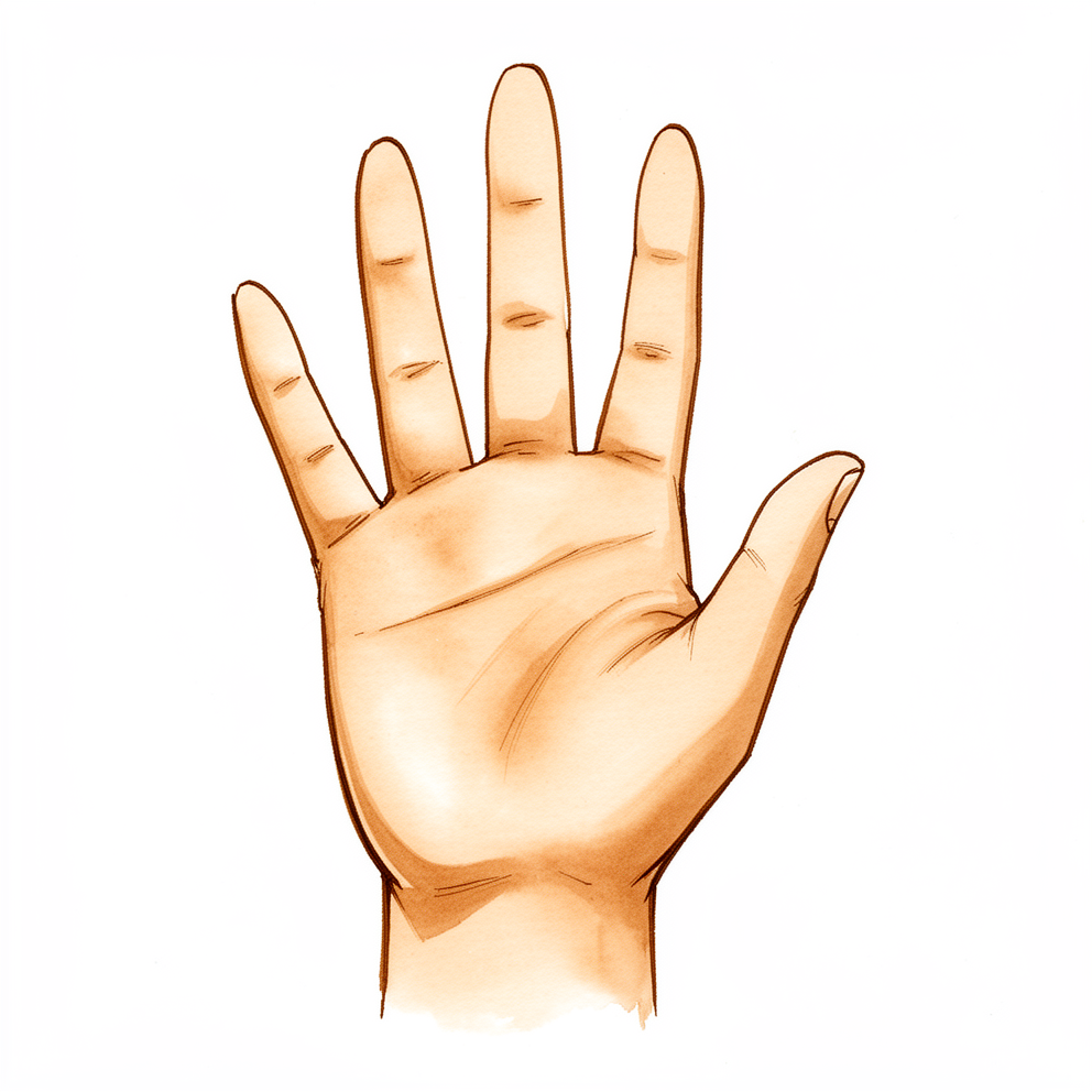
நீங்கள் உணர்வது
STT மூட்டு, மணிக்கட்டின் ஸ்கேஃபாய்டு (scaphoid) எலும்பு, ட்ரப்பீசியம் (trapezium), ட்ரப்பீசாய்டு (trapezoid) எனப்படும் இரண்டு சிறிய எலும்புகளைச் சந்திக்கும் இடத்தில், உங்கள் கட்டைவிரலின் அடிப்பகுதியில் உள்ள மூட்டுக்குச் சற்றுப் பின்னால் அமைந்துள்ளது. இங்கே ஏற்படும் மூட்டழற்சி (arthritis), கட்டைவிரலின் அடிப்பகுதியிலும் மணிக்கட்டின் கட்டைவிரல் பக்கத்திலும் வலியையோ குடைச்சலையோ ஏற்படுத்துகிறது. இறுகப் பற்றும்போதும் கிள்ளிப் பிடிக்கும்போதும் (pinching) அது மோசமாகிறது.
அன்றாட வேலைகள் இதைத் தூண்டலாம்: சாவியைத் திருப்புவது, ஜாடியைத் திறப்பது, எழுதுவது, கடைப் பையைச் சுமப்பது, அல்லது ஒரு கோப்பை தேநீரைப் பிடிப்பது.
இந்த மூட்டில் ஏற்படும் தேய்மானம் எக்ஸ்-ரேக்களில் பொதுவாகக் காணப்படுகிறது, குறிப்பாக வயதான பெண்களிடம். இந்த எக்ஸ்-ரே மாற்றங்கள் உள்ள பலருக்கு வலியே இருப்பதில்லை. உங்கள் வலிக்குக் காரணம் இந்த மூட்டுதானா என்பதற்கும் எக்ஸ்-ரே ஒரு மோசமான வழிகாட்டி: எவ்வளவு தேய்மானம் இருக்கிறது என்பதை அது குறைத்தோ மிகைப்படுத்தியோ மதிப்பிடக்கூடும். அதனால்தான், எக்ஸ்-ரே அல்லது அல்ட்ராசவுண்ட் வழிகாட்டுதலுடன், மூட்டுக்குள் உள்ளூர் மயக்க மருந்தும் (local anaesthetic) ஸ்டீராய்டும் (steroid) கலந்த ஊசியை உங்கள் அறுவை சிகிச்சை நிபுணர் பரிந்துரைக்கலாம். ஊசி வலியைத் தணித்தால், வலியின் மூலம் STT மூட்டுதான் என்பதை அது சுட்டிக்காட்டுகிறது.
STT மூட்டழற்சி பெரும்பாலும் கட்டைவிரலின் அடிப்பகுதியில் உள்ள மூட்டின் மூட்டழற்சியுடன் சேர்ந்தே வருகிறது; இரண்டையும் வேறுபடுத்திப் பார்ப்பது கடினமாக இருக்கலாம். இரண்டு மூட்டுகளும் தேய்ந்திருக்கும்போது, அது பான்ட்ரப்பீசியல் மூட்டழற்சி (pantrapezial arthritis) எனப்படுகிறது. எந்த மூட்டுகள் பாதிக்கப்பட்டுள்ளன என்பதை அறிவது முக்கியம்; ஏனெனில் எந்த அறுவை சிகிச்சைகள் பொருத்தமானவை என்பதை அதுவே தீர்மானிக்கிறது.
STT மூட்டழற்சி உள்ள சிலருக்கு, மணிக்கட்டு எலும்புகள் பின்னோக்கிச் சாய்கின்றன; இந்த அமைப்பு DISI (dorsal intercalated segment instability என்பதன் சுருக்கம்) எனப்படுகிறது. இது திட்டத்தை மாற்றக்கூடும் என்பதால், ஓர் அறுவை சிகிச்சையைத் தேர்ந்தெடுப்பதற்கு முன் உங்கள் அறுவை சிகிச்சை நிபுணர் எக்ஸ்-ரேயில் இதைச் சரிபார்க்கிறார்.
தேய்ந்த மூட்டு, ஃபிளெக்சர் கார்பை ரேடியாலிஸ் (flexor carpi radialis) எனப்படும் ஒரு மணிக்கட்டுத் தசையின் தசைநாணுக்கு (tendon) மிக அருகில் அமைந்துள்ளது. இந்தத் தசைநாண் அழற்சியடைந்து வலிக்கக்கூடும். மிக அரிதாக, அது அறுந்துவிடலாம்.
உங்கள் அறிகுறிகள் தணியவில்லை என்றால், வாரக்கணக்கில் மோசமடைந்தால், இரவில் உங்களை எழுப்பினால், அல்லது நீங்கள் வேலை செய்வதையோ உங்கள் கையைப் பயன்படுத்துவதையோ தடுத்தால், உங்கள் GP-யைப் பாருங்கள். உங்கள் கைப்பகுதியோ (hand) கையோ (arm) சூடாகவும், சிவந்தும், வீங்கியும், வலியுடனும் ஆனால், குறிப்பாகக் காய்ச்சலுடன் இருந்தால், அதே நாளில் அவசர சிகிச்சைப் பிரிவுக்குச் செல்லுங்கள்; ஏனெனில் இது அதே நாளில் சிகிச்சை தேவைப்படும் ஒரு தொற்றாக இருக்கலாம்.
உண்மையில் என்ன நடக்கிறது
மூட்டழற்சி என்பது ஒரு மூட்டின் உட்பரப்பில் ஏற்படும் தேய்மானம். இந்த நிலையில், தேய்ந்த மூட்டு STT மூட்டு: உங்கள் மணிக்கட்டின் ஆழத்தில் உள்ள சிறிய எலும்புகளில் ஒன்றான ஸ்கேஃபாய்டு, உங்கள் கட்டைவிரலின் அடிப்பகுதியை உருவாக்க உதவும் இரண்டு சிறிய எலும்புகளான ட்ரப்பீசியம், ட்ரப்பீசாய்டு ஆகியவற்றைச் சந்திக்கும் இடம். இந்த எலும்புகளை, உங்கள் கட்டைவிரல் சுழலவும், பற்றவும், கிள்ளிப் பிடிக்கவும் உதவும் சிறிய கட்டைகளின் ஒரு குட்டைச் சங்கிலியாக நினைத்துக்கொள்ளுங்கள். கட்டைகள் ஒன்றோடொன்று உரசும் இடங்களில், மென்மையான பரப்பு தேய்ந்து மெலிதாகலாம். அப்படி ஆகும்போது, அசைவின்போது கீழே உள்ள வெறும் எலும்பு அரைபடுகிறது; உங்கள் கட்டைவிரலின் அடிப்பகுதியிலும் மணிக்கட்டின் கட்டைவிரல் பக்கத்திலும் நீங்கள் உணரும் குடைச்சல் அந்த அரைபடுதல்தான்.
தசைநார்கள் (ligaments) எனப்படும் பட்டைகளின் தொகுப்பு மூட்டை ஒன்றாகப் பிடித்து வைத்திருக்கிறது. இந்தப் பட்டைகள் நீண்டுவிட்டாலோ சேதமடைந்தாலோ, ஸ்கேஃபாய்டு அதன் இயல்பான நிலையிலிருந்து சாய்ந்துவிடலாம். இது எலும்புகள் சுமையைத் தாங்கும் விதத்தை மாற்றுவதால், தேய்மானம் மோசமாகிறது. சிலருக்கு இது, மணிக்கட்டு எலும்புகள் பின்னோக்கிச் சாயும் ஓர் அமைப்புக்கு வழிவகுக்கிறது — உங்கள் அறுவை சிகிச்சை நிபுணர் எக்ஸ்-ரேயில் தேடும் DISI அமைப்பு. தேய்ந்த மூட்டு ஒரு மணிக்கட்டுத் தசைநாணுக்கு மிக அருகிலும் அமைந்துள்ளது; அந்தத் தசைநாணும் தனியாகவே அழற்சியடைந்து வலிக்கக்கூடும்.
இங்கே ஏற்படும் தேய்மானம், கட்டைவிரலின் அடிப்பகுதியில் உள்ள மூட்டின் தேய்மானத்துடன் சேர்ந்தே பெரும்பாலும் காணப்படுகிறது. இரண்டும் தேய்ந்திருக்கும்போது, அது பான்ட்ரப்பீசியல் மூட்டழற்சி எனப்படுகிறது. இரண்டு மூட்டுகளும் மிக அருகருகே இருப்பதால், ஒன்றிலிருந்து வரும் வலி மற்றொன்றிலிருந்து வரும் வலியைப் போல உணரப்படலாம்; இது முக்கியம், ஏனெனில் உண்மையில் எந்த மூட்டு உங்கள் அறிகுறிகளை ஏற்படுத்துகிறது என்பதைக் கண்டறிய உங்கள் அறுவை சிகிச்சை நிபுணர் வழிகாட்டுதலுடன் போடப்படும் ஊசியைப் பயன்படுத்தக் காரணம் இதுதான்.
இதற்கு நாங்கள் என்ன செய்ய முடியும்
Mater Private Hospital Rockhampton-இல் மேல் அவயவ (upper-limb) அறுவை சிகிச்சை நிபுணரான டாக்டர் கியரன் ஹிர்பரா, உங்கள் நிலைக்குப் பொருத்தமான, உடலுக்குள் மிகக் குறைவாகத் தலையிடும் (least invasive) தேர்வுகளுடன் தொடங்குகிறார். சிகிச்சை பொதுவாக அறுவை சிகிச்சை இல்லாமலேயே தொடங்குகிறது. கட்டைவிரலையும் மணிக்கட்டையும் அசையாமல் பிடித்து வைக்கும் ஒரு சிம்பு (splint) குடைச்சலைத் தணிக்கலாம், குறிப்பாக நீங்கள் உங்கள் கைகளால் அதிக வேலை செய்யும்போது. நீங்கள் வேலைகளைச் செய்யும் விதத்தை மாற்றுவதும் உதவுகிறது: இறுகப் பற்றும், கிள்ளிப் பிடிக்கும் வேலைகளைக் குறைப்பது, அல்லது ஜாடிகளைத் திறக்கவும் சுமைகளைச் சுமக்கவும் வேறு வழிகளைக் கண்டறிவது. கை சிகிச்சையின் (hand therapy) நோக்கம், கட்டைவிரலையும் மணிக்கட்டையும் அசைவில் வைத்திருப்பதும், அவற்றைச் சுற்றியுள்ள தசைகளை வலுப்படுத்துவதும். வேறு எதையும் பற்றிப் பேசுவதற்கு முன், பல வாரங்களுக்கு இந்த நடவடிக்கைகளை முறையாக முயன்று பார்க்கும்படி பொதுவாகக் கேட்டுக்கொள்கிறோம்.
எளிய வலி நிவாரணியும், வீக்கத்தைத் தணிக்கும் வகையான அழற்சி எதிர்ப்பு (anti-inflammatory) மருந்தும் வலியின் கூர்மையைக் குறைக்கலாம். எக்ஸ்-ரே அல்லது அல்ட்ராசவுண்ட் வழிகாட்டுதலுடன் மூட்டுக்குள் போடப்படும் ஸ்டீராய்டு ஊசி, உங்கள் வலியின் மூலம் STT மூட்டுதானா என்பதைக் கண்டறிய உதவலாம். அது வலியைத் தணித்தால், அந்த மூட்டையே அது சுட்டிக்காட்டுகிறது. நிவாரணம் சிறிது காலம் நீடிக்கலாம்; அதன் விளைவு குறைந்துவிட்டால் ஊசியை மீண்டும் போடலாம்.
இந்த நடவடிக்கைகள் உங்களுக்குப் போதுமான முன்னேற்றத்தைத் தராமல், வலி இன்னும் நீங்கள் செய்வதைக் கட்டுப்படுத்தும்போது, அறுவை சிகிச்சை பரிசீலனைக்கு வருகிறது. எந்த அறுவை சிகிச்சை பொருத்தமானது என்பது, எந்த மூட்டுகள் தேய்ந்துள்ளன என்பதைப் பொறுத்தது. STT மூட்டு மட்டும் தனியாகத் தேய்ந்திருந்தால், ஸ்கேஃபாய்டின் தேய்ந்த முனையை அகற்றலாம், அல்லது தேய்ந்த பரப்பின் வேலையை ஏற்கும்படி மூட்டுக்குள் ஒரு சிறிய பைரோகார்பன் (pyrocarbon) இடைவெளிப் பொருத்தை (spacer) வைக்கலாம். உங்கள் கட்டைவிரலின் அடிப்பகுதியில் உள்ள மூட்டும் தேய்ந்திருந்தால், வழக்கமான அறுவை சிகிச்சை ட்ரப்பீசியத்தையும் அதனுடன் ட்ரப்பீசாய்டின் ஒரு பகுதியையும் அகற்றுகிறது. தேர்ந்தெடுப்பதற்கு முன், உங்கள் மணிக்கட்டு எலும்புகள் சாய்ந்துள்ளனவா என்பதை எக்ஸ்-ரேயில் சரிபார்க்கிறோம்; ஏனெனில் அது திட்டத்தை மாற்றக்கூடும்.
என்ன எதிர்பார்க்கலாம்
STT மூட்டழற்சி கடந்து போகும் ஒரு காயம் அல்ல; அது ஒரு நீண்டகால நிலை. உங்கள் கட்டைவிரலின் அடிப்பகுதியில் உள்ள குடைச்சல் வந்து போகும் போக்குடையது. கனமாக இறுகப் பற்றும்போதோ கிள்ளிப் பிடிக்கும்போதோ அது பெரும்பாலும் தீவிரமடைந்து, கைக்கு ஓய்வு கொடுக்கும்போது தணிகிறது. சிகிச்சை இல்லாமல், வலி பொதுவாக நீடிக்கிறது அல்லது திரும்பத் திரும்ப வருகிறது; உங்கள் கையால் செய்ய முடியும் என்று நீங்கள் உணர்வதை அது மெல்ல மெல்லக் கட்டுப்படுத்தலாம்.
அறுவை சிகிச்சை இல்லாத நடவடிக்கைகளால் பெரும்பாலானோர் முன்னேற்றம் காண்கிறார்கள். ஒரு சிம்பு, வேலைகளைச் செய்யும் விதத்தில் மாற்றங்கள், எளிய வலி நிவாரணி, ஸ்டீராய்டு ஊசி ஆகியவை சிறிது காலத்துக்கு வலியைத் தணிக்கலாம். ஊசியால் கிடைக்கும் நிவாரணம் குறைந்துவிடலாம்; அதை மீண்டும் போடலாம். பல வாரங்களுக்குப் பிறகும் இந்தப் படிகள் உங்களுக்குப் போதுமான முன்னேற்றத்தைத் தரவில்லை என்றால், அறுவை சிகிச்சையைப் பற்றிப் பேசுவது பயனுள்ளது.
நீங்கள் அறுவை சிகிச்சை செய்துகொண்டால், அதன் பிறகான கை சிகிச்சை திட்டத்தின் ஒரு பகுதி. உங்கள் சிகிச்சை Extend Rehabilitation-இல் உள்ள Ruby Doolan-இடம் நடைபெறும். Ruby ஒரு கை சிகிச்சையாளர் (hand therapist): உங்கள் மீட்சிப் பயிற்சிகளுக்கு அவர் வழிகாட்டுவார், வழியில் உங்களுக்குத் தேவையான சிம்பையும் செய்து தருவார்.
மீட்சி படிப்படியானது. குடைச்சல் நாட்களில் அல்லாமல் வாரங்களில் தணியும் என்றும், கையில் வலிமையையும் நம்பிக்கையையும் மாதக்கணக்கில் கட்டியெழுப்புவீர்கள் என்றும் எதிர்பாருங்கள். சிலருக்கு மற்றவர்களைவிட அதிக காலம் தேவைப்படும்; புதியது போல உணரப்படும் ஒரு கை அல்ல, குறைந்த வலியுடன் உங்களுக்குத் தேவையானதைச் செய்யும் ஒரு கையே நோக்கம்.
எப்போது மருத்துவரைப் பார்க்க வேண்டும்
கட்டைவிரல் அடிப்பகுதியில் ஏற்படும் பெரும்பாலான குடைச்சல்கள் ஓய்வு, சிம்பு, எளிய வலி நிவாரணி ஆகியவற்றால் தணிகின்றன. உங்கள் வலி வாரக்கணக்கில் தணியவில்லை என்றால், மோசமடைந்துகொண்டிருந்தால், இரவில் உங்களை எழுப்பினால், அல்லது நீங்கள் வேலை செய்வதையோ உங்கள் கையைப் பயன்படுத்துவதையோ தடுத்தால், உங்கள் GP-யைப் பாருங்கள். இறுகப் பற்றும்போதும் கிள்ளிப் பிடிக்கும்போதும் வலி திரும்பத் திரும்ப வந்தால், அல்லது நீங்கள் பொருட்களை முன்பைவிட அடிக்கடி கீழே போட்டால், நிபுணர் பரிசோதனையைக் கேளுங்கள். இந்த மூட்டின் தேய்மானம் பெரும்பாலும் கட்டைவிரல் அடிப்பகுதியின் தேய்மானத்துடன் சேர்ந்தே காணப்படுவதால், எந்த மூட்டு வலியை ஏற்படுத்துகிறது என்பதைக் கண்டறிய வழிகாட்டுதலுடன் போடப்படும் ஊசியை உங்கள் அறுவை சிகிச்சை நிபுணர் பரிந்துரைக்கலாம்.
உங்கள் கைப்பகுதியோ (hand) கையோ (arm) சூடாகவும், சிவந்தும், வீங்கியும், வலியுடனும் ஆனால், குறிப்பாகக் காய்ச்சலுடன் இருந்தால், அதே நாளில் அவசர சிகிச்சைப் பிரிவுக்குச் செல்லுங்கள். இது ஒரு தொற்றாக இருக்கலாம்; அதற்கு அதே நாளில் சிகிச்சை தேவை. பரிசோதிக்கப்படுவதற்குப் பரிந்துரைக் கடிதம் தேவையில்லை.
ஆழமான விளக்கம்
Advanced reading: the deeper science (optional)
இந்தப் பகுதி, உங்கள் சொந்தச் சிகிச்சை முடிவுகளுக்குத் தேவையானதைவிட ஆழமாகச் செல்கிறது. STT மூட்டழற்சி பற்றிக் கூடுதலாகப் படிப்பது இரண்டு காரணங்களுக்காகப் பயனுள்ளது: வலிப்பது இந்த மூட்டுதானா என்பதற்கு எக்ஸ்-ரே ஒரு மோசமான வழிகாட்டி; மேலும் இதற்கான ஒவ்வோர் அறுவை சிகிச்சையும், வலி நிவாரணத்துக்கு ஈடாக, மணிக்கட்டின் சிறிய எலும்புகள் அமைந்திருக்கும் விதத்தில் ஒரு மாற்றத்தை ஏற்படுத்துகிறது.
இது எவ்வளவு பொதுவானது, எக்ஸ்-ரே ஏன் தவறாக வழிநடத்துகிறது
STT மூட்டில் ஏற்படும் தேய்மானம் எக்ஸ்-ரேயில் அடிக்கடி காணப்படும் ஒன்று. ஏதேனும் ஒரு புகாருக்காக எக்ஸ்-ரே எடுக்கப்பட்ட, கை கிளினிக்கின் தொடர்ச்சியான 700 நோயாளிகளில், 111 (16%) பேருக்கு STT மூட்டழற்சி இருந்தது; வயது அதிகரிக்கும்போதும், ஸ்கேஃபாய்டுக்கும் லூனேட்டுக்கும் (lunate) இடையே இடைவெளி இருக்கும்போதும், கட்டைவிரல் அடிப்பகுதி மூட்டில் மூட்டழற்சி இருக்கும்போதும் இது அதிக வாய்ப்புடன் காணப்பட்டது [1]. 50 முதல் 89 வயதுடைய ஜப்பானியக் குடியிருப்பாளர்களின் ஒரு சீரற்ற மாதிரியில் (random sample), 5.3% மணிக்கட்டுகளில் இது காணப்பட்டது; இங்கும் இது கட்டைவிரல் அடிப்பகுதி மூட்டழற்சி, பெண் பாலினம், வயது ஆகியவற்றுடன் தொடர்புடையதாக இருந்தது [2]. இவர்களில் பெரும்பாலானோருக்கு STT மூட்டிலிருந்து எந்த அறிகுறியும் இல்லை.
எக்ஸ்-ரே இரு திசைகளிலும் நம்பகத்தன்மையற்றது. கட்டைவிரல் அடிப்பகுதி அறுவை சிகிச்சையின்போது அறுவை சிகிச்சை நிபுணர்கள் மூட்டை நேரடியாகப் பார்த்தபோது, 62% கட்டைவிரல்களில் ஸ்கேஃபாய்டுக்கும் ட்ரப்பீசாய்டுக்கும் இடையே மூட்டழற்சி இருந்தது; ஆனால் எக்ஸ்-ரே அவற்றில் 44%-ஐ மட்டுமே கண்டறிந்திருந்தது [3]. 257 நோயாளிகள் கொண்ட ஒரு பெரிய தொடர் ஆய்வு, அறுவை சிகிச்சையின்போது 31% பேரில் இறுதிநிலைத் தேய்மானத்தைக் கண்டறிந்தது; எக்ஸ்-ரேயின் உணர்திறன் (sensitivity) 47%, தனித்தன்மை (specificity) 94% [4]. ஒரு நீண்டகாலத் தொடர் ஆய்வில், கட்டைவிரல் அடிப்பகுதி அறுவை சிகிச்சையின்போது அப்படியே விடப்பட்ட மிதமான STT தேய்மானம், சராசரியாக 8 ஆண்டுகள் காலத்தில் முன்னேறவும் இல்லை, அறிகுறிகளை ஏற்படுத்தவும் இல்லை [5]. அதனால்தான் முடிவு எக்ஸ்-ரேயை மட்டும் சார்ந்திராமல், தொட்டால் வலிக்கும் இடம் எங்கே என்பதையும், பெரும்பாலும் வழிகாட்டுதலுடன் போடப்படும் ஊசியையும் சார்ந்திருக்கிறது.
மணிக்கட்டு எலும்புகளின் சாய்வு
STT மூட்டழற்சி, டார்சல் இன்டர்கலேட்டட் செக்மென்ட் இன்ஸ்டபிலிட்டி (dorsal intercalated segment instability, DISI) எனப்படும் மணிக்கட்டு எலும்புகளின் ஒரு குறிப்பிட்ட சாய்வுடன் தொடர்புடையது: லூனேட் பின்னோக்கிச் சாய்கிறது, ஸ்கேஃபாய்டு மேலும் நிமிர்ந்து அமர்கிறது. STT மூட்டழற்சி உள்ள 135 மணிக்கட்டுகளில், மிதமான நிலைகளில் 5%-இலும் கடுமையான நிலைகளில் 60%-இலும் DISI இருந்தது; கிழிந்த ஸ்கேஃபோலூனேட் தசைநாரை (scapholunate ligament) தொடர்ந்து வரும் சாய்வைப் போலன்றி, ஸ்கேஃபாய்டுக்கும் லூனேட்டுக்கும் இடையிலான இடைவெளி இயல்பாகவே இருந்தது; முதன்மை மணிக்கட்டு மூட்டும் தேயவில்லை [6]. STT மூட்டழற்சியால் உந்தப்படும் ஒரு சரிவு அமைப்பை (SOAC wrist), நன்கு அறியப்பட்ட SLAC wrist-இலிருந்து வேறுபட்டதாகச் சில ஆசிரியர்கள் இப்போது விவரிக்கின்றனர் [7].
அறுவை சிகிச்சைக்கு இது முக்கியம்; ஏனெனில் எலும்பை அகற்றும் ஓர் அறுவை சிகிச்சை சாய்வை அதிகரிக்கக்கூடும். பின்னர் ஒரு மூட்டு இணைப்பு (fusion) தேவைப்பட்ட, பதிவுசெய்யப்பட்ட ஒரு நோயாளியின் நிலையில் நடந்ததுபோல, எலும்புகளின் நடு வரிசையில் ஏற்கெனவே நிலையற்ற ஒரு மணிக்கட்டு, எளிய அகற்றலுக்குப் பிறகு சரிந்துவிடலாம் [8]. எனவே ஓர் அறுவை சிகிச்சையைத் தேர்ந்தெடுப்பதற்கு முன், அறுவை சிகிச்சை நிபுணர்கள் சாய்வையும் நடு மணிக்கட்டு நிலையின்மையையும் (midcarpal instability) தேடுகிறார்கள்.
ஸ்கேஃபாய்டின் கீழ்முனை அகற்றல்: நிவாரணம், ஒரு சாய்வின் விலையில்
ஸ்கேஃபாய்டின் முனையை அகற்றுவது பெரும்பாலான தொடர் ஆய்வுகளில் வலியைப் போக்குகிறது; அதன் பிறகு மணிக்கட்டு எலும்புகள் பொதுவாகச் சாய்கின்றன. முதல் தொடர் ஆய்வுகளில் ஒன்றில், 21-இல் 12 மணிக்கட்டுகளில் DISI உருவானது; எதிலும் மூட்டு மேலும் சீர்குலையவில்லை; சாயாத மணிக்கட்டுகளில் இருந்ததைப் போலவே திருப்தியும் இருந்தது [9]. பிந்தைய ஒரு தொடர் ஆய்வு 21-இல் 19 மணிக்கட்டுகளில் (90%; அந்த ஆய்வுக் கட்டுரை 95% எனத் தெரிவிக்கிறது) DISI-ஐக் கண்டறிந்தது; லூனேட்டின் கோணம் சராசரியாக 13° உயர்ந்தது; சராசரியாக 3 ஆண்டுகளில், சாய்வுக்கும் செயல்பாட்டு மதிப்பெண்களுக்கும் இடையே எந்தத் தொடர்பும் இல்லை [10].
எவ்வளவு எலும்பு அகற்றப்படுகிறது என்பது முக்கியம். 17 மணிக்கட்டுகளில் நுண்ணோக்கி வழி (arthroscopic) அகற்றலுக்குப் பிறகு, 10-க்கு வலி மதிப்பெண்கள் 6.1-இலிருந்து 1.7-ஆகக் குறைந்தன; கிள்ளுப் பிடி வலிமை 2.5 kg-இலிருந்து 4.4 kg-ஆக உயர்ந்தது; 3 mm-க்கு மேல் எலும்பு அகற்றப்பட்டபோது சாய்வு அதிக வாய்ப்புடன் ஏற்பட்டது [11]. இறந்தவர்களின் உடல்களிலிருந்து (cadaver) எடுக்கப்பட்ட மணிக்கட்டுகளில், ஸ்கேஃபாய்டின் நான்கில் ஒரு பங்கை அகற்றியது எலும்புகள் அசையும் விதத்தை அரிதாகவே மாற்றியது; பாதியை அகற்றியது தெளிவான சாய்வை ஏற்படுத்தியது; நான்கில் மூன்று பங்கை அகற்றியது மணிக்கட்டை நிலையற்றதாக்கியது [12]. ஸ்கேஃபோட்ரப்பீசியல் தசைநாரின் (scaphotrapezial ligament) ஆழமான பட்டை, ஸ்கேஃபாய்டின் முனையிலிருந்து சுமார் 3.3 mm தொலைவில் இணைகிறது என்று ஓர் உடற்கூறு ஆய்வு கண்டறிந்தது; அகற்றலைச் சுமார் 3 mm அளவுக்குள் வைத்திருப்பதற்கு இது ஓர் உடற்கூறு அடிப்படையிலான காரணத்தைத் தருகிறது [13]. இதே அறுவை சிகிச்சை பற்றிய, இறந்தவர் உடல்களில் செய்யப்பட்ட ஓர் ஆய்வு, கேபிடேட் (capitate) சுமார் 14°-உம் லூனேட் 8°-உம் பின்னோக்கிச் சுழல்வதை அளவிட்டது [14]. அரிதாக, ஸ்கேஃபாய்டில் விடப்பட்ட ஒரு கூர்மையான விளிம்பு ஒரு மடக்கும் தசைநாணை (flexor tendon) உரசித் தேய்த்து அறுத்துள்ளது [15].
கட்டைவிரல் அடிப்பகுதியும் தேய்ந்திருக்கும்போது
கட்டைவிரல் அடிப்பகுதியும் STT மூட்டும் இரண்டும் தேய்ந்திருக்கும்போது, வழக்கமான அறுவை சிகிச்சை ட்ரப்பீசியத்தையும் ட்ரப்பீசாய்டின் தேய்ந்த முனையையும் அகற்றுகிறது. 37 கட்டைவிரல்களில், ட்ரப்பீசாய்டின் பகுதியளவு அகற்றலைச் சேர்த்தது, ட்ரப்பீசியத்தை மட்டும் அகற்றியதுடன் ஒப்பிடும்போது கூடுதல் பிரச்சினைகள் எதையும் ஏற்படுத்தவில்லை [3]. ட்ரப்பீசாய்டில் எவ்வளவை அகற்ற வேண்டும் என்பதுதான் விவாதத்துக்குரிய விஷயம். இடைநிலையாக 92 மாதங்கள் பின்தொடரப்பட்ட 79 கைகளில், ட்ரப்பீசாய்டின் ஒரு பகுதியையோ முழுவதையுமோ அகற்றியது, லூனேட் 15° அல்லது அதற்கு மேல் பின்னோக்கிச் சாய்வதை அதிக வாய்ப்புள்ளதாக்கியது (பகுதியளவு அகற்றலுக்குப் பிறகு 29%, முழு அகற்றலுக்குப் பிறகு 26%); அந்தச் சாய்வு உள்ள மணிக்கட்டுகள் மோசமான மதிப்பெண்களைப் பெற்றன; ஆள்காட்டி விரலின் உள்ளங்கை எலும்பு (index metacarpal) அறிகுறிகளுடன் கீழிறங்குவது முழு அகற்றலுக்குப் பிறகு 18%-இலும் பகுதியளவு அகற்றலுக்குப் பிறகு 10%-இலும் ஏற்பட்டது; முழுமையாக அகற்றுவதற்கு எதிராக ஆசிரியர்கள் அறிவுறுத்தினர் [16]. மடக்குதல், பக்கவாட்டு அசைவுகளின்போது ஸ்கேஃபாய்டுக்கும் ட்ரப்பீசாய்டுக்கும் இடையிலான அழுத்தம் குறைவதற்கு முன், ட்ரப்பீசாய்டில் 4 முதல் 6 mm அகற்றப்பட வேண்டும் என்று இறந்தவர் உடல்களில் செய்யப்பட்ட ஓர் ஆய்வு கண்டறிந்தது [17]. சராசரியாக 13.5 ஆண்டுகள் பின்தொடரப்பட்ட 150 கட்டைவிரல்கள் கொண்ட ஒரு தொடர் ஆய்வில், ட்ரப்பீசியெக்டமியுடன் (trapeziectomy) பகுதியளவு ட்ரப்பீசாய்டெக்டமியும் ஒரு தசைநாண் மறுகட்டமைப்பும் செய்யப்பட்டது, சராசரி DASH இயலாமை மதிப்பெண் 8.7-ஐத் தந்தது; மறு அறுவை சிகிச்சைகள் (revisions) எதுவும் இல்லை [18].
பைரோகார்பன் உள்பொருத்துகள்
பைரோகார்பன் இடைவெளிப் பொருத்து, ஸ்கேஃபாய்டின் உயரத்தைப் பாதுகாத்துக்கொண்டே வலியைப் போக்குவதையும், அதன் மூலம் சாய்வைக் கட்டுப்படுத்துவதையும் நோக்கமாகக் கொண்டது. பைரோகார்டன் (Pyrocardan) வட்டுடன், சராசரியாக 3.8 ஆண்டுகள் பின்தொடரப்பட்ட 20 நோயாளிகளுக்கு (22 உள்பொருத்துகள்) வலி குறைந்தது, மதிப்பெண்கள் மேம்பட்டன, கிள்ளுப் பிடி வலுவானது; மணிக்கட்டை நீட்டுவதில் (extension) ஓரளவு இழப்பு இருந்தது, இடப்பெயர்வுகள் (dislocations) எதுவும் இல்லை; கேபிடோலூனேட் கோணம் (capitolunate angle) மோசமாவதற்குப் பதிலாக மேம்பட்டது [19]. சராசரியாக 5 ஆண்டுகள் பின்தொடரப்பட்ட ஒரு தொடர் ஆய்வு, 95% உள்பொருத்துகள் மறு அறுவை சிகிச்சையின்றி இருந்ததையும், வலியற்ற ஓர் இடப்பெயர்வையும், 4 மணிக்கட்டுகளில் மூட்டைச் சுற்றிக் கால்சியம் படிந்ததையும், 3-இல் நீடித்த, 1-இல் புதிதாகத் தோன்றிய ஒரு சாய்வையும் பதிவு செய்தது [20]. சீரற்ற முறையில் ஒதுக்கப்படாத (non-randomised) ஓர் ஆய்வில் STT மூட்டு இணைப்புடன் ஒப்பிட்டபோது, உள்பொருத்துடன் மணிக்கட்டை நீட்டுவது சிறப்பாக இருந்ததைத் (58° எதிர் 38°) தவிர, முடிவுகள் ஒரே மாதிரியாக இருந்தன; உள்பொருத்து வைக்கப்பட்ட மணிக்கட்டுகளில் 22%-இல் சாய்வு உருவானது; மூட்டு இணைப்புகளில் 9% ஒன்றிணையவில்லை [21]. இந்தத் தொடர் ஆய்வுகளில் பெரும்பாலானவை சில பிரெஞ்சு மையங்களிலிருந்து வந்தவை.
வேறு வடிவமைப்புகளுடன் கிடைத்த முடிவுகள் குறைவான நிலைத்தன்மை கொண்டவை. ஸ்கேஃபாய்டின் முனையை மாற்றும் வகையில் வடிவமைக்கப்பட்ட வேறொரு பைரோகார்பன் உள்பொருத்துடன் (STPI), 29-இல் 7 உள்பொருத்துகள், முக்கியமாக வலிக்காக, மாற்றியமைக்கப்பட்டன (revised); இடைநிலையாக 68 மாதங்களில் சாய்வு உள்ள மணிக்கட்டுகளின் எண்ணிக்கை 6-இலிருந்து 12-ஆக இரட்டிப்பானது [22]. ஒரு டென்னிஸ் வீரரிடம் இரண்டு முறை உடைந்த ஒரு பைரோகார்டனை ஒரு நோயாளி அறிக்கை (case report) விவரிக்கிறது; அதனால்தான், மணிக்கட்டு நீண்ட நிலையில் அதன் மீது கனமான சுமை ஏற்றுவது பற்றி அறுவை சிகிச்சைக்கு முன் பேசப்படுகிறது [23].
ஒப்பீட்டுக்கு, STT மூட்டு இணைப்பு
மூட்டு இணைப்பு மூன்று எலும்புகளையும் ஒன்றாக இணைக்கிறது. ஒரு முறைமையான ஆய்வில் (systematic review) 1,429 அறுவை சிகிச்சைகளில், 6.3%-இல் இணைப்பு ஒன்றிணையவில்லை; 4.2% முழு மணிக்கட்டு இணைப்பாக மாற்றப்பட்டன; பிடி வலிமை சராசரியாக மறு கையின் 76% அளவில் இருந்தது [24]. 10 ஆண்டுகள் பின்தொடரப்பட்ட 58 மணிக்கட்டுகளில், 10%-க்கு மேலும் ஓர் அறுவை சிகிச்சை தேவைப்பட்டது; 7%-இல் வலியுடன் கூடிய ஒன்றிணையாமை (non-union) இருந்தது [25]. மூட்டு இணைப்பு பொதுவாக மணிக்கட்டு அசைவில் சுமார் நான்கில் மூன்று பங்கைப் பாதுகாக்கிறது; இணைக்கப்பட்ட ஸ்கேஃபாய்டில் ஆரை எலும்பின் முனை (radial styloid) உரசுவதால் ஏற்படும் வலி போதுமான அளவு பொதுவானது என்பதால், சில அறுவை சிகிச்சை நிபுணர்கள் அதே நேரத்தில் அந்த முனையையும் சீர்செய்கிறார்கள் [26].
தெரியாதவை
இந்த அறுவை சிகிச்சைகளை ஒப்பிடும் சீரற்ற ஒதுக்கீட்டுச் சோதனைகள் (randomised trials) எதுவும் இல்லை. ஐரோப்பியக் கை அறுவை சிகிச்சை நிபுணர்களிடையே நடத்தப்பட்ட ஒரு கணக்கெடுப்பு, ஒரே பிரச்சினைக்கு விரும்பப்படும் அறுவை சிகிச்சைகளில் பரந்த வேறுபாட்டைக் கண்டறிந்தது [27]; எந்தத் தேர்வுக்கும் தெளிவான மேன்மைக்கான சான்று இல்லை என்று சமீபத்திய மதிப்பாய்வுகள் முடிவு செய்கின்றன; STT மூட்டு மட்டும் தனியாகத் தேய்ந்துள்ளதா, கட்டைவிரல் அடிப்பகுதியும் தேய்ந்துள்ளதா, மணிக்கட்டு எலும்புகள் ஏற்கெனவே சாய்ந்துள்ளனவா என்பதைப் பொறுத்தே தேர்வு அமைகிறது [28] [29]. பெரும்பாலான தொடர் ஆய்வுகள் 30-க்கும் குறைவான நோயாளிகளை 10 ஆண்டுகளுக்கும் குறைவான காலமே பின்தொடர்கின்றன; எனவே அகற்றலுக்குப் பிந்தைய சாய்வின் நீண்டகால விளைவும், பைரோகார்பன் உள்பொருத்துகளின் நீண்டகால நிலைப்பும் இன்னும் விடை தெரியாத கேள்விகளே.
மேற்கோள்கள்
[1] Scordino LE, Bernstein J, Nakashian M, et al. Radiographic prevalence of scaphotrapeziotrapezoid osteoarthrosis. J Hand Surg Am. 2014;39(9):1677-82. https://doi.org/10.1016/j.jhsa.2014.05.033
[2] Kitamura Y, Kato H, Hayashi M, et al. Prevalence and associated factors for primary osteoarthritis of the scaphotrapeziotrapezoid, radiocarpal, and distal radioulnar joints in the Japanese general elderly population. J Hand Surg Am. 2025;50(1):103.e1-103.e10. Epub 2023. https://doi.org/10.1016/j.jhsa.2023.05.009
[3] Tomaino MM, Vogt M, Weiser R. Scaphotrapezoid arthritis: prevalence in thumbs undergoing trapezium excision arthroplasty and efficacy of proximal trapezoid excision. J Hand Surg Am. 1999;24(6):1220-4. https://doi.org/10.1053/jhsu.1999.1220
[4] Akoon A, Grandizio LC, Mookerjee GG, et al. Comparison of radiographic and intraoperative visual assessment of scaphotrapezoid joint arthritis in patients with end-stage carpometacarpal arthritis of the thumb base. Hand (N Y). 2019;14(5):609-13. Epub 2018. https://doi.org/10.1177/1558944718765246
[5] Glickel SZ, Kornstein AN, Eaton RG. Long-term follow-up of trapeziometacarpal arthroplasty with coexisting scaphotrapezial disease. J Hand Surg Am. 1992;17(4):612-20. https://doi.org/10.1016/0363-5023(92)90303-7
[6] Hattori Y, Sasaki J, Sakamoto S, et al. Radiographic characteristics of carpal instability associated with scaphotrapeziotrapezoid osteoarthritis. J Hand Surg Am. 2026;51(3):259-67. https://doi.org/10.1016/j.jhsa.2025.11.018
[7] Rodriguez Fontan F, Netscher DT. Scaphotrapeziotrapezoid joint osteoarthritis: differentiating SOAC wrist from SLAC wrist. J Am Acad Orthop Surg. 2026. https://doi.org/10.5435/jaaos-d-25-01408
[8] Corbin C, Warwick D. Midcarpal instability after excision arthroplasty for scapho-trapezial-trapezoid (STT) arthritis. J Hand Surg Eur Vol. 2009;34(4):537-8. https://doi.org/10.1177/1753193408098903
[9] Garcia-Elias M, Lluch AL, Farreres A, et al. Resection of the distal scaphoid for scaphotrapeziotrapezoid osteoarthritis. J Hand Surg Br. 1999;24(4):448-52. https://doi.org/10.1054/jhsb.1999.0169
[10] Brouwers D, Decramer A, Cornu P, Le Viet D. Does the DISI matter after distal scaphoidectomy with tendon interposition for STT osteoarthritis? Hand Surg Rehabil. 2020;39(4):284-90. https://doi.org/10.1016/j.hansur.2020.03.005
[11] Iida A, Omokawa S, Kawamura K, et al. Arthroscopic distal scaphoid resection for isolated scaphotrapeziotrapezoid osteoarthritis. J Hand Surg Am. 2019;44(4):337.e1-337.e5. https://doi.org/10.1016/j.jhsa.2018.06.021
[12] Kadar A, Grewal R, Padmore CE, et al. How much scaphoid can be safely resected? A biomechanical analysis of the effects of distal scaphoid resection. Hand (N Y). 2022;17(5):885-92. Epub 2020. https://doi.org/10.1177/1558944720966717
[13] Yoshimura Y, Yokota A, Fujino K, et al. Anatomy of the scaphotrapezial ligament complex and its implications for distal scaphoid resection. J Hand Surg Am. 2026;51(1):25.e1-25.e8. https://doi.org/10.1016/j.jhsa.2025.09.010
[14] Paeffgen L, Riederer J, Adler T, et al. Carpal instability after partial trapeziectomy, total trapeziectomy and the resection of the distal scaphoid pole: a cadaveric study. Arch Orthop Trauma Surg. 2024;144(3):1443-51. Epub 2023. https://doi.org/10.1007/s00402-023-05134-z
[15] Deren ME, Mitchell CH, Weiss AC. Flexor tendon ruptures after distal scaphoid excision for scaphotrapeziotrapezoid osteoarthritis. Hand (N Y). 2017;12(5):NP152-6. https://doi.org/10.1177/1558944717708028
[16] Renfree KJ, Odgers R, Tillinghast C, Zhang N. Effect of partial and complete trapezoid excision on radiographic and functional results after abductor pollicis longus suspensionplasty. J Hand Surg Am. 2020;45(4):364.e1-364.e9. https://doi.org/10.1016/j.jhsa.2019.10.006
[17] Anz H, Roylance J, Ismaily S, et al. Correlation of scaphotrapezoid contact pressures with degree of trapezoid resection: does eliminating contact matter? J Hand Surg Glob Online. 2026;8(4):101049. https://doi.org/10.1016/j.jhsg.2026.101049
[18] Melone CP, Lovy AJ, Ciaglia M, Qin B. Trapeziectomy, partial trapezoidectomy, and abductor pollicis longus dual loop ligament reconstruction for advanced basal joint arthritis: long-term outcome of 150 cases. Hand (N Y). 2023;18(3):491-500. Epub 2022. https://doi.org/10.1177/15589447221092060
[19] Gauthier E, Truffandier M, Gaisne E, Bellemère P. Treatment of scaphotrapeziotrapezoid osteoarthritis with the Pyrocardan implant: results with a minimum follow-up of 2 years. Hand Surg Rehabil. 2017;36(2):113-21. https://doi.org/10.1016/j.hansur.2017.01.003
[20] Cholley-Roulleau M, Bouju Y, Lecoq FA, et al. Pyrocardan scaphotrapeziotrapezoid joint arthroplasty for isolated osteoarthritis: results after a mean follow-up of 5 years. J Wrist Surg. 2022;11(3):262-8. Epub 2021. https://doi.org/10.1055/s-0041-1733877
[21] Cholley-Roulleau M, Dautel G, Dap F, et al. Comparison between scaphotrapeziotrapezoid arthrodesis and Pyrocardan implant for isolated scaphotrapeziotrapezoid osteoarthritis. Orthop Traumatol Surg Res. 2025;111(5):103867. https://doi.org/10.1016/j.otsr.2024.103867
[22] Zander M, Ibsen-Sörensen A, Nilsson A, Björkman A. Retrospective analysis of scaphoid trapezium pyrocarbon implant intervention in STT arthritis: a 3-year follow-up study. J Plast Surg Hand Surg. 2024;59:40-5. https://doi.org/10.2340/jphs.v59.34985
[23] Sarigiannis CN, Axelsson P. Game, set… revision! A case report of a tennis player who smashed his scaphotrapeziotrapezoid-joint Pyrocardan implant twice. J Hand Surg Glob Online. 2026;8(3):100964. https://doi.org/10.1016/j.jhsg.2026.100964
[24] Stephens AR, Garcia BN, Rogers MJ, et al. Scaphotrapeziotrapezoid arthrodesis: systematic review. J Hand Surg Am. 2022;47(3):218-227.e2. https://doi.org/10.1016/j.jhsa.2021.09.029
[25] Rogers MJ, Lu C, Stephens AR, et al. Scaphotrapeziotrapezoid arthrodesis: a 10-year follow-up study of complications in 58 wrists. Hand (N Y). 2022;17(5):919-25. Epub 2020. https://doi.org/10.1177/1558944720964972
[26] Deans VM, Naqui Z, Muir LT. Scaphotrapeziotrapezoidal joint osteoarthritis: a systematic review of surgical treatment. J Hand Surg Asian Pac Vol. 2017;22(1):1-9. https://doi.org/10.1142/s0218810417300017
[27] Berkhout MJL, Yin Q, Ritt MJPF. Current trends in operative treatment of scaphotrapeziotrapezoid osteoarthritis: a survey among European hand surgeons. J Wrist Surg. 2020;9(2):94-9. https://doi.org/10.1055/s-0039-3402796
[28] Bellemère P, Ardouin L. Surgical options and outcomes for treatment of osteoarthritis of the scaphotrapeziotrapezoidal joint. J Hand Surg Eur Vol. 2025;50(1):58-67. https://doi.org/10.1177/17531934241295345
[29] Alder KD, Feroe AG, Karim KE. Management of scaphotrapeziotrapezoid osteoarthritis: a critical analysis review. JBJS Rev. 2023;11(10):e23.00093. https://doi.org/10.2106/JBJS.RVW.23.00093
Evidence & references
This is the clinical evidence summary written for health professionals. It is technical, and it lists the research this page was built from. You do not need to read it to understand your treatment or to make a decision about it.
Anatomy & Pathophysiology
Osseous Skeleton
- The skeleton of the hand and wrist consists of 27 bones, of which 19 are long bones [3].
- The skeleton is divided into five rays, each comprising a polyarticulated chain of metacarpals and phalanges [3].
- The base of each metacarpal articulates with the distal row of the carpus [3].
- The carpus articulates with the skeleton of the forearm through its proximal row [3].
- The radioulnocarpal articulation has two axes of movement, to which a third axis of pronation and supination from the forearm is added [3].
- The wrist has three axes of movement, permitting the hand to be positioned in any spatial configuration [3].
- The radial ray (first ray) is the shortest, made up of a metacarpal and two phalanges [3].
- The first ray continues the external column of the carpus formed by the scaphoid and trapezium [3].
- The trapezium is angled out in front of the carpal plane so that the first metacarpal makes an angle of about 45 degrees with the second metacarpal in the sagittal plane [3].
- The thumb metacarpal is the shortest, while the index metacarpal is by far the longest [3].
- The proximal and middle phalanges of the long and ring fingers are longer than those of the index finger [3].
- The epiphyseal plates are located at the proximal ends of the phalanges and the first metacarpal [3].
- The epiphyseal plates are located at the distal ends of the other metacarpals [3].
- The hand presents a longitudinal and transverse concavity, giving it the shape of a cup with a palmar concavity when the thumb is placed next to the index finger [3].
- The transverse axis of the palm corresponds to the metacarpophalangeal articulations and is not perpendicular to the longitudinal axis [3].
- The transverse axis forms an acute angle of approximately 75 degrees with the longitudinal axis [3].
Musculature
- Extrinsic muscles have their origin outside of the hand and their insertion on the hand or carpus [2].
- Intrinsic muscles have both origin and insertion within the hand [2].
- The extrinsic extensors run through six different fibroosseous retinacular compartments at the wrist level [2].
- The first extensor compartment contains the abductor pollicis longus and the extensor pollicis brevis [2].
- The abductor pollicis longus inserts at the base of the thumb metacarpal and radially abducts the thumb [2].
- The extensor pollicis brevis inserts on the dorsum of the proximal aspect of the proximal phalanx of the thumb and actively extends the metacarpophalangeal joint of the thumb [2].
- The second extensor compartment contains the extensor carpi radialis longus and the extensor carpi radialis brevis [2].
- The extensor carpi radialis longus inserts on the index metacarpal, dorsiflexes and radially deviates the wrist [2].
- The extensor carpi radialis brevis inserts into the base of the middle metacarpal and provides balanced wrist dorsiflexion [2].
- The third compartment contains the extensor pollicis longus, which turns abruptly radialward about Lister tubercle [2].
- The extensor pollicis longus inserts on the distal phalanx and provides forceful extension of the thumb interphalangeal joint [2].
- The fourth extensor compartment contains the extensor indicis proprius lying deep to the four tendons of the extensor digitorum communis [2].
- The fifth compartment contains the extensor digiti quinti [2].
- The principal bony insertion of the extrinsic digital extensors is on the dorsal proximal aspect of the middle phalanx [2].
- Metacarpophalangeal joint extension is provided by extrinsic extensor force transmitted through the sagittal bands [2].
- Distal interphalangeal joint extension is achieved through the conjoined lateral bands composed of tendinous slips from the extrinsic and intrinsic tendons [2].
- The extensor carpi ulnaris tendon runs through the sixth compartment and inserts at the base of the little finger metacarpal [2].
- The extensor carpi ulnaris provides wrist extension and ulnar deviation [2].
- The extensor digitorum communis tendons of the middle, ring, and little fingers are tethered together by juncturae tendinum over the dorsum of the hand proximal to the metacarpophalangeal joint [2].
- The digital extensor tendons are stabilized over the mid-line of the metacarpophalangeal joint by their attachment to sagittal band fibers [2].
- The sagittal band fibers insert onto the volar proximal phalanx and onto the lateral borders of the volar plate [2].
- The sagittal bands keep the extrinsic extensor as far as possible away from the center of rotation of the metacarpophalangeal joint, giving it the greatest mechanical efficiency [2].
- With rupture or attenuation of the sagittal band fibers, the extrinsic extensor tendon can sublux to the ulnar side of the metacarpal head causing ulnar deviation of the finger [2].
- The extrinsic finger flexors are the flexor digitorum profundus and the flexor digitorum superficialis [2].
- The flexor digitorum profundus inserts on the proximal volar aspect of the distal phalanx [2].
- The flexor digitorum profundus flexes the distal interphalangeal joint as well as the proximal interphalangeal and metacarpophalangeal joints [2].
- The flexor digitorum superficialis acts as a flexor of the proximal interphalangeal and metacarpophalangeal joints [2].
- There are seven interosseous muscles, four dorsal and three volar [6].
- The dorsal interossei are abductors [6].
- The dorsal interossei lie to the radial side of the index and middle fingers and the ulnar side of the middle and ring fingers [6].
- The little finger is abducted by the abductor digiti quinti [6].
- The volar interossei are adductors and lie to the ulnar side of the index finger and the radial side of the ring and little fingers [6].
- The middle finger has two dorsal interossei (abductors) and no volar interossei (adductors) because the central axis of the hand lies within it [6].
- Each dorsal interosseous muscle, with the exception of the third, has two muscle heads [6].
- The superficial head of the dorsal interosseous muscles abducts and weakly flexes the proximal phalanx [6].
- The deep head of each dorsal interosseous muscle forms a lateral tendon, or lateral band, at the level of the MP joint [6].
- The deep head flexes and weakly abducts the proximal phalanx while extending the middle and distal phalanges [6].
- Transverse fibers arch dorsally from each lateral band to join each other over the dorsum of the finger, flexing the proximal phalanx [6].
- Oblique fibers (spiral fibers) from the lateral bands insert onto the lateral tubercles at the base of the middle phalanx and extend the middle phalanx (PIP joint) [6].
- The lateral bands are joined by the lateral slips of the extensor tendon to form the conjoined lateral band [6].
- The two conjoined lateral bands to each finger unite at the distal third of the middle phalanx to form the terminal tendon [6].
- The terminal tendon inserts at the base of the distal phalanx to extend it [6].
- The flexor digiti quinti brevis is structurally and functionally similar to the deep head of the dorsal interossei, forming the ulnar lateral band of the little finger [6].
- Each volar interosseous muscle has only one muscle head and none of them insert onto the proximal phalanx [6].
- The volar interossei form the ulnar lateral band of the index finger and the radial lateral band of the ring and little fingers [6].
- The abductor digiti quinti inserts onto the ulnar lateral tubercle at the base of the proximal phalanx of the little finger [6].
- The flexor digiti quinti forms the ulnar lateral band [6].
- The opponens digiti quinti arises from the pisohamate ligament and the hook of the hamate and inserts onto the ulnar side of the diaphysis of the fifth metacarpal [6].
- The opponens digiti quinti flexes and supinates the fifth metacarpal [6].
- The flexor digitorum profundus originates from the proximal ulna and the interosseous membrane [13].
- The flexor digitorum profundus and the flexor pollicis longus muscles form the deep compartment of the volar forearm [13].
- As the flexor digitorum profundus and flexor pollicis longus tendons travel through the carpal tunnel, they occupy the floor of the carpal tunnel [13].
- The tenosynovial sheath of the flexor pollicis longus is continuous with the radial bursa [13].
- The tenosynovial sheath to the little finger is continuous with the ulnar digital bursa [13].
- In some patients, the radial and ulnar bursae communicate, allowing a horseshoe abscess to spread between the thumb and little finger if infection occurs in the flexor tendon sheath of either one of these digits [13].
- The lumbricals originate from the radial side of the index, middle, ring, and little fingers in the palm [13].
- The profundus tendon passes through the bifurcation of the flexor digitorum superficialis before inserting into the proximal palmar base of the distal phalanx [13].
- The innervation of the flexor digitorum profundus of the index and middle fingers is through the anterior interosseous branch of the median nerve [13].
- The profundus of the ring and little fingers is innervated by the ulnar nerve [13].
- The flexor digitorum superficialis has two heads: a radial head originating from the proximal shaft of the radius, and a humeral ulnar head originating from the medial humeral epicondyle and coronoid process of the ulna [13].
- As the superficialis tendons pass through the carpal tunnel, the tendons of the middle and ring fingers are more superficial and central than those of the index and little fingers [13].
- In the proximal aspect of the finger, the flexor digitorum superficialis tendon bifurcates around the flexor digitorum profundus at the beginning of the A2 pulley [13].
- The flexor digitorum superficialis tendon slips reunite distally at the Camper chiasm, with approximately half of the fibers staying on the ipsilateral side and half crossing to the contralateral side of the finger [13].
- The flexor digitorum superficialis inserts via radial and ulnar slips into the proximal metaphysis of the middle phalanx [13].
- The entire flexor digitorum superficialis muscle receives innervation from the median nerve [13].
- The primary function of the superficialis is digital flexion at the proximal interphalangeal joint [13].
- The flexor pollicis longus originates from two heads: a radial head from the proximal radius and interosseous membrane, and an accessory head from the coronoid process of the ulna and medial epicondyle of the humerus [13].
- In the palm, the flexor pollicis longus tendon transverses between the abductor pollicis brevis and the flexor pollicis brevis [13].
- The flexor pollicis longus inserts into the proximal base of the thumb distal phalanx [13].
- The flexor pollicis longus is innervated by the anterior interosseous branch of the median nerve [13].
- The flexor pollicis longus flexes both the interphalangeal and metacarpophalangeal joints of the thumb [13].
Ligaments and Fascia
- The second to fifth metacarpals are all bound together by various fibrous structures, the most distal of which is the deep transverse intermetacarpal ligament [9].
- The deep transverse intermetacarpal ligament is better named the interglenoid ligament because it ties together the anterior "glenoid ligaments" of the metacarpophalangeal articulations, known as the "volar plates" [9].
- The volar plates are interconnected by the transverse interglenoid ligament [9].
- The stability of the metacarpophalangeal joints is essential to the support of the longitudinal arch as well as of the transverse metacarpal arch [9].
- The superficial palmar fascia lies in a coronal plane beneath the palmar subcutaneous tissue [16].
- The superficial palmar fascia covers a triangular area of the central palm, with the proximal corner facing directly proximal [16].
- The palmaris longus tendon, when present, terminates in continuity with the fibers of this proximal corner [16].
- From this common point, four central bands of fascia extend distally toward each of the fingers [16].
- There is no central band for the thumb [16].
- At the distal palmar crease level, the central bands are bridged transversely by the superficial transverse palmar ligament [16].
- Fibers from the central bands pass above, below, and through the superficial transverse palmar ligament [16].
- Superficial fibers of the central bands stay superficial and merge with vertical retinacular fibers at the dermis’s undersurface in the distal palm in areas between skin flexion creases [16].
- Intermediate fibers split transversely into two sections, which extend toward the lateral border of the digit base [16].
- These intermediate fibers are the spiral band that tracks around the neurovascular bundle [16].
- Proximally, the spiral band is central and superficial to the neurovascular bundle; distally, it is lateral and deep beneath it [16].
- Spiral neurovascular bundles develop when spiral bands shorten [16].
- Deep fibers continue dorsally to merge with sagittal interosseous fascia fibers and pierce the transverse deep intermetacarpal ligament to merge with fibers of the sagittal bands of the extensor mechanism [16].
- The digital neurovascular structures are surrounded by a diffuse network of thin transverse oblique fibers [16].
- Fibers dorsal to the neurovascular bundle are collectively called Cleland ligament [16].
- Fibers palmar to the neurovascular bundle are called Grayson ligament [16].
- Undisturbed, these "ligaments" are a mesh of nonparallel individual fibers [16].
- The appearance of a ligament is a dissection artifact from releasing skin attachments [16].
- These fibers have a common origin in continuity with the fingers’ retinacular ligaments, the floor of the flexor tendon sheath, and retinacular fibers attaching the palmar digital skin to deeper structures [16].
- On the small finger’s ulnar border, lateral fascial attachments are in continuity with the abductor digiti minimi fascia and tendon [16].
- A subdermal fascial layer borders the periphery of the web spaces from roughly the radial thumb sesamoid to the ulnar side of the small finger’s proximal flexion crease [16].
- These border fibers form the distal first web space ligament between thumb and index in continuity with the natatory ligament that spans the remaining web spaces [16].
- Fibers from the natatory ligament extend distally at each finger’s lateral base in continuity with Grayson ligament fibers and the lateral digit dermis [16].
Vascular Anatomy
- The "princeps pollicis" artery, the terminal branch of the radial artery, crosses the first intermetacarpal space [10].
- The "princeps pollicis" artery runs along the ulnar side of the first metacarpal bone and along the volar surface of the adductor muscle [10].
- The "princeps pollicis" artery emerges onto the subcutaneous palmar tissue at the level of the cutaneous flexion crease of the metacarpophalangeal joint [10].
- At the metacarpophalangeal joint level, the "princeps pollicis" divides into two terminal rami, namely the collateral palmar arteries of the thumb [10].
- The collateral palmar arteries run along the digital tunnel symmetrically and are of equal caliber [10].
- The collateral palmar arteries head distally to finally unite in the pulp arcade [10].
- During their transit in the digital tunnel, the collateral palmar arteries break off into numerous collateral branches, either cutaneous, articular or osseous [10].
- An arcade located deep in the flexor tendon joins together the two arteries at the level of the distal metaphysis of the first phalanx [10].
- From this arcade, vessels originate which enter the "vincula" and irrigate the flexor tendon [10].
- In anatomical studies, only 15% of dissections fall into the classical category of palmar artery layout [10].
- In
Clinical Presentation
- Inflammatory arthritis is a collection of different diseases that cause inflammation in the joints [20].
- The diagnosis of inflammatory arthritis requires a complete review of the disease process profile [20].
- There are no simple clinical tests that can be used to diagnose inflammatory arthritis diseases [20].
- The rheumatologic history and physical examination play a critical role in the diagnosis and treatment of inflammatory arthritis [20].
- The location of the problems is an important disease feature for diagnosing inflammatory arthritis [20].
- The symmetry of presentation is an important disease feature for diagnosing inflammatory arthritis [20].
- Rheumatoid arthritis has a predilection for the wrists and the proximal joints of the hands and feet [20].
- Psoriatic arthritis involves the distal interphalangeal joint of the hands [20].
- The physical findings with rheumatoid arthritis tend to be symmetrical [20].
- Other inflammatory arthritis conditions do not tend to present with symmetrical physical findings [20].
- The onset of the disease is an important feature for diagnosing inflammatory arthritis [20].
- The chronology of the disease is an important feature for diagnosing inflammatory arthritis [20].
- The onset of rheumatoid arthritis tends to present in a subacute manner [20].
- Septic arthritis has a rapid onset in several hours [20].
- Age can aid in the diagnosis of inflammatory arthritis [20].
- Gender can aid in the diagnosis of inflammatory arthritis [20].
- Precipitating factors can aid in the diagnosis of inflammatory arthritis [20].
Investigations
- Clinical evaluation of the hand and wrist requires combining patient history with a careful physical examination to narrow the scope of possible pathologic processes [1].
- Diagnostic tests such as imaging and serum laboratory studies are useful in determining pathology but can be expensive, time-consuming, and often nonspecific [1].
- A careful physical examination is essential to direct care and future testing if indicated [1].
- The radioulnocarpal articulation has two axes of movement, to which a third axis—pronation and supination from the forearm—is added [3].
- The transverse axis of the palm forms an acute angle of approximately 75 degrees with the longitudinal axis [3].
- An 8-MHz Doppler tone assessment may be used to identify superficially displaced neurovascular bundles when Dupuytren cords lie beneath soft fleshy prominences [19].
- False-negatives are possible with 8-MHz Doppler tone assessment for identifying neurovascular bundles [19].
- MR assessment of Dupuytren’s disease is hindered by the resolution of current equipment, orientation issues due to multiplanar deformities of the fingers, and lack of intraoperative availability [19].
- MRI is probably most useful in identifying additional pathology such as flexor tendon bowstringing [19].
- MRI may be helpful in providing a quantitative noninvasive measure of cellularity of affected areas, which is an index of biologic activity [19].
References
[1] Orthopaedic Knowledge Update 13 Ebook Without Multimedia. Anatomy, Evaluation, Clinical Examination, and Imaging > Evaluation and Clinical Examination: Current Concepts.
[2] A Lange Medical Book Current Diagnosis Treatment In Orthopedics Fifth Edition. 9Hand Surgery > Image DISORDERS OF THE MUSCULATURE OF THE HAND.
[3] Exam Of The Hand Wrist 2Ed. 1.1 SKELETON OF THE HAND > The osseous skeleton.
[6] Green S Operative Hand Surgery. Interosseous and Hypothenar Muscles.
[9] Exam Of The Hand Wrist 2Ed. The arches of the hand > The metacarpal arch.
[10] Exam Of The Hand Wrist 2Ed. Techniques of investigation of the arterial supply by J P Melki > Vascularization of the thumb > Palmar aspect.
[13] A Lange Medical Book Current Diagnosis Treatment In Orthopedics Fifth Edition. 9Hand Surgery > FLEXOR TENDON INJURY.
[16] Green S Operative Hand Surgery. PERTINENT ANATOMY.
[19] Dupuytren S Disease And Related Hyperproliferative Disorders. 54. The Future of Dupuytren’s Research and Treatment > 54.4 Mechanical Measurements and Procedures > 54.4.4 Imaging.
[20] Orthopaedic Knowledge Update 13 Ebook Without Multimedia. Degenerative Conditions of the Knee > Inflammatory Arthritis.