Clinicians › Hand
Tendon and Nerve Repair
Hand tendon/nerve repair: primary vs secondary reconstruction, Zone II management, and rehabilitation protocols for optimal functional recovery.
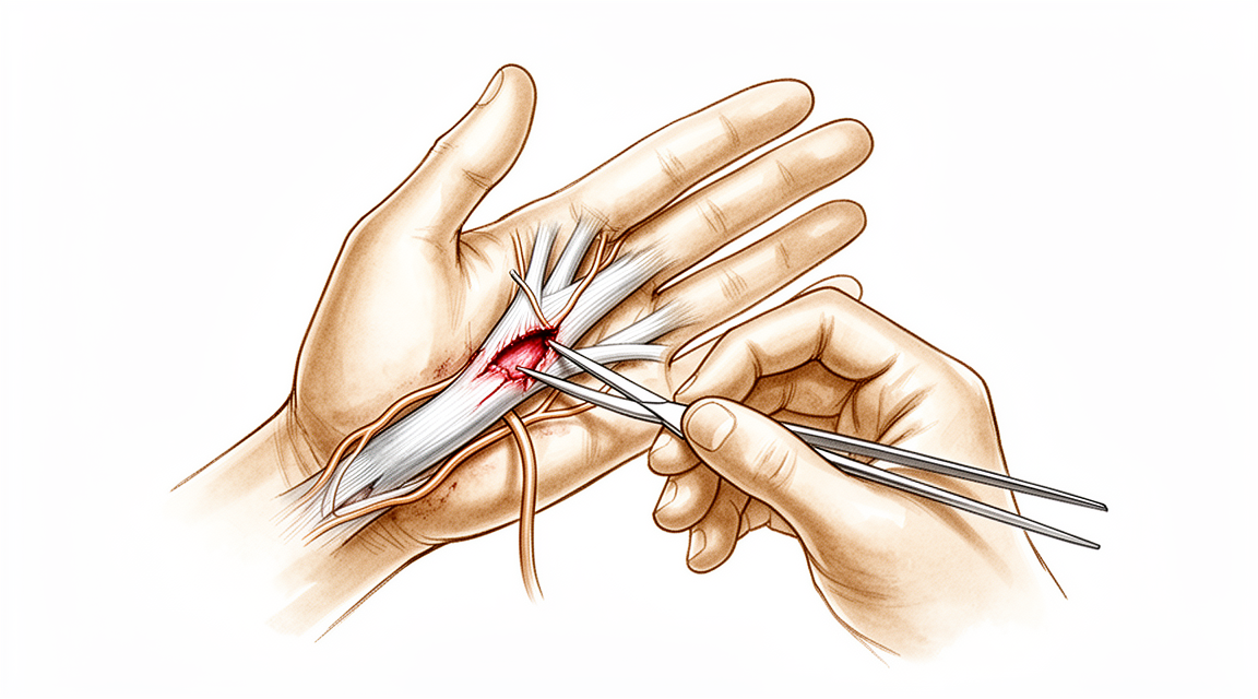
For patients: a plain-language version of this topic is available. See the patient guide.
Overview
Tendon and nerve repair procedures address functional deficits arising from peripheral nerve injuries, neuromuscular disorders, and congenital or traumatic lesions [21]. Nerve reconstruction strategies include end-to-end repair of fascicular groups, which yields superior results compared to nerve grafts for partial lacerations [11], and nerve conduits, indicated primarily for bridging digital sensory nerve gaps of 3 cm or less to avoid donor site morbidity [65]. Nerve transfers have become a first-line reconstructive technique, guided by principles of donor site location, strength, safety, and efficacy [25]. Selection of donor nerves for heterotopic transfers requires careful weighing against other alternatives to mitigate the risk of donor impairment and preserve future reconstructive options [16]. For high peripheral nerve injuries or secondary cases where intrinsic hand muscle recovery is not expected after end-to-end repair or grafting, the median-ulnar end-to-side bridge nerve graft technique is mainly indicated [4].
Tendon transfer serves as a useful option to restore function after radial, median, or ulnar nerve injury when surgical nerve repair does not result in useful function or is not possible [5]. It is specifically indicated for longstanding, irreparable, isolated radial nerve lesions [36]. Primary tendon repair is permissible only when strict criteria are met, including early presentation, minimal contamination, and favorable wound conditions; otherwise, secondary repair via tendon graft is recommended [15]. The tendon with Z-lengthening technique offers an alternative for patients with tendon deficiency and nerve injuries, avoiding the donor site morbidity associated with autograft harvest [42]. Vascularized tendon grafting has a real place in the armamentarium for zone 2 tendon injuries not amenable to direct repair, offering advantages in recovery time and complications over classic two-staged grafts in select cases [46].
Comparative outcomes between nerve and tendon procedures remain nuanced. Overall, better outcomes were observed in patients who underwent nerve transfer versus tendon transfer for radial nerve paralysis reconstruction [1]. Conversely, pooled analysis indicates that tendon transfers had higher rates of superior clinical outcomes compared with nerve transfers and nerve grafts for isolated radial nerve palsy [12]. Tendon transfer offers an important alternative, possibly the procedure of choice, to microsurgical nerve reconstruction, particularly when early professional and social reintegration is important [7]. Patients who present early and can tolerate longer time to functional recovery are optimal candidates for nerve transfers [14]. Even for large gaps within 8 months, an attempt at nerve reconstruction before proceeding to tendon transfers appears indicated for high radial nerve injury with defects of nine centimeters or greater [3]. Simultaneous nerve repair and tendon transfer showed no detrimental results and may provide improved function over tendon transfer alone for peroneal nerve injuries [2]. Further prospective studies using standardized outcome measures are needed to define the precise role of nerve transfers in reconstructive upper limb surgery for tetraplegia [35], and more participants with longer follow-up are required to fully demonstrate the superiority of combined nerve and tendon procedures for grasp and release function in these patients [6].
Anatomy & Pathophysiology
Nerve Injury and Regeneration
Following peripheral nerve injury, the distal stump undergoes Wallerian degeneration, a process mediated by Ca2+ influx and calpain activation followed by macrophage and Schwann cell clearance of debris [68]. Effective peripheral nerve repair requires an ultrastructural and biochemical understanding of nerve injury, microsurgical techniques for managing nerve gaps, and familiarity with experimental advances in regeneration [26]. Post-operative therapy is essential for splinting, optimizing nerve gliding, preventing secondary deformity, and assisting with sensory re-education [26].
A painful neuroma is defined as the biologic response of the proximal stump after division when regenerating axons are impeded from re-entering the distal stump [30]. Neuroma formation results from abnormal nerve regeneration following a peripheral nerve lesion, often involving fascicular escape and scarring as nerve ends attempt to connect with end organs [30]. Certain nerves, such as the superficial radial nerve, are more prone to the development of a painful neuroma [30].
End-to-side nerve suture has not demonstrated a reliable role as a substitute for standard reconstructive techniques in brachial plexus surgery [27]. In a series of 11 patients with traumatic brachial plexus injuries, electromyographical studies showed a contribution from end-to-side neurotisation in only three patients with shoulder abduction recovery [27]. The intrinsic hand muscles have motor evoked potentials at consistent distances from bony landmarks both dorsally and volarly [124].
Tendon Healing and Biology
Tendon healing comprises three phases: inflammatory, fibroblastic, and remodelling [221]. During the inflammatory phase, the strength of the repair is solely through the surgical suture and possibly the fibrin clot [221]. During the fibroblastic phase, tendon strength is increased by the synthesis of the extracellular matrix [221]. Adhesions form between tendon and sheath, inhibiting natural gliding and movement [221].
Tendon remodeling begins 6–8 weeks after injury, characterized by decreased cellularity, reduced matrix synthesis, decreased type III collagen, and increased type I collagen synthesis [69]. Type I collagen fibers organize longitudinally along the tendon axis during remodeling and are responsible for the mechanical strength of the regenerate tissue [69]. Repair tissue never achieves the characteristics of normal tendon [69].
Extrinsic tendon healing involves fibroblasts and inflammatory cells moving from peripheral tissue sources to invade the healing site, a process that includes initial adhesion formation [69]. The extrinsic healing mechanism is activated earlier than the intrinsic mechanism and is responsible for initial adhesion formation and a disorganized collagen matrix with high cellularity [69]. Intrinsic tendon healing occurs through the migration and proliferation of cells from the endotenon and epitenon into the injury site to establish an extracellular matrix and internal neovascular network [69]. The intrinsic healing mechanism is responsible for the reorganization of collagen [69].
The tenosynovium lining the fibroosseous tunnel supplies nutrition and lubrication to poorly vascularized flexor tendons [83]. Within the flexor tendon sheath, tendon vascularity is supplied via the vincula system, specifically the vinculum longus and brevis [83]. Decellularized flexor tendon-bone grafts can exceed the strength and excursion needed for hand therapy immediately after reconstruction at the time of repair [116].
Hand Anatomy and Biomechanics
The hand is an organ designed to obtain information and an organ of execution, with specialized anatomy expressing these two functions [37]. The hand consists of 19 bones, 17 articulations, and 19 muscles situated entirely within the hand, along with approximately the same number of tendons activated by forearm muscles [37]. The skeleton of the hand and wrist consists of 27 bones, of which 19 are long bones [41].
The wrist has three axes of movement, permitting the hand to be positioned in any spatial configuration for grasping [41]. The thumb metacarpal makes an angle of about 45 degrees with the second metacarpal in the sagittal plane, allowing the thumb to oppose the other four digital rays [41]. The transverse axis of the palm forms an acute angle of approximately 75 degrees with the longitudinal axis [41].
Control of digital posture requires a complex balance of extrinsic and intrinsic muscle forces [40]. Extrinsic extensor muscles run through six different fibroosseous retinacular compartments at the wrist level [40]. The sagittal bands stabilize the digital extensor tendons over the midline of the metacarpophalangeal joint, transmitting proximal extrinsic extensor tension to the proximal phalanx [40]. Rupture or attenuation of sagittal band fibers allows the extrinsic extensor tendon to sublux to the ulnar side of the metacarpal head, causing ulnar deviation of the finger [40].
The flexor digitorum superficialis tendon bifurcates around the flexor digitorum profundus at the beginning of the A2 pulley and reunites distally at the Camper chiasm [83]. The A2 and A4 pulleys are the most essential in maintaining the mechanical advantage of the flexor tendons [83].
The dorsal interossei are abductors, while the volar interossei are adductors [77]. The middle finger has two dorsal interossei and no volar interossei because the central axis of the hand lies within it [77]. The deep head of each dorsal interosseous muscle forms a lateral band that flexes and weakly abducts the proximal phalanx while extending the middle and distal phalanges [77]. The volar interossei form the ulnar lateral band of the index finger and the radial lateral band of the ring and little fingers [77].
The dorsal skin of the hand is thin, possesses a normal pilosebaceous system, and has loose connections with deeper planes allowing free gliding [85]. Flexion of the fingers produces a significant lengthening of the dorsal skin, with an average increase of 3 cm in the middle finger from extension to full flexion [85]. The palmar skin is subdivided into functional units, with the radial portion being mobile and well vascularized, and the ulnar/distal portion having poor mobility and vascularity [76]. Incisions made along the sides of the diamond-shaped cutaneous contact zones in flexed digits present a minimal chance of retraction [76].
The superficial palmar fascia covers a triangular area of the central palm, with four central bands extending distally toward each finger but no central band for the thumb [86]. The digital neurovascular structures are surrounded by a diffuse network of thin transverse oblique fibers, with dorsal fibers called Cleland ligament and palmar fibers called Grayson ligament [86].
The metacarpal arch is adaptable due to the mobility of the peripheral metacarpals, which can deepen the palmar concavity as they approach each other [79]. The fifth metacarpal has a range of flexion–extension of approximately 20 degrees [79]. The stability of the metacarpophalangeal joints is essential to the support of the longitudinal arch and the transverse metacarpal arch [79].
The thumb A1 pulley and digital neurovascular structures can be localized using specific hand surface landmarks [130]. The pulley system of the thumb is composed of 4 components, as opposed to the traditional view of only 3 [132].
Classification
Seddon and Sunderland: The original framework for classifying nerve injuries was proposed by Seddon in 1943 with three degrees of injury and by Sunderland in 1951 with five degrees of injury [192].
Mackinnon: The Mackinnon classification expanded the nerve injury classification to include a sixth category representing a mixed injury pattern [192]. In this system, first-, second-, and third-degree injuries have the potential for recovery and for the most part do not require surgical intervention [192]. A first-degree nerve injury involves temporary loss of conductive signaling activity while the axonal bundle remains intact, with function recovering within three months [192]. A second-degree nerve injury involves axonal damage with intact surrounding connective tissue, allowing complete regeneration at a rate of 1 inch per month [192]. Third-degree nerve injuries involve injury to the endoneurium while sparing the epineurium and perineurium, and do not recover well without surgical intervention [192]. Fourth-degree nerve injuries involve intact epineurium only and will not recover without surgical intervention [192]. Fifth-degree nerve injuries involve complete transection of the nerve and will not recover without surgical intervention [192]. A sixth-degree nerve injury represents a combination of any of the previous five levels of injury [192].
International Classification for Surgery of the Hand in Tetraplegia (ICSHT): Created in 1986, the ICSHT defines the level of injury and provides information about potential tendon transfer options [197]. In the ICSHT classification, muscles below the elbow with a Medical Research Council (MRC) grade of 4 or more are registered as potential tendon transfer donors [197]. ICSHT Groups 0, 1, and 2 are associated with very limited reconstructive options [197].
Türker: The Türker classification system for accessory extensor pollicis longus tendons has been modified to include a Type 3 category for rare findings of two radial-sided accessory extensor tendons in the same individual [163].
Other Considerations: A simpler classification system for extensor tendon zones has been proposed to align with current treatment strategies, such as conservative splinting for closed injuries and strong surgical repair for open injuries [170]. A panel recommends adapting a simpler classification system resembling that for flexor tendons for extensor tendon repairs to facilitate surgical decision-making and rehabilitation [131]. Survey results suggest that a revisit of popular extensor tendon zone classification systems may be necessary for future clinical practice and communication among surgeons [165].
Clinical Presentation
A thorough history must elucidate both the cause and the probable location of the injury [48]. Patients presenting with an injury pattern that may lead to nerve injury warrant prompt referral to an upper extremity specialist to optimize outcomes [47]. The presence of fractures is associated with a higher incidence of nerve and tendon injuries [18], and involvement of these structures is linked to an increased risk of long-term disability [18].
Physical Examination
Assessment of motor and sensory deficits is required during evaluation [48]. The strength of potential donor muscles should be assessed during evaluation [48]. Passive joint motion should be assessed during evaluation [48]. A migrating Tinel's phenomenon is a good prognostic indicator [48].
Diagnostic Modalities
MRI and ultrasound have been of limited use for objective measures in nerve injury evaluation [48]. Electromyography and nerve conduction velocity studies are rarely helpful acutely except when continuity is unknown [48]. Slowed but intact conduction indicates some continuity [48]. Electromyography and nerve conduction velocity study is recommended around 3 to 4 months after injury [48]. Larger polyphasic motor action potentials of longer duration may be seen before clinical recovery [48]. If the amplitude of motor nerve conduction velocity is low, less than 0.3 mV, exploration can be undertaken [48]. Repair remains a viable option even 5 to 6 months after injury if the amplitude of motor nerve conduction velocity is low [48].
Prognostic Factors and Sequelae
Factors affecting the results after flexor tendon repair in zone 2 include the mechanism of injury (clean-cut versus crush) [23], associated fractures [23], the number of digits/tendons injured [23], age [23], the experience of the surgeon [23], and the type of postoperative mobilization programme [23]. A large part of the variance in the outcome of flexor tendon repair in zone 2 is thought to be related to the psychological and biologic characteristics of the patient [23]. The outcome of a flexor tendon repair is influenced by many factors that cannot be controlled intraoperatively [29]. The painful neuroma is an often debilitating sequela of nerve injury about the hand [30]. Painful neuromas can be associated with blunt trauma or retraction of a nerve when the nerve is not actually divided [30].
Specific Injury Patterns
This study demonstrates a distinct clinical presentation of brachial plexus trauma, characterized by preserved finger flexion despite complete plexus injury [117]. This case contributes further to our understanding of the clinical presentation of hand function following high median nerve transection [45].
Investigations
Clinical Examination: A systematic method is required for the physical examination of the injured or dysfunctional hand and wrist [38]. A careful physical examination is essential to direct care and future testing if indicated [38]. Diagnostic tests such as imaging and serum laboratory studies are useful in determining pathology but can be expensive, time consuming, and often nonspecific [38].
MRI: Magnetic resonance imaging provides important preoperative information for surgical decision-making and planning in patients who present late with closed flexor tendon injuries of the hand [168]. MRI is probably most useful in identifying additional pathology such as flexor tendon bowstringing [89]. MR assessment of Dupuytren’s disease is hindered by the resolution of current equipment, orientation issues due to multiplanar deformities of the fingers, and lack of intraoperative availability [89].
CT: 3D CT volume rendering has been used to diagnose flexor tendon entrapment [219].
Ultrasonography: Ultrasonographic assessment may serve as a valuable complementary tool for objectively evaluating nerve recovery [205].
Doppler Imaging: Doppler imaging is a promising improvement for identifying superficially displaced neurovascular bundles, though higher resolution imaging technology is needed [89].
Other Considerations: Although diagnosing the cause of ulnar collateral ligament locking may be complicated by the lack of evidence in imaging studies, open surgical treatment has traditionally been the most often used with a high success rate [224]. Surgical exploration confirms the diagnosis of closed partial flexor digitorum profundus rupture and allows for excision of the damaged segment to return normal movement without compromising strength [227].
Treatment
Nerve Repair and Reconstruction
Non-Operative Management: In the absence of a space-occupying lesion, a trial of nonoperative management is advisable for atraumatic posterior interosseous nerve palsy; however, exploration is recommended if there is no sign of muscle recovery after 6 weeks of observation or if progressive weakness occurs [175]. Electromyography and nerve conduction velocity studies are rarely helpful acutely except when continuity is unknown, with study recommended around 3 to 4 months after injury [48]. If the amplitude of motor nerve conduction velocity is low, less than 0.3 mV, exploration can be undertaken, with repair remaining a viable option even 5 to 6 months after injury [48]. A migrating Tinel's phenomenon is a good prognostic indicator for radial nerve injuries [48].
Operative Indications and Timing: Delay in repair of less than 6 months, defect length of less than 5 cm, or grafting with three or more donor nerve cables achieved better recovery following high radial nerve injury [13]. For large gaps of nine centimeters or greater in high radial nerve injury, an attempt at nerve reconstruction before proceeding to tendon transfers appears to be indicated within 8 months [3]. Adequate sensory recovery without any nerve repair had occurred by the 2-year follow-up in artery-only fingertip replantations [64].
Reconstructive Techniques: Collagen conduits reliably provide a repair that restores nerve function for nerve gaps measuring less than 2 cm [28], and type I collagen conduit is a reliable alternative to nerve grafting for gaps up to 10 mm in length [191]. Processed nerve allografts offer a safe and effective method of reconstructing peripheral nerve gaps from 5 to 50 mm in length [71]; however, nerve regeneration is significantly impaired as the gap distance between the distal end of the allograft and the distal nerve stump increases to 5 mm [72]. Good nerve regeneration can be achieved with short nerve defects using direct gradual lengthening of the nerve stumps, a method that does not require the sacrifice of healthy nerves from the donor site or leave surgical scars [99]. Nerve transfers have become a first line reconstructive technique in the restoration of function, with fundamental principles such as donor site location, strength, safety, and efficacy remaining integral to contemporary nerve surgery [25]. End-to-side nerve suture must not be a substitute for standard reconstructive techniques in brachial plexus surgery, though it may occasionally represent a support to standard procedures in severe injuries with few undamaged donor nerves [27]. There was no difference in outcomes between nerve transfer and nerve graft groups at 1 or 2 or more years follow-up for late microsurgical nerve reconstruction of brachial plexus birth injury [19].
Technical Considerations: Wrapping the nerve with fibrin sealant before division and immediate fixation resulted in less protrusion of the nerve end [186]. The key to effective peripheral nerve repair includes an ultrastructural and biochemical understanding of the process, effective microsurgical techniques, and familiarity with experimental advances in enhancing nerve regeneration [26].
Post-Operative Care: The post-operative role of the therapist is essential in splintage, optimising nerve gliding, preventing secondary deformity, and assisting with sensory re-education for peripheral nerve repair [26].
Tendon Repair and Reconstruction
Operative Indications: Flexor tendon grafting is the preferred method of treatment for patients with neglected digital flexor tendon lacerations and after the failure of flexor tendon repair [17], although successful flexor tendon repair has narrowed the indications for flexor tendon grafting [17]. Surgical treatment consisting of flexor tendon sheath release is effective in restoring motion with minimal risks of recurrence and neurovascular complication for pediatric trigger thumb [55]. Open surgery resulted in more reliable and rapid outcomes compared with nonoperative treatment for idiopathic congenital talipes equinovarus [199]. Surgical treatment for chronic tendon mallet injury provides anatomical reconstruction, is indicated regardless of the condition of the terminal tendon and the interval from the initial injury, and allows for easy determination of graft tension [97].
Surgical Approach and Technique: Factors affecting the results after flexor tendon repair in zone 2 include the mechanism of injury, associated fractures, the number of digits/tendons injured, age, the experience of the surgeon, and the type of postoperative mobilization programme [23]. The six-strand double-loop technique for zone II flexor tendon repair appears better without an increased rate of rupture but with a shorter rehabilitation period compared with a two-strand technique [180]. The TWZL technique offers an alternative treatment option for patients with tendon deficiency and nerve injuries, avoiding donor site morbidity associated with autograft harvest [42]. Traditional tendon-to-tendon repair is more robust compared with interference screw fixation for FDL tendon transfer, with significantly greater load to failure [215]. With modern anesthesia techniques, tendon lacerations in a healthy neonate can be managed in a timely and safe manner [174]. The internal suture technique for mallet finger fracture provides fixation without an external button or transfixion of the fragment, with all fractures uniting and no neuromas [208].
Biological Healing: Tendon stumps appear to fuse by fibrous bridging after 2 weeks’ time when the collagen fibers are positioned perpendicularly to the tendon fibrils [67]. Within the next 2 weeks after tendon injury, the remodeling process ensures a progressive parallel organization of collagen fibers representing the realignment of the scar tissue [67].
Rehabilitation and Analgesia: Tendon grafts will tolerate early motion therapy if the proximal and distal tenorrhaphy junctures are strong enough to withstand these forces of active finger motion [172]. Despite a decline in opioid quantity per patient, most patients still receive opioids after zone II flexor tendon repair [213].
Combined Nerve and Tendon Procedures
Comparative Outcomes: Overall, better outcomes were observed in those who underwent nerve transfer versus tendon transfer procedures for radial nerve paralysis reconstruction [1]. The tendon transfer offers an important alternative – possibly the procedure of choice – to microsurgical nerve reconstruction, particularly when early professional and social reintegration is important for radial nerve palsy [7, 10]. Patients who present early and can tolerate longer time to functional recovery would be optimal candidates for nerve transfers in radial nerve palsy [14].
Surgical Approach: The anterior approach for supinator nerve transfer to the posterior interosseous nerve yields similar results to the posterior approach and has the advantage of allowing easier access for simultaneously performing nerve or tendon transfers to reconstruct grasp and pinch in tetraplegia [90]. Intrinsic hand muscle reinnervation by median-ulnar end-to-side bridge nerve graft is mainly indicated in high peripheral nerve injury or secondary cases when recovery of the intrinsic muscle of the hand is not expected after end-to-end repair or graft [4].
Neuroma Management
Pathology and Treatment: The painful neuroma is an often debilitating sequela of nerve injury about the hand, resulting from abnormal nerve regeneration following a peripheral nerve lesion [30]. Neurolysis of the superficial radial nerve offers the opportunity for pain relief but does not reliably produce success [200]. Neurolysis and fascial nerve wrap surgery resulted in complete resolution of all pain modalities in nine of 14 patients, with only one patient continuing to have severe pain for neuromas-in-continuity and scarred median and ulnar nerves [209]. A skin island approach using neurovascular island flaps provided good pain control and recovery of hand function after painful neuromas at the digit tip [212].
Complications
Nerve Repair and Reconstruction
Timing and Regeneration: Delaying nerve repair for more than 6 months substantially reduces the number of regenerating axons and their response to growth factors [49]. Even under optimal repair or reconstruction conditions, distal motor end plates often degenerate during the critical months required for regeneration and reinnervation [49]. Follow-up time and age significantly influence outcomes following median or ulnar nerve repair [60].
Surgical Technique and Donor Selection: End-to-side nerve suture must not substitute for standard reconstructive techniques in brachial plexus surgery [27]. Selecting a donor nerve for transfer requires weighing the risk of donor nerve impairment against the potential narrowing of future reconstructive options [16]. Evidence for good nerve recovery or improved function following single digital nerve repair is poor, with only 24% of repaired nerves regaining sensory recovery close to or equivalent to estimated pre-injury levels [9].
Injury Timing and Specific Procedures: Patients with sharp major nerve injuries required grafting more frequently after several days from injury, whereas primary repair of common and proper digital nerves could be achieved up to two weeks or greater after injury [33]. Although rare, unrecovered nerve injury adversely affects outcome following acute distal biceps tendon repair [32]. In one case of ulnar collateral ligament reconstruction, damage to a branch of the superficial radial nerve resulted in a painful neuroma on the dorso-ulnar aspect of the thumb [203]. Dysaesthesia of the dorsal sensory ulnar nerve occurred in 4% of wrists following ulnotriquetral split tear repair [206]. Nerve injury or persistent numbness occurred in 0.6% of patients following open trigger finger release [216]. There is a potential risk of ulnar nerve injury following reconstruction of the extensor carpi ulnaris subsheath using a fascia lata allograft [214].
Tendon Repair and Reconstruction
Flexor Tendon Outcomes: The overall rate of reoperation after flexor tendon repair in all zones is 6% [176]. The rate of repair rupture reported in the literature is 4% [176], while the rate of reoperation for repair rupture in New York state is 2.3% [176]. The reported rate of tendon adhesions after flexor tendon repair is 4% [176], and the rate of tenolysis performed after flexor tendon repair in New York state is 3.6% [176]. Other possible complications include triggering, pulley failure, quadriga, and lumbrical plus deformity [176]. In a study of 45 patients using a six-strand figure-of-eight suture for zone 2 flexor tendon repair, one repair (2%) ruptured on the seventh day after surgery [113]. No complications such as infection or complex regional pain syndrome were noted in the remaining 49 repairs in that study [113]. Delayed repair of tendons at the wrist often requires grafts with far from perfect results [240].
Extensor Tendon and Trigger Finger Complications: Potential complications of extensor tendon centralization at the metacarpophalangeal joint include failure of reconstruction, stiffness, and infection [189]. Potential complications of a reconstructive stabilization technique for extensor tendon subluxation include failure of reconstruction, joint stiffness, and infection [190]. Potential risks of extensor carpi ulnaris subsheath reconstruction include tendinitis, recurrent ECU instability, and tendon rupture [214]. Stiffness occurred in 8.6% of patients following open trigger finger release [216]. Recurrence of triggering occurred in 2.2% of patients following open trigger finger release [216]. Delayed wound healing occurred in 1.2% of patients following open trigger finger release [216]. Development of Dupuytren contracture occurred in 0.8% of patients following open trigger finger release [216]. 4.9% of patients required secondary operations following open trigger finger release [216]. At the time of surgery for rheumatoid hand tendon transfers, the underlying cause of the rupture should be addressed to prevent future additional tendon ruptures [234].
Combined and General Outcomes
Ballistic Injuries: The presence of fractures is associated with a higher incidence of nerve and tendon injuries, and involvement of these structures is linked to an increased risk of long-term disability in ballistic hand injuries [18].
Ulnotriquetral Split Tear Repair: Surgical complications occurred in 8% of cases following ulnotriquetral split tear repair [206]. Recurrent pain occurred in 3% of wrists following ulnotriquetral split tear repair [206]. Superficial infection occurred in one case (1%) following ulnotriquetral split tear repair [206]. Five wrists (5%) underwent revision operations following ulnotriquetral split tear repair [206].
Ulnar Collateral Ligament Reconstruction: The thumb metacarpophalangeal joint usually ached after surgery for ulnar collateral ligament reconstruction, taking several months to settle completely [203]. Five patients experienced persistent aching of the metacarpophalangeal joint when pinching after ulnar collateral ligament reconstruction, which was mild and not disabling [203].
Recovery
Light activity (weeks): The evidence provided does not specify a typical week range for the resumption of desk work, driving, or light activities of daily living.
Full activity (months): The evidence provided does not specify a month range for the return to manual work, sport, or full range of motion and strength.
Complete recovery / outcome plateau (months): The evidence provided does not specify a month range for the stabilization of pain, strength, and final functional outcomes.
Rehabilitation protocol: Nerve transfer rehabilitation requires a collaborative surgeon-therapist relationship to communicate operative details, expected timelines, and realistic expectations [157]. A rehabilitative protocol that is individualized to fit each patient's tendon pathology and surgery is essential following extensor mechanism surgery [135]. The results suggest that the central nervous system component should be considered in the development of new treatment protocols for flexor tendon injuries [63].
Functional milestones: In approximately 90% of patients, distal nerve transfers resulted in functional recovery of shoulder abduction, elbow flexion or extension, and wrist extension in C5–C8 (T1 Hand) brachial plexus paralysis [62]. Active pick-up function was successfully restored in ten patients after undergoing multiple nerve transfers combined with additional secondary functional hand reconstructions for total brachial plexus avulsion injuries [226]. Only 24% of repaired single digital nerves regained sensory recovery close to or equivalent to estimated pre-injury levels [9]. Regeneration to a satisfactory degree was observed in all patients, and the majority achieved full recovery of sensory and motor functions following autologous grafts and neurolysis for radial nerve lesions [160]. Both groups in a comparison of a novel atraumatic polymer-assisted peripheral nerve repair device and microsurgical neurorrhaphy showed signs of nerve conduction as early as 2 months, with muscle regrowth reaching 60% of the nonoperated contralateral leg by 3 months [162]. This technique restores good function in most patients with zone 1 and 2 flexor tendon injuries, in which primary tendon repair has not been performed or was unsuccessful, and where pulley reconstruction is not required [73]. All six cases achieved good or excellent results with no tendon ruptures during rehabilitation using the flexor digitorum profundus “demi-tendon” technique [149]. The final combination of repair and early active mobilization for primary repair of FPL tendons compares favourably with previous methods of treatment [144].
Other Considerations: Processed nerve allografts of up to 5 mm in diameter appear capable of supporting successful nerve regeneration, based on a small subset of patients [24]. Nerve regeneration was significantly impaired as the gap distance between the distal end of the allograft and the distal nerve stump increased to 5 mm [72]. The supercharged end-to-side anterior interosseous to ulnar motor nerve transfer procedure has broad clinical utility for augmenting partial recovery and preserving motor end plates in second- and third-degree axonotmetic nerve injuries [225]. Findings from health-related quality of life and functional outcomes following nerve transfers for traumatic upper brachial plexus injuries provide key prognostic information for patients and peripheral nerve surgeons [161].
Key Evidence
- [L4] Overall, we observed better outcomes in those who underwent nerve transfer versus tendon transfer procedures. [1] (10.1016/j.jhsa.2019.12.009)
- [L4] The results of our limited case series for this rare condition indicate that simultaneous nerve repair and tendon transfer showed no detrimental results and may provide improved function over tendon transfer alone. [2] (10.1186/s13018-014-0067-6)
- [L4] Even for large gaps, within 8 months, an attempt at nerve reconstruction before proceeding to tendon transfers appears to be indicated. [3] (10.1016/j.jhsa.2007.10.004)
- [Case_report] The technique is mainly indicated in high peripheral nerve injury or secondary cases when recovery of the intrinsic muscle of the hand is not expected after end-to-end repair or graft. [4] (10.1016/j.jhsa.2009.10.033)
- [L3] More participants with a longer follow-up are needed to fully demonstrate the superiority of combined nerve and tendon procedures. [6] (10.1177/17531934251381202)
- [L4] Accordingly, the tendon transfer offers an important alternative – possibly the procedure of choice – to microsurgical nerve reconstruction, particularly when early professional and social reintegration is important. [7] (10.1016/s0363-5023(09)60108-8)
- [L2] Evidence for good nerve recovery or improved function following nerve repair is poor, with only 24% of repaired nerves regaining sensory recovery close to or equivalent to estimated pre-injury levels. [9] (10.1177/1753193419846761)
- [L4] The tendon transfer offers an important alternative—possibly the procedure of choice—to microsurgical nerve reconstruction, particularly when early professional and social reintegration is important. [10] (10.1016/j.jhsa.2008.11.012)
- [L4] End-to-end repair of fascicular groups provides better results than repair using nerve grafts. [11] (10.1016/j.jhsa.2014.01.026)
- [L4] On pooled analysis, tendon transfers had higher rates of superior clinical outcomes as compared with nerve transfers and nerve grafts. [12] (10.1177/15589447221150516)
- [L4] Delay in repair of less than 6 months, defect length of less than 5 cm, or grafting with three or more donor nerve cables achieved better recovery. [13] (10.1177/17531934221147651)
- [L3] Patients who present early and can tolerate longer time to functional recovery would be optimal candidates for nerve transfers. [14] (10.1177/1558944720988126)
- [L5] Primary tendon repair is permissible only when strict criteria are met, including early presentation, minimal contamination, and favorable wound conditions; otherwise, secondary repair via tendon graft is recommended. [15] (10.2106/00004623-195941040-00001)
- [L4] Selection of the donor nerve must be carefully weighed against other treatment alternatives, considering the risk of donor nerve impairment and the potential narrowing of future reconstructive options. [16] (10.1016/j.jhsa.2006.12.012)
- [L5] [17] (10.1016/j.jhsa.2010.03.042)
- [L4] The presence of fractures is associated with a higher incidence of nerve and tendon injuries, and involvement of these structures is linked to an increased risk of long-term disability. [18] (10.1177/15589447221092111)
- [L4] There was no difference in outcomes between nerve transfer and nerve graft groups at 1 or 2 or more years follow-up. [19] (10.1016/j.jhsa.2019.10.036)
- [L5] Tendon lengthening and transfer are indicated for neuromuscular disorders, nerve injuries, and congenital or traumatic lesions. [21] (10.1016/j.otsr.2014.07.033)
- [L4] [23] (10.1177/1753193410387333)
- [L4] Although based on a small subset of patients, processed nerve allografts of up to 5 mm in diameter appear capable of supporting successful nerve regeneration. [24] (10.1177/1558944716646782)
- [L4] Nerve transfers have become a first line reconstructive technique in the restoration of function, with fundamental principles such as donor site location, strength, safety, and efficacy introduced by Oberlin et al remaining integral to contemporary nerve surgery. [25] (10.1016/j.jhsa.2025.01.013)
- [L5] [26] (10.1177/175899839900400102)
- [L4] [27] (10.1177/1753193409104673)
- [L4] This study confirms that collagen conduits reliably provide a repair that restores nerve function for nerve gaps measuring less than 2 cm. [28] (10.1016/j.jhsa.2011.06.009)
- [L4] The outcome of a flexor tendon repair is influenced by many factors that cannot be controlled intraoperatively. [29] (10.1016/j.jhsa.2022.01.015)
- [L5] [30] (10.1016/j.jhsa.2009.12.019)
- [L3] Although rare, unrecovered nerve injury adversely affects outcome. [32] (10.1302/0301-620x.103b7.bjj-2020-2246.r1)
- [L4] Patients with sharp major nerve injuries required grafting more frequently after several days from injury, whereas primary repair of common and proper digital nerves could be achieved up to two weeks or greater after injury. [33] (10.1016/j.jhsa.2023.11.006)
- [L4] More prospective studies using standardized outcome measures are needed to define the precise role of nerve transfers. [35] (10.1177/1753193419886443)
- [L3] Tendon transfers are indicated in longstanding, irreparable, isolated radial nerve lesions. [36] (10.1016/j.jhsa.2007.10.015)
- [L4] The TWZL technique offers an alternative treatment option for patients with tendon deficiency and nerve injuries, avoiding donor site morbidity associated with autograft harvest. [42] (10.1016/j.jhsa.2022.12.016)
- [Case_report] This case contributes further to our understanding of the clinical presentation of hand function following high median nerve transection. [45] (10.1186/s12891-025-08469-3)
- [Commentary] Despite limitations regarding preoperative details, vascularized tendon grafting has a real place in the armamentarium for zone 2 tendon injuries not amenable to direct repair, offering advantages in recovery time and complications over classic two-staged grafts in select cases. [46] (10.1177/1753193414552944)
- [L3] Patients with an injury pattern that may lead to nerve injury warrant prompt referral to an upper extremity specialist in an effort to optimize outcomes. [47] (10.1177/1558944719866865)
- [L5] [48] (10.5435/jaaos-d-17-00325)
- [L4] Surgical treatment consisting of flexor tendon sheath release is effective in restoring motion with minimal risks of recurrence and neurovascular complication, although the optimal age for surgical treatment is unclear. [55] (10.1016/j.jhsa.2008.04.017)
- [L3] Follow-up time and age significantly influence the outcome following nerve repair, with significant improvements in the total score seen throughout the follow-up period. [60] (10.1054/jhsb.2001.0567)
- [L4] In approximately 90% of patients, distal nerve transfers resulted in functional recovery of shoulder abduction, elbow flexion or extension, and wrist extension. [62] (10.1016/j.jhsa.2021.11.014)
- [L3] The results suggest that the central nervous system component should be considered in the development of new treatment protocols for flexor tendon injuries. [63] (10.1177/1753193408096017)
- [L4] Furthermore, adequate sensory recovery without any nerve repair had occurred by the 2-year follow-up. [64] (10.1016/j.jhsa.2013.08.110)
- [L5] Nerve conduits are indicated primarily for bridging digital sensory nerve gaps of 3 cm or less, avoiding donor site morbidity associated with nerve autografts. [65] (10.1016/j.jhsa.2013.02.034)
- [L3] [67] (10.1016/j.jhsa.2014.06.140)
- [L5] [68] (10.1016/j.jhsa.2018.01.023)
- [L5] [69] (10.1016/j.jhsa.2007.09.007)
- [L3] Our data suggest that processed nerve allografts offer a safe and effective method of reconstructing peripheral nerve gaps from 5 to 50 mm in length. [71] (10.1016/j.jhsa.2012.08.028)
- [L5] Nerve regeneration was significantly impaired as the gap distance between the distal end of the allograft and the distal nerve stump increased to 5 mm. [72] (10.1177/1558944719828009)
- [L4] This technique restores good function in most patients with zone 1 and 2 flexor tendon injuries, in which primary tendon repair has not been performed or was unsuccessful, and where pulley reconstruction is not required. [73] (10.1177/1753193417737920)
- [L4] The anterior approach yields similar results to the posterior approach and has the advantage of allowing easier access for simultaneously performing nerve or tendon transfers to reconstruct grasp and pinch. [90] (10.1177/1753193421996987)
- [L4] This technique provides anatomical reconstruction, is indicated regardless of the condition of the terminal tendon and the interval from the initial injury, and allows for easy determination of graft tension. [97] (10.1016/j.jhsa.2018.03.020)
- [L4] Good nerve regeneration can be achieved with short nerve defects, and the method is promising as it does not require the sacrifice of healthy nerves from the donor site or leave surgical scars. [99] (10.1186/s12891-025-08301-y)
- [L3] [113] (10.1177/1753193408099818)
- [L5] At the time of repair, decellularized flexor tendon-bone grafts can exceed the strength and excursion needed for hand therapy immediately after reconstruction. [116] (10.1016/j.jhsa.2013.08.092)
- [L4] This study demonstrates a distinct clinical presentation of brachial plexus trauma, characterized by preserved finger flexion despite complete plexus injury. [117] (10.1016/j.jhsa.2024.11.025)
- [L5] The intrinsic hand muscles have MEPs at consistent distances from bony landmarks both dorsally and volarly. [124] (10.1016/j.jhsa.2020.04.019)
- [L5] The findings from our study clarify hand surface landmarks in localizing the thumb A1 pulley and digital neurovascular structures. [130] (10.1016/j.jhsa.2013.02.028)
- [L5] The panel recommends adapting a simpler classification system resembling that for flexor tendons and outlines specific treatment approaches for acute extensor tendon injuries in each zone to facilitate surgical decision-making and rehabilitation. [131] (10.1177/17531934251363138)
- [L4] The pulley system of the thumb is composed of 4 components, as opposed to the traditional view of only 3. [132] (10.1016/j.jhsa.2012.08.005)
- [L5] A rehabilitative protocol that is individualized to fit each patient's tendon pathology and surgery is essential. [135] (10.1016/j.jhsa.2015.04.043)
- [L4] The final combination of repair and early active mobilization for primary repair of FPL tendons compares favourably with previous methods of treatment. [144] (10.1054/jhsb.1999.0230)
- [L4] All six cases achieved good or excellent results with no tendon ruptures during rehabilitation. [149] (10.1054/jhsb.2001.0654)
- [L5] Nerve transfer rehabilitation requires a collaborative surgeon-therapist relationship to communicate operative details, expected timelines, and realistic expectations. [157] (10.1016/j.jhsa.2023.09.016)
- [L4] Regeneration to a satisfactory degree was observed in all patients, and the majority achieved full recovery of sensory and motor functions. [160] (10.3390/jcm9123823)
- [L3] These findings provide key prognostic information for patients and peripheral nerve surgeons embarking upon this intensive pathway to potential recovery. [161] (10.1177/1753193411432706)
- [L5] Both groups showed signs of nerve conduction as early as 2 months, with muscle regrowth reaching 60% of the nonoperated contralateral leg by 3 months. [162] (10.1016/j.jhsg.2025.100812)
- [L4] The authors propose an additional category (Type 3) to the Türker classification system to encompass rare findings of two radial-sided accessory extensor tendons in the same individual, which were not previously represented in existing classifications. [163] (10.1016/j.jhsg.2023.10.005)
- [L4] Based on the results of this survey, a revisit of the popular extensor tendon zone classifications systems may be necessary for future clinical practice and communication among surgeons. [165] (10.1177/17531934241253137)
- [L4] MRI provides important preoperative information for surgical decision-making and planning in patients who present late with closed flexor tendon injuries of the hand. [168] (10.1054/jhsb.1999.0306)
- [L5] The authors propose a simpler classification system for extensor tendon zones to align with current treatment strategies, such as conservative splinting for closed injuries and strong surgical repair for open injuries. [170] (10.1177/17531934241274112)
- [L5] [172] (10.1016/j.jhsa.2015.04.016)
- [L4] With modern anesthesia techniques, tendon lacerations in a healthy neonate can be managed in a timely and safe manner. [174] (10.1016/j.jhsa.2010.09.015)
- [L5] In the absence of a space-occupying lesion, a trial of nonoperative management is advisable, but exploration of the nerve is recommended if there is no sign of muscle recovery after 6 weeks of observation or if there is progressive weakness. [175] (10.1016/j.jhsa.2017.07.026)
- [L5] [176] (10.5435/jaaos-22-12-791)
- [L3] The study notes that while non-randomised, the technique appears better without an increased rate of rupture but with a shorter rehabilitation period. [180] (10.1177/1753193408091570)
- [L5] Wrapping the nerve with fibrin sealant before division and immediate fixation resulted in less protrusion of the nerve end. [186] (10.1054/jhsb.1999.0250)
- [L4] [189] (10.1016/j.jhsa.2010.04.029)
- [L4] [190] (10.1016/j.jhsa.2016.10.008)
- [L5] The use of type I collagen conduit is a reliable alternative to nerve grafting for gaps up to 10 mm in length. [191] (10.1016/j.jhsa.2007.07.015)
- [L5] [192] (10.1155/2016/4175293)
- [L4] [197] (10.1177/17531934251369277)
- [L4] Based on the low level of evidence available, it seems that open surgery resulted in more reliable and rapid outcomes compared with nonoperative treatment. [199] (10.1177/1753193414523245)
- [L4] Therefore, while neurolysis of the superficial radial nerve offers the opportunity for pain relief, it does not reliably produce success. [200] (10.1177/1753193407087892)
- [L4] [203] (10.1054/jhsb.2002.0838)
- [L4] Ultrasonographic assessment may serve as a valuable complementary tool for objectively evaluating nerve recovery. [205] (10.1177/17531934231174603)
- [L4] [206] (10.1177/1753193419876066)
- [L4] The internal suture technique provides fixation without an external button or transfixion of the fragment, with all fractures uniting and no neuromas. [208] (10.1054/jhsb.1999.0284)
- [L4] Neurolysis and fascial nerve wrap surgery resulted in complete resolution of all pain modalities in nine of 14 patients, with only one patient continuing to have severe pain. [209] (10.1177/1753193410366191)
- [L4] This skin island approach provided good pain control and recovery of hand function after painful neuromas. [212] (10.1016/j.jhsa.2007.12.002)
- [L2] Despite a decline in opioid quantity per patient, most patients still receive opioids after zone II flexor tendon repair. [213] (10.1016/j.jhsg.2026.100971)
- [L4] [214] (10.1016/j.jhsg.2026.100957)
- [L5] Traditional tendon-to-tendon repair is more robust compared with interference screw fixation for FDL tendon transfer, with significantly greater load to failure. [215] (10.1007/s00167-018-4936-0)
- [L3] [216] (10.1177/1558944718796559)
- [L4] We present a previously unreported site of flexor tendon entrapment as well as the novel use of CT 3D volume rendering to diagnose the entrapment. [219] (10.1177/15589447231185857)
- [L5] [221] (10.1177/1753193413509231)
- [L4] Although diagnosing the cause of UCL locking may be complicated by the lack of evidence in imaging studies, open surgical treatment has traditionally been the most often used with a high success rate. [224] (10.1016/j.jhsg.2022.08.003)
- [L4] The authors believe the procedure has broad clinical utility for augmenting partial recovery and preserving motor end plates in second- and third-degree axonotmetic nerve injuries. [225] (10.1016/j.jhsa.2012.07.022)
- [L4] Active pick-up function was successfully restored in ten patients after undergoing multiple nerve transfers combined with additional secondary functional hand reconstructions. [226] (10.1177/1753193417728405)
- [L4] Surgical exploration confirms the diagnosis and allows for excision of the damaged segment to return normal movement without compromising strength. [227] (10.1177/1558944716681950)
- [L5] At the time of surgery, the underlying cause of the rupture should be addressed to prevent future additional tendon ruptures. [234] (10.1016/j.hcl.2016.03.014)
- [L5] Primary suture of tendons at the wrist yields nearly normal restoration, whereas delayed repair often requires grafts with far from perfect results. [240] (10.2106/00004623-196547010-00007)
See Also
- Flexor tendon repair
- Mallet Finger
- Trigger finger release
- Trigger Finger
References
[1] Nerve Versus Tendon Transfer for Radial Nerve Paralysis Reconstruction. The Journal of Hand Surgery. 2020. DOI: 10.1016/j.jhsa.2019.12.009
[2] Treatment of peroneal nerve injuries with simultaneous tendon transfer and nerve exploration. Journal of Orthopaedic Surgery and Research. 2014. DOI: 10.1186/s13018-014-0067-6
[3] Sural Nerve Autografts for High Radial Nerve Injury With Nine Centimeter or Greater Defects. The Journal of Hand Surgery. 2008. DOI: 10.1016/j.jhsa.2007.10.004
[4] Intrinsic Hand Muscle Reinnervation by Median-Ulnar End-to-Side Bridge Nerve Graft: Case Report. The Journal of Hand Surgery. 2010. DOI: 10.1016/j.jhsa.2009.10.033
[5] Chapter 29 Tendon Transfers for Peripheral Nerve Injuries in the Upper Extremity. 2019.
[6] Combined nerve and tendon transfer (CNaTT) for grasp and release function in patients with tetraplegia: a matched prospective pilot study. Journal of Hand Surgery (European Volume). 2025. DOI: 10.1177/17531934251381202
[7] Functional Outcome after Tendon Transfer for Radial Nerve Palsy. The Journal of Hand Surgery. 2009. DOI: 10.1016/s0363-5023(09)60108-8
[9] Outcomes of surgical repair of a single digital nerve in adults. Journal of Hand Surgery (European Volume). 2019. DOI: 10.1177/1753193419846761
[10] Long-Term Results and the Disabilities of the Arm, Shoulder, and Hand Score Analysis After Modified Brooks and d'Aubigne Tendon Transfer for Radial Nerve Palsy. The Journal of Hand Surgery. 2009. DOI: 10.1016/j.jhsa.2008.11.012
[11] Partial Lacerations of Peripheral Nerves. The Journal of Hand Surgery. 2014. DOI: 10.1016/j.jhsa.2014.01.026
[12] Tendon Transfers, Nerve Grafts, and Nerve Transfers for Isolated Radial Nerve Palsy: A Systematic Review and Analysis. HAND. 2023. DOI: 10.1177/15589447221150516
[13] Outcomes and prognostic factors for nerve grafting following high radial nerve injury. Journal of Hand Surgery (European Volume). 2023. DOI: 10.1177/17531934221147651
[14] Radial Nerve Palsy: Nerve Transfer Versus Tendon Transfer to Restore Function. HAND. 2021. DOI: 10.1177/1558944720988126
[15] Primary Tendon Repair. The Journal of Bone & Joint Surgery. 1959. DOI: 10.2106/00004623-195941040-00001
[16] Heterotopic Nerve Transfers: Recent Trends With Expanding Indication. The Journal of Hand Surgery. 2007. DOI: 10.1016/j.jhsa.2006.12.012
[17] Flexor Tendon Reconstruction. The Journal of Hand Surgery. 2010. DOI: 10.1016/j.jhsa.2010.03.042
[18] Outcomes in Ballistic Injuries to the Hand: Fractures and Nerve/Tendon Damage as Predictors of Poor Outcomes. HAND. 2022. DOI: 10.1177/15589447221092111
[19] Outcomes of Late Microsurgical Nerve Reconstruction for Brachial Plexus Birth Injury. The Journal of Hand Surgery. 2020. DOI: 10.1016/j.jhsa.2019.10.036
[21] Tendon lengthening and transfer. Orthopaedics & Traumatology: Surgery & Research. 2015. DOI: 10.1016/j.otsr.2014.07.033
[23] Zone 2 lacerations of both flexor tendons of all fingers in the same patient. Journal of Hand Surgery (European Volume). 2010. DOI: 10.1177/1753193410387333
[24] A Preliminary Assessment of the Utility of Large-Caliber Processed Nerve Allografts for the Repair of Upper Extremity Nerve Injuries. HAND. 2016. DOI: 10.1177/1558944716646782
[25] Establishing the Foundation for Success With Nerve Transfers. The Journal of Hand Surgery. 2025. DOI: 10.1016/j.jhsa.2025.01.013
[26] Nerve Repair and Regeneration. The British Journal of Hand Therapy. 1999. DOI: 10.1177/175899839900400102
[27] End-to-side nerve suture in traumatic injuries of brachial plexus: review of the literature and personal case series. Journal of Hand Surgery (European Volume). 2009. DOI: 10.1177/1753193409104673
[28] Reconstruction of Digital Nerves With Collagen Conduits. The Journal of Hand Surgery. 2011. DOI: 10.1016/j.jhsa.2011.06.009
[29] Outcomes of Wide-Awake Flexor Tendon Repairs in 58 Fingers and 9 Thumbs. The Journal of Hand Surgery. 2023. DOI: 10.1016/j.jhsa.2022.01.015
[30] Neuromas of the Hand and Upper Extremity. The Journal of Hand Surgery. 2010. DOI: 10.1016/j.jhsa.2009.12.019
[32] Acute distal biceps tendon repair using cortical button fixation results in excellent short- and long-term outcomes. The Bone & Joint Journal. 2021. DOI: 10.1302/0301-620x.103b7.bjj-2020-2246.r1
[33] At What Time Interval After a Sharp Nerve Laceration Is Primary Nerve Repair No Longer Possible?. The Journal of Hand Surgery. 2025. DOI: 10.1016/j.jhsa.2023.11.006
[35] Evidence for efficacy of new developments in reconstructive upper limb surgery for tetraplegia. Journal of Hand Surgery (European Volume). 2019. DOI: 10.1177/1753193419886443
[36] An Analysis of Results After Selective Tendon Transfers Through the Interosseous Membrane to Provide Selective Finger and Thumb Extension in Chronic Irreparable Radial Nerve Lesions. The Journal of Hand Surgery. 2008. DOI: 10.1016/j.jhsa.2007.10.015
[37] Exam Of The Hand Wrist 2Ed. INTRODUCTION.
[38] Orthopaedic Knowledge Update 13 Ebook Without Multimedia. Anatomy, Evaluation, Clinical Examination, and Imaging > Evaluation and Clinical Examination: Current Concepts.
[40] A Lange Medical Book Current Diagnosis Treatment In Orthopedics Fifth Edition. 9Hand Surgery > Image DISORDERS OF THE MUSCULATURE OF THE HAND.
[41] Exam Of The Hand Wrist 2Ed. 1.1 SKELETON OF THE HAND > The osseous skeleton.
[42] Application of Tendon With Z-Lengthening Technique. The Journal of Hand Surgery. 2023. DOI: 10.1016/j.jhsa.2022.12.016
[45] Preserved finger flexion following high median nerve transection: a rare case report and review of literature. BMC Musculoskeletal Disorders. 2025. DOI: 10.1186/s12891-025-08469-3
[46] Commentary on P. C. Cavadas, A. Pérez-García, A. Thione and C. Lorca-García, Single-stage reconstruction of flexor tendons with vascularized tendon transfers. Journal of Hand Surgery (European Volume). 2015. DOI: 10.1177/1753193414552944
[47] Risk Factors for a False-Negative Examination in Complete Upper Extremity Nerve Lacerations. HAND. 2019. DOI: 10.1177/1558944719866865
[48] Updates on and Controversies Related to Management of Radial Nerve Injuries. Journal of the American Academy of Orthopaedic Surgeons. 2019. DOI: 10.5435/jaaos-d-17-00325
[49] Chapter 28 Nerve Injuries and Nerve Transfers. 2019.
[55] Pediatric Trigger Thumb. The Journal of Hand Surgery. 2008. DOI: 10.1016/j.jhsa.2008.04.017
[60] The Long Term Recovery Curve in Adults after Median or Ulnar Nerve Repair: A Reference Interval. Journal of Hand Surgery. 2001. DOI: 10.1054/jhsb.2001.0567
[62] Reconstruction of C5–C8 (T1 Hand) Brachial Plexus Paralysis in a Series of 52 Patients. The Journal of Hand Surgery. 2022. DOI: 10.1016/j.jhsa.2021.11.014
[63] Changes in Speed of Information Processing in the Brain Following Tendon Repair. Journal of Hand Surgery (European Volume). 2008. DOI: 10.1177/1753193408096017
[64] Artery-Only Fingertip Replantations Using a Controlled Nailbed Bleeding Protocol. The Journal of Hand Surgery. 2013. DOI: 10.1016/j.jhsa.2013.08.110
[65] Nerve Conduits: An Update on Tubular Nerve Repair and Reconstruction. The Journal of Hand Surgery. 2013. DOI: 10.1016/j.jhsa.2013.02.034
[67] Soft-Tissue Mallet Injuries: A Comparison of Early and Delayed Treatment. The Journal of Hand Surgery. 2014. DOI: 10.1016/j.jhsa.2014.06.140
[68] Emerging Strategies on Adjuvant Therapies for Nerve Recovery. The Journal of Hand Surgery. 2018. DOI: 10.1016/j.jhsa.2018.01.023
[69] Tendon: Biology, Biomechanics, Repair, Growth Factors, and Evolving Treatment Options. The Journal of Hand Surgery. 2008. DOI: 10.1016/j.jhsa.2007.09.007
[71] Functional Outcome Following Nerve Repair in the Upper Extremity Using Processed Nerve Allograft. The Journal of Hand Surgery. 2012. DOI: 10.1016/j.jhsa.2012.08.028
[72] Implantation of Acellular Nerve Allograft Using Nerve Connectors. HAND. 2019. DOI: 10.1177/1558944719828009
[73] Results of heterodigital flexor digitorum profundus hemi-tendon transfer for 23 flexor tendon injuries in zones 1 or 2. Journal of Hand Surgery (European Volume). 2017. DOI: 10.1177/1753193417737920
[76] Exam Of The Hand Wrist 2Ed. Functional cutaneous units.
[77] Green S Operative Hand Surgery. Interosseous and Hypothenar Muscles.
[79] Exam Of The Hand Wrist 2Ed. The arches of the hand > The metacarpal arch.
[83] A Lange Medical Book Current Diagnosis Treatment In Orthopedics Fifth Edition. 9Hand Surgery > FLEXOR TENDON INJURY.
[85] Exam Of The Hand Wrist 2Ed. The dorsal skin.
[86] Green S Operative Hand Surgery. PERTINENT ANATOMY.
[89] Dupuytren S Disease And Related Hyperproliferative Disorders. 54. The Future of Dupuytren’s Research and Treatment > 54.4 Mechanical Measurements and Procedures > 54.4.4 Imaging.
[90] Transfer of the supinator nerve to the posterior interosseous nerve for hand opening in tetraplegia through an anterior approach. Journal of Hand Surgery (European Volume). 2021. DOI: 10.1177/1753193421996987
[97] Surgical Treatment for Chronic Tendon Mallet Injury. The Journal of Hand Surgery. 2018. DOI: 10.1016/j.jhsa.2018.03.020
[99] Repair of peripheral nerve defect with direct gradual lengthening of the nerve stumps: first clinical case series. BMC Musculoskeletal Disorders. 2025. DOI: 10.1186/s12891-025-08301-y
[113] Flexor Tendon Repair in Zone 2 Using A Six-Strand ‘Figure of Eight’ Suture. Journal of Hand Surgery (European Volume). 2009. DOI: 10.1177/1753193408099818
[116] Decellularized Human Tendon–Bone Grafts for Composite Flexor Tendon Reconstruction: A Cadaveric Model of Initial Mechanical Properties. The Journal of Hand Surgery. 2013. DOI: 10.1016/j.jhsa.2013.08.092
[117] Preserved Finger Flexion in Brachial Plexus Total Lesion: A Study of 16 Cases Among 989 Surgical Patients. The Journal of Hand Surgery. 2025. DOI: 10.1016/j.jhsa.2024.11.025
[124] Targeted Muscle Reinnervation in the Hand: An Anatomical Feasibility Study for Neuroma Treatment and Prevention. The Journal of Hand Surgery. 2020. DOI: 10.1016/j.jhsa.2020.04.019
[130] Hand Surface Landmarks and Measurements in the Treatment of Trigger Thumb. The Journal of Hand Surgery. 2013. DOI: 10.1016/j.jhsa.2013.02.028
[131] Extensor tendon repairs: consensus, current guidelines and recommendations. Journal of Hand Surgery (European Volume). 2025. DOI: 10.1177/17531934251363138
[132] Varied Anatomy of the Thumb Pulley System: Implications for Successful Trigger Thumb Release. The Journal of Hand Surgery. 2012. DOI: 10.1016/j.jhsa.2012.08.005
[135] Hand Therapy Modalities Following Extensor Mechanism Surgery. The Journal of Hand Surgery. 2015. DOI: 10.1016/j.jhsa.2015.04.043
[144] Early Active Mobilization of Primary Repairs of the Flexor Pollicis Longus Tendon. Journal of Hand Surgery. 1999. DOI: 10.1054/jhsb.1999.0230
[149] The Flexor Digitorum Profundus “Demi-Tendon” – A New Technique for Passage of the Flexor Profundus Tendon through the A4 Pulley. Journal of Hand Surgery. 2001. DOI: 10.1054/jhsb.2001.0654
[157] Key Considerations for Nerve Transfer Rehabilitation After Surgical Reconstruction for Brachial Plexus and Peripheral Nerve Injuries. The Journal of Hand Surgery. 2024. DOI: 10.1016/j.jhsa.2023.09.016
[160] Management of Radial Nerve Lesions after Trauma or Iatrogenic Nerve Injury: Autologous Grafts and Neurolysis. Journal of Clinical Medicine. 2020. DOI: 10.3390/jcm9123823
[161] Health-related quality of life and functional outcomes following nerve transfers for traumatic upper brachial plexus injuries. Journal of Hand Surgery (European Volume). 2011. DOI: 10.1177/1753193411432706
[162] A Novel Atraumatic Polymer-Assisted Peripheral Nerve Repair Device Compared With Microsurgical Neurorrhaphy. Journal of Hand Surgery Global Online. 2025. DOI: 10.1016/j.jhsg.2025.100812
[163] Accessory Extensor Pollicis Longus Tendon Classification Modification. Journal of Hand Surgery Global Online. 2024. DOI: 10.1016/j.jhsg.2023.10.005
[165] Extensor tendon zones: a survey of views in a hand surgery department. Journal of Hand Surgery (European Volume). 1934. DOI: 10.1177/17531934241253137
[168] The Role of Magnetic Resonance Imaging in Late Presentation of Isolated Injuries of the Flexor Digitorum Profundus Tendon in the Finger. Journal of Hand Surgery. 2000. DOI: 10.1054/jhsb.1999.0306
[170] Extensor tendons: can we design a simpler classification zone system?. Journal of Hand Surgery (European Volume). 2024. DOI: 10.1177/17531934241274112
[172] Single-Stage Flexor Tendon Grafting: Refining the Steps. The Journal of Hand Surgery. 2015. DOI: 10.1016/j.jhsa.2015.04.016
[174] Extensor Tendon Injury During Cesarean Delivery. The Journal of Hand Surgery. 2011. DOI: 10.1016/j.jhsa.2010.09.015
[175] Management of Atraumatic Posterior Interosseous Nerve Palsy. The Journal of Hand Surgery. 2017. DOI: 10.1016/j.jhsa.2017.07.026
[176] Update on Zone II Flexor Tendon Injuries. Journal of the American Academy of Orthopaedic Surgeons. 2014. DOI: 10.5435/jaaos-22-12-791
[180] Clinical Results of Flexor Tendon Repair in Zone II Using a Six-Strand Double-Loop Technique Compared with a Two-Strand Technique. Journal of Hand Surgery (European Volume). 2008. DOI: 10.1177/1753193408091570
[186] The Influence of Delay and the Effect of Fibrin Sealant on the Cut Surface of the Peripheral Nerve. Journal of Hand Surgery. 1999. DOI: 10.1054/jhsb.1999.0250
[189] Extensor Tendon Centralization at the Metacarpophalangeal Joint: Surgical Technique. The Journal of Hand Surgery. 2010. DOI: 10.1016/j.jhsa.2010.04.029
[190] A Reconstructive Stabilization Technique for Nontraumatic or Chronic Traumatic Extensor Tendon Subluxation. The Journal of Hand Surgery. 2017. DOI: 10.1016/j.jhsa.2016.10.008
[191] A Comparison of Polyglycolic Acid Versus Type 1 Collagen Bioabsorbable Nerve Conduits in a Rat Model: An Alternative to Autografting. The Journal of Hand Surgery. 2007. DOI: 10.1016/j.jhsa.2007.07.015
[192] The Role of Current Techniques and Concepts in Peripheral Nerve Repair. Plastic Surgery International. 2016. DOI: 10.1155/2016/4175293
[197] Extending reconstructive possibilities in tetraplegia; the results of 137 nerve transfers. Journal of Hand Surgery (European Volume). 2025. DOI: 10.1177/17531934251369277
[199] Open surgery versus nonoperative treatments for paediatric trigger thumb: a systematic review. Journal of Hand Surgery (European Volume). 2014. DOI: 10.1177/1753193414523245
[200] Neurolysis of the Distal Superficial Radial Nerve for Dysaesthesia Due to Nerve Tethering. Journal of Hand Surgery (European Volume). 2008. DOI: 10.1177/1753193407087892
[203] Treatment of “Gamekeeper's Thumb” by Reconstruction of the Ulnar Collateral Ligament. Journal of Hand Surgery. 2002. DOI: 10.1054/jhsb.2002.0838
[205] Role of ultrasonography for evaluation of nerve recovery in repaired median nerve lacerations. Journal of Hand Surgery (European Volume). 2023. DOI: 10.1177/17531934231174603
[206] Outcomes of ulnotriquetral split tear repair: a report of 96 patients. Journal of Hand Surgery (European Volume). 2019. DOI: 10.1177/1753193419876066
[208] Internal Suture for Mallet Finger Fracture. Journal of Hand Surgery. 1999. DOI: 10.1054/jhsb.1999.0284
[209] Relief of the pain of neuromas-in-continuity and scarred median and ulnar nerves in the distal forearm and wrist by neurolysis, wrapping in vascularized forearm fascial flaps and adjunctive procedures. Journal of Hand Surgery (European Volume). 2010. DOI: 10.1177/1753193410366191
[212] Treatment of Posttraumatic Painful Neuromas at the Digit Tip Using Neurovascular Island Flaps. The Journal of Hand Surgery. 2008. DOI: 10.1016/j.jhsa.2007.12.002
[213] Opioid Prescription Patterns Following Zone II Flexor Tendon Repairs: A National Database Study. Journal of Hand Surgery Global Online. 2026. DOI: 10.1016/j.jhsg.2026.100971
[214] Reconstruction of the Extensor Carpi Ulnaris Subsheath Using a Fascia Lata Allograft. Journal of Hand Surgery Global Online. 2026. DOI: 10.1016/j.jhsg.2026.100957
[215] Flexor digitorum longus tendon transfer to the navicular: tendon-to-tendon repair is stronger compared with interference screw fixation. Knee Surgery, Sports Traumatology, Arthroscopy. 2018. DOI: 10.1007/s00167-018-4936-0
[216] Steroid Injection and Open Trigger Finger Release Outcomes: A Retrospective Review of 999 Digits. HAND. 2018. DOI: 10.1177/1558944718796559
[219] Diagnosis of a Rare Flexor Tendon Entrapment Using 3D CT Imaging Techniques. HAND. 2023. DOI: 10.1177/15589447231185857
[221] The growth factors involved in flexor tendon repair and adhesion formation. Journal of Hand Surgery (European Volume). 2013. DOI: 10.1177/1753193413509231
[224] Middle Finger Metacarpophalangeal Joint Locked in Flexion Caused by Entrapped Ulnar Collateral Ligament. Journal of Hand Surgery Global Online. 2022. DOI: 10.1016/j.jhsg.2022.08.003
[225] Supercharged End-to-Side Anterior Interosseous to Ulnar Motor Nerve Transfer for Intrinsic Musculature Reinnervation. The Journal of Hand Surgery. 2012. DOI: 10.1016/j.jhsa.2012.07.022
[226] Restoration of active pick-up function in patients with total brachial plexus avulsion injuries. Journal of Hand Surgery (European Volume). 2017. DOI: 10.1177/1753193417728405
[227] Closed Partial Flexor Digitorum Profundus Rupture: An Unusual Cause of Pediatric Trigger Finger. HAND. 2016. DOI: 10.1177/1558944716681950
[234] Tendon Transfers in the Rheumatoid Hand for Reconstruction. Hand Clinics. 2016. DOI: 10.1016/j.hcl.2016.03.014
[240] Problems of Flexor-Tendon Surgery of the Hand. The Journal of Bone & Joint Surgery. 1965. DOI: 10.2106/00004623-196547010-00007