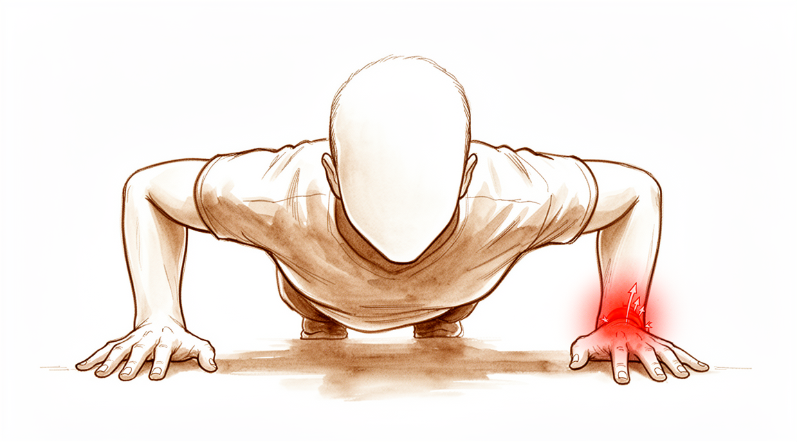
Why this operation has been suggested
Dr Kieran Hirpara, an upper-limb surgeon at Mater Private Hospital Rockhampton, starts with the least invasive options that suit your condition. The TFCC is a cartilage and ligament structure on the little-finger side of your wrist. Repair means stitching it back where it tore. We usually try hand therapy, splinting and activity changes first, and consider surgery when pain or instability continues after that. Patients are generally referred to our clinic by their GP; if a physiotherapist has suggested you see us, you will still need a referral from your GP in order to be eligible for the Medicare rebate. A clinic assessment, with your history, examination and imaging where needed, establishes the diagnosis.
This operation is offered for tears that can be repaired, when there are no arthritic changes in the joint. It aims to ease pain, restore stability to the joint between the two forearm bones, and keep your wrist and forearm moving. Repair is done as keyhole surgery, usually as day surgery under general anaesthetic. About three in four people return to full activity; some do not. We will talk through whether this operation suits you, and decide together.
Before the operation
Once surgery is booked, our rooms will confirm your admission details and fasting instructions. You will need to stop eating and drinking seven hours before your operation, so we can bring you forward if the theatre list runs early. Tell us about any blood thinners or regular medicines you take, as some need to pause beforehand. Bring a written list of your current medicines, and wear loose, comfortable clothing with a sleeve that fits over a bandaged wrist. You will need someone to drive you home, as the general anaesthetic makes driving unsafe for the rest of the day. Imaging such as an X-ray or MRI may already be done, or we may arrange it to plan the operation. If you have other medical conditions, you may need blood tests or a review with the anaesthetist.
On the day
You arrive at the hospital's surgical admissions unit, where staff check you in and prepare you for theatre. You will meet the anaesthetist, the doctor who puts you to sleep and looks after you during the operation. This operation is done under general anaesthetic. You will be fully asleep for the operation. Some patients may also have a regional nerve block for post-operative pain relief; the anaesthetist decides on the day based on your individual circumstances. You are then taken into the operating theatre, where the operation is performed.
When it is finished, you wake up in the recovery area. Nurses watch over you while the anaesthetic wears off. Once you are stable, you either move to a ward or go home, depending on the procedure and how your recovery is going.
What the operation involves
Your surgeon starts with keyhole surgery. A tiny camera is placed into your wrist through small cuts on the back of the wrist, so the tear can be seen clearly. The exact repair depends on what that shows, so the method cannot be promised beforehand.
If the tear is at the outer rim of the TFCC, it is stitched back to the capsule, the lining of the joint. If the tear is where the ligament has pulled off the ulna, the small bone on the little-finger side of your forearm, it is reattached to the bone. This can be done with a small anchor placed in the bone, or through a small tunnel drilled in the bone, sometimes through a short open cut. A tear that cannot be repaired is tidied instead, meaning the rough edges are trimmed away. Your surgeon will explain what was done once you are awake.
The cuts are closed with stitches and covered with a dressing.
After the operation
You wake up in the recovery area with nurses nearby while the anaesthetic wears off. This is usually a day case, so you can expect to go home the same day, although occasionally patients stay overnight. Your wrist will be in a below-elbow cast or firm splint, with your forearm turned partly palm-up and your elbow free to move. Pain is managed with medicines you can take at home; some discomfort is normal. Keep your hand raised on pillows when resting to help settle swelling. Wiggle your fingers when you can. If you had a nerve block, numbness and weakness in the first 24 hours are expected; once it wears off, call the clinic if you have numbness or cannot move your hand or fingers. Someone should stay with you for the first 24 hours. We leave the dressing on for about 10 days; please do not take it off before then unless we tell you to. We change or remove it when we see you.
Recovery
Your wrist will be in a below-elbow cast or firm splint for six weeks, with your forearm turned partly palm-up and your elbow free. This protects the repair while it heals. You cannot drive while the cast or splint is on. Once it is off and your surgeon clears you, you can drive again; the lighter removable splint you wear after that does not stop you driving by itself. For more detail, see Driving after upper-limb surgery.
After the six weeks, you will move into a removable splint while your wrist regains movement and strength. Your rehabilitation is hand therapy, not physiotherapy. It is directed by Ruby Doolan at Extend Rehabilitation. Ruby is a hand therapist: she guides your exercises and makes any splint you need.
Desk work and light daily tasks usually resume at six to eight weeks. Manual work, sport and the gym take longer, at four to six months. The final result, when pain and strength stop improving, comes at about 12 months.
Pain usually improves before strength and function do. Your wrist may feel easier to use well before it feels strong, so it is worth being patient with heavier tasks.
Almost half of people have a temporary setback after reaching their peak activity. This settles with time and does not change the final result. Knowing it can happen makes it less worrying if it happens to you.
What can go wrong
No operation is free of risk, and it is fair to know what can happen.
The repair can tear again, or the joint between the two forearm bones can stay unstable or become unstable again. Either of these can mean a further operation. Some people are left with stiffness or weakness in the wrist, even when the repair itself has healed.
The small nerve on the back of your hand, on the little-finger side, can be irritated by the surgery. This causes tingling or numbness in that patch of skin. It usually settles within a few months.
If your repair used a tunnel through the bone, the stitch knot can sometimes sit under the skin and cause irritation. This may need a small operation to remove it. The tendons on the little-finger side of your wrist can also become irritated.
Infection is rare. Call the clinic the same day if you have a fever, spreading redness around the wound, or fluid or pus leaking from it, or if your pain keeps getting worse despite your pain medicines.
When to call us
Most problems after this operation are easy to treat when they are picked up early. Call us if you are worried about anything at all.
Go to the emergency department if you have swelling or pain in your calf, or shortness of breath or chest pain. These can be signs of a blood clot. Go to the emergency department if your fingers turn pale, cold, white, blue or dark, as this can be a circulation problem.
Call the clinic the same day if you have a fever, spreading redness around the wound, or fluid or pus leaking from it. Call the same day too if your pain keeps getting worse despite your pain medicines.
Numbness and weakness in the first 24 hours after a nerve block are expected. Once the block has worn off, call the clinic if you have numbness or cannot move your hand or fingers.
If you cannot reach the clinic, out of hours or on a weekend, go to your nearest emergency department.
In more depth
Advanced reading: the deeper science (optional)
This section goes further than you need for your own treatment decisions. TFCC repair is worth the extra reading because the choices that attract the most debate have not separated in comparative studies, while recovery runs slower and less evenly than early pain relief suggests.
Arthroscopic or open, anchor or tunnel
For foveal tears, a 2026 meta-analysis of 3 studies and 157 patients found no statistically significant difference between arthroscopic and open repair in function, pain, grip strength or complications [1]. The small number of patients is the main limit on that conclusion.
Suture anchors and bone tunnels were compared across 904 patients in a systematic review. Both improved function, pain and grip strength, and reoperation was uncommon in both [2]. What differs is the kind of problem that follows. With anchors, the most common complication was irritation of the small sensory nerve on the back of the hand, usually settling within 2 to 4 months. With tunnels, it was an irritating suture knot [2]. In one randomised trial of 60 patients, 5 of 30 (16.7%) in the tunnel group needed a second operation to remove the knot [3].
One randomised trial of 40 patients added a capsular repair to a tunnel repair. Grip strength recovered to 94.2% of the other hand, compared with 84.2% with the tunnel repair alone. Pain, DASH and wrist scores were not significantly different [4]. This is a single small trial, and grip was the only measure that separated.
Waiting does not appear to close the door
It seems natural to expect a late repair to do worse, but the evidence is weaker than that. In 80 patients with foveal tears, results were similar whether surgery took place within 6 months, between 6 and 12 months, or after 12 months, and 78 (97%) had a stable joint at 2 years [5]. The authors note that the study lacked statistical power to prove equivalence. In 210 foveal repairs, the time between injury and surgery did not predict movement, grip or pain [6].
A much larger group points the other way, but only slightly. In 274 open repairs, each extra month of symptoms added 0.14 points to a 100-point wrist score at 12 months [7]. That is statistically real, and small: a year's delay would add less than two points.
The ulnar-plus wrist
A longer ulna loads the little-finger side of the wrist, so it might be expected to undermine a repair. In 50 arthroscopic repairs of peripheral tears, ulnar-plus variance did not significantly affect the result. A second tear of the central disc did: it was the only significant risk factor for failure, though with a very wide confidence interval, as there were only 4 failures [8]. In 36 foveal repairs, ulnar-positive wrists also did as well, and the authors concluded that variance alone may not justify adding an ulnar shortening operation [9].
The picture changes if the tear is only tidied. In 163 debridements, 40% of people with a positive ulnar variance went on to ulnar shortening, with the hazard rising 1.8-fold per millimetre [10]. The overall reoperation rate was 19%, so the extra operation is worth discussing where the ulna is long and the cartilage is already worn.
Recovery is slower than the pain suggests
In a 2026 series of 46 foveal repairs through a bone tunnel, pain had improved significantly by 12 weeks, but arm function and physical function had not. Those took longer, and all had improved by a median of 32 months [11]. At that point 77% had returned to full sport and activity, which leaves about one in four who had not.
The same series recorded something rarely reported: 45% of people had a temporary setback after reaching their peak activity. The authors now tell patients to expect this, as these setbacks were time-limited and did not affect the final result [11].
How long to protect the repair is unresolved
Published aftercare ranges from 2 to 9 weeks, often without a stated reason [12]. A meta-analysis of 288 patients found no significant difference in pain, DASH or grip between immobilisation that also restricted the elbow and immobilisation that left it free. Limiting forearm rotation for about 4 to 6 weeks gave better results than rotating immediately [13]. A short-arm cast in semisupination restricted pronation as well as a long-arm cast in 40 patients, with less restriction of daily activities [14]. That is the approach used in this practice, for six weeks.
The balance is not simple. In the 210 repairs above, longer forearm immobilisation predicted poorer movement, while a shorter period of wrist immobilisation predicted a higher risk of the repair tearing, which happened in 22 (10%) [6]. The authors suggested that forearm rotation might start before wrist movement. A randomised trial of 3 against 6 weeks after peripheral repairs has been registered [12], so this advice may change.
Joint laxity also matters. Among 120 foveal repairs, instability returned in 17.7% of people with generally loose joints and 5.8% of others [15].
References for the advanced reading
- Pan B, Chen W, Luo H. Arthroscopic versus open repair for triangular fibrocartilage complex foveal tears: a systematic review and meta-analysis. EFORT Open Rev. 2026;11(10):1151-8.
- Ma H, Wang J, Yang C. Effectiveness of suture anchor and transosseous suture technique in arthroscopic foveal repair of the triangular fibrocartilage complex: a systematic review. J Orthop Surg Res. 2024;19(1).
- Afifi A, Abdel-Ati EA, Abdel-Wahed M, et al. Arthroscopic-assisted foveal reattachment of triangular fibrocartilage complex tears with distal radioulnar joint instability: a comparison of suture anchors and transosseous sutures. J Hand Surg Am. 2022;47(6):507-16.
- Liao L, Xiong F, Gu F, et al. Combined transosseous and capsular repair improves grip strength in triangular fibrocartilage complex tears: a randomized controlled trial. Arthroscopy. 2025;41(11):4505-14.
- Park JH, Lim JW, Kwon YW, et al. Functional outcomes are similar after early and late arthroscopic one-tunnel transosseous repair of triangular fibrocartilage complex foveal tears. Arthroscopy. 2020;36(7):1845-52.
- McCarron L, Coombes BK, Bindra R, et al. Predictive factors for patient recovery following triangular fibrocartilage foveal repair surgery: a retrospective case-series. Hand (N Y). 2025;21(4):645-56.
- Feitz R, Stip D, van der Oest M, et al. Prognostic factors in open triangular fibrocartilage complex (TFCC) repair. J Hand Surg Glob Online. 2021;3(4):176-81.
- Kwon BC, Lee JH, Lee SY. What is the effect of the ulnar-plus variance on the outcomes of arthroscopic repair of the peripheral ulnar-side triangular fibrocartilage complex tear? Arthroscopy. 2020;36(9):2415-22.
- Won JH, Shin JW, Kwon J, et al. Effect of ulnar positive variance on outcomes after arthroscopic transosseous repair of palmer 1B TFCC foveal tears. J Orthop Surg Res. 2026;21(1).
- Verhiel SHWL, Ritt MJPF, Chen NC. Predictors of secondary ulnar shortening and reoperation after arthroscopic TFCC debridement. Hand (N Y). 2021;17(6):1147-53.
- Hong DY, Bailey KN, Goldfarb CA. Intermediate-term patient-reported outcomes after foveal TFCC repair: timing of recovery and successful return to sports or work. J Hand Surg Am. 2026.
- Paraj D, Bhat AK, Acharya A, Kunder MA. Comparison of the clinical and functional outcomes of two immobilisation protocols after arthroscopic peripheral triangular fibrocartilage complex (TFCC) repair in adults: a single-centre, double-blinded randomised controlled trial protocol. BMJ Open. 2025;15(5):e095855.
- Lee JK, Lee TC, Lee S, et al. Postoperative immobilization after foveal triangular fibrocartilage complex repair: a systematic review and meta-analysis of comparative studies. J Hand Surg Am. 2026;51(5):512.e1-512.e11.
- Jung H, Park J, Park H, et al. Postoperative immobilization using a short-arm cast in the semisupination position is appropriate after arthroscopic triangular fibrocartilage complex foveal repair. Bone Joint J. 2022;104-B(2):249-56.
- Kim J, Kim K, Lee S, et al. Generalized joint laxity increases the risk of recurrence of distal radioulnar joint instability after arthroscopic foveal repair of the triangular fibrocartilage complex. Arthroscopy. 2024;41(7):2309-16.
Evidence & references
This is the clinical evidence summary written for health professionals. It is technical, and it lists the research this page was built from. You do not need to read it to understand your treatment or to make a decision about it.
Anatomy & Pathophysiology
Anatomical Components
- The triangular fibrocartilage complex (TFCC) consists of the dorsal and volar radioulnar ligaments, the articular disc, a meniscus homologue, the extensor carpi ulnaris (ECU) tendon, and the ulnolunate and ulnotriquetral ligaments [13].
- The TFCC is subjected to considerable axial loading and shear stresses [9].
- The ulnar attachment of the TFCC is a three-dimensional complex comprising proximal radioulnar ligaments, a distal hammock structure (fibrocartilage disk, meniscus homologue, and ulnocarpal ligaments), and a functional ulnar collateral ligament (UCL) consisting of the ECU tendon subsheath and thickened ulnar capsule [9].
- The distal hammock structure and the UCL constitute the distal component of the TFCC (dc-TFCC), while the radioulnar ligament represents the proximal component (pc-TFCC) [9].
- The dorsal and volar radioulnar ligaments span from the dorsal and volar corners of the distal radius to a broad area of the fovea at the base of the ulnar styloid [9].
- A more superficial component of the radioulnar ligaments runs obliquely and distally to the ulnar styloid [9].
- The deep foveal components are considered the true radioulnar ligaments that stabilize the distal radioulnar joint (DRUJ) [9].
- Frank DRUJ instability can occur when the proximal foveal component is injured, even if the distal component remains intact [9].
- The ulnar styloid provides attachments for portions of the ulnocarpal ligaments, the ECU tendon sheath, and superficial limbs of the radioulnar ligaments [12].
- The deep limbs of the radioulnar ligaments insert into the fovea of the ulnar head [12].
- The tip of the ulnar styloid is devoid of soft tissue attachments [12].
- The ulnotriquetral (UT) ligament arises from the palmar radioulnar ligament and attaches to the palmar and ulnar triquetrum, forming an essential element of the TFCC [6].
Biomechanics and Load Distribution
- In a wrist with neutral variance, 80% of compressive loads are transmitted through the radius and 20% through the ulna [13].
- In a wrist with +2 mm ulnar variance, 60% of compressive loads are transmitted through the radius and 40% through the ulna [13].
- The most important stabilizers of the DRUJ during pronosupination are the palmar and dorsal radioulnar ligaments [4].
- Damage to either or both of the palmar and dorsal radioulnar ligaments generates pain and instability during forearm rotation [4].
- Biomechanical analysis indicates that traumatic disruptions of the distal radioulnar ligaments from the fovea occur predominantly from tension, though a combination of forces is required to destabilize the joint [4].
Injury Mechanisms and Clinical Presentation
- TFCC injuries typically occur with extension and pronation of the axially loaded wrist [9].
- The most common mechanism of TFCC injury is a fall on an outstretched hand [9].
- Peripheral tears of the articular disk are common athletic injuries involving racket sports, golf, or activities requiring rapid twisting and loading to the ulnar side of the wrist [9].
- Peripheral ulnar-sided tears of the articular disk are common work injuries, often resulting from traction or torquing of the forearm when a motorized drill bit binds [9].
- Patients with peripheral TFCC tears complain of deep diffuse pain across the ulnar side of the wrist [9].
- Patients with peripheral TFCC tears frequently report weakness when gripping and a clicking sensation with forearm rotation [9].
- Pain in peripheral TFCC tears is accentuated by passive pronation and supination of the wrist [9].
- Local tenderness in peripheral TFCC tears is located over the dorsal TFCC between the dorsoulnar head and triquetrum and/or over the ulnar snuffbox in supination at the foveal area [9].
- A dorsally prominent ulnar head relative to the radius can be seen in foveal detachments [9].
- Ballottement of the DRUJ reveals increased translation of the joint associated with pain in TFCC injuries [9].
- A class 1A tear typically manifests as ulnar-sided wrist pain aggravated by power grip, especially with ulnar deviation or forearm rotation [7].
- Class 1A tears are confined to the disc, located 2 to 3 mm ulnar to its radial attachment, and oriented volar to dorsal [7].
- Class 1A tears do not cause DRUJ instability [7].
- Symptoms and physical findings associated with a class 1B tear are similar to those of a class 1A tear, although a click is usually absent [12].
- Point tenderness in class 1B tears is characteristically volar to the ulnar styloid, coinciding anatomically with the fovea [12].
- Tenderness at the fovea is termed the fovea sign [12].
- Evaluation of DRUJ stability in class 1B tears is likely to produce pain even if the DRUJ is stable [12].
- A positive ulnar fovea sign was reported to be 90% sensitive and 88% specific in detecting a split tear of the UT ligament [8].
Classification
- The Palmer classification categorizes TFCC tears into traumatic (class 1) or degenerative (class 2) [1].
- Subtypes in the Palmer classification are based on the specific location within the TFCC [1].
- The class and location of the tear have important implications for treatment [1].
- Type I TFCC tears denote traumatic injuries, while type II lesions are degenerative in origin [9].
- Type I injuries are further classified into four types [9].
- Except for type 1A tears, which imply a central articular disk perforation with a stable margin, the other three types of Type I injuries are considered peripheral tears that may cause instability of the DRUJ or ulnocarpal joint, or both [9].
- Estrella and Ho described a dorsal type of tear located at the junction of the dorsal radioulnar ligament and the joint capsule just radial to the ECU tendon subsheath [9].
- Most peripheral lesions of the TFCC are traumatic in origin, though they may also occur in degenerative status due to chronic repetitive loading [9].
- Type 1B tears are among the most common peripheral tears of the TFCC leading to chronic ulnar wrist pain and impairment [9].
- Type 1B tears broadly include all injuries of the TFCC at the ulnar attachment, with or without an accompanied ulnar styloid fracture [9].
- Class 1A injuries are characterized by central perforation or tear [1].
- Class 1B injuries are characterized by ulnar avulsion with or without ulnar styloid fracture [1].
- Class 1C injuries are characterized by distal avulsion involving the origins of the ulnolunate and ulnotriquetral ligaments [1].
- Class 1D injuries are characterized by radial avulsion involving the dorsal and/or volar radioulnar ligaments [1].
- Class 2A degenerative tears are characterized by TFCC wear or thinning [1].
- Class 2B degenerative tears are characterized by TFCC wear plus lunate and/or ulnar chondromalacia [1].
- Class 2C degenerative tears are characterized by TFCC perforation plus lunate and/or ulnar chondromalacia [1].
- Class 2D degenerative tears are characterized by TFCC perforation, lunate and/or ulnar chondromalacia, and lunotriquetral ligament disruption [1].
- Class 2E degenerative tears are characterized by TFCC perforation, lunate and/or ulnar chondromalacia, lunotriquetral ligament disruption, and ulnocarpal and DRUJ arthritis [1].
- A class 1B injury is a partial or complete avulsion of the TFCC from its ulnar attachments, with or without an ulnar styloid fracture [12].
- A class 1D tear is a partial or complete avulsion of the TFCC from the radius, with or without a bone fragment, and may involve one or both radioulnar ligaments [10].
- Class 1C tears involve the distal attachment of the articular disk to the lunate, triquetrum, and lunotriquetral ligaments [6].
- Complete tears of the ulnocarpal ligaments in class 1C injuries can result in ulnar carpal instability and/or volar translocation of the ulnar carpus in relation to the radius [6].
Vascularity and Healing Potential
- The central area of the TFCC is devoid of vascularity and unable to heal [1].
- The peripheral rim of the TFCC is well vascularized, akin to the meniscus within the knee [1].
- Studies showed that the outer 10% to 40% of the articular disk was well perfused and suggested a healing potential of these injured areas upon repair [9].
- Failure to reattach the TFCC to the foveal footprint may induce synovitis, which inhibits the healing of the foveal fibers, setting up a vicious cycle and promoting the persistence of painful DRUJ instability [11].
Classification
- TFCC tear subtypes are defined based on the specific location within the TFCC [1].
- The class and location of a TFCC tear have important implications for treatment [1].
- Class 1A traumatic TFCC injury is characterized by central perforation or tear [1].
- Class 1B traumatic TFCC injury is characterized by ulnar avulsion with or without ulnar styloid fracture [1].
- Class 1C traumatic TFCC injury is characterized by distal avulsion involving the origins of the ulnolunate and ulnotriquetral ligaments [1].
- Class 1D traumatic TFCC injury is characterized by radial avulsion involving the dorsal and/or volar radioulnar ligaments [1].
- Class 2A degenerative TFCC tear is characterized by TFCC wear or thinning [1].
- Class 2B degenerative TFCC tear is characterized by Class 2A features plus lunate and/or ulnar chondromalacia [1].
- Class 2C degenerative TFCC tear is characterized by TFCC perforation plus lunate and/or ulnar chondromalacia [1].
- Class 2D degenerative TFCC tear is characterized by Class 2C features plus lunotriquetral ligament disruption [1].
- Class 2E degenerative TFCC tear is characterized by Class 2D features plus ulnocarpal and distal radioulnar joint arthritis [1].
- Arthroscopy is the gold standard for detection of TFCC tears [1].
- The arthroscopic trampoline test assesses TFCC resiliency by balloting the central portion with a small probe [1].
- The arthroscopic hook test can be used to demonstrate peripheral detachment of the TFCC [1].
- The arthroscopic suction test can show laxity of the TFCC when peripherally scarred in or foveal detachment when the distal radioulnar joint is clinically unstable [1].
- MRI is controversial for TFCC pathology, but newer innovations suggest value in detection and localization [1].
Indications
- Acute traumatic Palmar type 1A TFCC tears are an indication for arthroscopic TFCC debridement [3].
- Degenerative central tears of the TFCC with ulnar neutral or negative variance are an indication for arthroscopic TFCC debridement [3].
- Failure to respond to conservative treatment with splint and medication for more than 3 months is an indication for arthroscopic TFCC debridement [3].
Preoperative Evaluation
- Clinical correlation with provocative signs on the ulnar wrist is required during preoperative evaluation [3].
- Ulnar variance must be checked during preoperative evaluation [3].
- Wrist MRI is used to check for edema of the lunate in cases of ulnar positive variance or suspected impaction [3].
Investigations
Classification and Diagnostic Modalities
- Subtypes within the Palmer classification are based on the specific location of the tear within the TFCC [1].
- MRI is considered controversial for TFCC pathology, but newer innovations suggest value in detection and localization [1].
- Arthroscopy is the gold standard for the detection of TFCC tears [1].
Arthroscopic Diagnostic Tests
- The arthroscopic suction test can show laxity of the TFCC when it is peripherally scarred in or foveal detachment when the DRUJ is clinically unstable [1].
- Observation for swelling of the DRUJ when injecting saline at the 3-4 portal before inserting the arthroscope suggests a free flow perforation at the TFCC [3].
- The ulnar fovea sign is a provocative maneuver aimed at eliciting tenderness at the ulnar fovea [6].
Preoperative Evaluation
- Preoperative evaluation for arthroscopic TFCC debridement includes clinical correlation with provocative signs on the ulnar wrist [3].
- Preoperative evaluation for arthroscopic TFCC debridement includes checking ulnar variance [3].
- MRI of the wrist is used to check for edema of the lunate in cases of ulnar positive variance or suspected impaction [3].
- Preoperative evaluation for TFCC reconstruction with tendon graft includes clinical symptoms and signs of DRUJ instability [5].
- Preoperative evaluation for TFCC reconstruction with tendon graft includes radiographs to check ulnar variance and forearm alignment [5].
- Preoperative evaluation for TFCC reconstruction with tendon graft includes diagnostic arthroscopy or high-resolution MRI to evaluate the potential for TFCC repair [5].
- Preoperative evaluation for Class 1B TFCC tear repair includes positive provocative tests for peripheral TFCC tear [8].
- Preoperative evaluation for Class 1B TFCC tear repair includes DRUJ instability on ballottement test [8].
- Preoperative evaluation for Class 1B TFCC tear repair includes X-ray of the wrist to rule out ulnar styloid fracture [8].
- Preoperative evaluation for Class 1B TFCC tear repair includes MRI of the wrist [8].
Clinical Presentation and Physical Findings
- Class 1A tears typically manifest as ulnar-sided wrist pain aggravated by power grip, especially with ulnar deviation or forearm rotation [7].
- Class 1A tears may cause pain and mechanical symptoms such as clicking but do not cause DRUJ instability [7].
- Class 1A tears are typically confined to the disc, located 2 to 3 mm ulnar to its radial attachment, and oriented volar to dorsal [7].
- Patients with Class 1C tears complain of tenderness over the ulnar snuffbox and pain with grip [6].
- The arthroscopic appearance of UT ligament split tears is predominantly noted superficially as synovitis [8].
- Debridement of synovitis reveals the longitudinal split tear in the long axis of the UT ligament [8].
Treatment
Indications and Contraindications
- TFCC reconstruction with tendon graft is indicated in irreparable TFCC injuries with symptomatic DRUJ instability [4].
- TFCC reconstruction with tendon graft is indicated in neglected chronic injuries [4].
- TFCC reconstruction with tendon graft is indicated after suboptimal healing following nonoperative or surgical repair [4].
- Skeletal malalignment responsible for DRUJ instability, including ulnar incongruity, malunion of the sigmoid notch, and dorsal tilt or coronal shift of the radius, should be addressed concomitantly with TFCC reconstruction [4].
- Osteoarthritis of the DRUJ is a contraindication to TFCC reconstruction [4].
- Axial instability of the forearm due to interosseous membrane injury is a contraindication to TFCC reconstruction [4].
Biomechanical Rationale
- Biomechanical analysis by Adams et al. concluded that traumatic disruptions of the distal radioulnar ligaments from the fovea occurred predominantly from tension [4].
- Adams et al. proposed that a combination of forces was required to destabilize the DRUJ [4].
- The method aims to restore normal DRUJ kinematics by using a single tendon graft with uniform tension [4].
Operative Technique: General Setup and Arthroscopy
- The procedure is performed under general or regional anesthesia [4].
- An image intensifier is used to locate the isometric point for tunnel drilling [4].
- An arm tourniquet is placed [4].
- Radiocarpal joint arthroscopy is conducted through the standard 3-4 viewing and 4-5 working portals to evaluate and debride the TFCC lesion and associated synovitis [4].
- If the central part of the articular disk is intact, a central perforation of 4-5 mm is created with an arthroscopic knife or RF ablation to expose the ulnar head [4].
- The wrist is taken off the traction tower for subsequent tendon graft and bone tunnel preparation [4].
Operative Technique: Volar Incision and Tendon Harvest
- A 2 cm volar longitudinal incision is made midway between the palmaris longus tendon and the radial border of the FCU tendon, extending proximally from the proximal wrist crease [4].
- The palmaris longus tendon is harvested in full length with a tendon stripper [4].
- The flexor tendons and median nerve are retracted radially [4].
- The FCU, ulnar nerve, and artery are retracted ulnarly to expose the distal radius [4].
- The distal border of the pronator quadratus marks the approximate level of the radial tunnel [4].
Operative Technique: Dorsal Incision and Radial Tunnel
- A 2 cm dorsal longitudinal incision is made extending proximally from the 4-5 portal [4].
- The extensor retinaculum is split longitudinally over the fifth dorsal compartment [4].
- The EDM is exposed and retracted as a cotton sling [4].
- The septum of the fourth extensor compartment is stripped subperiosteally to expose the edge of the sigmoid notch [4].
- A 1.1 mm guide pin is inserted on the dorsal surface of the distal radius radial to the sigmoid notch, 5 mm proximal to the articular surface of lunate to fossa and 5 mm radial to the sigmoid fossa [4].
- The guide pin is inserted with a volar tilt of 10 to 15 degrees, parallel to the surface of the lunate fossa [4].
- Tendons and neurovascular structures on the volar side are gently retracted while the guide pin is advanced with direct visualization of the tip [4].
- Guide pin position is confirmed by fluoroscopy [4].
- The tunnel is enlarged from 2.0 to 2.5 mm using cannulated drill bits and soft tissue protection at both entry and exit sites [4].
Operative Technique: Ulnar Incision and Foveal Tunnel
- A 3 cm longitudinal incision is made over the subcutaneous border of ulna beginning 1 cm proximal to the tip of ulnar styloid [4].
- A 1.1 mm guide pin is inserted at the middiaphyseal point from 1.5 to 2 cm proximal to the tip of ulnar styloid [4].
- The guide pin is inserted at an acute angle of about 60 degrees targeting the fovea of the ulnar head and parallel to the ulna shaft in the sagittal plane under fluoroscopic guidance [4].
- The surgeon can place their thumb on the ulnar styloid as a reference to facilitate entry point decision [4].
- The wrist can be suspended in a vertical position with the forearm in neutral rotation position to monitor the foveal exit site of the guide pin under arthroscopy [4].
- Arthroscopic monitoring ensures the guide pin exits at the isometric point of the insertion of the distal radioulnar ligament [4].
References
[1] Miller S Review Of Orthopaedics. DISTAL RADIOULNAR JOINT, TRIANGULAR FIBROCARTILAGE COMPLEX, AND WRIST ARTHROSCOPY > 2. TFCC tears.
[3] Green S Operative Hand Surgery. AUTHOR'S PREFERRED METHOD OF TREATMENT: ARTHROSCOPIC PARTIAL WRIST FUSION > Arthroscopic TFCC Debridement.
[4] Green S Operative Hand Surgery. AUTHOR'S PREFERRED METHOD OF TREATMENT: ARTHROSCOPIC PARTIAL WRIST FUSION > TFCC Reconstruction with Tendon Graft.
[5] Green S Operative Hand Surgery. AUTHOR'S PREFERRED METHOD OF TREATMENT: ARTHROSCOPIC PARTIAL WRIST FUSION > Arthroscopic TFCC Reconstruction With Tendon Graft.
[6] Green S Operative Hand Surgery. AUTHOR'S PREFERRED METHOD OF TREATMENT: ARTHROSCOPIC PARTIAL WRIST FUSION > Arthroscopic-Assisted Suture Anchor Technique (Atzei) > Arthroscopic Repair of Class IC TFCC Tears.
[7] Green S Operative Hand Surgery. Chronic Perilunate Instability (SL Plus LT Instability) > Palmer’s Classification of TFCC Lesions > Class 1A Tear.
[8] Green S Operative Hand Surgery. AUTHOR'S PREFERRED METHOD OF TREATMENT: ARTHROSCOPIC PARTIAL WRIST FUSION > Arthroscopic Repair of Class 1B TFCC Tears.
[9] Green S Operative Hand Surgery. AUTHOR'S PREFERRED METHOD OF TREATMENT: ARTHROSCOPIC PARTIAL WRIST FUSION > TFCC Repair > Classification, Functional Anatomy, Clinical Features, and Indication for Repair.
[10] Green S Operative Hand Surgery. Chronic Perilunate Instability (SL Plus LT Instability) > Palmer’s Classification of TFCC Lesions > Class 1D Tear.
[11] Green S Operative Hand Surgery. AUTHOR'S PREFERRED METHOD OF TREATMENT: ARTHROSCOPIC PARTIAL WRIST FUSION > Arthroscopic Repair of Class 1B TFCC Capsular Tears > Arthroscopic Reattachment of Class 1B TFCC Foveal Tear.
[12] Green S Operative Hand Surgery. Chronic Perilunate Instability (SL Plus LT Instability) > Palmer’s Classification of TFCC Lesions > Class 1B Tear.
[13] Miller S Review Of Orthopaedics. DRUJ, TFCC, WRIST ARTHROSCOPY.