Clinicians › Wrist
TFCC Repair
For patients: a plain-language version of this topic is available. See the patient guide.
Overview
Arthroscopic ligament-specific repair of the triangular fibrocartilage complex (TFCC) is a reliable and useful technique that offers significant improvements in wrist motion, grip strength, pain, and patient-reported outcomes at a minimum 2-year follow-up [1]. The long-term efficacy of this approach has become widely accepted [2], with arthroscopic treatment of TFCC lesions resulting in 80% to 85% good to excellent results, characterized by pain relief and unrestricted return to work or sports [3]. These arthroscopic-assisted repair techniques provide detailed visualization and facilitate the repair of TFCC injuries and associated pathologies with minimally invasive methods [4]. Current evidence demonstrates that TFCC repair achieves good clinical outcomes with low complication rates [8], leading to satisfactory functional outcomes [15]. In high-demand athletes, arthroscopic repair is becoming the treatment of choice to obtain optimum physiologic strength, complete range of motion, stability, and the shortest possible postoperative period [29].
While open surgical TFCC repair is a safe and reliable therapeutic procedure in children and adolescent populations [19], there was no statistical difference in clinical outcomes after open versus arthroscopic repair [7]. A systematic review indicates a current lack of high-quality evidence required to draw firm conclusions on the merits of arthroscopic versus open repair of 1B TFCC tears [11]. Specific techniques, such as all-arthroscopic pre-tied suture device repair of isolated Palmer type 1B tears, have demonstrated excellent clinical function at a mean follow-up of 7 years [24]. Similarly, all-arthroscopic TFCC repair using the FasT-Fix results in clinical outcomes comparable with previously described techniques [30], and the all-arthroscopic technique for TFCC ligamentoplasty serves as a good alternative with promising results [53]. With appropriate indications and patient selection, arthroscopic foveal repair of the TFCC may restore distal radioulnar joint stability and provide satisfactory results without significant complications [80].
Several factors influence the clinical context and outcomes of TFCC repair. TFCC repair varies substantially from surgeon-to-surgeon, suggesting repairs are discretionary and preference sensitive [13]. A systematic review did not demonstrate strong evidence regarding the association between ulnar variance and outcomes after TFCC repair [23]. In a study of arthroscopically diagnosed but untreated traumatic TFCC tears in distal radial fractures, no evidence was found that a TFCC injury influences long-term outcome, though trends suggested complete tears might have inferior outcomes [9]. Adjunctive therapies may also impact recovery; patients treated with 4 weeks of electroacupuncture after arthroscopic TFCC repair had better wrist range of motion and DASH scores than patients in the control group [10].
Anatomy & Pathophysiology
Anatomical Components and Classification
The triangular fibrocartilage complex (TFCC) comprises the dorsal and volar radioulnar ligaments, the articular disc, a meniscus homologue, the extensor carpi ulnaris (ECU), and the ulnolunate and ulnotriquetral ligaments [140]. The ulnar side of the TFCC is arranged in a complex three-dimensional manner and separated into three components: the proximal triangular ligament, the distal hammock structure, and the ulnar collateral ligament (UCL) [18]. The distal hammock structure and the UCL constitute the distal component of the TFCC (dc-TFCC), while the proximal triangular ligament represents the proximal component (pc-TFCC) [18]. The proximal triangular ligament originates from the fovea ulnaris and spans to the ulnar corners of the distal radius with palmar and dorsal limbs, functioning as the true radioulnar ligament that stabilizes the distal radioulnar joint (DRUJ) [18]. The UCL is considered to be a part of the extensor carpi ulnaris (ECU) sheath floor [18].
The Palmer classification divides TFCC disorders into traumatic (class 1) and degenerative (class 2) categories, further subdivided by tear location and associated chondromalacial changes [18]. Class 1 traumatic lesions are subdivided into four types, with Type 1-B injuries defined as peripheral tears located on the ulnar side of the TFCC [18]. Class 1B injuries are partial or complete avulsions of the TFCC from its ulnar attachments, with or without an ulnar styloid fracture [122]. Class 1D tears involve partial or complete avulsion of the TFCC from the radius, with or without a bone fragment, and may involve one or both radioulnar ligaments [118]. Class 1A tears are central perforations or tears of the articular disc [32]. Class 2 degenerative tears are associated with ulnocarpal impaction syndrome and include subtypes ranging from TFCC wear to arthritis [32].
Biomechanics and Load Distribution
In neutral variance, 80% of compressive loads are transmitted through the radius and 20% through the ulna [140]. In +2 mm variance, 60% of compressive loads are transmitted through the radius and 40% through the ulna [140]. The TFCC acts as the primary stabilizer of the distal radioulnar joint (DRUJ) during forearm rotation while providing a smooth articular surface and partially absorbing axial load from the radiocarpal joint [93]. The palmar and dorsal radioulnar ligaments are the most important stabilizers of the DRUJ during pronosupination [46]. Traumatic disruptions of the distal radioulnar ligaments from the fovea occur predominantly from tension, though a combination of forces is required to destabilize the joint [46]. The foveal insertion has a greater effect on DRUJ stability than the styloid insertion due to its closer relationship to the rotational axis of the forearm [147]. The ulnocarpal ligaments are likely to be stretched tensely during wrist radial extension and wrist extension [116]. Distal radioulnar joint instability with an arthroscopically verified TFCC injury is associated with a significant loss of both pronation and supination torque [138].
Pathophysiology and Clinical Presentation
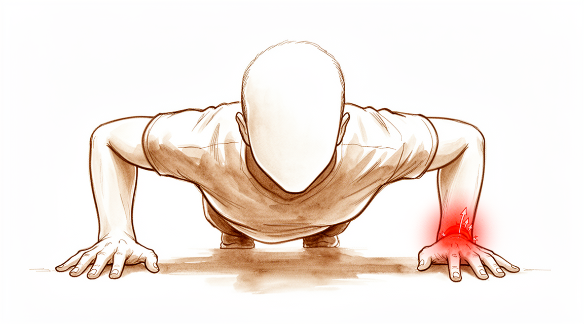
TFCC injuries typically occur with extension and pronation of the axially loaded wrist, with the most common mechanism being a fall on an outstretched hand [97]. Peripheral tears of the articular disk are common athletic injuries involving racket sports, golf, or activities requiring rapid twisting and loading to the ulnar side of the wrist [97]. Patients with peripheral TFCC tears complain of deep diffuse pain across the ulnar side of the wrist, weakness when gripping, and a clicking sensation with forearm rotation [97]. Local tenderness over the dorsal TFCC between the dorsoulnar head and triquetrum, or over the ulnar snuffbox in supination at the foveal area, is characteristic of peripheral tears [97]. A dorsally prominent ulnar head in relation to the radius can be seen in foveal detachments [97]. Ballottement of the DRUJ reveals increased translation of the joint associated with pain in TFCC injuries [97].
Class 1A tears typically manifest as ulnar-sided wrist pain aggravated by power grip, especially with ulnar deviation or forearm rotation, but do not cause DRUJ instability [81]. Class 1B tears present with point tenderness characteristically volar to the ulnar styloid, coinciding anatomically with the fovea (fovea sign) [122]. Tears of the TFCC superficial fibers with the deep fibers intact present with ulnar-sided wrist pain but without distal radioulnar joint instability [72]. Structurally intact but functionally incompetent foveal TFCC injuries represent a spectrum of tears not significant enough to produce gross DRUJ instability [83].
Signal abnormalities and structural defects of the TFCC are common, asymptomatic findings on MRI, more so with advancing age [28]. There is no evidence that clinical or radiological tests, including MRI and arthroscopy, can distinguish acute, traumatic lesions from pre-existing, previously asymptomatic lesions [28]. The rationale for TFCC repair in patients without DRUJ instability is difficult because the mechanisms by which a TFCC abnormality generates pain are not fully understood [28]. Not all TFCC tears are symptomatic, and the prevalence of TFCC lesions is high in asymptomatic wrists [91].
The outer 10% to 40% of the articular disk is well perfused, suggesting a healing potential for injured areas upon repair [97]. The central area of the TFCC is devoid of vascularity and unable to heal [32]. The peripheral rim of the TFCC is well vascularized, akin to the meniscus within the knee, making it amenable to repair [32]. Failure to reattach the TFCC to the foveal footprint may induce synovitis, which inhibits the healing of the foveal fibers and promotes persistent painful DRUJ instability [120].
Classification
Palmer Classification
The Palmer classification divides triangular fibrocartilage complex (TFCC) disorders into two basic categories: traumatic (Class 1) and degenerative (Class 2) [18, 32, 33, 82, 105]. Class 1 traumatic lesions are subdivided into four types based on the location of the tear [18, 32, 33, 82, 105]. Class 1A injuries involve a central perforation or tear of the avascular portion of the articular disk [32, 33, 82]. Class 1B injuries are ulnar avulsions with or without ulnar styloid fracture [32, 33, 82]. Class 1C injuries involve distal avulsion at the origins of the ulnolunate and ulnotriquetral ligaments [32, 33, 82]. Class 1D injuries are radial avulsions involving the dorsal and/or volar radioulnar ligaments [32, 33, 82]. Class 2 degenerative tears are staged A to E depending on the presence or absence of perforation, lunate and ulnar chondromalacia, lunotriquetral interosseous ligament involvement, and degenerative radiocarpal arthritis [32, 33, 82]. The Palmer classification does not completely classify all peripheral TFCC tears, particularly dorsal tears [21]. In a study of 106 patients, 44.3% presented peripheral lesions (Palmer 1b and 1c) and 68.9% showed central TFCC alterations (Palmer 1a, 1d and 2a–e) [129].
Atzei-Luchetti Classification
The Atzei-Luchetti classification categorizes TFCC peripheral tears into five types: repairable distal tears, repairable complete tears, repairable proximal tears, non-repairable tears, and tears associated with DRUJ arthritis [105]. Atzei Class 1 tears are sutured [105]. Atzei Class 2 and 3 tears are associated with DRUJ instability and require TFCC reattachment to the fovea [105]. Atzei Class 4 tears require reconstruction using a tendon graft [105]. Atzei Class 5 tears require an arthroplasty [105]. A series of patients presented with combined central disc and foveal TFCC tears, a pattern that does not fit current classification schemes [137].
Diagnostic Reliability
Interobserver reliability for the Palmer classification was 0.25 in Round 1 and 0.32 in Round 2 [130]. Interobserver reliability for the Atzei-Luchetti classification was 0.19 in Round 1 and 0.33 in Round 2 [130]. Interobserver reliability for overall treatment decision or method was 0.10 in Round 1 and 0.14 in Round 2 [130].
Clinical Presentation
Diagnostic Challenges and Natural History
Signal abnormalities and structural defects of the triangular fibrocartilage complex (TFCC) are common, asymptomatic findings on MRI, with prevalence increasing with advancing age [SHORTCOMINGS]. Consequently, there is no evidence that clinical or radiological tests, including MRI and arthroscopy, can distinguish acute traumatic lesions from pre-existing, previously asymptomatic lesions [SHORTCOMINGS]. Surgical visualization performed many months after symptom onset or after corticosteroid injections may be unreliable [SHORTCOMINGS]. Furthermore, there is no evidence that the distinction between Palmer types 1 and 2 lesions or between their subtypes is reliable [SHORTCOMINGS].
The rationale for TFCC repair in patients without distal radioulnar joint (DRUJ) instability is difficult, as treatments are based on the unproven rationale that eliminating inflammation and subtle instability addresses pain [SHORTCOMINGS]. Many TFCC lesions may be incidental findings, and there is little evidence to support surgical recommendations for ulnar-sided wrist pain without DRUJ instability [110]. A study of traumatic TFCC tears in distal radial fractures found no evidence that the injury influences long-term outcome, though trends suggested complete tears might have inferior outcomes [9]. Approximately 40% of patients sustaining a TFCC tear without DRUJ instability still had pain and disability at 1 year [16].
Careful history and physical examination are required to determine whether a TFCC tear is symptomatic [35]. It is important to quantify the severity of symptoms related to TFCC pathology to determine whether surgical treatment is necessary [35]. A stable distal radioulnar joint upon clinical examination and normal MRI findings do not rule out foveal TFCC injury [65]. A high index of clinical suspicion is needed when managing patients with ulnar-sided wrist pain to identify foveal TFCC injury [65].
Arthroscopy is a more accurate diagnostic technique than MRI studies for TFCC tears [75]. Wrist arthroscopy allows the clearest visualization of the TFCC and related intra-articular pathology [68]. Arthroscopy is effective in obtaining both correct diagnosis and treatment of peripheral TFCC tears [25]. However, arthroscopic diagnostic signs for TFCC lesions do not demonstrate equivalent reliability [54]. In the adolescent population, TFCC lesions are difficult to evaluate on an arthro-MRI and results are often inconclusive [64]. MRI findings in patients undergoing TFCC repairs may reflect damage to the TFCC altering relationships of the DRUJ and ECU subsheath, or various pathologies causing ulnar-sided wrist pain [41].
Indications for Repair
Arthroscopic TFCC repair is indicated for reparable tears without the presence of arthritic changes, irrespective of chronicity [58]. Operative treatment of TFCC injuries in adolescent and pediatric athletes after failure of conservative treatment allowed return to sport at the previous level of participation [43]. Return to play may be delayed in athletes with concomitant ulnar-sided wrist injuries [31].
Investigations
Imaging and Diagnostic Modalities
MRI: Conventional wrist MRI remains controversial for the detection and localization of TFCC pathology, though newer innovations suggest value [32]. MR arthrography offers a more sensitive and specific method for diagnosing TFCC tears compared to conventional wrist MRI [132]. High-resolution MRI using a microscopy surface coil allows assessment of each TFCC component and demonstrates higher accuracy for diagnosing injuries to the radial attachment and the disc of the TFC compared with previous studies [154]. When combining MRI and dynamic CT imaging of the DRUJ, the sensitivity of detecting a foveal TFCC tear approaches 88% compared with a gold standard of wrist arthroscopy [158]. A stable distal radioulnar joint upon clinical examination and normal MRI findings do not rule out foveal TFCC injury [65]. Postoperative MRI helps to analyze the integrity of TFCC postrepair and adds to understanding the natural course of its healing [69].
CT: Dynamic CT imaging of the DRUJ, when combined with MRI, contributes to a sensitivity of approximately 88% for detecting foveal TFCC tears [158].
Arthroscopy: Arthroscopy is the gold standard for the detection of TFCC tears [32]. Advances in arthroscopic equipment and technique have made wrist arthroscopy a valuable tool in the management of wrist disorders [47]. Arthroscopy is effective in obtaining both correct diagnosis and treatment of peripheral TFCC tear [25]. Radiological instability signs in the distal radioulnar joint are no replacement for a thorough clinical examination but may be helpful for indicating diagnostic wrist arthroscopy in ambiguous cases [87].
Arthroscopic Diagnostic Tests
The arthroscopic trampoline test is performed to assess TFCC resiliency by balloting the central portion with a small probe [32]. The arthroscopic hook test can be used to demonstrate peripheral detachment of the TFCC [32]. The arthroscopic suction test can show laxity of the TFCC when peripherally scarred in or foveal detachment when the DRUJ is clinically unstable [32]. Arthroscopic diagnostic signs for TFCC lesions do not demonstrate equivalent interobserver reliability [54].
Preoperative Evaluation
Preoperative Evaluation: Preoperative evaluation for arthroscopic TFCC debridement includes clinical correlation with provocative signs on the ulnar wrist, checking ulnar variance, and MRI of the wrist to check for edema of the lunate in cases of ulnar positive variance or suspected impaction [42]. Preoperative evaluation for TFCC reconstruction with tendon graft includes clinical symptoms and signs of DRUJ instability, radiographs to check ulnar variance and forearm alignment, and diagnostic arthroscopy or high-resolution MRI to evaluate the potential for TFCC repair [70]. Preoperative evaluation for arthroscopic repair of Class 1B TFCC tears includes positive provocative tests for peripheral TFCC tear, DRUJ instability on ballottement test, X-ray of the wrist to rule out ulnar styloid fracture, and MRI of the wrist [88].
Treatment
Non-Operative
Nonsurgical treatment is moderately successful for treating patients with TFCC tears without distal radioulnar joint (DRUJ) instability [39]. However, approximately 40% of patients sustaining a TFCC tear without DRUJ instability still experience pain and disability at 1 year [16]. Careful history and physical examination are required to determine whether a TFCC tear is symptomatic, and it is important to quantify the severity of symptoms related to TFCC pathology to determine whether surgical treatment is necessary [35].
Operative
Indications: TFCC reconstruction with tendon graft is indicated in irreparable TFCC injuries with symptomatic DRUJ instability, neglected chronic injuries, or after suboptimal healing following nonoperative or surgical repair [46]. The rationale for TFCC repair in patients without DRUJ instability is difficult [28]. Skeletal malalignment that may be responsible for the DRUJ instability should be addressed concomitantly, including ulnar incongruity, malunion of the sigmoid notch, and dorsal tilt or coronal shift of the radius [46]. Osteoarthritis of the DRUJ and an axial instability of the forearm due to interosseous membrane injury are contraindications to TFCC reconstruction [46]. Increased preoperative pain, less preoperative function, and a longer duration of complaints are factors that were associated with more pain and less function at 12 months after open surgery for TFCC [153].
Surgical Approach / Technique: Arthroscopic-assisted repair techniques have revolutionized surgical management, providing detailed visualization and facilitating the repair of TFCC injuries and associated pathologies with minimally invasive techniques [4]. The outside-in technique was the most commonly reported method for ulnar TFCC repair throughout the analyzed period [55]. Anatomic dissections were used to show the safe regions around the TFCC where tears are amenable to this type of repair [61]. For arthroscopic management, the 3–4 portal and 6R (or 6U) portal were created [163]. All patients underwent an arthroscopic wrist examination, and the lesions of the TFCC tears were identified and classified according to Palmer’s classification [163]. The tears and inflammatory synovium were debrided using a small joint power shaver [163]. The fovea region of the distal ulnar head was identified and prepared [163].
In a specific arthroscopic technique, wrist arthroscopy was performed on all patients using distraction with a wrist arthroscopy tower [165]. Local synovectomy is performed using a 3.0 mm oscillating full radius shaver placed in the 4-5 portal [165]. The shaver is also used to freshen the leading edge of the torn peripheral TFCC [165]. The adjacent edge of the ulnotriquetral ligament can also be freshened using the shaver to improve healing potential [165]. Percutaneous placement of an 18-gauge spinal needle is then placed in the safe zone along the ulnar aspect of the wrist between flexor carpi ulnaris and extensor carpi ulnaris [165]. The tip of the spinal needle pierces the ulnotriquetral ligament and the leading edge of the torn peripheral TFCC [165]. A shuttle relay is then passed through the spinal needle and brought out the cannula in the 4-5 portal using a grasper [165]. The shuttle relay is then used to pull one end of the suture through the peripheral tear and out the 4-5 portal cannula [165]. Braided absorbable suture is employed for the repair [165]. A second spinal needle is then placed in a parallel fashion to the first and the shuttle relay is passed through this needle [165]. The shuttle relay is grasped within the wrist joint and again brought out through the 4-5 portal [165]. The second arm of the suture is passed through the shuttle relay wire loop and percutaneously pulled back through the safe zone along the ulnar aspect of the wrist [165]. This creates a mattress suture repair [165]. The skin bridge between the suture entry points in the safe zone is then divided and gentle spreading of tissue with a hemostat is carried out down to the capsule [165]. The sutures are then tied with the wrist in a prona [165].
For open reconstruction with tendon graft, the procedure is performed under general or regional anesthesia with the aid of an image intensifier to locate the isometric point for tunnel drilling [46]. Radiocarpal joint arthroscopy through the standard 3-4 viewing and 4-5 working portals is conducted to evaluate and debride the TFCC lesion and associated synovitis [46]. If the central part of the articular disk is intact, a central perforation of 4-5 mm is created with an arthroscopic knife or preferably RF ablation to expose the ulnar head [46]. A 2 cm volar longitudinal incision is made midway between palmaris longus (PL) tendon and radial border of the FCU tendon extending proximally from the proximal wrist crease [46]. The PL tendon is harvested in full length with a tendon stripper [46]. The second incision is a 2 cm dorsal longitudinal incision extending proximally from the 4-5 portal [46]. A 1.1 mm guide pin is inserted on the dorsal surface of the distal radius radial to the sigmoid notch, 5 mm proximal to the articular surface of lunate to fossa and 5 mm radial to the sigmoid fossa with a volar tilt of 10 to 15 degrees [46]. The tunnel is enlarged from 2.0 to 2.5 mm using cannulated drill bits and soft tissue protection at both entry and exit sites [46]. The third incision is a 3 cm longitudinal incision made over the subcutaneous border of ulna beginning 1 cm proximal to the tip of ulnar styloid [46]. A 1.1 mm guide pin is inserted at the middiaphyseal point from 1.5 to 2 cm proximal to the tip of ulnar styloid at an acute angle of about 60 degrees targeting the fovea of the ulnar head and parallel to the ulna shaft in the sagittal plane under fluoroscopic guidance [46]. The surgical approach and the anatomic structures at risk are illustrated in the provided figure for TFCC reconstruction with tendon graft [46].
Another technique involves a longitudinal skin incision 2 cm in length produced on the lateral aspect of the ulna from the tip of the ulnar styloid process to the proximal location between the extensor carpi ulnaris and flexor carpi ulnaris [95]. A specially designed targeting device (C-ring aiming guide; Arthrex, Naples, FL, USA) was placed through the 6R portal by positioning the sharp pointed tip on the ulnar fovea [95]. The acceptable trajectory point was at approximately 1.5 cm proximal to the tip of the ulnar styloid process [95]. A 1.1-mm K-wire was introduced through the targeting guide to the center of the footprint of the torn fibers of the proximal component of the TFCC [95]. The paper describes a surgical technique incorporating ligament remnant preservation, utilizing suture anchors inserted into the radial wall of the fovea for anatomical repair of the deep triangular fibrocartilage complex (TFCC) insertion [150].
Implant Selection: Both the all-inside arthroscopic suture anchor technique and the arthroscopic transosseous suture technique are appropriate treatments to treat patients with TFCC [128]. The technique allows for low-profile fixation for distal radioulnar joint instability and provides excellent biomechanical fixation [36]. This technique provides a good suture construct for TFCC foveal reattachment and an anatomic contact surface between the torn TFCC and its foveal footprint for healing [78]. The authors state that their experience with its use in TFCC repair may represent a broader application for the future of orthopedic surgery, particularly as arthroscopic techniques advance [6].
Adjuncts: The surgery was performed with the patients under general anesthesia with an upper arm tourniquet applied, and the pressure set between 200 and 250 mmHg [163]. The wrist was elevated and distracted with a traction towel and finger traps applied over the index and middle fingers [163].
Other Considerations: The TRAAK procedure is a simple, minimally invasive technique that repairs the TFCC to foveal bone with strong outcomes and does not add significant time to surgery [101]. Arthroscopic foveal repair of the TFCC may restore DRUJ stability and provide satisfactory results without significant complications with appropriate indications and patient selection [80]. The clinical results of the TFCC reconstructive procedure with the ulnotriquetral ligament were excellent; therefore, it might be an alternative to the conventional procedure [102]. Arthroscopic-assisted suture anchor reattachment of the TFCC in patients with traumatic TFCC foveal avulsion can prevent or reduce distal radioulnar joint instability and reduce pain even in chronic cases with positive UV [156]. Arthroscopic one-tunnel transosseous approach is effective for chronic foveal tears of the TFCC with intact radioulnar ligament remnants, providing pain relief, improved joint stability, and remarkable functional ratings [52]. The all-arthroscopic technique for the TFCC ligamentoplasty can be a good alternative to other techniques and with promising results [53]. This method offers a minimally invasive and easily reproducible solution, addressing a challenging set of ulnar wrist injuries [56]. Based on this small sample, the method seems to have good clinical outcomes and is offered as an alternative for arthroscopic TFCC repair, particularly because of the technical ease with which it can be accomplished [59]. Arthroscopic debridement of TFCC tears in adolescents efficiently reduced wrist pain and yielded good to excellent results in the long term, though results can be unpredictable and further surgery may be necessary [63]. The study evaluated the long-term clinical and radiological outcomes of arthroscopic-assisted foveal repair of proximal triangular fibrocartilage complex (TFCC) tears with an anchor [51]. Results of this study support surgical reattachment of the traumatically avulsed TFCC to the fovea at the base of the distal ulna to reestablish normal mechanics and the integral support system for the ulnar aspect of the wrist [22]. Patients treated with 4 weeks of EA after the arthroscopic TFCC repair had better wrist ROM and DASH scores than patients of control group [10].
Outcomes and Efficacy: The technique provides pain relief while restoring a stable DRUJ without a loss of forearm rotation [26]. Central traumatic TFCC lesions can safely be treated by arthroscopic debridement, showing sustained pain relief, significantly improved quality of life (DASH score) and wrist motion, resulting in high patient satisfaction [38]. In a retrospective comparison of 76 patients, there were no statistical differences in clinical outcomes between the open and arthroscopic repair groups [28]. Fifty-seven percent of patients improved at least 1 level compared to preoperative MMWS pain scores, but the authors did not consider the average 6.5-point improvement to be clinically meaningful [28]. Arthroscopic management included an outside-in repair using 3 absorbable sutures [28]. Both open and arthroscopic repairs were immobilized for a mean of 5 weeks [28]. At the final follow-up, all patients had significant reduction of wrist pain [164]. Six patients were free from pain [164]. Five patients had mild pain with vigorous activities [164]. The remaining patient had moderate wrist pain with strenuous activities [164]. The mean visual analog scale score for wrist pain was significantly reduced from 72.1 17.5 preoperatively to 10.0 13.1 postoperatively (P < .0001) [164].
Complications and Limitations: Vigilance to anatomical details and careful dissection can help to reduce complications that may result in disturbing pain and functional loss [94]. There is evidence that signal abnormalities and structural defects of the TFCC are common, asymptomatic findings on MRI, more so with advancing age [28]. There is no evidence that clinical or radiological tests, including both MRI and arthroscopy, can distinguish acute, traumatic lesions from pre-existing, previously asymptomatic lesions [28]. Surgical visualization many months after the onset of symptoms and, in many cases, after corticosteroid injections might be particularly unreliable [28]. There is no evidence that the distinction between Palmer types 1 and 2 lesions or between any of the subtypes is reliable [28]. Until we understand the mechanisms by which a TFCC abnormality generates pain, our treatments (debridement, repair, reconstruction, and ulnar shortening) are based on the unproven rationale that eliminating inflammation and, perhaps, subtle instability addresses the pain [28]. The evidence for surgical treatment consists of a small number of retrospective case [28]. The study found no evidence that a TFCC injury influences long-term outcome, though trends suggested complete tears might have inferior outcomes [9].
Complications
Nerve Injury: Paresthesias in the region of the dorsal sensory branch of the ulnar nerve occurred in 5 patients (11%) following arthroscopic repair of Palmer 1B lesions [161]. These symptoms were most likely caused by the subcutaneous knot [161]. All sensory symptoms related to the dorsal sensory branch of the ulnar nerve resolved within 6 months of surgery [161]. Patients who experienced paresthesias continued to experience pain when pressure was applied to the area of the surgical approach [161]. There were no reported cases of injury or neuropraxia of the cutaneous branch of the ulnar nerve in a cohort treated with the styloid ulna window approach [103].
Hardware and Soft Tissue Complications: One patient had prominent hardware (tension band) necessitating removal after union of the osteotomy in a styloid ulna window approach cohort [103]. Pain was relieved after tension band removal in a patient with prominent hardware [103]. One patient developed a hypertrophied scar following arthroscopic repair of Palmer 1B lesions, which was treated with a silicone gel dressing [161].
Bony Complications: One of the ulnar-shortening patients experienced a delayed union following arthroscopic repair of Palmer 1B lesions [161]. No ulna nonunions occurred in a cohort of patients undergoing arthroscopic repair of Palmer 1B lesions [161].
Joint Stability and Recurrence: At a median follow-up of 106 months, 9 patients (30%) had a dynamically unstable distal radioulnar joint (DRUJ) and 2 patients (6.7%) had a statically unstable DRUJ after arthroscopically assisted transosseous TFCC refixation [99]. Three patients sustained wrist fracture or ligament injury after surgery for chronic TFCC injury with severe DRUJ instability [112]. The recurrence rate for wrist ganglions was 17% after 1 year in a prospective study of arthroscopically-treated patients [108].
Recovery
Light activity (weeks): Evidence indicates that 50% of patients return to work within 12 weeks following open TFCC reinsertion [71]. This phase encompasses desk work and light activities of daily living.
Full activity (months): By the end of the first year, 91% of patients have returned to work [71]. This timeline supports the resumption of manual labor and sport, contingent on individual healing and pain reduction.
Complete recovery / outcome plateau (months): Pain reduction is significant in the postoperative period, even in the presence of associated wrist pathology [40]. Long-term functional outcomes stabilize over time, though specific plateau months are not defined in the provided evidence.
Rehabilitation protocol: Rehabilitation follows a staggered commencement of wrist and forearm range-of-motion (ROM) exercises, with forearm rotation exercises initiated earlier than wrist exercises [60]. A wrist sensorimotor rehabilitation program serves as a potential treatment approach for TFCC rehabilitation [166]. Adjunctive therapies, such as 4 weeks of electroacupuncture following arthroscopic repair, have been associated with better wrist ROM and DASH scores compared to control groups [10].
Functional milestones: Validated outcome measures, including DASH scores, improve with specific rehabilitation protocols [10]. However, further prospective research is recommended to explore whether patient-related or rehabilitation factors influence outcomes following TFCC repair [44].
Other Considerations: A postoperative MRI serves as a noninvasive tool to analyze the integrity of the TFCC post-repair and aids in understanding the natural course of healing [69]. While a study found no evidence that a TFCC injury influences long-term outcome, trends suggested that complete tears might have inferior outcomes [9]. Additional research is recommended to comprehensively evaluate the association between wrist and/or forearm ROM and the rate of adverse events for this complex condition [45]. Furthermore, the staged wrist sensorimotor rehabilitation program requires further evaluation in prospective randomized controlled trials recruiting a larger group of patients with TFCC injury [90].
Key Evidence
- [L4] Arthroscopic ligament-specific repair of the TFCC offers significant improvements in wrist motion, grip strength, pain, and patient-reported outcomes at a minimum 2-year follow-up. [1] (10.1177/1753193420957901)
- [L4] The long-term efficacy of arthroscopic TFCC repair has become widely accepted. [2] (10.1053/j.oto.2007.01.007)
- [L5] Arthroscopic treatment of TFCC lesions will result in 80% to 85% good to excellent results, with relief of pain and unrestricted return to work or sports. [3] (10.1016/s0749-0712(21)00183-9)
- [L5] Arthroscopic-assisted repair techniques have revolutionized surgical management, providing detailed visualization and facilitating the repair of TFCC injuries and associated pathologies with minimally invasive techniques. [4] (10.1016/j.jhsg.2024.03.011)
- [L5] The authors state that their experience with its use in TFCC repair may represent a broader application for the future of orthopedic surgery, particularly as arthroscopic techniques advance. [6] (10.1016/j.jhsa.2006.05.016)
- [L3] There was no statistical difference in clinical outcomes after open versus arthroscopic TFCC repair. [7] (10.1016/j.jhsa.2008.01.020)
- [L4] Current evidence demonstrates that TFCC repair achieves good clinical outcomes, with low complication rates. [8] (10.1055/s-0040-1718913)
- [L2] The study found no evidence that a TFCC injury influences long-term outcome, though trends suggested complete tears might have inferior outcomes. [9] (10.1016/j.jhsa.2012.05.032)
- [L2] Patients treated with 4 weeks of EA after the arthroscopic TFCC repair had better wrist ROM and DASH scores than patients of control group. [10] (10.1186/s13018-021-02361-1)
- [L4] This SR demonstrates a current lack of high-quality evidence required to draw firm conclusions on the merits of arthroscopic versus open repair of 1B TFCC tears. [11] (10.1177/1558944718815244)
- [L4] Arthroscopical repair of the TFCC is a reliable and useful technique. [12] (10.1016/s1297-3203(02)00135-x)
- [L4] TFCC repair varies substantially from surgeon-to-surgeon, suggesting repairs are discretionary and preference sensitive. [13] (10.1055/s-0038-1625953)
- [L4] Arthroscopic treatment of TFCC lesions leads to satisfactory functional outcomes. [15] (10.1055/s-0039-3400454)
- [L4] About 40% of patients sustaining TFCC tear without DRUJ instability still had pain and disability at 1 year. [16] (10.1016/j.jhsa.2018.06.064)
- [L5] [18] (10.1097/bth.0b013e3181901b1)
- [L4] This study emphasizes that open surgical TFCC repair is a safe and reliable therapeutic procedure in children and adolescent populations. [19] (10.1142/s2424835518500509)
- [L4] The Palmer classification does not completely classify all peripheral TFCC tears, particularly dorsal tears. [21] (10.1016/j.arthro.2007.01.026)
- [L4] Results of this study support surgical reattachment of the traumatically avulsed TFCC to the fovea at the base of the distal ulna to reestablish normal mechanics and the integral support system for the ulnar aspect of the wrist. [22] (10.1016/s0363-5023(10)80123-6)
- [L4] This systematic review did not demonstrate strong evidence regarding the association between ulnar variance and outcomes after TFCC repair. [23] (10.1142/s2424835518300025)
- [L4] The cohort of patients following all-arthroscopic pre-tied suture device repair of isolated Palmer type 1B TFCC tears demonstrated excellent clinical function at a mean follow-up of 7 years. [24] (10.1055/s-0039-1688949)
- [L4] Arthroscopy is effective in obtaining both correct diagnosis and treatment of peripheral TFCC tear. [25] (10.2174/1874325001711010525)
- [L5] The technique provides pain relief while restoring a stable DRUJ without a loss of forearm rotation. [26] (10.1016/j.hcl.2011.05.002)
- [L5] [28] (10.1016/j.jhsa.2010.07.033)
- [L4] In high-demand athletes, arthroscopic repair of TFCC tears is becoming the treatment of choice to obtain optimum physiologic strength, complete range of motion, stability, and the shortest possible postoperative period. [29] (10.1016/j.hcl.2009.05.011)
- [L4] This new technique of all-arthroscopic TFCC repair using the FasT-Fix has been shown to result in clinical outcomes that are comparable with previously described techniques. [30] (10.1016/j.hcl.2011.05.004)
- [L4] Return to play may be delayed in athletes with concomitant ulnar-sided wrist injuries. [31] (10.1177/0363546508325921)
- [L4] Careful history and physical examination are required to determine whether a TFCC tear is symptomatic, and it is important to quantify the severity of symptoms related to TFCC pathology to determine whether surgical treatment is necessary. [35] (10.5435/jaaos-d-20-00998)
- [L5] The technique allows for low-profile fixation for distal radioulnar joint instability and provides excellent biomechanical fixation. [36] (10.1016/j.eats.2024.103349)
- [L4] Central traumatic TFCC lesions can safely be treated by arthroscopic debridement, showing sustained pain relief, significantly improved quality of life (DASH score) and wrist motion, resulting in high patient satisfaction. [38] (10.1007/s00402-018-2910-4)
- [L3] Nonsurgical treatment is moderately successful for treating patients with TFCC tears without DRUJ instability. [39] (10.1097/corr.0000000000000533)
- [L4] Furthermore, the reduction of pain is significant despite some associated pathology in the wrist. [40] (10.1016/s0266-7681(05)80046-4)
- [L3] This may be due to the damage to the TFCC itself altering relationships of the DRUJ and the ECU subsheath, or it may reflect various pathologies that cause ulnar-sided wrist pain and drive patients toward surgery. [41] (10.1177/1558944720937369)
- [L4] Operative treatment of TFCC injuries in adolescent and pediatric athletes after failure of conservative treatment allowed return to sport at the previous level of participation. [43] (10.1097/bpo.0000000000001243)
- [L4] Further prospective research is recommended to explore whether patient-related or rehabilitation factors influence outcomes following TFCC repair. [44] (10.1016/j.jht.2023.08.009)
- [L4] Additional research is recommended to comprehensively evaluate the association between wrist and/or forearm ROM and the rate of adverse events for this complex and multifaceted condition. [45] (10.1016/j.jht.2021.10.004)
- [Paper] Advances in arthroscopic equipment and technique have made wrist arthroscopy a valuable tool in the management of wrist disorders. [47] (10.1097/00130911-199709000-00004)
- [L4] The study evaluated the long-term clinical and radiological outcomes of arthroscopic-assisted foveal repair of proximal triangular fibrocartilage complex (TFCC) tears with an anchor. [51] (10.1016/j.hansur.2020.01.001)
- [L4] This simple arthroscopic one-tunnel transosseous approach is effective for chronic foveal tears of the TFCC with intact radioulnar ligament remnants, providing pain relief, improved joint stability, and remarkable functional ratings. [52] (10.1177/17531934211056854)
- [L4] The all-arthroscopic technique for the TFCC ligamentoplasty can be a good alternative to other techniques and with promising results. [53] (10.1097/bth.0000000000000222)
- [L4] Arthroscopic diagnostic signs for TFCC lesions do not demonstrate equivalent reliability. [54] (10.1016/j.jhsa.2026.02.036)
- [L4] The outside-in technique was the most commonly reported method for ulnar TFCC repair throughout the analyzed period. [55] (10.1016/j.hcl.2017.07.010)
- [L5] This method offers a minimally invasive and easily reproducible solution, addressing a challenging set of ulnar wrist injuries. [56] (10.1016/j.eats.2024.102995)
- [L4] [58] (10.1177/1753193417708980)
- [L4] Based on this small sample, the method seems to have good clinical outcomes and is offered as an alternative for arthroscopic TFCC repair, particularly because of the technical ease with which it can be accomplished. [59] (10.1016/j.jhsa.2012.03.037)
- [L4] These findings support a staggered commencement of wrist and forearm ROM exercises, whereby forearm rotation exercises could commence earlier than wrist exercises. [60] (10.1177/15589447251325821)
- [L5] Anatomic dissections were used to show the safe regions around the TFCC where tears are amenable to this type of repair. [61] (10.1016/s0363-5023(97)80074-3)
- [L4] Arthroscopic debridement of TFCC tears in adolescents efficiently reduced wrist pain and yielded good to excellent results in the long term, though results can be unpredictable and further surgery may be necessary. [63] (10.1055/s-0037-1604394)
- [L4] TFCC lesions in the adolescent population are difficult to evaluate on an arthro-MRI and results are often inconclusive. [64] (10.1016/j.main.2014.10.040)
- [L4] Having a stable distal radioulnar joint upon clinical examination and normal MRI findings does not rule out foveal TFCC injury, and a high index of clinical suspicion is needed when managing patients with ulnar sided wrist pain. [65] (10.1177/17531934231206426)
- [L5] Wrist arthroscopy allows the clearest visualization of the TFCC and related intra-articular pathology. [68] (10.1016/s0749-0712(21)00587-4)
- [L5] A postoperative MRI as a noninvasive tool helps to analyze the integrity of TFCC postrepair and adds to our understanding on the natural course of its healing. [69] (10.1016/j.eats.2025.103568)
- [L3] In the first year after open TFCC reinsertion, 91% of the patients returned to work, including 50% within 12 weeks. [71] (10.1016/j.hansur.2021.03.012)
- [L4] Tears of the TFCC superficial fibers with the deep fibers intact present with ulnar-sided wrist pain but without distal radioulnar joint instability. [72] (10.1016/j.jhsa.2011.12.023)
- [L3] Arthroscopy is more accurate diagnostic technique than MRI studies and besides it is a therapeutic technique for management of the TFCC tears. [75] (10.1016/s0363-5023(03)80388-x)
- [Paper] This technique provides a good suture construct for TFCC foveal reattachment and an anatomic contact surface between the torn TFCC and its foveal footprint for healing. [78] (10.1016/j.eats.2016.09.004)
- [L4] With appropriate indications and patient selection, arthroscopic foveal repair of the TFCC may restore DRUJ stability and provide satisfactory results without significant complications. [80] (10.1055/s-0035-1544226)
- [L4] [82] (10.1016/j.jhsa.2011.11.012)
- [L5] [83] (10.1302/0301-620x.105b1.bjj-2022-0908.r1)
- [L3] These measurement techniques are no replacement for a thorough clinical examination but may be helpful for indicating diagnostic wrist arthroscopy in ambiguous cases. [87] (10.1007/s00402-020-03470-y)
- [Case_report] There is a need to further evaluate the program in prospective randomized controlled trial recruiting a larger group of patients with TFCC injury. [90] (10.1016/j.jht.2018.04.003)
- [L4] [91] (10.1016/s0363-5023(97)80130-x)
- [Paper] [93] (10.1055/s-0040-1713580)
- [L4] Vigilance to anatomical details and careful dissection can help to reduce complications that may result in disturbing pain and functional loss. [94] (10.1177/17531934231218608)
- [Paper] [95] (10.1007/s00402-020-03613-1)
- [L4] [99] (10.1016/j.arthro.2021.01.056)
- [L4] The TRAAK procedure is a simple, minimally invasive technique that repairs the TFCC to foveal bone with strong outcomes and does not add significant time to surgery. [101] (10.1016/j.jhsg.2025.100742)
- [L4] The clinical results of the TFCC reconstructive procedure with the ulnotriquetral ligament were excellent; therefore, it might be an alternative to the conventional procedure. [102] (10.1016/j.jhsa.2018.06.065)
- [L4] [103] (10.1016/j.jhsa.2022.10.010)
- [L5] [105] (10.1177/1753193409100120)
- [L4] [108] (10.1016/j.jhsa.2012.04.042)
- [L5] This CORR Insights commentary notes that many TFCC lesions may be incidental findings and that there is little evidence to support surgical recommendations for ulnar-sided wrist pain without distal radioulnar joint instability, suggesting many patients may not need surgery. [110] (10.1097/corr.0000000000000587)
- [L4] [112] (10.1016/j.jhsa.2026.07.007)
- [L4] The ulnocarpal ligaments are likely to be stretched tensely in wrist radial extension and wrist extension. [116] (10.1016/j.jhsa.2008.04.033)
- [L3] Both the all-inside arthroscopic suture anchor technique and the arthroscopic transosseous suture technique are appropriate treatments to treat patients with TFCC. [128] (10.1186/s13018-021-02752-4)
- [L3] [129] (10.1186/s12891-020-03321-2)
- [L4] [130] (10.1016/j.jhsa.2026.06.011)
- [L3] MR arthrography is more sensitive and specific method in terms of the diagnosis of TFCC tears compared to conventional wrist MRI. [132] (10.1016/j.injury.2019.07.032)
- [L4] We present a series of patients with combined central disc and foveal TFCC tears, a pattern that does not fit current classification schemes. [137] (10.1016/j.jhsa.2022.03.010)
- [L4] Distal radioulnar joint instability with an arthroscopically verified injury to the triangular fibrocartilage complex is associated with a significant loss of both pronation and supination torque. [138] (10.1177/1753193415622342)
- [L5] [147] (10.1097/bth.0b013e3181bd8319)
- [L5] The paper describes a surgical technique incorporating ligament remnant preservation, utilizing suture anchors inserted into the radial wall of the fovea for anatomical repair of the deep triangular fibrocartilage complex (TFCC) insertion. [150] (10.1002/atn2.70146)
- [L2] Increased preoperative pain, less preoperative function, and a longer duration of complaints are factors that were associated with more pain and less function at 12 months after open surgery for TFCC. [153] (10.1016/j.jhsg.2021.05.005)
- [L3] High-resolution MRI using a microscopy surface coil allowed assessment of each TFCC component and showed a higher accuracy for diagnosing injuries to the radial attachment and the disc of the TFC compared with previous studies. [154] (10.1016/j.jhsa.2006.05.001)
- [L4] Arthroscopic-assisted suture anchor reattachment of the TFCC in patients with traumatic TFCC foveal avulsion can prevent or reduce distal radioulnar joint instability and reduce pain even in chronic cases with positive UV. [156] (10.1016/j.arthro.2013.08.022)
- [L4] When combining MRI and dynamic CT imaging of the DRUJ, the sensitivity of detecting a foveal TFCC tear approaches 88% compared with a gold standard of wrist arthroscopy. [158] (10.1177/15589447241232012)
- [L4] [161] (10.1016/j.jhsa.2012.08.027)
- [L4] [163] (10.1186/s13018-021-02827-2)
- [L4] [164] (10.1016/j.arthro.2011.05.020)
- [L4] [165] (10.1016/j.arthro.2011.03.049)
- [L4] [166] (10.1016/j.jhsa.2014.06.046)
See Also
- TFCC Injury
- Wrist Arthroscopy
References
[1] Arthroscopic ligament-specific repair for triangular fibrocartilage complex foveal avulsions: a minimum 2-year follow-up study. Journal of Hand Surgery (European Volume). 2020. DOI: 10.1177/1753193420957901
[2] Arthroscopic Repair of Ulnar-Sided Triangular Fibrocartilage Complex Tears. Operative Techniques in Orthopaedics. 2007. DOI: 10.1053/j.oto.2007.01.007
[3] ARTHROSCOPIC TREATMENT OF TRIANGULAR FIBROCARTILAGE TEARS. Hand Clinics. 1999. DOI: 10.1016/s0749-0712(21)00183-9
[4] Arthroscopic-Assisted Repair of the Triangular Fibrocartilage Complex. Journal of Hand Surgery Global Online. 2024. DOI: 10.1016/j.jhsg.2024.03.011
[6] Arthroscopic Repair of Peripheral Triangular Fibrocartilage Complex Tears With Suture Welding: A Technical Report. The Journal of Hand Surgery. 2006. DOI: 10.1016/j.jhsa.2006.05.016
[7] Clinical Comparison of Arthroscopic Versus Open Repair of Triangular Fibrocartilage Complex Tears. The Journal of Hand Surgery. 2008. DOI: 10.1016/j.jhsa.2008.01.020
[8] Surgical Repair of Triangular Fibrocartilage Complex Tears: A Systematic Review. Journal of Wrist Surgery. 2020. DOI: 10.1055/s-0040-1718913
[9] The Natural Course of Traumatic Triangular Fibrocartilage Complex Tears in Distal Radial Fractures: A 13–15 Year Follow-up of Arthroscopically Diagnosed but Untreated Injuries. The Journal of Hand Surgery. 2012. DOI: 10.1016/j.jhsa.2012.05.032
[10] Short-term effect of electroacupuncture on rehabilitation after arthroscopic triangular fibrocartilage complex repair: a randomised study. Journal of Orthopaedic Surgery and Research. 2021. DOI: 10.1186/s13018-021-02361-1
[11] Open Versus Arthroscopic Repair of 1B Ulnar-Sided Triangular Fibrocartilage Complex Tears: A Systematic Review. HAND. 2019. DOI: 10.1177/1558944718815244
[12] Outcome study of arthroscopic suturing of ulnar avulsions of the triangular fibrocartilage complex of the wrist. Chirurgie de la Main. 2002. DOI: 10.1016/s1297-3203(02)00135-x
[13] Variation in Repair of the Triangular Fibrocartilage Complex. Journal of Wrist Surgery. 2018. DOI: 10.1055/s-0038-1625953
[15] Patient-Reported Outcomes following Arthroscopic Triangular Fibrocartilage Complex Repair. Journal of Wrist Surgery. 2019. DOI: 10.1055/s-0039-3400454
[16] The Natural Course of Triangular Fibrocartilage Complex Tear without Distal Radioulnar Joint Instability. The Journal of Hand Surgery. 2018. DOI: 10.1016/j.jhsa.2018.06.064
[18] Arthroscopic Foveal Repair of Triangular Fibrocartilage Complex Peripheral Lesion With Distal Radioulnar Joint Instability. Techniques in Hand & Upper Extremity Surgery. 2008. DOI: 10.1097/bth.0b013e3181901b1
[19] TFCC Lesions in Children and Adolescents: Open Treatment. The Journal of Hand Surgery (Asian-Pacific Volume). 2018. DOI: 10.1142/s2424835518500509
[21] Arthroscopic Repair of Triangular Fibrocartilage Complex Tears. Arthroscopy. 2007. DOI: 10.1016/j.arthro.2007.01.026
[22] Management of chronic peripheral tears of the triangular fibrocartilage complex. The Journal of Hand Surgery. 1991. DOI: 10.1016/s0363-5023(10)80123-6
[23] Triangular Fibrocartilage Complex Repair and Ulna Variance: A Systematic Review. The Journal of Hand Surgery (Asian-Pacific Volume). 2018. DOI: 10.1142/s2424835518300025
[24] Long-Term Outcomes of All-Arthroscopic Pre-Tied Suture Device Triangular Fibrocartilage Complex Repair. Journal of Wrist Surgery. 2019. DOI: 10.1055/s-0039-1688949
[25] Results of Arthroscopic Repair of Peripheral Triangular Fibrocartilage Complex Tear With Exploration of Dorsal Sensory Branch of Ulnar Nerve. The Open Orthopaedics Journal. 2017. DOI: 10.2174/1874325001711010525
[26] Repair of Foveal Detachment of the Triangular Fibrocartilage Complex: Open and Arthroscopic Transosseous Techniques. Hand Clinics. 2011. DOI: 10.1016/j.hcl.2011.05.002
[28] Arthroscopic Management of Traumatic Peripheral Triangular Fibrocartilage Complex Tears. The Journal of Hand Surgery. 2011. DOI: 10.1016/j.jhsa.2010.07.033
[29] Repair of Arthroscopic Triangular Fibrocartilage Complex Tears in Athletes. Hand Clinics. 2009. DOI: 10.1016/j.hcl.2009.05.011
[30] All-Arthroscopic Repair of Peripheral Triangular Fibrocartilage Complex Tears Using FasT-Fix. Hand Clinics. 2011. DOI: 10.1016/j.hcl.2011.05.004
[31] Arthroscopic Treatment of Triangular Fibrocartilage Wrist Injuries in the Athlete. The American Journal of Sports Medicine. 2008. DOI: 10.1177/0363546508325921
[32] Miller S Review Of Orthopaedics. DISTAL RADIOULNAR JOINT, TRIANGULAR FIBROCARTILAGE COMPLEX, AND WRIST ARTHROSCOPY > 2. TFCC tears.
[33] Miller S Review Of Orthopaedics. SECTION 16 PATELLAR TRACKING IN TOTAL KNEE ARTHROPLASTY > DISTAL RADIOULNAR JOINT, TRIANGULAR FIBROCARTILAGE COMPLEX, AND WRIST ARTHROSCOPY > 2. TFCC tears.
[35] Open and Arthroscopic Triangular Fibrocartilage Complex (TFCC) Repair. Journal of the American Academy of Orthopaedic Surgeons. 2021. DOI: 10.5435/jaaos-d-20-00998
[36] Arthroscopic Triangular Fibrocartilage Complex Repair Using Modified Outside‐In Technique. Arthroscopy Techniques. 2025. DOI: 10.1016/j.eats.2024.103349
[38] Results after arthroscopic treatment of central traumatic lesions of the triangular fibrocartilage complex. Archives of Orthopaedic and Trauma Surgery. 2018. DOI: 10.1007/s00402-018-2910-4
[39] What is the Natural History of the Triangular Fibrocartilage Complex Tear Without Distal Radioulnar Joint Instability?. Clinical Orthopaedics & Related Research. 2018. DOI: 10.1097/corr.0000000000000533
[40] A New Technique of Reattachment after Traumatic Avulsion of the TFCC at Its Ulnar Insertion. Journal of Hand Surgery. 1995. DOI: 10.1016/s0266-7681(05)80046-4
[41] MRI Findings in Patients Undergoing Triangular Fibrocartilage Complex Repairs Versus Patients Without Ulnar-Sided Wrist Pain. HAND. 2020. DOI: 10.1177/1558944720937369
[42] Green S Operative Hand Surgery. AUTHOR'S PREFERRED METHOD OF TREATMENT: ARTHROSCOPIC PARTIAL WRIST FUSION > Arthroscopic TFCC Debridement.
[43] Outcomes of Operative Treatment of Triangular Fibrocartilage Tears in Pediatric and Adolescent Athletes. Journal of Pediatric Orthopaedics. 2018. DOI: 10.1097/bpo.0000000000001243
[44] Current rehabilitation recommendations following primary triangular fibrocartilage complex foveal repair surgery: A survey of Australian hand therapists. Journal of Hand Therapy. 2023. DOI: 10.1016/j.jht.2023.08.009
[45] Wrist and forearm range of motion commencement time following primary triangular fibrocartilage complex foveal repair surgery: A scoping review. Journal of Hand Therapy. 2023. DOI: 10.1016/j.jht.2021.10.004
[46] Green S Operative Hand Surgery. AUTHOR'S PREFERRED METHOD OF TREATMENT: ARTHROSCOPIC PARTIAL WRIST FUSION > TFCC Reconstruction with Tendon Graft.
[47] Arthroscopic Treatment of TFCC Tears. Techniques in Hand & Upper Extremity Surgery. 1997. DOI: 10.1097/00130911-199709000-00004
[51] Arthroscopic-assisted 6U approach for foveal reattachment of triangular fibrocartilage complex with an anchor: Clinical and radiographic outcomes at 4 years’ mean follow-up. Hand Surgery and Rehabilitation. 2020. DOI: 10.1016/j.hansur.2020.01.001
[52] Arthroscopic one-tunnel transosseous reconstruction of chronic triangular fibrocartilage complex foveal tears: outcomes in 12 patients. Journal of Hand Surgery (European Volume). 2021. DOI: 10.1177/17531934211056854
[53] All-Arthroscopic Triangular Fibrocartilage Complex Ligamentoplasty for Chronic DRUJ Instability. Techniques in Hand & Upper Extremity Surgery. 2019. DOI: 10.1097/bth.0000000000000222
[54] Interobserver Reliability of the Trampoline Test, Hook Test, and Suction Test in Arthroscopic Assessment of Triangular Fibrocartilage Complex Lesions. The Journal of Hand Surgery. 2026. DOI: 10.1016/j.jhsa.2026.02.036
[55] Chronologic and Geographic Trends of Triangular Fibrocartilage Complex Repair. Hand Clinics. 2017. DOI: 10.1016/j.hcl.2017.07.010
[56] Arthroscopic Repair of Combined Triangular Fibrocartilage Complex, Lunotriquetral Ligament, and Ulnocarpal Ligament Tears. Arthroscopy Techniques. 2024. DOI: 10.1016/j.eats.2024.102995
[58] Indications, techniques, and outcomes of arthroscopic repair of scapholunate ligament and triangular fibrocartilage complex. Journal of Hand Surgery (European Volume). 2017. DOI: 10.1177/1753193417708980
[59] Another Light in the Dark: Review of a New Method for the Arthroscopic Repair of Triangular Fibrocartilage Complex. The Journal of Hand Surgery. 2012. DOI: 10.1016/j.jhsa.2012.03.037
[60] Predictive Factors for Patient Recovery Following Triangular Fibrocartilage Foveal Repair Surgery: A Retrospective Case-Series. HAND. 2025. DOI: 10.1177/15589447251325821
[61] Operative technique for inside-out repair of the triangular fibrocartilage complex. The Journal of Hand Surgery. 1997. DOI: 10.1016/s0363-5023(97)80074-3
[63] Outcomes after Arthroscopic Debridement of the Triangular Fibrocartilage Complex in Adolescents. Journal of Wrist Surgery. 2017. DOI: 10.1055/s-0037-1604394
[64] La prise en charge des lésions du TFCC chez les adolescents par arthroscopie et mini voie d’abord. Chirurgie de la Main. 2014. DOI: 10.1016/j.main.2014.10.040
[65] Foveal triangular fibrocartilage complex pathology: a potentially under-recognized injury. Journal of Hand Surgery (European Volume). 2023. DOI: 10.1177/17531934231206426
[68] THE ROLE OF ARTHROSCOPY IN THE EVALUATION AND TREATMENT OF TRIANGULAR FIBROCARTILAGE COMPLEX INJURIES IN ATHLETES. Hand Clinics. 2000. DOI: 10.1016/s0749-0712(21)00587-4
[69] Bucket‐Handle Tear of the Triangular Fibrocartilage Complex: Surgical Technique With Clinical and Radiologic Outcome. Arthroscopy Techniques. 2025. DOI: 10.1016/j.eats.2025.103568
[70] Green S Operative Hand Surgery. AUTHOR'S PREFERRED METHOD OF TREATMENT: ARTHROSCOPIC PARTIAL WRIST FUSION > Arthroscopic TFCC Reconstruction With Tendon Graft.
[71] Factors associated with return to work after open reinsertion of the triangular fibrocartilage. Hand Surgery and Rehabilitation. 2021. DOI: 10.1016/j.hansur.2021.03.012
[72] Arthroscopic Treatment of Peripheral Triangular Fibrocartilage Complex Tears With the Deep Fibers Intact. The Journal of Hand Surgery. 2012. DOI: 10.1016/j.jhsa.2011.12.023
[75] Evaluation of the 42 TFCC tears by arthroscopy and MRI. The Journal of Hand Surgery. 2003. DOI: 10.1016/s0363-5023(03)80388-x
[78] Arthroscopically Assisted Transosseous Foveal Repair of Triangular Fibrocartilage Complex. Arthroscopy Techniques. 2017. DOI: 10.1016/j.eats.2016.09.004
[80] Arthroscopic Foveal Repair of the Triangular Fibrocartilage Complex. Journal of Wrist Surgery. 2015. DOI: 10.1055/s-0035-1544226
[81] Green S Operative Hand Surgery. Chronic Perilunate Instability (SL Plus LT Instability) > Palmer’s Classification of TFCC Lesions > Class 1A Tear.
[82] Arthroscopic Knotless Peripheral Triangular Fibrocartilage Repair. The Journal of Hand Surgery. 2012. DOI: 10.1016/j.jhsa.2011.11.012
[83] Structurally intact and functionally incompetent foveal triangular fibrocartilage complex injuries. The Bone & Joint Journal. 2023. DOI: 10.1302/0301-620x.105b1.bjj-2022-0908.r1
[87] Evaluation of radiological instability signs in the distal radioulnar joint in children and adolescents with arthroscopically-verified TFCC tears. Archives of Orthopaedic and Trauma Surgery. 2020. DOI: 10.1007/s00402-020-03470-y
[88] Green S Operative Hand Surgery. AUTHOR'S PREFERRED METHOD OF TREATMENT: ARTHROSCOPIC PARTIAL WRIST FUSION > Arthroscopic Repair of Class 1B TFCC Tears.
[90] A novel staged wrist sensorimotor rehabilitation program for a patient with triangular fibrocartilage complex injury: A case report. Journal of Hand Therapy. 2019. DOI: 10.1016/j.jht.2018.04.003
[91] Ulna-shortening osteotomy after failed arthroscopic debridement of the triangular fibrocartilage complex. The Journal of Hand Surgery. 1997. DOI: 10.1016/s0363-5023(97)80130-x
[93] A Systematic Review and Analysis of Palmer Type I Triangular Fibrocartilage Complex Injuries: Outcomes of Treatment. Journal of Hand and Microsurgery. 2020. DOI: 10.1055/s-0040-1713580
[94] Complications after arthroscopic triangular fibrocartilage complex (TFCC) surgery. Journal of Hand Surgery (European Volume). 2024. DOI: 10.1177/17531934231218608
[95] Revisional triangular fibrocartilage complex (TFCC) repair using arthroscopic one-tunnel transosseous suture: preliminary results. Archives of Orthopaedic and Trauma Surgery. 2020. DOI: 10.1007/s00402-020-03613-1
[97] Green S Operative Hand Surgery. AUTHOR'S PREFERRED METHOD OF TREATMENT: ARTHROSCOPIC PARTIAL WRIST FUSION > TFCC Repair > Classification, Functional Anatomy, Clinical Features, and Indication for Repair.
[99] Mid‐ and Long‐Term Outcome After Arthroscopically Assisted Transosseous Triangular Fibrocartilage Complex Refixation—Good to Excellent Results in Spite of Some Loss of Stability of the Distal Radioulnar Joint. Arthroscopy. 2021. DOI: 10.1016/j.arthro.2021.01.056
[101] Triangular Fibrocartilage Complex Repair All Arthroscopic Knotless. Journal of Hand Surgery Global Online. 2025. DOI: 10.1016/j.jhsg.2025.100742
[102] New TFCC Reconstructive Technique Using Ulnotriquetral Ligament for the Ulnar Foveal Tear of TFCC. The Journal of Hand Surgery. 2018. DOI: 10.1016/j.jhsa.2018.06.065
[103] Styloid Ulna Window Approach for Repair of Avulsed Foveal Attachment of Triangular Fibrocartilage Complex. The Journal of Hand Surgery. 2024. DOI: 10.1016/j.jhsa.2022.10.010
[105] New trends in arthroscopic management of type 1-B TFCC injuries with DRUJ instability. Journal of Hand Surgery (European Volume). 2009. DOI: 10.1177/1753193409100120
[108] Ganglions of the Wrist and Associated Triangular Fibrocartilage Lesions: A Prospective Study in Arthroscopically-treated Patients. The Journal of Hand Surgery. 2012. DOI: 10.1016/j.jhsa.2012.04.042
[110] CORR Insights®: What is the Natural History of the Triangular Fibrocartilage Complex Tear Without Distal Radioulnar Joint Instability?. Clinical Orthopaedics & Related Research. 2019. DOI: 10.1097/corr.0000000000000587
[112] The Efficacy of Transosseous Foveal Repair Combined With Dorsal Reinforcement in the Treatment of Chronic Triangular Fibrocartilage Complex Injury With Severe Distal Radioulnar Joint Instability. The Journal of Hand Surgery. 2026. DOI: 10.1016/j.jhsa.2026.07.007
[116] Change in the Length of the Ulnocarpal Ligaments During Radiocarpal Motion: Possible Impact on Triangular Fibrocartilage Complex Foveal Tears. The Journal of Hand Surgery. 2008. DOI: 10.1016/j.jhsa.2008.04.033
[118] Green S Operative Hand Surgery. Chronic Perilunate Instability (SL Plus LT Instability) > Palmer’s Classification of TFCC Lesions > Class 1D Tear.
[120] Green S Operative Hand Surgery. AUTHOR'S PREFERRED METHOD OF TREATMENT: ARTHROSCOPIC PARTIAL WRIST FUSION > Arthroscopic Repair of Class 1B TFCC Capsular Tears > Arthroscopic Reattachment of Class 1B TFCC Foveal Tear.
[122] Green S Operative Hand Surgery. Chronic Perilunate Instability (SL Plus LT Instability) > Palmer’s Classification of TFCC Lesions > Class 1B Tear.
[128] Comparative outcomes between all-inside arthroscopic suture anchor technique versus arthroscopic transosseous suture technique in patients with triangular fibrocartilage complex tear: a retrospective comparative study. Journal of Orthopaedic Surgery and Research. 2021. DOI: 10.1186/s13018-021-02752-4
[129] The importance of radial multiplanar reconstructions for assessment of triangular fibrocartilage complex injury in CT arthrography of the wrist. BMC Musculoskeletal Disorders. 2020. DOI: 10.1186/s12891-020-03321-2
[130] Inter- and Intraobserver Reliability of Wrist Arthroscopy for TFCC Tears: An International Study. The Journal of Hand Surgery. 2026. DOI: 10.1016/j.jhsa.2026.06.011
[132] Comparison between conventional MRI and MR arthrography in the diagnosis of triangular fibrocartilage tears and correlation with arthroscopic findings. Injury. 2019. DOI: 10.1016/j.injury.2019.07.032
[137] Foveal Triangular Fibrocartilage Complex Tears: Recognition of a Combined Tear Pattern. The Journal of Hand Surgery. 2023. DOI: 10.1016/j.jhsa.2022.03.010
[138] Patients with triangular fibrocartilage complex injuries and distal radioulnar joint instability have reduced rotational torque in the forearm. Journal of Hand Surgery (European Volume). 2016. DOI: 10.1177/1753193415622342
[140] Miller S Review Of Orthopaedics. DRUJ, TFCC, WRIST ARTHROSCOPY.
[147] Advantages of Open Repair of a Foveal Tear of the Triangular Fibrocartilage Complex Via a Palmar Surgical Approach. Techniques in Hand & Upper Extremity Surgery. 2009. DOI: 10.1097/bth.0b013e3181bd8319
[150] Arthroscopic Anatomic Repair of the Triangular Fibrocartilage Complex Foveal Tears With Suture Anchors With Preservation of the Ligament Remnant. Arthroscopy Techniques. 2026. DOI: 10.1002/atn2.70146
[153] Prognostic Factors in Open Triangular Fibrocartilage Complex (TFCC) Repair. Journal of Hand Surgery Global Online. 2021. DOI: 10.1016/j.jhsg.2021.05.005
[154] Comparison Between High-Resolution MRI With a Microscopy Coil and Arthroscopy in Triangular Fibrocartilage Complex Injury. The Journal of Hand Surgery. 2006. DOI: 10.1016/j.jhsa.2006.05.001
[156] Arthroscopically Assisted Reconstruction of Triangular Fibrocartilage Complex Foveal Avulsion in the Ulnar Variance‐Positive Patient. Arthroscopy. 2013. DOI: 10.1016/j.arthro.2013.08.022
[158] Dynamic Computed Tomography of the Distal Radioulnar Joint Versus Magnetic Resonance Imaging in Detecting Foveal Tears of the Triangular Fibrocartilage Complex. HAND. 2024. DOI: 10.1177/15589447241232012
[161] Arthroscopic Repair of Ulnar-Sided Triangular Fibrocartilage Complex (Palmer Type 1B) Tears: A Comparison Between Short- and Midterm Results. The Journal of Hand Surgery. 2012. DOI: 10.1016/j.jhsa.2012.08.027
[163] Arthroscopic triangular fibrocartilage complex reconstruction with free tendon graft for chronic distal radioulnar joint instability. Journal of Orthopaedic Surgery and Research. 2021. DOI: 10.1186/s13018-021-02827-2
[164] Arthroscopic‐Assisted Repair of Avulsed Triangular Fibrocartilage Complex to the Fovea of the Ulnar Head: A 2‐ to 4‐Year Follow‐up Study. Arthroscopy. 2011. DOI: 10.1016/j.arthro.2011.05.020
[165] Arthroscopic_Repair_of_Peripheral_TFCC_Tears;Long-Term_Follow-Up(SS-45)_S0749806311002660. Arthroscopy. 2011. DOI: 10.1016/j.arthro.2011.03.049
[166] Nonrepairable Foveal Avulsions of the TFCC: How to Make the Most of What Is Left. The Journal of Hand Surgery. 2014. DOI: 10.1016/j.jhsa.2014.06.046