Patients › Hand
ਮੈਲੇਟ ਫਿੰਗਰ (Mallet Finger)
Mallet finger causes fingertip drooping after extensor tendon injury; splinting is key, surgery occasionally needed.
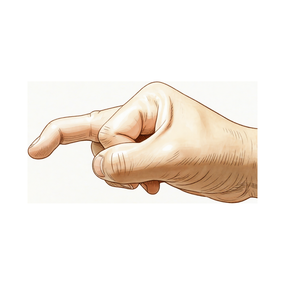
ਤੁਸੀਂ ਕੀ ਮਹਿਸੂਸ ਕਰਦੇ ਹੋ
ਮੈਲੇਟ ਫਿੰਗਰ (mallet finger) ਵਿੱਚ, ਤੁਹਾਡੀ ਉਂਗਲ ਦਾ ਆਖ਼ਰੀ ਜੋੜ ਹੇਠਾਂ ਵੱਲ ਲਟਕ ਜਾਂਦਾ ਹੈ ਅਤੇ ਤੁਸੀਂ ਆਪਣੇ ਆਪ ਪੋਟੇ ਨੂੰ ਸਿੱਧਾ ਨਹੀਂ ਕਰ ਸਕਦੇ। ਉਂਗਲ ਆਮ ਤੌਰ 'ਤੇ ਅਜੇ ਵੀ ਆਮ ਵਾਂਗ ਹੇਠਾਂ ਵੱਲ ਮੁੜ ਜਾਂਦੀ ਹੈ। ਇਹ ਸੱਟ ਉਦੋਂ ਲੱਗਦੀ ਹੈ ਜਦੋਂ ਪੋਟੇ ਨੂੰ ਸਿੱਧਾ ਕਰਨ ਵਾਲਾ ਟੈਂਡਨ (tendon, ਪੱਠੇ ਨੂੰ ਹੱਡੀ ਨਾਲ ਜੋੜਨ ਵਾਲੀ ਮਜ਼ਬੂਤ ਤੰਦ) ਪਾਟ ਕੇ ਵੱਖ ਹੋ ਜਾਂਦਾ ਹੈ, ਕਈ ਵਾਰ ਆਪਣੇ ਨਾਲ ਹੱਡੀ ਦਾ ਇੱਕ ਛੋਟਾ ਟੁਕੜਾ ਵੀ ਲੈ ਜਾਂਦਾ ਹੈ। ਇਹ ਅਕਸਰ ਸਿੱਧੀ ਉਂਗਲ ਦੇ ਸਿਰੇ ਉੱਤੇ ਗੇਂਦ ਵੱਜਣ ਤੋਂ ਬਾਅਦ ਹੁੰਦੀ ਹੈ, ਇਸੇ ਕਰਕੇ ਇਹ ਫ਼ੁੱਟਬਾਲ ਵਿੱਚ ਲੱਗਣ ਵਾਲੀ ਆਮ ਸੱਟ ਹੈ।
ਦਰਦ ਆਖ਼ਰੀ ਜੋੜ ਦੇ ਪਿਛਲੇ ਪਾਸੇ, ਨਹੁੰ ਦੇ ਬਿਲਕੁਲ ਹੇਠਾਂ ਹੁੰਦਾ ਹੈ। ਸੱਟ ਤੋਂ ਬਾਅਦ ਪਹਿਲੇ ਦਿਨਾਂ ਵਿੱਚ ਇਹ ਆਮ ਤੌਰ 'ਤੇ ਸਭ ਤੋਂ ਵੱਧ ਦੁਖਦਾ ਹੈ, ਜਦੋਂ ਉਂਗਲ ਸੁੱਜੀ ਹੁੰਦੀ ਹੈ ਅਤੇ ਛੂਹਣ ਨਾਲ ਦੁਖਦੀ ਹੈ। ਪੋਟੇ ਨੂੰ ਮੋੜਨ ਨਾਲ ਦਰਦ ਵਧਦਾ ਹੈ। ਇੱਕ ਵਾਰ ਉਂਗਲ ਉੱਤੇ ਸਪਲਿੰਟ ਲੱਗ ਜਾਵੇ ਅਤੇ ਉਹ ਸਿੱਧੀ ਰੱਖੀ ਜਾਵੇ, ਤਾਂ ਦਰਦ ਆਮ ਤੌਰ 'ਤੇ ਸ਼ਾਂਤ ਹੋ ਜਾਂਦਾ ਹੈ। ਲਟਕਣ ਆਪਣੇ ਆਪ ਵਿੱਚ ਨਹੀਂ ਦੁਖਦੀ, ਪਰ ਇਸ ਨਾਲ ਉਂਗਲ ਦੇ ਕੰਮ ਕਰਨ ਦਾ ਤਰੀਕਾ ਬਦਲ ਜਾਂਦਾ ਹੈ।
ਰੋਜ਼ਾਨਾ ਦੇ ਉਹ ਕੰਮ ਔਖੇ ਹੋ ਜਾਂਦੇ ਹਨ ਜਿਨ੍ਹਾਂ ਵਿੱਚ ਪੋਟੇ ਦੇ ਟਿਕੇ ਰਹਿਣ ਦੀ ਲੋੜ ਹੁੰਦੀ ਹੈ। ਛੋਟੇ ਸਿੱਕੇ ਚੁੱਕਣਾ, ਸੂਈ ਵਿੱਚ ਧਾਗਾ ਪਾਉਣਾ, ਛੋਟੀ ਚਾਬੀ ਘੁਮਾਉਣਾ ਜਾਂ ਬਟਨ ਦਬਾਉਣਾ ਬੇਢੰਗਾ ਹੋ ਸਕਦਾ ਹੈ, ਕਿਉਂਕਿ ਪੋਟਾ ਪੱਧਰਾ ਹੋ ਕੇ ਦਬਾ ਨਹੀਂ ਸਕਦਾ। ਲਿਖਣਾ ਜਾਂ ਫ਼ੋਨ ਫੜਨਾ ਵੀ ਵੱਖਰਾ ਮਹਿਸੂਸ ਹੋ ਸਕਦਾ ਹੈ। ਲਟਕਦੇ ਪੋਟੇ ਦੀ ਕਮੀ ਪੂਰੀ ਕਰਨ ਲਈ ਉਂਗਲ ਦਾ ਵਿਚਕਾਰਲਾ ਜੋੜ ਵੀ ਆਮ ਨਾਲੋਂ ਵੱਧ ਪਿੱਛੇ ਵੱਲ ਮੁੜ ਸਕਦਾ ਹੈ।
ਕੁਝ ਲੋਕ ਦੇਖਦੇ ਹਨ ਕਿ ਰਾਤ ਭਰ ਹੱਥ ਦੇ ਸਥਿਰ ਰਹਿਣ ਤੋਂ ਬਾਅਦ ਸਵੇਰੇ ਉਂਗਲ ਜ਼ਿਆਦਾ ਖ਼ਰਾਬ ਦਿਸਦੀ ਹੈ, ਅਤੇ ਇਸ ਨੂੰ ਹਿਲਾਉਣਾ ਸ਼ੁਰੂ ਕਰਨ ਤੋਂ ਬਾਅਦ ਇਹ ਢਿੱਲੀ ਪੈ ਜਾਂਦੀ ਹੈ।
ਕੁਝ ਚੇਤਾਵਨੀ ਵਾਲੀਆਂ ਨਿਸ਼ਾਨੀਆਂ ਉੱਤੇ ਛੇਤੀ ਕਾਰਵਾਈ ਦੀ ਲੋੜ ਹੁੰਦੀ ਹੈ। ਜੇ ਤੁਹਾਡੀ ਉਂਗਲ ਗਰਮ, ਲਾਲ, ਸੁੱਜੀ ਹੋਈ ਅਤੇ ਦਰਦ ਵਾਲੀ ਹੋ ਜਾਵੇ, ਖ਼ਾਸ ਕਰਕੇ ਬੁਖ਼ਾਰ ਦੇ ਨਾਲ, ਤਾਂ ਉਸੇ ਦਿਨ ਐਮਰਜੈਂਸੀ ਵਿਭਾਗ ਜਾਓ। ਜੇ ਸੱਟ ਵਾਲੇ ਜੋੜ ਦੇ ਉੱਪਰ ਚਮੜੀ ਫਟੀ ਹੋਈ ਹੈ, ਜਾਂ ਉਂਗਲ ਸਾਫ਼ ਤੌਰ 'ਤੇ ਵਿਗੜੀ ਹੋਈ (ਟੇਢੀ) ਦਿਸਦੀ ਹੈ, ਤਾਂ ਉਸੇ ਦਿਨ ਉੱਥੇ ਜਾਓ। ਜੇ ਤੁਹਾਡੀ ਉਂਗਲ ਫਿੱਕੀ, ਠੰਢੀ, ਸਫ਼ੈਦ ਜਾਂ ਨੀਲੀ ਪੈ ਜਾਵੇ, ਜਾਂ ਸੱਟ ਤੋਂ ਬਾਅਦ ਅਚਾਨਕ ਉਸ ਵਿੱਚ ਅਹਿਸਾਸ ਜਾਂ ਹਿਲਜੁਲ ਖ਼ਤਮ ਹੋ ਜਾਵੇ, ਤਾਂ ਉਸੇ ਦਿਨ ਉੱਥੇ ਜਾਓ। ਜੇ ਲੱਛਣ ਸ਼ਾਂਤ ਨਹੀਂ ਹੋ ਰਹੇ, ਹਫ਼ਤਿਆਂ ਦੇ ਨਾਲ ਵਿਗੜ ਰਹੇ ਹਨ, ਜਾਂ ਤੁਹਾਨੂੰ ਕੰਮ ਕਰਨ ਜਾਂ ਹੱਥ ਵਰਤਣ ਤੋਂ ਰੋਕਦੇ ਹਨ, ਤਾਂ ਆਪਣੇ ਜੀਪੀ (ਫ਼ੈਮਿਲੀ ਡਾਕਟਰ) ਨੂੰ ਮਿਲੋ ਜਾਂ ਸਪੈਸ਼ਲਿਸਟ ਤੋਂ ਜਾਂਚ ਕਰਵਾਉਣ ਲਈ ਕਹੋ।
ਅਸਲ ਵਿੱਚ ਕੀ ਹੋ ਰਿਹਾ ਹੈ
ਤੁਹਾਡੇ ਪੋਟੇ ਦੇ ਪਿਛਲੇ ਪਾਸੇ ਟੈਂਡਨ ਦੀ ਇੱਕ ਪਤਲੀ ਡੋਰੀ ਚੱਲਦੀ ਹੈ, ਯਾਨੀ ਰੇਸ਼ਿਆਂ ਦੀ ਇੱਕ ਰੱਸੀ ਜੋ ਮਾਸਪੇਸ਼ੀ ਨੂੰ ਹੱਡੀ ਨਾਲ ਜੋੜਦੀ ਹੈ। ਇਸ ਦੇ ਸਭ ਤੋਂ ਹੇਠਲੇ ਰੇਸ਼ੇ ਨਹੁੰ ਦੇ ਬਿਲਕੁਲ ਹੇਠਾਂ, ਉਂਗਲ ਦੀ ਆਖ਼ਰੀ ਛੋਟੀ ਹੱਡੀ ਨਾਲ ਜੁੜੇ ਹੁੰਦੇ ਹਨ। ਜਦੋਂ ਤੁਸੀਂ ਆਪਣੀ ਉਂਗਲ ਸਿੱਧੀ ਕਰਦੇ ਹੋ, ਤਾਂ ਉਹ ਡੋਰੀ ਹੱਡੀ ਨੂੰ ਖਿੱਚਦੀ ਹੈ। ਮੈਲੇਟ ਫਿੰਗਰ ਵਿੱਚ, ਉਹ ਡੋਰੀ ਆਪਣੀ ਜੁੜਨ ਵਾਲੀ ਥਾਂ ਤੋਂ ਟੁੱਟ ਕੇ ਵੱਖ ਹੋ ਗਈ ਹੁੰਦੀ ਹੈ। ਕਈ ਵਾਰ ਇਹ ਆਪਣੇ ਨਾਲ ਹੱਡੀ ਦਾ ਇੱਕ ਛੋਟਾ ਟੁਕੜਾ ਵੀ ਪਾੜ ਕੇ ਲੈ ਜਾਂਦੀ ਹੈ।
ਜਦੋਂ ਡੋਰੀ ਜੁੜੀ ਨਹੀਂ ਰਹਿੰਦੀ, ਤਾਂ ਪੋਟੇ ਨੂੰ ਉੱਪਰ ਚੁੱਕ ਕੇ ਰੱਖਣ ਵਾਲਾ ਕੁਝ ਨਹੀਂ ਬਚਦਾ। ਪੋਟੇ ਨੂੰ ਹੇਠਾਂ ਮੋੜਨ ਵਾਲੀ ਮਾਸਪੇਸ਼ੀ ਅਜੇ ਵੀ ਕੰਮ ਕਰਦੀ ਹੈ, ਅਤੇ ਦੂਜੇ ਪਾਸੇ ਖਿੱਚਣ ਵਾਲਾ ਕੁਝ ਨਾ ਹੋਣ ਕਰਕੇ, ਉਹੀ ਭਾਰੀ ਪੈਂਦੀ ਹੈ। ਪੋਟਾ ਲਟਕਿਆ ਰਹਿੰਦਾ ਹੈ ਅਤੇ ਤੁਸੀਂ ਇਸ ਨੂੰ ਆਪਣੇ ਆਪ ਸਿੱਧਾ ਨਹੀਂ ਕਰ ਸਕਦੇ, ਹਾਲਾਂਕਿ ਤੁਸੀਂ ਅਜੇ ਵੀ ਆਪਣੇ ਦੂਜੇ ਹੱਥ ਨਾਲ ਧੱਕ ਕੇ ਇਸ ਨੂੰ ਸਿੱਧਾ ਕਰ ਸਕਦੇ ਹੋ। ਇਸ ਦਾ ਕਾਰਨ ਇਹ ਹੈ ਕਿ ਮੋੜਨ ਵਾਲੀ ਮਾਸਪੇਸ਼ੀ ਠੀਕ-ਠਾਕ ਹੈ, ਪਰ ਸਿੱਧਾ ਕਰਨ ਵਾਲੀ ਡੋਰੀ ਨਹੀਂ।
ਇਹ ਸੱਟ ਆਮ ਤੌਰ 'ਤੇ ਉਦੋਂ ਲੱਗਦੀ ਹੈ ਜਦੋਂ ਸਿੱਧੇ ਪੋਟੇ ਨੂੰ ਅਚਾਨਕ ਜ਼ੋਰ ਨਾਲ ਮੋੜ ਦਿੱਤਾ ਜਾਂਦਾ ਹੈ, ਜਿਵੇਂ ਕਿਸੇ ਚੀਜ਼ ਵੱਲ ਹੱਥ ਵਧਾਉਂਦਿਆਂ ਉਂਗਲ ਦਾ ਜੇਬ ਜਾਂ ਚਾਦਰ ਵਿੱਚ ਅੜ ਜਾਣਾ। ਜੋੜ ਦੀ ਸਤ੍ਹਾ ਵੀ ਪ੍ਰਭਾਵਿਤ ਹੋ ਸਕਦੀ ਹੈ। ਜੇ ਪਾਟ ਕੇ ਵੱਖ ਹੋਇਆ ਹੱਡੀ ਦਾ ਟੁਕੜਾ ਜੋੜ ਦੀ ਸਤ੍ਹਾ ਦੇ ਤੀਜੇ ਹਿੱਸੇ ਤੋਂ ਵੱਧ ਹੈ, ਜਾਂ ਆਖ਼ਰੀ ਹੱਡੀ ਸੇਧ ਤੋਂ ਖਿਸਕ ਜਾਂਦੀ ਹੈ, ਤਾਂ ਆਮ ਤੌਰ 'ਤੇ ਸਰਜਰੀ ਦੀ ਸਿਫ਼ਾਰਸ਼ ਕੀਤੀ ਜਾਂਦੀ ਹੈ। ਛੋਟੇ ਟੁਕੜਿਆਂ ਦਾ ਇਲਾਜ ਆਮ ਤੌਰ 'ਤੇ ਸਿਰਫ਼ ਸਪਲਿੰਟ ਨਾਲ ਕੀਤਾ ਜਾਂਦਾ ਹੈ।
ਇਲਾਜ ਚੰਗੀ ਤਰ੍ਹਾਂ ਸਫਲ ਹੋਣ ਤੋਂ ਬਾਅਦ ਵੀ ਕੁਝ ਲਟਕਣ ਬਾਕੀ ਰਹਿ ਸਕਦੀ ਹੈ, ਅਤੇ ਜ਼ਿਆਦਾਤਰ ਲੋਕ ਬਿਨਾਂ ਬਹੁਤੀ ਮੁਸ਼ਕਲ ਦੇ ਇਸ ਦੇ ਆਦੀ ਹੋ ਜਾਂਦੇ ਹਨ। ਜੇ ਸਪਲਿੰਟ ਨੇ ਕੰਮ ਨਹੀਂ ਕੀਤਾ, ਤਾਂ ਸਰਜਰੀ ਇੱਕ ਵਿਕਲਪ ਹੈ, ਹਾਲਾਂਕਿ ਸਰਜਰੀ ਦਾ ਕੋਈ ਫ਼ਾਇਦਾ ਅਜੇ ਸਪਸ਼ਟ ਤੌਰ 'ਤੇ ਸਾਬਤ ਨਹੀਂ ਹੋਇਆ।
ਅਸੀਂ ਇਸ ਬਾਰੇ ਕੀ ਕਰ ਸਕਦੇ ਹਾਂ
ਡਾ. ਕੀਰਨ ਹਿਰਪਰਾ, ਮਾਟਰ ਪ੍ਰਾਈਵੇਟ ਹਸਪਤਾਲ ਰੌਕਹੈਂਪਟਨ ਵਿੱਚ ਉੱਪਰਲੇ ਅੰਗ (ਮੋਢੇ ਤੋਂ ਹੱਥ ਤੱਕ ਦੀ ਬਾਂਹ) ਦੇ ਸਰਜਨ, ਇਲਾਜ ਨੂੰ ਤੁਹਾਡੀ ਖ਼ਾਸ ਸੱਟ ਦੇ ਅਨੁਸਾਰ ਢਾਲਦੇ ਹਨ। ਕੁਝ ਮੈਲੇਟ ਫਿੰਗਰ ਓਪਰੇਸ਼ਨ ਤੋਂ ਬਿਨਾਂ ਠੀਕ ਹੋ ਜਾਂਦੀਆਂ ਹਨ, ਅਤੇ ਕੁਝ ਨੂੰ ਛੇਤੀ ਸਰਜਰੀ ਦੀ ਲੋੜ ਹੁੰਦੀ ਹੈ, ਇਸ ਲਈ ਬਿਨਾਂ ਦੇਰੀ ਜਾਂਚ ਕਰਵਾਉਣਾ ਮਹੱਤਵਪੂਰਨ ਹੈ। ਮਰੀਜ਼ਾਂ ਨੂੰ ਆਮ ਤੌਰ 'ਤੇ ਉਨ੍ਹਾਂ ਦਾ ਜੀਪੀ (ਫ਼ੈਮਿਲੀ ਡਾਕਟਰ) ਸਾਡੇ ਕਲੀਨਿਕ ਵਿੱਚ ਭੇਜਦਾ ਹੈ; ਜੇ ਕਿਸੇ ਫਿਜ਼ੀਓਥੈਰੇਪਿਸਟ ਨੇ ਤੁਹਾਨੂੰ ਸਾਨੂੰ ਮਿਲਣ ਦੀ ਸਲਾਹ ਦਿੱਤੀ ਹੈ, ਤਾਂ ਵੀ ਮੈਡੀਕੇਅਰ ਰਿਬੇਟ (Medicare rebate) ਦੇ ਯੋਗ ਹੋਣ ਲਈ ਤੁਹਾਨੂੰ ਆਪਣੇ ਜੀਪੀ ਤੋਂ ਰੈਫ਼ਰਲ ਦੀ ਲੋੜ ਪਵੇਗੀ। ਕਲੀਨਿਕ ਵਿੱਚ ਅਸੀਂ ਤੁਹਾਡਾ ਪਿਛੋਕੜ ਪੁੱਛਦੇ ਹਾਂ, ਤੁਹਾਡੀ ਉਂਗਲ ਦੀ ਜਾਂਚ ਕਰਦੇ ਹਾਂ ਅਤੇ ਲੋੜ ਹੋਵੇ ਤਾਂ ਐਕਸ-ਰੇ ਦਾ ਪ੍ਰਬੰਧ ਕਰਦੇ ਹਾਂ। ਐਕਸ-ਰੇ ਦਿਖਾਉਂਦਾ ਹੈ ਕਿ ਕੀ ਟੈਂਡਨ ਦੇ ਨਾਲ ਹੱਡੀ ਦਾ ਕੋਈ ਟੁਕੜਾ ਵੱਖ ਹੋਇਆ ਹੈ, ਉਹ ਟੁਕੜਾ ਕਿੰਨਾ ਵੱਡਾ ਹੈ, ਅਤੇ ਕੀ ਉਂਗਲ ਦੀ ਆਖ਼ਰੀ ਹੱਡੀ ਸੇਧ ਤੋਂ ਖਿਸਕ ਗਈ ਹੈ।
ਜ਼ਿਆਦਾਤਰ ਸੱਟਾਂ ਲਈ, ਇਲਾਜ ਇੱਕ ਸਪਲਿੰਟ ਨਾਲ ਸ਼ੁਰੂ ਹੁੰਦਾ ਹੈ ਜੋ ਪੋਟੇ ਨੂੰ ਸਿੱਧਾ ਰੱਖਦਾ ਹੈ। ਸਪਲਿੰਟ ਨੂੰ 6 ਤੋਂ 8 ਹਫ਼ਤਿਆਂ ਤੱਕ ਲਗਾਤਾਰ ਪਾ ਕੇ ਰੱਖਣ ਦੀ ਲੋੜ ਹੈ, ਕਿਉਂਕਿ ਕੁਝ ਮਿੰਟਾਂ ਲਈ ਵੀ ਪੋਟਾ ਮੁੜ ਜਾਵੇ, ਜਿਵੇਂ ਹੱਥ ਧੋਣ ਵੇਲੇ, ਤਾਂ ਜੁੜਨ ਦਾ ਕੀਤਾ-ਕਰਾਇਆ ਕੰਮ ਖ਼ਰਾਬ ਹੋ ਸਕਦਾ ਹੈ। ਸਾਡੀ ਹੈਂਡ ਥੈਰੇਪਿਸਟ, ਐਕਸਟੈਂਡ ਰੀਹੈਬਿਲੀਟੇਸ਼ਨ (Extend Rehabilitation) ਦੀ ਰੂਬੀ ਡੂਲਨ (Ruby Doolan), ਤੁਹਾਡਾ ਸਪਲਿੰਟ ਫਿੱਟ ਕਰਦੀ ਹੈ ਅਤੇ ਤੁਹਾਡੀਆਂ ਕਸਰਤਾਂ ਵਿੱਚ ਤੁਹਾਡੀ ਅਗਵਾਈ ਕਰਦੀ ਹੈ। ਹੈਂਡ ਥੈਰੇਪਿਸਟ ਇਨ੍ਹਾਂ ਸੱਟਾਂ ਦਾ ਇਲਾਜ ਸਰਜਨ ਜਿੰਨੀ ਹੀ ਚੰਗੀ ਤਰ੍ਹਾਂ ਕਰ ਸਕਦਾ ਹੈ, ਅਤੇ ਸਪਲਿੰਟ ਨਾਲ ਚਮੜੀ ਦੀਆਂ ਸਮੱਸਿਆਵਾਂ ਬਹੁਤ ਘੱਟ ਹੁੰਦੀਆਂ ਹਨ। ਜੇ ਸਪਲਿੰਟ ਉਤਾਰਨ ਤੋਂ ਬਾਅਦ ਵੀ ਤੁਹਾਡੀ ਉਂਗਲ ਲਟਕਦੀ ਹੈ, ਤਾਂ ਸਪਲਿੰਟ ਦੇ ਵਾਧੂ ਹਫ਼ਤੇ ਯੋਜਨਾ ਦਾ ਹਿੱਸਾ ਹਨ, ਇਸ ਗੱਲ ਦੀ ਨਿਸ਼ਾਨੀ ਨਹੀਂ ਕਿ ਇਲਾਜ ਅਸਫਲ ਹੋ ਗਿਆ ਹੈ। ਇਸ ਦੇ ਨਾਲ-ਨਾਲ ਰਾਤ ਵਾਲਾ ਸਪਲਿੰਟ ਪਾਉਣ ਨਾਲ ਕੋਈ ਵਾਧੂ ਫ਼ਾਇਦਾ ਨਹੀਂ ਹੁੰਦਾ। ਜੇ ਤੁਸੀਂ ਖੇਡਾਂ ਖੇਡਦੇ ਹੋ, ਤਾਂ ਜਦੋਂ ਤੱਕ ਉਂਗਲ ਉੱਤੇ ਸਪਲਿੰਟ ਲੱਗਾ ਹੈ, ਤੁਹਾਨੂੰ ਉਸ ਖੇਡ ਤੋਂ ਬਚਣਾ ਪਵੇਗਾ ਜਿਸ ਨਾਲ ਸੱਟ ਲੱਗੀ ਸੀ। ਜਿਨ੍ਹਾਂ ਸੱਟਾਂ ਵੱਲ ਇਲਾਜ ਤੋਂ ਪਹਿਲਾਂ 2 ਤੋਂ 4 ਹਫ਼ਤਿਆਂ ਤੱਕ ਧਿਆਨ ਨਹੀਂ ਦਿੱਤਾ ਗਿਆ ਸੀ, ਉਹ ਵੀ ਸਪਲਿੰਟ ਨਾਲ ਚੰਗੀ ਤਰ੍ਹਾਂ ਠੀਕ ਹੋ ਸਕਦੀਆਂ ਹਨ।
ਆਮ ਤੌਰ 'ਤੇ ਸਰਜਰੀ ਦੀ ਸਿਫ਼ਾਰਸ਼ ਉਦੋਂ ਕੀਤੀ ਜਾਂਦੀ ਹੈ ਜਦੋਂ ਪਾਟ ਕੇ ਵੱਖ ਹੋਇਆ ਹੱਡੀ ਦਾ ਟੁਕੜਾ ਜੋੜ ਦੀ ਸਤ੍ਹਾ ਦੇ ਤੀਜੇ ਹਿੱਸੇ ਤੋਂ ਵੱਧ ਹੋਵੇ, ਜਾਂ ਜਦੋਂ ਉਂਗਲ ਦੀ ਆਖ਼ਰੀ ਹੱਡੀ ਸੇਧ ਤੋਂ ਹਥੇਲੀ ਵੱਲ ਖਿਸਕ ਗਈ ਹੋਵੇ। ਕਦੇ-ਕਦਾਈਂ ਇਹ ਸਪਲਿੰਟ ਤੋਂ ਬਾਅਦ ਵੀ ਬਣੀ ਰਹਿੰਦੀ ਲਟਕਣ ਲਈ, ਜਾਂ ਕਿਸੇ ਅਜਿਹੀ ਪੁਰਾਣੀ ਸੱਟ ਲਈ ਜਿਸ ਦਾ ਕਦੇ ਇਲਾਜ ਨਹੀਂ ਹੋਇਆ, ਵੀ ਸੁਝਾਈ ਜਾਂਦੀ ਹੈ। ਜੇ ਸਪਲਿੰਟ ਨੇ ਕੰਮ ਨਹੀਂ ਕੀਤਾ, ਤਾਂ ਸਰਜਰੀ ਇੱਕ ਵਿਕਲਪ ਹੈ, ਹਾਲਾਂਕਿ ਸਰਜਰੀ ਦਾ ਕੋਈ ਫ਼ਾਇਦਾ ਅਜੇ ਸਪਸ਼ਟ ਤੌਰ 'ਤੇ ਸਾਬਤ ਨਹੀਂ ਹੋਇਆ। ਓਪਰੇਸ਼ਨ ਟੈਂਡਨ ਜਾਂ ਹੱਡੀ ਦੇ ਟੁਕੜੇ ਨੂੰ ਉਸ ਦੀ ਥਾਂ ਉੱਤੇ ਜਕੜ ਕੇ ਰੱਖਦਾ ਹੈ ਤਾਂ ਜੋ ਪੋਟਾ ਦੁਬਾਰਾ ਸਿੱਧਾ ਹੋ ਸਕੇ, ਅਤੇ ਇਸ ਦਾ ਆਪਣਾ ਵੱਖਰਾ ਪੰਨਾ ਹੈ।
ਕੀ ਉਮੀਦ ਰੱਖੀਏ
ਜ਼ਿਆਦਾਤਰ ਮੈਲੇਟ ਫਿੰਗਰ ਸਪਲਿੰਟ ਨਾਲ ਠੀਕ ਹੋ ਜਾਂਦੀਆਂ ਹਨ। ਲਗਭਗ ਅੱਧੀਆਂ ਸੱਟਾਂ ਸਿਰਫ਼ ਸਪਲਿੰਟ ਨਾਲ ਠੀਕ ਹੋ ਜਾਂਦੀਆਂ ਹਨ ਜਾਂ ਉਨ੍ਹਾਂ ਵਿੱਚ ਸਾਫ਼ ਸੁਧਾਰ ਹੁੰਦਾ ਹੈ। ਸਪਲਿੰਟ 6 ਤੋਂ 8 ਹਫ਼ਤਿਆਂ ਲਈ ਪੋਟੇ ਨੂੰ ਸਿੱਧਾ ਰੱਖਦਾ ਹੈ, ਅਤੇ ਟੈਂਡਨ ਉਸੇ ਸਥਿਤੀ ਵਿੱਚ ਜੁੜ ਕੇ ਠੀਕ ਹੁੰਦਾ ਹੈ। ਜੇ ਸਪਲਿੰਟ ਉਤਾਰਨ ਵੇਲੇ ਵੀ ਤੁਹਾਡੀ ਉਂਗਲ ਲਟਕਦੀ ਹੈ, ਤਾਂ ਸਪਲਿੰਟ ਦੇ ਹੋਰ ਹਫ਼ਤੇ ਯੋਜਨਾ ਦਾ ਹਿੱਸਾ ਹਨ, ਇਸ ਗੱਲ ਦੀ ਨਿਸ਼ਾਨੀ ਨਹੀਂ ਕਿ ਇਲਾਜ ਅਸਫਲ ਹੋ ਗਿਆ ਹੈ।
ਤੁਸੀਂ ਕਿੰਨੀ ਚੰਗੀ ਤਰ੍ਹਾਂ ਠੀਕ ਹੁੰਦੇ ਹੋ, ਇਹ ਬਹੁਤ ਹੱਦ ਤੱਕ ਇਸ ਗੱਲ ਉੱਤੇ ਨਿਰਭਰ ਕਰਦਾ ਹੈ ਕਿ ਤੁਸੀਂ ਸਪਲਿੰਟ ਬਿਲਕੁਲ ਹਦਾਇਤਾਂ ਅਨੁਸਾਰ ਪਾਉਂਦੇ ਹੋ। ਜੋ ਲੋਕ ਸਪਲਿੰਟ ਦੀਆਂ ਹਦਾਇਤਾਂ ਦੀ ਪਾਲਣਾ ਕਰਦੇ ਹਨ, ਉਨ੍ਹਾਂ ਦੇ ਨਤੀਜੇ ਇਸ ਨੂੰ ਉਤਾਰ ਦੇਣ ਵਾਲਿਆਂ ਨਾਲੋਂ ਬਿਹਤਰ ਹੁੰਦੇ ਹਨ। ਕੁਝ ਮਿੰਟਾਂ ਲਈ ਵੀ ਪੋਟਾ ਮੁੜ ਜਾਵੇ ਤਾਂ ਜੁੜਨ ਦਾ ਕੀਤਾ-ਕਰਾਇਆ ਕੰਮ ਖ਼ਰਾਬ ਹੋ ਸਕਦਾ ਹੈ, ਇਸੇ ਲਈ ਸਪਲਿੰਟ ਲਗਾਤਾਰ ਪਾ ਕੇ ਰੱਖਿਆ ਜਾਂਦਾ ਹੈ। ਜਿਨ੍ਹਾਂ ਸੱਟਾਂ ਦਾ ਇਲਾਜ ਸੱਟ ਲੱਗਣ ਤੋਂ ਦੋ ਹਫ਼ਤਿਆਂ ਦੇ ਅੰਦਰ ਹੋ ਜਾਂਦਾ ਹੈ, ਉਨ੍ਹਾਂ ਤੋਂ ਬਾਅਦ ਘੱਟ ਹੀ ਕੋਈ ਸਥਾਈ ਅਪੰਗਤਾ ਰਹਿੰਦੀ ਹੈ। ਜਿਨ੍ਹਾਂ ਸੱਟਾਂ ਦਾ 2 ਤੋਂ 4 ਹਫ਼ਤਿਆਂ ਤੱਕ ਇਲਾਜ ਨਹੀਂ ਹੋਇਆ, ਉਹ ਵੀ ਸਪਲਿੰਟ ਨਾਲ ਚੰਗੀ ਤਰ੍ਹਾਂ ਠੀਕ ਹੋ ਸਕਦੀਆਂ ਹਨ, ਇਸ ਲਈ ਦੇਰ ਨਾਲ ਸ਼ੁਰੂਆਤ ਹਿੰਮਤ ਹਾਰਨ ਦਾ ਕਾਰਨ ਨਹੀਂ ਹੈ।
ਇਲਾਜ ਚੰਗੀ ਤਰ੍ਹਾਂ ਸਫਲ ਹੋਣ ਦੇ ਬਾਵਜੂਦ ਵੀ ਕੁਝ ਲਟਕਣ ਬਾਕੀ ਰਹਿ ਸਕਦੀ ਹੈ। ਹੋ ਸਕਦਾ ਹੈ ਪੋਟਾ ਪੂਰੀ ਤਰ੍ਹਾਂ ਸਿੱਧਾ ਨਾ ਟਿਕੇ, ਅਤੇ ਜ਼ਿਆਦਾਤਰ ਲੋਕ ਬਿਨਾਂ ਬਹੁਤੀ ਮੁਸ਼ਕਲ ਦੇ ਇਸ ਦੇ ਆਦੀ ਹੋ ਜਾਂਦੇ ਹਨ। ਪਕੜਨ ਦੇ ਕੰਮ ਵਿੱਚ ਆਖ਼ਰੀ ਜੋੜ ਦਾ ਯੋਗਦਾਨ ਥੋੜ੍ਹਾ ਹੁੰਦਾ ਹੈ, ਇਸ ਲਈ ਥੋੜ੍ਹੀ ਜਿਹੀ ਸਥਾਈ ਲਟਕਣ ਆਮ ਤੌਰ 'ਤੇ ਦਿਸਣ ਨਾਲੋਂ ਘੱਟ ਮਾਇਨੇ ਰੱਖਦੀ ਹੈ। ਜੇ ਸਪਲਿੰਟ ਨੇ ਕੰਮ ਨਹੀਂ ਕੀਤਾ, ਤਾਂ ਸਰਜਰੀ ਇੱਕ ਵਿਕਲਪ ਹੈ, ਹਾਲਾਂਕਿ ਸਰਜਰੀ ਦਾ ਕੋਈ ਫ਼ਾਇਦਾ ਅਜੇ ਸਪਸ਼ਟ ਤੌਰ 'ਤੇ ਸਾਬਤ ਨਹੀਂ ਹੋਇਆ।
ਜੇ ਬਿਲਕੁਲ ਇਲਾਜ ਨਾ ਕੀਤਾ ਜਾਵੇ, ਤਾਂ ਮੈਲੇਟ ਫਿੰਗਰ ਦੀ ਲਟਕਣ ਆਮ ਤੌਰ 'ਤੇ ਬਣੀ ਰਹਿੰਦੀ ਹੈ। ਟੈਂਡਨ ਬਿਨਾਂ ਮਦਦ ਦੇ ਆਪਣੇ ਆਪ ਮੁੜ ਨਹੀਂ ਜੁੜਦਾ। ਕੁਝ ਲੋਕ ਲਟਕਣ ਦੇ ਨਾਲ ਹੀ ਰਹਿੰਦੇ ਹਨ ਅਤੇ ਠੀਕ ਗੁਜ਼ਾਰਾ ਕਰ ਲੈਂਦੇ ਹਨ, ਜਦਕਿ ਦੂਜਿਆਂ ਨੂੰ ਮਹੀਨਿਆਂ ਦੌਰਾਨ ਬੇਢੰਗਾ ਪੋਟਾ ਨਿਰਾਸ਼ ਕਰਦਾ ਹੈ। ਜੇ ਤੁਹਾਡੇ ਲੱਛਣ ਸ਼ਾਂਤ ਨਹੀਂ ਹੋ ਰਹੇ, ਹਫ਼ਤਿਆਂ ਦੇ ਨਾਲ ਵਿਗੜ ਰਹੇ ਹਨ, ਜਾਂ ਤੁਹਾਨੂੰ ਕੰਮ ਕਰਨ ਜਾਂ ਹੱਥ ਵਰਤਣ ਤੋਂ ਰੋਕਦੇ ਹਨ, ਤਾਂ ਆਪਣੇ ਜੀਪੀ ਨੂੰ ਮਿਲੋ ਜਾਂ ਸਪੈਸ਼ਲਿਸਟ ਤੋਂ ਜਾਂਚ ਕਰਵਾਉਣ ਲਈ ਕਹੋ।
ਡਾਕਟਰ ਨੂੰ ਕਦੋਂ ਮਿਲਣਾ ਚਾਹੀਦਾ ਹੈ
ਜ਼ਿਆਦਾਤਰ ਮੈਲੇਟ ਫਿੰਗਰ ਸਿੱਧੀ ਉਂਗਲ ਦੇ ਸਿਰੇ ਉੱਤੇ ਗੇਂਦ ਵੱਜਣ ਨਾਲ ਹੁੰਦੀਆਂ ਹਨ, ਅਕਸਰ ਫ਼ੁੱਟਬਾਲ ਵਿੱਚ। ਪੋਟਾ ਲਟਕ ਜਾਂਦਾ ਹੈ ਅਤੇ ਆਪਣੇ ਆਪ ਸਿੱਧਾ ਨਹੀਂ ਹੁੰਦਾ, ਜੋ ਆਮ ਤੌਰ 'ਤੇ ਡਾਕਟਰ ਕੋਲ ਜਾਣ ਲਈ ਕਾਫ਼ੀ ਕਾਰਨ ਹੁੰਦਾ ਹੈ। ਕੁਝ ਵੱਡੇ ਫ੍ਰੈਕਚਰ (ਹੱਡੀ ਦਾ ਟੁੱਟਣਾ) ਵੱਖਰਾ ਵਿਹਾਰ ਕਰਦੇ ਹਨ: ਹੱਡੀ ਦਾ ਟੁਕੜਾ ਵੱਖ ਹੋਣ ਦੇ ਬਾਵਜੂਦ ਪੋਟਾ ਅਜੇ ਵੀ ਸਿੱਧਾ ਟਿਕਿਆ ਰਹਿ ਸਕਦਾ ਹੈ, ਇਸ ਲਈ ਸਿਰੇ ਉੱਤੇ ਸੱਟ ਲੱਗਣ ਤੋਂ ਬਾਅਦ ਜੋ ਦੁਖਦੀ ਉਂਗਲ ਠੀਕ ਨਹੀਂ ਹੁੰਦੀ, ਉਸ ਦਾ ਵੀ ਐਕਸ-ਰੇ ਕਰਵਾਉਣਾ ਚਾਹੀਦਾ ਹੈ। ਹੱਡੀ ਵਾਲੀਆਂ ਮੈਲੇਟ ਸੱਟਾਂ ਆਮ ਤੌਰ 'ਤੇ ਉਂਗਲ ਦੇ ਜ਼ੋਰ ਨਾਲ ਪਿੱਛੇ ਵੱਲ ਮੁੜਨ ਨਾਲ ਲੱਗਦੀਆਂ ਹਨ, ਅੱਗੇ ਵੱਲ ਨਹੀਂ।
ਕੁਝ ਨਿਸ਼ਾਨੀਆਂ ਉੱਤੇ ਛੇਤੀ ਕਾਰਵਾਈ ਦੀ ਲੋੜ ਹੁੰਦੀ ਹੈ। ਜੇ ਤੁਹਾਡੀ ਉਂਗਲ ਗਰਮ, ਲਾਲ, ਸੁੱਜੀ ਹੋਈ ਅਤੇ ਦਰਦ ਵਾਲੀ ਹੋ ਜਾਵੇ, ਖ਼ਾਸ ਕਰਕੇ ਬੁਖ਼ਾਰ ਦੇ ਨਾਲ, ਤਾਂ ਉਸੇ ਦਿਨ ਐਮਰਜੈਂਸੀ ਵਿਭਾਗ ਜਾਓ। ਜੇ ਸੱਟ ਵਾਲੇ ਜੋੜ ਦੇ ਉੱਪਰ ਚਮੜੀ ਫਟੀ ਹੋਈ ਹੈ, ਜਾਂ ਉਂਗਲ ਸਾਫ਼ ਤੌਰ 'ਤੇ ਵਿਗੜੀ ਹੋਈ (ਟੇਢੀ) ਦਿਸਦੀ ਹੈ, ਤਾਂ ਉਸੇ ਦਿਨ ਉੱਥੇ ਜਾਓ। ਜੇ ਤੁਹਾਡੀ ਉਂਗਲ ਫਿੱਕੀ, ਠੰਢੀ, ਸਫ਼ੈਦ ਜਾਂ ਨੀਲੀ ਪੈ ਜਾਵੇ, ਜਾਂ ਸੱਟ ਤੋਂ ਬਾਅਦ ਅਚਾਨਕ ਉਸ ਵਿੱਚ ਅਹਿਸਾਸ ਜਾਂ ਹਿਲਜੁਲ ਖ਼ਤਮ ਹੋ ਜਾਵੇ, ਤਾਂ ਉਸੇ ਦਿਨ ਉੱਥੇ ਜਾਓ। ਜੇ ਲੱਛਣ ਸ਼ਾਂਤ ਨਹੀਂ ਹੋ ਰਹੇ, ਹਫ਼ਤਿਆਂ ਦੇ ਨਾਲ ਵਿਗੜ ਰਹੇ ਹਨ, ਜਾਂ ਤੁਹਾਨੂੰ ਕੰਮ ਕਰਨ ਜਾਂ ਹੱਥ ਵਰਤਣ ਤੋਂ ਰੋਕਦੇ ਹਨ, ਤਾਂ ਆਪਣੇ ਜੀਪੀ ਨੂੰ ਮਿਲੋ ਜਾਂ ਸਪੈਸ਼ਲਿਸਟ ਤੋਂ ਜਾਂਚ ਕਰਵਾਉਣ ਲਈ ਕਹੋ।
ਵਿਸਥਾਰ ਵਿੱਚ
Advanced reading: the deeper science (optional)
ਇਹ ਭਾਗ ਤੁਹਾਡੇ ਆਪਣੇ ਇਲਾਜ ਦੇ ਫ਼ੈਸਲਿਆਂ ਲਈ ਲੋੜ ਤੋਂ ਅੱਗੇ ਜਾਂਦਾ ਹੈ। ਮੈਲੇਟ ਫਿੰਗਰ ਬਾਰੇ ਵਾਧੂ ਪੜ੍ਹਨਾ ਲਾਭਦਾਇਕ ਹੈ ਕਿਉਂਕਿ ਜਿਨ੍ਹਾਂ ਦੋ ਹਾਲਤਾਂ ਨੂੰ ਆਮ ਤੌਰ 'ਤੇ ਸਰਜਰੀ ਦੇ ਸਪਸ਼ਟ ਕਾਰਨ ਮੰਨਿਆ ਜਾਂਦਾ ਹੈ — ਫ੍ਰੈਕਚਰ ਦਾ ਵੱਡਾ ਟੁਕੜਾ, ਅਤੇ ਸਪਲਿੰਟ ਤੋਂ ਬਾਅਦ ਵੀ ਬਣੀ ਰਹਿੰਦੀ ਲਟਕਣ, ਉਹ ਉਸ ਭਰੋਸੇ ਜਿੰਨੀਆਂ ਸਪਸ਼ਟ ਤੌਰ 'ਤੇ ਸਥਾਪਿਤ ਨਹੀਂ ਹਨ ਜਿਸ ਨਾਲ ਉਨ੍ਹਾਂ ਨੂੰ ਬਿਆਨ ਕੀਤਾ ਜਾਂਦਾ ਹੈ।
ਦੋਵੇਂ ਇਲਾਜ ਚੰਗੇ ਨਤੀਜੇ ਦਿੰਦੇ ਹਨ, ਅਤੇ ਕੋਈ ਨਹੀਂ ਜਾਣਦਾ ਕਿ ਹੱਦ ਕਿੱਥੇ ਹੈ
ਸਰਜਰੀ ਅਤੇ ਬਿਨਾਂ ਸਰਜਰੀ ਵਾਲੇ ਇਲਾਜ ਦੀ ਇੱਕ ਯੋਜਨਾਬੱਧ ਸਮੀਖਿਆ (systematic review) ਇੱਕ ਅਸਾਧਾਰਨ ਤੌਰ 'ਤੇ ਸਿੱਧੇ ਸਿੱਟੇ 'ਤੇ ਪਹੁੰਚੀ: ਦੋਵਾਂ ਨਾਲ ਚੰਗੇ ਕਲੀਨਿਕਲ ਨਤੀਜੇ ਮਿਲਦੇ ਹਨ, ਅਤੇ ਇਹ ਤੈਅ ਕਰਨ ਲਈ ਲੋੜੀਂਦੇ ਸਬੂਤ ਮੌਜੂਦ ਨਹੀਂ ਹਨ ਕਿ ਸਰਜਰੀ ਕਦੋਂ ਕਰਨੀ ਚਾਹੀਦੀ ਹੈ [1]।
ਦੂਜਾ ਹਿੱਸਾ ਹੀ ਮਹੱਤਵਪੂਰਨ ਹੈ। ਬਹਿਸ ਇਸ ਬਾਰੇ ਨਹੀਂ ਕਿ ਕੋਈ ਇਲਾਜ ਕੰਮ ਕਰਦਾ ਹੈ ਜਾਂ ਨਹੀਂ। ਗੱਲ ਇਹ ਹੈ ਕਿ ਸਰਜਰੀ ਚੁਣਨ ਦੀ ਹੱਦ, ਯਾਨੀ ਉਹ ਬਿੰਦੂ ਜਿੱਥੇ ਫ੍ਰੈਕਚਰ ਨੂੰ ਬਹੁਤ ਵੱਡਾ, ਜਾਂ ਜੋੜ ਨੂੰ ਬਹੁਤ ਜ਼ਿਆਦਾ ਖਿਸਕਿਆ (subluxed) ਮੰਨਿਆ ਜਾਂਦਾ ਹੈ, ਤੁਲਨਾਤਮਕ ਸਬੂਤਾਂ ਦੀ ਬਜਾਏ ਰਿਵਾਜ 'ਤੇ ਟਿਕੀ ਹੋਈ ਹੈ।
ਜਿੱਥੇ ਟੁਕੜਾ ਜੋੜ ਦੀ ਸਤ੍ਹਾ ਦੇ ਤੀਜੇ ਹਿੱਸੇ ਤੋਂ ਵੱਧ ਹੋਵੇ, ਜਾਂ ਉਂਗਲ ਦੀ ਸਿਰੇ ਵਾਲੀ ਹੱਡੀ (distal phalanx) ਖਿਸਕ ਗਈ ਹੋਵੇ, ਉੱਥੇ ਆਮ ਤੌਰ 'ਤੇ ਸਰਜਰੀ ਕੀਤੀ ਜਾਂਦੀ ਹੈ, ਪਰ ਉਨ੍ਹਾਂ ਗੁੰਝਲਦਾਰ ਮਾਮਲਿਆਂ ਵਿੱਚ ਵੀ ਸਰਜਰੀ ਦਾ ਕੋਈ ਖ਼ਾਸ ਫ਼ਾਇਦਾ ਅਜੇ ਸਪਸ਼ਟ ਤੌਰ 'ਤੇ ਸਾਬਤ ਨਹੀਂ ਹੋਇਆ [2]।
ਸਪਲਿੰਟ ਕੰਮ ਕਰਦਾ ਹੈ, ਅਤੇ ਮਾਇਨੇ ਰੱਖਣ ਵਾਲਾ ਅੰਕੜਾ ਇਹ ਹੈ ਕਿ ਕਿੰਨੇ ਸਮੇਂ ਲਈ
ਸਪਲਿੰਟ (orthotic) ਬਾਰੇ ਸਬੂਤ ਜ਼ਿਆਦਾ ਠੋਸ ਹਨ। ਤਿੰਨ ਵਿੱਚੋਂ ਦੋ ਅਧਿਐਨਾਂ ਵਿੱਚ ਸਪਲਿੰਟ ਵਾਲੇ ਇਲਾਜ ਦਾ ਵੱਡਾ ਅਸਰ (effect size) ਮਿਲਿਆ, 2.17 ਤੋਂ 12.12 ਤੱਕ, ਅਤੇ ਸਥਿਰ ਰੱਖਣ ਦਾ ਸਿਫ਼ਾਰਸ਼ ਕੀਤਾ ਸਮਾਂ 6 ਤੋਂ 8 ਹਫ਼ਤੇ ਸੀ, ਅਤੇ ਜਿੱਥੇ ਪੋਟਾ ਪੂਰਾ ਸਿੱਧਾ ਨਾ ਹੋਣ ਦੀ ਕਮੀ (lag) ਬਣੀ ਰਹੇ ਉੱਥੇ ਵਾਧੂ ਹਫ਼ਤੇ [3]।
ਇਸ ਤੋਂ ਦੋ ਅਮਲੀ ਗੱਲਾਂ ਨਿਕਲਦੀਆਂ ਹਨ। ਪਹਿਲੀ ਇਹ ਕਿ ਸਪਲਿੰਟ ਨੂੰ ਪੋਟੇ ਨੂੰ ਲਗਾਤਾਰ ਸਿੱਧਾ ਰੱਖਣਾ ਚਾਹੀਦਾ ਹੈ, ਟੈਂਡਨ ਦੇ ਸਿਰੇ ਸਿਰਫ਼ ਸਥਿਤੀ ਰਾਹੀਂ ਹੀ ਆਪਸ ਵਿੱਚ ਮਿਲੇ ਰਹਿੰਦੇ ਹਨ, ਅਤੇ ਧੋਣ ਵੇਲੇ ਕੁਝ ਮਿੰਟਾਂ ਲਈ ਵੀ ਪੋਟਾ ਮੁੜ ਜਾਵੇ ਤਾਂ ਸਮਾਂ ਮੁੜ ਸ਼ੁਰੂ ਤੋਂ ਗਿਣਿਆ ਜਾਂਦਾ ਹੈ। ਦੂਜੀ ਇਹ ਕਿ "ਜਿੱਥੇ ਕਮੀ ਬਣੀ ਰਹੇ ਉੱਥੇ ਵਾਧੂ ਹਫ਼ਤੇ" ਇਲਾਜ ਦੇ ਤਰੀਕੇ ਦਾ ਹਿੱਸਾ ਹੈ, ਅਸਫਲਤਾ ਦੀ ਨਿਸ਼ਾਨੀ ਨਹੀਂ।
ਬਾਕੀ ਰਹਿ ਗਈ ਥੋੜ੍ਹੀ ਲਟਕਣ ਅਕਸਰ ਸਵੀਕਾਰਯੋਗ ਕਿਉਂ ਹੈ
ਇਲਾਜ ਤੋਂ ਬਾਅਦ ਪੋਟੇ ਦਾ ਥੋੜ੍ਹਾ ਜਿਹਾ ਸਥਾਈ ਤੌਰ 'ਤੇ ਪੂਰਾ ਸਿੱਧਾ ਨਾ ਹੋਣਾ (extension lag) ਆਮ ਹੈ, ਅਤੇ ਇਹ ਆਮ ਤੌਰ 'ਤੇ ਹੱਥ ਦੀ ਸਧਾਰਨ ਵਰਤੋਂ ਵਿੱਚ ਰੁਕਾਵਟ ਨਹੀਂ ਬਣਦਾ। ਆਖ਼ਰੀ ਜੋੜ ਪਕੜ ਵਿੱਚ ਮੁਕਾਬਲਤਨ ਘੱਟ ਯੋਗਦਾਨ ਪਾਉਂਦਾ ਹੈ, ਅਤੇ ਜ਼ਿਆਦਾਤਰ ਲੋਕ ਕੁਝ ਡਿਗਰੀਆਂ ਦੀ ਲਟਕਣ ਦੇ ਆਦੀ ਹੋ ਜਾਂਦੇ ਹਨ ਅਤੇ ਕੰਮਕਾਜ ਵਿੱਚ ਇਸ ਨੂੰ ਮਹਿਸੂਸ ਵੀ ਨਹੀਂ ਕਰਦੇ।
ਇਹ ਗੱਲ ਉਦੋਂ ਮਾਇਨੇ ਰੱਖਦੀ ਹੈ ਜਦੋਂ ਕਿਸੇ ਅਧੂਰੇ ਨਤੀਜੇ ਲਈ ਸਰਜਰੀ ਬਾਰੇ ਸੋਚਿਆ ਜਾਂਦਾ ਹੈ, ਕਿਉਂਕਿ ਇਸ ਜੋੜ ਦਾ ਓਪਰੇਸ਼ਨ ਬਿਨਾਂ ਕੀਮਤ ਦੇ ਨਹੀਂ ਹੁੰਦਾ: ਟੁਕੜਾ ਛੋਟਾ ਹੁੰਦਾ ਹੈ, ਚਮੜੀ ਪਤਲੀ ਹੁੰਦੀ ਹੈ, ਅਤੇ ਇੰਨੇ ਛੋਟੇ ਜੋੜ ਨੂੰ ਪਿੰਨ ਜਾਂ ਤਾਰ ਨਾਲ ਜੋੜਨ ਵਿੱਚ ਲਾਗ (ਇਨਫੈਕਸ਼ਨ), ਨਹੁੰ ਦੀ ਬਣਤਰ ਵਿਗੜਨ ਅਤੇ ਜੋੜ ਦੇ ਅਕੜਾਅ ਦੇ ਖ਼ਤਰੇ ਹੁੰਦੇ ਹਨ, ਜਿਨ੍ਹਾਂ ਨੂੰ ਦਿੱਖ ਵਿੱਚ ਸੁਧਾਰ ਤੋਂ ਲੈ ਕੇ ਕੰਮਕਾਜ ਵਿੱਚ ਥੋੜ੍ਹੇ ਸੁਧਾਰ ਤੱਕ ਦੇ ਫ਼ਾਇਦੇ ਦੇ ਮੁਕਾਬਲੇ ਤੋਲਣਾ ਪੈਂਦਾ ਹੈ। ਕਿਉਂਕਿ ਉੱਪਰਲੀਆਂ ਸਮੀਖਿਆਵਾਂ ਗੁੰਝਲਦਾਰ ਮਾਮਲਿਆਂ ਵਿੱਚ ਵੀ ਸਰਜਰੀ ਦਾ ਫ਼ਾਇਦਾ ਨਹੀਂ ਦਿਖਾ ਸਕੀਆਂ, ਇਸ ਲਈ ਥੋੜ੍ਹੀ ਜਿਹੀ ਕਮੀ ਨੂੰ ਸਵੀਕਾਰ ਕਰਨਾ ਸਮਝੌਤਾ ਨਹੀਂ, ਸਗੋਂ ਸਬੂਤਾਂ ਦੇ ਅਨੁਕੂਲ ਚੋਣ ਹੈ।
ਉਹ ਵਿਗਾੜ ਜੋ ਮੈਲੇਟ ਫਿੰਗਰ ਨਹੀਂ ਹੈ
ਮੈਲੇਟ ਫਿੰਗਰ ਸਿੱਧਾ ਕਰਨ ਵਾਲੇ ਢਾਂਚੇ (extensor mechanism) ਦੀਆਂ ਬੰਦ (ਚਮੜੀ ਫਟੇ ਬਿਨਾਂ) ਸੱਟਾਂ ਦੇ ਇੱਕ ਪਰਿਵਾਰ ਵਿੱਚੋਂ ਇੱਕ ਹੈ, ਜਿਨ੍ਹਾਂ ਵਿੱਚ ਫ਼ਰਕ ਮੁੱਖ ਤੌਰ 'ਤੇ ਇਸ ਗੱਲ ਤੋਂ ਹੁੰਦਾ ਹੈ ਕਿ ਟੈਂਡਨ ਦੇ ਨਾਲ-ਨਾਲ ਕਿੱਥੇ ਨੁਕਸ ਪੈਂਦਾ ਹੈ, ਮੈਲੇਟ ਪੋਟੇ 'ਤੇ, ਬੂਟਨੀਅਰ (boutonnière) ਵਿਚਕਾਰਲੇ ਜੋੜ 'ਤੇ, ਅਤੇ ਸੈਜਿਟਲ ਬੈਂਡ (sagittal band) ਦੀ ਸੱਟ ਉਂਗਲ ਦੀ ਗੰਢ (knuckle) 'ਤੇ [4]। ਸ਼ੁਰੂ ਵਿੱਚ, ਜਦੋਂ ਸੋਜ ਪੈਟਰਨ ਨੂੰ ਲੁਕਾ ਦਿੰਦੀ ਹੈ, ਇਨ੍ਹਾਂ ਨੂੰ ਅਕਸਰ ਇੱਕ-ਦੂਜੇ ਨਾਲ ਉਲਝਾ ਲਿਆ ਜਾਂਦਾ ਹੈ, ਅਤੇ ਹਰ ਇੱਕ ਲਈ ਸਪਲਿੰਟ ਦੀ ਸਥਿਤੀ ਵੱਖਰੀ ਹੁੰਦੀ ਹੈ। ਬੂਟਨੀਅਰ ਨੂੰ ਮੈਲੇਟ ਸਮਝ ਕੇ ਸਪਲਿੰਟ ਲਗਾਉਣ ਨਾਲ ਗ਼ਲਤ ਜੋੜ ਸਥਿਰ ਰਹਿੰਦਾ ਹੈ, ਇਸੇ ਕਰਕੇ ਛੇ ਹਫ਼ਤਿਆਂ ਲਈ ਸਥਿਰ ਰੱਖਣ ਦਾ ਫ਼ੈਸਲਾ ਕਰਨ ਤੋਂ ਪਹਿਲਾਂ ਨਿਦਾਨ ਦੀ ਪੁਸ਼ਟੀ ਕਰਨਾ ਲਾਭਦਾਇਕ ਹੈ।
ਹਵਾਲੇ
[1] Lin JS, Samora JB. Surgical and nonsurgical management of mallet finger: a systematic review. J Hand Surg Am. 2018;43(2):146-163.e2. https://doi.org/10.1016/j.jhsa.2017.10.004
[2] Lamaris GA, Matthew MK. The diagnosis and management of mallet finger injuries. Hand (N Y). 2016;12(3):223-8. https://doi.org/10.1177/1558944716642763
[3] Valdes K, Naughton N, Algar L. Conservative treatment of mallet finger: a systematic review. J Hand Ther. 2015;28(3):237-46. https://doi.org/10.1016/j.jht.2015.03.001
[4] Lin JD, Strauch RJ. Closed soft tissue extensor mechanism injuries (mallet, boutonniere, and sagittal band). J Hand Surg Am. 2014;39(5):1005-11. https://doi.org/10.1016/j.jhsa.2013.11.018
Evidence & references
This is the clinical evidence summary written for health professionals. It is technical, and it lists the research this page was built from. You do not need to read it to understand your treatment or to make a decision about it.
Overview
- Most mallet finger injuries can be managed non-surgically with splinting [6].
- Surgery is occasionally recommended for acute or chronic mallet finger cases or for salvage of failed prior treatment [6].
- Mallet fingers can be cured or significantly improved in approximately 50% of cases by simple splintage [3, 9].
- After a mallet-finger injury treated within two weeks by either internal or external splintage, few patients have significant persistent disability [8].
- Both surgical and nonsurgical treatments of mallet finger injuries lead to excellent clinical outcomes [7].
- There was insufficient evidence from randomised controlled trials to establish the relative effectiveness of different custom-made or off-the-shelf finger splints used for treating mallet finger injury [15].
- The perforated mallet finger splint can produce consistently good results even in patients who would not tolerate a conventional splint [33, 34].
- Compliant patients have significantly better outcomes than noncompliant patients in the treatment for mallet finger injuries [22].
- Specialist hand therapists could treat mallet finger by themselves, providing that the patients fit the inclusion criteria for non-operative treatment [17].
- The authors propose to treat all cases of mallet finger with a dorsal glued splint except for stage IV mallet finger, which they treat with extra-articular pinning [1].
- Absolute indications for surgical intervention for mallet fingers in the pediatric population remain unclear [2].
- Simple surgery such as Kirschner wire fixation of the DIP joint or tenodermodesis is of benefit in most patients with mallet finger who fail to be cured by simple splintage [11].
- Delayed surgical management of bony mallet fingers demonstrated adequate functional outcome with minimal complications when compared with prior literature [4].
- Complication rates were low, suggesting that large-fragment mallet finger cases can be effectively managed conservatively [5].
- Surgery is generally indicated in the case of mallet fractures involving more than one-third of the articular surface as well as in all patients who develop volar subluxation of the distal phalanx [46].
- A significant advantage of surgical management even in complicated mallet fracture cases has yet to be clearly proven [46].
- The role of surgery for mallet fractures of the thumb remains unclear due to their uncommon nature and favorable radiographic findings [18].
- Primary surgical repair is the preferred treatment for mallet thumb, but conservative treatment should be the first choice if there is no skin wound or the conditions at first consultation are not appropriate for surgery [32].
- Central slip tenotomy is the best choice for mallet finger deformity because of the minimal amount of time required to return to function [14].
- The authors recommend the central slip release as an alternative to reconstruction in certain types of chronic mallet finger deformity [24].
- A safe and simple technique for chronic mallet fingers is proposed if deformity exceeds 30°, for patients untreated after the second month, or when splinting has failed [13].
- Today most authorities splint only the distal joint for mallet finger deformities [10].
Anatomy & Pathophysiology
Anatomy
- The extensor apparatus of the fingers includes the interosseous muscle, extensor digitorum communis tendon, lumbrical muscle, flexor tendon sheath, sagittal bands, transverse metacarpal ligament, interosseous hood, interosseous hood oblique fibers, extensor lateral band, extensor middle band, interosseous middle band, interosseous lateral band, oblique retinacular ligament, central extensor lateral, spiral fibers, transverse retinacular ligament, lateral extensor tendon, triangular lamina, and terminal extensor tendon [27].
- The terminal extensor tendon inserts onto the distal phalanx [27].
- The mallet finger deformity reflects the loss of normal extensor force transmission via the terminal tendon insertion onto the distal phalanx [41].
- The unopposed flexor digitorum profundus pulls the distal joint into flexion in mallet finger deformity [41].
- Displacement of the epiphysis of the distal phalanx can cause the digit to assume a mallet finger posture [27].
- Hyperextension of the phalanx usually affords satisfactory reduction of a displaced epiphysis in mallet finger [27].
Pathophysiology
- Mallet finger involves loss of continuity of the extensor tendon over the distal interphalangeal joint [28].
- Mallet finger results in a flexion deformity of the distal finger joint [28].
- Mallet finger may lead to an imbalance between flexion and extension forces more proximally in the digit [28].
- The mallet finger deformity is characterized by a loss of active distal interphalangeal joint extension with full passive range of motion evident [41].
- The usual mechanism of injury involves sudden passive flexion of the actively extended distal interphalangeal joint [41].
- Disruption of the terminal tendon may be entirely confined to the tendon or may involve an avulsed fracture fragment from the dorsal lip of the distal phalanx proximal articular surface [41].
- Because the avulsed fragment includes the terminal tendon insertion, the clinical appearance of soft tissue and bony mallet fingers is similar [41].
- The distal joint rests in flexion, a posture that cannot be actively changed in mallet finger [41].
- Full passive extension of the distal interphalangeal joint is possible in mallet finger [41].
- Mallet finger is due to a disruption of the extensor mechanism from its insertion at the dorsal base of the distal phalanx [57].
- Closed mallet injuries are usually from a sudden forced flexion of a previously extended distal interphalangeal joint [57].
- Mallet finger most commonly involves a closed rupture of the terminal tendon with or without associated fracture of the distal phalanx [91].
- Snagging the extending finger on a pants cuff, a bedsheet, or other object that suddenly flexes the extending distal interphalangeal joint is a frequent cause of mallet finger [91].
- A forceful hyperextension injury of the distal interphalangeal joint may result in a large fracture of the base of the distal phalanx involving one-third or more of the articular surface [91].
- Elderly patients with osteoarthritis of the distal interphalangeal joint may have mallet deformities that are not related to trauma [91].
- Individuals with hyperlax joints may have multiple pseudomallet swan neck postures that are unrelated to trauma [91].
- Open mallet injuries are uncommon [91].
- The most frequently involved digits in mallet finger are the small, ring, and middle fingers of the dominant hand [91].
- Mallet finger has a male predominance [91].
- Tendinous mallet fingers have been reported to occur from age 11 onward [91].
- In skeletally immature individuals, a transepiphyseal plate fracture may be seen in mallet finger [91].
- There may be a familial predisposition to mallet fingers [91].
- An epidemiologic survey of 24 members of a three-generation family revealed an unusually high incidence of mallet fingers [12].
- Twenty mallet fingers were found in seven family members by clinical examination, suggesting a familial predisposition to develop this deformity [12].
- Multiple mallet fingers occurred in the surveyed family, with a range of two to six per member [12].
- The term "mallet finger" refers to a disruption to the terminal extensor mechanism with a lack of full or active extension through the distal interphalangeal joint [53].
- The term "mallet finger" is a misnomer, as rarely do these digits actually resemble a mallet [53].
- Other descriptions for mallet finger injuries include "baseball finger," "cricket finger," and "drop finger" [53].
- The direction of force on the fingertip that results in a mallet fracture has yet to be determined [115].
- The precise mechanism of injury leading to different sizes of mallet fracture fragments might be difficult to delineate due to multiple variables, including tendon tension at the time of injury and the strain-rate-dependent mechanical properties of the affected bone and soft tissues [115].
- Distal interphalangeal joint subluxation is expected with a mallet fracture fragment involving more than one-half of the joint surface [115].
- The distal interphalangeal joint has a remarkable ability to remodel [115].
Classification
- The Doyle classification system describes four types of mallet finger injuries [119].
- In the Doyle classification, Type IV represents mallet fractures and is further broken into three subtypes according to the size of articular involvement [119].
- The Wehbe and Schneider classification describes DIP joint subluxation and epiphyseal and physeal injuries [119].
- In the Wehbe and Schneider classification, articular injuries are subdivided into type A (less than one-third), type B (between one-third and two-thirds), and type C (larger than two-thirds of the joint) [119].
- The Doyle classification is proposed to be modified to make it more encompassing and less prone to interobserver error [53].
- The modified Mallet classification demonstrated strong to excellent agreement and interrater reliability across face-to-face, live tele-assessment, and video-based conditions [113].
- The modified Mallet classification is suggested to be appropriate for remote medical follow-up [113].
- The interrater reliability of the Kellgren & Lawrence and OARSI classification systems for post-traumatic osteoarthritis in the distal interphalangeal joint after mallet finger fractures is considerably lower than initially assumed [102].
- There is a need for better randomized, prospective studies to separate the various types of mallet injuries and their treatments [31].
Clinical Presentation
- Mallet injuries can be classified into four types based on skin integrity and the presence or absence of bony involvement [28].
- The diagnosis of bony mallet finger is usually straightforward based on the patient’s history, typical findings observed on physical examination (i.e., drop finger deformity and lack of DIPJ extension) and identification of an avulsion fracture on lateral finger radiographs [66].
- Mallet finger is often associated with a dorsal fracture of the base of the terminal phalanx [67].
- In cases of mallet finger with fracture, the avulsed fragment is typically small [67].
- In cases of mallet finger with fracture, there may be hyperextension at the proximal interphalangeal joint [67].
- In some cases of dorsal basal fracture, the fragment is appreciably larger and widely separated but there is no dropping of the fingertip [67].
- In cases with a larger dorsal basal fragment and no dropping of the fingertip, there is no associated hyperextension at the proximal interphalangeal joint [67].
- In cases with a larger dorsal basal fragment and no dropping of the fingertip, there may be volar subluxation of the terminal phalanx [67].
- Bony mallet injuries are typically caused by hyperextension rather than hyperflexion [44].
- Mallet finger injuries are frequent in football [43].
- Mallet deformity accounts for a minority of sporting injuries [36].
- Multiple mallet fingers occurred in the surveyed family, with a range of two to six per individual [12].
Investigations
- A radiograph should be obtained to determine whether a fracture is present [41].
- If a fracture is present, a radiograph should determine whether the dorsal fragment is large and whether the distal phalanx is subluxed palmarward [41].
- The clinical appearance of soft tissue and bony mallet fingers is similar, with the distal joint resting in a flexion posture that cannot be actively changed [41].
- Full passive extension of the distal interphalangeal joint is possible in mallet finger deformities [41].
Treatment
Non-Operative Management
- Splinting of the distal interphalangeal joint for 6 to 8 weeks has yielded good results while minimizing morbidity in the majority of patients [28].
- Non-operative management of mallet fractures, regardless of fracture classification, joint congruence or pre-existing degenerate change in the DIP joint, is safe and yields predictably good outcomes in most patients [30].
- This study supports the rationale for nonsurgical treatment of closed and displaced mallet finger fractures with greater than one-third articular surface involvement [39].
- The author argues that mallet fractures can be treated by nonoperative methods, stating that nonoperative treatment provided good results without the need for manipulation, open or closed, and without the use of pins [99].
- Conservative management of tendinous mallet finger injuries that have been neglected for 2 to 4 weeks can be treated as well as those injuries in patients presenting within the first 2 weeks of injury with low long-term complication rates [38].
- A hand therapist can treat mallet finger injuries of type 1 as effectively as a surgeon, with a method of immobilisation that offers practically no complications regarding skin condition [86].
- A hand therapist can treat type 1 mallet finger injuries as effectively as a surgeon [97].
- The management of uncomplicated mallet fingers by the hand therapy team achieved satisfactory results, indicating that specialist hand therapists could treat mallet finger by themselves, providing that the patients fit the inclusion criteria for non-operative treatment [17].
- Supplemental night splinting does not improve the outcome of mallet finger in terms of extensor lag, disability, or satisfaction with treatment [52].
- There was insufficient evidence from comparisons tested within randomised controlled trials to establish the relative effectiveness of different, either custom-made or off-the-shelf, finger splints used for treating mallet finger injury [15].
- Conservative therapeutic management of acute, closed mallet finger is diverse and varied, with exercises and interventions supplementary to splinting commonly utilised [82].
- Mallet deformity accounts for a minority of sporting injuries, but excellent functional outcome can be achieved with splintage and avoidance of the causative sport while splinted [36].
- Two patients with closed mallet deformities of the thumb were treated conservatively with satisfactory results [19].
- The prevailing opinion of most authors is to treat a closed mallet thumb injury nonsurgically with splinting [111].
- A retrospective study examined the outcomes of acute bony mallet injuries treated with 4 weeks of static immobilization in a splint, followed by graduated mobilization [44].
- The clinical efficacy of the proposed method of elastic taping for the treatment of mallet finger injuries remains to be tested vigorously [76].
- The study recommends a simple splint as an alternative means of treating mallet finger [23].
Operative Management
- Surgery is occasionally recommended for acute or chronic cases or for salvage of failed prior treatment [6].
- It is concluded that simple surgery such as Kirschner wire fixation of the DIP joint or tenodermodesis is of benefit in most patients with mallet finger who fail to be cured by simple splintage [11].
- Although surgery is generally indicated in the case of mallet fractures involving more than one-third of the articular surface as well as in all patients who develop volar subluxation of the distal phalanx, a significant advantage of surgical management even in those complicated cases has yet to be clearly proven [46].
- The indications for surgical treatment of bony mallet injuries include dorsal fracture fragments of more than one-third of the joint surface on the lateral radiograph or volar subluxation of the main remainder of the distal phalanx [109].
- It is a challenging surgery performed in treatment of bony mallet finger [16].
- We propose this safe and simple technique for chronic mallet fingers if deformity exceeds 30°, for patients untreated (after the second month), or when splinting has failed [13].
- The method is a suitable alternative in patients with annoying chronic mallet finger who refuse arthrodesis or further conservative treatment [45].
- The method is simple and very effective for treatment of chronic mallet deformity [50].
- This method seems to be a new reliable alternative in the treatment of chronic mallet finger [55].
- Scar overlapping suture for treating chronic tendinous mallet finger in children is safe and effective [89].
Complications
- The complication rate after operative treatment of mallet fracture was 41% [117].
- The high complication rate after operative treatment of mallet fracture is likely attributable to anatomical factors such as thin extensor tendon and poor blood supply [117].
- Delayed surgical management of bony mallet fingers demonstrated minimal complications when compared with prior literature [4].
- Complication rates were low in large-fragment mallet finger cases managed conservatively [5].
- Conservative management of tendinous mallet finger injuries neglected for 2 to 4 weeks is associated with low long-term complication rates [38].
- A series of acute combined tendon and bone mallet fingers treated with pullout wire fixation and Kirschner wire stabilization reported no complications such as infection, nonunion, or nail deformity [59].
Recovery
Non-Operative Management
- Treatment by splintage produces a cure or improvement in half the patients studied [9].
- Most authorities splint only the distal joint for mallet finger deformities [10].
- The management of uncomplicated mallet fingers by a hand therapy team achieved satisfactory results, indicating that specialist hand therapists could treat mallet finger by themselves provided patients fit inclusion criteria for non-operative treatment [17].
- Conservative management of tendinous mallet finger injuries neglected for 2 to 4 weeks can be treated as well as those presenting within the first 2 weeks of injury with low long-term complication rates [38].
- Nonsurgical treatment is supported for closed and displaced mallet finger fractures with greater than one-third articular surface involvement [39].
- Large-fragment mallet finger cases can be effectively managed conservatively with low complication rates [5].
Operative Management
- The chronic mallet deformity was corrected in each of the 12 cases reported in a series using oblique retinacular ligament reconstruction [25].
- Tenodermodesis is a useful surgical procedure in cases of mallet fingers where conservative treatment has failed [49].
- Mallet finger injuries are frequent in football and difficult to treat during the competitive season, often requiring delayed treatment or internal splinting [43].
Key Evidence
- [L5] The authors propose to treat all cases of mallet finger with a dorsal glued splint except for stage IV mallet finger, which they treat with extra-articular pinning. [1] (10.5999/aps.2016.43.2.134)
- [L4] Absolute indications for surgical intervention for mallet fingers in this population remain unclear. [2] (10.1016/j.jhsa.2018.03.037)
- [L1] Mallet fingers can be cured or significantly improved in approximately 50% of cases by simple splintage. [3] (10.1016/0266-7681(88)90124-6)
- [L4] Delayed surgical management of bony mallet fingers demonstrated adequate functional outcome with minimal complications when compared with prior literature. [4] (10.1177/1558944719840749)
- [L4] Complication rates were low, suggesting that large-fragment mallet finger cases can be effectively managed conservatively. [5] (10.1186/s12891-026-09787-w)
- [L5] Most mallet finger injuries can be managed non-surgically with splinting, although surgery is occasionally recommended for acute or chronic cases or for salvage of failed prior treatment. [6] (10.1007/s11552-014-9609-y)
- [L4] Both surgical and nonsurgical treatments of mallet finger injuries lead to excellent clinical outcomes. [7] (10.1016/j.jhsa.2017.10.004)
- [L1] After a mallet-finger injury treated within two weeks by either method few patients have significant persistent disability. [8] (10.1016/s0072-968x(82)80011-9)
- [L1] Our study shows that treatment by splintage is worthwhile in cases of mallet finger, producing a cure or improvement in half the patients studied. [9] (10.1016/0266-7681_88_90124-6)
- [L5] Today most authorities splint only the distal joint for mallet finger deformities. [10] (10.1016/s0749-0712(21)00059-7)
- [L4] It is concluded that simple surgery such as Kirschner wire fixation of the DIP joint or tenodermodesis is of benefit in most patients with mallet finger who fail to be cured by simple splintage. [11] (10.1016/0266-7681(88)90127-1)
- [L4] [12] (10.1016/s0363-5023(88)80003-0)
- [L4] We propose this safe and simple technique for chronic mallet fingers if deformity exceeds 30°, for patients untreated (after the second month), or when splinting has failed. [13] (10.1016/s1297-3203(02)00008-2)
- [L4] Central slip tenotomy is the best choice for mallet finger deformity because of the minimal amount of time required to return to function. [14] (10.1016/s0363-5023(87)80205-8)
- [L1] There was insufficient evidence from comparisons tested within randomised controlled trials to establish the relative effectiveness of different, either custom-made or off-the-shelf, finger splints used for treating mallet finger injury. [15] (10.1002/14651858.cd004574.pub2)
- [L4] It is a challenging surgery performed in treatment of bony mallet finger. [16] (10.1177/1753193414553139)
- [L3] The management of these uncomplicated mallet fingers by the hand therapy team achieved satisfactory results, indicating that specialist hand therapists could treat mallet finger by themselves, providing that the patients fit the inclusion criteria for non-operative treatment. [17] (10.1016/s0266-7681(03)00220-1)
- [L3] The role of surgery for mallet fractures of the thumb remains unclear due to their uncommon nature and favorable radiographic findings. [18] (10.1177/1558944716672192)
- [L4] Two patients with closed mallet deformities of the thumb were treated conservatively with satisfactory results. [19] (10.1016/s0363-5023(86)80058-2)
- [L4] Compliant patients have significantly better outcomes than noncompliant patients in the treatment for mallet finger injuries. [22] (10.1016/s0894-1130(12)80037-8)
- [L2] The study recommends this splint as an alternative means of treating mallet finger. [23] (10.1136/emj.10.3.244)
- [L4] The authors recommend the central slip release as an alternative to reconstruction in certain types of chronic mallet finger deformity. [24] (10.1016/s0363-5023(78)80042-2)
- [L4] The chronic mallet deformity was corrected in each of the 12 cases reported in this series. [25] (10.1016/s0363-5023(84)80231-2)
- [L5] [28] (10.5435/00124635-200509000-00007)
- [L3] Non-operative management of mallet fractures, regardless of fracture classification, joint congruence or pre-existing degenerate change in the DIP joint, is safe and yields predictably good outcomes in most patients. [30] (10.1177/1753193421992986)
- [L4] Primary surgical repair is the preferred treatment for mallet thumb, but conservative treatment should be the first choice if there is no skin wound or the conditions at first consultation are not appropriate for surgery. [32] (10.1016/s0363-5023(86)80057-0)
- [L1] The perforated mallet finger splint can produce consistently good results even in those patients who would not tolerate a conventional splint. [33] (10.1016/0266-7681(86)90276-7)
- [L1] The perforated mallet finger splint can produce consistently good results even in those patients who would not tolerate a conventional splint. [34] (10.1016/0266-7681_86_90276-7)
- [L4] Mallet deformity accounts for a minority of sporting injuries, but excellent functional outcome can be achieved with splintage and avoidance of the causative sport while splinted. [36] (10.1054/jhsb.2000.0484)
- [L3] Conservative management of tendinous mallet finger injuries that have been neglected for 2 to 4 weeks can be treated as well as those injuries in patients presenting within the first 2 weeks of injury with low long-term complication rates. [38] (10.1016/j.jhsa.2014.06.140)
- [L4] This study supports the rationale for nonsurgical treatment of closed and displaced mallet finger fractures with greater than one-third articular surface involvement. [39] (10.1016/j.jhsa.2005.02.010)
- [L5] Mallet finger injuries are frequent in football and difficult to treat during the competitive season, often requiring delayed treatment or internal splinting. [43] (10.1016/j.hcl.2012.05.043)
- [L4] [44] (10.1177/17531934251382017)
- [L4] The method is a suitable alternative in patients with annoying chronic mallet finger who refuse arthrodesis or further conservative treatment. [45] (10.1016/0266-7681(89)90102-2)
- [L4] Although surgery is generally indicated in the case of mallet fractures involving more than one-third of the articular surface as well as in all patients who develop volar subluxation of the distal phalanx, a significant advantage of surgical management even in those complicated cases has yet to be clearly proven. [46] (10.1177/1558944716642763)
- [L4] Tenodermodesis is a useful surgical procedure in cases of mallet fingers where conservative treatment has failed. [49] (10.1016/s0363-5023(77)80095-6)
- [L5] The method is simple and very effective for treatment of chronic mallet deformity. [50] (10.1097/00130911-200403000-00006)
- [L1] Supplemental night splinting does not improve the outcome of mallet finger in terms of extensor lag, disability, or satisfaction with treatment. [52] (10.1007/s11552-013-9600-z)
- [L4] [53] (10.1016/j.jhsa.2022.10.013)
- [Paper] This method seems to be a new reliable alternative in the treatment of chronic mallet finger. [55] (10.1016/j.injury.2013.01.013)
- [L3] [57] (10.1016/0363-5023(94)90200-3)
- [L4] The study describes a surgical technique for acute combined tendon and bone mallet fingers and reports good to excellent long-term results with no reported complications such as infection, nonunion, or nail deformity in the series. [59] (10.1016/j.jhsa.2014.11.011)
- [L4] [66] (10.1016/j.hansur.2020.08.008)
- [L4] [67] (10.1016/0020-1383(81)90167-4)
- [L4] The clinical efficacy of the proposed method of elastic taping for the treatment of mallet finger injuries remains to be tested vigorously. [76] (10.1016/j.jht.2014.02.005)
- [L4] Conservative therapeutic management of acute, closed mallet finger is diverse and varied, with exercises and interventions supplementary to splinting commonly utilised. [82] (10.1177/1758998316664822)
- [L4] A hand therapist can treat mallet finger injuries of type 1 as effectively as a surgeon, with a method of immobilisation that offers practically no complications regarding skin condition. [86] (10.1177/175899830501000103)
- [L4] Scar overlapping suture for treating chronic tendinous mallet finger in children is safe and effective. [89] (10.1186/s13018-019-1106-0)
- [L4] A hand therapist can treat type 1 mallet finger injuries as effectively as a surgeon. [97] (10.1197/j.jht.2008.04.002)
- [L5] The author argues that mallet fractures can be treated by nonoperative methods, stating that nonoperative treatment provided good results without the need for manipulation, open or closed, and without the use of pins. [99] (10.1016/j.jhsa.2005.01.011)
- [L4] The interrater reliability of the Kellgren & Lawrence and OARSI classification systems for post-traumatic osteoarthritis in the distal interphalangeal joint after mallet finger fractures is considerably lower than initially assumed. [102] (10.1016/j.jhsa.2024.03.012)
- [L3] [109] (10.1177/1753193415581517)
- [L5] [111] (10.1016/j.jhsa.2013.02.001)
- [L3] The modified Mallet classification demonstrated strong to excellent agreement and interrater reliability across face-to-face, live tele-assessment, and video-based conditions, suggesting it is appropriate for remote medical follow-up. [113] (10.1177/17531934231196118)
- [L5] [115] (10.1016/j.jhsa.2008.04.014)
- [L4] The complication rate after operative treatment of mallet fracture was 41%, likely attributable to anatomical factors such as thin extensor tendon and poor blood supply. [117] (10.1054/jhsb.2000.0440)
- [L5] [119] (10.1177/1753193414554772)
References
[1] Review of Acute Traumatic Closed Mallet Finger Injuries in Adults. Archives of Plastic Surgery. 2016. DOI: 10.5999/aps.2016.43.2.134
[2] Outcomes of Splinting in Pediatric Mallet Finger. The Journal of Hand Surgery. 2018. DOI: 10.1016/j.jhsa.2018.03.037
[3] Mallet finger: A trial of two splints. The Journal of Hand Surgery: Journal of the British Society for Surgery of the Hand. 1988. DOI: 10.1016/0266-7681(88)90124-6
[4] Delayed Extension Block Pinning in 27 Patients With Mallet Fracture. HAND. 2019. DOI: 10.1177/1558944719840749
[5] Surgical versus conservative management of Doyle type 4c mallet finger: a comparative study. BMC Musculoskeletal Disorders. 2026. DOI: 10.1186/s12891-026-09787-w
[6] Current Concepts: Mallet Finger. HAND. 2014. DOI: 10.1007/s11552-014-9609-y
[7] Surgical and Nonsurgical Management of Mallet Finger: A Systematic Review. The Journal of Hand Surgery. 2018. DOI: 10.1016/j.jhsa.2017.10.004
[8] Mallet-Finger Injuries: A Prospective, Controlled Trial of Internal and External Splintage.. Hand. 1982. DOI: 10.1016/s0072-968x(82)80011-9
[9] Mallet Finger: A Trial of Two Splints. Journal of Hand Surgery. 1988. DOI: 10.1016/0266-7681_88_90124-6
[10] EXTENSOR TENDON INJURIES AT THE DISTAL INTERPHALANGEAL JOINT. Hand Clinics. 1995. DOI: 10.1016/s0749-0712(21)00059-7
[11] Mallet finger : Comparison between operative and conservative management in those cases failing to be cured by splintage. The Journal of Hand Surgery: Journal of the British Society for Surgery of the Hand. 1988. DOI: 10.1016/0266-7681(88)90127-1
[12] Epidemiologie study of the mallet finger deformity. The Journal of Hand Surgery. 1988. DOI: 10.1016/s0363-5023(88)80003-0
[13] Traitement chirurgical du doigt en maillet invétéré par accourcissement–suture du cal tendineux. À propos de 66 cas. Chirurgie de la Main. 2003. DOI: 10.1016/s1297-3203(02)00008-2
[14] Central slip tenotomy for chronic mallet finger deformity. The Journal of Hand Surgery. 1987. DOI: 10.1016/s0363-5023(87)80205-8
[15] Interventions for treating mallet finger injuries. Cochrane Database of Systematic Reviews. 2004. DOI: 10.1002/14651858.cd004574.pub2
[16] The treatment of bony mallet fingers using a triple K-wire fixation technique. Journal of Hand Surgery (European Volume). 2014. DOI: 10.1177/1753193414553139
[17] A Model for the Conservative Management of Mallet Finger. Journal of Hand Surgery. 2004. DOI: 10.1016/s0266-7681(03)00220-1
[18] Mallet Fractures of the Thumb Compared With Mallet Fractures of the Fingers. HAND. 2016. DOI: 10.1177/1558944716672192
[19] Conservative treatment of two cases of mallet thumb. The Journal of Hand Surgery. 1986. DOI: 10.1016/s0363-5023(86)80058-2
[22] The Impact of Cormpliance on the Rehabilitation of Patients with Mallet Finger Injuries. Journal of Hand Therapy. 1994. DOI: 10.1016/s0894-1130(12)80037-8
[23] The conservative treatment of mallet finger with a simple splint: a case report.. Emergency Medicine Journal. 1993. DOI: 10.1136/emj.10.3.244
[24] Chronic mallet finger: The use of Fowler's central slip release. The Journal of Hand Surgery. 1978. DOI: 10.1016/s0363-5023(78)80042-2
[25] Oblique retinacular ligament reconstruction for chronic mallet finger deformity. The Journal of Hand Surgery. 1984. DOI: 10.1016/s0363-5023(84)80231-2
[27] Campbell S Operative Orthopaedics 4 Volume Set. RECONSTRUCTION OF FINGER FLEXORS: SINGLE-STAGE TENDON GRAFT > CHRONIC MALLET FINGER (SECONDARY REPAIR).
[28] Mallet Finger. Journal of the American Academy of Orthopaedic Surgeons. 2005. DOI: 10.5435/00124635-200509000-00007
[30] The non-operative management of bony mallet injuries. Journal of Hand Surgery (European Volume). 2021. DOI: 10.1177/1753193421992986
[31] In_Reply_S0363502310000766. n.d..
[32] Conservative treatment for a ruptured extensor tendon on the dorsum of the proximal phalanges of the thumb (mallet thumb). The Journal of Hand Surgery. 1986. DOI: 10.1016/s0363-5023(86)80057-0
[33] A comparative controlled trial of a new perforated splint and a traditional splint in the treatment of mallet finger. The Journal of Hand Surgery: Journal of the British Society for Surgery of the Hand. 1986. DOI: 10.1016/0266-7681(86)90276-7
[34] A Comparative Controlled Trial of a New Perforated Splint and a Traditional Splint in the Treatment of Mallet Finger. Journal of Hand Surgery. 1986. DOI: 10.1016/0266-7681_86_90276-7
[36] Mallet Deformity in Sport. Journal of Hand Surgery. 2001. DOI: 10.1054/jhsb.2000.0484
[38] Soft-Tissue Mallet Injuries: A Comparison of Early and Delayed Treatment. The Journal of Hand Surgery. 2014. DOI: 10.1016/j.jhsa.2014.06.140
[39] Nonsurgical Treatment of Closed Mallet Finger Fractures. The Journal of Hand Surgery. 2005. DOI: 10.1016/j.jhsa.2005.02.010
[41] A Lange Medical Book Current Diagnosis Treatment In Orthopedics Fifth Edition. 9Hand Surgery > 3. Mallet Finger.
[43] Tendon Ruptures: Mallet, FDP in Football. Hand Clinics. 2012. DOI: 10.1016/j.hcl.2012.05.043
[44] Four-week splint with early mobilization protocol for the management of bony mallet injuries. Journal of Hand Surgery (European Volume). 2025. DOI: 10.1177/17531934251382017
[45] Abbrevatio: A new operation for chronic mallet finger. The Journal of Hand Surgery: Journal of the British Society for Surgery of the Hand. 1989. DOI: 10.1016/0266-7681(89)90102-2
[46] The Diagnosis and Management of Mallet Finger Injuries. HAND. 2016. DOI: 10.1177/1558944716642763
[49] A simplified technique for treating mallet fingers: Tenodermodesis. The Journal of Hand Surgery. 1977. DOI: 10.1016/s0363-5023(77)80095-6
[50] The Brooks and Graner Procedure for Treatment of Chronic Tendinous Mallet Finger Deformity. Techniques in Hand and Upper Extremity Surgery. 2004. DOI: 10.1097/00130911-200403000-00006
[52] A Prospective Randomized Controlled Trial Comparing Night Splinting with No Splinting after Treatment of Mallet Finger. HAND. 2014. DOI: 10.1007/s11552-013-9600-z
[53] Acute Mallet Finger Injuries—A Review. The Journal of Hand Surgery. 2023. DOI: 10.1016/j.jhsa.2022.10.013
[55] A new surgical treatment for mallet finger deformity: Deepithelialised pedicled skin flap technique. Injury. 2013. DOI: 10.1016/j.injury.2013.01.013
[57] Mallet finger: Results of early versus delayed closed treatment. The Journal of Hand Surgery. 1994. DOI: 10.1016/0363-5023(94)90200-3
[59] Pullout Wire Fixation Together With Distal Interphalangeal Joint Kirschner Wire Stabilization for Acute Combined Tendon and Bone (Double Level) Mallet Finger Injury. The Journal of Hand Surgery. 2015. DOI: 10.1016/j.jhsa.2014.11.011
[66] Reliability of subluxation and articular involvement measurements during the assessment of bony mallet finger. Hand Surgery and Rehabilitation. 2021. DOI: 10.1016/j.hansur.2020.08.008
[67] Mallet finger and fractures. Injury. 1981. DOI: 10.1016/0020-1383(81)90167-4
[76] A novel way of treating mallet finger injuries. Journal of Hand Therapy. 2014. DOI: 10.1016/j.jht.2014.02.005
[82] How do hand therapists conservatively manage acute, closed mallet finger? A survey of members of the British Association of Hand Therapists. Hand Therapy. 2016. DOI: 10.1177/1758998316664822
[86] Hand Therapist-led Management of Mallet Finger. The British Journal of Hand Therapy. 2005. DOI: 10.1177/175899830501000103
[89] Scar overlapping suture for treating chronic tendinous mallet finger in children. Journal of Orthopaedic Surgery and Research. 2019. DOI: 10.1186/s13018-019-1106-0
[91] Green S Operative Hand Surgery. CASE STUDY 5.2 Unusual Mallet Finger Presentation.
[97] Hand Therapist-led Management of Mallet Finger. Journal of Hand Therapy. 2008. DOI: 10.1197/j.jht.2008.04.002
[99] A Simple Fixation Method for Unstable Bony Mallet Finger. The Journal of Hand Surgery. 2005. DOI: 10.1016/j.jhsa.2005.01.011
[102] Rater Agreement of Post-Traumatic Osteoarthritis of the Distal Interphalangeal Joint 12 Years After a Mallet Finger Fracture. The Journal of Hand Surgery. 2024. DOI: 10.1016/j.jhsa.2024.03.012
[109] Clinical comparison of hook plate fixation versus extension block pinning for bony mallet finger: a retrospective comparison study. Journal of Hand Surgery (European Volume). 2015. DOI: 10.1177/1753193415581517
[111] Mallet Thumb. The Journal of Hand Surgery. 2013. DOI: 10.1016/j.jhsa.2013.02.001
[113] Interrater reliability of face-to-face, tele- and video-based assessments with the modified Mallet classification in brachial plexus birth injuries. Journal of Hand Surgery (European Volume). 2023. DOI: 10.1177/17531934231196118
[115] A Biomechanical Study of Distal Interphalangeal Joint Subluxation After Mallet Fracture Injury. The Journal of Hand Surgery. 2008. DOI: 10.1016/j.jhsa.2008.04.014
[117] Complications of Operative Treatment for Mallet Fractures of the Distal Phalanx. Journal of Hand Surgery. 2001. DOI: 10.1054/jhsb.2000.0440
[119] Mallet fingers with bone avulsion and DIP joint subluxation. Journal of Hand Surgery (European Volume). 2014. DOI: 10.1177/1753193414554772