Patients › Hand
ਮਿਊਕਸ ਸਿਸਟ
Mucous cysts – common bumps near finger joints, often linked to arthritis, and treatment options.
ਤੁਸੀਂ ਕੀ ਮਹਿਸੂਸ ਕਰ ਰਹੇ ਹੋ
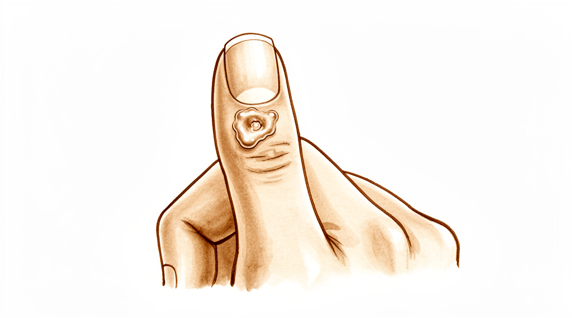
ਤੁਸੀਂ ਉਂਗਲੀ ਦੇ ਉੱਪਰਲੇ ਹਿੱਸੇ 'ਤੇ, ਆਮ ਤੌਰ 'ਤੇ ਆਖਰੀ ਜੋੜ ਦੇ ਨੇੜੇ, ਇੱਕ ਛੋਟਾ, ਸਖ਼ਤ ਗੁੱਛਾ ਨੋਟ ਕਰ ਸਕਦੇ ਹੋ। ਇਹ ਗੁੱਛਾ ਇੱਕ ਮਿਊਕਸ ਸਿਸਟ ਹੈ। ਇਹ ਆਮ ਤੌਰ 'ਤੇ ਚਮੜੀ ਦੇ ਹੇਠਾਂ ਇੱਕ ਛੋਟੀ ਮਟਰ ਵਾਂਗ ਮਹਿਸੂਸ ਹੁੰਦਾ ਹੈ। ਜੇਕਰ ਉਸ ਥਾਂ 'ਤੇ ਤੁਹਾਡੀ ਚਮੜੀ ਪਤਲੀ ਹੈ, ਤਾਂ ਤੁਸੀਂ ਇਸਨੂੰ ਸਪਸ਼ਟ ਤੌਰ 'ਤੇ ਦੇਖ ਸਕਦੇ ਹੋ। ਸਿਸਟ ਕਾਰਨ ਉਸ ਉੱਪਰਲੀ ਚਮੜੀ ਚਮਕਦਾਰ ਜਾਂ ਖਿੱਚੀ ਹੋਈ ਦਿਖਾਈ ਦੇ ਸਕਦੀ ਹੈ। ਕੁਝ ਮਾਮਲਿਆਂ ਵਿੱਚ, ਇਹ ਉਂਗਲੀ ਦੇ ਨੱਖ਼ਸ਼ (fingernail) ਦੇ ਲਹਿਰਦਾਰ ਜਾਂ ਖੱਚਰਦਾਰ ਪੈਟਰਨ ਵਿੱਚ ਵਧਣ ਦਾ ਕਾਰਨ ਬਣ ਸਕਦਾ ਹੈ।
ਸਿਸਟ ਦੇ ਆਲੇ-ਦੁਆਲੇ ਦਾ ਖੇਤਰ ਸੰਵੇਦਨਸ਼ੀਲ ਜਾਂ ਦਰਦਨਾਕ ਮਹਿਸੂਸ ਹੋ ਸਕਦਾ ਹੈ। ਜਦੋਂ ਤੁਸੀਂ ਇਸਨੂੰ ਦਬਾਉਂਦੇ ਹੋ ਜਾਂ ਉਂਗਲੀ ਨੂੰ ਮੋੜਦੇ ਹੋ, ਤਾਂ ਤੁਸੀਂ ਦਰਦ ਮਹਿਸੂਸ ਕਰ ਸਕਦੇ ਹੋ। ਰੋਜ਼ਾਨਾ ਦੇ ਕੰਮ ਮੁਸ਼ਕਲ ਹੋ ਸਕਦੇ ਹਨ। ਵਸਤੂਆਂ ਨੂੰ ਛੂਹਣਾ, ਟਾਈਪਿੰਗ ਕਰਨਾ, ਜਾਂ ਔਜ਼ਾਰਾਂ ਨੂੰ ਫੜਨਾ ਦਰਦਨਾਕ ਹੋ ਸਕਦਾ ਹੈ। ਤੁਸੀਂ ਬਟਨ ਬੰਦ ਕਰਨ ਜਾਂ ਜੈਕਟ ਦਾ ਜ਼ਿਪਰ ਬੰਦ ਕਰਨ ਵਿੱਚ ਮੁਸ਼ਕਲ ਮਹਿਸੂਸ ਕਰ ਸਕਦੇ ਹੋ। ਕੁਝ ਲੋਕਾਂ ਨੇ ਰਿਪੋਰਟ ਕੀਤੀ ਹੈ ਕਿ ਦਰਦ ਰਾਤ ਨੂੰ ਜਾਂ ਲੰਬੇ ਸਮੇਂ ਤੱਕ ਹੱਥਾਂ ਦੀ ਵਰਤੋਂ ਕਰਨ ਤੋਂ ਬਾਅਦ ਵਧੇਰੇ ਹੁੰਦਾ ਹੈ। ਜੇਕਰ ਤੁਸੀਂ ਉਸ ਹੱਥ 'ਤੇ ਸੌਂਦੇ ਹੋ, ਤਾਂ ਅਸੁਵਿਧਾ ਨੀਂਦ ਵਿੱਚ ਰੁਕਾਵਟ ਪਾ ਸਕਦੀ ਹੈ।
ਤੁਹਾਡਾ ਸਰਜਨ ਸਿਸਟ ਦੀ ਜਾਂਚ ਕਰੇਗਾ ਅਤੇ ਦੇਖੇਗਾ ਕਿ ਇਹ ਤੁਹਾਡੀ ਉਂਗਲੀ ਦੀ ਗਤੀ ਨੂੰ ਕਿਵੇਂ ਪ੍ਰਭਾਵਿਤ ਕਰਦਾ ਹੈ। ਦਰਦ ਅਕਸਰ ਅੰਦਰੂਨੀ ਜੋੜ ਦੇ ਬਦਲਾਵਾਂ ਕਾਰਨ ਹੁੰਦਾ ਹੈ, ਨਾ ਕਿ ਸਿਰਫ਼ ਗੁੱਛੇ ਕਾਰਨ। ਜੇਕਰ ਹੱਡੀ ਦਾ ਸਪੂਰ (osteophyte) ਬਾਕੀ ਰਹਿੰਦਾ ਹੈ, ਤਾਂ ਸਿਰਫ਼ ਸਿਸਟ ਨੂੰ ਹਟਾਉਣ ਨਾਲ ਦਰਦ ਰੁਕ ਸਕਦਾ ਹੈ। ਤੁਹਾਡਾ ਸਰਜਨ ਜੋੜ ਅਤੇ ਚਮੜੀ 'ਤੇ ਦਬਾਅ ਘਟਾਉਣ ਲਈ ਹੱਡੀ ਦੇ ਸਪੂਰ ਨੂੰ ਹਟਾਉਣ ਬਾਰੇ ਚਰਚਾ ਕਰ ਸਕਦਾ ਹੈ। ਇਹ ਪਹੁੰਚ ਚਮੜੀ ਦੇ ਬਿਹਤਰ ਭਰਪਾਈ ਵਿੱਚ ਮਦਦ ਕਰ ਸਕਦੀ ਹੈ ਅਤੇ ਸਿਸਟ ਦੇ ਦੁਬਾਰਾ ਆਉਣ ਦੇ ਖਤਰੇ ਨੂੰ ਘਟਾ ਸਕਦੀ ਹੈ।
ਕਈ ਮਾਮਲਿਆਂ ਵਿੱਚ, ਹੱਡੀ ਦੇ ਸਪੂਰ ਦੇ ਇਲਾਜ ਨਾਲ ਸਿਸਟ ਪੂਰੀ ਤਰ੍ਹਾਂ ਠੀਕ ਹੋ ਜਾਂਦਾ ਹੈ। ਤੁਹਾਨੂੰ ਜਟਿਲ ਸਰਜਰੀ ਦੀ ਲੋੜ ਨਹੀਂ ਹੋ ਸਕਦੀ। ਸਿਸਟ ਅਤੇ ਹੱਡੀ ਦੇ ਸਪੂਰ ਦੋਵਾਂ ਨੂੰ ਸਰਲਤਾ ਨਾਲ ਹਟਾਉਣ ਦੇ ਨਾਲ ਦੁਬਾਰਾ ਆਉਣ ਦੀ ਦਰ ਬਹੁਤ ਹੀ ਦੁਰਲੱਭ ਹੈ। ਇੱਥੋਂ ਤੱਕ ਕਿ ਘੱਟ ਹਮਲਾਵਰ ਵਿਕਲਪ, ਜਿਵੇਂ ਕਿ ਸਿਰਫ਼ ਹੱਡੀ ਦੇ ਸਪੂਰ ਨੂੰ ਹਟਾਉਣਾ, ਜ਼ਿਆਦਾਤਰ ਮਾਮਲਿਆਂ ਵਿੱਚ ਪੂਰੀ ਤਰ੍ਹਾਂ ਠੀਕ ਕਰ ਸਕਦਾ ਹੈ। ਜੇਕਰ ਚਮੜੀ ਦੇ ਫਲੈਪ ਦੀ ਲੋੜ ਹੁੰਦੀ ਹੈ, ਤਾਂ ਸਕਾਰ ਬਾਰੇ ਮਰੀਜ਼ ਦੀ ਸੰਤੁਸ਼ਟੀ ਉੱਚੀ ਹੁੰਦੀ ਹੈ। ਤੁਸੀਂ ਦਿਖਾਵੇ ਤੋਂ ਸੰਤੁਸ਼ਟ ਹੋਣ ਦੀ ਸੰਭਾਵਨਾ ਹੈ ਅਤੇ ਇਸ ਪ੍ਰਕਿਰਿਆ ਨੂੰ ਦੁਬਾਰਾ ਕਰਵਾਉਣ ਲਈ ਤਿਆਰ ਹੋਵੋਗੇ।
ਤੁਹਾਡਾ ਸਰਜਨ ਯੋਜਨਾ ਨੂੰ ਤੁਹਾਡੀਆਂ ਖਾਸ ਲੋੜਾਂ ਅਨੁਸਾਰ ਢਾਲੇਗਾ। ਉਦੇਸ਼ ਤੁਹਾਡੇ ਦਰਦ ਨੂੰ ਘਟਾਉਣਾ ਅਤੇ ਕਾਰਜਸ਼ੀਲਤਾ ਨੂੰ ਬਹਾਲ ਕਰਨਾ ਹੈ। ਜ਼ਿਆਦਾਤਰ ਮਰੀਜ਼ਾਂ ਨੂੰ ਇਲਾਜ ਤੋਂ ਬਾਅਦ ਮਹਿਸੂਸ ਹੁੰਦਾ ਹੈ ਕਿ ਉਹਨਾਂ ਦੀ ਉਂਗਲੀ ਵਧੇਰੇ ਆਰਾਮਦਾਇਕ ਅਤੇ ਬਿਹਤਰ ਦਿਖਾਈ ਦਿੰਦੀ ਹੈ। ਮੁੱਖ ਸਮੱਸਿਆ ਨੂੰ ਹੱਲ ਕਰਨ ਤੋਂ ਬਾਅਦ ਚਮੜੀ ਵਿੱਚ ਭਰਪਾਈ ਦੀ ਵਧੀਆ ਸਮਰੱਥਾ ਹੁੰਦੀ ਹੈ। ਤੁਸੀਂ ਸਾਵਧਾਨ ਘਾਅ ਦੇਖਭਾਲ ਨਾਲ ਇੱਕ ਸਰਲ ਭਰਪਾਈ ਦੀ ਉਮੀਦ ਕਰ ਸਕਦੇ ਹੋ।
ਅਸਲ ਵਿੱਚ ਕੀ ਹੋ ਰਿਹਾ ਹੈ
ਮਿਊਕਸ ਸਿਸਟ (Mucous cyst) ਉਂਗਲੀ 'ਤੇ ਬਣਨ ਵਾਲਾ ਇੱਕ ਛੋਟਾ, ਤਰਲ ਪਦਾਰਥ ਨਾਲ ਭਰਿਆ ਥੈਲਾ ਹੁੰਦਾ ਹੈ। ਇਹ ਆਮ ਤੌਰ 'ਤੇ ਉਂਗਲੀ ਦੇ ਸਿਰੇ ਦੇ ਨੇੜੇ, ਨਖ ਦੇ ਬੈੱਡ ਦੇ ਨਾਲ ਪ੍ਰਗਟ ਹੁੰਦਾ ਹੈ। ਸਿਸਟ ਉਸ ਜੋੜ ਕੈਪਸੂਲ ਦੇ ਉੱਪਰ ਹੁੰਦਾ ਹੈ, ਜੋ ਕਿ ਇੱਕ ਮਜ਼ਬੂਤ, ਫਾਈਬਰਸ ਢਾਂਚਾ ਹੈ ਜੋ ਤੁਹਾਡੇ ਜੋੜ ਨੂੰ ਘੇਰ ਕੇ ਸਥਿਰ ਰੱਖਦਾ ਹੈ।
ਇਸਦਾ ਮੂਲ ਕਾਰਨ ਜੋੜ ਵਿੱਚ ਘਸਾਅ-ਸੰਬੰਧੀ ਆਰਥਰਾਈਟਸ (wear-and-tear arthritis) ਹੈ। ਜਦੋਂ ਕਾਰਟੀਲੇਜ—ਹੱਡੀਆਂ ਦੇ ਸਿਰਿਆਂ 'ਤੇ ਮੌਜੂਦ ਚਿਕਨਾ ਪਰਤ—ਟੁੱਟਦੀ ਹੈ, ਤਾਂ ਤੁਹਾਡਾ ਸਰੀਰ ਨੁਕਸਾਨ ਨੂੰ ਠੀਕ ਕਰਨ ਦੀ ਕੋਸ਼ਿਸ਼ ਕਰਦਾ ਹੈ। ਇਸ ਪ੍ਰਕਿਰਿਆ ਦੌਰਾਨ ਅਕਸਰ ਹੱਡੀ ਦੇ ਸਪੂਰ (bone spurs), ਜਿਨ੍ਹਾਂ ਨੂੰ ਓਸਟੀਓਫਾਈਟਸ (osteophytes) ਕਿਹਾ ਜਾਂਦਾ ਹੈ, ਬਣਦੇ ਹਨ। ਇਹ ਜੋੜ ਦੀ ਸਤਹ ਤੋਂ ਬਾਹਰ ਵੱਢੇ ਜਾਣ ਵਾਲੇ ਹੱਡੀ ਦੇ ਛੋਟੇ, ਖੁਰਦਰੇ ਟੁਕੜੇ ਹੁੰਦੇ ਹਨ।
ਜੋੜ ਕੈਪਸੂਲ ਨੂੰ ਇੱਕ ਗੈਸਕੈਟ ਜਾਂ ਸੀਲ (gasket or seal) ਵਾਂਗ ਸੋਚੋ। ਜਦੋਂ ਹੱਡੀ ਦਾ ਸਪੂਰ ਇਸ ਸੀਲ ਨਾਲ ਰਗੜਦਾ ਹੈ, ਤਾਂ ਇਹ ਟਿਸ਼ੂ ਨੂੰ ਉਤੇਜਿਤ ਕਰਦਾ ਹੈ। ਇਸ ਉਤੇਜਨਾ ਕਾਰਨ ਜੋੜ ਦੀ ਪਰਤ ਵਿੱਚੋਂ ਸਾਈਨੋਵੀਅਲ ਤਰਲ (synovial fluid) ਲੀਕ ਹੋਣ ਲੱਗ ਪੈਂਦਾ ਹੈ, ਜੋ ਕਿ ਇੱਕ ਕੁਦਰਤੀ ਲੁਬਰੀਕੈਂਟ ਹੈ ਜੋ ਤੁਹਾਡੇ ਜੋੜ ਨੂੰ ਚਲਾਉਣ ਵਿੱਚ ਮੁਲਾਇਮ ਬਣਾਈ ਰੱਖਦਾ ਹੈ। ਤਰਲ ਪਦਾਰਥ ਕੈਪਸੂਲ ਦੇ ਕਮਜ਼ੋਰ ਹਿੱਸੇ ਵਿੱਚੋਂ ਲੰਘ ਕਾਰਨ ਚਮੜੀ 'ਤੇ ਦਿਖਣ ਵਾਲਾ ਉਭਾਰ ਬਣ ਜਾਂਦਾ ਹੈ।
ਕਿਉਂਕਿ ਸਿਸਟ ਜੋੜ ਨਾਲ ਜੁੜਿਆ ਹੁੰਦਾ ਹੈ, ਇਹ ਉਸੇ ਲੁਬਰੀਕੇਟਿੰਗ ਤਰਲ ਨਾਲ ਭਰਿਆ ਹੁੰਦਾ ਹੈ। ਤਰਲ ਦੇ ਦਬਾਅ ਕਾਰਨ ਸਿਸਟ ਦੇ ਉੱਪਰਲੀ ਚਮੜੀ ਪਤਲੀ ਅਤੇ ਕਮਜ਼ੋਰ ਹੋ ਸਕਦੀ ਹੈ। ਕੁਝ ਮਾਮਲਿਆਂ ਵਿੱਚ, ਸਿਸਟ ਨੇੜਲੀਆਂ ਨਸਾਂ (nerves) 'ਤੇ ਦਬਾਅ ਪਾ ਸਕਦਾ ਹੈ ਜਾਂ ਨਖ ਮੈਟ੍ਰਿਕਸ ਨੂੰ ਪ੍ਰਭਾਵਿਤ ਕਰ ਸਕਦਾ ਹੈ, ਜੋ ਕਿ ਤੁਹਾਡੀ ਕਟੀਕਲ (cuticle) ਦੇ ਹੇਠਾਂ ਉਹ ਟਿਸ਼ੂ ਹੈ ਜੋ ਤੁਹਾਡੀ ਨਖ ਨੂੰ ਵਧਾਉਂਦਾ ਹੈ। ਇਹੀ ਦਬਾਅ ਤੁਹਾਡੀ ਨਖ ਵਿੱਚ ਦਿਖਣ ਵਾਲੀਆਂ ਖਾਚਿਆਂ ਜਾਂ ਰੇਖਾਵਾਂ ਦਾ ਕਾਰਨ ਬਣਦਾ ਹੈ।
ਸਿਰਫ਼ ਸਿਸਟ ਨੂੰ ਹਟਾਉਣ ਨਾਲ ਅਕਸਰ ਇਹ ਦੁਬਾਰਾ ਪੈਦਾ ਹੋ ਜਾਂਦਾ ਹੈ ਕਿਉਂਕਿ ਹੱਡੀ ਦਾ ਸਪੂਰ ਉੱਥੇ ਹੀ ਰਹਿੰਦਾ ਹੈ। ਸਪੂਰ ਜੋੜ ਦੀ ਪਰਤ ਨੂੰ ਲਗਾਤਾਰ ਉਤੇਜਿਤ ਕਰਦਾ ਰਹਿੰਦਾ ਹੈ, ਜਿਸ ਕਾਰਨ ਹੋਰ ਤਰਲ ਲੀਕ ਹੁੰਦਾ ਹੈ। ਇਸ ਚੱਕਰ ਨੂੰ ਰੋਕਣ ਲਈ, ਅਧਿਨਿਹਾਰੀ ਹੱਡੀ ਦੇ ਸਪੂਰ ਨੂੰ ਹੱਲ ਕਰਨਾ ਜ਼ਰੂਰੀ ਹੈ। ਜਦੋਂ ਸਪੂਰ ਨੂੰ ਹਟਾ ਦਿੱਤਾ ਜਾਂਦਾ ਹੈ, ਤਾਂ ਉਤੇਜਨਾ ਬੰਦ ਹੋ ਜਾਂਦੀ ਹੈ ਅਤੇ ਜੋੜ ਦੀ ਪਰਤ ਠੀਕ ਹੋ ਸਕਦੀ ਹੈ। ਇਹੀ ਕਾਰਨ ਹੈ ਕਿ ਸਿਸਟ ਨੂੰ ਹਟਾਉਣ ਦੇ ਨਾਲ-ਨਾਲ ਹੱਡੀ ਦੇ ਇਲਾਜ ਨੂੰ ਵੀ ਇੰਨਾ hi ਮਹੱਤਵਪੂਰਨ ਮੰਨਿਆ ਜਾਂਦਾ ਹੈ।
ਇਸ ਬਾਰੇ ਅਸੀਂ ਕੀ ਕਰ ਸਕਦੇ ਹਾਂ
ਤੁਹਾਡਾ ਸਫ਼ਰ ਆਮ ਤੌਰ 'ਤੇ ਸਰਲ ਆਤਮ-ਦੇਖਭਾਲ ਅਤੇ ਪੇਸ਼ੇਵਰ ਮਾਰਗਦਰਸ਼ਨ ਨਾਲ ਸ਼ੁਰੂ ਹੁੰਦਾ ਹੈ। ਤੁਸੀਂ ਉਂਗਲ ਨੂੰ ਆਰਾਮ ਦੇਣ ਅਤੇ ਉਸ ਸਿਸਟ 'ਤੇ ਦਬਾਅ ਪਾਉਣ ਵਾਲੀਆਂ ਗਤੀਵਿਧੀਆਂ ਤੋਂ ਬਚਣ ਦੀ ਕੋਸ਼ਿਸ਼ ਕਰ ਸਕਦੇ ਹੋ। ਭੌਤਿਕ ਚਿਕਿਤਸਾ (Physiotherapy) ਤੁਹਾਨੂੰ ਜੋੜ ਵਿੱਚ ਚਾਲ ਬਣਾਈ ਰੱਖਣ ਅਤੇ ਆਲੇ-ਦੁਆਲੇ ਦੇ ਮਾਸਪੇਸ਼ੀਆਂ ਨੂੰ ਮਜ਼ਬੂਤ ਰੱਖਣ ਵਿੱਚ ਮਦਦ ਕਰਦੀ ਹੈ। ਇਸ ਪਹੁੰਚ ਦਾ ਉਦੇਸ਼ ਬਿਨਾਂ ਆਕ੍ਰਮਕ ਕਦਮਾਂ ਦੇ ਜ਼ਖ਼ਮੀ ਹੋਣ ਨੂੰ ਘਟਾਉਣਾ ਅਤੇ ਕਾਰਜਕੁਸ਼ਲਤਾ ਨੂੰ ਬਿਹਤਰ ਬਣਾਉਣਾ ਹੈ। ਤੁਹਾਨੂੰ ਇਸ ਸੰਭਾਲ ਭਰਪੂਰ (conservative) ਦੇਖਭਾਲ ਨੂੰ ਇੱਕ ਨਿਆਂਯੁਕਤ ਪਰਖ ਲਈ ਦੇਣਾ ਚਾਹੀਦਾ ਹੈ ਤਾਂ ਜੋ ਦੇਖਿਆ ਜਾ ਸਕੇ ਕਿ ਕੀ ਲੱਛਣ ਠੀਕ ਹੋ ਜਾਂਦੇ ਹਨ। ਬਹੁਤ ਸਾਰੇ ਮਰੀਜ਼ਾਂ ਨੂੰ ਲੱਗਦਾ ਹੈ ਕਿ ਰੋਜ਼ਾਨਾ ਆਦਤਾਂ ਦਾ ਪ੍ਰਬੰਧਨ ਕਰਨਾ ਅਤੇ ਇੱਕ ਹਲਕੀ ਵਰਕਆਊਟ ਯੋਜਨਾ ਦੀ ਪਾਲਣਾ ਕਰਨਾ ਅੱਗੇ ਵਾਲੇ ਦਖਲ-ਅੰਦਾਜ਼ੀ ਤੋਂ ਬਚਣ ਲਈ ਕਾਫ਼ੀ ਰਾਹਤ ਪ੍ਰਦਾਨ ਕਰਦਾ ਹੈ।
ਜੇਕਰ ਆਤਮ-ਦੇਖਭਾਲ ਕਾਫ਼ੀ ਨਹੀਂ ਹੈ, ਤਾਂ ਤੁਹਾਡਾ ਸਰਜਨ ਦਰਦ ਅਤੇ ਸੋਜ ਨੂੰ ਕੰਟਰੋਲ ਕਰਨ ਲਈ ਦਵਾਈਆਂ ਦੀ ਸਿਫ਼ਾਰਿਸ਼ ਕਰ ਸਕਦਾ ਹੈ। ਕਾਰਟੀਕੋਸਟੀਰਾਇਡ ਇੰਜੈਕਸ਼ਨ ਇਹਨਾਂ ਸਿਸਟਾਂ ਲਈ ਇੱਕ ਆਮ ਵਿਕਲਪ ਹਨ। ਇੱਕ ਵੋਲਰ ਕਾਰਟੀਕੋਸਟੀਰਾਇਡ ਇੰਜੈਕਸ਼ਨ ਜੋੜ ਵਿੱਚ ਸੂਈ ਨੂੰ ਆਸਾਨੀ ਅਤੇ ਸਥਿਰਤਾ ਨਾਲ ਰੱਖਣ ਦੀ ਆਗਿਆ ਦਿੰਦਾ ਹੈ। ਹੋਰ ਤਕਨੀਕਾਂ ਦੇ ਮੁਕਾਬਲੇ, ਇਹ ਵਿਧੀ ਸੰਭਾਵੀ ਨਰਮ ਟਿਸ਼ੂ ਦੇ ਨੁਕਸਾਨ ਅਤੇ ਸੰਕਰਮਣ ਦੇ ਜੋਖਮ ਨੂੰ ਘਟਾਉਂਦੀ ਹੈ। ਇੰਜੈਕਸ਼ਨ ਸਿਸਟ ਦੇ ਵਧਣ ਦਾ ਕਾਰਨ ਬਣਨ ਵਾਲੀ ਸੋਜ ਨੂੰ ਸ਼ਾਂਤ ਕਰਨ ਵਿੱਚ ਮਦਦ ਕਰਦਾ ਹੈ। ਜਦੋਂ ਕਿ ਸਬੂਤ ਇਸ ਪਹੁੰਚ ਦੀ ਸੁਰੱਖਿਆ ਅਤੇ ਆਸਾਨੀ ਨੂੰ ਉਜਾਗਰ ਕਰਦੇ ਹਨ, ਇਹ ਸਮਝਣਾ ਜ਼ਰੂਰੀ ਹੈ ਕਿ ਇੰਜੈਕਸ਼ਨ ਆਮ ਤੌਰ 'ਤੇ ਲੱਛਣਾਂ ਦਾ ਪ੍ਰਬੰਧਨ ਕਰਦੇ ਹਨ ਨਾ ਕਿ ਸਿਸਟ ਨੂੰ ਸਦਾ ਲਈ ਹਟਾਉਂਦੇ ਹਨ। ਇਸਦਾ ਅਸਰ ਕੁਝ ਸਮੇਂ ਲਈ ਰਹਿੰਦਾ ਹੈ, ਪਰ ਅਧੀਨ ਵਾਲਾ ਘਸਾਈ-ਪਹਿਣੀ (wear-and-tear) ਆਰਥਰਾਈਟਿਸ ਬਰਕਰਾਰ ਰਹਿੰਦਾ ਹੈ। ਤੁਸੀਂ ਅਤੇ ਤੁਹਾਡਾ ਸਰਜਨ ਇਹ ਤੈਅ ਕਰਨਗੇ ਕਿ ਕੀ ਇਹ ਅਸਥਾਈ ਰਾਹਤ ਕਾਫ਼ੀ ਹੈ ਜਾਂ ਕੀ ਅੱਗੇ ਵਾਲੇ ਕਦਮਾਂ ਦੀ ਲੋੜ ਹੈ।
ਜਦੋਂ ਸੰਭਾਲ ਭਰਪੂਰ ਦੇਖਭਾਲ ਆਪਣੀ ਸੀਮਾ ਤੱਕ ਪਹੁੰਚ ਜਾਂਦੀ ਹੈ ਅਤੇ ਸਿਸਟ ਲਗਾਤਾਰ ਦਰਦ, ਵਿਗਾੜ, ਜਾਂ ਕਾਰਜਕੁਸ਼ਲਤਾ ਦੀਆਂ ਸਮੱਸਿਆਵਾਂ ਦਾ ਕਾਰਨ ਬਣਦਾ ਹੈ, ਤਾਂ ਸਰਜਰੀ 'ਤੇ ਵਿਚਾਰ ਕੀਤਾ ਜਾਂਦਾ ਹੈ। ਤੁਹਾਡਾ ਸਰਜਨ ਤੁਹਾਡੇ ਖਾਸ ਮਾਮਲੇ ਲਈ ਸਭ ਤੋਂ ਵਧੀਆ ਸਰਜਰੀ ਵਿਕਲਪ ਬਾਰੇ ਚਰਚਾ ਕਰੇਗਾ। ਇਸਦਾ ਉਦੇਸ਼ ਸਿਸਟ ਨੂੰ ਹਟਾਉਣਾ ਅਤੇ ਮੂਲ ਕਾਰਨ ਨੂੰ ਹੱਲ ਕਰਨਾ ਹੈ, ਜੋ ਅਕਸਰ ਆਰਥਰਾਈਟਿਸ ਤੋਂ ਹੱਡੀ ਦੇ ਸਪੂਰ (osteophytes) ਹੁੰਦੇ ਹਨ। ਇਹਨਾਂ ਹੱਡੀ ਦੇ ਸਪੂਰ ਨੂੰ ਹਟਾਉਣਾ ਜ਼ਰੂਰੀ ਹੈ ਕਿਉਂਕਿ ਇਹ ਸਿਸਟ ਦੇ ਵਾਪਸ ਆਉਣ ਦੇ ਚਾਂਸ ਨੂੰ ਘਟਾਉਂਦਾ ਹੈ। ਕੁਝ ਤਕਨੀਕਾਂ ਵਿੱਚ ਸਹੀ ਭਰਨ ਲਈ ਸਿਸਟ ਦੇ ਨਾਲ-ਨਾਲ ਚਮੜੀ ਦੇ ਇੱਕ ਛੋਟੇ ਫਲੈਪ ਨੂੰ ਹਟਾਉਣਾ ਸ਼ਾਮਲ ਹੁੰਦਾ ਹੈ, ਜਦੋਂ ਕਿ ਹੋਰੇ ਮੁੱਖ ਤੌਰ 'ਤੇ ਹੱਡੀ 'ਤੇ ਧਿਆਨ ਕੇਂਦਰਿਤ ਕਰਦੀਆਂ ਹਨ। ਤੁਹਾਡਾ ਸਰਜਨ ਉਸ ਵਿਧੀ ਨੂੰ ਚੁਣੇਗਾ ਜੋ ਤੁਹਾਡੀ ਹੱਥ ਲਈ ਵਾਪਸੀ ਦਰਾਂ ਅਤੇ ਚੰਗੀ ਕੋਸਮੈਟਿਕ ਨਤੀਜਿਆਂ ਵਿੱਚ ਸਭ ਤੋਂ ਵਧੀਆ ਸੰਤੁਲਨ ਪੇਸ਼ ਕਰਦੀ ਹੈ।
ਤੁਹਾਡੀ ਉਮੀਦ ਕੀ ਹੋ ਸਕਦੀ ਹੈ
ਮਿਊਕਸ ਸਿਸਟ (Mucous cysts) ਛੋਟੇ, ਤਰਲ ਪਦਾਰਥ ਨਾਲ ਭਰੇ ਹੋਏ ਗੁੱਛੇ ਹੁੰਦੇ ਹਨ ਜੋ ਅਕਸਰ ਤੁਹਾਡੀ ਉਂਗਲ ਜਾਂ ਅੰਗੂਠੇ ਦੇ ਅੰਤਿਮ ਜੋੜ ਦੇ ਨੇੜੇ ਦਿਖਾਈ ਦਿੰਦੇ ਹਨ। ਇਹ ਉਸ ਜੋੜ ਵਿੱਚ ਘਸਾਈ-ਪਹਿਰਾਵੇ ਵਾਲੇ ਗਠੀਏ (wear-and-tear arthritis) ਨਾਲ ਘੱਨੀ ਤੌਰ 'ਤੇ ਜੁੜੇ ਹੁੰਦੇ ਹਨ। ਕਿਉਂਕਿ ਇਹ ਅਧੋਲੇ ਜੋੜ ਦੇ ਬਦਲਾਅ ਤੋਂ ਪੈਦਾ ਹੁੰਦੇ ਹਨ, ਜੇਕਰ ਮੂਲ ਕਾਰਨ ਨੂੰ ਦੂਰ ਨਹੀਂ ਕੀਤਾ ਜਾਂਦਾ, ਤਾਂ ਸਿਸਟ ਖੁਦ ਜਾਰੀ ਰਹਿ ਸਕਦਾ ਹੈ ਜਾਂ ਵਾਪਸ ਆ ਸਕਦਾ ਹੈ। ਹਾਲਾਂਕਿ, ਸਹੀ ਇਲਾਜ ਨਾਲ, ਭਵਿੱਖਬਾਣੀ ਆਮ ਤੌਰ 'ਤੇ ਬਹੁਤ ਸਕਾਰਾਤਮਕ ਹੁੰਦੀ ਹੈ।
ਤੁਹਾਡਾ ਸਰਜਨ ਸਿਸਟ ਨੂੰ ਹਟਾਉਣ ਅਤੇ ਇਸਨੂੰ ਪੈਦਾ ਕਰਨ ਵਾਲੇ ਹੱਡੀ ਦੇ ਬਾਹਰ ਨਿਕਲੇ ਹਿੱਸਿਆਂ (osteophytes) ਨੂੰ ਹਟਾਉਣ ਦੀ ਸਿਫਾਰਸ਼ ਕਰ ਸਕਦਾ ਹੈ। ਇਸ ਤਰੀਕੇ ਨਾਲ ਮਿਊਕਸ ਸਿਸਟਾਂ ਨੂੰ ਬਹੁਤ ਹੀ ਘੱਟ ਦੁਬਾਰਾ ਆਉਣ ਦੀ ਦਰ ਨਾਲ ਪੂਰੀ ਤਰ੍ਹਾਂ ਖਤਮ ਕੀਤਾ ਜਾ ਸਕਦਾ ਹੈ। ਜੇਕਰ ਤੁਹਾਡਾ ਸਰਜਨ ਸਥਾਨਕ ਅਗਰਾਹਤ ਚਮੜੀ ਦੇ ਫਲੈਪ (local advancement skin flap) ਵਾਲੀ ਤਕਨੀਕ ਦੀ ਚੋਣ ਕਰਦਾ ਹੈ, ਤਾਂ ਦੁਬਾਰਾ ਆਉਣ ਦੀ ਦਰ 1.4% ਹੁੰਦੀ ਹੈ। ਕੁਝ ਮਾਮਲਿਆਂ ਵਿੱਚ, ਤੁਹਾਡਾ ਸਰਜਨ ਸਿਰਫ਼ ਹੱਡੀ ਦਾ ਬਾਹਰ ਨਿਕਲਿਆ ਹਿੱਸਾ ਹਟਾ ਸਕਦਾ ਹੈ ਬਿਨਾਂ ਸਿਸਟ ਨੂੰ ਕੱਢੇ। ਇਹ ਘੱਟ ਆਕ੍ਰਮਕ ਤਰੀਕਾ ਜ਼ਿਆਦਾਤਰ ਮਾਮਲਿਆਂ ਵਿੱਚ ਪੂਰੀ ਤਰ੍ਹਾਂ ਠੀਕ ਹੋਣ ਦਾ ਨਤੀਜਾ ਦਿੰਦਾ ਹੈ। ਨਰਮ ਟਿਸ਼ੂਆਂ ਲਈ ਵਿਸ਼ੇਸ਼ ਯੋਜਨਾ ਭਾਵੇਂ ਕੁਝ ਵੀ ਹੋਵੇ, ਤੁਹਾਡਾ ਸਰਜਨ ਸਿਸਟ ਦੇ ਵਾਪਸ ਆਉਣ ਤੋਂ ਰੋਕਣ ਲਈ ਹੱਡੀ ਦਾ ਬਾਹਰ ਨਿਕਲਿਆ ਹਿੱਸਾ ਹਟਾਵੇਗਾ।
ਤੁਸੀਂ ਨਤੀਜਿਆਂ ਨਾਲ ਉੱਚ ਸੰਤੁਸ਼ਟੀ ਦੀ ਉਮੀਦ ਕਰ ਸਕਦੇ ਹੋ, ਖਾਸ ਕਰਕੇ ਸਕਾਰ (scar) ਦੀ ਦਿੱਖ ਦੇ ਸਬੰਧ ਵਿੱਚ। ਬਹੁਤ ਸਾਰੇ ਮਰੀਜ਼ਾਂ ਨੇ ਰਿਪੋਰਟ ਕੀਤਾ ਹੈ ਕਿ ਉਹ ਇਸ ਪ੍ਰਕਿਰਿਆ ਨੂੰ ਦੁਬਾਰਾ ਕਰਵਾਉਣਗੇ। ਵਰਤੀਆਂ ਗਈਆਂ ਸਰਜੀਕਲ ਤਕਨੀਕਾਂ ਨੂੰ ਸਰਲ ਅਤੇ ਭਰੋਸੇਯੋਗ ਬਣਾਉਣ ਲਈ ਡਿਜ਼ਾਈਨ ਕੀਤਾ ਗਿਆ ਹੈ। ਉਦਾਹਰਣ ਵਜੋਂ, ਕੁਝ ਤਰੀਕੇ ਪਤਲੀ ਹੋਈ ਚਮੜੀ ਨੂੰ ਹਟਾਉਣ ਦੀ ਆਗਿਆ ਦਿੰਦੇ ਹਨ ਬਿਨਾਂ ਤੁਹਾਡੇ ਨੇਲ ਮੈਟ੍ਰਿਕਸ (nail matrix) ਨੂੰ ਕੋਈ ਜੋਖਮ ਪਹੁੰਚਾਏ। ਦੂਜੇ ਚਮੜੀ ਦੇ ਗ੍ਰਾਫਟਸ ਦੀ ਵਰਤੋਂ ਕਰਦੇ ਹਨ ਜੋ ਸੰਤੁਸ਼ਟ ਕਾਰਜਸ਼ੀਲ ਨਤੀਜੇ ਅਤੇ ਸਵੀਕਾਰਯੋਗ ਦੁਬਾਰਾ ਆਉਣ ਦੀਆਂ ਦਰਾਂ ਪ੍ਰਦਾਨ ਕਰਦੇ ਹਨ। ਜਟਿਲ ਮਾਮਲੇ, ਜਿਵੇਂ ਕਿ ਡਿਜੀਟਲ ਨਰਵ (digital nerve) ਦੇ ਅੰਦਰ ਵਧ ਰਹੇ ਸਿਸਟ, ਵੀ ਸਫਲ ਨਤੀਜਿਆਂ ਵੱਲ ਲੈ ਜਾ ਸਕਦੇ ਹਨ।
ਜੇਕਰ ਇਸਦਾ ਇਲਾਜ ਨਹੀਂ ਕੀਤਾ ਜਾਂਦਾ, ਤਾਂ ਸਿਸਟ ਜਾਰੀ ਰਹਿ ਸਕਦਾ ਹੈ ਜਾਂ ਵਧ ਸਕਦਾ ਹੈ, ਜਿਸ ਨਾਲ ਚਮੜੀ ਪਤਲੀ ਹੋ ਸਕਦੀ ਹੈ ਅਤੇ ਸੰਕਰਮਣ ਦਾ ਜੋਖਮ ਵਧ ਸਕਦਾ ਹੈ। ਸਿਸਟ ਅਤੇ ਅਧੋਲੇ ਗਠੀਏ ਦੋਵਾਂ ਨੂੰ ਦੂਰ ਕਰਕੇ, ਤੁਹਾਡਾ ਸਰਜਨ ਇੱਕ ਟਿਕਾਊ ਹੱਲ ਦੀ ਕੋਸ਼ਿਸ਼ ਕਰਦਾ ਹੈ। ਜ਼ਿਆਦਾਤਰ ਮਰੀਜ਼ਾਂ ਨੂੰ ਲੱਗਦਾ ਹੈ ਕਿ ਪ੍ਰਕਿਰਿਆ ਤੋਂ ਬਾਅਦ ਉਹਨਾਂ ਦੀ ਉਂਗਲ ਵਧੀਆ ਦਿਖਾਈ ਦਿੰਦੀ ਹੈ ਅਤੇ ਮਹਿਸੂਸ ਹੁੰਦੀ ਹੈ। ਟੀਚਾ ਸਿਰਫ਼ ਗੁੱਛੇ ਨੂੰ ਹਟਾਉਣਾ ਨਹੀਂ ਹੈ, ਬਲਕਿ ਇਸਨੂੰ ਵਾਪਸ ਆਉਣ ਤੋਂ ਰੋਕਣਾ ਹੈ। ਤੁਸੀਂ ਇੱਕ ਸਰਲ ਰਿਕਵਰੀ ਪ੍ਰਕਿਰਿਆ ਦੀ ਉਮੀਦ ਕਰ ਸਕਦੇ ਹੋ ਜੋ ਸਰਜੀਕਲ ਸਥਾਨ ਦੀ ਰੱਖਿਆ ਕਰਨ 'ਤੇ ਧਿਆਨ ਕੇਂਦਰਿਤ ਕਰਦੀ ਹੈ ਜਦੋਂ ਕਿ ਤੁਹਾਡੇ ਜੋੜ ਦੇ ਠੀਕ ਹੋਣ ਦੀ ਆਗਿਆ ਦਿੰਦੀ ਹੈ।
ਕਦੋਂ ਕਿਸੇ ਡਾਕਟਰ ਨੂੰ ਦਿਖਾਉਣਾ ਹੈ
ਜੇਕਰ ਤੁਹਾਡੀ ਉਂਗਲੀ ਵਿੱਚ ਨਖ਼ ਦੇ ਨੇੜੇ ਕੋਈ ਛੋਟਾ ਫੁੱਲਣਾ ਦਿਖਾਈ ਦਿੰਦਾ ਹੈ, ਤਾਂ ਆਪਣੇ ਆਮ ਪਰਿਵਾਰਕ ਡਾਕਟਰ (GP) ਨੂੰ ਦਿਖਾਓ। ਜੇਕਰ ਸਿਸਟ (cyst) ਕਾਰਨ ਲਗਾਤਾਰ ਦਰਦ ਹੁੰਦਾ ਹੈ ਜੋ ਆਰਾਮ ਨਾਲ ਠੀਕ ਨਹੀਂ ਹੁੰਦਾ, ਤਾਂ ਵਿਸ਼ੇਸ਼ਜ਼ ਦੀ ਸਲਾਹ ਲਓ। ਜੇਕਰ ਜੋੜ ਵਿੱਚ ਕਮਜ਼ੋਰੀ ਜਾਂ ਅਸਥਿਰਤਾ ਮਹਿਸੂਸ ਹੁੰਦੀ ਹੈ, ਤਾਂ ਇਲਾਜ ਲਈ ਜਾਓ। ਬੰਦ ਹੋਣ ਜਾਂ ਫਿਸਲਣ ਦੀ ਅਹਿਸਾਸ ਵੱਲ ਧਿਆਨ ਦਿਓ। ਜੇਕਰ ਲੱਛਣ ਨੀਂਦ ਜਾਂ ਕੰਮ ਵਿੱਚ ਰੁਕਾਵਟ ਪਾਉਂਦੇ ਹਨ, ਤਾਂ ਆਪਣੇ ਸਰਜਨ ਨਾਲ ਸੰਪਰਕ ਕਰੋ। ਇਲਾਜ ਵਾਲੇ ਖੇਤਰ ਵਿੱਚ ਅਚਾਨਕ ਖਰਾਬ ਹੋਣ 'ਤੇ ਵੀ ਜਾਂਚ ਕਰਵਾਓ। ਸਮੇਂ ਸਿਰ ਮੁਲਾਂਕਣ ਜਟਿਲਤਾਵਾਂ ਨੂੰ ਰੋਕਣ ਵਿੱਚ ਮਦਦ ਕਰਦਾ ਹੈ। ਤੁਹਾਡਾ ਸਰਜਨ ਸਿਸਟ ਅਤੇ ਹੱਡੀ ਦੇ ਕਿਸੇ ਵੀ ਬਾਹਰ ਨਿਕਲੇ ਹਿੱਸੇ (bone spurs) ਨੂੰ ਹਟਾਉਣ ਵਰਗੇ ਵਿਕਲਪਾਂ ਬਾਰੇ ਚਰਚਾ ਕਰ ਸਕਦਾ ਹੈ। ਇਸ ਤਰੀਕੇ ਨਾਲ ਅਕਸਰ ਸੰਤੁਸ਼ਟੀ ਦੀ ਦਰ ਉੱਚ ਅਤੇ ਦੁਬਾਰਾ ਹੋਣ ਦੀ ਦਰ ਘੱਟ ਹੁੰਦੀ ਹੈ। ਅਧਾਰ ਕਾਰਨ ਦਾ ਇਲਾਜ ਕਰਨ ਨਾਲ ਚਮੜੀ ਕੁਦਰਤੀ ਤੌਰ 'ਤੇ ਠੀਕ ਹੋ ਜਾਂਦੀ ਹੈ।
Evidence & references
Overview
- Scientific data regarding mucous cysts consist almost entirely of retrospective studies [1].
- Much of the management or recommendations for mucous cysts is based on expert opinion [1].
- Total dorsal capsulectomy alone is a simple treatment for mucous cysts that does not lead to any recurrence [2].
- Excision of the cyst combined with complete removal of the marginal osteophyte eradicates mucous cysts with extremely rare recurrence [3].
- Osteophyte excision without cyst excision may be a good treatment choice for mucous cysts of the finger, providing a less invasive method with complete resolution in most cases [5].
- Osteophyte removal results in a low cyst recurrence rate, indicating it should be undertaken regardless of the surgeon's plan for the soft tissues [13].
- The Zitelli bilobed flap allows excision of the cyst and thinned skin with no added risk to the nail matrix [6].
- The use of a Wolfe graft for mucous cysts is simple, easy to perform, and provides satisfactory cosmesis with acceptable recurrence rates [7].
- Surgical excision with a local advancement skin flap is a reliable treatment for digital mucous cysts, demonstrating a low recurrence rate of 1.4% and high patient satisfaction regarding the scar and willingness to undergo the procedure again [9].
- A surgical technique involving excision of the cyst, synovectomy, and débridement of osteophytes with rotational flap closure resulted in no recurrences in thirty-six patients [10].
- Pathohistological analysis is useful in cases where doubts arise about the initial diagnosis of a benign tumorous lesion [4].
- Eccrine porocarcinomas have a substantial risk of metastasis, high risk of local recurrence, and are potentially fatal [8].
Anatomy & Pathophysiology
- Scientific data regarding mucous cysts consist almost entirely of retrospective studies [1].
- Much of the management or recommendations for mucous cysts is based on expert opinion [1].
- Mucous cysts are associated with marginal osteophytes at the distal interphalangeal joint [3].
- The primary pathology in mucous cysts involves osteophytes, and removal of these osteophytes allows for skin recovery potential [20].
- Ultrasound is a powerful modality for evaluating pathologic conditions in the hand and wrist [16].
- Ultrasound provides a cost-effective and expedient alternative or adjunct to MRI for hand and wrist evaluation [16].
- Ultrasound is best used when there is a specific clinical question regarding a well-localized abnormality [16].
- Pathohistological analysis is useful in cases where doubts arise about the initial diagnosis of a benign tumorous lesion [4].
- Subungual keratoacanthoma may show locally aggressive behaviour but does not metastasize [14].
Classification
- Scientific data regarding mucous cysts consist almost entirely of retrospective studies [1].
- Much of the management or recommendations for mucous cysts is based on expert opinion [1].
- Total dorsal capsulectomy alone is a simple treatment for mucous cysts that does not lead to any recurrence [2].
- Excision of the cyst combined with complete removal of the marginal osteophyte eradicates mucous cysts with extremely rare recurrence [3].
- Pathohistological analysis is useful in cases where doubts arise about the initial diagnosis of a benign tumorous lesion [4].
- Osteophyte excision without cyst excision may be a good treatment choice for mucous cyst of the finger, providing a less invasive method with complete resolution in most cases [5].
- The Zitelli bilobed flap allows excision of the cyst and thinned skin with no added risk to the nail matrix [6].
- The use of a Wolfe graft is simple, easy to perform, and provides satisfactory cosmesis with acceptable recurrence rates [7].
- Eccrine porocarcinomas have a substantial risk of metastasis, high risk of local recurrence, and are potentially fatal [8].
- Surgical excision with a local advancement skin flap is a reliable treatment for digital mucous cysts, demonstrating a low recurrence rate of 1.4% and high patient satisfaction regarding the scar and willingness to undergo the procedure again [9].
- A surgical technique involving excision of the cyst, synovectomy, and débridement of osteophytes with rotational flap closure resulted in no recurrences in thirty-six patients [10].
- There is a statistically significant difference in recurrence rates between Type I giant cell tumours of the tendon sheath (0%) and Type II tumours (38%) [11].
- Recurrence in Type II giant cell tumours of the tendon sheath is likely due to undetected satellite lesions or incomplete excision [11].
- Incomplete excision of a granular cell nerve tumor can lead to recurrence [12].
- Osteophyte removal results in a low cyst recurrence rate [13].
- Osteophyte removal should be undertaken regardless of the surgeon's plan for the soft tissues [13].
Clinical Presentation
- Scientific data regarding mucous cysts consist almost entirely of retrospective studies [1].
- Much of the management or recommendations for mucous cysts is based on expert opinion [1].
- Malignant natural-killer cell neoplasms can present as a mucous cyst on the distal interphalangeal joint of the finger [4].
- Eccrine porocarcinomas can present as a hand cyst [8].
- Subungual keratoacanthoma may present as a condition masquerading as flexor tenosynovitis in the finger [14].
- Ultrasound is a powerful modality for the evaluation of pathologic conditions in the hand and wrist [16].
- Ultrasound provides a cost-effective and expedient alternative and/or adjunct to MRI for hand and wrist evaluation [16].
- Ultrasound is best used when there is a specific clinical question regarding a well-localized abnormality in the hand or wrist [16].
Investigations
- Scientific data regarding mucous cysts consist almost entirely of retrospective studies [1].
- Much of the management of mucous cysts is based on expert opinion [1].
- Pathohistological analysis is useful when doubts arise about the initial diagnosis of a benign tumorous lesion [4].
- Eccrine porocarcinomas have a substantial risk of metastasis, high risk of local recurrence, and are potentially fatal [8].
- Ultrasound is a powerful modality for evaluation of pathologic conditions in the hand and wrist [16].
- Ultrasound provides a cost-effective and expedient alternative and/or adjunct to MRI [16].
- Ultrasound is best used when there is a specific clinical question regarding a well-localized abnormality [16].
Treatment
- Scientific data regarding mucous cysts consist almost entirely of retrospective studies, and much of what is done or recommended is based on expert opinion [1].
- Total dorsal capsulectomy alone is a simple treatment for mucous cysts that does not lead to any recurrence [2].
- Excision of the cyst and complete removal of the marginal osteophyte eradicates mucous cysts with extremely rare recurrence [3].
- Osteophyte excision without cyst excision may be a good treatment choice for mucous cyst of the finger, providing a less invasive method with complete resolution in most cases [5].
- Osteophyte removal results in a low cyst recurrence rate, indicating that it should be undertaken regardless of the surgeon's plan for the soft tissues [13].
- The Zitelli bilobed flap allows excision of the cyst and thinned skin with no added risk to the nail matrix [6].
- Use of Wolfe graft is simple, easy to perform, and provides satisfactory cosmesis with acceptable recurrence rates [7].
- Surgical excision with a local advancement skin flap is a reliable treatment for digital mucous cysts, demonstrating a low recurrence rate of 1.4% and high patient satisfaction regarding the scar and willingness to undergo the procedure again [9].
- A surgical technique involving excision of the cyst, synovectomy, and débridement of osteophytes with rotational flap closure resulted in no recurrences in thirty-six patients [10].
- Pathohistological analysis is useful in cases where doubts arise about the initial diagnosis of a benign tumorous lesion [4].
Complications
- Scientific data regarding mucous cysts consist almost entirely of retrospective studies, with many recommendations based on expert opinion [1].
- Total dorsal capsulectomy alone for mucous cysts did not lead to any recurrence [2].
- Excision of the cyst and complete removal of the marginal osteophyte eradicates mucous cysts with extremely rare recurrence [3].
- Osteophyte excision without cyst excision may provide complete resolution in most cases [5].
- The Zitelli bilobed flap allows excision of the cyst and thinned skin with no added risk to the nail matrix [6].
- Use of a Wolfe graft provides satisfactory cosmesis with acceptable recurrence rates [7].
- Surgical excision with a local advancement skin flap demonstrates a low recurrence rate of 1.4% and high patient satisfaction regarding the scar [9].
- A surgical technique involving excision of the cyst, synovectomy, and débridement of osteophytes with rotational flap closure resulted in no recurrences in thirty-six patients [10].
- Incomplete excision can lead to recurrence of granular cell nerve tumors [12].
- Type II giant cell tumors of the tendon sheath have a 38% recurrence rate, likely due to undetected satellite lesions or incomplete excision [11].
- Malignant natural-killer cell neoplasms can present as mucous cysts on the distal interphalangeal joint [4].
- Pathohistological analysis is useful in cases where doubts arise about the initial diagnosis of a benign tumorous lesion [4].
- Eccrine porocarcinomas have a substantial risk of metastasis, high risk of local recurrence, and are potentially fatal [8].
Recovery
- Scientific data regarding mucous cysts consist almost entirely of retrospective studies [1].
- Much of the treatment for mucous cysts is based on expert opinion [1].
- Total dorsal capsulectomy alone is a simple treatment for mucous cysts that does not lead to any recurrence [2].
- Excision of the cyst and complete removal of the marginal osteophyte eradicates mucous cysts with extremely rare recurrence [3].
- Osteophyte excision without cyst excision may be a good treatment choice for mucous cysts of the finger, providing a less invasive method with complete resolution in most cases [5].
- The Zitelli bilobed flap allows excision of the cyst and thinned skin with no added risk to the nail matrix [6].
- The use of a Wolfe graft is simple, easy to perform, and provides satisfactory cosmesis with acceptable recurrence rates [7].
- Surgical excision with a local advancement skin flap is a reliable treatment for digital mucous cysts, demonstrating a low recurrence rate of 1.4% and high patient satisfaction regarding the scar and willingness to undergo the procedure again [9].
- Pathohistological analysis is useful in cases where doubts arise about the initial diagnosis of a benign tumorous lesion [4].
- Eccrine porocarcinomas have a substantial risk of metastasis, high risk of local recurrence, and are potentially fatal [8].
- Incomplete excision can lead to recurrence in granular cell nerve tumors [12].
Key Evidence
- [L4] The scientific data regarding mucous cysts consist almost entirely of retrospective studies, and much of what is done or recommended is based on expert opinion. [1] (10.1016/j.jhsa.2010.01.029)
- [L4] A total dorsal capsulectomy alone was a simple treatment for mucous cysts and did not lead to any recurrence. [2] (10.1016/j.jhsa.2014.03.004)
- [L4] Excision of the cyst and complete removal of the marginal osteophyte eradicates mucous cysts with extremely rare recurrence. [3] (10.2106/00004623-197355030-00013)
- [L5] This case emphasizes the utility of a pathohistological analysis in cases where doubts arise about the initial diagnosis of a benign tumorous lesion. [4] (10.1007/s00402-008-0794-4)
- [L4] Osteophyte excision without cyst excision may be a good treatment choice for mucous cyst of the finger, providing a less invasive method with complete resolution in most cases. [5] (10.1177/1753193413478549)
- [L4] It allows excision of the cyst and thinned skin with no added risk to the nail matrix. [6] (10.1016/j.jhsa.2017.03.013)
- [L4] The technique is simple, easy to perform, and provides satisfactory cosmesis with acceptable recurrence rates. [7] (10.1177/1753193408103498)
- [L4] Prompt recognition and appropriate treatment are critical because eccrine porocarcinomas have a substantial risk of metastasis, high risk of local recurrence, and are potentially fatal. [8] (10.1016/j.jhsa.2016.07.112)
- [L4] Surgical excision with a local advancement skin flap is a reliable treatment for digital mucous cysts, demonstrating a low recurrence rate of 1.4% and high patient satisfaction regarding the scar and willingness to undergo the procedure again. [9] (10.1177/1753193413508540)
- [L4] A new surgical technique involving excision of the cyst, synovectomy, and débridement of osteophytes with rotational flap closure resulted in no recurrences in thirty-six patients. [10] (10.2106/00004623-197254070-00008)
- [L3] The study found a statistically significant difference in recurrence rates between Type I tumours (0%) and Type II tumours (38%), with recurrence in Type II likely due to undetected satellite lesions or incomplete excision. [11] (10.1054/jhsb.2000.0522)
- [Case_report] The author notes that while the true recurrence rate is unknown, incomplete excision can lead to recurrence. [12] (10.1016/j.jhsa.2009.05.011)
- [Commentary] The article shows that osteophyte removal results in a low cyst recurrence rate, indicating that it should be undertaken regardless of the surgeon's plan for the soft tissues. [13] (10.1177/1753193413510663)
- [L4] Subungual keratoacanthoma may show locally aggressive behaviour but does not metastasize. [14] (10.1177/1753193409360605)
- [L5] Ultrasound is a powerful modality for evaluation of pathologic conditions in the hand and wrist, providing a cost-effective and expedient alternative and/or adjunct to MRI, best used when there is a specific clinical question regarding a well-localized abnormality. [16] (10.1016/j.jhsa.2009.02.010)
- [L5] The authors of the original study believe that extensive damage to the skin is unnecessary and that the skin has recovery potential once the main problem (osteophytes) is removed, favoring a less invasive approach over techniques requiring skin flaps. [20] (10.1177/1753193414546443)
References
[1] Mucous Cysts. The Journal of Hand Surgery. 2010. DOI: 10.1016/j.jhsa.2010.01.029 [2] Total Dorsal Capsulectomy for the Treatment of Mucous Cysts. The Journal of Hand Surgery. 2014. DOI: 10.1016/j.jhsa.2014.03.004 [3] Marginal Osteophyte Excision in Treatment of Mucous Cysts. The Journal of Bone & Joint Surgery. 1973. DOI: 10.2106/00004623-197355030-00013 [4] Malignant Natural-Killer cell neoplasm presenting as a mucous cyst on the distal interphalangeal joint of the finger. Archives of Orthopaedic and Trauma Surgery. 2008. DOI: 10.1007/s00402-008-0794-4 [5] Osteophyte excision without cyst excision for a mucous cyst of the finger. Journal of Hand Surgery (European Volume). 2013. DOI: 10.1177/1753193413478549 [6] The Zitelli Bilobed Flap on Skin Coverage After Mucous Cyst Excision: A Retrospective Cohort of 33 Cases. The Journal of Hand Surgery. 2017. DOI: 10.1016/j.jhsa.2017.03.013 [7] Use of Wolfe Graft for the Treatment of Mucous Cysts. Journal of Hand Surgery (European Volume). 2009. DOI: 10.1177/1753193408103498 [8] Eccrine Porocarcinoma Presenting as a Hand Cyst. The Journal of Hand Surgery. 2016. DOI: 10.1016/j.jhsa.2016.07.112 [9] A reliable surgical treatment for digital mucous cysts. Journal of Hand Surgery (European Volume). 2013. DOI: 10.1177/1753193413508540 [10] Etiology and Treatment of the So-Called Mucous Cyst of the Finger. The Journal of Bone & Joint Surgery. 1972. DOI: 10.2106/00004623-197254070-00008 [11] Giant Cell Tumours of Tendon Sheath: Classification and Recurrence Rate. Journal of Hand Surgery. 2001. DOI: 10.1054/jhsb.2000.0522 [12] Granular Cell Nerve Tumor in the Hand: Case Report. The Journal of Hand Surgery. 2009. DOI: 10.1016/j.jhsa.2009.05.011 [13] Commentary on Lee et al. Osteophyte excision without cyst excision for a mucous cyst of the finger. Journal of Hand Surgery (European Volume). 2014. DOI: 10.1177/1753193413510663 [14] Metastases to the finger masquerading as flexor tenosynovitis. Journal of Hand Surgery (European Volume). 2010. DOI: 10.1177/1753193409360605 [16] Ultrasound of the Hand and Wrist. The Journal of Hand Surgery. 2009. DOI: 10.1016/j.jhsa.2009.02.010 [20] Re: Lee HJ, Kim PT, Jeon IH, et al. Osteophyte excision without cyst excision for a mucous cyst of the finger. J Hand Surg Eur. 2014, 39: 258–61. Journal of Hand Surgery (European Volume). 2014. DOI: 10.1177/1753193414546443