Patients › Hand
ਟੈਂਡਨ ਦੀ ਝਿੱਲੀ ਦੀ ਜਾਇੰਟ ਸੈੱਲ ਰਸੌਲੀ (ਉਂਗਲ ਜਾਂ ਅੰਗੂਠੇ ਉੱਤੇ ਗੰਢ)
A giant cell tumour of tendon sheath is a common, benign (non-cancerous) lump on a finger or thumb. What causes it, how it is diagnosed and removed, and how often it comes back.
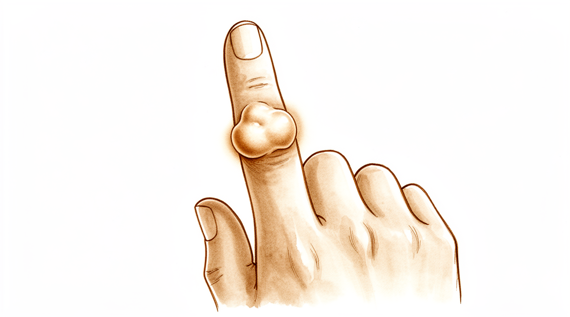
ਤੁਸੀਂ ਕੀ ਮਹਿਸੂਸ ਕਰਦੇ ਹੋ
ਜ਼ਿਆਦਾਤਰ ਲੋਕਾਂ ਨੂੰ ਕਿਸੇ ਉਂਗਲ ਜਾਂ ਅੰਗੂਠੇ ਉੱਤੇ ਇੱਕ ਗੰਢ ਨਜ਼ਰ ਆਉਂਦੀ ਹੈ ਜੋ ਦੁਖਦੀ ਨਹੀਂ। ਇਹ ਆਮ ਤੌਰ 'ਤੇ ਸਖ਼ਤ ਹੁੰਦੀ ਹੈ, ਅਤੇ ਹੌਲੀ-ਹੌਲੀ ਵਧਦੀ ਹੈ। ਕੁਝ ਗੰਢਾਂ ਬਿਲਕੁਲ ਕਿਸੇ ਜੋੜ ਦੇ ਨਾਲ ਹੁੰਦੀਆਂ ਹਨ, ਅਕਸਰ ਪੋਟੇ ਦੇ ਸਭ ਤੋਂ ਨੇੜੇ ਵਾਲੇ ਜੋੜ ਦੇ ਨਾਲ, ਅਤੇ ਇਹ ਉਂਗਲ ਦੇ ਉੱਪਰਲੇ ਪਾਸੇ, ਪਾਸੇ ਵੱਲ ਜਾਂ ਹਥੇਲੀ ਵਾਲੇ ਪਾਸੇ ਹੋ ਸਕਦੀਆਂ ਹਨ। ਸ਼ਹਾਦਤ ਦੀ ਉਂਗਲ ਸਭ ਤੋਂ ਆਮ ਥਾਂ ਹੈ।
ਗੰਢ ਆਪਣੇ ਆਪ ਵਿੱਚ ਲਗਭਗ ਹਰ ਮਾਮਲੇ ਵਿੱਚ ਦਰਦ-ਰਹਿਤ ਹੁੰਦੀ ਹੈ। ਜਦੋਂ ਕੋਈ ਤਕਲੀਫ਼ ਹੁੰਦੀ ਹੈ, ਤਾਂ ਉਹ ਆਮ ਤੌਰ 'ਤੇ ਇਸ ਕਰਕੇ ਹੁੰਦੀ ਹੈ ਕਿ ਵਧਦੀ ਹੋਈ ਗੰਢ ਨੇੜਲੀਆਂ ਬਣਤਰਾਂ ਉੱਤੇ ਦਬਾਅ ਪਾਉਂਦੀ ਹੈ। ਜੇ ਗੰਢ ਕਿਸੇ ਨਸ (nerve) ਉੱਤੇ ਦਬਾਅ ਪਾਉਂਦੀ ਹੈ, ਤਾਂ ਥੋੜ੍ਹੇ ਜਿਹੇ ਲੋਕਾਂ ਨੂੰ ਉਂਗਲ ਵਿੱਚ ਝਰਨਾਹਟ ਜਾਂ ਸੁੰਨਪਨ ਮਹਿਸੂਸ ਹੁੰਦਾ ਹੈ। ਕੁਝ ਲੋਕਾਂ ਨੂੰ ਲੱਗਦਾ ਹੈ ਕਿ ਹਿੱਲਣ ਵੇਲੇ ਉਂਗਲ ਅਟਕਦੀ ਹੈ ਜਾਂ ਕਲਿੱਕ ਕਰਦੀ ਹੈ, ਕੁਝ ਹੱਦ ਤੱਕ ਟ੍ਰਿਗਰ ਫਿੰਗਰ (ਉਂਗਲ ਦਾ ਅਟਕਣਾ) ਵਾਂਗ, ਜਿਸ ਵਿੱਚ ਉਂਗਲ ਥੋੜ੍ਹੀ ਦੇਰ ਲਈ ਫਸ ਜਾਂਦੀ ਹੈ ਅਤੇ ਫਿਰ ਝਟਕੇ ਨਾਲ ਖੁੱਲ੍ਹ ਜਾਂਦੀ ਹੈ। ਜੇ ਗੰਢ ਕਿਸੇ ਜੋੜ ਦੇ ਨੇੜੇ ਹੈ, ਤਾਂ ਉਸ ਉਂਗਲ ਨੂੰ ਪੂਰੀ ਤਰ੍ਹਾਂ ਮੋੜਨਾ ਔਖਾ ਹੋ ਸਕਦਾ ਹੈ ਕਿਉਂਕਿ ਗੰਢ ਰੁਕਾਵਟ ਪਾਉਂਦੀ ਹੈ।
ਉਸ ਉਂਗਲ ਨਾਲ ਕੀਤੇ ਜਾਣ ਵਾਲੇ ਰੋਜ਼ਾਨਾ ਦੇ ਕੰਮ ਔਖੇ ਹੋ ਸਕਦੇ ਹਨ। ਜੇ ਗੰਢ ਉਸ ਥਾਂ ਹੈ ਜਿੱਥੇ ਤੁਹਾਡੀ ਪਕੜ ਪੈਂਦੀ ਹੈ, ਤਾਂ ਛੋਟੇ ਸਿੱਕੇ ਚੁੱਕਣਾ, ਬਟਨ ਲਾਉਣਾ, ਪੈੱਨ ਫੜਨਾ ਜਾਂ ਮਰਤਬਾਨ ਦਾ ਢੱਕਣ ਫੜਨਾ ਬੇਢੰਗਾ ਲੱਗ ਸਕਦਾ ਹੈ। ਗੰਢ ਦਸਤਾਨਿਆਂ ਜਾਂ ਜੇਬਾਂ ਵਿੱਚ ਵੀ ਅੜ ਸਕਦੀ ਹੈ।
ਅਕਸਰ ਗੰਢ ਇੰਨੀ ਤੰਗ ਕਰਨ ਤੋਂ ਪਹਿਲਾਂ ਕਿ ਤੁਸੀਂ ਇਸ ਬਾਰੇ ਪੁੱਛੋ, ਕਾਫ਼ੀ ਲੰਮੇ ਸਮੇਂ ਤੋਂ ਮੌਜੂਦ ਹੁੰਦੀ ਹੈ। ਬਹੁਤ ਸਾਰੇ ਲੋਕ ਇਸ ਨੂੰ ਹੌਲੀ-ਹੌਲੀ ਬਦਲਦਾ ਦੇਖਦੇ ਹੋਏ ਮਹੀਨਿਆਂ ਜਾਂ ਸਾਲਾਂ ਤੱਕ ਉਡੀਕ ਕਰਦੇ ਹਨ।
ਇਸ ਸਥਿਤੀ ਬਾਰੇ ਕੁਝ ਗੱਲਾਂ ਜਾਣਨੀਆਂ ਚੰਗੀਆਂ ਹਨ। ਇਹ ਨੁਕਸਾਨ-ਰਹਿਤ (benign) ਹੈ, ਜਿਸ ਦਾ ਮਤਲਬ ਹੈ ਕਿ ਇਹ ਸਰੀਰ ਦੇ ਹੋਰ ਹਿੱਸਿਆਂ ਵਿੱਚ ਨਹੀਂ ਫੈਲਦੀ। ਇਹ ਮਰਦਾਂ ਨਾਲੋਂ ਔਰਤਾਂ ਵਿੱਚ ਥੋੜ੍ਹੀ ਜ਼ਿਆਦਾ ਆਮ ਹੈ, ਅਤੇ ਆਮ ਤੌਰ 'ਤੇ 32 ਤੋਂ 51 ਸਾਲ ਦੀ ਉਮਰ ਦੇ ਵਿਚਕਾਰ ਸਾਹਮਣੇ ਆਉਂਦੀ ਹੈ। ਜ਼ਿਆਦਾਤਰ ਲੋਕਾਂ ਨੂੰ ਇੱਕੋ ਗੰਢ ਹੁੰਦੀ ਹੈ, ਹਾਲਾਂਕਿ ਕੁਝ ਲੋਕਾਂ ਨੂੰ ਵੱਖ-ਵੱਖ ਥਾਵਾਂ 'ਤੇ ਇੱਕ ਤੋਂ ਵੱਧ ਗੰਢਾਂ ਹੁੰਦੀਆਂ ਹਨ।
ਧਿਆਨ ਰੱਖੋ ਕਿ ਕਿਤੇ ਉਂਗਲ ਜਾਂ ਹੱਥ ਗਰਮ, ਲਾਲ, ਸੁੱਜਿਆ ਹੋਇਆ ਅਤੇ ਦਰਦ ਵਾਲਾ ਤਾਂ ਨਹੀਂ ਹੋ ਗਿਆ, ਖ਼ਾਸ ਕਰਕੇ ਬੁਖ਼ਾਰ ਦੇ ਨਾਲ। ਜੇ ਅਜਿਹਾ ਹੁੰਦਾ ਹੈ, ਤਾਂ ਉਸੇ ਦਿਨ ਐਮਰਜੈਂਸੀ ਵਿਭਾਗ ਜਾਓ। ਜੇ ਗੰਢ ਵਧਦੀ ਰਹੇ, ਦੁਖਣ ਲੱਗ ਪਵੇ, ਜਾਂ ਉਂਗਲ ਠੀਕ ਤਰ੍ਹਾਂ ਕੰਮ ਨਾ ਕਰ ਰਹੀ ਹੋਵੇ, ਤਾਂ ਆਪਣੇ ਜੀਪੀ (ਫ਼ੈਮਿਲੀ ਡਾਕਟਰ) ਨੂੰ ਮਿਲੋ ਜਾਂ ਮਾਹਰ ਡਾਕਟਰ ਤੋਂ ਜਾਂਚ ਕਰਵਾਉਣ ਲਈ ਕਹੋ।
ਅਸਲ ਵਿੱਚ ਕੀ ਹੋ ਰਿਹਾ ਹੈ
ਤੁਹਾਡੇ ਟੈਂਡਨ (tendon) ਉਹ ਮਜ਼ਬੂਤ ਡੋਰੀਆਂ ਹਨ ਜੋ ਮਾਸਪੇਸ਼ੀ ਨੂੰ ਹੱਡੀ ਨਾਲ ਜੋੜਦੀਆਂ ਹਨ ਅਤੇ ਤੁਹਾਡੀਆਂ ਉਂਗਲਾਂ ਨੂੰ ਮੁੜਨ ਦਿੰਦੀਆਂ ਹਨ। ਹਰ ਟੈਂਡਨ ਇੱਕ ਪਤਲੀ ਖੋਲ ਦੇ ਅੰਦਰ ਚੱਲਦਾ ਹੈ ਜਿਸ ਨੂੰ ਟੈਂਡਨ ਦੀ ਝਿੱਲੀ (tendon sheath) ਕਹਿੰਦੇ ਹਨ, ਜੋ ਕੁਝ ਹੱਦ ਤੱਕ ਰੱਸੀ ਦੁਆਲੇ ਲੱਗੇ ਗੈਸਕਟ (gasket, ਸੀਲ ਕਰਨ ਵਾਲਾ ਛੱਲਾ) ਵਾਂਗ ਕੰਮ ਕਰਦੀ ਹੈ ਅਤੇ ਡੋਰੀ ਨੂੰ ਤਿਲਕਵੀਂ ਰੱਖਦੀ ਹੈ ਤਾਂ ਜੋ ਉਂਗਲ ਦੇ ਹਿੱਲਣ ਵੇਲੇ ਇਹ ਖਿਸਕ ਸਕੇ।
ਇਸ ਸਥਿਤੀ ਵਿੱਚ, ਉਸ ਖੋਲ ਦੇ ਸੈੱਲ ਲੋੜ ਨਾਲੋਂ ਵੱਧ ਵਧਣ ਲੱਗਦੇ ਹਨ। ਇਸ ਦਾ ਨਤੀਜਾ ਨਰਮ ਟਿਸ਼ੂ ਦੀ ਇੱਕ ਗੰਢ ਹੁੰਦਾ ਹੈ ਜੋ ਟੈਂਡਨ ਉੱਤੇ ਜਾਂ ਉਸ ਦੇ ਨਾਲ ਹੁੰਦੀ ਹੈ, ਅਕਸਰ ਕਿਸੇ ਜੋੜ ਦੇ ਨੇੜੇ। ਇਹ ਨੁਕਸਾਨ-ਰਹਿਤ ਹੈ, ਜਿਸ ਦਾ ਮਤਲਬ ਹੈ ਕਿ ਇਹ ਫੈਲਦੀ ਨਹੀਂ, ਅਤੇ ਇਹ ਹੌਲੀ-ਹੌਲੀ ਵਧਦੀ ਹੈ। ਜ਼ਿਆਦਾਤਰ ਗੰਢਾਂ ਛੋਟੀਆਂ ਹੁੰਦੀਆਂ ਹਨ, ਔਸਤਨ ਲਗਭਗ 1.35 ਸੈਂਟੀਮੀਟਰ, ਹਾਲਾਂਕਿ ਕੁਝ ਇਸ ਤੋਂ ਵੱਡੀਆਂ ਹੋ ਜਾਂਦੀਆਂ ਹਨ।
ਗੰਢ ਕੁਝ ਚੀਜ਼ਾਂ ਦੇ ਇਕੱਠੇ ਭੀੜੇ ਹੋਣ ਨਾਲ ਬਣਦੀ ਹੈ: ਸੋਜਸ਼ (inflammation) ਵਾਲੇ ਸੈੱਲ, ਜਾਇੰਟ ਸੈੱਲ (giant cells, ਜੋ ਬਸ ਕਈ ਨਿਊਕਲੀਅਸ ਵਾਲੇ ਸੈੱਲ ਹੁੰਦੇ ਹਨ), ਅਤੇ ਹੀਮੋਸਿਡਰਿਨ (haemosiderin) ਨਾਮ ਦਾ ਇੱਕ ਗੂੜ੍ਹਾ ਰੰਗਦਾਰ ਪਦਾਰਥ, ਉਹੀ ਚੀਜ਼ ਜੋ ਪੁਰਾਣੇ ਨੀਲ ਨੂੰ ਰੰਗ ਦਿੰਦੀ ਹੈ। ਇਸੇ ਰੰਗਦਾਰ ਪਦਾਰਥ ਕਰਕੇ, ਜੇ ਇਹ ਗੰਢਾਂ ਕਦੇ ਸਰਜਰੀ ਦੌਰਾਨ ਦੇਖੀਆਂ ਜਾਣ, ਤਾਂ ਇਹ ਭੂਰੀਆਂ ਜਿਹੀਆਂ ਦਿਸ ਸਕਦੀਆਂ ਹਨ।
ਉੱਪਰਲੇ ਹਿੱਸੇ ਵਿੱਚ ਦੱਸੇ ਜਿਹੜੇ ਲੱਛਣ ਤੁਸੀਂ ਮਹਿਸੂਸ ਕੀਤੇ, ਉਹ ਗੰਢ ਦੇ ਰਾਹ ਵਿੱਚ ਆਉਣ ਕਾਰਨ ਹੁੰਦੇ ਹਨ। ਜਿਵੇਂ-ਜਿਵੇਂ ਇਹ ਵਧਦੀ ਹੈ, ਇਹ ਟੈਂਡਨ, ਜੋੜ ਜਾਂ ਕਿਸੇ ਨਸ ਉੱਤੇ ਦਬਾਅ ਪਾ ਸਕਦੀ ਹੈ, ਜਿਸ ਤੋਂ ਅਟਕਣ, ਅਕੜਾਅ ਅਤੇ ਕਦੇ-ਕਦਾਈਂ ਹੋਣ ਵਾਲੀ ਝਰਨਾਹਟ ਦੀ ਸਮਝ ਆਉਂਦੀ ਹੈ। ਜ਼ਿਆਦਾਤਰ ਸਮਾਂ ਇਹ ਬਿਲਕੁਲ ਨਹੀਂ ਦੁਖਦੀ, ਅਤੇ ਸਿਰਫ਼ ਲਗਭਗ ਹਰ 100 ਵਿੱਚੋਂ 16 ਲੋਕ ਦਰਦ ਦੱਸਦੇ ਹਨ।
ਕੁਝ ਗੱਲਾਂ ਜਾਣਨ ਯੋਗ ਹਨ। ਜ਼ਿਆਦਾਤਰ ਲੋਕਾਂ ਨੂੰ ਇੱਕੋ ਗੰਢ ਹੁੰਦੀ ਹੈ, ਪਰ ਲਗਭਗ ਹਰ 100 ਵਿੱਚੋਂ 21 ਲੋਕਾਂ ਨੂੰ ਦੋ ਜਾਂ ਵੱਧ ਵੱਖਰੀਆਂ ਗੰਢਾਂ ਹੁੰਦੀਆਂ ਹਨ। ਗੰਢ ਆਮ ਤੌਰ 'ਤੇ ਕਿਸੇ ਜੋੜ ਦੇ ਨਾਲ ਹੁੰਦੀ ਹੈ, ਅਤੇ ਉਂਗਲਾਂ ਦੇ ਲਗਭਗ ਇੱਕ ਤਿਹਾਈ ਮਾਮਲਿਆਂ ਵਿੱਚ ਇਹ ਪੋਟੇ ਦੇ ਸਭ ਤੋਂ ਨੇੜੇ ਵਾਲੇ ਜੋੜ ਉੱਤੇ ਹੁੰਦੀ ਹੈ। ਬਹੁਤ ਹੀ ਘੱਟ ਮਾਮਲਿਆਂ ਵਿੱਚ, ਗੰਢ ਹੱਡੀ ਦੇ ਅੰਦਰ ਵੱਲ ਦਬ ਸਕਦੀ ਹੈ, ਜੋ ਐਕਸ-ਰੇ ਉੱਤੇ ਦਿਸ ਜਾਂਦਾ ਹੈ।
ਇਸੇ ਸਥਿਤੀ ਦਾ ਇੱਕ ਘੱਟ ਮਿਲਣ ਵਾਲਾ, ਵਧੇਰੇ ਫੈਲਿਆ ਹੋਇਆ (diffuse) ਰੂਪ ਵੀ ਹੈ, ਜੋ ਇੱਕ ਸਾਫ਼-ਸੁਥਰੀ ਗੰਢ ਬਣਾਉਣ ਦੀ ਬਜਾਏ ਪਤਲੀਆਂ ਟਾਹਣੀਆਂ ਵਾਂਗ ਟੈਂਡਨ ਦੇ ਨਾਲ-ਨਾਲ ਫੈਲਦਾ ਹੈ। ਉਂਗਲ ਵਿੱਚ ਇਹ ਰੂਪ ਬਹੁਤ ਘੱਟ ਮਿਲਦਾ ਹੈ ਅਤੇ ਇਸ ਦਾ ਵਿਹਾਰ ਅਕਸਰ ਵੱਖਰਾ ਹੁੰਦਾ ਹੈ, ਇਸੇ ਲਈ ਕੋਈ ਵੀ ਯੋਜਨਾ ਬਣਾਉਣ ਤੋਂ ਪਹਿਲਾਂ ਤੁਹਾਡਾ ਸਰਜਨ ਸਾਫ਼-ਸਾਫ਼ ਜਾਣਨਾ ਚਾਹੇਗਾ ਕਿ ਇਹ ਬਿਲਕੁਲ ਕਿਸ ਕਿਸਮ ਦੀ ਗੰਢ ਹੈ।
ਅਸੀਂ ਇਸ ਬਾਰੇ ਕੀ ਕਰ ਸਕਦੇ ਹਾਂ
ਡਾ. ਕੀਰਨ ਹਿਰਪਰਾ, ਮਾਟਰ ਪ੍ਰਾਈਵੇਟ ਹਸਪਤਾਲ ਰੌਕਹੈਂਪਟਨ ਵਿੱਚ ਉੱਪਰਲੇ ਅੰਗ (ਮੋਢੇ ਤੋਂ ਹੱਥ ਤੱਕ ਦੀ ਬਾਂਹ) ਦੇ ਸਰਜਨ, ਤੁਹਾਡੀ ਸਥਿਤੀ ਦੇ ਅਨੁਕੂਲ ਸਭ ਤੋਂ ਘੱਟ ਚੀਰ-ਫਾੜ ਵਾਲੇ ਵਿਕਲਪਾਂ ਤੋਂ ਸ਼ੁਰੂਆਤ ਕਰਦੇ ਹਨ। ਮਰੀਜ਼ਾਂ ਨੂੰ ਆਮ ਤੌਰ 'ਤੇ ਉਨ੍ਹਾਂ ਦਾ ਜੀਪੀ (ਫ਼ੈਮਿਲੀ ਡਾਕਟਰ) ਸਾਡੇ ਕਲੀਨਿਕ ਵਿੱਚ ਭੇਜਦਾ ਹੈ; ਜੇ ਕਿਸੇ ਫਿਜ਼ੀਓਥੈਰੇਪਿਸਟ ਨੇ ਤੁਹਾਨੂੰ ਸਾਡੇ ਕੋਲ ਆਉਣ ਦੀ ਸਲਾਹ ਦਿੱਤੀ ਹੈ, ਤਾਂ ਵੀ Medicare ਦੀ ਛੋਟ (rebate) ਦੇ ਯੋਗ ਹੋਣ ਲਈ ਤੁਹਾਨੂੰ ਆਪਣੇ ਜੀਪੀ ਤੋਂ ਰੈਫ਼ਰਲ (referral) ਲੈਣਾ ਪਵੇਗਾ। ਕਲੀਨਿਕ ਵਿੱਚ ਅਸੀਂ ਤੁਹਾਡੀ ਤਕਲੀਫ਼ ਦਾ ਪੂਰਾ ਵੇਰਵਾ ਪੁੱਛਦੇ ਹਾਂ, ਤੁਹਾਡੇ ਹੱਥ ਦੀ ਜਾਂਚ ਕਰਦੇ ਹਾਂ, ਅਤੇ ਇਹ ਪਤਾ ਲਗਾਉਣ ਲਈ ਕਿ ਇਹ ਬਿਲਕੁਲ ਕਿਸ ਕਿਸਮ ਦੀ ਗੰਢ ਹੈ, ਜਿੱਥੇ ਲੋੜ ਹੋਵੇ ਉੱਥੇ ਇਮੇਜਿੰਗ (ਐਕਸ-ਰੇ ਜਾਂ ਸਕੈਨ) ਦਾ ਪ੍ਰਬੰਧ ਕਰਦੇ ਹਾਂ।
ਕਿਉਂਕਿ ਇਹ ਗੰਢ ਨੁਕਸਾਨ-ਰਹਿਤ ਹੈ ਅਤੇ ਹੌਲੀ-ਹੌਲੀ ਵਧਦੀ ਹੈ, ਇਸ ਲਈ ਕੁਝ ਨਾ ਕਰਨਾ ਵੀ ਇੱਕ ਅਸਲ ਵਿਕਲਪ ਹੈ। ਛੋਟੀ, ਦਰਦ-ਰਹਿਤ ਗੰਢ ਲਈ, ਸਿਰਫ਼ ਇਸ ਉੱਤੇ ਨਜ਼ਰ ਰੱਖਣਾ ਸਹੀ ਚੋਣ ਹੋ ਸਕਦੀ ਹੈ। ਜੇ ਗੰਢ ਕੰਮਾਂ ਵਿੱਚ ਰੁਕਾਵਟ ਪਾ ਰਹੀ ਹੈ, ਤਾਂ ਹੈਂਡ ਥੈਰੇਪੀ (ਹੱਥ ਦੀ ਥੈਰੇਪੀ) ਅਕੜਾਅ ਅਤੇ ਅਟਕਣ ਵਿੱਚ ਮਦਦ ਕਰ ਸਕਦੀ ਹੈ। ਥੈਰੇਪੀ ਦਾ ਮਕਸਦ ਉਂਗਲ ਨੂੰ ਹਿਲਦਾ ਰੱਖਣਾ ਅਤੇ ਰੋਜ਼ਾਨਾ ਦੇ ਕੰਮਾਂ ਨੂੰ ਸੌਖਾ ਬਣਾਉਣਾ ਹੈ। ਸਰਜਰੀ ਬਾਰੇ ਸੋਚਣ ਤੋਂ ਪਹਿਲਾਂ ਅਸੀਂ ਆਮ ਤੌਰ 'ਤੇ ਇਸ ਨੂੰ ਪੂਰਾ ਮੌਕਾ ਦੇਣ ਦੀ ਸਲਾਹ ਦਿੰਦੇ ਹਾਂ।
ਇਸ ਸਥਿਤੀ ਲਈ ਕੋਈ ਅਜਿਹੀ ਦਵਾਈ ਨਹੀਂ ਹੈ ਜੋ ਗੰਢ ਨੂੰ ਸੁੰਗੇੜ ਦੇਵੇ, ਇਸ ਲਈ ਦਰਦ ਦੀਆਂ ਗੋਲੀਆਂ ਅਤੇ ਸੋਜਸ਼-ਰੋਧੀ ਦਵਾਈਆਂ (anti-inflammatories) ਸਿਰਫ਼ ਉਸ ਤਕਲੀਫ਼ ਨੂੰ ਸ਼ਾਂਤ ਕਰਦੀਆਂ ਹਨ ਜੋ ਗੰਢ ਦੇ ਨੇੜਲੇ ਟਿਸ਼ੂ ਉੱਤੇ ਦਬਾਅ ਪਾਉਣ ਕਾਰਨ ਹੁੰਦੀ ਹੈ। ਇਹ ਗੰਢ ਨੂੰ ਛੋਟਾ ਨਹੀਂ ਕਰਦੀਆਂ।
ਜਦੋਂ ਗੰਢ ਵੱਡੀ ਹੋਵੇ, ਦੁਖਦੀ ਹੋਵੇ, ਜਾਂ ਉਂਗਲ ਨੂੰ ਠੀਕ ਤਰ੍ਹਾਂ ਕੰਮ ਕਰਨ ਤੋਂ ਰੋਕ ਰਹੀ ਹੋਵੇ, ਤਾਂ ਸਰਜਰੀ ਆਮ ਇਲਾਜ ਹੈ। ਮਕਸਦ ਪੂਰੀ ਗੰਢ ਨੂੰ ਟੈਂਡਨ ਦੀ ਖੋਲ ਉੱਤੇ ਉਸ ਦੀ ਜੁੜਨ ਵਾਲੀ ਥਾਂ ਸਮੇਤ ਕੱਢਣਾ ਹੈ, ਕਿਉਂਕਿ ਜੁੜਨ ਵਾਲੀ ਉਸ ਥਾਂ ਨੂੰ ਪਿੱਛੇ ਛੱਡ ਦੇਣ ਨਾਲ ਹੀ ਇਹ ਦੁਬਾਰਾ ਉੱਗ ਸਕਦੀ ਹੈ। ਅਸੀਂ ਓਪਰੇਸ਼ਨ ਤੋਂ ਪਹਿਲਾਂ ਧਿਆਨ ਨਾਲ ਯੋਜਨਾ ਬਣਾਉਂਦੇ ਹਾਂ, ਓਪਰੇਸ਼ਨ ਦੌਰਾਨ ਵੱਡਦਰਸ਼ੀ ਸ਼ੀਸ਼ੇ (magnification) ਵਰਤਦੇ ਹਾਂ, ਅਤੇ ਗੰਢ ਨੂੰ ਪੂਰੀ ਤਰ੍ਹਾਂ ਕੱਢਦੇ ਹਾਂ। ਦੁਬਾਰਾ ਉੱਗਣਾ (recurrence) ਇਸ ਸਥਿਤੀ ਦਾ ਮੁੱਖ ਖ਼ਤਰਾ ਹੈ, ਅਤੇ ਇਹ ਆਮ ਤੌਰ 'ਤੇ ਕੱਢਣ ਤੋਂ 36 ਮਹੀਨਿਆਂ ਦੇ ਅੰਦਰ ਹੁੰਦਾ ਹੈ। ਉਂਗਲ ਦੀ ਗੰਢ ਕਢਵਾਉਣ ਵਾਲੇ 605 ਲੋਕਾਂ ਦੇ ਇੱਕ ਸਮੂਹ ਵਿੱਚ, 14.8% ਲੋਕਾਂ ਦੀ ਗੰਢ ਵਾਪਸ ਆ ਗਈ। ਇਸ ਨੂੰ ਇਵੇਂ ਵੀ ਕਹਿ ਸਕਦੇ ਹਾਂ: ਜ਼ਿਆਦਾਤਰ ਲੋਕਾਂ ਦੀ, ਲਗਭਗ ਹਰ 100 ਵਿੱਚੋਂ 85 ਦੀ, ਗੰਢ ਵਾਪਸ ਨਹੀਂ ਆਉਂਦੀ। ਜੇ ਇਹ ਵਾਪਸ ਆ ਜਾਵੇ, ਤਾਂ ਦੁਬਾਰਾ ਉੱਗਣ ਨੂੰ ਰੋਕਣ ਲਈ ਵਾਧੂ ਕਦਮਾਂ ਨਾਲ ਇਸ ਨੂੰ ਫਿਰ ਤੋਂ ਕੱਢਣ ਨਾਲ ਵੀ ਸਮੱਸਿਆ ਹੱਲ ਹੋ ਸਕਦੀ ਹੈ।
ਕੀ ਉਮੀਦ ਰੱਖੀਏ
ਇਸ ਗੰਢ ਦਾ ਭਵਿੱਖ ਨਾਟਕੀ ਹੋਣ ਦੀ ਬਜਾਏ ਸਥਿਰ ਹੁੰਦਾ ਹੈ। ਇਹ ਨੁਕਸਾਨ-ਰਹਿਤ ਹੈ, ਇਸ ਲਈ ਇਹ ਤੁਹਾਡੇ ਸਰੀਰ ਵਿੱਚ ਕਿਤੇ ਹੋਰ ਨਹੀਂ ਫੈਲਦੀ। ਜੇ ਇਵੇਂ ਹੀ ਛੱਡ ਦਿੱਤੀ ਜਾਵੇ, ਤਾਂ ਛੋਟੀ ਦਰਦ-ਰਹਿਤ ਗੰਢ ਆਮ ਤੌਰ 'ਤੇ ਹੌਲੀ-ਹੌਲੀ ਵਧਦੀ ਰਹਿੰਦੀ ਹੈ, ਜਾਂ ਬਸ ਜਿਵੇਂ ਹੈ ਉਵੇਂ ਹੀ ਰਹਿੰਦੀ ਹੈ। ਇਹ ਆਪਣੇ ਆਪ ਬਹੁਤ ਘੱਟ ਠੀਕ ਹੁੰਦੀ ਹੈ, ਅਤੇ ਇਹ ਕਿਸੇ ਖ਼ਤਰਨਾਕ ਚੀਜ਼ ਵਿੱਚ ਨਹੀਂ ਬਦਲਦੀ।
ਸਮੇਂ ਦੇ ਨਾਲ ਇਹ ਸਥਿਤੀ ਮੁੱਖ ਤੌਰ 'ਤੇ ਜੋ ਕਰਦੀ ਹੈ, ਉਹ ਹੈ ਕੱਢਣ ਤੋਂ ਬਾਅਦ ਵਾਪਸ ਆਉਣਾ। ਦੁਬਾਰਾ ਉੱਗਣਾ ਇਸ ਗੰਢ ਦਾ ਮੁੱਢਲਾ ਖ਼ਤਰਾ ਹੈ, ਅਤੇ ਇਹ ਆਮ ਤੌਰ 'ਤੇ ਕੱਢਣ ਤੋਂ 36 ਮਹੀਨਿਆਂ ਦੇ ਅੰਦਰ ਹੁੰਦਾ ਹੈ। ਕੁਝ ਗੰਢਾਂ ਦੇ ਵਾਪਸ ਆਉਣ ਦੀ ਸੰਭਾਵਨਾ ਦੂਜੀਆਂ ਨਾਲੋਂ ਜ਼ਿਆਦਾ ਹੁੰਦੀ ਹੈ: ਉਹ ਗੰਢਾਂ ਜੋ ਦੋ ਜਾਂ ਵੱਧ ਵੱਖਰੇ ਟੁਕੜਿਆਂ ਤੋਂ ਬਣੀਆਂ ਹੋਣ ਜੋ ਆਪਸ ਵਿੱਚ ਜੁੜੇ ਨਹੀਂ ਹੁੰਦੇ, ਅਤੇ ਉਹ ਗੰਢਾਂ ਜੋ ਖ਼ੁਦ ਟੈਂਡਨ ਵਿੱਚ ਜਾਂ ਜੋੜ ਦੀ ਅੰਦਰਲੀ ਪਰਤ ਵਿੱਚ ਵਧ ਗਈਆਂ ਹੋਣ। ਜਦੋਂ ਪੂਰੀ ਗੰਢ ਨੂੰ ਉਸ ਦੀ ਜੁੜਨ ਵਾਲੀ ਥਾਂ ਸਮੇਤ ਕੱਢ ਦਿੱਤਾ ਜਾਂਦਾ ਹੈ, ਤਾਂ ਇਹ ਖ਼ਤਰਾ ਕਾਫ਼ੀ ਘਟ ਜਾਂਦਾ ਹੈ।
ਇਲਾਜ ਤੋਂ ਬਾਅਦ ਕੰਮਕਾਜ ਆਮ ਤੌਰ 'ਤੇ ਚੰਗਾ ਰਹਿੰਦਾ ਹੈ। ਜਿਨ੍ਹਾਂ ਲੋਕਾਂ ਦੀ ਉਂਗਲ ਤੋਂ ਇਨ੍ਹਾਂ ਵਿੱਚੋਂ ਕੋਈ ਗੰਢ ਕੱਢੀ ਗਈ ਹੈ, ਉਹ ਬਾਂਹ ਦੇ ਕੰਮਕਾਜ ਨੂੰ ਮਾਪਣ ਵਾਲੇ ਇੱਕ ਮਿਆਰੀ ਪੈਮਾਨੇ ਉੱਤੇ ਔਸਤਨ ਆਮ ਕੰਮਕਾਜ ਦਾ 92% ਅੰਕ ਹਾਸਲ ਕਰਦੇ ਹਨ। ਦਰਜ ਕੀਤੇ ਗਏ ਮਾਮਲਿਆਂ ਵਿੱਚ, ਲੋਕਾਂ ਦੀ ਉਂਗਲ ਦੀ ਪੂਰੀ ਹਿਲਜੁਲ ਬਣੀ ਰਹੀ, ਅਤੇ ਦੋ ਸਾਲ ਵਾਲੀ ਜਾਂਚ ਤੱਕ ਐਕਸ-ਰੇ ਉੱਤੇ ਉਂਗਲ ਦੀ ਹੱਡੀ ਦੀ ਸ਼ਕਲ ਮੁੜ ਆਮ ਵਰਗੀ ਹੋ ਗਈ।
ਜੇ ਗੰਢ ਉਹ ਘੱਟ ਮਿਲਣ ਵਾਲੀ, ਵਧੇਰੇ ਫੈਲਣ ਵਾਲੀ ਕਿਸਮ ਦੀ ਹੈ ਜੋ ਟਾਹਣੀਆਂ ਵਾਂਗ ਟੈਂਡਨ ਦੁਆਲੇ ਲਿਪਟ ਜਾਂਦੀ ਹੈ, ਤਾਂ ਇਸ ਨੂੰ ਪੂਰੀ ਤਰ੍ਹਾਂ ਕੱਢਣਾ ਔਖਾ ਹੁੰਦਾ ਹੈ ਅਤੇ ਇਸ ਦੇ ਵਾਪਸ ਆਉਣ ਦੀ ਸੰਭਾਵਨਾ ਜ਼ਿਆਦਾ ਹੁੰਦੀ ਹੈ। ਅਜਿਹੇ ਮਾਮਲਿਆਂ ਵਿੱਚ, ਉਸ ਥਾਂ ਉੱਤੇ ਕੇਂਦਰਿਤ ਰੇਡੀਏਸ਼ਨ ਥੈਰੇਪੀ (radiation therapy, ਰੇਡੀਏਸ਼ਨ ਨਾਲ ਇਲਾਜ) ਤੁਹਾਡੇ ਹੱਥ ਨੂੰ ਕੰਮ ਕਰਦਾ ਰੱਖਦੇ ਹੋਏ ਗੰਢ ਨੂੰ ਕਾਬੂ ਵਿੱਚ ਰੱਖਣ ਵਿੱਚ ਮਦਦ ਕਰ ਸਕਦੀ ਹੈ।
ਜੇ ਤੁਸੀਂ ਛੋਟੀ, ਦਰਦ-ਰਹਿਤ ਗੰਢ ਉੱਤੇ ਸਿਰਫ਼ ਨਜ਼ਰ ਰੱਖਣ ਦੀ ਚੋਣ ਕਰਦੇ ਹੋ, ਤਾਂ ਹਕੀਕੀ ਤਸਵੀਰ ਇਹ ਹੈ ਕਿ ਗੰਢ ਹੌਲੀ-ਹੌਲੀ ਵਧੇਗੀ ਜਾਂ ਉਵੇਂ ਹੀ ਰਹੇਗੀ, ਅਤੇ ਹੌਲੀ-ਹੌਲੀ ਉਂਗਲ ਨੂੰ ਮੋੜਨਾ ਜਾਂ ਛੋਟੇ ਕੰਮਾਂ ਲਈ ਵਰਤਣਾ ਹੋਰ ਔਖਾ ਬਣਾ ਸਕਦੀ ਹੈ। ਜੇ ਇਹ ਦੁਖਣ ਲੱਗ ਪਵੇ, ਵਧਦੀ ਰਹੇ, ਜਾਂ ਉਂਗਲ ਠੀਕ ਤਰ੍ਹਾਂ ਕੰਮ ਕਰਨਾ ਬੰਦ ਕਰ ਦੇਵੇ, ਤਾਂ ਆਮ ਤੌਰ 'ਤੇ ਇਹੀ ਉਹ ਮੋੜ ਹੈ ਜਿੱਥੇ ਇਲਾਜ ਕਰਵਾਉਣਾ ਫ਼ਾਇਦੇਮੰਦ ਬਣਦਾ ਹੈ।
ਡਾਕਟਰ ਨੂੰ ਕਦੋਂ ਮਿਲਣਾ ਚਾਹੀਦਾ ਹੈ
ਇਸ ਤਰ੍ਹਾਂ ਦੀਆਂ ਜ਼ਿਆਦਾਤਰ ਗੰਢਾਂ ਲਈ ਜਲਦਬਾਜ਼ੀ ਦੀ ਲੋੜ ਨਹੀਂ ਹੁੰਦੀ, ਅਤੇ ਇਲਾਜ ਕਦੇ ਵੀ ਕੋਈ ਦੌੜ ਨਹੀਂ ਹੁੰਦਾ। ਆਪਣੇ ਜੀਪੀ ਨੂੰ ਮਿਲੋ ਜੇ ਗੰਢ ਠੀਕ ਨਹੀਂ ਹੋ ਰਹੀ, ਹਫ਼ਤਿਆਂ ਦੌਰਾਨ ਵਧ ਰਹੀ ਹੈ, ਦੁਖਣ ਲੱਗ ਪਈ ਹੈ, ਜਾਂ ਤੁਹਾਨੂੰ ਉਂਗਲ ਜਾਂ ਹੱਥ ਨੂੰ ਆਮ ਵਾਂਗ ਵਰਤਣ ਤੋਂ ਰੋਕਦੀ ਹੈ। ਮਾਹਰ ਡਾਕਟਰ ਤੋਂ ਜਾਂਚ ਕਰਵਾਉਣ ਲਈ ਕਹੋ ਜੇ ਹਿੱਲਣ ਵੇਲੇ ਉਂਗਲ ਅਟਕਦੀ ਜਾਂ ਕਲਿੱਕ ਕਰਦੀ ਹੈ, ਜੇ ਤੁਹਾਨੂੰ ਝਰਨਾਹਟ ਜਾਂ ਸੁੰਨਪਨ ਮਹਿਸੂਸ ਹੁੰਦਾ ਹੈ, ਜਾਂ ਜੇ ਗੰਢ ਦੇ ਰਾਹ ਵਿੱਚ ਆਉਣ ਕਾਰਨ ਉਂਗਲ ਨੂੰ ਪੂਰੀ ਤਰ੍ਹਾਂ ਮੋੜਨਾ ਔਖਾ ਹੋ ਗਿਆ ਹੈ। ਇਹ ਉਹ ਗੱਲਾਂ ਹਨ ਜਿਨ੍ਹਾਂ ਦੀ ਦੇਰ ਕਰਨ ਦੀ ਬਜਾਏ ਜਲਦੀ ਜਾਂਚ ਕਰਵਾਉਣੀ ਚੰਗੀ ਹੈ, ਕਿਉਂਕਿ ਜੋ ਗੰਢ ਟੈਂਡਨ ਜਾਂ ਜੋੜ ਦੀ ਅੰਦਰਲੀ ਪਰਤ ਵਿੱਚ ਵਧ ਗਈ ਹੋਵੇ, ਉਸ ਨੂੰ ਪੂਰੀ ਤਰ੍ਹਾਂ ਕੱਢਣਾ ਔਖਾ ਹੁੰਦਾ ਹੈ ਅਤੇ ਉਸ ਦੇ ਵਾਪਸ ਆਉਣ ਦੀ ਸੰਭਾਵਨਾ ਜ਼ਿਆਦਾ ਹੁੰਦੀ ਹੈ।
ਪਰ ਲੱਛਣਾਂ ਦੇ ਇੱਕ ਸਮੂਹ ਲਈ ਉਸੇ ਦਿਨ ਇਲਾਜ ਦੀ ਲੋੜ ਹੁੰਦੀ ਹੈ। ਜੇ ਤੁਹਾਡੀ ਉਂਗਲ ਜਾਂ ਹੱਥ ਗਰਮ, ਲਾਲ, ਸੁੱਜਿਆ ਹੋਇਆ ਅਤੇ ਦਰਦ ਵਾਲਾ ਹੋ ਜਾਵੇ, ਖ਼ਾਸ ਕਰਕੇ ਬੁਖ਼ਾਰ ਦੇ ਨਾਲ, ਤਾਂ ਐਮਰਜੈਂਸੀ ਵਿਭਾਗ ਜਾਓ। ਤੁਹਾਨੂੰ ਪਹਿਲਾਂ ਜੀਪੀ ਦੇ ਰੈਫ਼ਰਲ ਦੀ ਲੋੜ ਨਹੀਂ ਹੈ। ਜੇ ਤੁਸੀਂ ਕਲੀਨਿਕ ਦੇ ਸਮੇਂ ਤੋਂ ਬਾਅਦ ਜਾਂ ਹਫ਼ਤੇ ਦੇ ਅਖ਼ੀਰ ਵਿੱਚ ਕਲੀਨਿਕ ਨਾਲ ਸੰਪਰਕ ਨਹੀਂ ਕਰ ਸਕਦੇ, ਤਾਂ ਆਪਣੇ ਸਭ ਤੋਂ ਨੇੜਲੇ ਐਮਰਜੈਂਸੀ ਵਿਭਾਗ ਜਾਓ।
ਵਿਸਥਾਰ ਵਿੱਚ
Advanced reading: the deeper science (optional)
ਇਹ ਹਿੱਸਾ ਤੁਹਾਡੇ ਆਪਣੇ ਇਲਾਜ ਬਾਰੇ ਫ਼ੈਸਲਿਆਂ ਲਈ ਲੋੜ ਤੋਂ ਵੱਧ ਡੂੰਘਾਈ ਵਿੱਚ ਜਾਂਦਾ ਹੈ। ਟੈਂਡਨ ਦੀ ਝਿੱਲੀ ਦੀ ਜਾਇੰਟ ਸੈੱਲ ਰਸੌਲੀ ਬਾਰੇ ਵਾਧੂ ਪੜ੍ਹਨਾ ਇਸ ਲਈ ਫ਼ਾਇਦੇਮੰਦ ਹੈ ਕਿਉਂਕਿ ਇਸ ਦੀ ਮੁੱਖ ਸਮੱਸਿਆ ਇਸ ਦਾ ਦੁਬਾਰਾ ਉੱਗਣਾ (recurrence) ਹੈ, ਅਤੇ ਸਬੂਤ ਦੱਸਦੇ ਹਨ ਕਿ ਦੁਬਾਰਾ ਉੱਗਣਾ ਇਸ ਨੂੰ ਕੱਢਣ ਦੇ ਤਰੀਕੇ ਦੀ ਕਿਸੇ ਗੱਲ ਨਾਲੋਂ ਜ਼ਿਆਦਾ ਹਰ ਰਸੌਲੀ ਦੀ ਆਪਣੀ ਜੀਵ-ਵਿਗਿਆਨਕ ਪ੍ਰਕਿਰਤੀ (biology) ਉੱਤੇ ਨਿਰਭਰ ਕਰਦਾ ਹੈ।
ਦੁਬਾਰਾ ਉੱਗਣਾ ਰਸੌਲੀ ਦੀ ਆਪਣੀ ਖ਼ਾਸੀਅਤ ਹੈ, ਸਿਰਫ਼ ਸਰਜਰੀ ਦੀ ਨਹੀਂ
ਗੰਢ ਦੇ ਵਾਪਸ ਆਉਣ ਦੀ ਸਹਿਜ ਵਿਆਖਿਆ ਇਹ ਹੈ ਕਿ ਕੁਝ ਹਿੱਸਾ ਪਿੱਛੇ ਰਹਿ ਗਿਆ ਸੀ। ਉਂਗਲਾਂ ਦੇ 605 ਮਾਮਲਿਆਂ ਦੀ ਇੱਕ ਯੋਜਨਾਬੱਧ ਸਮੀਖਿਆ (systematic review) ਨੇ ਕੁਝ ਹੋਰ ਸਿੱਟਾ ਕੱਢਿਆ: ਦੁਬਾਰਾ ਉੱਗਣ ਵਿੱਚ ਰਸੌਲੀ ਦੀ ਅੰਦਰੂਨੀ ਜੀਵ-ਵਿਗਿਆਨਕ ਪ੍ਰਕਿਰਤੀ, ਰਸੌਲੀ ਦੀ ਥਾਂ ਜਾਂ ਆਲੇ-ਦੁਆਲੇ ਵਿੱਚ ਧਸਣ ਦੀ ਸਮਰੱਥਾ ਨਾਲੋਂ ਜ਼ਿਆਦਾ ਬੁਨਿਆਦੀ ਭੂਮਿਕਾ ਨਿਭਾਉਂਦੀ ਜਾਪਦੀ ਹੈ, ਅਤੇ ਲੇਖਕਾਂ ਨੇ ਇਹ ਪਛਾਣਨ ਲਈ ਵੱਡੇ ਅਗਾਂਹਵਧੂ ਅਧਿਐਨਾਂ (prospective studies) ਦੀ ਮੰਗ ਕੀਤੀ ਕਿ ਕਿਹੜੀਆਂ ਰਸੌਲੀਆਂ ਦੇ ਦੁਬਾਰਾ ਉੱਗਣ ਦਾ ਰੁਝਾਨ ਹੁੰਦਾ ਹੈ [1]।
ਓਪਰੇਸ਼ਨ ਤੋਂ ਪਹਿਲਾਂ ਇਹ ਜਾਣਨਾ ਸੱਚਮੁੱਚ ਲਾਭਦਾਇਕ ਹੈ। ਚੰਗੀ ਤਰ੍ਹਾਂ ਕੀਤੇ ਓਪਰੇਸ਼ਨ ਤੋਂ ਬਾਅਦ ਦੁਬਾਰਾ ਉੱਗਣਾ ਇਸ ਰਸੌਲੀ ਦਾ ਇੱਕ ਜਾਣਿਆ-ਪਛਾਣਿਆ ਵਿਹਾਰ ਹੈ, ਨਾ ਕਿ ਇਸ ਗੱਲ ਦਾ ਸਬੂਤ ਕਿ ਕੁਝ ਗ਼ਲਤ ਹੋ ਗਿਆ।
ਇਲਾਜ ਚੋਣਵਾਂ ਹੈ, ਅਤੇ ਕੁਝ ਨਾ ਕਰਨਾ ਇੱਕ ਅਸਲ ਵਿਕਲਪ ਹੈ
ਜਦੋਂ ਓਪਰੇਸ਼ਨ ਦੀ ਯੋਜਨਾ ਬਣ ਰਹੀ ਹੋਵੇ, ਤਾਂ ਇਹ ਗੱਲ ਭੁੱਲਣੀ ਸੌਖੀ ਹੈ। ਪਹਿਲੀ ਕਤਾਰ ਦੇ ਇਲਾਜ ਦਾ ਸਿਧਾਂਤ ਗੰਢ ਨੂੰ ਪੂਰੀ ਤਰ੍ਹਾਂ ਕੱਢਣਾ ਹੈ, ਪਰ ਇਲਾਜ ਕਦੇ ਵੀ ਤੁਰੰਤ ਕਰਨ ਦੀ ਲੋੜ ਨਹੀਂ ਹੁੰਦੀ, ਅਤੇ ਇਲਾਜ ਦੇ ਕਾਰਨ ਨੂੰ ਲੱਛਣਾਂ, ਵਾਧੇ, ਥਾਂ ਅਤੇ ਤੁਹਾਡੇ ਆਪਣੇ ਹਾਲਾਤਾਂ ਦੇ ਮੁਕਾਬਲੇ ਤੋਲਿਆ ਜਾਣਾ ਚਾਹੀਦਾ ਹੈ [4]।
ਛੋਟੀ, ਦਰਦ-ਰਹਿਤ, ਹੌਲੀ-ਹੌਲੀ ਵਧਣ ਵਾਲੀ ਗੰਢੀ ਲਈ, ਇਸ ਉੱਤੇ ਨਜ਼ਰ ਰੱਖਣਾ ਇੱਕ ਜਾਇਜ਼ ਚੋਣ ਹੈ। ਰਸੌਲੀ ਨੁਕਸਾਨ-ਰਹਿਤ ਹੈ ਅਤੇ ਫੈਲਦੀ ਨਹੀਂ, ਇਸ ਲਈ ਓਪਰੇਸ਼ਨ ਕਰਨ ਦੀ ਦਲੀਲ ਕੰਮਕਾਜ, ਆਕਾਰ ਅਤੇ ਪਰੇਸ਼ਾਨੀ ਬਾਰੇ ਹੈ — ਖ਼ਤਰੇ ਬਾਰੇ ਨਹੀਂ।
ਪਰ ਸਰਜਰੀ ਦੀਆਂ ਦੋ ਗੱਲਾਂ ਸੱਚਮੁੱਚ ਫ਼ਰਕ ਪਾਉਂਦੀਆਂ ਹਨ
ਜੀਵ-ਵਿਗਿਆਨ ਹੀ ਪੂਰੀ ਕਹਾਣੀ ਨਹੀਂ ਹੈ। ਇੱਕ ਥਾਂ ਸੀਮਤ ਕਿਸਮ ਦੀ ਟੀਨੋਸਾਈਨੋਵੀਅਲ ਜਾਇੰਟ ਸੈੱਲ ਰਸੌਲੀ (tenosynovial giant cell tumour) ਵਾਲੇ 941 ਮਰੀਜ਼ਾਂ ਵਿੱਚ, ਕੱਢਣ ਤੋਂ ਬਾਅਦ ਦੁਬਾਰਾ ਉੱਗਣ ਨਾਲ ਜੁੜੇ ਕਾਰਕ ਸਨ ਰਸੌਲੀ ਦਾ ਵੱਡਾ ਆਕਾਰ ਅਤੇ ਪਹਿਲਾ ਇਲਾਜ ਦੂਰਬੀਨ ਵਾਲੀ (ਆਰਥਰੋਸਕੋਪਿਕ) ਸਰਜਰੀ ਨਾਲ ਹੋਣਾ। ਪੇਚੀਦਗੀਆਂ ਦੀਆਂ ਮੁਕਾਬਲਤਨ ਘੱਟ ਦਰਾਂ ਅਤੇ ਕੰਮਕਾਜ ਦੇ ਚੰਗੇ ਨਤੀਜਿਆਂ ਨੂੰ ਦੇਖਦੇ ਹੋਏ, ਲੇਖਕ ਜ਼ਿਆਦਾ ਖ਼ਤਰੇ ਵਾਲੇ ਮਾਮਲਿਆਂ ਵਿੱਚ ਦੁਬਾਰਾ ਉੱਗਣ ਨੂੰ ਘਟਾਉਣ ਲਈ, ਜਿੱਥੇ ਸੰਭਵ ਹੋਵੇ, ਖੁੱਲ੍ਹੀ ਸਰਜਰੀ ਰਾਹੀਂ ਪੂਰੀ ਤਰ੍ਹਾਂ ਕੱਢਣ ਦੀ ਸਿਫ਼ਾਰਸ਼ ਕਰਦੇ ਹਨ [2]।
ਦੂਜਾ ਪੱਖ ਇਹ ਹੈ ਕਿ 1,448 ਮਰੀਜ਼ਾਂ ਦੀ ਇੱਕ ਸਮੀਖਿਆ ਵਿੱਚ ਦੂਰਬੀਨ ਵਾਲੀ ਸਰਜਰੀ ਨਾਲ ਕੱਢਣਾ ਇੱਕ ਥਾਂ ਸੀਮਤ ਕਿਸਮ ਲਈ ਚਾਰ ਜੋੜਾਂ ਵਿੱਚ ਅਸਰਦਾਰ ਦਿਖਾਇਆ ਗਿਆ ਹੈ, ਜਦਕਿ ਫੈਲੀ ਹੋਈ (diffuse) ਕਿਸਮ ਵਿੱਚ, ਦੂਰਬੀਨ ਨਾਲ ਜੋੜ ਦੀ ਅੰਦਰਲੀ ਪਰਤ ਕੱਢਣਾ (arthroscopic synovectomy) ਸਿਰਫ਼ ਗੋਡੇ ਵਿੱਚ ਅਸਰਦਾਰ ਸਾਬਤ ਹੋਇਆ ਹੈ [3]।
ਇਨ੍ਹਾਂ ਨੂੰ ਮਿਲਾ ਕੇ ਦੇਖੀਏ ਤਾਂ: ਛੋਟੀ, ਇੱਕ ਥਾਂ ਸੀਮਤ, ਸਾਫ਼ ਕਿਨਾਰਿਆਂ ਵਾਲੀ ਗੰਢ ਲਈ, ਕੋਈ ਵੀ ਤਰੀਕਾ ਕੰਮ ਕਰ ਸਕਦਾ ਹੈ। ਜਿਵੇਂ-ਜਿਵੇਂ ਆਕਾਰ ਵਧਦਾ ਹੈ, ਅਤੇ ਫੈਲੇ ਹੋਏ ਰੂਪ ਲਈ, ਖੁੱਲ੍ਹੀ ਸਰਜਰੀ ਨਾਲ ਪੂਰੀ ਤਰ੍ਹਾਂ ਕੱਢਣ ਦੇ ਹੱਕ ਵਿੱਚ ਜ਼ਿਆਦਾ ਸਬੂਤ ਹਨ। ਕਾਰਨ ਮਕੈਨੀਕਲ ਹੈ: ਇਹ ਰਸੌਲੀ ਟੈਂਡਨ, ਨਸ ਅਤੇ ਜੋੜ ਦੇ ਦੁਆਲੇ ਪੱਤਿਆਂ ਵਾਂਗ ਟਾਹਣੀਆਂ ਬਣਾ ਕੇ ਫੈਲਦੀ ਹੈ, ਅਤੇ ਜਿਹੜੇ ਹਿੱਸੇ ਸਭ ਤੋਂ ਸੌਖਿਆਂ ਰਹਿ ਜਾਂਦੇ ਹਨ ਉਹ ਉਹੀ ਹਨ ਜੋ ਉਨ੍ਹਾਂ ਬਣਤਰਾਂ ਦੇ ਪਿੱਛੇ ਲੁਕੇ ਹੁੰਦੇ ਹਨ ਜਿਨ੍ਹਾਂ ਨੂੰ ਚੁੱਕ ਕੇ ਸਿੱਧਾ ਦੇਖਣਾ ਪੈਂਦਾ ਹੈ।
ਨਾਮ ਕਿਉਂ ਅਹਿਮ ਹੈ
ਇਹ ਸਥਿਤੀ ਹੁਣ ਟੀਨੋਸਾਈਨੋਵੀਅਲ ਜਾਇੰਟ ਸੈੱਲ ਰਸੌਲੀ ਦੇ ਨਾਮ ਹੇਠ ਆਉਂਦੀ ਹੈ, ਜਿਸ ਵਿੱਚ ਹੱਥ ਦਾ ਇੱਕ ਥਾਂ ਸੀਮਤ ਰੂਪ ਅਤੇ ਜੋੜ ਦੇ ਅੰਦਰ ਫੈਲਿਆ ਹੋਇਆ ਰੂਪ, ਦੋਵੇਂ ਸ਼ਾਮਲ ਹਨ, ਜਿਸ ਨੂੰ ਪਹਿਲਾਂ ਪਿਗਮੈਂਟਿਡ ਵਿਲੋਨੋਡਿਊਲਰ ਸਾਈਨੋਵਾਈਟਿਸ (pigmented villonodular synovitis) ਕਿਹਾ ਜਾਂਦਾ ਸੀ [4]। ਇਹ ਵੱਖ-ਵੱਖ ਥਾਵਾਂ ਅਤੇ ਵਾਧੇ ਦੇ ਵੱਖ-ਵੱਖ ਢੰਗਾਂ ਵਿੱਚ ਇੱਕੋ ਹੀ ਬਿਮਾਰੀ ਹੈ।
ਜੇ ਤੁਸੀਂ ਇਸ ਵਿਸ਼ੇ ਬਾਰੇ ਹੋਰ ਪੜ੍ਹਦੇ ਹੋ ਤਾਂ ਇਹ ਜਾਣਨਾ ਲਾਭਦਾਇਕ ਹੈ, ਕਿਉਂਕਿ ਖੋਜ ਕਰਨ 'ਤੇ ਗੋਡਿਆਂ ਅਤੇ ਚੂਲਿਆਂ (ਕੂਲ੍ਹੇ ਦਾ ਜੋੜ, hip) ਬਾਰੇ ਸਮੱਗਰੀ ਮਿਲੇਗੀ ਜੋ ਤੁਹਾਡੀ ਸਥਿਤੀ ਦੇ ਫੈਲੇ ਹੋਏ ਰੂਪ ਦਾ ਵਰਣਨ ਕਰ ਰਹੀ ਹੈ, ਅਤੇ ਫੈਲੀ ਹੋਈ ਬਿਮਾਰੀ ਲਈ ਦੱਸੀਆਂ ਗਈਆਂ ਦੁਬਾਰਾ ਉੱਗਣ ਦੀਆਂ ਦਰਾਂ ਉਂਗਲ ਦੀ ਇੱਕ ਥਾਂ ਸੀਮਤ ਗੰਢ ਦੀਆਂ ਦਰਾਂ ਨਾਲੋਂ ਕਾਫ਼ੀ ਜ਼ਿਆਦਾ ਹਨ। ਗੋਡੇ ਦੇ ਅੰਕੜਿਆਂ ਨੂੰ ਉਂਗਲ ਦੀ ਗੰਢ ਉੱਤੇ ਲਾਗੂ ਕਰਨਾ ਖ਼ਤਰੇ ਨੂੰ ਵਧਾ-ਚੜ੍ਹਾ ਕੇ ਦੱਸਦਾ ਹੈ।
ਰੇਡੀਓਥੈਰੇਪੀ ਦੀ ਥਾਂ ਕਿੱਥੇ ਹੈ
ਜੋ ਫੈਲੀ ਹੋਈ ਬਿਮਾਰੀ ਦੁਬਾਰਾ ਉੱਗ ਆਈ ਹੈ ਜਾਂ ਪੂਰੀ ਤਰ੍ਹਾਂ ਕੱਢੀ ਨਹੀਂ ਜਾ ਸਕਦੀ, ਉਸ ਲਈ ਕਈ ਵਾਰ ਸਰਜਰੀ ਦੇ ਨਾਲ ਸਹਾਇਕ ਰੇਡੀਓਥੈਰੇਪੀ (adjuvant radiotherapy) ਬਾਰੇ ਸੋਚਿਆ ਜਾਂਦਾ ਹੈ। ਇੱਕ ਮੈਟਾ-ਵਿਸ਼ਲੇਸ਼ਣ (meta-analysis) ਵਿੱਚ ਪਾਇਆ ਗਿਆ ਕਿ ਫੈਲੇ ਹੋਏ ਪਿਗਮੈਂਟਿਡ ਵਿਲੋਨੋਡਿਊਲਰ ਸਾਈਨੋਵਾਈਟਿਸ ਵਿੱਚ, ਖੁੱਲ੍ਹੀ ਸਰਜਰੀ ਨਾਲ ਜੋੜ ਦੀ ਅੰਦਰਲੀ ਪਰਤ ਕੱਢਣਾ (open synovectomy), ਜਾਂ ਇਸ ਦੇ ਨਾਲ ਓਪਰੇਸ਼ਨ ਦੇ ਆਲੇ-ਦੁਆਲੇ ਦੇ ਸਮੇਂ ਵਿੱਚ ਰੇਡੀਓਥੈਰੇਪੀ, ਦੁਬਾਰਾ ਉੱਗਣ ਦੀ ਘੱਟ ਦਰ ਨਾਲ ਜੁੜੀ ਹੋਈ ਸੀ, ਹਾਲਾਂਕਿ ਲੇਖਕਾਂ ਨੇ ਇਸ ਦੀ ਪੁਸ਼ਟੀ ਲਈ ਵੱਡੇ ਅਤੇ ਲੰਬੇ ਸਮੇਂ ਦੇ ਅਗਾਂਹਵਧੂ ਅਧਿਐਨਾਂ ਦੀ ਮੰਗ ਕੀਤੀ [5]।
ਉਂਗਲ ਦੀ ਇੱਕ ਥਾਂ ਸੀਮਤ ਰਸੌਲੀ ਲਈ, ਜੋ ਹੱਥ ਦੇ ਬਹੁਤ ਜ਼ਿਆਦਾ ਮਾਮਲਿਆਂ ਵਿੱਚ ਹੁੰਦੀ ਹੈ, ਇਹ ਸਵਾਲ ਪੈਦਾ ਹੀ ਨਹੀਂ ਹੁੰਦਾ। ਇਹ ਬਿਮਾਰੀ ਦੇ ਫੈਲੇ ਹੋਏ, ਮੁੜ-ਮੁੜ ਉੱਗਣ ਵਾਲੇ, ਜੋੜ-ਅਧਾਰਿਤ ਸਿਰੇ ਨਾਲ ਸਬੰਧਤ ਹੈ, ਅਤੇ ਇੱਥੇ ਸਿਰਫ਼ ਇਸ ਲਈ ਦੱਸਿਆ ਗਿਆ ਹੈ ਕਿਉਂਕਿ ਸਥਿਤੀ ਦੇ ਨਾਮ ਨਾਲ ਖੋਜ ਕਰਨ 'ਤੇ ਇਹ ਸਾਹਮਣੇ ਆਵੇਗਾ।
ਇਹ ਕੀ ਨਹੀਂ ਹੈ
ਨਾਮ ਦੇ ਬਾਵਜੂਦ, ਇਹ ਇੱਕ ਨੁਕਸਾਨ-ਰਹਿਤ ਰਸੌਲੀ ਹੈ। ਇਹ ਸਰੀਰ ਵਿੱਚ ਕਿਤੇ ਹੋਰ ਨਹੀਂ ਫੈਲਦੀ। "ਰਸੌਲੀ" (tumour) ਸ਼ਬਦ ਇੱਥੇ ਉਸ ਤੋਂ ਵੱਧ ਡਰ ਪੈਦਾ ਕਰਦਾ ਹੈ ਜਿੰਨਾ ਉਸ ਨੂੰ ਕਰਨਾ ਚਾਹੀਦਾ ਹੈ, ਅਤੇ ਦੁਬਾਰਾ ਉੱਗਣ ਦੀ ਚਿੰਤਾ ਵਾਰ-ਵਾਰ ਦੀ ਸਥਾਨਕ ਸਰਜਰੀ, ਅਕੜਾਅ ਅਤੇ ਨਸਾਂ ਦੇ ਨੇੜੇ ਹੋਣ ਬਾਰੇ ਹੈ, ਕੈਂਸਰ ਬਾਰੇ ਨਹੀਂ।
ਹਵਾਲੇ
[1] Fotiadis E, Papadopoulos A, Svarnas T, Akritopoulos P, Sachinis NP, Chalidis BE. Giant cell tumour of tendon sheath of the digits. A systematic review. Hand (N Y). 2011;6(3):244-9. https://doi.org/10.1007/s11552-011-9341-9
[2] Mastboom M, Staals E, Verspoor F, Rueten-Budde A, Stacchiotti S, Palmerini E, et al. Surgical treatment of localized-type tenosynovial giant cell tumors of large joints: a study based on a multicenter-pooled database of 31 international sarcoma centers. J Bone Joint Surg Am. 2019;101(14):1309-18. https://doi.org/10.2106/JBJS.18.01147
[3] Noailles T, Brulefert K, Briand S, Longis P, Andrieu K, Chalopin A, et al. Giant cell tumor of tendon sheath: open surgery or arthroscopic synovectomy? A systematic review of the literature. Orthop Traumatol Surg Res. 2017;103(5):809-14. https://doi.org/10.1016/j.otsr.2017.03.016
[4] Gouin F, Noailles T. Localized and diffuse forms of tenosynovial giant cell tumor (formerly giant cell tumor of the tendon sheath and pigmented villonodular synovitis). Orthop Traumatol Surg Res. 2017;103(1):S91-S97. https://doi.org/10.1016/j.otsr.2016.11.002
[5] Mollon B, Lee A, Busse JW, Griffin AM, Ferguson PC, Wunder JS, et al. The effect of surgical synovectomy and radiotherapy on the rate of recurrence of pigmented villonodular synovitis of the knee. Bone Joint J. 2015;97-B(4):550-7. https://doi.org/10.1302/0301-620X.97B4.34907
Evidence & references
This is the clinical evidence summary written for health professionals. It is technical, and it lists the research this page was built from. You do not need to read it to understand your treatment or to make a decision about it.
Overview
- The diagnosis and treatment of suspected hand neoplasms should proceed in an orderly fashion similar to accepted methods applied to tumors in other locations of the extremities [1].
- Giant cell tumors of the tendon sheaths in the hand are benign lesions where recurrence is the primary risk [3, 4].
- Recurrence of giant cell tumors of the tendon sheaths typically occurs within 36 months of excision [3].
- The fundamental consideration in the management of hand tumours is to reconcile adequate tumour clearance with the maintenance of function and appearance [20].
- Complete surgical resection remains the treatment of choice for most patients with tenosynovial giant cell tumors [15].
- Diffuse tenosynovial giant cell tumor disease presents challenges due to high recurrence rates [15].
- Curettage, cryosurgery, and cementation performed by experienced surgeons appears to be a safe, effective, and reliable method for treating selected giant cell tumors of the hand [5].
- In cases of infiltrative giant cell tumor of the tendon sheath, radiation therapy may provide local tumor control with preservation of hand function [12].
- En bloc resection of the tumour and reconstruction with a fibular graft where necessary should be considered as the treatment of choice in giant cell tumour of the hand [10].
- En bloc resection of the tumour and reconstruction with an autograft should be considered in the treatment of recurrent giant cell tumour of the hand [8].
- En bloc resection and matched nonvascularized toe phalangeal transfer for Campanacci Grade 2 or 3 giant cell tumor of the phalanges resulted in a functional tumor-free digit with a low complication rate and no recurrences [35].
- Well-designed studies combining the recurrence rates of several hand surgery centers implementing a standardized treatment are needed to better demonstrate the associated risk factors for recurrence [28].
Anatomy & Pathophysiology
Clinical Presentation and Demographics
- Giant cell tumour of tendon sheath (GCTTS) is the second most common benign proliferative tumour in the upper extremities after ganglion cysts [102].
- GCTTS are usually slow-growing, painless, benign soft tissue tumours [102].
- GCTTS are most commonly found in the fingers and among women in their fourth and fifth decades [102].
- The male-to-female ratio for GCTTS of the digits is 1:1.47 [23].
- The mean age range for GCTTS of the digits is 32 to 51 years [23].
- Pain was reported in 15.7% of GCTTS cases and sensory disturbances in 4.57% of cases [23].
- A definite history of trauma was recorded in 5% of GCTTS lesions [23].
- The most frequent tumour location for GCTTS of the digits is the index finger (29.7%) [23].
- In a series of 64 cases, the most frequent location for GCTTS was the long finger (23.5%), followed by the thumb, index finger, and hand (20.3% each) [49].
- Patients with GCTTS arising in the fingers complained of a painless mass in almost every instance [24].
- The duration of symptoms for finger GCTTS ranged from two weeks to fifteen years, with an average duration of thirty-eight months [24].
- GCTTS are usually firm, lobulated, and non-tender masses [24].
- As the tumour grows, patients may present with swelling, pain, and limitation of movement [102].
Anatomical Distribution and Morphology
- Fifty-three xanthomas (GCTTS) arose adjacent to joints in the fingers, whereas twenty-nine bore no relation to joints [24].
- Thirty-one of ninety-one finger GCTTS occurred at the distal joints [24].
- Of the thirty-one distal joint GCTTS, eighteen were on the dorsal aspect and thirteen were distributed evenly on the radial, ulnar, and volar aspects [24].
- Type I GCTTS (single lesions) were more frequently detected (78.7%) than Type II tumours (two or more distinct tumours that were not joined together) (21.3%) [23].
- Type 1 GCTTS includes a nodular or multinodular lesion surrounded by a capsule, while Type 2 describes tumours with no connective tissue membrane and satellite, diffuse, or multicentric nodules [102].
- The average size of GCTTS was 1.35 cm, with a range of 0.3 cm to 5 cm [49].
- Bone erosion was found in 3 patients (4.7%) in a series of 64 GCTTS cases [49].
- Tendon involvement with flexors/extensors ratio of 4:3 was found in 7 cases (10.9%) in a series of 64 GCTTS cases [49].
- Involvement of the neurovascular bundle was presented in 7 patients (10.9%) in a series of 64 GCTTS cases [49].
- A case report describes the first patient with three separate GCTTS developing from both flexor and extensor tendons in a single digit [7].
Histology and Pathogenesis
- Microscopically, all GCTTS contained multinucleated giant cells, histiocytes, and haemosiderin deposits [49].
- GCTTS originates from the synovial membrane, tendon sheath, or synovial bursa [102].
- Inflammation resulting from reactive or regenerative hyperplasia is the generally accepted theory of pathogenesis for GCTTS [102].
- Genetic factors have been observed in previous studies regarding GCTTS pathogenesis [102].
- Fibroma of tendon sheath is histologically distinct from giant cell tumor of tendon sheath, a lesion with which it is commonly confused [16].
Diagnostic Imaging
- Radiography can be helpful in evaluating cortical destruction but is not helpful in the definitive diagnosis of GCTTS [102].
- Magnetic resonance imaging (MRI) is the most useful examination for the diagnosis and treatment planning of GCTTS [102].
- Al-Qattan classified GCTTS with MRI into two types according to the risk of recurrence [102].
Classification
- Giant cell tumors of the synovial sheaths in the hand are benign lesions in which recurrence is the primary risk [4].
- Recurrence for giant cell tumors of the tendon sheaths typically occurs within 36 months of excision [3].
- The Al-Qattan classification divides giant cell tumours of tendon sheath into two main types based on whether the entire tumour is surrounded by one pseudocapsule as assessed by the surgeon during surgery [90].
- Type I tumours are defined as those where the entire tumour is surrounded by one pseudocapsule [90].
- Type II tumours are defined as those where the entire tumour is not surrounded by one pseudocapsule [90].
- Type I tumours are sub-classified according to the thickness of the capsule, lobulation of the tumour, the presence of satellite lesions, and the diffuse or multicenteric nature of the tumour [90].
- Type II tumours are sub-classified according to the thickness of the capsule, lobulation of the tumour, the presence of satellite lesions, and the diffuse or multicenteric nature of the tumour [90].
- In a prospective study of 43 consecutive cases, none of the type I tumours (n=30) recurred [90].
- In a prospective study of 43 consecutive cases, recurrence occurred in five out of 13 type II tumours [90].
- Second recurrences were seen with type II B and C tumours, but not type II A tumours [90].
- Type I tumours (single lesions) were more frequently detected (78.7%) than type II tumours (two or more distinct tumours that were not joined together) (21.3%) in a systematic review of 605 patients [23].
- Type II tumours were associated with higher recurrence rates compared to Type I tumours in a systematic review of 605 patients [23].
- Direct involvement of the extensor tendons, flexor tendons, or joint capsule puts patients in a high-risk category with respect to recurrence [43].
- Preoperative diagnosis and meticulous surgical technique were found to be the only predictive factor of recurrence in a series of 14 cases [34].
- The overall recurrence rate in a systematic review of 605 patients was 14.8% [23].
Clinical Presentation
- Patients with fibrous xanthomas arising in the fingers complained of a painless mass in almost every instance [24].
- The duration of symptoms for finger tumors ranged from two weeks to fifteen years, with an average duration of thirty-eight months [24].
- The tumors were usually firm, lobulated, and non-tender [24].
- Fifty-three xanthomas arose adjacent to joints in the fingers, whereas twenty-nine bore no relation to joints [24].
- Thirty-one of the ninety-one tumors in the fingers occurred at the distal joints [24].
- Eighteen of the thirty-one distal joint tumors were on the dorsal aspect of the joint, and thirteen were distributed about evenly on the radial, ulnar, and volar aspects of the joint [24].
- The most frequent tumour location for giant cell tumour of tendon sheath of the digits was the index finger (29.7%) [23].
- Pain was reported in 15.7% of cases and sensory disturbances in 4.57% of cases [23].
- A definite history of trauma was recorded in 5% of lesions [23].
- Type I tumours (single lesions) were more frequently detected (78.7%) than type II tumours (two or more distinct tumours that were not joined together) (21.3%) [23].
- The male-to-female ratio for giant cell tumour of tendon sheath of the digits was 1:1.47 [23].
- The mean age for giant cell tumour of tendon sheath of the digits ranged from 32 to 51 years [23].
- A tendon tumour can present as a trigger finger [18].
- Trigger wrist caused by a giant cell tumour of tendon sheath has been reported [26].
- The trigger phenomenon in trigger wrist caused by a giant cell tumour of tendon sheath probably occurred when the tumour entered and left the distal carpal tunnel [26].
- Fibroma of tendon sheath is a benign soft tissue tumor that has a predilection for the hand [16].
- Fibroma of tendon sheath may be more common than has been previously recognized [16].
- The differential diagnosis of a soft tissue tumor in the digits and hand is usually limited to a ganglion, an inclusion cyst, or a giant cell tumor of tendon sheath (xanthofibroma) [16].
- Fibroma of tendon sheath should be included in the differential diagnosis of a soft tissue tumor in the digits and hand [16].
- A case of fibroma of tendon sheath presented as a 6-year history of a slowly enlarging palmar mass of the left index finger [16].
- Examination of a fibroma of tendon sheath case demonstrated a soft, compressible, lobulated, nontender mass extending from the second web space to the tip of the finger [16].
- Flexion of the digit was limited by the bulge in a case of fibroma of tendon sheath [16].
- The unusual location of a fibroma of the tendon sheath and atypical nature of the triggering phenomenon can lead to it being reported as a rare cause of median nerve compression at the wrist [50].
- Giant cell tumor of tendon sheath and pigmented villonodular synovitis are benign, locally invasive tumors involving synovium, tendon sheaths, and bursae [47].
- Patients with giant cell tumor of tendon sheath or pigmented villonodular synovitis may present with a discrete mass or with joint swelling, pain, or locking or catching [47].
- The knee and digits of the hand are the most common locations of lesions for giant cell tumor of tendon sheath and pigmented villonodular synovitis [47].
- Multifocal lesions of giant cell tumor of tendon sheath and pigmented villonodular synovitis are rare [47].
- The authors report the uniqueness of a case of multicentric giant cell tumor in the upper extremity [9].
- Radiological changes in the form of bony indentation was seen in only 2 cases of giant cell tumor of tendon sheath in a series of 12 patients [34].
- The most common presentation for giant cell tumor of tendon sheath in a series of 12 patients was with a mass over the hand, with a predilection to the thumb [34].
- A 65-year-old man presented with a slowly enlarging mass on the dorsal aspect of the left thumb consistent with a recurrent giant cell tumor of the tendon sheath [33].
Investigations
- The differential diagnosis of a soft tissue tumor in the digits and hand is usually limited to a ganglion, an inclusion cyst, or a giant cell tumor of tendon sheath [16].
- Fibroma of tendon sheath should be included in the differential diagnosis as it may be more common than has been previously recognized [16].
- Fibro-osseous pseudotumor of the digit should be considered in the differential diagnosis of rapidly enlarging digital lesions [64].
- Although MRI findings and location might help in the diagnosis of tenosynovial giant cell tumors, careful assessment is mandatory, especially in unusual locations [129].
Treatment
General Principles
Surgical Excision
- Complete surgical resection remains the treatment of choice for most patients with tenosynovial giant cell tumors, though diffuse disease presents challenges due to high recurrence rates [15].
- En bloc resection of metacarpal giant cell tumors with reconstruction by iliac graft appears to be an effective treatment [14].
- En bloc resection and matched nonvascularized toe phalangeal transfer resulted in a functional tumor-free digit with a low complication rate and no recurrences for Campanacci Grade 2 or 3 giant cell tumors of the phalanges [35].
- Total surgical excision ensuring removal of the attachment site (flexor sheath/palmar fascia) resulted in no local recurrences at a mean follow-up of 3.2 years for fibroma of tendon sheath [11].
- Local excision of the tumour appears to be a satisfactory method of treatment for Nora’s lesion [109].
- Preoperative planning aided by a tissue diagnosis with fine needle aspiration cytology, wide surgical exposure, and meticulous dissection with help of magnification are imperative for a successful outcome in giant cell tumor of tendon sheath [34].
- In all cases of giant cell tumor of tendon sheath, magnifying loupe or operating microscope was used during surgical excision [49].
- In cases of infiltrative giant cell tumor of tendon sheath, radiation therapy may provide local tumor control with preservation of hand function [12].
Recurrence and Adjuvant Therapy
- Giant cell tumors of the tendon sheaths in the hand are benign lesions where recurrence is the primary risk, typically occurring within 36 months of excision [3].
- In a study of 64 cases of giant cell tumor of tendon sheath, the recurrence rate was 4.7% (n=3) [49].
- In a systematic review of 605 patients with giant cell tumor of tendon sheath of the digits, 14.8% of patients had tumour recurrence [23].
- Repeated curettage with adjuvants eventually resulted in the cure for all patients and is therefore a reasonable treatment for both primary and recurrent giant cell tumor of the small bones of the hands and feet [105].
- Intralesional excision with cautery and methylmethacrylate provides a reliable method of treatment of giant cell tumors with good long-term functional results [71].
- Intralesional excision with local adjuvant therapy is recommended for the treatment of giant cell tumor of bone because it results in a good functional outcome compared to extralesional excision [98].
- Intralesional excision remains a viable, and likely the standard, mode of treatment for most giant cell tumors of the distal radius unless there is extensive bone loss [29].
- Based on data obtained from the number of studies available, intralesional excision appears to be more appropriate for the treatment of local lesions (eg, Grades 1 and 2) than Grade 3 giant cell tumors of the distal radius [52].
- Well-designed studies combining the recurrence rates of several hand surgery centers implementing a standardized treatment are needed to better demonstrate the associated risk factors for recurrence in giant cell tumor of tendon sheath [28].
Reconstruction and Resection Options
- Both curettage and resection/amputation are acceptable treatment options for the rare condition of giant cell tumour of bone in the hand, with a need to individualize treatment decisions based on the site and extent of disease to minimize treatment morbidity while maximizing disease control [6].
- Aggressive and malignant bone tumors of the second through fifth metacarpals generally require en bloc bone excision [17].
- Ray excision is usually best for lesions of the second and fifth metacarpals [17].
- If bone graft has been used for reconstruction, carpometacarpal arthrodesis and MCP ligament reconstruction or silicone arthroplasty may be practical [17].
- Whatever level is chosen for reconstruction, it is essential that an adequate, safe margin of normal tissue first be excised en bloc with the tumor [17].
- Aggressive tumors of the second through fifth metacarpals that have invaded soft tissue or sustained pathologic fractures often require excision of not only the involved ray but also the contiguous ray or rays radial and ulnar to the involved digit [17].
- If a malignant tumor has broken into the midpalm and extends across the metacarpals, removal of all digital rays may be needed to gain an adequate soft tissue margin [17].
- Retention of a sensate and relatively mobile thumb may be considerably more esthetic and functionally satisfactory than a forearm- or wrist-level amputation [17].
- If a tumor extends proximally from the metacarpal level, a more proximal level of hand, wrist, or forearm amputation is required for safe tumor management [17].
- Malignant soft tissue tumors in the palm or carpal tunnel often require at least partial hand amputation [17].
- If treated by only local or limited excision, the chance of recurrence is extremely high particularly in the setting of a positive margin for malignant soft tissue tumors in the palm or carpal tunnel [17].
- Below-elbow amputation is necessary to treat larger tumors of the palm or carpal tunnel [17].
- Wide en bloc excision of soft tissue sarcomas with negative margins is required to achieve local control of the lesion [17].
- At a minimum, aggressive soft tissue tumors, such as bone tumors, require ray resection or removal of multiple rays [17].
- Central palmar lesions more likely require sacrifice of three rays; those on the border are more likely than those in the center to be salvageable by removing just two rays [17].
- In the presence of proximal, broader, and larger lesions, all four digits or the entire hand may have to be sacrificed to save the patient [17].
- The use of a massive biocompatible bipolar unconstrained prosthesis is a viable treatment option for distal radius reconstruction after en-bloc resection of a giant cell tumour, offering rapid functional improvement without donor-site morbidity [68].
- Reconstruction after wide excision by nonvascularized fibular graft is a viable alternative for giant cell tumors of the lower end of radius though it is a challenging procedure and may be accompanied by major complications [111].
- En bloc resection is recommended for aggressive giant cell tumours (GCT) of the bone involving the distal end of the radius to minimize the risk of recurrence [91].
- The distal ulna may be widely resected with or without stabilization of the residual ulnar stump, yielding satisfactory local disease control and functional outcome [48].
- This is a simple and effective modality of reconstruction after resection of distal radial tumors using centralization of the ulna [30].
Complications
- Recurrence is the primary risk associated with giant cell tumors of the tendon sheaths in the hand [3].
- The overall recurrence rate for giant cell tumour of tendon sheath of the digits is 14.8% [23].
- Type II tumours (two or more distinct tumours that were not joined together) are associated with a higher risk of recurrence compared to Type I tumours [23].
- Direct involvement of the extensor tendons, flexor tendons, or joint capsule places patients in a high-risk category for recurrence [43].
- Surgical treatment of pigmented villonodular synovitis led to good functional results with an average Enneking score of 92% of normal limb function [32].
- A tendon tumour arising from the superficialis tendon can present as a trigger finger [18].
- A giant cell tumour of the tendon sheath can cause a trigger wrist phenomenon when the tumour enters and leaves the distal carpal tunnel [26].
- A fibroma of the tendon sheath in an unusual location can cause a triggering phenomenon and median nerve compression at the wrist [50].
- Total surgical excision ensuring removal of the attachment site (flexor sheath/palmar fascia) for fibroma of tendon sheath resulted in no local recurrences at a mean follow-up of 3.2 years [11].
- Patients with giant cell tumor of bone in the hand who are at higher risk of recurrence should be clinically followed more closely [41].
- Both curettage and resection/amputation are acceptable treatment options for giant cell tumour of bone in the hand, with a need to individualize treatment decisions based on the site and extent of disease to minimize treatment morbidity while maximizing disease control [6].
- Below-elbow amputation is necessary to treat larger malignant soft tissue tumors in the palm or carpal tunnel [17].
- Patients who received preoperative radiotherapy for hand tumors had a decrease in functional outcomes and grip strength that averaged 66% compared with the contralateral hand [55].
- None of the patients who underwent single-ray amputation for tumors of the hand, including giant cell tumors of bone, had local recurrences [55].
- A 29-year-old patient with recurrent giant-cell tumor of the digit was treated by phalangeal excision and toe phalanx transplant [54].
Recovery
- Recurrence of giant cell tumors of the tendon sheaths in the hand typically occurs within 36 months of excision [3].
- Total surgical excision ensuring removal of the attachment site (flexor sheath/palmar fascia) resulted in no local recurrences at a mean follow-up of 3.2 years [11].
- In cases of infiltrative GCTTS, radiation therapy may provide local tumor control with preservation of hand function [12].
- At 2-year final follow-up examination, no recurrence was noted, finger range of motion was full, and the x-ray film contour of the phalanx had returned to normal [13].
- Surgical treatment led to good functional results with an average Enneking score of 92% of normal limb function [32].
- En bloc resection and matched nonvascularized toe phalangeal transfer resulted in a functional tumor-free digit with a low complication rate and no recurrences [35].
Key Evidence
- [L5] The diagnosis and treatment of suspected hand neoplasms should proceed in an orderly fashion similar to accepted methods applied to tumors in other locations of the extremities. [1] (10.1016/s0363-5023(83)80277-9)
- [L4] Giant cell tumors of the tendon sheaths in the hand are benign lesions where recurrence is the primary risk, typically occurring within 36 months of excision. [3] (10.1016/j.otsr.2013.03.008)
- [L4] Giant cell tumors of the synovial sheaths in the hand are benign lesions in which recurrence is the primary risk. [4] (10.1016/j.jhsa.2013.08.051)
- [L4] Curettage, cryosurgery, and cementation performed by experienced surgeons appears to be a safe, effective, and reliable method for treating selected giant cell tumors of the hand. [5] (10.1053/jhsu.2001.22525)
- [L4] Both curettage and resection/amputation are acceptable treatment options for the rare condition of giant cell tumour of bone in the hand, with a need to individualize treatment decisions based on the site and extent of disease to minimize treatment morbidity while maximizing disease control. [6] (10.1177/17531934211007820)
- [L5] We believe this case report describes the first patient with three separate GCTTS developing from both flexor and extensor tendons in a single digit. [7] (10.1142/s2424835518720189)
- [L4] En bloc resection of the tumour and reconstruction with an autograft should be considered in the treatment of recurrent giant cell tumour of the hand. [8] (10.1016/s0266-7681(98)80197-6)
- [L5] The authors report the uniqueness of this case of multicentric giant cell tumor in the upper extremity. [9] (10.1016/j.main.2011.11.006)
- [L4] En bloc resection of the tumour and reconstruction with a fibular graft where necessary should be considered as the treatment of choice in giant cell tumour of the hand. [10] (10.1016/s0266-7681(96)80161-6)
- [L4] Total surgical excision ensuring removal of the attachment site (flexor sheath/palmar fascia) resulted in no local recurrences at a mean follow-up of 3.2 years. [11] (10.1177/1753193412469146)
- [L4] In cases of infiltrative GCTTS, radiation therapy may provide local tumor control with preservation of hand function. [12] (10.1016/j.jhsa.2012.01.011)
- [L5] At 2-year final follow-up examination, no recurrence was noted, finger range of motion was full, and the x-ray film contour of the phalanx had returned to normal. [13] (10.1016/s0363-5023(05)80151-0)
- [L5] En bloc resection of metacarpal giant cell tumors with reconstruction by iliac graft appears to be an effective treatment. [14] (10.1016/s1297-3203(03)00042-8)
- [L5] Complete surgical resection remains the treatment of choice for most patients with tenosynovial giant cell tumors, though diffuse disease presents challenges due to high recurrence rates. [15] (10.5435/jaaos-d-24-01255)
- [L5] [16] (10.1016/s0363-5023(84)80031-3)
- [L5] At operation a tumour arising from the superficials tendon was found and removed. [18] (10.1016/0266-7681(86)90283-4)
- [L5] The fundamental consideration in the management of hand tumours is to reconcile adequate tumour clearance with the maintenance of function and appearance. [20] (10.1016/0266-7681(91)90159-l)
- [L1] [23] (10.1007/s11552-011-9341-9)
- [L4] [24] (10.2106/00004623-196951010-00005)
- [L5] [26] (10.1016/s0266-7681(85)80038-3)
- [L3] Well-designed studies combining the recurrence rates of several hand surgery centers implementing a standardized treatment are needed to better demonstrate the associated risk factors for recurrence. [28] (10.1186/s12891-019-2866-8)
- [L3] Intralesional excision remains a viable, and likely the standard, mode of treatment for most giant cell tumors of the distal radius unless there is extensive bone loss. [29] (10.1007/s11999-014-4054-3)
- [L4] This is a simple and effective modality of reconstruction after resection of distal radial tumors. [30] (10.1016/j.jhsa.2022.05.011)
- [L4] Surgical treatment led to good functional results with an average Enneking score of 92% of normal limb function. [32] (10.1097/01.blo.0000224051.01873.fb)
- [L4] [33] (10.1016/j.jhsa.2012.11.001)
- [L4] [34] (10.1007/s12593-010-0020-9)
- [L4] En bloc resection and matched nonvascularized toe phalangeal transfer resulted in a functional tumor-free digit with a low complication rate and no recurrences. [35] (10.1016/j.jhsa.2024.06.013)
- [L4] Our observations suggest there are subsets of patients with giant cell tumor of bone who are at higher risk of recurrence and should be clinically followed more closely. [41] (10.1007/s11999-011-2172-8)
- [L3] Direct involvement of the extensor tendons, flexor tendons, or joint capsule puts patients in a high-risk category with respect to recurrence. [43] (10.1016/j.jhsa.2009.12.004)
- [Case_report] [47] (10.1016/j.jhsa.2014.11.010)
- [L3] The distal ulna may be widely resected with or without stabilization of the residual ulnar stump, yielding satisfactory local disease control and functional outcome. [48] (10.1177/1558944717743598)
- [L4] [49] (10.11138/gchir/2013.34.5.149)
- [L5] The unusual location of the fibroma of the tendon sheath (FGT) and the atypical nature of the triggering phenomenon led to the reporting of this observation as a rare cause of median nerve compression at the wrist. [50] (10.1016/j.main.2006.03.001)
- [L3] Based on data obtained from the number of studies available, intralesional excision appears to be more appropriate for the treatment of local lesions (eg, Grades 1 and 2) than Grade 3 GCTs of the distal radius. [52] (10.1007/s11999-012-2464-7)
- [L5] [54] (10.1016/s0363-5023(79)80134-3)
- [Paper] Fibro-osseous pseudotumor of the digit should be considered in the differential diagnosis of rapidly enlarging digital lesions. [64] (10.1177/15589447261481209)
- [Case_report] The use of a massive biocompatible bipolar unconstrained prosthesis is a viable treatment option for distal radius reconstruction after en-bloc resection of a giant cell tumour, offering rapid functional improvement without donor-site morbidity. [68] (10.1016/j.otsr.2013.04.001)
- [L4] Intralesional excision with cautery and methylmethacrylate provides a reliable method of treatment of giant cell tumors with good long-term functional results. [71] (10.1097/01.blo.0000128280.59965.e3)
- [L3] [90] (10.1054/jhsb.2000.0522)
- [L4] [91] (10.1177/17531934211068622)
- [L3] Intralesional excision with local adjuvant therapy is recommended for the treatment of giant cell tumor of bone because it results in a good functional outcome compared to extralesional excision. [98] (10.1007/s004020100317)
- [L4] [102] (10.1177/17531934231222401)
- [L3] Repeated curettage with adjuvants eventually resulted in the cure for all patients and is therefore a reasonable treatment for both primary and recurrent GCT of the small bones of the hands and feet. [105] (10.1302/0301-620x.95b6.30876)
- [L5] Local excision of the tumour appears to be a satisfactory method of treatment. [109] (10.1016/s0266-7681(97)80269-0)
- [L4] Reconstruction after wide excision by nonvascularized fibular graft is a viable alternative for giant cell tumors of the lower end of radius though it is a challenging procedure and may be accompanied by major complications. [111] (10.1007/s00402-010-1059-6)
- [L4] Although MRI findings and location might help in the diagnosis of a T-GCT, careful assessment is mandatory, especially in unusual locations. [129] (10.1186/s12891-016-1050-7)
References
[1] Tumors in the hand. The Journal of Hand Surgery. 1983. DOI: 10.1016/s0363-5023(83)80277-9
[3] Giant cell tumors of the tendon sheaths in the hand: Review of 96 patients with an average follow-up of 12 years. Orthopaedics & Traumatology: Surgery & Research. 2013. DOI: 10.1016/j.otsr.2013.03.008
[4] Giant Cell Tumors of the Tendon Sheaths in the Hand: Review of 96 Patients With an Average Follow-Up of 12 Years. The Journal of Hand Surgery. 2013. DOI: 10.1016/j.jhsa.2013.08.051
[5] Giant cell tumor of the hand: Superior results with curettage, cryosurgery, and cementation. The Journal of Hand Surgery. 2001. DOI: 10.1053/jhsu.2001.22525
[6] Giant cell tumour of hand bones: outcomes of treatment. Journal of Hand Surgery (European Volume). 2021. DOI: 10.1177/17531934211007820
[7] Multiple Giant Cell Tumor of Tendon Sheath Involving Both Flexor and Extensor Tendons in a Single Digit: A Case Report and Review of the Literatures. The Journal of Hand Surgery (Asian-Pacific Volume). 2018. DOI: 10.1142/s2424835518720189
[8] Recurrent Metacarpal Giant Cell Tumour Treated by En Bloc Resection and Metatarsal Transfer. Journal of Hand Surgery. 1998. DOI: 10.1016/s0266-7681(98)80197-6
[9] Tumeurs à cellules géantes à localisations multiples du membre supérieur. Chirurgie de la Main. 2012. DOI: 10.1016/j.main.2011.11.006
[10] Giant Cell Tumours of the Hand. Journal of Hand Surgery. 1996. DOI: 10.1016/s0266-7681(96)80161-6
[11] Fibroma of tendon sheath of the hand: a series of 20 patients with 23 tumours. Journal of Hand Surgery (European Volume). 2012. DOI: 10.1177/1753193412469146
[12] Radiation Therapy for Infiltrative Giant Cell Tumor of the Tendon Sheath. The Journal of Hand Surgery. 2012. DOI: 10.1016/j.jhsa.2012.01.011
[13] Vascular leiomyoma of the finger causing bone erosion. The Journal of Hand Surgery. 1995. DOI: 10.1016/s0363-5023(05)80151-0
[14] Tumeur à cellules géantes du troisième métacarpien : à propos d’un cas. Chirurgie de la Main. 2003. DOI: 10.1016/s1297-3203(03)00042-8
[15] Tenosynovial Giant Cell Tumor and Pigmented Villonodular Synovitis. Journal of the American Academy of Orthopaedic Surgeons. 2025. DOI: 10.5435/jaaos-d-24-01255
[16] Fibroma of tendon sheath. The Journal of Hand Surgery. 1984. DOI: 10.1016/s0363-5023(84)80031-3
[17] Green S Operative Hand Surgery. BOX 59.1 Ganglions of the Hand and Wrist > Finger Metacarpals.
[18] A tendon tumour presenting as a trigger finger. The Journal of Hand Surgery: Journal of the British Society for Surgery of the Hand. 1986. DOI: 10.1016/0266-7681(86)90283-4
[20] Soft Tissue Tumours of the Hand. Journal of Hand Surgery. 1991. DOI: 10.1016/0266-7681(91)90159-l
[23] Giant Cell Tumour of Tendon Sheath of the Digits. A Systematic Review. HAND. 2011. DOI: 10.1007/s11552-011-9341-9
[24] Fibrous Xanthoma of Synovium (Giant-Cell Tumor of Tendon Sheath, Pigmented Nodular Synovitis). The Journal of Bone & Joint Surgery. 1969. DOI: 10.2106/00004623-196951010-00005
[26] Trigger Wrist Caused by a Giant Cell Tumour of Tendon Sheath. Journal of Hand Surgery. 1985. DOI: 10.1016/s0266-7681(85)80038-3
[28] Giant cell tumor of tendon sheath in the hand: analysis of risk factors for recurrence in 50 cases. BMC Musculoskeletal Disorders. 2019. DOI: 10.1186/s12891-019-2866-8
[29] Is Intralesional Treatment of Giant Cell Tumor of the Distal Radius Comparable to Resection With Respect to Local Control and Functional Outcome?. Clinical Orthopaedics & Related Research. 2015. DOI: 10.1007/s11999-014-4054-3
[30] Functional Outcomes of Centralization of the Ulna as a Method of Reconstruction Following Resection of Campanacci Grade 3 Giant Cell Tumor of the Distal Radius. The Journal of Hand Surgery. 2024. DOI: 10.1016/j.jhsa.2022.05.011
[32] What Affects the Recurrence and Clinical Outcome of Pigmented Villonodular Synovitis?. Clinical Orthopaedics and Related Research. 2006. DOI: 10.1097/01.blo.0000224051.01873.fb
[33] Giant Cell Tumor of the Tendon Sheath. The Journal of Hand Surgery. 2013. DOI: 10.1016/j.jhsa.2012.11.001
[34] Giant Cell Tumor of Tendon Sheath: Case Series and Review of Literature. Journal of Hand and Microsurgery. 2010. DOI: 10.1007/s12593-010-0020-9
[35] Campanacci Grade 2 or 3 Giant Cell Tumor of the Phalanges: En Bloc Resection and Matched Nonvascularized Toe Phalangeal Transfer. The Journal of Hand Surgery. 2025. DOI: 10.1016/j.jhsa.2024.06.013
[41] Giant Cell Tumor of Bone: Are We Stratifying Results Appropriately?. Clinical Orthopaedics & Related Research. 2012. DOI: 10.1007/s11999-011-2172-8
[43] Recurrence of Giant Cell Tumors in the Hand: A Prospective Study. The Journal of Hand Surgery. 2010. DOI: 10.1016/j.jhsa.2009.12.004
[47] Recurrent Pigmented Villonodular Synovitis and Multifocal Giant Cell Tumor of the Tendon Sheath: Case Report. The Journal of Hand Surgery. 2015. DOI: 10.1016/j.jhsa.2014.11.010
[48] Extensor Carpi Ulnaris Tenodesis Versus No Stabilization After Wide Resection of Distal Ulna Giant Cell Tumors. HAND. 2017. DOI: 10.1177/1558944717743598
[49] Giant cell tumor of tendon sheath: study of 64 cases and review of literature. Giornale di Chirurgia - Journal of Surgery. 2013. DOI: 10.11138/gchir/2013.34.5.149
[50] Fibrome de la gaine tendineuse occasionnant un « poignet à ressaut » et un syndrome du canal carpien. Chirurgie de la Main. 2006. DOI: 10.1016/j.main.2006.03.001
[52] Which Treatment is the Best for Giant Cell Tumors of the Distal Radius? A Meta-analysis. Clinical Orthopaedics & Related Research. 2012. DOI: 10.1007/s11999-012-2464-7
[54] Treatment of recurrent giant-cell tumor of the digit by phalangeal excision and toe phalanx transplant: A case report. The Journal of Hand Surgery. 1979. DOI: 10.1016/s0363-5023(79)80134-3
[55] Green S Operative Hand Surgery. Index Finger Ray Amputation.
[64] Recurrent Fibro-Osseous Pseudotumour of the Digit Mimicking Infection: A Narrative Review and Case Report. HAND. 2026. DOI: 10.1177/15589447261481209
[68] Massive wrist prosthesis for giant cell tumour of the distal radius: A case report with a 3-year follow-up. Orthopaedics & Traumatology: Surgery & Research. 2013. DOI: 10.1016/j.otsr.2013.04.001
[71] Results of Giant Cell Tumor of Bone Treated With Intralesional Excision. Clinical Orthopaedics & Related Research. 2004. DOI: 10.1097/01.blo.0000128280.59965.e3
[90] Giant Cell Tumours of Tendon Sheath: Classification and Recurrence Rate. Journal of Hand Surgery. 2001. DOI: 10.1054/jhsb.2000.0522
[91] En bloc resection and vascularized ulnar pedicle graft reconstruction with plate fixation for giant cell tumour of the distal radius. Journal of Hand Surgery (European Volume). 2022. DOI: 10.1177/17531934211068622
[98] Oncologic and functional results after treatment of giant cell tumors of bone. Archives of Orthopaedic and Trauma Surgery. 2001. DOI: 10.1007/s004020100317
[102] The effect of surgical factors on recurrence of tendon sheath giant cell tumours. Journal of Hand Surgery (European Volume). 2024. DOI: 10.1177/17531934231222401
[105] Giant cell tumours of the small bones of the hands and feet. The Bone & Joint Journal. 2013. DOI: 10.1302/0301-620x.95b6.30876
[109] Nora’s Lesion. Journal of Hand Surgery. 1997. DOI: 10.1016/s0266-7681(97)80269-0
[111] Autogenous non-vascularized fibula for treatment of giant cell tumor of distal end radius. Archives of Orthopaedic and Trauma Surgery. 2010. DOI: 10.1007/s00402-010-1059-6
[129] Tenosynovial giant cell tumors in unusual locations detected by positron emission tomography imaging confused with malignant tumors: report of two cases. BMC Musculoskeletal Disorders. 2016. DOI: 10.1186/s12891-016-1050-7