Patients › Hand
ਫਲੈਕਸਰ ਟੈਂਡਨ ਦੀ ਝਿੱਲੀ ਦਾ ਗੈਂਗਲੀਅਨ
A benign, fluid-filled cyst arising from the flexor tendon sheath – often painless, but can cause clicking or limited finger movement.
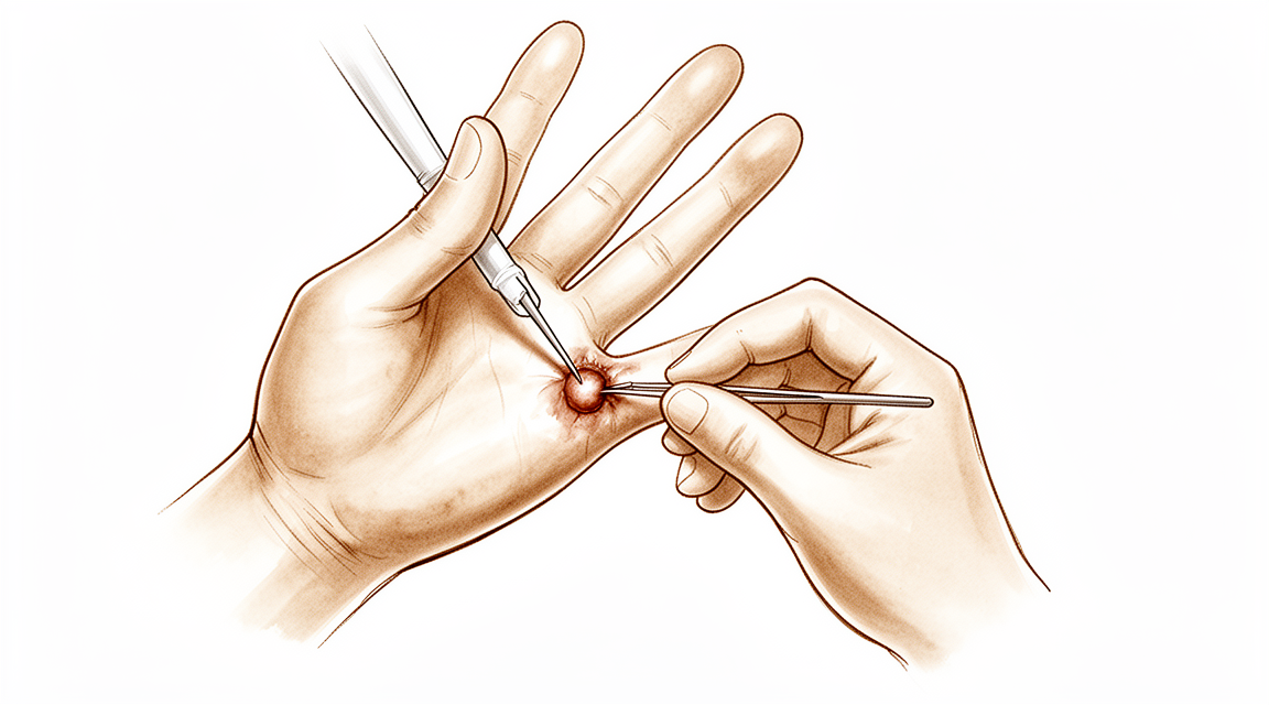
ਤੁਸੀਂ ਕੀ ਮਹਿਸੂਸ ਕਰਦੇ ਹੋ
ਫਲੈਕਸਰ ਟੈਂਡਨ ਦੀ ਝਿੱਲੀ ਦਾ ਗੈਂਗਲੀਅਨ (flexor tendon sheath ganglion) ਹਥੇਲੀ ਵਿੱਚ, ਉਂਗਲ ਦੀ ਜੜ੍ਹ ਉੱਤੇ ਇੱਕ ਛੋਟੀ, ਸਖ਼ਤ ਗੰਢ ਹੈ। ਇਹ ਆਮ ਤੌਰ 'ਤੇ ਸਿਰਫ਼ ਕੁਝ ਮਿਲੀਮੀਟਰ ਚੌੜੀ ਹੁੰਦੀ ਹੈ, ਪਰ ਇੰਨੀ ਛੋਟੀ ਚੀਜ਼ ਤੋਂ ਜਿੰਨੀ ਉਮੀਦ ਹੋਵੇ, ਉਸ ਨਾਲੋਂ ਵੱਧ ਦੁਖ ਸਕਦੀ ਹੈ। ਦਰਦ ਉਦੋਂ ਹੁੰਦਾ ਹੈ ਜਦੋਂ ਤੁਸੀਂ ਕਿਸੇ ਸਖ਼ਤ ਅਤੇ ਪਤਲੀ ਚੀਜ਼ ਨੂੰ ਪਕੜਦੇ ਹੋ, ਜਿਵੇਂ ਸਟੀਅਰਿੰਗ ਵ੍ਹੀਲ, ਗੋਲਫ਼ ਦੀ ਸੋਟੀ, ਸਾਈਕਲ ਦਾ ਹੈਂਡਲ ਜਾਂ ਸੌਦੇ ਵਾਲੇ ਥੈਲੇ ਦਾ ਹੱਥਾ। ਗੰਢ ਉਸ ਚੀਜ਼ ਅਤੇ ਤੁਹਾਡੀ ਉਂਗਲ ਦੀ ਹੱਡੀ ਦੇ ਵਿਚਕਾਰ ਦੱਬੀ ਜਾਂਦੀ ਹੈ, ਅਤੇ ਉਸ ਨੂੰ ਬਚਾਉਣ ਲਈ ਵਿਚਕਾਰ ਕੋਈ ਨਰਮ ਗੱਦੀ ਨਹੀਂ ਹੁੰਦੀ।
ਪਕੜਨ ਵਾਲੇ ਇਨ੍ਹਾਂ ਕੰਮਾਂ ਦੌਰਾਨ ਜਾਂ ਬਾਅਦ ਵਿੱਚ ਦਰਦ ਅਕਸਰ ਵਧ ਜਾਂਦਾ ਹੈ। ਭਾਰੀ ਸੌਦਾ ਚੁੱਕਣਾ, ਔਜ਼ਾਰ ਫੜਨਾ, ਜਾਂ ਧੋਤੇ ਕੱਪੜੇ ਨਿਚੋੜਨਾ ਵਰਗੇ ਕੰਮ ਤਕਲੀਫ਼ ਵਾਲੇ ਬਣ ਸਕਦੇ ਹਨ। ਕੁਝ ਲੋਕ ਦੇਖਦੇ ਹਨ ਕਿ ਚੀਜ਼ ਛੱਡਣ ਤੋਂ ਬਾਅਦ ਦੁਖਣਾ ਸ਼ਾਂਤ ਹੋ ਜਾਂਦਾ ਹੈ। ਗੰਢ ਆਪਣੇ ਆਪ ਵਿੱਚ ਨਰਮ ਹੋਣ ਦੀ ਬਜਾਏ ਸਖ਼ਤ ਹੁੰਦੀ ਹੈ, ਅਤੇ ਬਿਲਕੁਲ ਉੱਥੇ ਹੁੰਦੀ ਹੈ ਜਿੱਥੇ ਤੁਹਾਡੀਆਂ ਉਂਗਲਾਂ ਮੁੜ ਕੇ ਹਥੇਲੀ ਨਾਲ ਮਿਲਦੀਆਂ ਹਨ।
ਕਿਉਂਕਿ ਦਰਦ ਪਕੜਨ ਨਾਲ ਜੁੜਿਆ ਹੈ, ਇਹ ਉਸ ਹੱਥ ਨਾਲ ਤੁਹਾਡੇ ਕੰਮਾਂ ਨੂੰ ਸੀਮਤ ਕਰਨ ਲੱਗ ਸਕਦਾ ਹੈ। ਹੋ ਸਕਦਾ ਹੈ ਤੁਸੀਂ ਕੁਝ ਕੰਮਾਂ ਤੋਂ ਬਚਣ ਲੱਗੋ, ਜਾਂ ਉਸ ਥਾਂ ਉੱਤੇ ਦਬਾਅ ਨਾ ਪਵੇ, ਇਸ ਲਈ ਚੀਜ਼ਾਂ ਫੜਨ ਦਾ ਤਰੀਕਾ ਬਦਲ ਲਓ।
ਜੇ ਤੁਹਾਡੀ ਉਂਗਲ, ਹੱਥ ਜਾਂ ਬਾਂਹ ਕਦੇ ਵੀ ਗਰਮ, ਲਾਲ, ਸੁੱਜੀ ਹੋਈ ਅਤੇ ਦਰਦ ਵਾਲੀ ਹੋ ਜਾਵੇ, ਖ਼ਾਸ ਕਰਕੇ ਬੁਖ਼ਾਰ ਦੇ ਨਾਲ, ਤਾਂ ਉਸੇ ਦਿਨ ਐਮਰਜੈਂਸੀ ਵਿਭਾਗ ਵਿੱਚ ਇਲਾਜ ਕਰਵਾਉਣਾ ਜ਼ਰੂਰੀ ਹੈ। ਉੱਥੇ ਜਾਣ ਲਈ ਤੁਹਾਨੂੰ ਆਪਣੇ ਜੀਪੀ (ਫ਼ੈਮਿਲੀ ਡਾਕਟਰ) ਤੋਂ ਰੈਫ਼ਰਲ ਦੀ ਲੋੜ ਨਹੀਂ ਹੈ।
ਅਸਲ ਵਿੱਚ ਕੀ ਹੋ ਰਿਹਾ ਹੈ
ਤੁਹਾਡੀ ਉਂਗਲ ਦੋ ਟੈਂਡਨਾਂ (tendons, ਪੱਠੇ ਨੂੰ ਹੱਡੀ ਨਾਲ ਜੋੜਨ ਵਾਲੀਆਂ ਮਜ਼ਬੂਤ ਤੰਦਾਂ) ਕਾਰਨ ਮੁੜਦੀ ਹੈ, ਜੋ ਤੁਹਾਡੀ ਬਾਂਹ ਦੇ ਹੇਠਲੇ ਹਿੱਸੇ (forearm) ਤੋਂ ਉਂਗਲ ਦੇ ਹਥੇਲੀ ਵਾਲੇ ਪਾਸੇ ਦੇ ਨਾਲ-ਨਾਲ ਚੱਲਦੇ ਹਨ ਅਤੇ ਉਂਗਲ ਦੀ ਹੱਡੀ ਨਾਲ ਜੁੜੇ ਹੁੰਦੇ ਹਨ। ਇਨ੍ਹਾਂ ਨੂੰ ਰੱਸੀਆਂ ਵਾਂਗ ਸਮਝੋ ਜੋ ਉਂਗਲ ਮੋੜਨ ਵੇਲੇ ਖਿੱਚਦੀਆਂ ਹਨ। ਇਨ੍ਹਾਂ ਰੱਸੀਆਂ ਨੂੰ ਹੱਡੀ ਤੋਂ ਦੂਰ ਕਮਾਨ ਵਾਂਗ ਉੱਭਰਨ ਦੀ ਬਜਾਏ ਹੱਡੀ ਦੇ ਨੇੜੇ ਰੱਖਣ ਲਈ, ਇਹ ਸਖ਼ਤ ਪਟਿਆਂ ਦੀ ਬਣੀ ਇੱਕ ਸੁਰੰਗ ਦੇ ਅੰਦਰ ਚੱਲਦੇ ਹਨ, ਜਿਨ੍ਹਾਂ ਪਟਿਆਂ ਨੂੰ ਪੁਲੀਆਂ (pulleys) ਕਿਹਾ ਜਾਂਦਾ ਹੈ। ਇਸ ਸੁਰੰਗ ਦੇ ਅੰਦਰ ਇੱਕ ਪਤਲੀ, ਤਿਲਕਣੀ ਪਰਤ ਹੁੰਦੀ ਹੈ ਜੋ ਟੈਂਡਨਾਂ ਨੂੰ ਖ਼ੁਰਾਕ ਪਹੁੰਚਾਉਂਦੀ ਹੈ ਅਤੇ ਉਨ੍ਹਾਂ ਨੂੰ ਸੁਚੱਜੇ ਢੰਗ ਨਾਲ ਖਿਸਕਦਾ ਰੱਖਦੀ ਹੈ।
ਫਲੈਕਸਰ ਟੈਂਡਨ ਦੀ ਝਿੱਲੀ ਦਾ ਗੈਂਗਲੀਅਨ ਗਾੜ੍ਹੇ, ਜੈਲੀ ਵਰਗੇ ਤਰਲ ਦੀ ਇੱਕ ਛੋਟੀ ਥੈਲੀ ਹੈ ਜੋ ਉਸ ਸੁਰੰਗ ਵਿੱਚੋਂ ਬਾਹਰ ਵੱਲ ਉੱਭਰ ਆਉਂਦੀ ਹੈ। ਇਹ ਥੈਲੀ ਝਿੱਲੀ ਦੀ ਆਪਣੀ ਅੰਦਰਲੀ ਪਰਤ ਤੋਂ ਭਰਦੀ ਹੈ, ਇਸ ਲਈ ਇਹ ਦਬਾਅ ਹੇਠ ਇੱਕ ਛੋਟੇ ਰਿਸਾਅ ਵਾਂਗ ਵਿਹਾਰ ਕਰਦੀ ਹੈ: ਜੇ ਇਸ ਦਾ ਤਰਲ ਕੱਢਿਆ ਜਾਵੇ ਤਾਂ ਇਹ ਫਿਰ ਭਰ ਜਾਂਦੀ ਹੈ, ਕਿਉਂਕਿ ਰਿਸਾਅ ਅਜੇ ਵੀ ਮੌਜੂਦ ਹੈ। ਥੈਲੀ ਹਥੇਲੀ ਵਿੱਚ ਉਂਗਲ ਦੀ ਬਿਲਕੁਲ ਜੜ੍ਹ ਉੱਤੇ ਹੁੰਦੀ ਹੈ, ਜਿੱਥੇ ਸੁਰੰਗ ਹੱਡੀ ਨਾਲ ਕੱਸ ਕੇ ਬੱਝੀ ਹੁੰਦੀ ਹੈ ਅਤੇ ਉਸ ਦੇ ਉੱਪਰ ਕੋਈ ਨਰਮ ਗੱਦੀ ਨਹੀਂ ਹੁੰਦੀ। ਇਸੇ ਲਈ ਸਿਰਫ਼ ਕੁਝ ਮਿਲੀਮੀਟਰ ਚੌੜੀ ਗੰਢ ਪਕੜਨ ਵੇਲੇ ਇੰਨੀ ਜ਼ਿਆਦਾ ਦੁਖ ਸਕਦੀ ਹੈ: ਤੁਸੀਂ ਜੋ ਵੀ ਫੜਿਆ ਹੋਵੇ, ਗੰਢ ਉਸ ਚੀਜ਼ ਅਤੇ ਹੇਠਲੀ ਹੱਡੀ ਦੇ ਵਿਚਕਾਰ ਭੀਂਚੀ ਜਾਂਦੀ ਹੈ।
ਇਹੀ ਥਾਂ ਉਨ੍ਹਾਂ ਹੋਰ ਗੱਲਾਂ ਦਾ ਕਾਰਨ ਵੀ ਸਮਝਾਉਂਦੀ ਹੈ ਜੋ ਸ਼ਾਇਦ ਤੁਸੀਂ ਮਹਿਸੂਸ ਕੀਤੀਆਂ ਹੋਣ। ਕਿਉਂਕਿ ਗੰਢ ਇੱਕ ਤੰਗ ਸੁਰੰਗ ਦੇ ਅੰਦਰ ਥਾਂ ਘੇਰਦੀ ਹੈ, ਇਹ ਖਿਸਕਦੇ ਹੋਏ ਟੈਂਡਨ ਲਈ ਥਾਂ ਤੰਗ ਕਰ ਸਕਦੀ ਹੈ, ਇਸੇ ਲਈ ਇਸ ਥਾਂ ਉੱਤੇ ਗੰਢ ਵਾਲੇ ਕੁਝ ਲੋਕਾਂ ਦੀ ਉਂਗਲ ਅਟਕਦੀ ਜਾਂ ਕਲਿੱਕ ਵੀ ਕਰਦੀ ਹੈ। ਉਂਗਲ ਦੀਆਂ ਨੇੜਲੀਆਂ ਨਸਾਂ (nerves) ਝਿੱਲੀ ਦੇ ਬਿਲਕੁਲ ਨਾਲ-ਨਾਲ ਚੱਲਦੀਆਂ ਹਨ, ਹਰ ਪਾਸੇ ਇੱਕ, ਇਸ ਲਈ ਨਸ ਉੱਤੇ ਦਬਾਅ ਪਾਉਣ ਵਾਲੀ ਗੰਢ ਸਿਰਫ਼ ਦਰਦ ਹੀ ਨਹੀਂ, ਸਗੋਂ ਝਰਨਾਹਟ ਜਾਂ ਸੁੰਨਪਨ ਵੀ ਪੈਦਾ ਕਰ ਸਕਦੀ ਹੈ।
ਇਹ ਜਾਣਨਾ ਚੰਗਾ ਹੈ ਕਿ ਬਿਲਕੁਲ ਇਸੇ ਥਾਂ ਉੱਤੇ ਗੰਢ ਹੋਰ ਕਾਰਨਾਂ ਕਰਕੇ ਵੀ ਹੋ ਸਕਦੀ ਹੈ, ਜਿਵੇਂ ਕੋਈ ਟੈਂਡਨ ਜੋ ਖਿਸਕਣ ਵੇਲੇ ਅਟਕਦਾ ਹੈ, ਜਾਂ ਹਥੇਲੀ ਦੇ ਟਿਸ਼ੂ ਦਾ ਮੋਟਾ ਹੋਣਾ ਜੋ ਉਂਗਲ ਨੂੰ ਹਥੇਲੀ ਵੱਲ ਖਿੱਚਦਾ ਹੈ। ਤੁਹਾਡਾ ਸਰਜਨ ਆਮ ਤੌਰ 'ਤੇ ਤੁਹਾਡੇ ਹੱਥ ਦੀ ਜਾਂਚ ਕਰਕੇ ਇਨ੍ਹਾਂ ਵਿੱਚ ਫ਼ਰਕ ਪਛਾਣ ਸਕਦਾ ਹੈ, ਅਤੇ ਹਰ ਇੱਕ ਦਾ ਇਲਾਜ ਵੱਖਰਾ ਹੁੰਦਾ ਹੈ।
ਅਸੀਂ ਇਸ ਬਾਰੇ ਕੀ ਕਰ ਸਕਦੇ ਹਾਂ
ਡਾ. ਕੀਰਨ ਹਿਰਪਰਾ, ਮਾਟਰ ਪ੍ਰਾਈਵੇਟ ਹਸਪਤਾਲ ਰੌਕਹੈਂਪਟਨ ਵਿੱਚ ਉੱਪਰਲੇ ਅੰਗ (ਮੋਢੇ ਤੋਂ ਹੱਥ ਤੱਕ ਦੀ ਬਾਂਹ) ਦੇ ਸਰਜਨ, ਤੁਹਾਡੀ ਸਥਿਤੀ ਦੇ ਅਨੁਕੂਲ ਸਭ ਤੋਂ ਘੱਟ ਚੀਰ-ਫਾੜ ਵਾਲੇ ਵਿਕਲਪਾਂ ਤੋਂ ਸ਼ੁਰੂਆਤ ਕਰਦੇ ਹਨ। ਮਰੀਜ਼ਾਂ ਨੂੰ ਆਮ ਤੌਰ 'ਤੇ ਉਨ੍ਹਾਂ ਦਾ ਜੀਪੀ (ਫ਼ੈਮਿਲੀ ਡਾਕਟਰ) ਸਾਡੇ ਕਲੀਨਿਕ ਵਿੱਚ ਭੇਜਦਾ ਹੈ; ਜੇ ਕਿਸੇ ਫਿਜ਼ੀਓਥੈਰੇਪਿਸਟ ਨੇ ਤੁਹਾਨੂੰ ਸਾਡੇ ਕੋਲ ਆਉਣ ਦੀ ਸਲਾਹ ਦਿੱਤੀ ਹੈ, ਤਾਂ ਵੀ Medicare ਦੀ ਛੋਟ (rebate) ਦੇ ਯੋਗ ਹੋਣ ਲਈ ਤੁਹਾਨੂੰ ਆਪਣੇ ਜੀਪੀ ਤੋਂ ਰੈਫ਼ਰਲ (referral) ਲੈਣਾ ਪਵੇਗਾ। ਤੁਹਾਡੀ ਮੁਲਾਕਾਤ ਵੇਲੇ ਅਸੀਂ ਤੁਹਾਡੀ ਤਕਲੀਫ਼ ਦਾ ਵੇਰਵਾ ਪੁੱਛਦੇ ਹਾਂ, ਤੁਹਾਡੇ ਹੱਥ ਦੀ ਜਾਂਚ ਕਰਦੇ ਹਾਂ, ਅਤੇ ਜੇ ਇਹ ਪੱਕਾ ਕਰਨ ਲਈ ਲੋੜ ਹੋਵੇ ਕਿ ਗੰਢ ਕੀ ਹੈ, ਤਾਂ ਅਲਟਰਾਸਾਊਂਡ ਸਕੈਨ ਵਰਗੇ ਸਕੈਨ ਕਰਵਾਉਂਦੇ ਹਾਂ।
ਕਿਉਂਕਿ ਇਹ ਗੰਢ ਨੁਕਸਾਨ-ਰਹਿਤ ਹੈ ਅਤੇ ਵਧਦੀ ਨਹੀਂ ਰਹਿੰਦੀ, ਇਲਾਜ ਪੂਰੀ ਤਰ੍ਹਾਂ ਇਸ ਗੱਲ ਉੱਤੇ ਨਿਰਭਰ ਕਰਦਾ ਹੈ ਕਿ ਇਹ ਤੁਹਾਨੂੰ ਕਿੰਨੀ ਤੰਗ ਕਰਦੀ ਹੈ। ਜਿਹੜੀ ਗੰਢ ਤੁਹਾਨੂੰ ਮਹਿਸੂਸ ਤਾਂ ਹੁੰਦੀ ਹੈ ਪਰ ਆਮ ਪਕੜ ਵੇਲੇ ਦੁਖਦੀ ਨਹੀਂ, ਉਸ ਲਈ ਸ਼ਾਇਦ ਕੁਝ ਵੀ ਕਰਨ ਦੀ ਲੋੜ ਨਾ ਪਵੇ। ਜੇ ਕੋਈ ਖ਼ਾਸ ਪਕੜ ਵਾਰ-ਵਾਰ ਦਰਦ ਕਰਦੀ ਹੈ, ਤਾਂ ਚੀਜ਼ਾਂ ਫੜਨ ਦਾ ਤਰੀਕਾ ਬਦਲਣਾ ਜਾਂ ਕੁਝ ਸਮੇਂ ਲਈ ਉਹ ਕੰਮ ਘਟਾਉਣਾ ਕਾਫ਼ੀ ਹੋ ਸਕਦਾ ਹੈ। ਹੈਂਡ ਥੈਰੇਪੀ ਵੀ ਤੁਹਾਨੂੰ ਹੱਥ ਵਰਤਣ ਦੇ ਅਜਿਹੇ ਤਰੀਕੇ ਲੱਭਣ ਵਿੱਚ ਮਦਦ ਕਰ ਸਕਦੀ ਹੈ ਜਿਨ੍ਹਾਂ ਨਾਲ ਉਸ ਥਾਂ ਉੱਤੇ ਘੱਟ ਦਬਾਅ ਪਵੇ।
ਸੂਈ ਨਾਲ ਗੰਢ ਦਾ ਤਰਲ ਕੱਢਣਾ, ਜਿਸ ਨੂੰ ਐਸਪੀਰੇਸ਼ਨ (aspiration) ਕਿਹਾ ਜਾਂਦਾ ਹੈ, ਅਕਸਰ ਸਰਜਰੀ ਤੋਂ ਪਹਿਲਾਂ ਅਜ਼ਮਾਇਆ ਜਾਂਦਾ ਹੈ। ਇਸ ਨਾਲ ਥੈਲੀ ਤਰਲ ਤੋਂ ਖ਼ਾਲੀ ਹੋ ਜਾਂਦੀ ਹੈ, ਪਰ ਕਿਉਂਕਿ ਥੈਲੀ ਨੂੰ ਟੈਂਡਨ ਦੀ ਝਿੱਲੀ ਤੋਂ ਤਰਲ ਮਿਲਦਾ ਹੈ, ਇਹ ਆਮ ਤੌਰ 'ਤੇ ਫਿਰ ਭਰ ਜਾਂਦੀ ਹੈ। ਕੁਝ ਗੰਢਾਂ ਸਮੇਂ ਨਾਲ ਆਪਣੇ ਆਪ ਵੀ ਠੀਕ ਹੋ ਜਾਂਦੀਆਂ ਹਨ। ਅਸੀਂ ਤੁਹਾਡੇ ਨਾਲ ਗੱਲ ਕਰਾਂਗੇ ਕਿ ਤੁਹਾਡੇ ਮਾਮਲੇ ਵਿੱਚ ਤਰਲ ਕਢਵਾਉਣਾ ਅਜ਼ਮਾਉਣ ਦੇ ਲਾਇਕ ਹੈ ਜਾਂ ਨਹੀਂ।
ਜੇ ਇਨ੍ਹਾਂ ਸਧਾਰਨ ਕਦਮਾਂ ਨਾਲ ਤੁਹਾਨੂੰ ਕਾਫ਼ੀ ਰਾਹਤ ਨਹੀਂ ਮਿਲੀ, ਤਾਂ ਸਰਜਰੀ ਬਾਰੇ ਸੋਚਿਆ ਜਾ ਸਕਦਾ ਹੈ। ਓਪਰੇਸ਼ਨ ਵਿੱਚ ਉਂਗਲ ਦੀ ਜੜ੍ਹ ਉੱਤੇ ਇੱਕ ਛੋਟੇ ਚੀਰੇ ਰਾਹੀਂ ਗੰਢ ਕੱਢੀ ਜਾਂਦੀ ਹੈ, ਅਤੇ ਨਾਲ ਹੀ ਟੈਂਡਨ ਦੀ ਝਿੱਲੀ ਦਾ ਉਹ ਛੋਟਾ ਟੁਕੜਾ ਵੀ ਜਿਸ ਤੋਂ ਗੰਢ ਉੱਗਦੀ ਹੈ, ਅਤੇ ਇਹੀ ਗੰਢ ਨੂੰ ਵਾਪਸ ਆਉਣ ਤੋਂ ਰੋਕਦਾ ਹੈ। ਅਸੀਂ ਆਮ ਤੌਰ 'ਤੇ ਸਰਜਰੀ ਦਾ ਸੁਝਾਅ ਉਦੋਂ ਦਿੰਦੇ ਹਾਂ ਜਦੋਂ ਦਰਦ ਕਾਰਨ ਤੁਹਾਡੀ ਰੋਜ਼ਾਨਾ ਜ਼ਿੰਦਗੀ ਦਾ ਕੋਈ ਖ਼ਾਸ, ਵਾਰ-ਵਾਰ ਕੀਤਾ ਜਾਣ ਵਾਲਾ ਕੰਮ ਸੀਮਤ ਹੋ ਰਿਹਾ ਹੋਵੇ, ਅਤੇ ਇਹ ਫ਼ੈਸਲਾ ਅਸੀਂ ਤੁਹਾਡੇ ਨਾਲ ਮਿਲ ਕੇ ਕਰਦੇ ਹਾਂ।
ਜੇ ਤੁਹਾਡੀ ਉਂਗਲ, ਹੱਥ ਜਾਂ ਬਾਂਹ ਕਦੇ ਵੀ ਗਰਮ, ਲਾਲ, ਸੁੱਜੀ ਹੋਈ ਅਤੇ ਦਰਦ ਵਾਲੀ ਹੋ ਜਾਵੇ, ਖ਼ਾਸ ਕਰਕੇ ਬੁਖ਼ਾਰ ਦੇ ਨਾਲ, ਤਾਂ ਉਸੇ ਦਿਨ ਐਮਰਜੈਂਸੀ ਵਿਭਾਗ ਜਾਓ।
ਕੀ ਉਮੀਦ ਰੱਖੀਏ
ਇਸ ਤਰ੍ਹਾਂ ਦੇ ਜ਼ਿਆਦਾਤਰ ਗੈਂਗਲੀਅਨ ਨੁਕਸਾਨ-ਰਹਿਤ ਹੁੰਦੇ ਹਨ ਅਤੇ ਵਧਦੇ ਨਹੀਂ ਰਹਿੰਦੇ। ਕੁਝ ਸਮੇਂ ਨਾਲ ਆਪਣੇ ਆਪ ਠੀਕ ਹੋ ਜਾਂਦੇ ਹਨ, ਅਤੇ ਕੁਝ ਆਉਂਦੇ-ਜਾਂਦੇ ਰਹਿੰਦੇ ਹਨ। ਜੇ ਤੁਹਾਡਾ ਗੈਂਗਲੀਅਨ ਦੁਖਦਾ ਨਹੀਂ, ਤਾਂ ਹੋ ਸਕਦਾ ਹੈ ਤੁਹਾਨੂੰ ਕਦੇ ਵੀ ਇਲਾਜ ਦੀ ਲੋੜ ਨਾ ਪਵੇ।
ਜੇ ਪਕੜਨ ਵੇਲੇ ਗੰਢ ਦੁਖਦੀ ਹੈ, ਤਾਂ ਸਧਾਰਨ ਕਦਮ ਮਦਦ ਕਰ ਸਕਦੇ ਹਨ। ਸੂਈ ਨਾਲ ਤਰਲ ਕੱਢਣ ਨਾਲ ਥੈਲੀ ਖ਼ਾਲੀ ਹੋ ਜਾਂਦੀ ਹੈ, ਪਰ ਕਿਉਂਕਿ ਥੈਲੀ ਨੂੰ ਟੈਂਡਨ ਦੀ ਝਿੱਲੀ ਤੋਂ ਤਰਲ ਮਿਲਦਾ ਹੈ, ਇਹ ਆਮ ਤੌਰ 'ਤੇ ਫਿਰ ਭਰ ਜਾਂਦੀ ਹੈ। ਜਦੋਂ ਤੁਸੀਂ ਇਹ ਦੇਖਣ ਦੀ ਉਡੀਕ ਕਰਦੇ ਹੋ ਕਿ ਗੰਢ ਠੀਕ ਹੁੰਦੀ ਹੈ ਜਾਂ ਨਹੀਂ, ਤਾਂ ਚੀਜ਼ਾਂ ਫੜਨ ਦਾ ਤਰੀਕਾ ਬਦਲਣਾ, ਜਾਂ ਜਿਹੜਾ ਕੰਮ ਦਰਦ ਵਧਾਉਂਦਾ ਹੈ ਉਸ ਨੂੰ ਘਟਾਉਣਾ, ਕਾਫ਼ੀ ਹੋ ਸਕਦਾ ਹੈ।
ਜੇ ਇਨ੍ਹਾਂ ਕਦਮਾਂ ਨਾਲ ਕਾਫ਼ੀ ਮਦਦ ਨਹੀਂ ਮਿਲੀ, ਤਾਂ ਸਰਜਰੀ ਵਿੱਚ ਗੰਢ ਨੂੰ ਝਿੱਲੀ ਦੇ ਉਸ ਛੋਟੇ ਟੁਕੜੇ ਸਮੇਤ ਕੱਢਿਆ ਜਾਂਦਾ ਹੈ ਜਿਸ ਤੋਂ ਇਹ ਉੱਗਦੀ ਹੈ, ਅਤੇ ਇਹੀ ਇਸ ਨੂੰ ਵਾਪਸ ਆਉਣ ਤੋਂ ਰੋਕਦਾ ਹੈ। ਜ਼ਿਆਦਾਤਰ ਲੋਕ ਮਹਿਸੂਸ ਕਰਦੇ ਹਨ ਕਿ ਜਦੋਂ ਉਹ ਥਾਂ ਹੋਰ ਨਹੀਂ ਭੀਂਚੀ ਜਾਂਦੀ, ਤਾਂ ਉਨ੍ਹਾਂ ਦੀ ਪਕੜ ਵਧੇਰੇ ਆਰਾਮਦਾਇਕ ਲੱਗਦੀ ਹੈ। ਹੱਥ ਦੇ ਕਿਸੇ ਵੀ ਓਪਰੇਸ਼ਨ ਵਾਂਗ, ਇਸ ਦੇ ਵੀ ਕੁਝ ਖ਼ਤਰੇ ਹਨ ਜਿਨ੍ਹਾਂ ਨੂੰ ਤੋਲਣਾ ਪੈਂਦਾ ਹੈ, ਜਿਨ੍ਹਾਂ ਵਿੱਚ ਉਂਗਲ ਦੇ ਇੱਕ ਪਾਸੇ ਦੇ ਨਾਲ-ਨਾਲ ਸੁੰਨ ਥਾਂ, ਅਕੜਾਅ, ਲਾਗ (ਇਨਫੈਕਸ਼ਨ) ਅਤੇ ਮੋਟਾ ਜਾਂ ਛੂਹਣ ਨਾਲ ਦੁਖਣ ਵਾਲਾ ਦਾਗ਼ ਸ਼ਾਮਲ ਹਨ। ਫ਼ੈਸਲਾ ਕਰਨ ਤੋਂ ਪਹਿਲਾਂ ਤੁਹਾਡਾ ਸਰਜਨ ਤੁਹਾਡੇ ਨਾਲ ਇਨ੍ਹਾਂ ਬਾਰੇ ਗੱਲ ਕਰੇਗਾ।
ਸਰਜਰੀ ਤੋਂ ਬਾਅਦ ਠੀਕ ਹੋਣ ਵਿੱਚ ਦਿਨ ਨਹੀਂ, ਸਗੋਂ ਹਫ਼ਤੇ ਲੱਗਦੇ ਹਨ। ਸ਼ੁਰੂ ਵਿੱਚ ਤੁਹਾਡਾ ਹੱਥ ਦੁਖੇਗਾ, ਅਤੇ ਉਂਗਲ ਦੀ ਜੜ੍ਹ ਉੱਤੇ ਛੋਟੇ ਚੀਰੇ ਨੂੰ ਠੀਕ ਹੋਣ ਵਿੱਚ ਸਮਾਂ ਲੱਗੇਗਾ। ਸਰਜਰੀ ਤੋਂ ਬਾਅਦ ਹੈਂਡ ਥੈਰੇਪੀ ਐਕਸਟੈਂਡ ਰੀਹੈਬਿਲੀਟੇਸ਼ਨ (Extend Rehabilitation) ਵਿਖੇ ਰੂਬੀ ਡੂਲਨ (Ruby Doolan) ਨਾਲ ਹੁੰਦੀ ਹੈ। ਰੂਬੀ ਇੱਕ ਹੈਂਡ ਥੈਰੇਪਿਸਟ ਹੈ: ਜਦੋਂ ਤੁਹਾਡੀ ਉਂਗਲ ਠੀਕ ਹੋ ਰਹੀ ਹੋਵੇਗੀ, ਉਹ ਤੁਹਾਡੀਆਂ ਕਸਰਤਾਂ ਦੀ ਸੇਧ ਦੇਵੇਗੀ ਅਤੇ ਤੁਹਾਨੂੰ ਲੋੜੀਂਦਾ ਕੋਈ ਵੀ ਸਪਲਿੰਟ ਬਣਾਏਗੀ। ਜ਼ਿਆਦਾਤਰ ਲੋਕ ਕੁਝ ਹਫ਼ਤਿਆਂ ਦੇ ਅੰਦਰ ਆਪਣੇ ਆਮ ਪਕੜਨ ਵਾਲੇ ਕੰਮਾਂ ਉੱਤੇ ਵਾਪਸ ਆ ਜਾਂਦੇ ਹਨ, ਹਾਲਾਂਕਿ ਦਾਗ਼ ਉੱਤੇ ਛੂਹਣ ਨਾਲ ਹੋਣ ਵਾਲਾ ਕੁਝ ਦੁਖਣਾ ਥੋੜ੍ਹਾ ਲੰਬਾ ਰਹਿ ਸਕਦਾ ਹੈ।
ਜੇ ਤੁਹਾਡੇ ਲੱਛਣ ਸ਼ਾਂਤ ਨਹੀਂ ਹੋ ਰਹੇ, ਹਫ਼ਤਿਆਂ ਦੌਰਾਨ ਵਿਗੜ ਰਹੇ ਹਨ, ਤੁਹਾਨੂੰ ਰਾਤ ਨੂੰ ਨੀਂਦ ਤੋਂ ਜਗਾਉਂਦੇ ਹਨ, ਜਾਂ ਤੁਹਾਨੂੰ ਕੰਮ ਕਰਨ ਜਾਂ ਹੱਥ ਵਰਤਣ ਤੋਂ ਰੋਕਦੇ ਹਨ, ਤਾਂ ਆਪਣੇ ਜੀਪੀ ਨੂੰ ਮਿਲੋ ਜਾਂ ਮਾਹਰ ਡਾਕਟਰ ਤੋਂ ਜਾਂਚ ਦੀ ਮੰਗ ਕਰੋ।
ਡਾਕਟਰ ਨੂੰ ਕਦੋਂ ਮਿਲਣਾ ਚਾਹੀਦਾ ਹੈ
ਉਂਗਲ ਦੀ ਜੜ੍ਹ ਉੱਤੇ ਜ਼ਿਆਦਾਤਰ ਗੰਢਾਂ ਨੁਕਸਾਨ-ਰਹਿਤ ਗੈਂਗਲੀਅਨ ਹੁੰਦੀਆਂ ਹਨ, ਪਰ ਉਸ ਥਾਂ ਉੱਤੇ ਕੁਝ ਹੋਰ ਚੀਜ਼ਾਂ ਵੀ ਬਿਲਕੁਲ ਇਸੇ ਤਰ੍ਹਾਂ ਦੀਆਂ ਲੱਗ ਸਕਦੀਆਂ ਹਨ। ਮੋੜਨ ਵੇਲੇ ਫਸਣ ਜਾਂ ਕਲਿੱਕ ਕਰਨ ਵਾਲੀ ਉਂਗਲ, ਜਾਂ ਹਥੇਲੀ ਵਿੱਚ ਇੱਕ ਸਖ਼ਤ ਡੋਰੀ ਜੋ ਉਂਗਲ ਨੂੰ ਹਥੇਲੀ ਵੱਲ ਖਿੱਚ ਰਹੀ ਹੈ, ਅਜਿਹੀਆਂ ਸਥਿਤੀਆਂ ਹਨ ਜਿਨ੍ਹਾਂ ਵਿੱਚ ਤੁਹਾਡਾ ਸਰਜਨ ਤੁਹਾਡੇ ਹੱਥ ਦੀ ਜਾਂਚ ਕਰਕੇ ਫ਼ਰਕ ਪਛਾਣ ਸਕਦਾ ਹੈ। ਕਿਸੇ ਵੀ ਨਵੀਂ ਗੰਢ ਦੀ ਜਾਂਚ ਕਰਵਾਉਣੀ ਚੰਗੀ ਹੈ, ਤਾਂ ਜੋ ਤੁਹਾਨੂੰ ਪਤਾ ਲੱਗੇ ਕਿ ਤੁਹਾਨੂੰ ਇਨ੍ਹਾਂ ਵਿੱਚੋਂ ਕਿਹੜੀ ਹੈ।
ਜੇ ਪਕੜਨ ਵੇਲੇ ਗੰਢ ਦੁਖਦੀ ਹੈ, ਜੇ ਤੁਹਾਨੂੰ ਉਂਗਲ ਵਿੱਚ ਝਰਨਾਹਟ ਜਾਂ ਸੁੰਨਪਨ ਮਹਿਸੂਸ ਹੁੰਦਾ ਹੈ, ਜਾਂ ਜੇ ਉਂਗਲ ਅਟਕਦੀ ਜਾਂ ਕਲਿੱਕ ਕਰਦੀ ਹੈ, ਤਾਂ ਮਾਹਰ ਡਾਕਟਰ ਤੋਂ ਜਾਂਚ ਦੀ ਮੰਗ ਕਰੋ। ਜੇ ਤੁਹਾਡੇ ਲੱਛਣ ਸ਼ਾਂਤ ਨਹੀਂ ਹੋ ਰਹੇ, ਹਫ਼ਤਿਆਂ ਦੌਰਾਨ ਵਿਗੜ ਰਹੇ ਹਨ, ਤੁਹਾਨੂੰ ਰਾਤ ਨੂੰ ਨੀਂਦ ਤੋਂ ਜਗਾਉਂਦੇ ਹਨ, ਜਾਂ ਤੁਹਾਨੂੰ ਕੰਮ ਕਰਨ ਜਾਂ ਹੱਥ ਵਰਤਣ ਤੋਂ ਰੋਕਦੇ ਹਨ, ਤਾਂ ਵੀ ਜਾਂਚ ਦੀ ਮੰਗ ਕਰੋ।
ਜੇ ਤੁਹਾਡੀ ਉਂਗਲ, ਹੱਥ ਜਾਂ ਬਾਂਹ ਕਦੇ ਵੀ ਗਰਮ, ਲਾਲ, ਸੁੱਜੀ ਹੋਈ ਅਤੇ ਦਰਦ ਵਾਲੀ ਹੋ ਜਾਵੇ, ਖ਼ਾਸ ਕਰਕੇ ਬੁਖ਼ਾਰ ਦੇ ਨਾਲ, ਤਾਂ ਉਸੇ ਦਿਨ ਐਮਰਜੈਂਸੀ ਵਿਭਾਗ ਜਾਓ। ਉੱਥੇ ਜਾਣ ਲਈ ਤੁਹਾਨੂੰ ਆਪਣੇ ਜੀਪੀ ਤੋਂ ਰੈਫ਼ਰਲ ਦੀ ਲੋੜ ਨਹੀਂ ਹੈ।
ਵਿਸਥਾਰ ਵਿੱਚ
Advanced reading: the deeper science (optional)
ਇਹ ਹਿੱਸਾ ਤੁਹਾਡੇ ਆਪਣੇ ਇਲਾਜ ਬਾਰੇ ਫ਼ੈਸਲਿਆਂ ਲਈ ਲੋੜ ਤੋਂ ਵੱਧ ਡੂੰਘਾਈ ਵਿੱਚ ਜਾਂਦਾ ਹੈ। ਫਲੈਕਸਰ ਟੈਂਡਨ ਦੀ ਝਿੱਲੀ ਦਾ ਗੈਂਗਲੀਅਨ (flexor tendon sheath ganglion) ਵਾਧੂ ਪੜ੍ਹਨ ਦੇ ਲਾਇਕ ਹੈ ਕਿਉਂਕਿ ਇਹ ਹੱਥ ਦੀ ਸਰਜਰੀ ਦੀ ਸਭ ਤੋਂ ਛੋਟੀ ਗੰਢ ਹੈ ਜੋ ਲਗਾਤਾਰ ਆਪਣੇ ਆਕਾਰ ਦੇ ਮੁਕਾਬਲੇ ਕਿਤੇ ਜ਼ਿਆਦਾ ਤਕਲੀਫ਼ ਦਿੰਦੀ ਹੈ, ਅਤੇ ਕਿਉਂਕਿ ਇਹ ਜਿਸ ਚੀਜ਼ ਨਾਲ ਜੁੜੀ ਹੈ, ਉਸੇ ਤੋਂ ਤੈਅ ਹੁੰਦਾ ਹੈ ਕਿ ਇਹ ਕਿਉਂ ਦੁਖਦੀ ਹੈ ਅਤੇ ਸਧਾਰਨ ਇਲਾਜ ਕਿਉਂ ਨਾਕਾਮ ਹੁੰਦੇ ਹਨ।
ਮਟਰ ਜਿੰਨੀ ਗੰਢ ਜੋ ਆਪਣੇ ਆਕਾਰ ਤੋਂ ਕਿਤੇ ਜ਼ਿਆਦਾ ਦੁਖਦੀ ਹੈ
ਇਹ ਸਿਸਟ, ਜਿਨ੍ਹਾਂ ਨੂੰ ਰੈਟੀਨੈਕੂਲਰ ਸਿਸਟ (retinacular cysts) ਜਾਂ ਵੋਲਰ ਰੈਟੀਨੈਕੂਲਰ ਗੈਂਗਲੀਆ (volar retinacular ganglia) ਵੀ ਕਹਿੰਦੇ ਹਨ, ਉਸ ਰੇਸ਼ੇਦਾਰ ਝਿੱਲੀ ਤੋਂ ਪੈਦਾ ਹੁੰਦੇ ਹਨ ਜੋ ਫਲੈਕਸਰ ਟੈਂਡਨਾਂ ਨੂੰ ਉਂਗਲ ਦੀਆਂ ਹੱਡੀਆਂ ਨਾਲ ਲੱਗਾ ਕੇ ਰੱਖਦੀ ਹੈ [1]। ਇਹ ਹਥੇਲੀ ਵਿੱਚ ਉਂਗਲ ਦੀ ਜੜ੍ਹ ਉੱਤੇ ਹੁੰਦੇ ਹਨ, ਆਮ ਤੌਰ 'ਤੇ ਸਿਰਫ਼ ਕੁਝ ਮਿਲੀਮੀਟਰ ਚੌੜੇ ਹੁੰਦੇ ਹਨ, ਅਤੇ ਨਰਮ ਹੋਣ ਦੀ ਬਜਾਏ ਸਖ਼ਤ ਹੁੰਦੇ ਹਨ।
ਇਸ ਦਾ ਲੱਛਣ ਖ਼ਾਸ ਕਿਸਮ ਦਾ ਹੈ ਅਤੇ ਬਣਤਰ ਸਮਝ ਆਉਣ ਤੋਂ ਬਾਅਦ ਆਪਣੇ ਆਪ ਸਮਝ ਆ ਜਾਂਦਾ ਹੈ: ਕਿਸੇ ਸਖ਼ਤ ਅਤੇ ਪਤਲੀ ਚੀਜ਼ ਨੂੰ ਫੜਨ ਵੇਲੇ ਦਰਦ, ਜਿਵੇਂ ਸਟੀਅਰਿੰਗ ਵ੍ਹੀਲ, ਗੋਲਫ਼ ਦੀ ਸੋਟੀ, ਸਾਈਕਲ ਦਾ ਹੈਂਡਲ, ਜਾਂ ਸੌਦੇ ਵਾਲੇ ਥੈਲੇ ਦਾ ਹੱਥਾ। ਸਿਸਟ ਚੀਜ਼ ਅਤੇ ਹੇਠਲੀ ਹੱਡੀ ਦੇ ਵਿਚਕਾਰ ਫਸ ਜਾਂਦੀ ਹੈ, ਅਤੇ ਇਸ ਨੂੰ ਗੱਦੀ ਦੇਣ ਲਈ ਕੋਈ ਨਰਮ ਟਿਸ਼ੂ ਨਹੀਂ ਹੁੰਦਾ ਕਿਉਂਕਿ ਝਿੱਲੀ ਸਿੱਧੀ ਉਂਗਲ ਦੀ ਹੱਡੀ (phalanx) ਨਾਲ ਬੱਝੀ ਹੁੰਦੀ ਹੈ।
ਇਸੇ ਲਈ ਆਕਾਰ ਅਤੇ ਲੱਛਣਾਂ ਦਾ ਆਪਸ ਵਿੱਚ ਬਹੁਤ ਘੱਟ ਸਬੰਧ ਹੁੰਦਾ ਹੈ। ਗੁੱਟ ਦੇ ਪਿਛਲੇ ਪਾਸੇ ਦੀ ਇੱਕ ਵੱਡੀ ਨਰਮ ਸੋਜ ਦਰਦ-ਰਹਿਤ ਹੋ ਸਕਦੀ ਹੈ, ਜਦਕਿ ਉਂਗਲ ਦੀ ਜੜ੍ਹ ਉੱਤੇ ਉਸ ਤੋਂ ਕਿਤੇ ਛੋਟੀ ਗੰਢ ਸੱਚਮੁੱਚ ਕੰਮਾਂ ਨੂੰ ਸੀਮਤ ਕਰ ਸਕਦੀ ਹੈ।
ਹਰ ਦੂਜੇ ਗੈਂਗਲੀਅਨ ਵਾਲਾ ਹੀ ਨਿਯਮ
ਇਸ ਦਾ ਵਿਹਾਰ ਇਸ ਗੱਲ ਤੋਂ ਤੈਅ ਹੁੰਦਾ ਹੈ ਕਿ ਇਹ ਕਿਸ ਨਾਲ ਜੁੜੀ ਹੈ। ਸਿਸਟ ਟੈਂਡਨ ਦੀ ਝਿੱਲੀ ਤੋਂ ਪੈਦਾ ਹੁੰਦੀ ਹੈ ਅਤੇ ਉਸੇ ਤੋਂ ਭਰਦੀ ਹੈ, ਜਿਸ ਦਾ ਮਤਲਬ ਹੈ ਕਿ ਇਸ ਦਾ ਕੰਮ ਕਰਨ ਦਾ ਢੰਗ ਗੁੱਟ ਦੇ ਗੈਂਗਲੀਅਨ ਅਤੇ ਮਿਊਕਸ ਸਿਸਟ (mucous cyst, ਉਂਗਲ ਦੇ ਸਿਰੇ ਵਾਲੇ ਜੋੜ ਦੀ ਸਿਸਟ) ਵਰਗਾ ਹੀ ਹੈ: ਥੈਲੀ ਸਮੱਸਿਆ ਦਾ ਦਿਸਣ ਵਾਲਾ ਸਿਰਾ ਹੈ, ਉਸ ਦੀ ਜੜ੍ਹ ਨਹੀਂ।
ਨਤੀਜੇ ਵਜੋਂ, ਇਸ ਨੂੰ ਸੂਈ ਨਾਲ ਫੋੜਨਾ ਜਾਂ ਤਰਲ ਖਿੱਚ ਕੇ ਕੱਢਣਾ (aspiration) ਸੋਜ ਦਾ ਇਲਾਜ ਕਰਦਾ ਹੈ, ਰਿਸਾਵ ਦਾ ਨਹੀਂ। ਸਿਰਫ਼ ਤਰਲ ਕੱਢਣ ਤੋਂ ਬਾਅਦ ਗੰਢ ਦਾ ਵਾਪਸ ਆਉਣਾ ਆਮ ਹੈ, ਅਤੇ ਪੱਕੇ ਇਲਾਜ ਦਾ ਮਤਲਬ ਹੈ ਸਿਸਟ ਨੂੰ ਝਿੱਲੀ ਦੇ ਉਸ ਪ੍ਰਭਾਵਿਤ ਹਿੱਸੇ ਸਮੇਤ ਕੱਟ ਕੇ ਕੱਢਣਾ ਜਿਸ ਤੋਂ ਇਹ ਪੈਦਾ ਹੁੰਦੀ ਹੈ।
ਓਪਰੇਸ਼ਨ ਜਿੰਨਾ ਸੁਣਨ ਵਿੱਚ ਲੱਗਦਾ ਹੈ ਉਸ ਤੋਂ ਛੋਟਾ ਕਿਉਂ ਹੈ, ਪਰ ਮਾਮੂਲੀ ਨਹੀਂ
ਗੰਢ ਕੱਢਣਾ ਉਂਗਲ ਦੀ ਜੜ੍ਹ ਉੱਤੇ ਛੋਟੇ ਚੀਰੇ ਰਾਹੀਂ ਕੀਤੀ ਜਾਣ ਵਾਲੀ ਇੱਕ ਛੋਟੀ ਪ੍ਰਕਿਰਿਆ ਹੈ, ਅਤੇ ਇਸ ਨਾਲ ਆਮ ਤੌਰ 'ਤੇ ਸਮੱਸਿਆ ਖ਼ਤਮ ਹੋ ਜਾਂਦੀ ਹੈ। ਸਰੀਰ ਦੀ ਬਣਤਰ ਨਾਲ ਜੁੜੀਆਂ ਦੋ ਗੱਲਾਂ ਕਰਕੇ ਉਸ ਥਾਂ ਉੱਤੇ ਸਾਵਧਾਨੀ ਜ਼ਰੂਰੀ ਹੈ।
ਉਂਗਲਾਂ ਦੀਆਂ ਨਸਾਂ (digital nerves) ਫਲੈਕਸਰ ਝਿੱਲੀ ਦੇ ਬਿਲਕੁਲ ਨਾਲ-ਨਾਲ ਚੱਲਦੀਆਂ ਹਨ, ਇੱਕ ਹਰ ਪਾਸੇ, ਅਤੇ ਉਂਗਲ ਦੀ ਜੜ੍ਹ ਉੱਤੇ ਇਹ ਚਮੜੀ ਦੇ ਨੇੜੇ ਹੁੰਦੀਆਂ ਹਨ ਅਤੇ ਵਿਚਕਾਰ ਵਾਲੀਆਂ ਉਨ੍ਹਾਂ ਬਣਤਰਾਂ ਦੇ ਨੇੜੇ ਹੁੰਦੀਆਂ ਹਨ ਜਿਨ੍ਹਾਂ ਨੂੰ ਕੱਢਿਆ ਜਾ ਰਿਹਾ ਹੁੰਦਾ ਹੈ। ਉਂਗਲ ਦੇ ਇੱਕ ਕੰਢੇ ਦੇ ਨਾਲ-ਨਾਲ ਸੁੰਨ ਥਾਂ ਇਸ ਥਾਂ ਉੱਤੇ ਕੀਤੇ ਛੋਟੇ ਓਪਰੇਸ਼ਨ ਦਾ ਇੱਕ ਜਾਣਿਆ-ਪਛਾਣਿਆ ਖ਼ਤਰਾ ਹੈ।
ਦੂਜਾ, ਝਿੱਲੀ ਦਾ ਸਿਰਫ਼ ਵਾਧੂ ਹਿੱਸਾ ਹੀ ਕੱਢਿਆ ਜਾ ਸਕਦਾ ਹੈ। ਜਿਹੜੀਆਂ ਪੁਲੀਆਂ (pulleys, ਛੱਲੇ ਵਰਗੀਆਂ ਬਣਤਰਾਂ) ਟੈਂਡਨਾਂ ਨੂੰ ਹੱਡੀ ਨਾਲ ਲੱਗਾ ਕੇ ਰੱਖਦੀਆਂ ਹਨ, ਉਹ ਭਾਰ ਚੁੱਕਣ ਵਾਲੀਆਂ ਬਣਤਰਾਂ ਹਨ, ਅਤੇ ਕਿਸੇ ਅਹਿਮ ਪੁਲੀ ਦੇ ਗੁਆਚ ਜਾਣ ਨਾਲ ਟੈਂਡਨ ਕਮਾਨ ਦੀ ਡੋਰੀ ਵਾਂਗ ਉਂਗਲ ਤੋਂ ਦੂਰ ਉੱਭਰ ਆਉਂਦਾ ਹੈ (bowstring), ਜਿਸ ਨਾਲ ਪਕੜ ਕਮਜ਼ੋਰ ਹੋ ਜਾਂਦੀ ਹੈ। ਇਸ ਲਈ ਗੰਢ ਕੱਢਣ ਦਾ ਕੰਮ ਜਾਣ-ਬੁੱਝ ਕੇ ਝਿੱਲੀ ਦੇ ਉਸ ਹਿੱਸੇ ਤੱਕ ਸੀਮਤ ਰੱਖਿਆ ਜਾਂਦਾ ਹੈ ਜੋ ਇਹ ਕੰਮ ਨਹੀਂ ਕਰ ਰਿਹਾ।
ਇਸ ਨੂੰ ਕਦੋਂ ਇਵੇਂ ਹੀ ਛੱਡ ਦੇਣਾ ਚਾਹੀਦਾ ਹੈ
ਕਿਉਂਕਿ ਇਹ ਗੰਢ ਨੁਕਸਾਨ-ਰਹਿਤ (ਕੈਂਸਰ ਨਹੀਂ) ਹੈ ਅਤੇ ਬੇਹਿਸਾਬ ਨਹੀਂ ਵਧਦੀ, ਇਸ ਲਈ ਇਲਾਜ ਦਾ ਫ਼ੈਸਲਾ ਸਿਰਫ਼ ਲੱਛਣਾਂ ਦੇ ਆਧਾਰ ਉੱਤੇ ਹੁੰਦਾ ਹੈ। ਜੋ ਸਿਸਟ ਦਿਸਦੀ ਤਾਂ ਹੈ ਪਰ ਰੋਜ਼ਾਨਾ ਚੀਜ਼ਾਂ ਫੜਨ ਵੇਲੇ ਦੁਖਦੀ ਨਹੀਂ, ਉਸ ਲਈ ਕੁਝ ਕਰਨ ਦੀ ਲੋੜ ਨਹੀਂ। ਜਿੱਥੇ ਇਸ ਦਾ ਇਲਾਜ ਕੀਤਾ ਜਾਂਦਾ ਹੈ, ਉੱਥੇ ਕਾਰਨ ਇਹ ਹੁੰਦਾ ਹੈ ਕਿ ਕੋਈ ਖ਼ਾਸ ਅਤੇ ਵਾਰ-ਵਾਰ ਕੀਤਾ ਜਾਣ ਵਾਲਾ ਕੰਮ ਪ੍ਰਭਾਵਿਤ ਹੁੰਦਾ ਹੈ, ਜੋ ਜ਼ਿਆਦਾਤਰ ਮਾਮਲਿਆਂ ਨਾਲੋਂ ਵਧੇਰੇ ਸਪਸ਼ਟ ਕਾਰਨ ਹੈ, ਕਿਉਂਕਿ ਦਰਦ ਛੇੜਨ ਵਾਲੀ ਪਕੜ ਨੂੰ ਵਿਅਕਤੀ ਆਮ ਤੌਰ 'ਤੇ ਸੌਖਿਆਂ ਪਛਾਣ ਲੈਂਦਾ ਹੈ।
ਉਂਗਲ ਦੇ ਸਿਰੇ ਵਾਲੇ ਜੋੜ ਅਤੇ ਗੁੱਟ ਦੀਆਂ ਸਬੰਧਤ ਸਿਸਟਾਂ ਬਾਰੇ ਉਨ੍ਹਾਂ ਦੇ ਆਪਣੇ ਸਫ਼ਿਆਂ ਉੱਤੇ ਦੱਸਿਆ ਗਿਆ ਹੈ; ਤਿੰਨਾਂ ਵਿੱਚ ਸਾਂਝਾ ਸਿਧਾਂਤ ਇਹ ਹੈ ਕਿ ਥੈਲੀ ਨਹੀਂ, ਸਗੋਂ ਉਸ ਦੀ ਡੰਡੀ (stalk) ਤੈਅ ਕਰਦੀ ਹੈ ਕਿ ਸਮੱਸਿਆ ਵਾਪਸ ਆਵੇਗੀ ਜਾਂ ਨਹੀਂ।
ਹਵਾਲੇ
[1] Foret AL, Chhabra AB. Volar retinacular ganglions. J Hand Surg Am. 2012;37(3):566-7. https://doi.org/10.1016/j.jhsa.2011.05.013
Evidence & references
This is the clinical evidence summary written for health professionals. It is technical, and it lists the research this page was built from. You do not need to read it to understand your treatment or to make a decision about it.
Overview
Epidemiology and Natural History
- Pediatric ganglions are more commonly found on the volar surface of the hand and wrist compared to adults [7].
- Pediatric ganglions more commonly have a tendon sheath origin compared to adults [7].
- Approximately 40% of wrist ganglion lesions decrease over the first 6 years after evaluation by a hand surgeon [25].
Diagnosis
- Ultrasound is useful for assisting in the diagnosis of suspected flexor tendon sheath ganglions [1].
- Ultrasound is useful for determining whether patients with suspected flexor tendon sheath ganglions should undergo aspiration or surgical excision [1].
- Routine wrist radiography is not cost-effective in the evaluation and treatment decision-making process for patients with a wrist ganglion due to a low prevalence of therapeutically significant findings [21].
Non-Operative Management
- A nonsurgical approach to flexor tendon sheath ganglions is both cost-effective and successful [3].
- Most ganglions recur after aspiration [25].
- Percutaneous puncture is a practical option for managing flexor tendon sheath ganglions given its low cost, lack of downtime, and low recurrence rate [5].
- No recurrences were observed after a second puncture in the cohort assessed for percutaneous puncture of flexor sheath ganglions [5].
Operative Management
- Surgical excision is a simple, safe, and effective method for treating a painful ganglion of the digital flexor tendon sheath [2].
- Surgical intervention for wrist ganglions has about a 10% recurrence rate [25].
- Surgical intervention for wrist ganglions leaves scars and has some risk for adverse events [25].
- Ultrasound-guided excision of flexor tendon sheath ganglion using a 2-mm portal under local anaesthesia was reliable and efficient without specific morbidity [4].
- Endoscopic ganglionectomy via flexor carpi radialis tendoscopy is a minimally invasive approach that offers better cosmesis and less soft tissue trauma [9].
- Endoscopic ganglionectomy via flexor carpi radialis tendoscopy allows for the management of concomitant FCR tendon pathology [9].
- Endoscopic ganglionectomy via flexor carpi radialis tendoscopy carries risks of recurrence and nerve or vessel injury [9].
- Operation-related complications after arthroscopic volar wrist ganglionectomy are associated with the anatomical location of the ganglion being distal to the bifurcation of the radial artery and concurrently penetrated up to the superficial fascia layer [60].
Pediatric Management
- There is no consensus within the literature regarding the best management of pediatric wrist ganglia [22].
- No single treatment modality confers a particular advantage or disadvantage over another for pediatric wrist ganglia [22].
Anatomy & Pathophysiology
Flexor Tendon Sheath Anatomy
- The fibroosseous tunnel, or digital flexor sheath, extends distally from the metacarpal neck to the proximal aspect of the distal phalanx [75].
- The tendinous sheath consists of annular pulleys that provide mechanical stability and cruciate pulleys that provide flexibility [75].
- The A2 and A4 pulleys are situated over the middle portion of the proximal and middle phalanges, respectively [75].
- The A2 and A4 pulleys are the most essential in maintaining the mechanical advantage of the flexor tendons [75].
- The tenosynovium lining the fibroosseous tunnel supplies both nutrition and lubrication to the poorly vascularized flexor tendons [75].
- Within the sheath, tendon vascularity is supplied via the vincula system, specifically the vinculum longus and brevis [75].
- Anatomical studies have identified two annular and one oblique pulley in the thumb [91].
- A branch from the digital nerve enters the flexor tendon sheath at the same place as the transverse branch of the digital artery [8].
- The nerve branch entering the flexor tendon sheath supplies the nerve fibres found within the vinculum [8].
- The transverse branch of the digital artery enters the edge of the sheath to reach the area of the vinculum brevis [8].
Gliding Mechanisms and Biomechanics
- In narrow crowded areas, the gliding mechanism is assured by the synovial sheath, which allows a considerable amplitude of movement [76].
- Fibrous sheaths surrounding synovial sheaths keep the tendon close to the skeleton, particularly when the tendon crosses an articular angle [76].
- The fibrous sheath assumes the role of a pulley when the tendon changes direction [76].
- Each synovial sheath has a visceral and parietal component separated by a potential synovial cavity containing a very thin layer of synovial fluid [76].
- This synovial fluid constitutes the basic gliding and nutritional mechanism for the tendon [76].
- Diffusion is a significant nutrient pathway to the flexor tendon, supporting the importance of sheath closure at the time of tendon suture [36, 37].
- At half the total tendon excursion, the force required to flex the digit through half its range is always less than a third of the force required to flex the digit fully [46].
- The force required to fully flex the digit decreased with more extensive A1 pulley release, becoming significant when more than 50% of the pulley was released [97].
Pathophysiology of Triggering and Ganglions
- Triggering results from the loss of smooth gliding of a tendon within its tendon sheath [6].
- Inflammation of the synovium lining the tendon sheath interferes with the normal gliding mechanism [6].
- Interference with the gliding mechanism is especially troublesome at a point of change in direction of a tendon [6].
- Ganglions are defined as cystic swellings closely connected to joints or tendon sheaths which contain mucinous material [141].
- The etiology of ganglions is unknown [141].
- The articular (synovial) theory has gained worldwide acceptance as a scientifically proven explanation for the formation of intraneural ganglions [17].
- Triggering of the finger can occur secondary to a partial flexor tendon tear after closed direct injury [98].
Classification
- Volar wrist ganglions may arise from a variety of locations, unlike dorsal wrist ganglions which typically arise from a specific location at the dorsal margin of the scapholunate interosseous membrane [16].
- Approximately two thirds of anterior wrist ganglions reportedly arise from the radiocarpal joint [16].
- One third of anterior wrist ganglions reportedly arise from the scaphotrapezial joint [16].
- Ganglion cysts that arise from the flexor carpi radialis (FCR) sheath are often in a slightly more medial location, either just radial or palmar to the sheath [16].
- Ganglions arising from the FCR sheath are typically less mobile than ganglions arising from the wrist joint [16].
- Ganglions arising from the first extensor compartment tend to adhere to the sheath (extensor retinaculum) [16].
- Ganglions arising from the first extensor compartment tend to be quite firm and are less mobile [16].
- In children aged <10 years, ganglion cysts present on the volar aspect of the wrist [13].
- In patients aged >10 years, ganglions resemble those in the adult population and present on the dorsal aspect of the wrist [13].
Clinical Presentation
General Characteristics
- Ganglions are the most common cause of focal masses in the hand and foot [50].
- Ganglions commonly arise from the synovium of joints, tendon sheaths, or the epineurium of nerves [50].
- Ganglions are filled with synovial fluid that may become jelly-like over time [50].
- Most patients with intraneural ganglions present with a painless mass [17].
- Some patients with intraneural ganglions present with symptoms of nerve irritation or entrapment neuropathy [17].
- In many patients with intraneural ganglions, definitive diagnosis is made only at the time of surgery [17].
Volar Wrist and Hand
- Volar wrist ganglia tend to arise in the interval between the first extensor compartment and flexor carpi radialis (FCR) tendon sheath, just proximal to the wrist flexion crease [16].
- Volar wrist ganglia typically appear to be 1 to 2 cm in size but may be significantly larger at the time of surgical dissection [16].
- Patients with volar wrist ganglions often present with complaints of a mass that has been present for a number of months or years and is typically asymptomatic [16].
- Patients with volar wrist ganglions often note discomfort when the mass is "bumped" [16].
- Volar wrist ganglia are clinically compressible, slightly mobile, nontender, and visible when transilluminated [16].
- Volar wrist ganglia are nonpulsatile but are often intimate with the radial artery, which is palpable alongside the lesion [16].
- Approximately one third of anterior wrist ganglions reportedly arise from the scaphotrapezial joint [16].
- Ganglion cysts that arise from the FCR sheath are often in a slightly more medial location, either just radial or palmar to the sheath [16].
- Ganglion cysts arising from the FCR sheath are typically less mobile than ganglions arising from the wrist joint [16].
Pediatric Population
- In children aged <10 years, ganglion cysts are generally amenable to observation with spontaneous regression [13].
- Ganglions in pediatric populations most commonly affect the dorsal wrist [47].
- Ganglions in pediatric populations demonstrate a female predilection [47].
- Pediatric ganglions of the hand have a greater rate of resolution than ganglions of the wrist [52].
Diagnostic Imaging
- Ultrasound is useful for assisting in diagnosis and determining whether patients should undergo aspiration or surgical excision for suspected flexor tendon sheath ganglions [1].
- MRI scans provide relatively good reliability in establishing the diagnosis of an occult dorsal wrist ganglion, with a sensitivity of 83% when using intra-operative findings as the standard [48].
- MRI is an excellent diagnostic modality to evaluate rapidly growing upper extremity masses and distinguish ganglions from malignant processes [33].
- The use of the 3-dimensional FSE extended echo train MRI sequence, called cube, has revolutionized the visualization of intraneural ganglions and articular branches that connect them to the joint [17].
Investigations
Imaging Modalities
- The architecture and relationship of the FDS to FDP within the flexor sheath is accurately defined using a non-invasive imaging technique and the pulley system can be clearly imaged in cross-section [58].
- Sonography-guided wrist arthroscopy provides visualization of the ganglions and ganglion stalk, as well as of the arthroscopic shaver and adjacent structures such as nerves, vessels, and tendons to perform surgery safely [124].
- Sonography-assisted arthroscopy allows clear visualization of ganglia, vessels, nerves, and tendons, enabling safe guidance of the arthroscopic shaver to the lesion without blind resection [126].
Diagnostic Limitations and Utility
- The study concludes that routinely performing wrist radiography is not cost-effective in the evaluation and treatment decision-making process for patients with a wrist ganglion due to a low prevalence of therapeutically significant findings [21].
- Radiologists are less likely to identify occult dorsal ganglion cysts on MRI if the cyst is smaller or if the requisition form does not mention a cyst [138].
Pathology and Histology
- In patients with the clinical diagnosis of wrist ganglion cyst, quality of care would not be compromised by abandoning the practice of routinely submitting surgical specimens for pathological examination after excision of the ganglion cyst [27, 31].
Treatment
Non-Operative Management
- Ultrasound imaging assists in determining whether patients with suspected flexor tendon sheath ganglions should undergo aspiration or surgical excision [1].
- Percutaneous puncture is a practical option for managing flexor tendon sheath ganglions due to its low cost, lack of downtime, and low recurrence rate [5].
- No recurrences were observed after a second percutaneous puncture in the cohort assessed for recurrence [5].
- Ganglion aspiration should be considered as a first-line intervention [55].
- Nonsurgical treatment is largely ineffective in treating ganglion cysts, though it may be considered for symptomatic relief in patients who do not want surgery [125].
- Aspiration, with or without cortisone injection, has an average success rate of 35% to 50% [122].
- Approximately 40% of lesions decrease over the first 6 years after evaluation by a hand surgeon [25].
Operative Management
- Ultrasound-guided excision of flexor tendon sheath ganglions using a 2-mm portal under local anaesthesia was reliable and efficient without specific morbidity [4].
- Open surgical excision offers a significantly lower chance of recurrence compared with aspiration in the treatment of wrist ganglions [135].
- Surgical intervention for ganglions has about a 10% recurrence rate [25].
- Surgical intervention for ganglions leaves scars and carries some risk for adverse events [25].
- In patients with a clinical diagnosis of wrist ganglion cyst, quality of care would not be compromised by abandoning the practice of routinely submitting surgical specimens for pathological examination after excision [27, 31].
- Arthroscopic resection is an effective treatment strategy for intra-articular synovial ganglions refractory to conservative measures, providing complete resolution of symptoms and functional recovery with minimal soft tissue damage [119].
- Arthroscopic ganglionectomy through an intrafocal cystic portal is a safe and efficacious option for the treatment of painful wrist ganglia [120].
- Arthroscopic ganglionectomy is a safe and reliable alternative to open resection [140].
- At 12 months follow-up, the rates of recurrence with arthroscopic dorsal ganglion excision are comparable with and not superior to those of open excision [24].
- Routine midcarpal joint exploration during arthroscopic excision of dorsal wrist ganglions appeared to reduce recurrence at 1 year without negatively impacting patient outcomes [35].
- Recurrent ganglion cysts originating from the midcarpal joint are not contraindications for arthroscopic resection [38].
- Arthroscopy allows for the simultaneous treatment of ganglions and other pathologies [29].
- Endoscopic ganglionectomy via flexor carpi radialis tendoscopy is a minimally invasive approach that offers better cosmesis, less soft tissue trauma, and the ability to manage concomitant FCR tendon pathology [9].
- There was no impairment of wrist motion and function in all patients following arthroscopic resection of palmar ganglions [51].
- The key to successful open treatment of dorsal wrist ganglions is excision of the ganglion stalk based at the scapholunate ligament and its associated dorsal-capsular attachments [122].
- Open ganglionectomy creates a defect in the capsule of 1 to 1.5 cm, which is left open [122].
- Stiffness has been reported from prolonged immobilization following open ganglionectomy [122].
- The rate of recurrence for open ganglionectomy can be as high as 40% [122].
- Indications for surgery include pain, restricted motion, and/or aesthetic complaints as a result of the ganglion cyst [113].
- Ultrasound, magnetic resonance imaging, or X-rays are performed to confirm the presence of a ganglion cyst or to rule out other conditions when surgery is indicated [113].
- The operation for dorsal wrist ganglion excision is generally performed under regional anaesthesia and a tourniquet [113].
- A dorsal incision distal to Lister’s tubercle is used, with the extensor retinaculum between the tendons of the second and fourth extensor compartments incised [113].
- A small arthrotomy is performed over the scapholunate ligament, where the dorsal wrist ganglion usually originates [113].
- Complete excision of the ganglion along with its capsule is performed with local synovectomy [113].
- The capsule is either fenestrated or sutured, depending on the surgeon [113].
- A compressive dressing is applied after skin closure and left in place for 3 days [113].
- Active mobilization of the wrist starts after the hand therapist changes the dressing [113].
- Sutures are removed 10 to 14 days postoperatively [113].
- Splints are not used on a regular basis [113].
- Patients start with a stabilizing and strength programme under the guidance of the hand therapist after 3 weeks [113].
- In pediatric wrist ganglion excision, a transverse incision is made over the ganglion to expose the cyst's wall [123].
- Fluid is aspirated and 0.3e0.5 mL of methylene blue is injected into the cavity to facilitate dye penetration into the cyst wall [123].
- The base of the ganglion is ligated with a 4e0 absorbable suture [123].
- All blue-stained ganglion tissue is thoroughly resected [123].
- The wrist is immobilized with a plaster cast for 7 to 10 days following pediatric excision [123].
- Followup examinations occur monthly for 6 months and every 3 months thereafter [123].
- Recurrence is assessed within 48 months' follow-up by palpation alone [123].
- Patients undergoing initial pediatric excision had been treated conservatively for more than 1 year [123].
- Recurrence of the wrist ganglion cyst occurred in 9% of patients in a cohort of 53 arthroscopic resections [116].
- PRWE scores were significantly higher in patients with a recurrence (30 vs. 12, p = 0.002) [116].
- Patients initially presenting with a new ganglion cyst had fewer recurrences at final follow-up compared with patients initially presenting with recurrence (4 vs. 15%) [116].
- Asymptomatic ganglion cysts were left untreated in the arthroscopic cohort [116].
- One patient experienced neuropraxia of the radial superficial nerve, which resolved spontaneously [116].
- One patient developed extensor carpi ulnaris (ECU) tendinitis, which was successfully treated with splinting [116].
- One patient experienced painful scar tissue, which was successfully removed surgically [116].
Complications
- Surgical intervention for wrist ganglions carries a recurrence rate of approximately 10% [25].
- Surgical intervention for wrist ganglions leaves scars and carries some risk for adverse events [25].
- Operation-related complications after arthroscopic volar wrist ganglionectomy are associated with the anatomical location of the cyst, specifically when it is distal to the bifurcation of the radial artery and concurrently penetrated up to the superficial fascia layer [60].
- Open surgical excision of pediatric wrist ganglions demonstrates minimal complications [13].
Recovery
Non-Operative Management
- No recurrences were observed after a second puncture in the cohort assessed for percutaneous puncture management [5].
- About 40% of lesions decrease over the first 6 years after evaluation by a hand surgeon [25].
- In a child with a wrist ganglion, if the cyst ultimately resolved, it usually did so within 18 months [20].
Operative Management
- Surgical intervention has about a 10% recurrence rate [25].
- Surgical intervention leaves scars and has some risk for adverse events [25].
Key Evidence
- [L4] Ultrasound is useful for assisting in diagnosis and determining whether patients should undergo aspiration or surgical excision for suspected flexor tendon sheath ganglions. [1] (10.1016/s0363-5023(97)80043-3)
- [L4] Surgical excision is a simple, safe, and effective method for treating a painful ganglion of the digital flexor tendon sheath. [2] (10.1007/s11552-007-9028-4)
- [L4] A nonsurgical approach to flexor tendon sheath ganglions is both cost-effective and successful. [3] (10.1053/jhsu.2002.34318)
- [L4] The ultrasound-guided excision of flexor tendon sheath ganglion, using a 2-mm portal, under local anaesthesia was reliable and efficient, without specific morbidity. [4] (10.1016/j.hansur.2018.10.116)
- [L4] Percutaneous puncture is a practical option for managing flexor tendon sheath ganglions given its low cost, lack of downtime, and low recurrence rate, with no recurrences observed after a second puncture in this cohort. [5] (10.1177/17531934221115983)
- [L5] [6] (10.1016/0266-7681(94)90139-2)
- [L4] Pediatric ganglions are more commonly found on the volar surface of the hand and wrist and more commonly have tendon sheath origin compared to adults. [7] (10.1007/s11552-008-9122-2)
- [L5] [8] (10.1016/0266-7681(91)90138-e)
- [Paper] Endoscopic ganglionectomy via flexor carpi radialis tendoscopy is a minimally invasive approach that offers better cosmesis, less soft tissue trauma, and the ability to manage concomitant FCR tendon pathology, though it carries risks of recurrence and nerve or vessel injury. [9] (10.1016/j.eats.2017.06.002)
- [L4] [13] (10.1016/j.jhsa.2021.12.015)
- [L5] [16] (10.1016/j.hcl.2004.03.015)
- [L4] [17] (10.1016/j.jhsa.2015.05.025)
- [L4] In a child with a wrist ganglion, if the cyst ultimately resolved, it usually did so within 18 months. [20] (10.1016/j.jhsa.2019.10.032)
- [L4] The study concludes that routinely performing wrist radiography is not cost-effective in the evaluation and treatment decision-making process for patients with a wrist ganglion due to a low prevalence of therapeutically significant findings. [21] (10.1007/s11552-007-9032-8)
- [L4] There is no consensus within the literature regarding the best management of pediatric wrist ganglia, and no single treatment modality confers a particular advantage or disadvantage over another. [22] (10.1177/1558944720966716)
- [L1] At 12 months follow-up, the rates of recurrence with arthroscopic dorsal ganglion excision are comparable with and not superior to those of open excision. [24] (10.1016/j.jhsa.2008.01.009)
- [L5] Current best evidence suggests that about 40% of lesions decrease over the first 6 years after evaluation by a hand surgeon, that most ganglions recur after aspiration, and that surgical intervention has about a 10% recurrence rate, leaves scars, and has some risk for adverse events. [25] (10.1016/j.jhsa.2010.11.048)
- [L3] This study suggests that, in patients with the clinical diagnosis of wrist ganglion cyst, quality of care would not be compromised by abandoning the practice of routinely submitting surgical specimens for pathological examination after excision of the ganglion cyst. [27] (10.1016/j.jhsa.2010.03.021)
- [L4] Arthroscopy allows for the simultaneous treatment of ganglions and other pathologies. [29] (10.1016/j.jhsa.2012.04.042)
- [L4] In patients with the clinical diagnosis of wrist ganglion cyst, the quality of care would not be compromised by abandoning the practice of routine submission of surgical specimens for pathological examination after excision of the ganglion cyst. [31] (10.1016/s0363-5023(10)60107-4)
- [L4] MRI is an excellent diagnostic modality to evaluate rapidly growing upper extremity masses and distinguish ganglions from malignant processes. [33] (10.1007/s11552-007-9083-x)
- [L3] Routine midcarpal joint exploration during arthroscopic excision of dorsal wrist ganglions appeared to reduce recurrence at 1 year without negatively impacting patient outcomes. [35] (10.1177/17531934251405730)
- [L5] The importance of sheath closure at the time of tendon suture is based in part on the evidence that diffusion is a significant nutrient pathway to the flexor tendon. [36] (10.1016/0266-7681(88)90077-0)
- [L5] The importance of sheath closure at the time of tendon suture is based in part on the evidence that diffusion is a significant nutrient pathway to the flexor tendon. [37] (10.1016/0266-7681_88_90077-0)
- [L4] Ganglion cysts also have a high association with certain interosseous laxities, and recurrent ganglion cysts originating from the midcarpal joint are not contraindications for arthroscopic resection. [38] (10.1016/j.jhsa.2008.11.025)
- [L5] At half the total tendon excursion, the force required to flex the digit through half its range is always less than a third of the force required to flex the digit fully. [46] (10.1016/0266-7681(90)90086-j)
- [L2] Ganglions in pediatric populations, which most commonly affect the dorsal wrist, demonstrate a female predilection. [47] (10.1016/j.jhsa.2021.02.026)
- [L3] MRI scans provide relatively good reliability in establishing the diagnosis of an occult dorsal wrist ganglion, with a sensitivity of 83% when using intra-operative findings as the standard. [48] (10.1177/1753193408092041)
- [Case_report] [50] (10.1007/s12593-014-0117-7)
- [L4] There was no impairment of wrist motion and function in all patients. [51] (10.1016/j.main.2006.07.028)
- [L4] Pediatric ganglions of the hand have a greater rate of resolution than ganglions of the wrist. [52] (10.1016/j.jhsa.2023.07.002)
- [L4] Ganglion aspiration should be considered as a first-line intervention, with surgical excision remaining an effective option for symptomatic cases. [55] (10.1177/1753193411434376)
- [L5] The architecture and relationship of the FDS to FDP within the flexor sheath is accurately defined using a non-invasive imaging technique and the pulley system can be clearly imaged in cross-section. [58] (10.1016/j.jhsb.2005.08.001)
- [L3] The operation-related complications after arthroscopic volar wrist ganglionectomy are associated with its anatomical location: distal to the bifurcation of the radial artery and concurrently penetrated up to the superficial fascia layer. [60] (10.1186/s12891-025-08766-x)
- [L5] Anatomical studies have identified two annular and one oblique pulley in the thumb. [91] (10.1016/s0363-5023(77)80101-9)
- [L5] The force required to fully flex the digit decreased with more extensive A1 pulley release, becoming significant when more than 50% of the pulley was released. [97] (10.1016/s0266-7681(98)80217-9)
- [L5] This illustrates another post-traumatic mechanical cause for triggering of the finger. [98] (10.1016/0363-5023(93)90059-c)
- [L2] [113] (10.1177/17531934231153029)
- [L4] [116] (10.1055/s-0040-1716509)
- [L4] Arthroscopic resection is an effective treatment strategy for intra-articular synovial ganglions refractory to conservative measures, providing complete resolution of symptoms and functional recovery with minimal soft tissue damage. [119] (10.1016/j.jhsg.2024.05.007)
- [L4] Arthroscopic ganglionectomy through an intrafocal cystic portal is a safe and efficacious option for the treatment of painful wrist ganglia. [120] (10.1016/j.arthro.2009.08.021)
- [L4] [122] (10.1016/s0749-0712(21)00020-2)
- [L2] [123] (10.1016/j.jhsa.2015.01.015)
- [L4] Sonography-guided wrist arthroscopy provides several advantages for surgeons, including visualization of the ganglions and ganglion stalk, as well as of the arthroscopic shaver and adjacent structures such as nerves, vessels, and tendons to perform surgery safely. [124] (10.1016/j.jhsa.2012.04.012)
- [L4] Nonsurgical treatment is largely ineffective in treating ganglion cysts, though it may be considered for symptomatic relief in patients who do not want surgery. [125] (10.1155/2013/940615)
- [Paper] Sonography-assisted arthroscopy allows clear visualization of ganglia, vessels, nerves, and tendons, enabling safe guidance of the arthroscopic shaver to the lesion without blind resection. [126] (10.1016/j.eats.2011.12.007)
- [L1] Open surgical excision offers significantly lower chance of recurrence compared with aspiration in the treatment of wrist ganglions. [135] (10.1016/j.jhsa.2014.12.014)
- [L3] Radiologists are less likely to identify occult dorsal ganglion cysts on MRI if the cyst is smaller or if the requisition form does not mention a cyst. [138] (10.1055/s-0039-1683847)
- [L4] Arthroscopic ganglionectomy is a safe and reliable alternative to open resection. [140] (10.1016/j.jhsa.2003.10.018)
- [L4] [141] (10.2106/00004623-197254070-00009)
References
[1] Treatment of flexor tendon sheath ganglions using ultrasound imaging. The Journal of Hand Surgery. 1997. DOI: 10.1016/s0363-5023(97)80043-3
[2] Flexor Tendon Sheath Ganglions: Results of Surgical Excision. HAND. 2007. DOI: 10.1007/s11552-007-9028-4
[3] Management of flexor tendon sheath ganglions: A cost analysis. The Journal of Hand Surgery. 2002. DOI: 10.1053/jhsu.2002.34318
[4] Flexor tendon sheath ganglions — A new procedure to perform excision under ultrasound-guidance. Hand Surgery and Rehabilitation. 2018. DOI: 10.1016/j.hansur.2018.10.116
[5] Percutaneous puncture of flexor sheath ganglions: an assessment of recurrence. Journal of Hand Surgery (European Volume). 2022. DOI: 10.1177/17531934221115983
[6] Extensor Tendon Sheath Stenosis Resulting in Triggering of the Little Finger. Journal of Hand Surgery. 1994. DOI: 10.1016/0266-7681(94)90139-2
[7] Pediatric Ganglion Cysts of the Hand and Wrist: An Epidemiologic Analysis. HAND. 2008. DOI: 10.1007/s11552-008-9122-2
[8] Anatomical Demonstration of the Nerve-Supply to the Flexor Tendon. Journal of Hand Surgery. 1991. DOI: 10.1016/0266-7681(91)90138-e
[9] Endoscopic Ganglionectomy of Palmar Ganglion via Flexor Carpi Radialis Tendoscopy. Arthroscopy Techniques. 2017. DOI: 10.1016/j.eats.2017.06.002
[13] Pediatric Ganglions of the Hand and Wrist: A Review of Current Literature. The Journal of Hand Surgery. 2022. DOI: 10.1016/j.jhsa.2021.12.015
[16] Ganglion cysts and other tumor related conditions of the hand and wrist. Hand Clinics. 2004. DOI: 10.1016/j.hcl.2004.03.015
[17] Intraneural Ganglions of the Hand and Wrist. The Journal of Hand Surgery. 2015. DOI: 10.1016/j.jhsa.2015.05.025
[20] Wrist Ganglia in Children: Nonsurgical Versus Surgical Treatment. The Journal of Hand Surgery. 2020. DOI: 10.1016/j.jhsa.2019.10.032
[21] The Use of Routine Wrist Radiography is Not Useful in the Evaluation of Patients with a Ganglion Cyst of the Wrist. HAND. 2007. DOI: 10.1007/s11552-007-9032-8
[22] Wrist Ganglion Cysts in Children: An Update and Review of the Literature. HAND. 2022. DOI: 10.1177/1558944720966716
[24] Arthroscopic Versus Open Dorsal Ganglion Excision: A Prospective, Randomized Comparison of Rates of Recurrence and of Residual Pain. The Journal of Hand Surgery. 2008. DOI: 10.1016/j.jhsa.2008.01.009
[25] Wrist Ganglions. The Journal of Hand Surgery. 2011. DOI: 10.1016/j.jhsa.2010.11.048
[27] Necessity of Routine Pathological Examination After Surgical Excision of Wrist Ganglions. The Journal of Hand Surgery. 2010. DOI: 10.1016/j.jhsa.2010.03.021
[29] Ganglions of the Wrist and Associated Triangular Fibrocartilage Lesions: A Prospective Study in Arthroscopically-treated Patients. The Journal of Hand Surgery. 2012. DOI: 10.1016/j.jhsa.2012.04.042
[31] Necessity of Routine Pathological Examination following Surgical Excision of Wrist Ganglions. The Journal of Hand Surgery. 2010. DOI: 10.1016/s0363-5023(10)60107-4
[33] Compression Neuropathy of the Radial Nerve Due to Ganglion Cysts. HAND. 2008. DOI: 10.1007/s11552-007-9083-x
[35] Arthroscopic resection of dorsal wrist ganglions with or without midcarpal exploration. Journal of Hand Surgery (European Volume). 2025. DOI: 10.1177/17531934251405730
[36] Flexor tendon healing. The Journal of Hand Surgery: Journal of the British Society for Surgery of the Hand. 1988. DOI: 10.1016/0266-7681(88)90077-0
[37] Flexor Tendon Healing. Journal of Hand Surgery. 1988. DOI: 10.1016/0266-7681_88_90077-0
[38] Prospective Outcomes and Associations of Wrist Ganglion Cysts Resected Arthroscopically. The Journal of Hand Surgery. 2009. DOI: 10.1016/j.jhsa.2008.11.025
[46] The Mechanical Effect of Partial Resection of the Digital Fibrous Flexor Sheath. Journal of Hand Surgery. 1990. DOI: 10.1016/0266-7681(90)90086-j
[47] Clinical Presentation and Characteristics of Hand and Wrist Ganglion Cysts in Children. The Journal of Hand Surgery. 2021. DOI: 10.1016/j.jhsa.2021.02.026
[48] Magnetic Resonance Imaging in the Diagnosis of Occult Dorsal Wrist Ganglions. Journal of Hand Surgery (European Volume). 2008. DOI: 10.1177/1753193408092041
[50] Intraneural Ganglion of Digital Nerve of Thumb: A Case Report and Review of Literature. Journal of Hand and Microsurgery. 2015. DOI: 10.1007/s12593-014-0117-7
[51] Résection des kystes synoviaux palmaires par arthroscopie. Chirurgie de la Main. 2006. DOI: 10.1016/j.main.2006.07.028
[52] Natural History of Pediatric Hand and Wrist Ganglion Cysts: Longitudinal Follow-Up of a Prospective, Dual-Center Cohort. The Journal of Hand Surgery. 2023. DOI: 10.1016/j.jhsa.2023.07.002
[55] Patient outcomes following wrist ganglion excision surgery. Journal of Hand Surgery (European Volume). 2012. DOI: 10.1177/1753193411434376
[58] Mri ‘Magic Angle’ Imaging of Finger Tendons. Journal of Hand Surgery. 2006. DOI: 10.1016/j.jhsb.2005.08.001
[60] Anatomical location of volar wrist ganglion in preoperative MRI is a risk factor for operation-related complications after arthroscopic ganglionectomy. BMC Musculoskeletal Disorders. 2025. DOI: 10.1186/s12891-025-08766-x
[75] A Lange Medical Book Current Diagnosis Treatment In Orthopedics Fifth Edition. 9Hand Surgery > FLEXOR TENDON INJURY.
[76] Exam Of The Hand Wrist 2Ed. 1.3 MOVEMENTS OF THE HAND AND WRIST > Gliding mechanisms.
[91] Anatomy of the flexor tendon sheath and pulleys of the thumb. The Journal of Hand Surgery. 1977. DOI: 10.1016/s0363-5023(77)80101-9
[97] The Effect of the Extent of A1 Pulley Release on the Force Required to Flex the Digits. Journal of Hand Surgery. 1998. DOI: 10.1016/s0266-7681(98)80217-9
[98] Triggering of the finger secondary to partial flexor tendon tear after closed direct injury. The Journal of Hand Surgery. 1993. DOI: 10.1016/0363-5023(93)90059-c
[113] Factors associated with self-reported pain and hand function following dorsal wrist ganglion excision. Journal of Hand Surgery (European Volume). 2023. DOI: 10.1177/17531934231153029
[116] Patient-Related Outcomes of Arthroscopic Resection of Ganglion Cysts of the Wrist. Journal of Wrist Surgery. 2020. DOI: 10.1055/s-0040-1716509
[119] Intra-articular Synovial Ganglion of the Wrist. Journal of Hand Surgery Global Online. 2024. DOI: 10.1016/j.jhsg.2024.05.007
[120] Arthroscopic Ganglionectomy Through an Intrafocal Cystic Portal for Wrist Ganglia. Arthroscopy. 2010. DOI: 10.1016/j.arthro.2009.08.021
[122] ARTHROSCOPIC RESECTION OF DORSAL GANGLION OF THE WRIST. Hand Clinics. 1995. DOI: 10.1016/s0749-0712(21)00020-2
[123] Visualization of the Wrist Ganglion Capsule by Methylene Blue Staining as an Aid for Complete Resection in Children. The Journal of Hand Surgery. 2015. DOI: 10.1016/j.jhsa.2015.01.015
[124] Sonography-guided Arthroscopy for Wrist Ganglion. The Journal of Hand Surgery. 2012. DOI: 10.1016/j.jhsa.2012.04.012
[125] Treatment of Ganglion Cysts. ISRN Orthopedics. 2013. DOI: 10.1155/2013/940615
[126] Sonography‐Assisted Arthroscopic Resection of Volar Wrist Ganglia: A New Technique. Arthroscopy Techniques. 2012. DOI: 10.1016/j.eats.2011.12.007
[135] Wrist Ganglion Treatment: Systematic Review and Meta-Analysis. The Journal of Hand Surgery. 2015. DOI: 10.1016/j.jhsa.2014.12.014
[138] Radiologist Identification of Occult Dorsal Wrist Ganglion Cysts on MRI. Journal of Wrist Surgery. 2019. DOI: 10.1055/s-0039-1683847
[140] Arthroscopic resection in the management of dorsal wrist ganglions: results with a minimum 2-year follow-up period. The Journal of Hand Surgery. 2004. DOI: 10.1016/j.jhsa.2003.10.018
[141] Ganglions of the Wrist and Hand. The Journal of Bone & Joint Surgery. 1972. DOI: 10.2106/00004623-197254070-00009