Patients › Hand
ਗਲੋਮਸ ਰਸੌਲੀ ਕੱਢਣ ਦਾ ਓਪਰੇਸ਼ਨ (Glomus Tumour Excision)
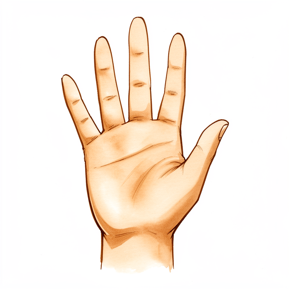
ਇਹ ਓਪਰੇਸ਼ਨ ਕਿਉਂ ਸੁਝਾਇਆ ਗਿਆ ਹੈ
ਡਾ. ਕੀਰਨ ਹਿਰਪਰਾ, ਮਾਟਰ ਪ੍ਰਾਈਵੇਟ ਹਸਪਤਾਲ ਰੌਕਹੈਂਪਟਨ ਵਿੱਚ ਉੱਪਰਲੇ ਅੰਗ (ਮੋਢੇ ਤੋਂ ਹੱਥ ਤੱਕ ਦੀ ਬਾਂਹ) ਦੇ ਸਰਜਨ, ਪਛਾਣ (diagnosis) ਪੱਕੀ ਹੋਣ ਤੋਂ ਬਾਅਦ ਗਲੋਮਸ ਰਸੌਲੀਆਂ (glomus tumours) ਦਾ ਇਲਾਜ ਉਨ੍ਹਾਂ ਨੂੰ ਪੂਰੀ ਤਰ੍ਹਾਂ ਕੱਢ ਕੇ ਕਰਦੇ ਹਨ। ਮਰੀਜ਼ਾਂ ਨੂੰ ਆਮ ਤੌਰ 'ਤੇ ਉਨ੍ਹਾਂ ਦਾ ਜੀਪੀ (ਫ਼ੈਮਿਲੀ ਡਾਕਟਰ) ਸਾਡੇ ਕਲੀਨਿਕ ਵਿੱਚ ਭੇਜਦਾ ਹੈ; ਜੇ ਕਿਸੇ ਫਿਜ਼ੀਓਥੈਰੇਪਿਸਟ ਨੇ ਤੁਹਾਨੂੰ ਸਾਨੂੰ ਮਿਲਣ ਦੀ ਸਲਾਹ ਦਿੱਤੀ ਹੈ, ਤਾਂ ਵੀ ਮੈਡੀਕੇਅਰ ਰਿਬੇਟ (Medicare rebate, ਸਰਕਾਰ ਵੱਲੋਂ ਫ਼ੀਸ ਦੀ ਵਾਪਸੀ) ਦੇ ਯੋਗ ਹੋਣ ਲਈ ਤੁਹਾਨੂੰ ਆਪਣੇ ਜੀਪੀ ਤੋਂ ਰੈਫ਼ਰਲ (referral) ਦੀ ਲੋੜ ਪਵੇਗੀ। ਗਲੋਮਸ ਰਸੌਲੀ ਪੋਟੇ ਦੇ ਨਰਮ ਟਿਸ਼ੂ ਵਿੱਚ ਇੱਕ ਛੋਟੀ, ਬਹੁਤ ਘੱਟ ਮਿਲਣ ਵਾਲੀ ਗੰਢ ਹੈ, ਜੋ ਆਮ ਤੌਰ 'ਤੇ ਨਹੁੰ ਦੇ ਹੇਠਾਂ ਜਾਂ ਨਹੁੰ ਦੇ ਨਾਲ ਹੁੰਦੀ ਹੈ। ਇਹ ਕੈਂਸਰ ਨਹੀਂ ਹੈ। ਇਹ ਹੱਥ ਨੂੰ ਪ੍ਰਭਾਵਿਤ ਕਰਨ ਵਾਲੀਆਂ ਸਭ ਤੋਂ ਦਰਦਨਾਕ ਹਾਲਤਾਂ ਵਿੱਚੋਂ ਇੱਕ ਹੈ, ਅਤੇ ਇਹ ਆਪਣੇ ਆਕਾਰ ਦੇ ਮੁਕਾਬਲੇ ਕਿਤੇ ਜ਼ਿਆਦਾ ਤਕਲੀਫ਼ ਦਿੰਦੀ ਹੈ। ਜ਼ਿਆਦਾਤਰ ਲੋਕਾਂ ਨੂੰ ਤਿੱਖਾ ਦਰਦ ਹੁੰਦਾ ਹੈ ਜੋ ਆਉਂਦਾ-ਜਾਂਦਾ ਰਹਿੰਦਾ ਹੈ, ਅਤੇ ਉਸ ਥਾਂ ਨੂੰ ਦਬਾਉਣ ਉੱਤੇ ਦਰਦ ਹੁੰਦਾ ਹੈ। ਠੰਢ ਨਾਲ ਵੀ ਦਰਦ ਛਿੜ ਸਕਦਾ ਹੈ।
ਜਦੋਂ ਜਾਂਚ, ਐਕਸ-ਰੇ ਅਤੇ MRI ਨਾਲ ਪਛਾਣ ਪੱਕੀ ਹੋ ਜਾਂਦੀ ਹੈ, ਤਾਂ ਰਸੌਲੀ ਨੂੰ ਕੱਢਣਾ ਹੀ ਇਲਾਜ ਹੈ। ਕੋਈ ਵੀ ਬਿਨਾਂ ਓਪਰੇਸ਼ਨ ਵਾਲਾ ਇਲਾਜ ਗਲੋਮਸ ਰਸੌਲੀ ਨੂੰ ਖ਼ਤਮ ਨਹੀਂ ਕਰਦਾ, ਇਸ ਲਈ ਅਸੀਂ ਤੁਹਾਨੂੰ ਪਹਿਲਾਂ ਹੋਰ ਵਿਕਲਪ ਅਜ਼ਮਾਉਣ ਲਈ ਨਹੀਂ ਕਹਿੰਦੇ। ਪੂਰੀ ਰਸੌਲੀ ਨੂੰ ਧਿਆਨ ਨਾਲ ਕੱਢਣ ਨਾਲ ਲਗਭਗ ਹਮੇਸ਼ਾ ਦਰਦ ਤੋਂ ਪੱਕੀ ਰਾਹਤ ਮਿਲਦੀ ਹੈ, ਅਤੇ ਦਰਦ ਆਮ ਤੌਰ 'ਤੇ ਉਸੇ ਵੇਲੇ ਚਲਾ ਜਾਂਦਾ ਹੈ। ਕੁਝ ਹਫ਼ਤਿਆਂ ਦੇ ਅੰਦਰ ਆਮ ਕੰਮਕਾਜ ਵਾਪਸ ਆ ਜਾਂਦਾ ਹੈ। ਅਸੀਂ ਤੁਹਾਡੇ ਸਕੈਨ ਦੇ ਨਤੀਜਿਆਂ ਬਾਰੇ ਤੁਹਾਡੇ ਨਾਲ ਗੱਲ ਕਰਾਂਗੇ ਅਤੇ ਮਿਲ ਕੇ ਫ਼ੈਸਲਾ ਕਰਾਂਗੇ ਕਿ ਇਹ ਓਪਰੇਸ਼ਨ ਤੁਹਾਡੇ ਲਈ ਢੁਕਵਾਂ ਹੈ ਜਾਂ ਨਹੀਂ।
ਓਪਰੇਸ਼ਨ ਤੋਂ ਪਹਿਲਾਂ
ਜਦੋਂ ਤੁਹਾਡੀ ਜਾਂਚ, ਐਕਸ-ਰੇ ਅਤੇ MRI ਨਾਲ ਪਛਾਣ ਪੱਕੀ ਹੋ ਜਾਂਦੀ ਹੈ, ਤਾਂ ਤੁਹਾਡੀ ਸਰਜਰੀ ਜਨਰਲ ਅਨੱਸਥੀਸੀਆ (ਬੇਹੋਸ਼ੀ ਵਾਲੀ ਦਵਾਈ) ਹੇਠ ਡੇਅ ਸਰਜਰੀ (ਉਸੇ ਦਿਨ ਘਰ ਜਾਣ ਵਾਲੀ ਸਰਜਰੀ) ਵਜੋਂ ਬੁੱਕ ਕੀਤੀ ਜਾਵੇਗੀ, ਇਸ ਲਈ ਤੁਸੀਂ ਉਸੇ ਦਿਨ ਘਰ ਜਾਓਗੇ। ਤੁਹਾਨੂੰ ਆਪਣੇ ਓਪਰੇਸ਼ਨ ਤੋਂ ਸੱਤ ਘੰਟੇ ਪਹਿਲਾਂ ਖਾਣਾ ਅਤੇ ਪੀਣਾ ਬੰਦ ਕਰਨਾ ਪਵੇਗਾ। ਅਸੀਂ ਛੇ ਦੀ ਬਜਾਏ ਸੱਤ ਘੰਟੇ ਮੰਗਦੇ ਹਾਂ ਤਾਂ ਜੋ ਜੇ ਓਪਰੇਸ਼ਨਾਂ ਦੀ ਸੂਚੀ ਜਲਦੀ ਚੱਲੇ ਤਾਂ ਤੁਹਾਨੂੰ ਪਹਿਲਾਂ ਬੁਲਾਇਆ ਜਾ ਸਕੇ। ਤੁਹਾਡਾ ਸਰਜਨ ਤੁਹਾਨੂੰ ਦੱਸੇਗਾ ਕਿ ਓਪਰੇਸ਼ਨ ਵਾਲੇ ਦਿਨ ਤੁਹਾਡੀਆਂ ਆਮ ਦਵਾਈਆਂ ਵਿੱਚੋਂ ਕਿਹੜੀਆਂ ਨਹੀਂ ਲੈਣੀਆਂ। ਜੋ ਵੀ ਤੁਸੀਂ ਲੈਂਦੇ ਹੋ ਉਸ ਸਭ ਦੀ ਸੂਚੀ ਨਾਲ ਲਿਆਓ, ਅਤੇ ਤੁਹਾਨੂੰ ਘਰ ਛੱਡਣ ਲਈ ਕਿਸੇ ਦਾ ਪ੍ਰਬੰਧ ਕਰੋ। ਢਿੱਲੇ, ਅਰਾਮਦਾਇਕ ਕੱਪੜੇ ਪਾਓ। ਜੇ ਤੁਹਾਨੂੰ ਹੋਰ ਬਿਮਾਰੀਆਂ ਹਨ, ਤਾਂ ਤੁਹਾਨੂੰ ਖ਼ੂਨ ਦੇ ਟੈਸਟਾਂ ਜਾਂ ਅਨੱਸਥੀਟਿਸਟ (ਬੇਹੋਸ਼ੀ ਵਾਲਾ ਡਾਕਟਰ) ਨਾਲ ਜਾਂਚ ਦੀ ਲੋੜ ਪੈ ਸਕਦੀ ਹੈ, ਪਰ ਜ਼ਿਆਦਾਤਰ ਲੋਕਾਂ ਨੂੰ ਇਹਨਾਂ ਵਿੱਚੋਂ ਕਿਸੇ ਦੀ ਵੀ ਲੋੜ ਨਹੀਂ ਪੈਂਦੀ।
ਓਪਰੇਸ਼ਨ ਵਾਲੇ ਦਿਨ
ਓਪਰੇਸ਼ਨ ਵਾਲੇ ਦਿਨ ਤੁਸੀਂ ਹਸਪਤਾਲ ਦੇ ਸਰਜੀਕਲ ਦਾਖ਼ਲਾ ਵਿਭਾਗ (surgical admissions unit) ਵਿੱਚ ਪਹੁੰਚਦੇ ਹੋ। ਉੱਥੇ ਤੁਹਾਡਾ ਦਾਖ਼ਲਾ ਕੀਤਾ ਜਾਂਦਾ ਹੈ ਅਤੇ ਤੁਹਾਨੂੰ ਓਪਰੇਸ਼ਨ ਥੀਏਟਰ ਲਈ ਤਿਆਰ ਕੀਤਾ ਜਾਂਦਾ ਹੈ। ਤੁਸੀਂ ਅਨੱਸਥੀਟਿਸਟ ਨੂੰ ਮਿਲੋਗੇ, ਜੋ ਓਪਰੇਸ਼ਨ ਦੌਰਾਨ ਤੁਹਾਡਾ ਧਿਆਨ ਰੱਖੇਗਾ। ਇਹ ਓਪਰੇਸ਼ਨ ਜਨਰਲ ਅਨੱਸਥੀਸੀਆ ਹੇਠ ਕੀਤਾ ਜਾਂਦਾ ਹੈ। ਓਪਰੇਸ਼ਨ ਦੌਰਾਨ ਤੁਸੀਂ ਪੂਰੀ ਤਰ੍ਹਾਂ ਬੇਹੋਸ਼ ਹੋਵੋਗੇ। ਕੁਝ ਮਰੀਜ਼ਾਂ ਨੂੰ ਓਪਰੇਸ਼ਨ ਤੋਂ ਬਾਅਦ ਦਰਦ ਤੋਂ ਰਾਹਤ ਲਈ ਰੀਜਨਲ ਨਰਵ ਬਲਾਕ (ਨਸ ਨੂੰ ਸੁੰਨ ਕਰਨ ਵਾਲਾ ਟੀਕਾ) ਵੀ ਦਿੱਤਾ ਜਾ ਸਕਦਾ ਹੈ; ਅਨੱਸਥੀਟਿਸਟ ਤੁਹਾਡੇ ਨਿੱਜੀ ਹਾਲਾਤ ਦੇ ਆਧਾਰ ਉੱਤੇ ਉਸੇ ਦਿਨ ਇਸ ਦਾ ਫ਼ੈਸਲਾ ਕਰਦਾ ਹੈ।
ਫਿਰ ਤੁਹਾਨੂੰ ਓਪਰੇਸ਼ਨ ਥੀਏਟਰ ਵਿੱਚ ਲਿਜਾਇਆ ਜਾਂਦਾ ਹੈ, ਜਿੱਥੇ ਤੁਹਾਡਾ ਸਰਜਨ ਰਸੌਲੀ ਕੱਢਦਾ ਹੈ। ਜਦੋਂ ਓਪਰੇਸ਼ਨ ਪੂਰਾ ਹੋ ਜਾਂਦਾ ਹੈ, ਤੁਸੀਂ ਰਿਕਵਰੀ ਵਾਲੀ ਥਾਂ ਵਿੱਚ ਜਾਗਦੇ ਹੋ। ਅਨੱਸਥੀਸੀਆ ਦਾ ਅਸਰ ਉਤਰਨ ਤੱਕ ਨਰਸਾਂ ਉੱਥੇ ਤੁਹਾਡੀ ਨਿਗਰਾਨੀ ਕਰਨਗੀਆਂ। ਜਦੋਂ ਤੁਸੀਂ ਸਥਿਰ ਹੋ ਜਾਂਦੇ ਹੋ, ਤਾਂ ਪ੍ਰਕਿਰਿਆ ਅਤੇ ਤੁਹਾਡੀ ਸਿਹਤਯਾਬੀ ਦੇ ਹਿਸਾਬ ਨਾਲ ਤੁਸੀਂ ਜਾਂ ਤਾਂ ਵਾਰਡ ਵਿੱਚ ਜਾਓਗੇ ਜਾਂ ਘਰ ਜਾਓਗੇ।
ਓਪਰੇਸ਼ਨ ਵਿੱਚ ਕੀ ਕੀਤਾ ਜਾਂਦਾ ਹੈ
ਓਪਰੇਸ਼ਨ ਤੁਹਾਡੇ ਪੋਟੇ ਉੱਤੇ ਇੱਕ ਛੋਟੇ ਚੀਰੇ ਰਾਹੀਂ ਰਸੌਲੀ ਨੂੰ ਕੱਢਦਾ ਹੈ। ਚੀਰਾ ਕਿੱਥੇ ਦਿੱਤਾ ਜਾਵੇ, ਇਹ ਇਸ ਗੱਲ ਉੱਤੇ ਨਿਰਭਰ ਕਰਦਾ ਹੈ ਕਿ ਤੁਹਾਡਾ MRI ਕੀ ਦਿਖਾਉਂਦਾ ਹੈ। ਜੇ ਰਸੌਲੀ ਨੇਲ ਬੈੱਡ (nail bed, ਨਹੁੰ ਦੇ ਹੇਠਾਂ ਵਾਲੀ ਚਮੜੀ) ਦੇ ਵਿਚਕਾਰ ਹੈ, ਤਾਂ ਤੁਹਾਡਾ ਸਰਜਨ ਨਹੁੰ ਨੂੰ ਚੁੱਕਦਾ ਹੈ, ਰਸੌਲੀ ਦੇ ਉੱਪਰੋਂ ਨੇਲ ਬੈੱਡ ਨੂੰ ਖੋਲ੍ਹਦਾ ਹੈ, ਅਤੇ ਨੇਲ ਬੈੱਡ ਦੀ ਟਾਂਕਿਆਂ ਨਾਲ ਮੁਰੰਮਤ ਕਰਦਾ ਹੈ। ਫਿਰ ਨਹੁੰ ਨੂੰ ਵਾਪਸ ਉਸ ਦੀ ਥਾਂ ਉੱਤੇ ਰੱਖ ਦਿੱਤਾ ਜਾਂਦਾ ਹੈ, ਜਿੱਥੇ ਇਹ ਠੀਕ ਹੋਣ ਦੌਰਾਨ ਸਪਲਿੰਟ ਵਾਂਗ ਕੰਮ ਕਰਦਾ ਹੈ। ਜੇ ਰਸੌਲੀ ਨਹੁੰ ਦੇ ਪਾਸੇ ਵੱਲ ਹੈ, ਤਾਂ ਤੁਹਾਡਾ ਸਰਜਨ ਤੁਹਾਡੇ ਪੋਟੇ ਦੇ ਪਾਸੇ ਦੇ ਨਾਲ-ਨਾਲ ਦਿੱਤੇ ਚੀਰੇ ਰਾਹੀਂ, ਨੇਲ ਬੈੱਡ ਦੇ ਹੇਠਾਂ ਤੋਂ, ਨੇਲ ਬੈੱਡ ਨੂੰ ਖੋਲ੍ਹੇ ਬਿਨਾਂ ਉਸ ਤੱਕ ਪਹੁੰਚਦਾ ਹੈ।
ਤੁਹਾਡਾ ਸਰਜਨ ਪੂਰੀ ਰਸੌਲੀ ਕੱਢਣ ਲਈ ਧਿਆਨ ਨਾਲ ਕੰਮ ਕਰਦਾ ਹੈ। ਕੱਢਿਆ ਗਿਆ ਟਿਸ਼ੂ ਲੈਬਾਰਟਰੀ ਵਿੱਚ ਭੇਜਿਆ ਜਾਂਦਾ ਹੈ, ਜਿੱਥੇ ਪਛਾਣ ਦੀ ਪੁਸ਼ਟੀ ਲਈ ਇਸ ਦੀ ਜਾਂਚ ਕੀਤੀ ਜਾਂਦੀ ਹੈ। ਫਿਰ ਜ਼ਖ਼ਮ ਨੂੰ ਟਾਂਕਿਆਂ ਨਾਲ ਬੰਦ ਕਰ ਦਿੱਤਾ ਜਾਂਦਾ ਹੈ।
ਬਾਅਦ ਵਿੱਚ, ਤੁਹਾਡੀ ਉਂਗਲ ਉੱਤੇ ਹਲਕੀ ਪੱਟੀ ਹੁੰਦੀ ਹੈ। ਤੁਹਾਨੂੰ ਸਪਲਿੰਟ ਦੀ ਲੋੜ ਨਹੀਂ ਹੁੰਦੀ।
ਓਪਰੇਸ਼ਨ ਤੋਂ ਬਾਅਦ
ਤੁਸੀਂ ਰਿਕਵਰੀ ਵਾਲੀ ਥਾਂ ਵਿੱਚ ਜਾਗੋਗੇ, ਜਿੱਥੇ ਅਨੱਸਥੀਸੀਆ ਦਾ ਅਸਰ ਉਤਰਨ ਤੱਕ ਨਰਸਾਂ ਤੁਹਾਡੀ ਨਿਗਰਾਨੀ ਕਰਦੀਆਂ ਹਨ। ਇਹ ਡੇਅ ਕੇਸ ਹੈ, ਇਸ ਲਈ ਤੁਸੀਂ ਉਸੇ ਦਿਨ ਘਰ ਜਾਓਗੇ। ਰਸੌਲੀ ਵਾਲਾ ਦਰਦ ਆਮ ਤੌਰ 'ਤੇ ਉਸੇ ਵੇਲੇ ਚਲਾ ਜਾਂਦਾ ਹੈ; ਜ਼ਖ਼ਮ ਆਪ ਇੱਕ-ਦੋ ਹਫ਼ਤੇ ਦੁਖਦਾ ਹੈ, ਅਤੇ ਦਰਦ ਦੀਆਂ ਸਾਦੀਆਂ ਦਵਾਈਆਂ ਆਮ ਤੌਰ 'ਤੇ ਕਾਫ਼ੀ ਹੁੰਦੀਆਂ ਹਨ। ਤੁਹਾਡੀ ਉਂਗਲ ਉੱਤੇ ਹਲਕੀ ਪੱਟੀ ਹੋਵੇਗੀ ਅਤੇ ਕੋਈ ਸਪਲਿੰਟ ਨਹੀਂ ਹੋਵੇਗਾ। ਅਸੀਂ ਪੱਟੀ ਲਗਭਗ 10 ਦਿਨ ਲੱਗੀ ਰਹਿਣ ਦਿੰਦੇ ਹਾਂ; ਕਿਰਪਾ ਕਰਕੇ ਉਸ ਤੋਂ ਪਹਿਲਾਂ ਇਸ ਨੂੰ ਨਾ ਉਤਾਰੋ, ਜਦੋਂ ਤੱਕ ਅਸੀਂ ਨਾ ਕਹੀਏ। ਜਦੋਂ ਅਸੀਂ ਤੁਹਾਨੂੰ ਮਿਲਦੇ ਹਾਂ ਤਾਂ ਅਸੀਂ ਇਸ ਨੂੰ ਬਦਲਦੇ ਜਾਂ ਉਤਾਰਦੇ ਹਾਂ। ਤੁਸੀਂ ਹਲਕੇ ਕੰਮਾਂ ਲਈ ਆਪਣਾ ਹੱਥ ਸ਼ੁਰੂ ਤੋਂ ਹੀ ਵਰਤ ਸਕਦੇ ਹੋ। ਪਹਿਲੇ 24 ਘੰਟਿਆਂ ਲਈ ਕਿਸੇ ਨੂੰ ਆਪਣੇ ਨਾਲ ਰਹਿਣ ਲਈ ਕਹੋ। ਜੇ ਤੁਹਾਨੂੰ ਬੁਖ਼ਾਰ ਹੋਵੇ, ਜ਼ਖ਼ਮ ਦੇ ਆਲੇ-ਦੁਆਲੇ ਲਾਲੀ ਫੈਲ ਰਹੀ ਹੋਵੇ, ਉਸ ਵਿੱਚੋਂ ਤਰਲ ਜਾਂ ਪਾਕ ਰਿਸ ਰਹੀ ਹੋਵੇ, ਜਾਂ ਦਰਦ ਦੀਆਂ ਦਵਾਈਆਂ ਦੇ ਬਾਵਜੂਦ ਦਰਦ ਵਧਦਾ ਹੀ ਜਾਵੇ, ਤਾਂ ਉਸੇ ਦਿਨ ਕਲੀਨਿਕ ਨੂੰ ਫ਼ੋਨ ਕਰੋ।
ਸਿਹਤਯਾਬੀ
ਰਸੌਲੀ ਵਾਲਾ ਡੂੰਘਾ, ਤਿੱਖਾ ਦਰਦ ਆਮ ਤੌਰ 'ਤੇ ਉਸੇ ਵੇਲੇ ਚਲਾ ਜਾਂਦਾ ਹੈ। ਬਾਅਦ ਵਿੱਚ ਜੋ ਤੁਸੀਂ ਮਹਿਸੂਸ ਕਰਦੇ ਹੋ ਉਹ ਜ਼ਖ਼ਮ ਆਪ ਹੈ: ਇੱਕ-ਦੋ ਹਫ਼ਤੇ ਦੁਖਦਾ ਹੈ, ਪੋਟੇ ਦੇ ਆਲੇ-ਦੁਆਲੇ ਕੁਝ ਸੋਜ ਅਤੇ ਛੂਹਣ ਉੱਤੇ ਦਰਦ ਦੇ ਨਾਲ। ਦਰਦ ਦੀਆਂ ਸਾਦੀਆਂ ਦਵਾਈਆਂ ਆਮ ਤੌਰ 'ਤੇ ਤੁਹਾਨੂੰ ਅਰਾਮ ਵਿੱਚ ਰੱਖਣ ਲਈ ਕਾਫ਼ੀ ਹੁੰਦੀਆਂ ਹਨ। ਅਰਾਮ ਕਰਦੇ ਸਮੇਂ ਆਪਣਾ ਹੱਥ ਗੱਦੀ ਉੱਤੇ ਉੱਚਾ ਰੱਖਣ ਨਾਲ ਸੋਜ ਘਟਣ ਵਿੱਚ ਮਦਦ ਮਿਲਦੀ ਹੈ।
ਤੁਸੀਂ ਹਲਕੇ ਕੰਮਾਂ ਲਈ ਆਪਣਾ ਹੱਥ ਸ਼ੁਰੂ ਤੋਂ ਹੀ ਵਰਤ ਸਕਦੇ ਹੋ, ਅਤੇ ਤੁਹਾਨੂੰ ਸਪਲਿੰਟ ਨਹੀਂ ਪਾਉਣਾ ਪਵੇਗਾ। ਤੁਹਾਡੀ ਉਂਗਲ ਉੱਤੇ ਹਲਕੀ ਪੱਟੀ ਹੁੰਦੀ ਹੈ ਜੋ ਸਾਡੇ ਮਿਲਣ ਤੱਕ ਲੱਗੀ ਰਹਿੰਦੀ ਹੈ। ਪੱਟੀ ਉਤਰਨ ਤੋਂ ਬਾਅਦ, ਉਂਗਲ ਦੀ ਹਲਕੀ ਹਿਲਜੁਲ ਇਸ ਨੂੰ ਅਕੜਨ ਤੋਂ ਬਚਾਉਂਦੀ ਹੈ। ਸਰਜਰੀ ਤੋਂ ਬਾਅਦ ਹੈਂਡ ਥੈਰੇਪੀ (ਹੱਥ ਦੀ ਕਸਰਤ ਵਾਲਾ ਇਲਾਜ) ਐਕਸਟੈਂਡ ਰੀਹੈਬਿਲੀਟੇਸ਼ਨ (Extend Rehabilitation) ਵਿੱਚ ਰੂਬੀ ਡੂਲਨ (Ruby Doolan) ਨਾਲ ਹੁੰਦੀ ਹੈ। ਰੂਬੀ ਇੱਕ ਹੈਂਡ ਥੈਰੇਪਿਸਟ ਹੈ: ਉਹ ਤੁਹਾਡੀਆਂ ਕਸਰਤਾਂ ਵਿੱਚ ਤੁਹਾਡੀ ਅਗਵਾਈ ਕਰੇਗੀ ਅਤੇ ਜਿਸ ਵੀ ਸਪਲਿੰਟ ਦੀ ਤੁਹਾਨੂੰ ਲੋੜ ਹੋਵੇ ਉਹ ਬਣਾਏਗੀ, ਹਾਲਾਂਕਿ ਜ਼ਿਆਦਾਤਰ ਲੋਕਾਂ ਨੂੰ ਘਰ ਵਿੱਚ ਹਲਕੀ ਵਰਤੋਂ ਤੋਂ ਵੱਧ ਕੁਝ ਖ਼ਾਸ ਦੀ ਲੋੜ ਨਹੀਂ ਪੈਂਦੀ।
ਜਦੋਂ ਜ਼ਖ਼ਮ ਅਰਾਮਦਾਇਕ ਹੋ ਜਾਂਦਾ ਹੈ, ਤਾਂ ਦਫ਼ਤਰ ਦਾ ਕੰਮ ਅਤੇ ਹਲਕੀਆਂ ਡਿਊਟੀਆਂ ਆਮ ਤੌਰ 'ਤੇ ਤੁਹਾਡੀ ਜ਼ਿੰਦਗੀ ਵਿੱਚ ਵਾਪਸ ਆ ਜਾਂਦੀਆਂ ਹਨ। ਗੱਡੀ ਚਲਾਉਣਾ ਵੀ ਲਗਭਗ ਉਸੇ ਸਮੇਂ ਵਾਪਸ ਆਉਂਦਾ ਹੈ, ਜਦੋਂ ਤੁਸੀਂ ਹੱਥ ਨੂੰ ਬਚਾਏ ਬਿਨਾਂ ਸਟੀਅਰਿੰਗ ਨੂੰ ਫੜ ਅਤੇ ਘੁਮਾ ਸਕੋ। ਹੋਰ ਵੇਰਵੇ ਲਈ ਉੱਪਰਲੇ ਅੰਗ ਦੀ ਸਰਜਰੀ ਤੋਂ ਬਾਅਦ ਗੱਡੀ ਚਲਾਉਣਾ ਬਾਰੇ ਸਾਡਾ ਪੰਨਾ ਦੇਖੋ। ਜਿਵੇਂ-ਜਿਵੇਂ ਪੋਟੇ ਨੂੰ ਛੂਹਣ ਉੱਤੇ ਦਰਦ ਹੋਣਾ ਬੰਦ ਹੁੰਦਾ ਹੈ, ਪਕੜਨਾ, ਭਾਰੀ ਕੰਮ ਅਤੇ ਖੇਡਾਂ ਵਾਪਸ ਆਉਂਦੀਆਂ ਹਨ, ਅਤੇ ਜ਼ਿਆਦਾਤਰ ਲੋਕ ਕੁਝ ਹਫ਼ਤਿਆਂ ਦੇ ਅੰਦਰ ਆਮ ਕੰਮਕਾਜ ਉੱਤੇ ਵਾਪਸ ਆ ਜਾਂਦੇ ਹਨ।
ਜੇ ਓਪਰੇਸ਼ਨ ਦੌਰਾਨ ਨਹੁੰ ਚੁੱਕਿਆ ਗਿਆ ਸੀ, ਤਾਂ ਨਵਾਂ ਨਹੁੰ ਕਈ ਮਹੀਨਿਆਂ ਵਿੱਚ ਉੱਗ ਕੇ ਅੱਗੇ ਆਉਂਦਾ ਹੈ। ਠੀਕ ਹੋਣ ਦਾ ਸਮਾਂ ਹਰ ਵਿਅਕਤੀ ਲਈ ਵੱਖਰਾ ਹੁੰਦਾ ਹੈ, ਅਤੇ ਤੁਹਾਡਾ ਸਮਾਂ ਵੱਖਰਾ ਹੋ ਸਕਦਾ ਹੈ। ਰਸਤੇ ਵਿੱਚ ਅਸੀਂ ਤੁਹਾਨੂੰ ਮਿਲਦੇ ਰਹਾਂਗੇ ਅਤੇ ਹਰ ਕਦਮ ਉੱਤੇ ਤੁਹਾਡੀ ਅਗਵਾਈ ਕਰਾਂਗੇ।
ਕੀ ਗ਼ਲਤ ਹੋ ਸਕਦਾ ਹੈ
ਜ਼ਿਆਦਾਤਰ ਮਰੀਜ਼ ਠੀਕ ਰਹਿੰਦੇ ਹਨ, ਪਰ ਕਦੇ-ਕਦਾਈਂ ਸਮੱਸਿਆਵਾਂ ਹੋ ਸਕਦੀਆਂ ਹਨ। ਤੁਹਾਡਾ ਸਰਜਨ ਅਤੇ ਟੀਮ ਕਿਸੇ ਵੀ ਮੁਸ਼ਕਲ ਨੂੰ ਜਲਦੀ ਫੜਨ ਲਈ ਤੁਹਾਡੀ ਨੇੜਿਓਂ ਨਿਗਰਾਨੀ ਕਰਦੇ ਹਨ।
ਇਸ ਓਪਰੇਸ਼ਨ ਤੋਂ ਬਾਅਦ ਜਿਨ੍ਹਾਂ ਦੋ ਸਮੱਸਿਆਵਾਂ ਉੱਤੇ ਨਜ਼ਰ ਰੱਖੀ ਜਾਂਦੀ ਹੈ, ਉਹ ਹਨ ਰਸੌਲੀ ਦਾ ਮੁੜ ਆਉਣਾ ਅਤੇ ਨਵੇਂ ਨਹੁੰ ਦੇ ਉੱਗਣ ਦੇ ਤਰੀਕੇ ਵਿੱਚ ਬਦਲਾਅ।
ਜੇ ਰਸੌਲੀ ਮੁੜ ਆ ਜਾਵੇ, ਤਾਂ ਤੁਹਾਨੂੰ ਪੁਰਾਣਾ ਦਰਦ ਵਾਪਸ ਆਉਂਦਾ ਮਹਿਸੂਸ ਹੋਵੇਗਾ: ਉਸੇ ਥਾਂ ਉੱਤੇ ਡੂੰਘਾ, ਤਿੱਖਾ ਦਰਦ, ਜੋ ਦਬਾਅ ਜਾਂ ਠੰਢ ਨਾਲ ਛਿੜਦਾ ਹੈ। ਇਹ ਆਮ ਤੌਰ 'ਤੇ ਇਸ ਲਈ ਹੁੰਦਾ ਹੈ ਕਿਉਂਕਿ ਰਸੌਲੀ ਦਾ ਕੋਈ ਛੋਟਾ ਟੁਕੜਾ ਪਿੱਛੇ ਰਹਿ ਗਿਆ ਸੀ, ਜਾਂ ਕਿਉਂਕਿ ਉੱਥੇ ਸ਼ੁਰੂ ਤੋਂ ਹੀ ਇੱਕ ਦੂਜੀ ਬਹੁਤ ਛੋਟੀ ਰਸੌਲੀ ਸੀ। ਜੇ ਓਪਰੇਸ਼ਨ ਤੋਂ ਪਹਿਲਾਂ ਵਾਲਾ ਦਰਦ ਵਾਪਸ ਆ ਜਾਵੇ, ਤਾਂ ਕਲੀਨਿਕ ਨੂੰ ਫ਼ੋਨ ਕਰੋ। ਅਸੀਂ ਇਸ ਦੀ ਜਾਂਚ ਕਰਾਂਗੇ ਅਤੇ, ਜੇ ਲੋੜ ਹੋਵੇ, ਇਹ ਪਤਾ ਲਗਾਉਣ ਲਈ ਸਕੈਨ ਦਾ ਪ੍ਰਬੰਧ ਕਰਾਂਗੇ ਕਿ ਕੀ ਹੋ ਰਿਹਾ ਹੈ।
ਜੇ ਓਪਰੇਸ਼ਨ ਦੌਰਾਨ ਨੇਲ ਬੈੱਡ ਖੋਲ੍ਹਿਆ ਗਿਆ ਸੀ, ਤਾਂ ਨਵਾਂ ਨਹੁੰ ਉੱਭਰੀ ਧਾਰੀ ਜਾਂ ਤਰੇੜ ਨਾਲ ਉੱਗ ਸਕਦਾ ਹੈ। ਇਹ ਤੁਹਾਨੂੰ ਸਰਜਰੀ ਤੋਂ ਬਾਅਦ ਦੇ ਮਹੀਨਿਆਂ ਵਿੱਚ ਨਹੁੰ ਦੇ ਉੱਗ ਕੇ ਅੱਗੇ ਆਉਣ ਨਾਲ, ਨਹੁੰ ਦੀ ਸਤਹ ਉੱਤੇ ਇੱਕ ਲਕੀਰ, ਉੱਭਰੀ ਧਾਰੀ ਜਾਂ ਤਰੇੜ ਵਜੋਂ ਦਿਸੇਗਾ। ਜੇ ਤੁਸੀਂ ਇਹ ਦੇਖੋ, ਤਾਂ ਆਪਣੀ ਅਗਲੀ ਜਾਂਚ ਵੇਲੇ ਇਸ ਬਾਰੇ ਦੱਸੋ। ਇਹ ਨਹੁੰ ਦੀ ਦਿੱਖ ਉੱਤੇ ਅਸਰ ਪਾਉਂਦਾ ਹੈ, ਅਤੇ ਜੇ ਇਹ ਤੁਹਾਨੂੰ ਪਰੇਸ਼ਾਨ ਕਰਦਾ ਹੈ ਤਾਂ ਅਸੀਂ ਕਈ ਤਰੀਕਿਆਂ ਨਾਲ ਮਦਦ ਕਰ ਸਕਦੇ ਹਾਂ।
ਜੇ ਤੁਸੀਂ ਵੇਰਵੇ ਜਾਣਨਾ ਚਾਹੁੰਦੇ ਹੋ, ਤਾਂ ਇਸ ਸਫ਼ੇ ਉੱਤੇ ਪੇਚੀਦਗੀਆਂ ਵਾਲੀ ਸਾਰਣੀ ਆਮ ਦਰਾਂ ਦੱਸਦੀ ਹੈ।
ਸਾਨੂੰ ਕਦੋਂ ਫ਼ੋਨ ਕਰਨਾ ਹੈ
ਜ਼ਿਆਦਾਤਰ ਸਮੱਸਿਆਵਾਂ ਛੇਤੀ ਫੜੀਆਂ ਜਾਣ ਤਾਂ ਸੌਖਿਆਂ ਠੀਕ ਹੋ ਜਾਂਦੀਆਂ ਹਨ। ਇਹ ਹੈ ਜਿਸ ਦਾ ਧਿਆਨ ਰੱਖਣਾ ਹੈ।
ਜੇ ਤੁਹਾਡੀ ਪਿੰਨੀ (ਲੱਤ ਦੀ ਪਿੰਨੀ, calf) ਵਿੱਚ ਸੋਜ ਜਾਂ ਦਰਦ ਹੋਵੇ, ਸਾਹ ਚੜ੍ਹੇ ਜਾਂ ਛਾਤੀ ਵਿੱਚ ਦਰਦ ਹੋਵੇ, ਤਾਂ ਐਮਰਜੈਂਸੀ ਵਿਭਾਗ ਜਾਓ। ਇਹ ਖ਼ੂਨ ਦੇ ਗਤਲੇ ਦੀਆਂ ਨਿਸ਼ਾਨੀਆਂ ਹੋ ਸਕਦੀਆਂ ਹਨ। ਜੇ ਤੁਹਾਡੀਆਂ ਉਂਗਲਾਂ, ਹੱਥ ਜਾਂ ਅੰਗੂਠਾ ਫਿੱਕੇ, ਠੰਢੇ, ਸਫ਼ੈਦ, ਨੀਲੇ ਜਾਂ ਗੂੜ੍ਹੇ ਰੰਗ ਦੇ ਪੈ ਜਾਣ, ਤਾਂ ਐਮਰਜੈਂਸੀ ਵਿਭਾਗ ਜਾਓ, ਕਿਉਂਕਿ ਇਸ ਦਾ ਮਤਲਬ ਖ਼ੂਨ ਦੇ ਵਹਾਅ ਵਿੱਚ ਸਮੱਸਿਆ ਹੋ ਸਕਦਾ ਹੈ।
ਜੇ ਤੁਹਾਨੂੰ ਬੁਖ਼ਾਰ ਹੈ, ਜ਼ਖ਼ਮ ਦੁਆਲੇ ਲਾਲੀ ਫੈਲ ਰਹੀ ਹੈ, ਜਾਂ ਉਸ ਵਿੱਚੋਂ ਤਰਲ ਜਾਂ ਪਾਕ ਦਾ ਰਿਸਾਵ ਹੋ ਰਿਹਾ ਹੈ, ਤਾਂ ਉਸੇ ਦਿਨ ਕਲੀਨਿਕ ਨੂੰ ਫ਼ੋਨ ਕਰੋ। ਜੇ ਦਰਦ ਦੀਆਂ ਦਵਾਈਆਂ ਦੇ ਬਾਵਜੂਦ ਦਰਦ ਵਧਦਾ ਹੀ ਜਾਂਦਾ ਹੈ, ਤਾਂ ਉਸੇ ਦਿਨ ਕਲੀਨਿਕ ਨੂੰ ਫ਼ੋਨ ਕਰੋ।
ਜੇ ਤੁਹਾਨੂੰ ਨਰਵ ਬਲਾਕ ਲਗਾਇਆ ਗਿਆ ਸੀ, ਤਾਂ ਪਹਿਲੇ 24 ਘੰਟਿਆਂ ਵਿੱਚ ਸੁੰਨਪਨ ਅਤੇ ਕਮਜ਼ੋਰੀ ਉਮੀਦ ਮੁਤਾਬਕ ਹਨ। ਜੇ ਲਗਭਗ 24 ਘੰਟਿਆਂ ਬਾਅਦ ਵੀ ਇਹ ਨਾ ਹਟਣ, ਤਾਂ ਕਲੀਨਿਕ ਨੂੰ ਫ਼ੋਨ ਕਰੋ। ਜੇ ਤੁਸੀਂ ਆਪਣੀ ਬਾਂਹ, ਹੱਥ ਜਾਂ ਉਂਗਲਾਂ ਨਾ ਹਿਲਾ ਸਕੋ, ਤਾਂ ਕਲੀਨਿਕ ਨੂੰ ਫ਼ੋਨ ਕਰੋ।
ਜੇ ਤੁਸੀਂ ਕਲੀਨਿਕ ਨਾਲ ਸੰਪਰਕ ਨਹੀਂ ਕਰ ਸਕਦੇ, ਜਿਵੇਂ ਦਫ਼ਤਰੀ ਸਮੇਂ ਤੋਂ ਬਾਅਦ ਜਾਂ ਹਫ਼ਤੇ ਦੇ ਅਖ਼ੀਰ ’ਤੇ, ਤਾਂ ਆਪਣੇ ਸਭ ਤੋਂ ਨੇੜਲੇ ਐਮਰਜੈਂਸੀ ਵਿਭਾਗ ਜਾਓ।
ਵਿਸਥਾਰ ਵਿੱਚ
Advanced reading: the deeper science (optional)
ਇਹ ਹਿੱਸਾ ਤੁਹਾਡੇ ਆਪਣੇ ਇਲਾਜ ਬਾਰੇ ਫ਼ੈਸਲਿਆਂ ਲਈ ਲੋੜ ਤੋਂ ਵੱਧ ਡੂੰਘਾਈ ਵਿੱਚ ਜਾਂਦਾ ਹੈ। ਇਸ ਓਪਰੇਸ਼ਨ ਬਾਰੇ ਵਾਧੂ ਪੜ੍ਹਨਾ ਇਸ ਲਈ ਲਾਹੇਵੰਦ ਹੈ ਕਿਉਂਕਿ ਇਸ ਦੇ ਦੋ ਅਹਿਮ ਨਤੀਜੇ, ਯਾਨੀ ਰਸੌਲੀ ਜੋ ਮੁੜ ਨਾ ਆਵੇ ਅਤੇ ਨਹੁੰ ਜੋ ਆਮ ਵਾਂਗ ਉੱਗੇ, ਸਰਜਨ ਨੂੰ ਉਲਟੀਆਂ ਦਿਸ਼ਾਵਾਂ ਵਿੱਚ ਖਿੱਚਦੇ ਹਨ, ਅਤੇ ਛਪੀਆਂ ਹੋਈਆਂ ਲੜੀਆਂ ਇਸ ਸੰਤੁਲਨ (trade-off) ਨੂੰ ਸਾਫ਼ ਦਿਖਾਉਂਦੀਆਂ ਹਨ।
ਅੰਦਰ ਜਾਣ ਦੇ ਦੋ ਰਾਹ, ਅਤੇ ਉਨ੍ਹਾਂ ਵਿਚਕਾਰ ਸੰਤੁਲਨ
ਨਹੁੰ ਦੇ ਹੇਠਾਂ ਵਾਲੀ ਰਸੌਲੀ ਤੱਕ ਮੋਟੇ ਤੌਰ 'ਤੇ ਦੋ ਤਰੀਕਿਆਂ ਨਾਲ ਪਹੁੰਚਿਆ ਜਾ ਸਕਦਾ ਹੈ [1]:
- ਨਹੁੰ ਰਾਹੀਂ (transungual)। ਨਹੁੰ ਚੁੱਕਿਆ ਜਾਂਦਾ ਹੈ, ਨੇਲ ਬੈੱਡ ਨੂੰ ਸਿੱਧਾ ਰਸੌਲੀ ਦੇ ਉੱਪਰੋਂ ਖੋਲ੍ਹਿਆ ਜਾਂਦਾ ਹੈ, ਰਸੌਲੀ ਨੂੰ ਸਾਹਮਣੇ ਦੇਖ ਕੇ ਕੱਢਿਆ ਜਾਂਦਾ ਹੈ, ਅਤੇ ਨੇਲ ਬੈੱਡ ਦੀ ਮੁਰੰਮਤ ਕੀਤੀ ਜਾਂਦੀ ਹੈ। ਨਜ਼ਾਰਾ ਸਭ ਤੋਂ ਵਧੀਆ ਮਿਲਦਾ ਹੈ, ਪਰ ਨੇਲ ਬੈੱਡ ਕੱਟਿਆ ਜਾਂਦਾ ਹੈ।
- ਪਾਸੇ ਤੋਂ (ਨਹੁੰ ਬਚਾਉਣ ਵਾਲਾ, nail-sparing)। ਪੋਟੇ ਦੇ ਪਾਸੇ ਦੇ ਨਾਲ-ਨਾਲ ਦਿੱਤੇ ਚੀਰੇ ਰਾਹੀਂ ਨੇਲ ਬੈੱਡ ਨੂੰ ਇੱਕ ਫ਼ਲੈਪ (flap, ਟਿਸ਼ੂ ਦੀ ਚੁੱਕੀ ਹੋਈ ਪਰਤ) ਵਾਂਗ ਹੱਡੀ ਤੋਂ ਚੁੱਕਿਆ ਜਾਂਦਾ ਹੈ, ਅਤੇ ਰਸੌਲੀ ਹੇਠਾਂ ਤੋਂ ਕੱਢੀ ਜਾਂਦੀ ਹੈ। ਨੇਲ ਬੈੱਡ ਕੱਟਿਆ ਨਹੀਂ ਜਾਂਦਾ, ਪਰ ਨਹੁੰ ਦੇ ਵਿਚਕਾਰ ਵਾਲੀ ਰਸੌਲੀ ਦਾ ਨਜ਼ਾਰਾ ਜ਼ਿਆਦਾ ਸੀਮਤ ਹੁੰਦਾ ਹੈ।
ਲੜੀਆਂ ਇਸ ਸੰਤੁਲਨ ਦੇ ਦੋਵਾਂ ਪਾਸਿਆਂ ਉੱਤੇ ਆਉਂਦੀਆਂ ਹਨ। 29 ਸਾਲਾਂ ਦੌਰਾਨ ਪਾਸੇ ਤੋਂ ਇਲਾਜ ਕੀਤੇ ਗਏ 19 ਮਰੀਜ਼ਾਂ ਵਿੱਚ, ਨਹੁੰ ਦਾ ਕੋਈ ਵਿਗਾੜ ਨਹੀਂ ਹੋਇਆ, ਪਰ ਮੁੜ ਆਉਣ ਦੀ ਦਰ 15.7% ਸੀ [2]। ਉਨ੍ਹਾਂ 53 ਮਰੀਜ਼ਾਂ ਵਿੱਚ, ਜਿਨ੍ਹਾਂ ਦਾ ਕਿਨਾਰੇ ਦੇ ਨੇੜੇ ਵਾਲੀਆਂ ਰਸੌਲੀਆਂ ਲਈ ਪਾਸੇ ਤੋਂ ਅਤੇ ਵਿਚਕਾਰ ਵਾਲੀਆਂ ਲਈ ਨਹੁੰ ਰਾਹੀਂ ਇਲਾਜ ਕੀਤਾ ਗਿਆ, ਕੋਈ ਵੀ ਰਸੌਲੀ ਮੁੜ ਨਹੀਂ ਆਈ; 10 (18.9%) ਵਿੱਚ ਨਵੇਂ ਨਹੁੰ ਉੱਤੇ ਲੰਬਾਈ ਵਾਲੀਆਂ ਬਾਰੀਕ ਲਕੀਰਾਂ ਦਿਸੀਆਂ, ਜੋ ਸਾਰੀਆਂ ਨਹੁੰ ਰਾਹੀਂ ਵਾਲੇ ਤਰੀਕੇ ਤੋਂ ਬਾਅਦ ਸਨ ਅਤੇ ਸਾਰੀਆਂ ਦਿੱਖ ਪੱਖੋਂ ਮਾਮੂਲੀ ਸਨ [3]। 72 ਮਰੀਜ਼ਾਂ ਦੇ ਇੱਕ ਬਹੁ-ਕੇਂਦਰੀ (multicentre) ਅਧਿਐਨ ਵਿੱਚ, ਨਹੁੰ ਬਚਾਉਣ ਵਾਲੇ ਤਰੀਕੇ ਨੇ ਨਹੁੰ ਵਿੱਚ ਬਦਲਾਅ ਦੀ ਦਰ ਨਹੀਂ ਘਟਾਈ, ਅਤੇ ਕੋਈ ਵੀ ਕਾਰਕ ਭਰੋਸੇ ਨਾਲ ਇਹ ਨਹੀਂ ਦੱਸ ਸਕਿਆ ਕਿ ਰਸੌਲੀ ਮੁੜ ਆਵੇਗੀ ਜਾਂ ਨਹੀਂ [4]।
ਥਾਂ ਦੇ ਹਿਸਾਬ ਨਾਲ ਤਰੀਕਾ ਚੁਣਨ ਦਾ ਇਹੀ ਆਧਾਰ ਹੈ, ਅਤੇ ਇਸ ਸਫ਼ੇ ਉੱਤੇ ਦੱਸਿਆ ਓਪਰੇਸ਼ਨ ਇਹੀ ਕਰਦਾ ਹੈ: MRI ਦਿਖਾਉਂਦਾ ਹੈ ਕਿ ਰਸੌਲੀ ਕਿੱਥੇ ਹੈ, ਕਿਨਾਰੇ ਵਾਲੀ ਰਸੌਲੀ ਤੱਕ ਪਾਸੇ ਤੋਂ ਪਹੁੰਚਿਆ ਜਾਂਦਾ ਹੈ, ਅਤੇ ਵਿਚਕਾਰ ਵਾਲੀ ਤੱਕ ਨਹੁੰ ਰਾਹੀਂ।
ਰਸੌਲੀਆਂ ਮੁੜ ਕਿਉਂ ਆਉਂਦੀਆਂ ਹਨ
ਵੱਡੀਆਂ ਆਧੁਨਿਕ ਲੜੀਆਂ ਵਿੱਚ ਕੱਢਣ ਤੋਂ ਬਾਅਦ ਮੁੜ ਆਉਣ ਦੀ ਦਰ ਲਗਭਗ 3 ਤੋਂ 7% ਹੈ: ਬਹੁ-ਕੇਂਦਰੀ ਅਧਿਐਨ ਵਿੱਚ 72 ਵਿੱਚੋਂ 5 (6.9%) [4], ਅਤੇ ਯੂਕੇ ਦੀ ਇੱਕ ਲੜੀ ਵਿੱਚ 73 ਵਿੱਚੋਂ 2 (2.7%) [5]। ਦੋ ਵੱਖ-ਵੱਖ ਚੀਜ਼ਾਂ ਨੂੰ ਮੁੜ ਆਉਣਾ ਕਿਹਾ ਜਾਂਦਾ ਹੈ।
- ਪਿੱਛੇ ਰਹਿ ਗਈ ਰਸੌਲੀ। ਯੂਕੇ ਦੀ ਲੜੀ ਵਿੱਚ ਲੈਬਾਰਟਰੀ ਨੇ 73 ਵਿੱਚੋਂ 11 (15%) ਵਿੱਚ ਕੱਢੇ ਗਏ ਟਿਸ਼ੂ ਦੇ ਕਿਨਾਰੇ ਉੱਤੇ ਰਸੌਲੀ ਦੱਸੀ, ਫਿਰ ਵੀ ਸਿਰਫ਼ 2 ਲੋਕਾਂ ਵਿੱਚ ਹੀ ਰਸੌਲੀ ਮੁੜ ਆਈ [5]। ਸਿੰਗਾਪੁਰ ਦੀ ਇੱਕ ਲੜੀ ਵਿੱਚ, ਕਿਨਾਰਿਆਂ ਉੱਤੇ ਰਸੌਲੀ ਵਾਲੀਆਂ 3 ਰਸੌਲੀਆਂ ਬਿਲਕੁਲ ਵੀ ਮੁੜ ਨਹੀਂ ਆਈਆਂ [6]। ਇਸ ਲਈ ਅਧੂਰਾ ਕੱਢੇ ਜਾਣ ਦੀ ਰਿਪੋਰਟ ਦਾ ਮਤਲਬ ਇਹ ਨਹੀਂ ਕਿ ਰਸੌਲੀ ਮੁੜ ਆਵੇਗੀ।
- ਦੂਜੀ ਰਸੌਲੀ। 12 ਮਰੀਜ਼ਾਂ ਦੀ ਇੱਕ ਲੜੀ ਵਿੱਚ, ਸਾਰੇ 5 ਮੁੜ ਆਉਣ ਵਾਲੇ ਮਾਮਲੇ ਅਸਲ ਥਾਂ ਦੀ ਬਜਾਏ ਨਵੀਆਂ, ਵੱਖਰੀਆਂ ਥਾਵਾਂ ਉੱਤੇ ਹੋਏ, ਜੋ ਮੁੜ ਵਾਧੇ ਦੀ ਬਜਾਏ ਪਹਿਲੇ ਓਪਰੇਸ਼ਨ ਵਿੱਚ ਖੁੰਝ ਗਈਆਂ ਛੋਟੀਆਂ ਦੂਜੀਆਂ ਰਸੌਲੀਆਂ ਵੱਲ ਇਸ਼ਾਰਾ ਕਰਦਾ ਹੈ [7]।
ਇਸ ਲਈ ਦਰਦ ਵਾਪਸ ਆਉਣ ਉੱਤੇ ਕਿਸੇ ਵੀ ਦੂਜੇ ਓਪਰੇਸ਼ਨ ਤੋਂ ਪਹਿਲਾਂ ਦੁਬਾਰਾ MRI ਕਰਵਾਉਣਾ ਲਾਹੇਵੰਦ ਹੈ, ਤਾਂ ਜੋ ਨਵੀਂ ਰਸੌਲੀ ਦੀ ਵੀ ਭਾਲ ਕੀਤੀ ਜਾ ਸਕੇ ਅਤੇ ਪੁਰਾਣੀ ਦੇ ਬਚੇ ਹਿੱਸੇ ਦੀ ਵੀ।
ਨਹੁੰ ਦੀ ਦੇਖਭਾਲ
ਨਹੁੰ ਨਹੁੰ ਦੇ ਮੁੱਢ ਉੱਤੇ ਚਮੜੀ ਦੀ ਤਹਿ ਦੇ ਹੇਠਾਂ ਵਾਲੇ ਮੈਟ੍ਰਿਕਸ (matrix, ਨਹੁੰ ਦੀ ਜੜ੍ਹ) ਤੋਂ ਉੱਗਦਾ ਹੈ, ਅਤੇ ਨੇਲ ਬੈੱਡ ਇਸ ਨੂੰ ਅੱਗੇ ਵੱਲ ਲੈ ਜਾਂਦਾ ਹੈ। ਨੇਲ ਬੈੱਡ ਵਿੱਚ ਕੋਈ ਕੱਟ ਜੋ ਉੱਚੇ-ਨੀਵੇਂ ਕਿਨਾਰੇ ਜਾਂ ਦਾਗ਼ ਨਾਲ ਭਰੇ, ਨਵੇਂ ਨਹੁੰ ਵਿੱਚ ਉੱਭਰੀ ਧਾਰੀ ਜਾਂ ਤਰੇੜ ਵਜੋਂ ਦਿਸ ਸਕਦਾ ਹੈ। ਧਿਆਨ ਨਾਲ ਮੁਰੰਮਤ ਕੀਤੇ ਨੇਲ ਬੈੱਡ ਉੱਤੇ ਨਹੁੰ ਦੀ ਪਲੇਟ ਨੂੰ ਵਾਪਸ ਰੱਖਣਾ ਠੀਕ ਹੋਣ ਦੌਰਾਨ ਸਪਲਿੰਟ ਵਾਂਗ ਕੰਮ ਕਰਦਾ ਹੈ। ਇੱਕ ਸ਼ੁਰੂਆਤੀ ਲੜੀ ਵਿੱਚ, ਜਿਨ੍ਹਾਂ 2 ਮਰੀਜ਼ਾਂ ਦਾ ਨਹੁੰ ਵਾਪਸ ਰੱਖੇ ਬਿਨਾਂ ਓਪਰੇਸ਼ਨ ਹੋਇਆ ਉਨ੍ਹਾਂ ਦਾ ਨਹੁੰ ਪਾਟ ਗਿਆ, ਜਦੋਂ ਕਿ ਜਿਨ੍ਹਾਂ 4 ਦਾ ਨਹੁੰ ਵਾਪਸ ਰੱਖਿਆ ਗਿਆ ਉਨ੍ਹਾਂ ਵਿੱਚੋਂ ਕਿਸੇ ਦਾ ਨਹੀਂ ਪਾਟਿਆ [8]। ਨਵੇਂ ਨਹੁੰ ਨੂੰ ਪੂਰਾ ਉੱਗਣ ਵਿੱਚ ਮਹੀਨੇ ਲੱਗਦੇ ਹਨ: ਇੱਕ ਲੜੀ ਵਿੱਚ ਲਗਭਗ 10 ਮਹੀਨਿਆਂ ਤੱਕ [9]।
ਦੂਜੇ ਖ਼ਤਰੇ ਕਿਹੋ ਜਿਹੇ ਹਨ
- ਸੁੰਨ ਜਾਂ ਸੰਵੇਦਨਸ਼ੀਲ ਪੋਟਾ। 72 ਵਿੱਚੋਂ 2 ਓਪਰੇਸ਼ਨਾਂ (2.8%) ਤੋਂ ਬਾਅਦ ਪੋਟੇ ਵਿੱਚ ਸੁੰਨਪਨ ਹੋਇਆ [4]। ਪਾਸੇ ਵਾਲੇ ਫ਼ਲੈਪ ਤੋਂ ਬਾਅਦ, 24 ਵਿੱਚੋਂ 11 ਲੋਕਾਂ ਨੇ ਦਾਗ਼ ਨੂੰ ਛੂਹਣ ਉੱਤੇ ਦਾਗ਼ ਦੇ ਉੱਪਰ ਹਲਕਾ ਸੁੰਨਪਨ ਮਹਿਸੂਸ ਕੀਤਾ, ਬਿਨਾਂ ਕਿਸੇ ਹੋਰ ਸਮੱਸਿਆ ਦੇ [10]। 73 ਵਿੱਚੋਂ 1 ਵਿੱਚ ਨਸ ਦਾ ਦਰਦ ਵਾਲਾ ਸਿਰਾ (neuroma) ਬਣ ਗਿਆ [5]।
- ਰਸੌਲੀ ਮੁੜ ਆਏ ਬਿਨਾਂ ਲਗਾਤਾਰ ਦਰਦ। 53 ਵਿੱਚੋਂ 3 ਲੋਕਾਂ ਨੂੰ ਰਸੌਲੀ ਮੁੜ ਆਉਣ ਦੀ ਕਿਸੇ ਨਿਸ਼ਾਨੀ ਤੋਂ ਬਿਨਾਂ ਵੀ ਕੁਝ ਦਰਦ ਰਿਹਾ [3]।
- ਲਾਗ (ਇਨਫੈਕਸ਼ਨ)। 53 ਵਿੱਚੋਂ 2 ਓਪਰੇਸ਼ਨਾਂ ਤੋਂ ਬਾਅਦ ਉੱਪਰਲੀ ਸਤ੍ਹਾ ਦੀ ਲਾਗ ਹੋਈ [3]।
ਤੁਹਾਡੇ ਫ਼ੈਸਲੇ ਲਈ ਇਸ ਦਾ ਕੀ ਮਤਲਬ ਹੈ
- ਰਸੌਲੀ ਕੱਢਣਾ ਹੀ ਇਲਾਜ ਹੈ, ਅਤੇ ਰਾਹਤ ਆਮ ਤੌਰ 'ਤੇ ਉਸੇ ਵੇਲੇ ਮਿਲਦੀ ਹੈ।
- ਤਰੀਕਾ ਸਕੈਨ ਦੇ ਆਧਾਰ ਉੱਤੇ ਚੁਣਿਆ ਜਾਂਦਾ ਹੈ ਤਾਂ ਜੋ ਪੂਰੀ ਰਸੌਲੀ ਕੱਢਣ ਅਤੇ ਨਹੁੰ ਨੂੰ ਬਚਾਉਣ ਵਿਚਕਾਰ ਸੰਤੁਲਨ ਬਣਾਇਆ ਜਾ ਸਕੇ। ਕੋਈ ਵੀ ਤਰੀਕਾ ਨਹੁੰ ਵਿੱਚ ਬਦਲਾਅ ਜਾਂ ਦਰਦ ਵਾਪਸ ਆਉਣ ਦਾ ਸਾਰਾ ਖ਼ਤਰਾ ਖ਼ਤਮ ਨਹੀਂ ਕਰਦਾ।
- ਕਿਨਾਰੇ ਉੱਤੇ ਰਸੌਲੀ ਹੋਣ ਦੀ ਲੈਬਾਰਟਰੀ ਰਿਪੋਰਟ ਦਾ ਮਤਲਬ ਇਹ ਨਹੀਂ ਕਿ ਰਸੌਲੀ ਮੁੜ ਆਵੇਗੀ।
- ਜੇ ਦਰਦ ਵਾਪਸ ਆਉਂਦਾ ਹੈ, ਤਾਂ ਦੂਜੀ ਛੋਟੀ ਰਸੌਲੀ ਇੱਕ ਆਮ ਕਾਰਨ ਹੈ, ਅਤੇ ਆਮ ਤੌਰ 'ਤੇ ਇਸ ਨੂੰ ਲੱਭ ਕੇ ਕੱਢਿਆ ਜਾ ਸਕਦਾ ਹੈ।
ਹਵਾਲੇ
[1] Falcone MO, Asmar G, Chassat R. Subungual glomus tumor. Hand Surg Rehabil. 2024;43S:101607. https://doi.org/10.1016/j.hansur.2023.10.001
[2] Vasisht B, Watson HK, Joseph E, Lionelli GT. Digital glomus tumors: a 29-year experience with a lateral subperiosteal approach. Plast Reconstr Surg. 2004;114(6):1486-9. https://doi.org/10.1097/01.prs.0000138752.36175.d5
[3] Madhar M, Bouslous J, Saidi H, Fikry T, Schuind F. Which approach is best for subungual glomus tumors? Transungual with microsurgical dissection of the nail bed or periungual? Chir Main. 2015;34(1):39-43. https://doi.org/10.1016/j.main.2014.09.006
[4] Kim YJ, Kim DH, Park JS, Baek JH, Kim KJ, Lee JH. Factors affecting surgical outcomes of digital glomus tumour: a multicentre study. J Hand Surg Eur Vol. 2018;43(6):652-8. https://doi.org/10.1177/1753193418774176
[5] Jabir S, Rodrigo T, Petkar M, Iwuagwu F. Glomus tumours of the upper limb and hand. A clinicopathological review of cases over two decades. J Hand Surg Eur Vol. 2021;47(4):419-20. https://doi.org/10.1177/17531934211049632
[6] Lee WQ, Li Y, Yeo NEM. Glomus tumour: an institutional experience of 31 cases. J Orthop Surg Res. 2023;18(1):741. https://doi.org/10.1186/s13018-023-04234-1
[7] Gandhi J, Yang SS, Hurd J. The Anatomic Location of Digital Glomus Tumor Recurrences. J Hand Surg Am. 2010;35(6):986-9. https://doi.org/10.1016/j.jhsa.2010.02.019
[8] Tada H, Hirayma T, Takemitsu Y. Prevention of postoperative nail deformity after subungual glomus resection. J Hand Surg Am. 1994;19(3):500-3. https://doi.org/10.1016/0363-5023(94)90070-1
[9] Dhingra M, Niraula BB, Regmi A, Bansal S, Singh V, Phulware RH. Glomus Tumour of Hand--A Commonly Misdiagnosed Pathology: A Case Series. J West Afr Coll Surg. 2022;12(4):39-45. https://doi.org/10.4103/jwas.jwas_171_22
[10] Asmar G, Mati T, Pop IA, Falcone M. The 'shark mouth' flap approach for digital glomus tumours in 24 patients: technique and clinical outcomes. J Hand Surg Eur Vol. 2019;45(2):153-9. https://doi.org/10.1177/1753193419867049
Evidence & references
This is the clinical evidence summary written for health professionals. It is technical, and it lists the research this page was built from. You do not need to read it to understand your treatment or to make a decision about it.
Anatomy & Pathophysiology
Osseous Skeleton
- The skeleton of the hand and wrist consists of 27 bones, of which 19 are long bones [3].
- The skeleton is divided into five rays, each ray making up a polyarticulated chain comprising the metacarpals and phalanges [3].
- The base of each metacarpal articulates with the distal row of the carpus [3].
- The carpus articulates with the skeleton of the forearm through its proximal row [3].
- The radioulnocarpal articulation has two axes of movement, to which is added a third axis of pronation and supination from the forearm [3].
- The wrist has three axes of movement, permitting the hand to be positioned in any spatial configuration [3].
- The radial ray or first ray is the shortest and is made up of only three bones: a metacarpal and two phalanges [3].
- The first ray continues the external column of the carpus formed by the scaphoid and trapezium [3].
- The trapezium is angled out in front of the carpal plane so that the first metacarpal makes an angle of about 45 degrees with the second metacarpal in the sagittal plane [3].
- The thumb metacarpal is the shortest, while the index metacarpal is by far the longest [3].
- The proximal and middle phalanges of the long and ring fingers are longer than those of the index finger [3].
- The epiphyseal plates are located at the proximal ends of the phalanges and the first metacarpal [3].
- The epiphyseal plates are located at the distal ends of the other metacarpals [3].
Musculature
- Extrinsic muscles have their origin outside of the hand and their insertion on the hand or carpus [2].
- Intrinsic muscles have both origin and insertion within the hand [2].
- Extrinsic muscles are either flexors or extensors, whereas intrinsic muscles contribute to both digital flexion and extension [2].
- The extrinsic extensors run through six different fibroosseous retinacular compartments at the wrist level [2].
- The first extensor compartment contains the abductor pollicis longus and the extensor pollicis brevis [2].
- The second extensor compartment contains the extensor carpi radialis longus and the extensor carpi radialis brevis [2].
- The third compartment contains the extensor pollicis longus, which turns abruptly radialward about Lister tubercle [2].
- The fourth extensor compartment contains the extensor indicis proprius lying deep to the four tendons of the extensor digitorum communis [2].
- The fifth compartment contains the extensor digiti quinti [2].
- The extensor carpi ulnaris tendon runs through the sixth compartment and inserts at the base of the little finger metacarpal [2].
- The extensor digitorum communis tendons of the middle, ring, and little fingers are tethered together by juncturae tendinum over the dorsum of the hand proximal to the metacarpophalangeal joint [2].
- The digital extensor tendons are stabilized over the midline of the metacarpophalangeal joint by their attachment to sagittal band fibers [2].
- The sagittal band fibers insert onto the volar proximal phalanx and onto the lateral borders of the volar plate [2].
- With rupture or attenuation of the sagittal band fibers, the extrinsic extensor tendon can sublux to the ulnar side of the metacarpal head causing ulnar deviation of the finger [2].
- The extrinsic finger flexors are the flexor digitorum profundus and the flexor digitorum superficialis [2].
- The flexor digitorum profundus inserts on the proximal volar aspect of the distal phalanx [2].
- The flexor digitorum profundus flexes the distal interphalangeal joint as well as the proximal interphalangeal and metacarpophalangeal joints [2].
- The flexor digitorum superficialis acts as a flexor of the proximal interphalangeal and metacarpophalangeal joints [2].
- There are seven interosseous muscles, four dorsal and three volar [6].
- The dorsal interossei are abductors [6].
- The volar interossei are adductors [6].
- The middle finger has two dorsal interossei and no volar interossei because the central axis of the hand lies within it [6].
- Each dorsal interosseous muscle, with the exception of the third, has two muscle heads [6].
- The superficial head of the dorsal interosseous muscles abducts and weakly flexes the proximal phalanx [6].
- The deep head of the dorsal interosseous muscles flexes and weakly abducts the proximal phalanx while extending the middle and distal phalanges [6].
- The three volar interossei arise from adjacent surfaces of contiguous metacarpal shafts [6].
- Each volar interosseous muscle has only one muscle head and none of them insert onto the proximal phalanx [6].
- The abductor digiti quinti and flexor digiti quinti brevis are similar in structure and function to the superficial and deep heads of the dorsal interossei, respectively [6].
- The opponens digiti quinti arises from the pisohamate ligament and the hook of the hamate and inserts onto the ulnar side of the diaphysis of the fifth metacarpal [6].
- The flexor digitorum profundus originates from the proximal ulna and the interosseous membrane [13].
- The flexor digitorum superficialis has two heads: a radial head originating from the proximal shaft of the radius, and a humeral ulnar head originating from the medial humeral epicondyle and coronoid process of the ulna [13].
- The flexor pollicis longus originates from two heads: a radial head from the proximal radius and interosseous membrane, and an accessory head from the coronoid process of the ulna and medial epicondyle of the humerus [13].
- The flexor pollicis longus inserts into the proximal base of the thumb distal phalanx [13].
- The flexor pollicis longus flexes both the interphalangeal and metacarpophalangeal joints of the thumb [13].
Vascular and Neural Anatomy
- The hand’s blood and nerve supplies are continuous with those of the rest of the limb [4].
- The "princeps pollicis" artery is the terminal branch of the radial artery and crosses the first intermetacarpal space [10].
- The princeps pollicis runs along the ulnar side of the first metacarpal bone and along the volar surface of the adductor muscle [10].
- The princeps pollicis emerges onto the subcutaneous palmar tissue at the level of the cutaneous flexion crease of the metacarpophalangeal joint [10].
- The princeps pollicis divides into two terminal rami, namely the collateral palmar arteries of the thumb [10].
- The collateral palmar arteries run along the digital tunnel symmetrically and are of equal caliber [10].
- An arcade located deep in the flexor tendon joins together the two arteries at the level of the distal metaphysis of the first phalanx [10].
- In anatomical studies, only 15% of dissections fall into the classical category of palmar artery layout [10].
- In the second segment of the thumb, the main artery is the ulnar collateral artery [10].
- The subtendinous anastomosis situated at the level of the neck of the first phalanx acts as a moderator between the two arteries [10].
- In the pulp segment, the two arteries are of similar size and run through the thick fatty subcutaneous padding [10].
- The dorsal arteries of the thumb vascularize the posterior area via two arteries originating from the palmar arteries at the level of the first metacarpal [10].
- The ulnar dorso-collateral artery generally stems from the "princeps pollicis" onto the medial border of the neck of the first metacarpal [10].
- The most important structure in the thenar area is the recurrent branch (motor) of the median nerve [11].
- Anatomic studies have shown that there is no single longitudinal incision in the proximal palm that completely avoids the palmar cutaneous branches of the median and ulnar nerves [11].
Soft Tissue and Skin
- The hand is both an organ designed to obtain information and an organ of execution [4].
- The posterior or dorsal aspect of the hand is convex, and the anterior, palmar or volar aspect is concave [4].
- The open hand, with fingers extended and in contact, forms a balanced graceful oval in its longitudinal axis [4].
- The hand consists of 19 bones, 17 articulations, and 19 muscles situated entirely within the hand [4].
- The skin of the radial portion of the palm covers the thenar eminence and the external part of the palm and is the mobile portion [5].
- The skin of the ulnar and distal portion covers the hypothenar eminence where the skin has poor mobility [5].
- The central triangular part of the palm has skin that is fixed and poorly vascularized, covering almost directly the superficial palmar aponeurosis [5].
- The dorsal covering of the interphalangeal articulations of the digits forms a unique cutaneous unit characterized by a considerable excess of skin when the digits are in extension [5].
- The dorsal integument of the distal phalanx is very special because of the nail bed with its matrix [5].
- The web spaces are formed from the union of two nonsymmetrical cutaneous surfaces [5].
- The dorsal slope of the web space has a gradual incline and its supple skin is not adherent to the subjacent region [5].
- The palmar surface of the web space is flat and precipitously interrupted, and the skin is densely adherent to the commissural skeleton [5].
- The commissural skeleton is formed by the interdigital palmar (natatory) ligament between the fingers and by the distal transverse ligament at the level of the thumb web [5].
- The necessary skin to cover the thumb distal to the metacarpophalangeal joint is about 9 cm wide and 8 cm long [8].
- Skin loss of thumb and first metacarpal is 13 cm wide and 12 cm long [8].
- The skin cover of both the palmar and dorsal surfaces of the hand is 12 cm by 10 cm [8].
- The skin covering each finger is 7 cm by 10 cm on both the palmar and dorsal aspects [8].
- The histological features of the dorsal skin include a papillary dermis where papillary ridges do not show on the surface because they are neutralized by the epidermis [15].
- The dorsal skin epidermis is thin and lined by a horny layer that is only 0.02 mm thick [15].
- The dorsal skin dermis is thinner and less resistant than the palmar dermis and has only loose connections with the deeper planes [15].
- The dorsal skin possesses a normal pilosebaceous system, unlike the palm [15].
- The dorsal skin becomes fragile in old age and has a greater vulnerability to factors causing cutaneous atrophy, such as steroid therapy [15].
- In the middle finger, the distance between the wrist and the ungual fold shows an average increase of 3 cm as the finger goes from extension to full flexion [15].
- Flexion at the metacarpophalangeal joint alone requires an average skin lengthening of 1.25 cm [15].
- Flexion of the proximal interphalangeal joint may occur at the expense of the segment of loose skin immediately above the joint, which provides a gain in length between 0.7 and 0.9 cm [15].
- The dorsal and palmar areas of skin are independent because of a system of adhesions that anchors their common boundary to the underlying plane [15].
- In the proximal part of the first phalanx, fixation occurs almost in a straight line in the plane of the commissural crest [15].
- Opposite the middle and distal phalanges, the adhesion band lies just posterior to the palmar collateral neurovascular bundle on the lateral side [15].
- The superficial palmar fascia lies in a coronal plane beneath the palmar subcutaneous tissue [16].
- The superficial palmar fascia covers a triangular area of the central palm, with the proximal corner facing directly proximal [16].
- The palmaris longus tendon, when present, terminates in continuity with the fibers of the proximal corner of the superficial palmar fascia [16].
- Four central bands of fascia extend distally from the common point toward each of the fingers, with no central band for the thumb [16].
- At the distal palmar crease level, the central bands are bridged transversely by the superficial transverse palmar ligament [16].
- Superficial fibers of the central bands stay superficial and merge with vertical retinacular fibers at the dermis’s undersurface in the distal palm [16].
- Intermediate fibers split transversely into two sections that extend toward the lateral border of the digit base, forming the spiral band that tracks around the neurovascular bundle [16].
- Deep fibers continue dorsally to merge with sagittal interosseous fascia fibers and pierce the transverse deep intermetacarpal ligament to merge with fibers of the sagittal bands of the extensor mechanism [16].
- Fibers dorsal to the digital neurovascular bundle are collectively called Cleland ligament [16].
- Fibers palmar to the digital neurovascular bundle are called Grayson ligament [16].
- A subdermal fascial layer borders the periphery of the web spaces from roughly the radial thumb sesamoid to the ulnar side of the small finger’s proximal flexion crease [16].
- These border fibers form the distal first web space ligament between thumb and index in continuity with the natatory ligament that spans the remaining web spaces [16].
Functional Mechanics
- The hand functions efficiently only if the proximal joints of the limb are stable and yet mobile [4].
- The wrist is usually in flexion when close to the trunk and in extension when placed at a distance for gripping [4].
- The digits converge in closing—that is, they flex and adduct—and diverge in opening—that is, they extend and abduct [4].
- When the fingers are extended and separated, the tips of the fingers lie on the circumference of a circle whose center is the head of the third metacarpal [4].
- The metacarpal arch is endowed with a great deal of adaptability because of the mobility of the peripheral metacarpals [9].
- The index metacarpal is the most firmly fixed [9].
- The ring metacarpal has about 10 degrees of mobility in flexion and extension [9].
- The fifth metacarpal has a range of flexion–extension of approximately 20 degrees [9].
- The second to fifth metacarpals are all bound together by various fibrous structures, the most distal of which is the deep transverse intermetacarpal ligament [9].
- The deep transverse intermetacarpal ligament is better named the interglenoid ligament because it ties together the anterior glenoid ligaments of the metacarpophalangeal articulations [9].
- The longitudinal arches are composed of a fixed portion, the carpometacarpal, and a mobile portion, the digits [9].
- The keystones of the longitudinal arches are the metacarpophalangeal articulations [9].
- The thick anterior glenoid capsules, the volar plates, prevent hyperextension at the metacarpophalangeal joints [9].
- Most of the structures in the hand glide in relation to the neighboring structures [14].
- The dorsal integument must be supple, elastic, and malleable to allow metacarpophalangeal joint flexion [14].
- In unrestricted areas where the tendon has a straight trajectory, it is surrounded by the paratenon and areolar connective tissue [14].
- In narrow crowded areas, the gliding mechanism is assured by the synovial sheath [14].
- The fibrous sheath assumes the role of a pulley when the tendon changes direction [14].
- There are three digitopalmar synovial sheaths for the flexor tendons of the index finger, long finger, and ring finger [14].
- The extensor synovial tendon sheaths are present only at the level of the wrist [14].
- The position of each articulation depends on the equilibrium of forces acting at that level, which is subject to the position of the immediately proximal articulation [14].
- The wrist influences the position of the metacarpophalangeal joint, which affects the position of the proximal interphalangeal joint, which in turn affects the distal interphalangeal joint [14].
- The fibroosseous tunnel extends distally to the proximal aspect of the distal phalanx [13].
- The tendinous sheath consists of annular pulleys, which provide mechanical stability, and cruciate pulleys, which provide flexibility [13].
- The first, third, and fifth annular pulleys (A1, A3, and A5) are located over
Investigations
- Clinical evaluation of the hand and wrist requires combining patient history with a careful physical examination to pinpoint or narrow the scope of possible pathologic processes [1].
- Diagnostic tests such as imaging and serum laboratory studies are useful in determining pathology but can be expensive, time consuming, and often nonspecific [1].
- A careful physical examination is essential to direct care and future testing if indicated [1].
- An 8-MHz Doppler tone assessment may be used to identify superficially displaced neurovascular bundles when cords lie beneath soft fleshy prominences [19].
- False-negatives are possible with 8-MHz Doppler tone assessment for identifying neurovascular bundles [19].
- Doppler imaging is a promising improvement over handheld Doppler tone assessment but higher resolution imaging technology is needed [19].
- MR assessment of palmar pathology is hindered by the resolution of current equipment, orientation issues due to multiplanar deformities of the fingers, and lack of intraoperative availability [19].
- MRI is probably most useful in identifying additional pathology such as flexor tendon bowstringing [19].
- MRI may be helpful in providing a quantitative noninvasive measure of cellularity of affected areas, which is an index of biologic activity [19].
References
[1] Orthopaedic Knowledge Update 13 Ebook Without Multimedia. Anatomy, Evaluation, Clinical Examination, and Imaging > Evaluation and Clinical Examination: Current Concepts.
[2] A Lange Medical Book Current Diagnosis Treatment In Orthopedics Fifth Edition. 9Hand Surgery > Image DISORDERS OF THE MUSCULATURE OF THE HAND.
[3] Exam Of The Hand Wrist 2Ed. 1.1 SKELETON OF THE HAND > The osseous skeleton.
[4] Exam Of The Hand Wrist 2Ed. INTRODUCTION.
[5] Exam Of The Hand Wrist 2Ed. Functional cutaneous units.
[6] Green S Operative Hand Surgery. Interosseous and Hypothenar Muscles.
[8] Exam Of The Hand Wrist 2Ed. Planning skin cover of the hand and forearm.
[9] Exam Of The Hand Wrist 2Ed. The arches of the hand > The metacarpal arch.
[10] Exam Of The Hand Wrist 2Ed. Techniques of investigation of the arterial supply by J P Melki > Vascularization of the thumb > Palmar aspect.
[11] Campbell S Operative Orthopaedics 4 Volume Set. RESULTS OF SUTURE OF THE SCIATIC NERVE > PALMAR INCISIONS.
[13] A Lange Medical Book Current Diagnosis Treatment In Orthopedics Fifth Edition. 9Hand Surgery > FLEXOR TENDON INJURY.
[14] Exam Of The Hand Wrist 2Ed. 1.3 MOVEMENTS OF THE HAND AND WRIST > Gliding mechanisms.
[15] Exam Of The Hand Wrist 2Ed. The dorsal skin.
[16] Green S Operative Hand Surgery. PERTINENT ANATOMY.
[19] Dupuytren S Disease And Related Hyperproliferative Disorders. 54. The Future of Dupuytren’s Research and Treatment > 54.4 Mechanical Measurements and Procedures > 54.4.4 Imaging.