ਇਹ ਓਪਰੇਸ਼ਨ ਕਿਉਂ ਸੁਝਾਇਆ ਗਿਆ ਹੈ
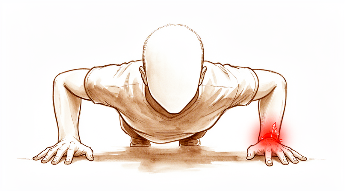
ਡਾ. ਕੀਰਨ ਹਿਰਪਰਾ, ਮਾਟਰ ਪ੍ਰਾਈਵੇਟ ਹਸਪਤਾਲ ਰੌਕਹੈਂਪਟਨ ਵਿੱਚ ਉੱਪਰਲੇ ਅੰਗ (ਮੋਢੇ ਤੋਂ ਹੱਥ ਤੱਕ ਦੀ ਬਾਂਹ) ਦੇ ਸਰਜਨ, ਉਨ੍ਹਾਂ ਸਭ ਤੋਂ ਘੱਟ ਚੀਰ-ਫਾੜ ਵਾਲੇ ਵਿਕਲਪਾਂ ਨਾਲ ਸ਼ੁਰੂਆਤ ਕਰਦੇ ਹਨ ਜੋ ਤੁਹਾਡੀ ਹਾਲਤ ਲਈ ਢੁਕਵੇਂ ਹੋਣ। TFCC (ਟ੍ਰਾਈਐਂਗੂਲਰ ਫ਼ਾਈਬ੍ਰੋਕਾਰਟੀਲੇਜ ਕੰਪਲੈਕਸ, triangular fibrocartilage complex) ਤੁਹਾਡੇ ਗੁੱਟ ਦੇ ਛੋਟੀ ਉਂਗਲ ਵਾਲੇ ਪਾਸੇ ਕਾਰਟੀਲੇਜ (cartilage) ਅਤੇ ਲਿਗਾਮੈਂਟ (ligament, ਹੱਡੀ ਨੂੰ ਹੱਡੀ ਨਾਲ ਜੋੜਨ ਵਾਲਾ ਬੰਧਨ) ਦੀ ਇੱਕ ਬਣਤਰ ਹੈ। ਮੁਰੰਮਤ ਦਾ ਮਤਲਬ ਹੈ ਇਸ ਨੂੰ ਟਾਂਕਿਆਂ ਨਾਲ ਉੱਥੇ ਵਾਪਸ ਜੋੜਨਾ ਜਿੱਥੋਂ ਇਹ ਪਾਟਿਆ ਸੀ। ਅਸੀਂ ਆਮ ਤੌਰ 'ਤੇ ਪਹਿਲਾਂ ਹੈਂਡ ਥੈਰੇਪੀ, ਸਪਲਿੰਟ ਅਤੇ ਸਰਗਰਮੀਆਂ ਵਿੱਚ ਬਦਲਾਅ ਅਜ਼ਮਾਉਂਦੇ ਹਾਂ, ਅਤੇ ਜਦੋਂ ਉਸ ਤੋਂ ਬਾਅਦ ਵੀ ਦਰਦ ਜਾਂ ਅਸਥਿਰਤਾ ਜਾਰੀ ਰਹੇ ਤਾਂ ਸਰਜਰੀ ਬਾਰੇ ਸੋਚਦੇ ਹਾਂ। ਮਰੀਜ਼ਾਂ ਨੂੰ ਆਮ ਤੌਰ 'ਤੇ ਉਨ੍ਹਾਂ ਦੇ ਜੀਪੀ (ਫ਼ੈਮਿਲੀ ਡਾਕਟਰ) ਵੱਲੋਂ ਸਾਡੇ ਕਲੀਨਿਕ ਵਿੱਚ ਭੇਜਿਆ ਜਾਂਦਾ ਹੈ; ਜੇ ਕਿਸੇ ਫਿਜ਼ੀਓਥੈਰੇਪਿਸਟ ਨੇ ਤੁਹਾਨੂੰ ਸਾਨੂੰ ਮਿਲਣ ਦੀ ਸਲਾਹ ਦਿੱਤੀ ਹੈ, ਤਾਂ ਵੀ ਮੈਡੀਕੇਅਰ ਰਿਬੇਟ (Medicare rebate, ਸਰਕਾਰ ਵੱਲੋਂ ਫ਼ੀਸ ਦੀ ਵਾਪਸੀ) ਦੇ ਯੋਗ ਹੋਣ ਲਈ ਤੁਹਾਨੂੰ ਆਪਣੇ ਜੀਪੀ ਤੋਂ ਰੈਫ਼ਰਲ (referral) ਦੀ ਲੋੜ ਪਵੇਗੀ। ਕਲੀਨਿਕ ਵਿੱਚ ਮੁਲਾਂਕਣ, ਜਿਸ ਵਿੱਚ ਤੁਹਾਡੀ ਤਕਲੀਫ਼ ਦਾ ਵੇਰਵਾ, ਜਾਂਚ ਅਤੇ ਲੋੜ ਹੋਵੇ ਤਾਂ ਸਕੈਨ ਸ਼ਾਮਲ ਹਨ, ਤਸ਼ਖ਼ੀਸ ਪੱਕੀ ਕਰਦਾ ਹੈ।
ਇਹ ਓਪਰੇਸ਼ਨ ਉਹਨਾਂ ਪਾਟਣਾਂ ਲਈ ਪੇਸ਼ ਕੀਤਾ ਜਾਂਦਾ ਹੈ ਜਿਨ੍ਹਾਂ ਦੀ ਮੁਰੰਮਤ ਹੋ ਸਕਦੀ ਹੈ, ਜਦੋਂ ਜੋੜ ਵਿੱਚ ਗਠੀਏ (ਆਰਥਰਾਈਟਿਸ) ਵਾਲੀਆਂ ਕੋਈ ਤਬਦੀਲੀਆਂ ਨਾ ਹੋਣ। ਇਸ ਦਾ ਮਕਸਦ ਦਰਦ ਘਟਾਉਣਾ, ਬਾਂਹ ਦੀਆਂ ਦੋ ਹੱਡੀਆਂ ਦੇ ਵਿਚਕਾਰਲੇ ਜੋੜ ਦੀ ਸਥਿਰਤਾ ਵਾਪਸ ਲਿਆਉਣਾ, ਅਤੇ ਤੁਹਾਡੇ ਗੁੱਟ ਅਤੇ ਬਾਂਹ ਦੇ ਹੇਠਲੇ ਹਿੱਸੇ (forearm) ਨੂੰ ਹਿੱਲਦਾ ਰੱਖਣਾ ਹੈ। ਮੁਰੰਮਤ ਦੂਰਬੀਨ ਵਾਲੀ (ਆਰਥਰੋਸਕੋਪਿਕ) ਸਰਜਰੀ ਵਜੋਂ ਕੀਤੀ ਜਾਂਦੀ ਹੈ, ਆਮ ਤੌਰ 'ਤੇ ਜਨਰਲ ਅਨੱਸਥੀਸੀਆ (ਬੇਹੋਸ਼ੀ ਵਾਲੀ ਦਵਾਈ) ਹੇਠ ਉਸੇ ਦਿਨ ਘਰ ਜਾਣ ਵਾਲੀ ਸਰਜਰੀ ਵਜੋਂ। ਹਰ ਚਾਰ ਵਿੱਚੋਂ ਲਗਭਗ ਤਿੰਨ ਲੋਕ ਪੂਰੀਆਂ ਸਰਗਰਮੀਆਂ ਵਿੱਚ ਵਾਪਸ ਆ ਜਾਂਦੇ ਹਨ; ਕੁਝ ਨਹੀਂ ਆਉਂਦੇ। ਅਸੀਂ ਇਸ ਬਾਰੇ ਗੱਲ ਕਰਾਂਗੇ ਕਿ ਕੀ ਇਹ ਓਪਰੇਸ਼ਨ ਤੁਹਾਡੇ ਲਈ ਢੁਕਵਾਂ ਹੈ, ਅਤੇ ਮਿਲ ਕੇ ਫ਼ੈਸਲਾ ਕਰਾਂਗੇ।
ਓਪਰੇਸ਼ਨ ਤੋਂ ਪਹਿਲਾਂ
ਸਰਜਰੀ ਦੀ ਬੁਕਿੰਗ ਹੋਣ ਤੋਂ ਬਾਅਦ, ਸਾਡਾ ਕਲੀਨਿਕ ਤੁਹਾਡੇ ਦਾਖ਼ਲੇ ਦੇ ਵੇਰਵਿਆਂ ਅਤੇ ਖਾਣਾ-ਪੀਣਾ ਬੰਦ ਕਰਨ ਦੀਆਂ ਹਿਦਾਇਤਾਂ ਦੀ ਪੁਸ਼ਟੀ ਕਰੇਗਾ। ਤੁਹਾਨੂੰ ਆਪਣੇ ਓਪਰੇਸ਼ਨ ਤੋਂ ਸੱਤ ਘੰਟੇ ਪਹਿਲਾਂ ਖਾਣਾ ਅਤੇ ਪੀਣਾ ਬੰਦ ਕਰਨਾ ਪਵੇਗਾ, ਤਾਂ ਜੋ ਜੇ ਓਪਰੇਸ਼ਨ ਥੀਏਟਰ ਦੀ ਸੂਚੀ ਸਮੇਂ ਤੋਂ ਪਹਿਲਾਂ ਚੱਲ ਰਹੀ ਹੋਵੇ ਤਾਂ ਅਸੀਂ ਤੁਹਾਡਾ ਓਪਰੇਸ਼ਨ ਪਹਿਲਾਂ ਕਰ ਸਕੀਏ। ਜੇ ਤੁਸੀਂ ਖ਼ੂਨ ਪਤਲਾ ਕਰਨ ਵਾਲੀ ਦਵਾਈ ਜਾਂ ਕੋਈ ਨਿਯਮਿਤ ਦਵਾਈਆਂ ਲੈਂਦੇ ਹੋ, ਤਾਂ ਸਾਨੂੰ ਦੱਸੋ, ਕਿਉਂਕਿ ਕੁਝ ਨੂੰ ਪਹਿਲਾਂ ਤੋਂ ਰੋਕਣਾ ਪੈਂਦਾ ਹੈ। ਆਪਣੀਆਂ ਮੌਜੂਦਾ ਦਵਾਈਆਂ ਦੀ ਲਿਖਤੀ ਸੂਚੀ ਨਾਲ ਲਿਆਓ, ਅਤੇ ਢਿੱਲੇ, ਅਰਾਮਦਾਇਕ ਕੱਪੜੇ ਪਾਓ ਜਿਨ੍ਹਾਂ ਦੀ ਬਾਂਹ ਪੱਟੀ ਵਾਲੇ ਗੁੱਟ ਉੱਤੋਂ ਲੰਘ ਸਕੇ। ਤੁਹਾਨੂੰ ਘਰ ਛੱਡਣ ਲਈ ਕਿਸੇ ਦੀ ਲੋੜ ਪਵੇਗੀ, ਕਿਉਂਕਿ ਜਨਰਲ ਅਨੱਸਥੀਸੀਆ ਕਰਕੇ ਬਾਕੀ ਦਿਨ ਗੱਡੀ ਚਲਾਉਣਾ ਸੁਰੱਖਿਅਤ ਨਹੀਂ ਹੁੰਦਾ। ਐਕਸ-ਰੇ ਜਾਂ MRI ਵਰਗੇ ਸਕੈਨ ਸ਼ਾਇਦ ਪਹਿਲਾਂ ਹੀ ਹੋ ਚੁੱਕੇ ਹੋਣ, ਜਾਂ ਓਪਰੇਸ਼ਨ ਦੀ ਯੋਜਨਾ ਬਣਾਉਣ ਲਈ ਅਸੀਂ ਇਹਨਾਂ ਦਾ ਪ੍ਰਬੰਧ ਕਰ ਸਕਦੇ ਹਾਂ। ਜੇ ਤੁਹਾਨੂੰ ਹੋਰ ਬਿਮਾਰੀਆਂ ਹਨ, ਤਾਂ ਤੁਹਾਨੂੰ ਖ਼ੂਨ ਦੇ ਟੈਸਟਾਂ ਜਾਂ ਅਨੱਸਥੀਟਿਸਟ (ਬੇਹੋਸ਼ੀ ਵਾਲਾ ਡਾਕਟਰ) ਨਾਲ ਜਾਂਚ ਦੀ ਲੋੜ ਪੈ ਸਕਦੀ ਹੈ।
ਓਪਰੇਸ਼ਨ ਵਾਲੇ ਦਿਨ
ਤੁਸੀਂ ਹਸਪਤਾਲ ਦੇ ਸਰਜੀਕਲ ਦਾਖ਼ਲਾ ਵਿਭਾਗ ਵਿੱਚ ਪਹੁੰਚਦੇ ਹੋ, ਜਿੱਥੇ ਸਟਾਫ਼ ਤੁਹਾਡਾ ਦਾਖ਼ਲਾ ਕਰਦਾ ਹੈ ਅਤੇ ਤੁਹਾਨੂੰ ਓਪਰੇਸ਼ਨ ਥੀਏਟਰ ਲਈ ਤਿਆਰ ਕਰਦਾ ਹੈ। ਤੁਸੀਂ ਅਨੱਸਥੀਟਿਸਟ ਨੂੰ ਮਿਲੋਗੇ, ਉਹ ਡਾਕਟਰ ਜੋ ਤੁਹਾਨੂੰ ਬੇਹੋਸ਼ ਕਰਦਾ ਹੈ ਅਤੇ ਓਪਰੇਸ਼ਨ ਦੌਰਾਨ ਤੁਹਾਡਾ ਧਿਆਨ ਰੱਖਦਾ ਹੈ। ਇਹ ਓਪਰੇਸ਼ਨ ਜਨਰਲ ਅਨੱਸਥੀਸੀਆ ਹੇਠ ਕੀਤਾ ਜਾਂਦਾ ਹੈ। ਓਪਰੇਸ਼ਨ ਦੌਰਾਨ ਤੁਸੀਂ ਪੂਰੀ ਤਰ੍ਹਾਂ ਬੇਹੋਸ਼ ਹੋਵੋਗੇ। ਕੁਝ ਮਰੀਜ਼ਾਂ ਨੂੰ ਓਪਰੇਸ਼ਨ ਤੋਂ ਬਾਅਦ ਦਰਦ ਤੋਂ ਰਾਹਤ ਲਈ ਰੀਜਨਲ ਨਰਵ ਬਲਾਕ (ਨਸ ਨੂੰ ਸੁੰਨ ਕਰਨ ਵਾਲਾ ਟੀਕਾ) ਵੀ ਦਿੱਤਾ ਜਾ ਸਕਦਾ ਹੈ; ਅਨੱਸਥੀਟਿਸਟ ਤੁਹਾਡੇ ਨਿੱਜੀ ਹਾਲਾਤ ਦੇ ਆਧਾਰ ਉੱਤੇ ਉਸੇ ਦਿਨ ਇਸ ਦਾ ਫ਼ੈਸਲਾ ਕਰਦਾ ਹੈ। ਫਿਰ ਤੁਹਾਨੂੰ ਓਪਰੇਸ਼ਨ ਥੀਏਟਰ ਵਿੱਚ ਲਿਜਾਇਆ ਜਾਂਦਾ ਹੈ, ਜਿੱਥੇ ਓਪਰੇਸ਼ਨ ਕੀਤਾ ਜਾਂਦਾ ਹੈ।
ਜਦੋਂ ਇਹ ਪੂਰਾ ਹੋ ਜਾਂਦਾ ਹੈ, ਤੁਸੀਂ ਰਿਕਵਰੀ ਵਾਲੀ ਥਾਂ ਵਿੱਚ ਜਾਗਦੇ ਹੋ। ਅਨੱਸਥੀਸੀਆ ਦਾ ਅਸਰ ਉਤਰਨ ਤੱਕ ਨਰਸਾਂ ਤੁਹਾਡਾ ਧਿਆਨ ਰੱਖਦੀਆਂ ਹਨ। ਜਦੋਂ ਤੁਹਾਡੀ ਹਾਲਤ ਸਥਿਰ ਹੋ ਜਾਂਦੀ ਹੈ, ਤਾਂ ਪ੍ਰਕਿਰਿਆ ਅਤੇ ਤੁਹਾਡੇ ਠੀਕ ਹੋਣ ਦੇ ਹਿਸਾਬ ਨਾਲ ਤੁਸੀਂ ਜਾਂ ਤਾਂ ਵਾਰਡ ਵਿੱਚ ਜਾਂਦੇ ਹੋ ਜਾਂ ਘਰ ਚਲੇ ਜਾਂਦੇ ਹੋ।
ਓਪਰੇਸ਼ਨ ਵਿੱਚ ਕੀ ਕੀਤਾ ਜਾਂਦਾ ਹੈ
ਤੁਹਾਡਾ ਸਰਜਨ ਦੂਰਬੀਨ ਵਾਲੀ ਸਰਜਰੀ ਨਾਲ ਸ਼ੁਰੂਆਤ ਕਰਦਾ ਹੈ। ਗੁੱਟ ਦੇ ਪਿਛਲੇ ਪਾਸੇ ਛੋਟੇ ਚੀਰਿਆਂ ਰਾਹੀਂ ਤੁਹਾਡੇ ਗੁੱਟ ਵਿੱਚ ਇੱਕ ਨਿੱਕਾ ਜਿਹਾ ਕੈਮਰਾ ਪਾਇਆ ਜਾਂਦਾ ਹੈ, ਤਾਂ ਜੋ ਪਾਟਣ ਨੂੰ ਸਾਫ਼ ਦੇਖਿਆ ਜਾ ਸਕੇ। ਮੁਰੰਮਤ ਦਾ ਠੀਕ-ਠੀਕ ਤਰੀਕਾ ਇਸ ਗੱਲ ਉੱਤੇ ਨਿਰਭਰ ਕਰਦਾ ਹੈ ਕਿ ਕੈਮਰਾ ਕੀ ਦਿਖਾਉਂਦਾ ਹੈ, ਇਸ ਲਈ ਪਹਿਲਾਂ ਤੋਂ ਤਰੀਕੇ ਦਾ ਵਾਅਦਾ ਨਹੀਂ ਕੀਤਾ ਜਾ ਸਕਦਾ।
ਜੇ ਪਾਟਣ TFCC ਦੇ ਬਾਹਰਲੇ ਕਿਨਾਰੇ ਉੱਤੇ ਹੈ, ਤਾਂ ਇਸ ਨੂੰ ਟਾਂਕਿਆਂ ਨਾਲ ਕੈਪਸੂਲ (capsule), ਯਾਨੀ ਜੋੜ ਦੀ ਅੰਦਰਲੀ ਪਰਤ, ਨਾਲ ਵਾਪਸ ਜੋੜਿਆ ਜਾਂਦਾ ਹੈ। ਜੇ ਪਾਟਣ ਉੱਥੇ ਹੈ ਜਿੱਥੇ ਲਿਗਾਮੈਂਟ ਅਲਨਾ (ulna), ਯਾਨੀ ਤੁਹਾਡੀ ਬਾਂਹ ਦੇ ਹੇਠਲੇ ਹਿੱਸੇ ਵਿੱਚ ਛੋਟੀ ਉਂਗਲ ਵਾਲੇ ਪਾਸੇ ਦੀ ਛੋਟੀ ਹੱਡੀ, ਤੋਂ ਉੱਖੜ ਗਿਆ ਹੈ, ਤਾਂ ਇਸ ਨੂੰ ਹੱਡੀ ਨਾਲ ਦੁਬਾਰਾ ਜੋੜਿਆ ਜਾਂਦਾ ਹੈ। ਇਹ ਹੱਡੀ ਵਿੱਚ ਲਗਾਏ ਇੱਕ ਛੋਟੇ ਸਿਊਚਰ ਐਂਕਰ (suture anchor, ਹੱਡੀ ਵਿੱਚ ਲਗਾਇਆ ਜਾਣ ਵਾਲਾ ਛੋਟਾ ਕਿੱਲ, ਜਿਸ ਨਾਲ ਟਾਂਕੇ ਬੰਨ੍ਹ ਕੇ ਟਿਸ਼ੂ ਨੂੰ ਹੱਡੀ ਨਾਲ ਜੋੜਿਆ ਜਾਂਦਾ ਹੈ) ਨਾਲ, ਜਾਂ ਹੱਡੀ ਵਿੱਚ ਡ੍ਰਿੱਲ ਕੀਤੀ ਇੱਕ ਛੋਟੀ ਸੁਰੰਗ ਰਾਹੀਂ ਕੀਤਾ ਜਾ ਸਕਦਾ ਹੈ, ਕਈ ਵਾਰ ਇੱਕ ਛੋਟੇ ਖੁੱਲ੍ਹੇ ਚੀਰੇ ਰਾਹੀਂ। ਜਿਸ ਪਾਟਣ ਦੀ ਮੁਰੰਮਤ ਨਹੀਂ ਹੋ ਸਕਦੀ, ਉਸ ਦੀ ਬਜਾਏ ਸਫ਼ਾਈ ਕੀਤੀ ਜਾਂਦੀ ਹੈ, ਯਾਨੀ ਖੁਰਦਰੇ ਕਿਨਾਰੇ ਛਾਂਗ ਦਿੱਤੇ ਜਾਂਦੇ ਹਨ। ਜਦੋਂ ਤੁਸੀਂ ਜਾਗ ਜਾਓਗੇ, ਤੁਹਾਡਾ ਸਰਜਨ ਸਮਝਾਏਗਾ ਕਿ ਕੀ ਕੀਤਾ ਗਿਆ।
ਚੀਰਿਆਂ ਨੂੰ ਟਾਂਕਿਆਂ ਨਾਲ ਬੰਦ ਕਰਕੇ ਪੱਟੀ ਨਾਲ ਢਕ ਦਿੱਤਾ ਜਾਂਦਾ ਹੈ।
ਓਪਰੇਸ਼ਨ ਤੋਂ ਬਾਅਦ
ਤੁਸੀਂ ਰਿਕਵਰੀ ਵਾਲੀ ਥਾਂ ਵਿੱਚ ਜਾਗਦੇ ਹੋ, ਜਿੱਥੇ ਅਨੱਸਥੀਸੀਆ ਦਾ ਅਸਰ ਉਤਰਨ ਦੌਰਾਨ ਨਰਸਾਂ ਨੇੜੇ ਹੁੰਦੀਆਂ ਹਨ। ਇਹ ਆਮ ਤੌਰ 'ਤੇ ਉਸੇ ਦਿਨ ਘਰ ਜਾਣ ਵਾਲਾ ਓਪਰੇਸ਼ਨ ਹੁੰਦਾ ਹੈ, ਇਸ ਲਈ ਤੁਸੀਂ ਉਸੇ ਦਿਨ ਘਰ ਜਾਣ ਦੀ ਉਮੀਦ ਰੱਖ ਸਕਦੇ ਹੋ, ਹਾਲਾਂਕਿ ਕਦੇ-ਕਦਾਈਂ ਮਰੀਜ਼ ਰਾਤ ਭਰ ਰੁਕਦੇ ਹਨ। ਤੁਹਾਡਾ ਗੁੱਟ ਕੂਹਣੀ ਤੋਂ ਹੇਠਾਂ ਤੱਕ ਦੇ ਪਲੱਸਤਰ (cast) ਜਾਂ ਪੱਕੇ ਸਪਲਿੰਟ ਵਿੱਚ ਹੋਵੇਗਾ, ਤੁਹਾਡੀ ਬਾਂਹ ਦਾ ਹੇਠਲਾ ਹਿੱਸਾ ਥੋੜ੍ਹਾ ਹਥੇਲੀ ਉੱਪਰ ਵੱਲ ਘੁੰਮਿਆ ਹੋਵੇਗਾ ਅਤੇ ਤੁਹਾਡੀ ਕੂਹਣੀ ਹਿੱਲਣ ਲਈ ਖੁੱਲ੍ਹੀ ਹੋਵੇਗੀ। ਦਰਦ ਨੂੰ ਉਹਨਾਂ ਦਵਾਈਆਂ ਨਾਲ ਸੰਭਾਲਿਆ ਜਾਂਦਾ ਹੈ ਜੋ ਤੁਸੀਂ ਘਰ ਵਿੱਚ ਲੈ ਸਕਦੇ ਹੋ; ਕੁਝ ਬੇਅਰਾਮੀ ਆਮ ਗੱਲ ਹੈ। ਸੋਜ ਘਟਾਉਣ ਵਿੱਚ ਮਦਦ ਲਈ ਅਰਾਮ ਕਰਨ ਵੇਲੇ ਆਪਣੇ ਹੱਥ ਨੂੰ ਸਿਰਹਾਣਿਆਂ ਉੱਤੇ ਉੱਚਾ ਰੱਖੋ। ਜਦੋਂ ਹੋ ਸਕੇ, ਆਪਣੀਆਂ ਉਂਗਲਾਂ ਹਿਲਾਓ। ਜੇ ਤੁਹਾਨੂੰ ਨਰਵ ਬਲਾਕ ਲਗਾਇਆ ਗਿਆ ਸੀ, ਤਾਂ ਪਹਿਲੇ 24 ਘੰਟਿਆਂ ਵਿੱਚ ਸੁੰਨਪਨ ਅਤੇ ਕਮਜ਼ੋਰੀ ਉਮੀਦ ਮੁਤਾਬਕ ਹਨ; ਇਸ ਦਾ ਅਸਰ ਉਤਰ ਜਾਣ ਤੋਂ ਬਾਅਦ, ਜੇ ਤੁਹਾਨੂੰ ਸੁੰਨਪਨ ਹੋਵੇ ਜਾਂ ਤੁਸੀਂ ਆਪਣਾ ਹੱਥ ਜਾਂ ਉਂਗਲਾਂ ਨਾ ਹਿਲਾ ਸਕੋ, ਤਾਂ ਕਲੀਨਿਕ ਨੂੰ ਫ਼ੋਨ ਕਰੋ। ਪਹਿਲੇ 24 ਘੰਟੇ ਕਿਸੇ ਨੂੰ ਤੁਹਾਡੇ ਨਾਲ ਰਹਿਣਾ ਚਾਹੀਦਾ ਹੈ। ਅਸੀਂ ਪੱਟੀ ਲਗਭਗ 10 ਦਿਨ ਲੱਗੀ ਰਹਿਣ ਦਿੰਦੇ ਹਾਂ; ਕਿਰਪਾ ਕਰਕੇ ਉਸ ਤੋਂ ਪਹਿਲਾਂ ਇਸ ਨੂੰ ਨਾ ਉਤਾਰੋ, ਜਦੋਂ ਤੱਕ ਅਸੀਂ ਤੁਹਾਨੂੰ ਨਾ ਕਹੀਏ। ਜਦੋਂ ਅਸੀਂ ਤੁਹਾਨੂੰ ਮਿਲਦੇ ਹਾਂ ਤਾਂ ਅਸੀਂ ਇਸ ਨੂੰ ਬਦਲਦੇ ਜਾਂ ਉਤਾਰਦੇ ਹਾਂ।
ਸਿਹਤਯਾਬੀ
ਤੁਹਾਡਾ ਗੁੱਟ ਛੇ ਹਫ਼ਤਿਆਂ ਲਈ ਕੂਹਣੀ ਤੋਂ ਹੇਠਾਂ ਤੱਕ ਦੇ ਪਲੱਸਤਰ ਜਾਂ ਪੱਕੇ ਸਪਲਿੰਟ ਵਿੱਚ ਰਹੇਗਾ, ਤੁਹਾਡੀ ਬਾਂਹ ਦਾ ਹੇਠਲਾ ਹਿੱਸਾ ਥੋੜ੍ਹਾ ਹਥੇਲੀ ਉੱਪਰ ਵੱਲ ਘੁੰਮਿਆ ਹੋਵੇਗਾ ਅਤੇ ਤੁਹਾਡੀ ਕੂਹਣੀ ਖੁੱਲ੍ਹੀ ਹੋਵੇਗੀ। ਇਹ ਮੁਰੰਮਤ ਦੇ ਠੀਕ ਹੋਣ ਦੌਰਾਨ ਉਸ ਦੀ ਰੱਖਿਆ ਕਰਦਾ ਹੈ। ਜਦੋਂ ਤੱਕ ਪਲੱਸਤਰ ਜਾਂ ਸਪਲਿੰਟ ਲੱਗਿਆ ਹੈ, ਤੁਸੀਂ ਗੱਡੀ ਨਹੀਂ ਚਲਾ ਸਕਦੇ। ਜਦੋਂ ਇਹ ਉਤਰ ਜਾਵੇ ਅਤੇ ਤੁਹਾਡਾ ਸਰਜਨ ਇਜਾਜ਼ਤ ਦੇ ਦੇਵੇ, ਤਾਂ ਤੁਸੀਂ ਦੁਬਾਰਾ ਗੱਡੀ ਚਲਾ ਸਕਦੇ ਹੋ; ਉਸ ਤੋਂ ਬਾਅਦ ਪਾਇਆ ਜਾਣ ਵਾਲਾ ਹਲਕਾ, ਉਤਾਰਨਯੋਗ ਸਪਲਿੰਟ ਆਪਣੇ ਆਪ ਵਿੱਚ ਤੁਹਾਨੂੰ ਗੱਡੀ ਚਲਾਉਣ ਤੋਂ ਨਹੀਂ ਰੋਕਦਾ। ਹੋਰ ਵੇਰਵੇ ਲਈ, ਉੱਪਰਲੇ ਅੰਗ ਦੀ ਸਰਜਰੀ ਤੋਂ ਬਾਅਦ ਗੱਡੀ ਚਲਾਉਣਾ ਦੇਖੋ।
ਛੇ ਹਫ਼ਤਿਆਂ ਤੋਂ ਬਾਅਦ, ਜਦੋਂ ਤੁਹਾਡਾ ਗੁੱਟ ਹਿਲਜੁਲ ਅਤੇ ਤਾਕਤ ਵਾਪਸ ਹਾਸਲ ਕਰਦਾ ਹੈ, ਤੁਸੀਂ ਇੱਕ ਉਤਾਰਨਯੋਗ ਸਪਲਿੰਟ ਵਿੱਚ ਆ ਜਾਓਗੇ। ਤੁਹਾਡਾ ਮੁੜ-ਵਸੇਬਾ (rehabilitation) ਹੈਂਡ ਥੈਰੇਪੀ ਹੈ, ਫਿਜ਼ੀਓਥੈਰੇਪੀ ਨਹੀਂ। ਇਸ ਦੀ ਅਗਵਾਈ ਐਕਸਟੈਂਡ ਰੀਹੈਬਿਲੀਟੇਸ਼ਨ (Extend Rehabilitation) ਵਿੱਚ ਰੂਬੀ ਡੂਲਨ (Ruby Doolan) ਕਰਦੀ ਹੈ। ਰੂਬੀ ਇੱਕ ਹੈਂਡ ਥੈਰੇਪਿਸਟ ਹੈ: ਉਹ ਤੁਹਾਡੀਆਂ ਕਸਰਤਾਂ ਵਿੱਚ ਤੁਹਾਡੀ ਅਗਵਾਈ ਕਰਦੀ ਹੈ ਅਤੇ ਜਿਸ ਵੀ ਸਪਲਿੰਟ ਦੀ ਤੁਹਾਨੂੰ ਲੋੜ ਹੋਵੇ ਉਹ ਬਣਾਉਂਦੀ ਹੈ।
ਦਫ਼ਤਰ ਦਾ ਕੰਮ ਅਤੇ ਰੋਜ਼ਾਨਾ ਦੇ ਹਲਕੇ ਕੰਮ ਆਮ ਤੌਰ 'ਤੇ ਛੇ ਤੋਂ ਅੱਠ ਹਫ਼ਤਿਆਂ ਉੱਤੇ ਦੁਬਾਰਾ ਸ਼ੁਰੂ ਹੁੰਦੇ ਹਨ। ਹੱਥੀਂ ਮਿਹਨਤ ਵਾਲੇ ਕੰਮ, ਖੇਡਾਂ ਅਤੇ ਜਿਮ ਵਿੱਚ ਜ਼ਿਆਦਾ ਸਮਾਂ ਲੱਗਦਾ ਹੈ, ਚਾਰ ਤੋਂ ਛੇ ਮਹੀਨੇ। ਅੰਤਿਮ ਨਤੀਜਾ, ਜਦੋਂ ਦਰਦ ਅਤੇ ਤਾਕਤ ਵਿੱਚ ਹੋਰ ਸੁਧਾਰ ਹੋਣਾ ਰੁਕ ਜਾਂਦਾ ਹੈ, ਲਗਭਗ 12 ਮਹੀਨਿਆਂ ਉੱਤੇ ਆਉਂਦਾ ਹੈ।
ਦਰਦ ਆਮ ਤੌਰ 'ਤੇ ਤਾਕਤ ਅਤੇ ਕੰਮਕਾਜ ਤੋਂ ਪਹਿਲਾਂ ਸੁਧਰਦਾ ਹੈ। ਤੁਹਾਡਾ ਗੁੱਟ ਮਜ਼ਬੂਤ ਮਹਿਸੂਸ ਹੋਣ ਤੋਂ ਕਾਫ਼ੀ ਪਹਿਲਾਂ ਵਰਤਣ ਵਿੱਚ ਸੌਖਾ ਲੱਗ ਸਕਦਾ ਹੈ, ਇਸ ਲਈ ਭਾਰੀ ਕੰਮਾਂ ਨਾਲ ਸਬਰ ਰੱਖਣਾ ਫ਼ਾਇਦੇਮੰਦ ਹੈ।
ਲਗਭਗ ਅੱਧੇ ਲੋਕਾਂ ਨੂੰ ਆਪਣੀ ਸਭ ਤੋਂ ਵੱਧ ਸਰਗਰਮੀ ਤੱਕ ਪਹੁੰਚਣ ਤੋਂ ਬਾਅਦ ਆਰਜ਼ੀ ਤੌਰ 'ਤੇ ਪਿੱਛੇ ਹਟਣਾ ਪੈਂਦਾ ਹੈ। ਇਹ ਸਮੇਂ ਨਾਲ ਠੀਕ ਹੋ ਜਾਂਦਾ ਹੈ ਅਤੇ ਅੰਤਿਮ ਨਤੀਜੇ ਨੂੰ ਨਹੀਂ ਬਦਲਦਾ। ਇਹ ਜਾਣਨਾ ਕਿ ਅਜਿਹਾ ਹੋ ਸਕਦਾ ਹੈ, ਜੇ ਤੁਹਾਡੇ ਨਾਲ ਹੋਵੇ ਤਾਂ ਇਸ ਨੂੰ ਘੱਟ ਚਿੰਤਾਜਨਕ ਬਣਾਉਂਦਾ ਹੈ।
ਕੀ ਗ਼ਲਤ ਹੋ ਸਕਦਾ ਹੈ
ਕੋਈ ਵੀ ਓਪਰੇਸ਼ਨ ਖ਼ਤਰੇ ਤੋਂ ਖ਼ਾਲੀ ਨਹੀਂ ਹੁੰਦਾ, ਅਤੇ ਇਹ ਜਾਣਨਾ ਵਾਜਬ ਹੈ ਕਿ ਕੀ ਹੋ ਸਕਦਾ ਹੈ।
ਮੁਰੰਮਤ ਮੁੜ ਪਾਟ ਸਕਦੀ ਹੈ, ਜਾਂ ਬਾਂਹ ਦੀਆਂ ਦੋ ਹੱਡੀਆਂ ਦੇ ਵਿਚਕਾਰਲਾ ਜੋੜ ਅਸਥਿਰ ਰਹਿ ਸਕਦਾ ਹੈ ਜਾਂ ਦੁਬਾਰਾ ਅਸਥਿਰ ਹੋ ਸਕਦਾ ਹੈ। ਇਹਨਾਂ ਵਿੱਚੋਂ ਕਿਸੇ ਦਾ ਵੀ ਮਤਲਬ ਇੱਕ ਹੋਰ ਓਪਰੇਸ਼ਨ ਹੋ ਸਕਦਾ ਹੈ। ਕੁਝ ਲੋਕਾਂ ਦੇ ਗੁੱਟ ਵਿੱਚ ਅਕੜਾਅ ਜਾਂ ਕਮਜ਼ੋਰੀ ਰਹਿ ਜਾਂਦੀ ਹੈ, ਭਾਵੇਂ ਖ਼ੁਦ ਮੁਰੰਮਤ ਠੀਕ ਹੋ ਗਈ ਹੋਵੇ।
ਤੁਹਾਡੇ ਹੱਥ ਦੇ ਪੁੱਠੇ ਪਾਸੇ, ਛੋਟੀ ਉਂਗਲ ਵਾਲੇ ਪਾਸੇ ਦੀ ਛੋਟੀ ਨਸ (nerve) ਸਰਜਰੀ ਨਾਲ ਚਿੜ (irritated) ਸਕਦੀ ਹੈ। ਇਸ ਨਾਲ ਚਮੜੀ ਦੇ ਉਸ ਹਿੱਸੇ ਵਿੱਚ ਝਰਨਾਹਟ ਜਾਂ ਸੁੰਨਪਨ ਹੁੰਦਾ ਹੈ। ਇਹ ਆਮ ਤੌਰ 'ਤੇ ਕੁਝ ਮਹੀਨਿਆਂ ਵਿੱਚ ਠੀਕ ਹੋ ਜਾਂਦਾ ਹੈ।
ਜੇ ਤੁਹਾਡੀ ਮੁਰੰਮਤ ਵਿੱਚ ਹੱਡੀ ਰਾਹੀਂ ਸੁਰੰਗ ਵਰਤੀ ਗਈ ਸੀ, ਤਾਂ ਟਾਂਕੇ ਦੀ ਗੰਢ ਕਈ ਵਾਰ ਚਮੜੀ ਹੇਠ ਟਿਕ ਕੇ ਚੁਭਣ ਜਾਂ ਰਗੜ ਪੈਦਾ ਕਰ ਸਕਦੀ ਹੈ। ਇਸ ਨੂੰ ਕੱਢਣ ਲਈ ਇੱਕ ਛੋਟੇ ਓਪਰੇਸ਼ਨ ਦੀ ਲੋੜ ਪੈ ਸਕਦੀ ਹੈ। ਤੁਹਾਡੇ ਗੁੱਟ ਦੇ ਛੋਟੀ ਉਂਗਲ ਵਾਲੇ ਪਾਸੇ ਦੇ ਟੈਂਡਨ (tendon, ਪੱਠੇ ਨੂੰ ਹੱਡੀ ਨਾਲ ਜੋੜਨ ਵਾਲੀ ਮਜ਼ਬੂਤ ਤੰਦ) ਵੀ ਚਿੜ ਸਕਦੇ ਹਨ।
ਲਾਗ (ਇਨਫੈਕਸ਼ਨ) ਬਹੁਤ ਘੱਟ ਹੁੰਦੀ ਹੈ। ਜੇ ਤੁਹਾਨੂੰ ਬੁਖ਼ਾਰ ਹੋਵੇ, ਜ਼ਖ਼ਮ ਦੇ ਆਲੇ-ਦੁਆਲੇ ਲਾਲੀ ਫੈਲ ਰਹੀ ਹੋਵੇ, ਜਾਂ ਜ਼ਖ਼ਮ ਵਿੱਚੋਂ ਤਰਲ ਜਾਂ ਪਾਕ ਰਿਸ ਰਹੀ ਹੋਵੇ, ਜਾਂ ਜੇ ਦਰਦ ਦੀਆਂ ਦਵਾਈਆਂ ਦੇ ਬਾਵਜੂਦ ਤੁਹਾਡਾ ਦਰਦ ਵਧਦਾ ਹੀ ਜਾਵੇ, ਤਾਂ ਉਸੇ ਦਿਨ ਕਲੀਨਿਕ ਨੂੰ ਫ਼ੋਨ ਕਰੋ।
ਸਾਨੂੰ ਕਦੋਂ ਫ਼ੋਨ ਕਰਨਾ ਹੈ
ਇਸ ਓਪਰੇਸ਼ਨ ਤੋਂ ਬਾਅਦ ਜ਼ਿਆਦਾਤਰ ਸਮੱਸਿਆਵਾਂ ਦਾ ਇਲਾਜ ਸੌਖਾ ਹੁੰਦਾ ਹੈ ਜਦੋਂ ਉਹ ਛੇਤੀ ਫੜੀਆਂ ਜਾਣ। ਜੇ ਤੁਹਾਨੂੰ ਕਿਸੇ ਵੀ ਗੱਲ ਦੀ ਚਿੰਤਾ ਹੋਵੇ, ਤਾਂ ਸਾਨੂੰ ਫ਼ੋਨ ਕਰੋ।
ਜੇ ਤੁਹਾਡੀ ਲੱਤ ਦੀ ਪਿੰਨੀ (calf) ਵਿੱਚ ਸੋਜ ਜਾਂ ਦਰਦ ਹੋਵੇ, ਜਾਂ ਸਾਹ ਚੜ੍ਹੇ ਜਾਂ ਛਾਤੀ ਵਿੱਚ ਦਰਦ ਹੋਵੇ, ਤਾਂ ਐਮਰਜੈਂਸੀ ਵਿਭਾਗ ਜਾਓ। ਇਹ ਖ਼ੂਨ ਦੇ ਗਤਲੇ ਦੀਆਂ ਨਿਸ਼ਾਨੀਆਂ ਹੋ ਸਕਦੀਆਂ ਹਨ। ਜੇ ਤੁਹਾਡੀਆਂ ਉਂਗਲਾਂ ਫਿੱਕੀਆਂ, ਠੰਢੀਆਂ, ਸਫ਼ੈਦ, ਨੀਲੀਆਂ ਜਾਂ ਗੂੜ੍ਹੇ ਰੰਗ ਦੀਆਂ ਪੈ ਜਾਣ, ਤਾਂ ਐਮਰਜੈਂਸੀ ਵਿਭਾਗ ਜਾਓ, ਕਿਉਂਕਿ ਇਹ ਖ਼ੂਨ ਦੇ ਦੌਰੇ ਦੀ ਸਮੱਸਿਆ ਹੋ ਸਕਦੀ ਹੈ।
ਜੇ ਤੁਹਾਨੂੰ ਬੁਖ਼ਾਰ ਹੋਵੇ, ਜ਼ਖ਼ਮ ਦੇ ਆਲੇ-ਦੁਆਲੇ ਲਾਲੀ ਫੈਲ ਰਹੀ ਹੋਵੇ, ਜਾਂ ਜ਼ਖ਼ਮ ਵਿੱਚੋਂ ਤਰਲ ਜਾਂ ਪਾਕ ਰਿਸ ਰਹੀ ਹੋਵੇ, ਤਾਂ ਉਸੇ ਦਿਨ ਕਲੀਨਿਕ ਨੂੰ ਫ਼ੋਨ ਕਰੋ। ਜੇ ਦਰਦ ਦੀਆਂ ਦਵਾਈਆਂ ਦੇ ਬਾਵਜੂਦ ਤੁਹਾਡਾ ਦਰਦ ਵਧਦਾ ਹੀ ਜਾਵੇ, ਤਾਂ ਵੀ ਉਸੇ ਦਿਨ ਫ਼ੋਨ ਕਰੋ।
ਨਰਵ ਬਲਾਕ ਤੋਂ ਬਾਅਦ ਪਹਿਲੇ 24 ਘੰਟਿਆਂ ਵਿੱਚ ਸੁੰਨਪਨ ਅਤੇ ਕਮਜ਼ੋਰੀ ਉਮੀਦ ਮੁਤਾਬਕ ਹਨ। ਬਲਾਕ ਦਾ ਅਸਰ ਉਤਰ ਜਾਣ ਤੋਂ ਬਾਅਦ, ਜੇ ਤੁਹਾਨੂੰ ਸੁੰਨਪਨ ਹੋਵੇ ਜਾਂ ਤੁਸੀਂ ਆਪਣਾ ਹੱਥ ਜਾਂ ਉਂਗਲਾਂ ਨਾ ਹਿਲਾ ਸਕੋ, ਤਾਂ ਕਲੀਨਿਕ ਨੂੰ ਫ਼ੋਨ ਕਰੋ।
ਜੇ ਤੁਸੀਂ ਕਲੀਨਿਕ ਨਾਲ ਸੰਪਰਕ ਨਹੀਂ ਕਰ ਸਕਦੇ, ਜਿਵੇਂ ਦਫ਼ਤਰੀ ਸਮੇਂ ਤੋਂ ਬਾਅਦ ਜਾਂ ਹਫ਼ਤੇ ਦੇ ਅਖ਼ੀਰ ’ਤੇ, ਤਾਂ ਆਪਣੇ ਸਭ ਤੋਂ ਨੇੜਲੇ ਐਮਰਜੈਂਸੀ ਵਿਭਾਗ ਜਾਓ।
ਵਿਸਥਾਰ ਵਿੱਚ
Advanced reading: the deeper science (optional)
ਇਹ ਹਿੱਸਾ ਤੁਹਾਡੇ ਆਪਣੇ ਇਲਾਜ ਬਾਰੇ ਫ਼ੈਸਲਿਆਂ ਲਈ ਲੋੜ ਤੋਂ ਵੱਧ ਵਿਸਥਾਰ ਵਿੱਚ ਜਾਂਦਾ ਹੈ। TFCC ਦੀ ਮੁਰੰਮਤ ਬਾਰੇ ਵਾਧੂ ਪੜ੍ਹਨਾ ਇਸ ਲਈ ਲਾਹੇਵੰਦ ਹੈ ਕਿਉਂਕਿ ਜਿਨ੍ਹਾਂ ਚੋਣਾਂ ਉੱਤੇ ਸਭ ਤੋਂ ਵੱਧ ਬਹਿਸ ਹੁੰਦੀ ਹੈ, ਉਹ ਤੁਲਨਾਤਮਕ ਅਧਿਐਨਾਂ ਵਿੱਚ ਇੱਕ ਦੂਜੇ ਤੋਂ ਵੱਖਰੀਆਂ ਨਹੀਂ ਨਿਕਲੀਆਂ, ਜਦਕਿ ਸਿਹਤਯਾਬੀ ਉਸ ਨਾਲੋਂ ਹੌਲੀ ਅਤੇ ਘੱਟ ਇੱਕਸਾਰ ਚੱਲਦੀ ਹੈ ਜਿੰਨਾ ਦਰਦ ਤੋਂ ਸ਼ੁਰੂਆਤੀ ਰਾਹਤ ਤੋਂ ਲੱਗਦਾ ਹੈ।
ਆਰਥਰੋਸਕੋਪਿਕ ਜਾਂ ਖੁੱਲ੍ਹੀ, ਐਂਕਰ ਜਾਂ ਸੁਰੰਗ
ਫ਼ੋਵੀਅਲ (foveal, ਅਲਨਾ ਉੱਤੇ ਲਿਗਾਮੈਂਟ ਦੀ ਡੂੰਘੀ ਜੁੜਨ-ਥਾਂ ਵਾਲੇ) ਪਾਟਣਾਂ ਲਈ, 3 ਅਧਿਐਨਾਂ ਅਤੇ 157 ਮਰੀਜ਼ਾਂ ਦੇ 2026 ਦੇ ਇੱਕ ਮੈਟਾ-ਵਿਸ਼ਲੇਸ਼ਣ (meta-analysis, ਕਈ ਅਧਿਐਨਾਂ ਨੂੰ ਇਕੱਠਾ ਕਰਕੇ ਕੀਤਾ ਵਿਸ਼ਲੇਸ਼ਣ) ਵਿੱਚ ਕੰਮਕਾਜ, ਦਰਦ, ਪਕੜ ਦੀ ਤਾਕਤ ਜਾਂ ਪੇਚੀਦਗੀਆਂ ਵਿੱਚ ਆਰਥਰੋਸਕੋਪਿਕ ਅਤੇ ਖੁੱਲ੍ਹੀ ਮੁਰੰਮਤ ਵਿਚਕਾਰ ਅੰਕੜਿਆਂ ਪੱਖੋਂ ਕੋਈ ਅਹਿਮ ਫ਼ਰਕ ਨਹੀਂ ਮਿਲਿਆ [1]। ਮਰੀਜ਼ਾਂ ਦੀ ਘੱਟ ਗਿਣਤੀ ਉਸ ਸਿੱਟੇ ਦੀ ਮੁੱਖ ਸੀਮਾ ਹੈ।
ਇੱਕ ਯੋਜਨਾਬੱਧ ਸਮੀਖਿਆ (systematic review) ਵਿੱਚ 904 ਮਰੀਜ਼ਾਂ ਵਿੱਚ ਸਿਊਚਰ ਐਂਕਰਾਂ ਅਤੇ ਹੱਡੀ ਦੀਆਂ ਸੁਰੰਗਾਂ ਦੀ ਤੁਲਨਾ ਕੀਤੀ ਗਈ। ਦੋਵਾਂ ਨੇ ਕੰਮਕਾਜ, ਦਰਦ ਅਤੇ ਪਕੜ ਦੀ ਤਾਕਤ ਵਿੱਚ ਸੁਧਾਰ ਕੀਤਾ, ਅਤੇ ਦੋਵਾਂ ਵਿੱਚ ਦੁਬਾਰਾ ਓਪਰੇਸ਼ਨ ਆਮ ਨਹੀਂ ਸੀ [2]। ਫ਼ਰਕ ਬਾਅਦ ਵਿੱਚ ਆਉਣ ਵਾਲੀ ਸਮੱਸਿਆ ਦੀ ਕਿਸਮ ਵਿੱਚ ਹੈ। ਐਂਕਰਾਂ ਨਾਲ, ਸਭ ਤੋਂ ਆਮ ਪੇਚੀਦਗੀ ਹੱਥ ਦੇ ਪੁੱਠੇ ਪਾਸੇ ਦੀ ਛੋਟੀ ਅਹਿਸਾਸ ਵਾਲੀ ਨਸ ਦਾ ਚਿੜ ਜਾਣਾ ਸੀ, ਜੋ ਆਮ ਤੌਰ 'ਤੇ 2 ਤੋਂ 4 ਮਹੀਨਿਆਂ ਵਿੱਚ ਠੀਕ ਹੋ ਜਾਂਦਾ ਸੀ। ਸੁਰੰਗਾਂ ਨਾਲ, ਇਹ ਚੁਭਣ ਵਾਲੀ ਟਾਂਕੇ ਦੀ ਗੰਢ ਸੀ [2]। 60 ਮਰੀਜ਼ਾਂ ਦੇ ਇੱਕ ਰੈਂਡਮਾਈਜ਼ਡ ਟ੍ਰਾਇਲ (ਬੇਤਰਤੀਬ ਨਿਯੰਤਰਿਤ ਪਰਖ) ਵਿੱਚ, ਸੁਰੰਗ ਵਾਲੇ ਸਮੂਹ ਵਿੱਚ 30 ਵਿੱਚੋਂ 5 (16.7%) ਨੂੰ ਗੰਢ ਕੱਢਣ ਲਈ ਦੂਜੇ ਓਪਰੇਸ਼ਨ ਦੀ ਲੋੜ ਪਈ [3]।
40 ਮਰੀਜ਼ਾਂ ਦੇ ਇੱਕ ਰੈਂਡਮਾਈਜ਼ਡ ਟ੍ਰਾਇਲ ਵਿੱਚ ਸੁਰੰਗ ਵਾਲੀ ਮੁਰੰਮਤ ਦੇ ਨਾਲ ਕੈਪਸੂਲ ਦੀ ਮੁਰੰਮਤ ਵੀ ਜੋੜੀ ਗਈ। ਪਕੜ ਦੀ ਤਾਕਤ ਦੂਜੇ ਹੱਥ ਦੇ 94.2% ਤੱਕ ਵਾਪਸ ਆਈ, ਜਦਕਿ ਸਿਰਫ਼ ਸੁਰੰਗ ਵਾਲੀ ਮੁਰੰਮਤ ਨਾਲ 84.2%। ਦਰਦ, DASH (ਬਾਂਹ, ਮੋਢੇ ਅਤੇ ਹੱਥ ਦੀ ਅਪਾਹਜਤਾ ਦਾ ਸਕੋਰ) ਅਤੇ ਗੁੱਟ ਦੇ ਸਕੋਰਾਂ ਵਿੱਚ ਕੋਈ ਅਹਿਮ ਫ਼ਰਕ ਨਹੀਂ ਸੀ [4]। ਇਹ ਇੱਕੋ ਛੋਟਾ ਟ੍ਰਾਇਲ ਹੈ, ਅਤੇ ਪਕੜ ਹੀ ਇੱਕੋ-ਇੱਕ ਮਾਪ ਸੀ ਜਿਸ ਵਿੱਚ ਫ਼ਰਕ ਨਿਕਲਿਆ।
ਉਡੀਕ ਕਰਨ ਨਾਲ ਰਾਹ ਬੰਦ ਹੁੰਦਾ ਨਹੀਂ ਲੱਗਦਾ
ਇਹ ਉਮੀਦ ਕਰਨਾ ਕੁਦਰਤੀ ਲੱਗਦਾ ਹੈ ਕਿ ਦੇਰ ਨਾਲ ਕੀਤੀ ਮੁਰੰਮਤ ਦੇ ਨਤੀਜੇ ਮਾੜੇ ਹੋਣਗੇ, ਪਰ ਸਬੂਤ ਇਸ ਤੋਂ ਕਮਜ਼ੋਰ ਹਨ। ਫ਼ੋਵੀਅਲ ਪਾਟਣਾਂ ਵਾਲੇ 80 ਮਰੀਜ਼ਾਂ ਵਿੱਚ, ਨਤੀਜੇ ਮਿਲਦੇ-ਜੁਲਦੇ ਸਨ ਭਾਵੇਂ ਸਰਜਰੀ 6 ਮਹੀਨਿਆਂ ਦੇ ਅੰਦਰ, 6 ਤੋਂ 12 ਮਹੀਨਿਆਂ ਦੇ ਵਿਚਕਾਰ, ਜਾਂ 12 ਮਹੀਨਿਆਂ ਤੋਂ ਬਾਅਦ ਹੋਈ ਹੋਵੇ, ਅਤੇ 2 ਸਾਲਾਂ ਉੱਤੇ 78 (97%) ਦਾ ਜੋੜ ਸਥਿਰ ਸੀ [5]। ਲੇਖਕ ਦੱਸਦੇ ਹਨ ਕਿ ਅਧਿਐਨ ਵਿੱਚ ਬਰਾਬਰੀ ਸਾਬਤ ਕਰਨ ਲਈ ਲੋੜੀਂਦੀ ਅੰਕੜਾ-ਸ਼ਕਤੀ (statistical power) ਨਹੀਂ ਸੀ। 210 ਫ਼ੋਵੀਅਲ ਮੁਰੰਮਤਾਂ ਵਿੱਚ, ਸੱਟ ਅਤੇ ਸਰਜਰੀ ਵਿਚਕਾਰਲੇ ਸਮੇਂ ਨੇ ਹਿਲਜੁਲ, ਪਕੜ ਜਾਂ ਦਰਦ ਦੀ ਭਵਿੱਖਬਾਣੀ ਨਹੀਂ ਕੀਤੀ [6]।
ਇੱਕ ਕਿਤੇ ਵੱਡਾ ਸਮੂਹ ਉਲਟ ਪਾਸੇ ਇਸ਼ਾਰਾ ਕਰਦਾ ਹੈ, ਪਰ ਸਿਰਫ਼ ਥੋੜ੍ਹਾ ਜਿਹਾ। 274 ਖੁੱਲ੍ਹੀਆਂ ਮੁਰੰਮਤਾਂ ਵਿੱਚ, ਲੱਛਣਾਂ ਦੇ ਹਰ ਵਾਧੂ ਮਹੀਨੇ ਨੇ 12 ਮਹੀਨਿਆਂ ਉੱਤੇ 100 ਅੰਕਾਂ ਵਾਲੇ ਗੁੱਟ ਦੇ ਸਕੋਰ ਵਿੱਚ 0.14 ਅੰਕ ਜੋੜੇ [7]। ਇਹ ਅੰਕੜਿਆਂ ਪੱਖੋਂ ਅਸਲ ਹੈ, ਅਤੇ ਛੋਟਾ ਹੈ: ਇੱਕ ਸਾਲ ਦੀ ਦੇਰੀ ਦੋ ਅੰਕਾਂ ਤੋਂ ਵੀ ਘੱਟ ਜੋੜੇਗੀ।
ਅਲਨਾ-ਪਲੱਸ ਗੁੱਟ
ਲੰਮੀ ਅਲਨਾ ਗੁੱਟ ਦੇ ਛੋਟੀ ਉਂਗਲ ਵਾਲੇ ਪਾਸੇ ਭਾਰ ਪਾਉਂਦੀ ਹੈ, ਇਸ ਲਈ ਉਮੀਦ ਕੀਤੀ ਜਾ ਸਕਦੀ ਹੈ ਕਿ ਇਹ ਮੁਰੰਮਤ ਨੂੰ ਕਮਜ਼ੋਰ ਕਰੇਗੀ। ਬਾਹਰਲੇ ਕਿਨਾਰੇ ਦੇ ਪਾਟਣਾਂ ਦੀਆਂ 50 ਆਰਥਰੋਸਕੋਪਿਕ ਮੁਰੰਮਤਾਂ ਵਿੱਚ, ਪੌਜ਼ਿਟਿਵ ਅਲਨਰ ਵੇਰੀਅੰਸ (ulnar-plus variance, ਅਲਨਾ ਦਾ ਰੇਡੀਅਸ ਨਾਲੋਂ ਲੰਮਾ ਹੋਣਾ) ਨੇ ਨਤੀਜੇ ਉੱਤੇ ਕੋਈ ਅਹਿਮ ਅਸਰ ਨਹੀਂ ਪਾਇਆ। ਵਿਚਕਾਰਲੀ ਡਿਸਕ (central disc) ਦੇ ਦੂਜੇ ਪਾਟਣ ਨੇ ਅਸਰ ਪਾਇਆ: ਇਹ ਅਸਫਲਤਾ ਦਾ ਇੱਕੋ-ਇੱਕ ਅਹਿਮ ਖ਼ਤਰੇ ਵਾਲਾ ਕਾਰਕ ਸੀ, ਹਾਲਾਂਕਿ ਬਹੁਤ ਚੌੜੇ ਭਰੋਸੇ ਦੇ ਅੰਤਰਾਲ (confidence interval) ਨਾਲ, ਕਿਉਂਕਿ ਸਿਰਫ਼ 4 ਅਸਫਲਤਾਵਾਂ ਸਨ [8]। 36 ਫ਼ੋਵੀਅਲ ਮੁਰੰਮਤਾਂ ਵਿੱਚ, ਅਲਨਾ-ਪੌਜ਼ਿਟਿਵ ਗੁੱਟਾਂ ਦੇ ਨਤੀਜੇ ਵੀ ਓਨੇ ਹੀ ਚੰਗੇ ਸਨ, ਅਤੇ ਲੇਖਕਾਂ ਨੇ ਸਿੱਟਾ ਕੱਢਿਆ ਕਿ ਸਿਰਫ਼ ਵੇਰੀਅੰਸ ਕਰਕੇ ਅਲਨਾ ਨੂੰ ਛੋਟਾ ਕਰਨ ਦਾ ਓਪਰੇਸ਼ਨ ਜੋੜਨਾ ਸ਼ਾਇਦ ਜਾਇਜ਼ ਨਾ ਹੋਵੇ [9]।
ਜੇ ਪਾਟਣ ਦੀ ਸਿਰਫ਼ ਸਫ਼ਾਈ ਕੀਤੀ ਜਾਵੇ ਤਾਂ ਤਸਵੀਰ ਬਦਲ ਜਾਂਦੀ ਹੈ। 163 ਸਫ਼ਾਈ ਵਾਲੇ ਓਪਰੇਸ਼ਨਾਂ (debridement) ਵਿੱਚ, ਪੌਜ਼ਿਟਿਵ ਅਲਨਰ ਵੇਰੀਅੰਸ ਵਾਲੇ 40% ਲੋਕਾਂ ਦਾ ਬਾਅਦ ਵਿੱਚ ਅਲਨਾ ਛੋਟੀ ਕਰਨ ਦਾ ਓਪਰੇਸ਼ਨ ਹੋਇਆ, ਅਤੇ ਖ਼ਤਰਾ ਹਰ ਮਿਲੀਮੀਟਰ ਪਿੱਛੇ 1.8 ਗੁਣਾ ਵਧਿਆ [10]। ਕੁੱਲ ਦੁਬਾਰਾ ਓਪਰੇਸ਼ਨ ਦੀ ਦਰ 19% ਸੀ, ਇਸ ਲਈ ਜਿੱਥੇ ਅਲਨਾ ਲੰਮੀ ਹੋਵੇ ਅਤੇ ਕਾਰਟੀਲੇਜ ਪਹਿਲਾਂ ਹੀ ਘਿਸ ਚੁੱਕੀ ਹੋਵੇ, ਉੱਥੇ ਵਾਧੂ ਓਪਰੇਸ਼ਨ ਬਾਰੇ ਗੱਲ ਕਰਨੀ ਬਣਦੀ ਹੈ।
ਸਿਹਤਯਾਬੀ ਦਰਦ ਤੋਂ ਲੱਗਣ ਵਾਲੇ ਅੰਦਾਜ਼ੇ ਨਾਲੋਂ ਹੌਲੀ ਹੈ
ਹੱਡੀ ਦੀ ਸੁਰੰਗ ਰਾਹੀਂ 46 ਫ਼ੋਵੀਅਲ ਮੁਰੰਮਤਾਂ ਦੀ 2026 ਦੀ ਇੱਕ ਲੜੀ ਵਿੱਚ, 12 ਹਫ਼ਤਿਆਂ ਤੱਕ ਦਰਦ ਵਿੱਚ ਅਹਿਮ ਸੁਧਾਰ ਹੋ ਗਿਆ ਸੀ, ਪਰ ਬਾਂਹ ਦੇ ਕੰਮਕਾਜ ਅਤੇ ਸਰੀਰਕ ਕੰਮਕਾਜ ਵਿੱਚ ਨਹੀਂ। ਇਹਨਾਂ ਵਿੱਚ ਜ਼ਿਆਦਾ ਸਮਾਂ ਲੱਗਿਆ, ਅਤੇ 32 ਮਹੀਨਿਆਂ ਦੇ ਮੱਧਮਾਨ (median) ਤੱਕ ਸਭ ਵਿੱਚ ਸੁਧਾਰ ਹੋ ਗਿਆ ਸੀ [11]। ਉਸ ਸਮੇਂ ਤੱਕ 77% ਪੂਰੀਆਂ ਖੇਡਾਂ ਅਤੇ ਸਰਗਰਮੀਆਂ ਵਿੱਚ ਵਾਪਸ ਆ ਚੁੱਕੇ ਸਨ, ਜਿਸ ਦਾ ਮਤਲਬ ਹੈ ਕਿ ਹਰ ਚਾਰ ਵਿੱਚੋਂ ਲਗਭਗ ਇੱਕ ਵਾਪਸ ਨਹੀਂ ਆਇਆ ਸੀ।
ਉਸੇ ਲੜੀ ਨੇ ਇੱਕ ਅਜਿਹੀ ਗੱਲ ਦਰਜ ਕੀਤੀ ਜੋ ਘੱਟ ਹੀ ਦੱਸੀ ਜਾਂਦੀ ਹੈ: 45% ਲੋਕਾਂ ਨੂੰ ਆਪਣੀ ਸਭ ਤੋਂ ਵੱਧ ਸਰਗਰਮੀ ਤੱਕ ਪਹੁੰਚਣ ਤੋਂ ਬਾਅਦ ਆਰਜ਼ੀ ਤੌਰ 'ਤੇ ਪਿੱਛੇ ਹਟਣਾ ਪਿਆ। ਲੇਖਕ ਹੁਣ ਮਰੀਜ਼ਾਂ ਨੂੰ ਇਸ ਦੀ ਉਮੀਦ ਰੱਖਣ ਲਈ ਕਹਿੰਦੇ ਹਨ, ਕਿਉਂਕਿ ਇਹ ਰੁਕਾਵਟਾਂ ਥੋੜ੍ਹੇ ਸਮੇਂ ਦੀਆਂ ਸਨ ਅਤੇ ਇਹਨਾਂ ਨੇ ਅੰਤਿਮ ਨਤੀਜੇ ਉੱਤੇ ਅਸਰ ਨਹੀਂ ਪਾਇਆ [11]।
ਮੁਰੰਮਤ ਦੀ ਕਿੰਨੀ ਦੇਰ ਰੱਖਿਆ ਕਰਨੀ ਹੈ, ਇਹ ਅਜੇ ਤੈਅ ਨਹੀਂ
ਛਪੀਆਂ ਹੋਈਆਂ ਓਪਰੇਸ਼ਨ ਤੋਂ ਬਾਅਦ ਦੀ ਦੇਖਭਾਲ ਦੀਆਂ ਹਿਦਾਇਤਾਂ 2 ਤੋਂ 9 ਹਫ਼ਤਿਆਂ ਤੱਕ ਹਨ, ਅਕਸਰ ਬਿਨਾਂ ਕੋਈ ਕਾਰਨ ਦੱਸੇ [12]। 288 ਮਰੀਜ਼ਾਂ ਦੇ ਇੱਕ ਮੈਟਾ-ਵਿਸ਼ਲੇਸ਼ਣ ਵਿੱਚ ਉਸ ਸਥਿਰ ਰੱਖਣ (immobilisation) ਜੋ ਕੂਹਣੀ ਨੂੰ ਵੀ ਰੋਕਦਾ ਸੀ ਅਤੇ ਉਸ ਸਥਿਰ ਰੱਖਣ ਜੋ ਇਸ ਨੂੰ ਖੁੱਲ੍ਹਾ ਛੱਡਦਾ ਸੀ, ਦੇ ਵਿਚਕਾਰ ਦਰਦ, DASH ਜਾਂ ਪਕੜ ਵਿੱਚ ਕੋਈ ਅਹਿਮ ਫ਼ਰਕ ਨਹੀਂ ਮਿਲਿਆ। ਬਾਂਹ ਦੇ ਹੇਠਲੇ ਹਿੱਸੇ ਦੇ ਘੁਮਾਅ ਨੂੰ ਲਗਭਗ 4 ਤੋਂ 6 ਹਫ਼ਤਿਆਂ ਲਈ ਸੀਮਤ ਕਰਨ ਨਾਲ ਤੁਰੰਤ ਘੁਮਾਉਣ ਨਾਲੋਂ ਬਿਹਤਰ ਨਤੀਜੇ ਮਿਲੇ [13]। 40 ਮਰੀਜ਼ਾਂ ਵਿੱਚ, ਹਥੇਲੀ ਅੱਧੀ ਉੱਪਰ ਵੱਲ ਮੁੜੀ ਹੋਈ ਸਥਿਤੀ (semisupination) ਵਿੱਚ ਕੂਹਣੀ ਤੋਂ ਹੇਠਾਂ ਤੱਕ ਦੇ ਪਲੱਸਤਰ ਨੇ ਹਥੇਲੀ ਨੂੰ ਹੇਠਾਂ ਵੱਲ ਘੁਮਾਉਣ (pronation) ਨੂੰ ਓਨਾ ਹੀ ਰੋਕਿਆ ਜਿੰਨਾ ਕੂਹਣੀ ਤੋਂ ਉੱਪਰ ਤੱਕ ਦੇ ਪਲੱਸਤਰ ਨੇ, ਅਤੇ ਰੋਜ਼ਾਨਾ ਦੀਆਂ ਸਰਗਰਮੀਆਂ ਉੱਤੇ ਘੱਟ ਪਾਬੰਦੀ ਲਾਈ [14]। ਇਸ ਪ੍ਰੈਕਟਿਸ ਵਿੱਚ ਇਹੀ ਤਰੀਕਾ ਵਰਤਿਆ ਜਾਂਦਾ ਹੈ, ਛੇ ਹਫ਼ਤਿਆਂ ਲਈ।
ਸੰਤੁਲਨ ਸੌਖਾ ਨਹੀਂ ਹੈ। ਉੱਪਰ ਦੱਸੀਆਂ 210 ਮੁਰੰਮਤਾਂ ਵਿੱਚ, ਬਾਂਹ ਦੇ ਹੇਠਲੇ ਹਿੱਸੇ ਨੂੰ ਲੰਮੇ ਸਮੇਂ ਤੱਕ ਸਥਿਰ ਰੱਖਣ ਨਾਲ ਮਾੜੀ ਹਿਲਜੁਲ ਦੀ ਭਵਿੱਖਬਾਣੀ ਹੋਈ, ਜਦਕਿ ਗੁੱਟ ਨੂੰ ਘੱਟ ਸਮੇਂ ਲਈ ਸਥਿਰ ਰੱਖਣ ਨਾਲ ਮੁਰੰਮਤ ਦੇ ਪਾਟਣ ਦੇ ਵੱਧ ਖ਼ਤਰੇ ਦੀ ਭਵਿੱਖਬਾਣੀ ਹੋਈ, ਜੋ 22 (10%) ਵਿੱਚ ਹੋਇਆ [6]। ਲੇਖਕਾਂ ਨੇ ਸੁਝਾਅ ਦਿੱਤਾ ਕਿ ਬਾਂਹ ਦੇ ਹੇਠਲੇ ਹਿੱਸੇ ਦਾ ਘੁਮਾਅ ਗੁੱਟ ਦੀ ਹਿਲਜੁਲ ਤੋਂ ਪਹਿਲਾਂ ਸ਼ੁਰੂ ਹੋ ਸਕਦਾ ਹੈ। ਬਾਹਰਲੇ ਕਿਨਾਰੇ ਦੀਆਂ ਮੁਰੰਮਤਾਂ ਤੋਂ ਬਾਅਦ 3 ਬਨਾਮ 6 ਹਫ਼ਤਿਆਂ ਦਾ ਇੱਕ ਰੈਂਡਮਾਈਜ਼ਡ ਟ੍ਰਾਇਲ ਦਰਜ ਕੀਤਾ ਗਿਆ ਹੈ [12], ਇਸ ਲਈ ਇਹ ਸਲਾਹ ਬਦਲ ਸਕਦੀ ਹੈ।
ਜੋੜਾਂ ਦਾ ਢਿੱਲਾਪਣ ਵੀ ਮਾਇਨੇ ਰੱਖਦਾ ਹੈ। 120 ਫ਼ੋਵੀਅਲ ਮੁਰੰਮਤਾਂ ਵਿੱਚ, ਆਮ ਤੌਰ 'ਤੇ ਢਿੱਲੇ ਜੋੜਾਂ ਵਾਲੇ 17.7% ਲੋਕਾਂ ਵਿੱਚ ਅਤੇ ਬਾਕੀਆਂ ਵਿੱਚੋਂ 5.8% ਵਿੱਚ ਅਸਥਿਰਤਾ ਮੁੜ ਆਈ [15]।
ਹਵਾਲੇ
[1] Pan B, Chen W, Luo H. Arthroscopic versus open repair for triangular fibrocartilage complex foveal tears: a systematic review and meta-analysis. EFORT Open Rev. 2026;11(10):1151-8. https://doi.org/10.1530/EOR-2026-0088
[2] Ma H, Wang J, Yang C. Effectiveness of suture anchor and transosseous suture technique in arthroscopic foveal repair of the triangular fibrocartilage complex: a systematic review. J Orthop Surg Res. 2024;19(1). https://doi.org/10.1186/s13018-024-04530-4
[3] Afifi A, Abdel-Ati EA, Abdel-Wahed M, et al. Arthroscopic-assisted foveal reattachment of triangular fibrocartilage complex tears with distal radioulnar joint instability: a comparison of suture anchors and transosseous sutures. J Hand Surg Am. 2022;47(6):507-16. https://doi.org/10.1016/j.jhsa.2022.01.021
[4] Liao L, Xiong F, Gu F, et al. Combined transosseous and capsular repair improves grip strength in triangular fibrocartilage complex tears: a randomized controlled trial. Arthroscopy. 2025;41(11):4505-14. https://doi.org/10.1016/j.arthro.2025.05.005
[5] Park JH, Lim JW, Kwon YW, et al. Functional outcomes are similar after early and late arthroscopic one-tunnel transosseous repair of triangular fibrocartilage complex foveal tears. Arthroscopy. 2020;36(7):1845-52. https://doi.org/10.1016/j.arthro.2020.03.026
[6] McCarron L, Coombes BK, Bindra R, et al. Predictive factors for patient recovery following triangular fibrocartilage foveal repair surgery: a retrospective case-series. Hand (N Y). 2025;21(4):645-56. https://doi.org/10.1177/15589447251325821
[7] Feitz R, Stip D, van der Oest M, et al. Prognostic factors in open triangular fibrocartilage complex (TFCC) repair. J Hand Surg Glob Online. 2021;3(4):176-81. https://doi.org/10.1016/j.jhsg.2021.05.005
[8] Kwon BC, Lee JH, Lee SY. What is the effect of the ulnar-plus variance on the outcomes of arthroscopic repair of the peripheral ulnar-side triangular fibrocartilage complex tear? Arthroscopy. 2020;36(9):2415-22. https://doi.org/10.1016/j.arthro.2020.05.012
[9] Won JH, Shin JW, Kwon J, et al. Effect of ulnar positive variance on outcomes after arthroscopic transosseous repair of palmer 1B TFCC foveal tears. J Orthop Surg Res. 2026;21(1). https://doi.org/10.1186/s13018-026-07010-z
[10] Verhiel SHWL, Ritt MJPF, Chen NC. Predictors of secondary ulnar shortening and reoperation after arthroscopic TFCC debridement. Hand (N Y). 2021;17(6):1147-53. https://doi.org/10.1177/1558944720977534
[11] Hong DY, Bailey KN, Goldfarb CA. Intermediate-term patient-reported outcomes after foveal TFCC repair: timing of recovery and successful return to sports or work. J Hand Surg Am. 2026. https://doi.org/10.1016/j.jhsa.2026.06.007
[12] Paraj D, Bhat AK, Acharya A, Kunder MA. Comparison of the clinical and functional outcomes of two immobilisation protocols after arthroscopic peripheral triangular fibrocartilage complex (TFCC) repair in adults: a single-centre, double-blinded randomised controlled trial protocol. BMJ Open. 2025;15(5):e095855. https://doi.org/10.1136/bmjopen-2024-095855
[13] Lee JK, Lee TC, Lee S, et al. Postoperative immobilization after foveal triangular fibrocartilage complex repair: a systematic review and meta-analysis of comparative studies. J Hand Surg Am. 2026;51(5):512.e1-512.e11. https://doi.org/10.1016/j.jhsa.2026.01.029
[14] Jung H, Park J, Park H, et al. Postoperative immobilization using a short-arm cast in the semisupination position is appropriate after arthroscopic triangular fibrocartilage complex foveal repair. Bone Joint J. 2022;104-B(2):249-56. https://doi.org/10.1302/0301-620x.104b2.bjj-2021-0592.r2
[15] Kim J, Kim K, Lee S, et al. Generalized joint laxity increases the risk of recurrence of distal radioulnar joint instability after arthroscopic foveal repair of the triangular fibrocartilage complex. Arthroscopy. 2024;41(7):2309-16. https://doi.org/10.1016/j.arthro.2024.10.040
Evidence & references
This is the clinical evidence summary written for health professionals. It is technical, and it lists the research this page was built from. You do not need to read it to understand your treatment or to make a decision about it.
Anatomy & Pathophysiology
Anatomical Components
- The triangular fibrocartilage complex (TFCC) consists of the dorsal and volar radioulnar ligaments, the articular disc, a meniscus homologue, the extensor carpi ulnaris (ECU) tendon, and the ulnolunate and ulnotriquetral ligaments [13].
- The TFCC is subjected to considerable axial loading and shear stresses [9].
- The ulnar attachment of the TFCC is a three-dimensional complex comprising proximal radioulnar ligaments, a distal hammock structure (fibrocartilage disk, meniscus homologue, and ulnocarpal ligaments), and a functional ulnar collateral ligament (UCL) consisting of the ECU tendon subsheath and thickened ulnar capsule [9].
- The distal hammock structure and the UCL constitute the distal component of the TFCC (dc-TFCC), while the radioulnar ligament represents the proximal component (pc-TFCC) [9].
- The dorsal and volar radioulnar ligaments span from the dorsal and volar corners of the distal radius to a broad area of the fovea at the base of the ulnar styloid [9].
- A more superficial component of the radioulnar ligaments runs obliquely and distally to the ulnar styloid [9].
- The deep foveal components are considered the true radioulnar ligaments that stabilize the distal radioulnar joint (DRUJ) [9].
- Frank DRUJ instability can occur when the proximal foveal component is injured, even if the distal component remains intact [9].
- The ulnar styloid provides attachments for portions of the ulnocarpal ligaments, the ECU tendon sheath, and superficial limbs of the radioulnar ligaments [12].
- The deep limbs of the radioulnar ligaments insert into the fovea of the ulnar head [12].
- The tip of the ulnar styloid is devoid of soft tissue attachments [12].
- The ulnotriquetral (UT) ligament arises from the palmar radioulnar ligament and attaches to the palmar and ulnar triquetrum, forming an essential element of the TFCC [6].
Biomechanics and Load Distribution
- In a wrist with neutral variance, 80% of compressive loads are transmitted through the radius and 20% through the ulna [13].
- In a wrist with +2 mm ulnar variance, 60% of compressive loads are transmitted through the radius and 40% through the ulna [13].
- The most important stabilizers of the DRUJ during pronosupination are the palmar and dorsal radioulnar ligaments [4].
- Damage to either or both of the palmar and dorsal radioulnar ligaments generates pain and instability during forearm rotation [4].
- Biomechanical analysis indicates that traumatic disruptions of the distal radioulnar ligaments from the fovea occur predominantly from tension, though a combination of forces is required to destabilize the joint [4].
Injury Mechanisms and Clinical Presentation
- TFCC injuries typically occur with extension and pronation of the axially loaded wrist [9].
- The most common mechanism of TFCC injury is a fall on an outstretched hand [9].
- Peripheral tears of the articular disk are common athletic injuries involving racket sports, golf, or activities requiring rapid twisting and loading to the ulnar side of the wrist [9].
- Peripheral ulnar-sided tears of the articular disk are common work injuries, often resulting from traction or torquing of the forearm when a motorized drill bit binds [9].
- Patients with peripheral TFCC tears complain of deep diffuse pain across the ulnar side of the wrist [9].
- Patients with peripheral TFCC tears frequently report weakness when gripping and a clicking sensation with forearm rotation [9].
- Pain in peripheral TFCC tears is accentuated by passive pronation and supination of the wrist [9].
- Local tenderness in peripheral TFCC tears is located over the dorsal TFCC between the dorsoulnar head and triquetrum and/or over the ulnar snuffbox in supination at the foveal area [9].
- A dorsally prominent ulnar head relative to the radius can be seen in foveal detachments [9].
- Ballottement of the DRUJ reveals increased translation of the joint associated with pain in TFCC injuries [9].
- A class 1A tear typically manifests as ulnar-sided wrist pain aggravated by power grip, especially with ulnar deviation or forearm rotation [7].
- Class 1A tears are confined to the disc, located 2 to 3 mm ulnar to its radial attachment, and oriented volar to dorsal [7].
- Class 1A tears do not cause DRUJ instability [7].
- Symptoms and physical findings associated with a class 1B tear are similar to those of a class 1A tear, although a click is usually absent [12].
- Point tenderness in class 1B tears is characteristically volar to the ulnar styloid, coinciding anatomically with the fovea [12].
- Tenderness at the fovea is termed the fovea sign [12].
- Evaluation of DRUJ stability in class 1B tears is likely to produce pain even if the DRUJ is stable [12].
- A positive ulnar fovea sign was reported to be 90% sensitive and 88% specific in detecting a split tear of the UT ligament [8].
Classification
- The Palmer classification categorizes TFCC tears into traumatic (class 1) or degenerative (class 2) [1].
- Subtypes in the Palmer classification are based on the specific location within the TFCC [1].
- The class and location of the tear have important implications for treatment [1].
- Type I TFCC tears denote traumatic injuries, while type II lesions are degenerative in origin [9].
- Type I injuries are further classified into four types [9].
- Except for type 1A tears, which imply a central articular disk perforation with a stable margin, the other three types of Type I injuries are considered peripheral tears that may cause instability of the DRUJ or ulnocarpal joint, or both [9].
- Estrella and Ho described a dorsal type of tear located at the junction of the dorsal radioulnar ligament and the joint capsule just radial to the ECU tendon subsheath [9].
- Most peripheral lesions of the TFCC are traumatic in origin, though they may also occur in degenerative status due to chronic repetitive loading [9].
- Type 1B tears are among the most common peripheral tears of the TFCC leading to chronic ulnar wrist pain and impairment [9].
- Type 1B tears broadly include all injuries of the TFCC at the ulnar attachment, with or without an accompanied ulnar styloid fracture [9].
- Class 1A injuries are characterized by central perforation or tear [1].
- Class 1B injuries are characterized by ulnar avulsion with or without ulnar styloid fracture [1].
- Class 1C injuries are characterized by distal avulsion involving the origins of the ulnolunate and ulnotriquetral ligaments [1].
- Class 1D injuries are characterized by radial avulsion involving the dorsal and/or volar radioulnar ligaments [1].
- Class 2A degenerative tears are characterized by TFCC wear or thinning [1].
- Class 2B degenerative tears are characterized by TFCC wear plus lunate and/or ulnar chondromalacia [1].
- Class 2C degenerative tears are characterized by TFCC perforation plus lunate and/or ulnar chondromalacia [1].
- Class 2D degenerative tears are characterized by TFCC perforation, lunate and/or ulnar chondromalacia, and lunotriquetral ligament disruption [1].
- Class 2E degenerative tears are characterized by TFCC perforation, lunate and/or ulnar chondromalacia, lunotriquetral ligament disruption, and ulnocarpal and DRUJ arthritis [1].
- A class 1B injury is a partial or complete avulsion of the TFCC from its ulnar attachments, with or without an ulnar styloid fracture [12].
- A class 1D tear is a partial or complete avulsion of the TFCC from the radius, with or without a bone fragment, and may involve one or both radioulnar ligaments [10].
- Class 1C tears involve the distal attachment of the articular disk to the lunate, triquetrum, and lunotriquetral ligaments [6].
- Complete tears of the ulnocarpal ligaments in class 1C injuries can result in ulnar carpal instability and/or volar translocation of the ulnar carpus in relation to the radius [6].
Vascularity and Healing Potential
- The central area of the TFCC is devoid of vascularity and unable to heal [1].
- The peripheral rim of the TFCC is well vascularized, akin to the meniscus within the knee [1].
- Studies showed that the outer 10% to 40% of the articular disk was well perfused and suggested a healing potential of these injured areas upon repair [9].
- Failure to reattach the TFCC to the foveal footprint may induce synovitis, which inhibits the healing of the foveal fibers, setting up a vicious cycle and promoting the persistence of painful DRUJ instability [11].
Classification
- TFCC tear subtypes are defined based on the specific location within the TFCC [1].
- The class and location of a TFCC tear have important implications for treatment [1].
- Class 1A traumatic TFCC injury is characterized by central perforation or tear [1].
- Class 1B traumatic TFCC injury is characterized by ulnar avulsion with or without ulnar styloid fracture [1].
- Class 1C traumatic TFCC injury is characterized by distal avulsion involving the origins of the ulnolunate and ulnotriquetral ligaments [1].
- Class 1D traumatic TFCC injury is characterized by radial avulsion involving the dorsal and/or volar radioulnar ligaments [1].
- Class 2A degenerative TFCC tear is characterized by TFCC wear or thinning [1].
- Class 2B degenerative TFCC tear is characterized by Class 2A features plus lunate and/or ulnar chondromalacia [1].
- Class 2C degenerative TFCC tear is characterized by TFCC perforation plus lunate and/or ulnar chondromalacia [1].
- Class 2D degenerative TFCC tear is characterized by Class 2C features plus lunotriquetral ligament disruption [1].
- Class 2E degenerative TFCC tear is characterized by Class 2D features plus ulnocarpal and distal radioulnar joint arthritis [1].
- Arthroscopy is the gold standard for detection of TFCC tears [1].
- The arthroscopic trampoline test assesses TFCC resiliency by balloting the central portion with a small probe [1].
- The arthroscopic hook test can be used to demonstrate peripheral detachment of the TFCC [1].
- The arthroscopic suction test can show laxity of the TFCC when peripherally scarred in or foveal detachment when the distal radioulnar joint is clinically unstable [1].
- MRI is controversial for TFCC pathology, but newer innovations suggest value in detection and localization [1].
Indications
- Acute traumatic Palmar type 1A TFCC tears are an indication for arthroscopic TFCC debridement [3].
- Degenerative central tears of the TFCC with ulnar neutral or negative variance are an indication for arthroscopic TFCC debridement [3].
- Failure to respond to conservative treatment with splint and medication for more than 3 months is an indication for arthroscopic TFCC debridement [3].
Preoperative Evaluation
- Clinical correlation with provocative signs on the ulnar wrist is required during preoperative evaluation [3].
- Ulnar variance must be checked during preoperative evaluation [3].
- Wrist MRI is used to check for edema of the lunate in cases of ulnar positive variance or suspected impaction [3].
Investigations
Classification and Diagnostic Modalities
- Subtypes within the Palmer classification are based on the specific location of the tear within the TFCC [1].
- MRI is considered controversial for TFCC pathology, but newer innovations suggest value in detection and localization [1].
- Arthroscopy is the gold standard for the detection of TFCC tears [1].
Arthroscopic Diagnostic Tests
- The arthroscopic suction test can show laxity of the TFCC when it is peripherally scarred in or foveal detachment when the DRUJ is clinically unstable [1].
- Observation for swelling of the DRUJ when injecting saline at the 3-4 portal before inserting the arthroscope suggests a free flow perforation at the TFCC [3].
- The ulnar fovea sign is a provocative maneuver aimed at eliciting tenderness at the ulnar fovea [6].
Preoperative Evaluation
- Preoperative evaluation for arthroscopic TFCC debridement includes clinical correlation with provocative signs on the ulnar wrist [3].
- Preoperative evaluation for arthroscopic TFCC debridement includes checking ulnar variance [3].
- MRI of the wrist is used to check for edema of the lunate in cases of ulnar positive variance or suspected impaction [3].
- Preoperative evaluation for TFCC reconstruction with tendon graft includes clinical symptoms and signs of DRUJ instability [5].
- Preoperative evaluation for TFCC reconstruction with tendon graft includes radiographs to check ulnar variance and forearm alignment [5].
- Preoperative evaluation for TFCC reconstruction with tendon graft includes diagnostic arthroscopy or high-resolution MRI to evaluate the potential for TFCC repair [5].
- Preoperative evaluation for Class 1B TFCC tear repair includes positive provocative tests for peripheral TFCC tear [8].
- Preoperative evaluation for Class 1B TFCC tear repair includes DRUJ instability on ballottement test [8].
- Preoperative evaluation for Class 1B TFCC tear repair includes X-ray of the wrist to rule out ulnar styloid fracture [8].
- Preoperative evaluation for Class 1B TFCC tear repair includes MRI of the wrist [8].
Clinical Presentation and Physical Findings
- Class 1A tears typically manifest as ulnar-sided wrist pain aggravated by power grip, especially with ulnar deviation or forearm rotation [7].
- Class 1A tears may cause pain and mechanical symptoms such as clicking but do not cause DRUJ instability [7].
- Class 1A tears are typically confined to the disc, located 2 to 3 mm ulnar to its radial attachment, and oriented volar to dorsal [7].
- Patients with Class 1C tears complain of tenderness over the ulnar snuffbox and pain with grip [6].
- The arthroscopic appearance of UT ligament split tears is predominantly noted superficially as synovitis [8].
- Debridement of synovitis reveals the longitudinal split tear in the long axis of the UT ligament [8].
Treatment
Indications and Contraindications
- TFCC reconstruction with tendon graft is indicated in irreparable TFCC injuries with symptomatic DRUJ instability [4].
- TFCC reconstruction with tendon graft is indicated in neglected chronic injuries [4].
- TFCC reconstruction with tendon graft is indicated after suboptimal healing following nonoperative or surgical repair [4].
- Skeletal malalignment responsible for DRUJ instability, including ulnar incongruity, malunion of the sigmoid notch, and dorsal tilt or coronal shift of the radius, should be addressed concomitantly with TFCC reconstruction [4].
- Osteoarthritis of the DRUJ is a contraindication to TFCC reconstruction [4].
- Axial instability of the forearm due to interosseous membrane injury is a contraindication to TFCC reconstruction [4].
Biomechanical Rationale
- Biomechanical analysis by Adams et al. concluded that traumatic disruptions of the distal radioulnar ligaments from the fovea occurred predominantly from tension [4].
- Adams et al. proposed that a combination of forces was required to destabilize the DRUJ [4].
- The method aims to restore normal DRUJ kinematics by using a single tendon graft with uniform tension [4].
Operative Technique: General Setup and Arthroscopy
- The procedure is performed under general or regional anesthesia [4].
- An image intensifier is used to locate the isometric point for tunnel drilling [4].
- An arm tourniquet is placed [4].
- Radiocarpal joint arthroscopy is conducted through the standard 3-4 viewing and 4-5 working portals to evaluate and debride the TFCC lesion and associated synovitis [4].
- If the central part of the articular disk is intact, a central perforation of 4-5 mm is created with an arthroscopic knife or RF ablation to expose the ulnar head [4].
- The wrist is taken off the traction tower for subsequent tendon graft and bone tunnel preparation [4].
Operative Technique: Volar Incision and Tendon Harvest
- A 2 cm volar longitudinal incision is made midway between the palmaris longus tendon and the radial border of the FCU tendon, extending proximally from the proximal wrist crease [4].
- The palmaris longus tendon is harvested in full length with a tendon stripper [4].
- The flexor tendons and median nerve are retracted radially [4].
- The FCU, ulnar nerve, and artery are retracted ulnarly to expose the distal radius [4].
- The distal border of the pronator quadratus marks the approximate level of the radial tunnel [4].
Operative Technique: Dorsal Incision and Radial Tunnel
- A 2 cm dorsal longitudinal incision is made extending proximally from the 4-5 portal [4].
- The extensor retinaculum is split longitudinally over the fifth dorsal compartment [4].
- The EDM is exposed and retracted as a cotton sling [4].
- The septum of the fourth extensor compartment is stripped subperiosteally to expose the edge of the sigmoid notch [4].
- A 1.1 mm guide pin is inserted on the dorsal surface of the distal radius radial to the sigmoid notch, 5 mm proximal to the articular surface of lunate to fossa and 5 mm radial to the sigmoid fossa [4].
- The guide pin is inserted with a volar tilt of 10 to 15 degrees, parallel to the surface of the lunate fossa [4].
- Tendons and neurovascular structures on the volar side are gently retracted while the guide pin is advanced with direct visualization of the tip [4].
- Guide pin position is confirmed by fluoroscopy [4].
- The tunnel is enlarged from 2.0 to 2.5 mm using cannulated drill bits and soft tissue protection at both entry and exit sites [4].
Operative Technique: Ulnar Incision and Foveal Tunnel
- A 3 cm longitudinal incision is made over the subcutaneous border of ulna beginning 1 cm proximal to the tip of ulnar styloid [4].
- A 1.1 mm guide pin is inserted at the middiaphyseal point from 1.5 to 2 cm proximal to the tip of ulnar styloid [4].
- The guide pin is inserted at an acute angle of about 60 degrees targeting the fovea of the ulnar head and parallel to the ulna shaft in the sagittal plane under fluoroscopic guidance [4].
- The surgeon can place their thumb on the ulnar styloid as a reference to facilitate entry point decision [4].
- The wrist can be suspended in a vertical position with the forearm in neutral rotation position to monitor the foveal exit site of the guide pin under arthroscopy [4].
- Arthroscopic monitoring ensures the guide pin exits at the isometric point of the insertion of the distal radioulnar ligament [4].
References
[1] Miller S Review Of Orthopaedics. DISTAL RADIOULNAR JOINT, TRIANGULAR FIBROCARTILAGE COMPLEX, AND WRIST ARTHROSCOPY > 2. TFCC tears.
[3] Green S Operative Hand Surgery. AUTHOR'S PREFERRED METHOD OF TREATMENT: ARTHROSCOPIC PARTIAL WRIST FUSION > Arthroscopic TFCC Debridement.
[4] Green S Operative Hand Surgery. AUTHOR'S PREFERRED METHOD OF TREATMENT: ARTHROSCOPIC PARTIAL WRIST FUSION > TFCC Reconstruction with Tendon Graft.
[5] Green S Operative Hand Surgery. AUTHOR'S PREFERRED METHOD OF TREATMENT: ARTHROSCOPIC PARTIAL WRIST FUSION > Arthroscopic TFCC Reconstruction With Tendon Graft.
[6] Green S Operative Hand Surgery. AUTHOR'S PREFERRED METHOD OF TREATMENT: ARTHROSCOPIC PARTIAL WRIST FUSION > Arthroscopic-Assisted Suture Anchor Technique (Atzei) > Arthroscopic Repair of Class IC TFCC Tears.
[7] Green S Operative Hand Surgery. Chronic Perilunate Instability (SL Plus LT Instability) > Palmer’s Classification of TFCC Lesions > Class 1A Tear.
[8] Green S Operative Hand Surgery. AUTHOR'S PREFERRED METHOD OF TREATMENT: ARTHROSCOPIC PARTIAL WRIST FUSION > Arthroscopic Repair of Class 1B TFCC Tears.
[9] Green S Operative Hand Surgery. AUTHOR'S PREFERRED METHOD OF TREATMENT: ARTHROSCOPIC PARTIAL WRIST FUSION > TFCC Repair > Classification, Functional Anatomy, Clinical Features, and Indication for Repair.
[10] Green S Operative Hand Surgery. Chronic Perilunate Instability (SL Plus LT Instability) > Palmer’s Classification of TFCC Lesions > Class 1D Tear.
[11] Green S Operative Hand Surgery. AUTHOR'S PREFERRED METHOD OF TREATMENT: ARTHROSCOPIC PARTIAL WRIST FUSION > Arthroscopic Repair of Class 1B TFCC Capsular Tears > Arthroscopic Reattachment of Class 1B TFCC Foveal Tear.
[12] Green S Operative Hand Surgery. Chronic Perilunate Instability (SL Plus LT Instability) > Palmer’s Classification of TFCC Lesions > Class 1B Tear.
[13] Miller S Review Of Orthopaedics. DRUJ, TFCC, WRIST ARTHROSCOPY.