Patients › Shoulder
ਦੂਰਬੀਨ ਨਾਲ ਮੋਢੇ ਵਿੱਚੋਂ ਕੈਲਸ਼ੀਅਮ ਦਾ ਜਮਾਅ ਕੱਢਣਾ (Arthroscopic Calcium Deposit Removal)
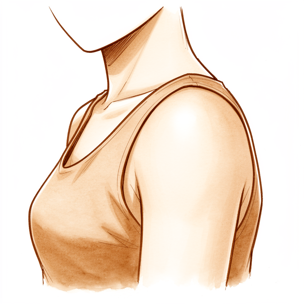
ਇਹ ਓਪਰੇਸ਼ਨ ਕਿਉਂ ਸੁਝਾਇਆ ਗਿਆ ਹੈ
ਡਾ. ਕੀਰਨ ਹਿਰਪਰਾ, ਮਾਟਰ ਪ੍ਰਾਈਵੇਟ ਹਸਪਤਾਲ ਰੌਕਹੈਂਪਟਨ ਵਿੱਚ ਉੱਪਰਲੇ ਅੰਗ (ਮੋਢੇ ਤੋਂ ਹੱਥ ਤੱਕ ਦੀ ਬਾਂਹ) ਦੇ ਸਰਜਨ, ਇਲਾਜ ਦੀ ਸ਼ੁਰੂਆਤ ਉਨ੍ਹਾਂ ਸਭ ਤੋਂ ਘੱਟ ਚੀਰ-ਫਾੜ ਵਾਲੇ ਤਰੀਕਿਆਂ ਨਾਲ ਕਰਦੇ ਹਨ ਜੋ ਤੁਹਾਡੀ ਹਾਲਤ ਲਈ ਢੁਕਵੇਂ ਹੋਣ। ਮੋਢੇ ਵਿੱਚ ਦਰਦ ਵਾਲੇ ਕੈਲਸ਼ੀਅਮ ਦੇ ਜਮਾਅ ਲਈ, ਇਸ ਦਾ ਮਤਲਬ ਹੈ ਫਿਜ਼ੀਓਥੈਰੇਪੀ ਅਤੇ ਸੋਜਸ਼-ਰੋਧੀ ਦਵਾਈਆਂ (anti-inflammatories), ਬਰਸਾ (ਤਰਲ ਨਾਲ ਭਰੀ ਛੋਟੀ ਗੱਦੀ) ਵਿੱਚ ਸਟੀਰੌਇਡ ਟੀਕਾ, ਅਲਟਰਾਸਾਊਂਡ ਦੀ ਮਦਦ ਨਾਲ ਜਮਾਅ ਵਿੱਚ ਸੂਈ ਮਾਰ ਕੇ ਉਸ ਨੂੰ ਤੋੜਨਾ (barbotage) ਅਤੇ ਸ਼ੌਕ ਵੇਵ ਥੈਰੇਪੀ (shock wave therapy, ਆਵਾਜ਼ ਦੀਆਂ ਤੇਜ਼ ਤਰੰਗਾਂ ਨਾਲ ਇਲਾਜ)। ਮਰੀਜ਼ਾਂ ਨੂੰ ਆਮ ਤੌਰ 'ਤੇ ਉਨ੍ਹਾਂ ਦੇ ਜੀਪੀ (ਫ਼ੈਮਿਲੀ ਡਾਕਟਰ) ਵੱਲੋਂ ਸਾਡੇ ਕਲੀਨਿਕ ਵਿੱਚ ਭੇਜਿਆ ਜਾਂਦਾ ਹੈ; ਜੇ ਕਿਸੇ ਫਿਜ਼ੀਓਥੈਰੇਪਿਸਟ ਨੇ ਤੁਹਾਨੂੰ ਸਾਨੂੰ ਮਿਲਣ ਦੀ ਸਲਾਹ ਦਿੱਤੀ ਹੈ, ਤਾਂ ਵੀ ਮੈਡੀਕੇਅਰ ਰਿਬੇਟ (Medicare rebate, ਸਰਕਾਰ ਵੱਲੋਂ ਫ਼ੀਸ ਦੀ ਵਾਪਸੀ) ਦੇ ਯੋਗ ਹੋਣ ਲਈ ਤੁਹਾਨੂੰ ਆਪਣੇ ਜੀਪੀ ਤੋਂ ਰੈਫ਼ਰਲ (referral) ਦੀ ਲੋੜ ਪਵੇਗੀ। ਅਸੀਂ ਇਸ ਓਪਰੇਸ਼ਨ ਬਾਰੇ ਉਦੋਂ ਸੋਚਦੇ ਹਾਂ ਜਦੋਂ ਉਨ੍ਹਾਂ ਇਲਾਜਾਂ ਨਾਲ ਤੁਹਾਨੂੰ ਕਾਫ਼ੀ ਰਾਹਤ ਨਹੀਂ ਮਿਲੀ।
ਇਹ ਓਪਰੇਸ਼ਨ ਦੂਰਬੀਨ ਵਾਲੀ (ਆਰਥਰੋਸਕੋਪਿਕ) ਸਰਜਰੀ ਹੈ ਜਿਸ ਵਿੱਚ ਤੁਹਾਡੇ ਰੋਟੇਟਰ ਕਫ਼ (rotator cuff) ਦੇ ਕਿਸੇ ਇੱਕ ਟੈਂਡਨ (tendon, ਪੱਠੇ ਨੂੰ ਹੱਡੀ ਨਾਲ ਜੋੜਨ ਵਾਲੀ ਮਜ਼ਬੂਤ ਤੰਦ) ਵਿੱਚ ਜੰਮੇ ਕੈਲਸ਼ੀਅਮ ਦੇ ਜਮਾਅ ਨੂੰ ਕੱਢਿਆ ਜਾਂਦਾ ਹੈ। ਇਹ ਜਮਾਅ ਲਗਾਤਾਰ ਦਰਦ ਅਤੇ ਅਕੜਾਅ ਪੈਦਾ ਕਰਦੇ ਹਨ ਜੋ ਮਹੀਨਿਆਂ ਜਾਂ ਸਾਲਾਂ ਤੱਕ ਰਹਿ ਸਕਦੇ ਹਨ। ਜਮਾਅ ਨੂੰ ਕੱਢਣ ਦਾ ਮਕਸਦ ਤੁਹਾਡਾ ਦਰਦ ਘਟਾਉਣਾ ਅਤੇ ਤੁਹਾਡੇ ਮੋਢੇ ਨੂੰ ਦੁਬਾਰਾ ਆਮ ਵਾਂਗ ਹਿੱਲਣ ਅਤੇ ਕੰਮ ਕਰਨ ਦੇਣਾ ਹੈ। ਇਹ ਓਪਰੇਸ਼ਨ ਕਰਵਾਉਣ ਵਾਲੇ ਜ਼ਿਆਦਾਤਰ ਲੋਕ ਨਤੀਜੇ ਤੋਂ ਸੰਤੁਸ਼ਟ ਹੁੰਦੇ ਹਨ, ਅਤੇ ਅਧਿਐਨ ਕੀਤੇ ਇੱਕ ਸਮੂਹ ਵਿੱਚ ਸੰਤੁਸ਼ਟੀ 93% ਦੱਸੀ ਗਈ। ਅਸੀਂ ਸਰਜਰੀ ਦਾ ਸੁਝਾਅ ਉਦੋਂ ਹੀ ਦੇਵਾਂਗੇ ਜਦੋਂ ਅਸੀਂ ਸਹਿਮਤ ਹੋਈਏ ਕਿ ਤੁਹਾਡਾ ਮੋਢਾ ਅਜੇ ਵੀ ਤੁਹਾਡੇ ਜਮਾਅ ਕਾਰਨ ਹੀ ਦੁਖਦਾ ਹੈ।
ਓਪਰੇਸ਼ਨ ਤੋਂ ਪਹਿਲਾਂ
ਸਰਜਰੀ ਤੋਂ ਪਹਿਲਾਂ ਓਪਰੇਸ਼ਨ ਦੀ ਯੋਜਨਾ ਬਣਾਉਣ ਲਈ ਤੁਹਾਨੂੰ ਕੁਝ ਸਕੈਨਾਂ ਦੀ ਲੋੜ ਪਵੇਗੀ। ਇਹ ਆਮ ਤੌਰ 'ਤੇ ਐਕਸ-ਰੇ ਅਤੇ ਅਲਟਰਾਸਾਊਂਡ ਹੁੰਦੇ ਹਨ, ਜੋ ਕੈਲਸ਼ੀਅਮ ਦੇ ਜਮਾਅ ਅਤੇ ਟੈਂਡਨਾਂ ਨੂੰ ਸਾਫ਼ ਦਿਖਾਉਂਦੇ ਹਨ, ਅਤੇ ਕਦੇ-ਕਦੇ ਨਾਲ MRI ਸਕੈਨ ਵੀ। ਜ਼ਿਆਦਾਤਰ ਲੋਕਾਂ ਨੂੰ ਹੋਰ ਕਿਸੇ ਚੀਜ਼ ਦੀ ਲੋੜ ਨਹੀਂ ਹੁੰਦੀ। ਜੇ ਤੁਹਾਨੂੰ ਹੋਰ ਬਿਮਾਰੀਆਂ ਹਨ, ਤਾਂ ਤੁਹਾਨੂੰ ਖ਼ੂਨ ਦੇ ਟੈਸਟਾਂ ਜਾਂ ਅਨੱਸਥੀਟਿਸਟ (ਬੇਹੋਸ਼ੀ ਵਾਲਾ ਡਾਕਟਰ) ਨਾਲ ਜਾਂਚ ਦੀ ਲੋੜ ਪੈ ਸਕਦੀ ਹੈ।
ਆਪਣੇ ਓਪਰੇਸ਼ਨ ਤੋਂ ਪਹਿਲਾਂ ਦੇ ਹਫ਼ਤਿਆਂ ਵਿੱਚ, ਉਹ ਇਲਾਜ ਜਾਰੀ ਰੱਖੋ ਜਿਨ੍ਹਾਂ ਨਾਲ ਮਦਦ ਮਿਲਦੀ ਹੈ, ਅਤੇ ਆਪਣੀਆਂ ਦਵਾਈਆਂ ਬਾਰੇ ਸਾਡੇ ਕਲੀਨਿਕ ਵੱਲੋਂ ਦਿੱਤੀ ਹਰ ਹਦਾਇਤ ਦੀ ਪਾਲਣਾ ਕਰੋ। ਤੁਹਾਨੂੰ ਆਪਣੇ ਓਪਰੇਸ਼ਨ ਤੋਂ ਸੱਤ ਘੰਟੇ ਪਹਿਲਾਂ ਤੱਕ ਕੁਝ ਨਾ ਖਾਣ ਅਤੇ ਨਾ ਪੀਣ ਲਈ ਕਿਹਾ ਜਾਵੇਗਾ, ਤਾਂ ਜੋ ਜੇ ਓਪਰੇਸ਼ਨ ਯੋਜਨਾ ਨਾਲੋਂ ਤੇਜ਼ੀ ਨਾਲ ਹੋ ਰਹੇ ਹੋਣ ਤਾਂ ਸੂਚੀ ਸਮੇਂ ਸਿਰ ਚੱਲ ਸਕੇ। ਤੁਹਾਨੂੰ ਗੱਡੀ ਵਿੱਚ ਘਰ ਛੱਡਣ ਲਈ ਕਿਸੇ ਦਾ ਪ੍ਰਬੰਧ ਕਰੋ, ਕਿਉਂਕਿ ਤੁਸੀਂ ਆਪ ਗੱਡੀ ਨਹੀਂ ਚਲਾ ਸਕੋਗੇ। ਆਪਣੀਆਂ ਮੌਜੂਦਾ ਦਵਾਈਆਂ ਦੀ ਸੂਚੀ ਨਾਲ ਲਿਆਓ, ਅਤੇ ਢਿੱਲੇ, ਆਰਾਮਦਾਇਕ ਕੱਪੜੇ ਪਾਓ ਜੋ ਸੌਖਿਆਂ ਬਦਲੇ ਜਾ ਸਕਣ।
ਓਪਰੇਸ਼ਨ ਵਾਲੇ ਦਿਨ
ਆਪਣੇ ਓਪਰੇਸ਼ਨ ਵਾਲੇ ਦਿਨ, ਤੁਸੀਂ ਹਸਪਤਾਲ ਦੇ ਸਰਜੀਕਲ ਦਾਖ਼ਲਾ ਵਿਭਾਗ ਵਿੱਚ ਹਾਜ਼ਰ ਹੁੰਦੇ ਹੋ। ਉੱਥੇ ਤੁਹਾਡਾ ਦਾਖ਼ਲਾ ਕੀਤਾ ਜਾਵੇਗਾ ਅਤੇ ਓਪਰੇਸ਼ਨ ਥੀਏਟਰ ਲਈ ਤਿਆਰ ਕੀਤਾ ਜਾਵੇਗਾ। ਇਹ ਓਪਰੇਸ਼ਨ ਜਨਰਲ ਅਨੱਸਥੀਸੀਆ (ਬੇਹੋਸ਼ੀ ਵਾਲੀ ਦਵਾਈ) ਦੇ ਨਾਲ ਰੀਜਨਲ ਨਰਵ ਬਲਾਕ (ਨਸ ਨੂੰ ਸੁੰਨ ਕਰਨ ਵਾਲਾ ਟੀਕਾ) ਮਿਲਾ ਕੇ ਕੀਤਾ ਜਾਂਦਾ ਹੈ। ਓਪਰੇਸ਼ਨ ਦੌਰਾਨ ਤੁਸੀਂ ਪੂਰੀ ਤਰ੍ਹਾਂ ਬੇਹੋਸ਼ ਹੋਵੋਗੇ, ਅਤੇ ਬਲਾਕ (ਤੁਹਾਡੇ ਜਾਗਣ ਤੋਂ ਪਹਿਲਾਂ ਬਾਂਹ ਦੀਆਂ ਨਸਾਂ ਨੂੰ ਸੁੰਨ ਕਰਨ ਵਾਲਾ ਟੀਕਾ) ਸਰਜਰੀ ਤੋਂ ਬਾਅਦ ਪਹਿਲੇ 12 ਤੋਂ 24 ਘੰਟਿਆਂ ਲਈ ਦਰਦ ਤੋਂ ਰਾਹਤ ਦਿੰਦਾ ਹੈ। ਅਨੱਸਥੀਟਿਸਟ ਓਪਰੇਸ਼ਨ ਤੋਂ ਪਹਿਲਾਂ ਤੁਹਾਨੂੰ ਮਿਲੇਗਾ ਅਤੇ ਦੋਵਾਂ ਹਿੱਸਿਆਂ ਬਾਰੇ ਤੁਹਾਨੂੰ ਸਮਝਾਏਗਾ।
ਫਿਰ ਤੁਹਾਨੂੰ ਓਪਰੇਸ਼ਨ ਥੀਏਟਰ ਵਿੱਚ ਲਿਜਾਇਆ ਜਾਂਦਾ ਹੈ, ਜਿੱਥੇ ਓਪਰੇਸ਼ਨ ਕੀਤਾ ਜਾਂਦਾ ਹੈ। ਜਦੋਂ ਇਹ ਪੂਰਾ ਹੋ ਜਾਂਦਾ ਹੈ, ਤੁਸੀਂ ਰਿਕਵਰੀ ਵਾਲੀ ਥਾਂ ਵਿੱਚ ਜਾਗੋਗੇ। ਅਨੱਸਥੀਸੀਆ ਦਾ ਅਸਰ ਉਤਰਨ ਤੱਕ ਉੱਥੇ ਨਰਸਾਂ ਤੁਹਾਡੀ ਨਿਗਰਾਨੀ ਕਰਨਗੀਆਂ। ਜਦੋਂ ਤੁਹਾਡੀ ਹਾਲਤ ਸਥਿਰ ਹੋ ਜਾਂਦੀ ਹੈ, ਤਾਂ ਪ੍ਰਕਿਰਿਆ ਅਤੇ ਤੁਹਾਡੇ ਠੀਕ ਹੋਣ ਦੇ ਹਿਸਾਬ ਨਾਲ ਤੁਸੀਂ ਜਾਂ ਤਾਂ ਵਾਰਡ ਵਿੱਚ ਜਾਓਗੇ ਜਾਂ ਘਰ ਚਲੇ ਜਾਓਗੇ।
ਓਪਰੇਸ਼ਨ ਵਿੱਚ ਕੀ ਕੀਤਾ ਜਾਂਦਾ ਹੈ
ਇਹ ਦੂਰਬੀਨ ਵਾਲੀ ਸਰਜਰੀ ਹੈ, ਜੋ ਤੁਹਾਡੇ ਮੋਢੇ ਦੇ ਆਲੇ-ਦੁਆਲੇ ਵੱਖ-ਵੱਖ ਥਾਵਾਂ ਉੱਤੇ ਲਗਾਏ ਕੁਝ ਛੋਟੇ ਚੀਰਿਆਂ ਰਾਹੀਂ ਕੀਤੀ ਜਾਂਦੀ ਹੈ, ਜਿਨ੍ਹਾਂ ਵਿੱਚੋਂ ਇੱਕ ਪਿਛਲੇ ਪਾਸੇ ਹੁੰਦਾ ਹੈ। ਸਰਜਨ ਇੱਕ ਚੀਰੇ ਰਾਹੀਂ ਇੱਕ ਛੋਟਾ ਕੈਮਰਾ ਅੰਦਰ ਪਾਉਂਦਾ ਹੈ ਤਾਂ ਜੋ ਉਹ ਸਕਰੀਨ ਉੱਤੇ ਤੁਹਾਡੇ ਮੋਢੇ ਦੇ ਅੰਦਰ ਦੇਖ ਸਕੇ। ਓਪਰੇਸ਼ਨ ਦੀ ਸ਼ੁਰੂਆਤ ਮੋਢੇ ਦੇ ਜੋੜ ਦੇ ਅੰਦਰ ਆਲੇ-ਦੁਆਲੇ ਦੇਖਣ ਨਾਲ ਹੁੰਦੀ ਹੈ, ਤਾਂ ਜੋ ਟੈਂਡਨ ਉੱਤੇ ਕੰਮ ਸ਼ੁਰੂ ਕਰਨ ਤੋਂ ਪਹਿਲਾਂ ਕਿਸੇ ਹੋਰ ਸਮੱਸਿਆ ਦੀ ਜਾਂਚ ਕੀਤੀ ਜਾ ਸਕੇ।
ਕੈਲਸ਼ੀਅਮ ਦਾ ਜਮਾਅ ਤੁਹਾਡੇ ਰੋਟੇਟਰ ਕਫ਼ ਦੇ ਕਿਸੇ ਇੱਕ ਟੈਂਡਨ ਦੇ ਅੰਦਰ ਹੁੰਦਾ ਹੈ, ਯਾਨੀ ਟੈਂਡਨਾਂ ਦਾ ਉਹ ਸਮੂਹ ਜੋ ਤੁਹਾਡੇ ਮੋਢੇ ਨੂੰ ਜੋੜ ਕੇ ਰੱਖਦਾ ਹੈ ਅਤੇ ਤੁਹਾਡੀ ਬਾਂਹ ਨੂੰ ਹਿਲਾਉਂਦਾ ਹੈ। ਇਸ ਨੂੰ ਲੱਭਣ ਵਿੱਚ ਕੁਝ ਧਿਆਨ ਲੱਗ ਸਕਦਾ ਹੈ। ਸਰਜਨ ਅਕਸਰ ਇੱਕ ਬਰੀਕ ਸੂਈ ਨਾਲ ਟੈਂਡਨ ਨੂੰ ਟੋਹ ਕੇ ਇਸ ਨੂੰ ਲੱਭਦਾ ਹੈ, ਕਈ ਵਾਰ ਓਪਰੇਸ਼ਨ ਦੌਰਾਨ ਅਲਟਰਾਸਾਊਂਡ ਦੀ ਮਦਦ ਨਾਲ। ਜਦੋਂ ਇਹ ਲੱਭ ਜਾਂਦਾ ਹੈ, ਤਾਂ ਜਮਾਅ ਨੂੰ ਕੱਢ ਦਿੱਤਾ ਜਾਂਦਾ ਹੈ, ਅਤੇ ਸੋਜਸ਼ (inflammation) ਵਾਲੇ ਬਰਸਾ, ਯਾਨੀ ਟੈਂਡਨ ਦੇ ਉੱਤੇ ਟਿਸ਼ੂ ਦੀ ਇੱਕ ਪਤਲੀ ਗੱਦੀ, ਨੂੰ ਸਾਫ਼ ਕਰਕੇ ਹਟਾ ਦਿੱਤਾ ਜਾਂਦਾ ਹੈ ਤਾਂ ਜੋ ਟੈਂਡਨ ਸਾਫ਼ ਦਿਸ ਸਕੇ।
ਜਮਾਅ ਨੂੰ ਕੱਢਣ ਨਾਲ ਟੈਂਡਨ ਵਿੱਚ ਖ਼ਾਲੀ ਥਾਂ ਰਹਿ ਸਕਦੀ ਹੈ। ਜੇ ਉਹ ਖ਼ਾਲੀ ਥਾਂ ਵੱਡੀ ਹੋਵੇ, ਜਾਂ ਜੇ ਟੈਂਡਨ ਪਾਟਿਆ ਹੋਇਆ ਮਿਲੇ, ਤਾਂ ਸਰਜਨ ਟਾਂਕਿਆਂ ਨਾਲ ਟੈਂਡਨ ਦੀ ਮੁਰੰਮਤ ਕਰਦਾ ਹੈ, ਅਤੇ ਲੋੜ ਹੋਵੇ ਤਾਂ ਟਾਂਕਿਆਂ ਨੂੰ ਟਿਕਾਉਣ ਲਈ ਹੱਡੀ ਵਿੱਚ ਛੋਟੇ ਐਂਕਰ (anchors, ਹੱਡੀ ਵਿੱਚ ਲਗਾਏ ਜਾਣ ਵਾਲੇ ਛੋਟੇ ਕਿੱਲ) ਵਰਤਦਾ ਹੈ। ਮੁਰੰਮਤ ਕੀਤੀ ਜਾਵੇ ਜਾਂ ਨਹੀਂ, ਇਹ ਓਪਰੇਸ਼ਨ ਦੌਰਾਨ ਤੈਅ ਕੀਤਾ ਜਾਂਦਾ ਹੈ, ਇਸ ਆਧਾਰ ਉੱਤੇ ਕਿ ਕੈਲਸ਼ੀਅਮ ਕੱਢਣ ਤੋਂ ਬਾਅਦ ਤੁਹਾਡਾ ਟੈਂਡਨ ਕਿਹੋ ਜਿਹਾ ਦਿਸਦਾ ਹੈ। ਪੂਰੇ ਸਮੇਂ ਮਕਸਦ ਤੰਦਰੁਸਤ ਟੈਂਡਨ ਨੂੰ ਕੱਟੇ ਬਿਨਾਂ ਜਮਾਅ ਦਾ ਜਿੰਨਾ ਹੋ ਸਕੇ ਓਨਾ ਹਿੱਸਾ ਕੱਢਣਾ ਹੁੰਦਾ ਹੈ।
ਜਦੋਂ ਕੰਮ ਪੂਰਾ ਹੋ ਜਾਂਦਾ ਹੈ, ਤਾਂ ਛੋਟੇ ਚੀਰਿਆਂ ਨੂੰ ਟਾਂਕਿਆਂ ਨਾਲ ਬੰਦ ਕਰਕੇ ਪੱਟੀ ਨਾਲ ਢਕ ਦਿੱਤਾ ਜਾਂਦਾ ਹੈ। ਜਦੋਂ ਤੁਸੀਂ ਜਾਗੋਗੇ, ਤੁਹਾਡੀ ਬਾਂਹ ਸਲਿੰਗ (ਬਾਂਹ ਨੂੰ ਸਹਾਰਾ ਦੇਣ ਵਾਲੀ ਪੱਟੀ) ਵਿੱਚ ਹੋਵੇਗੀ। ਜੇ ਤੁਹਾਡੇ ਟੈਂਡਨ ਦੀ ਮੁਰੰਮਤ ਨਹੀਂ ਕੀਤੀ ਗਈ, ਤਾਂ ਸਲਿੰਗ ਆਰਾਮ ਲਈ ਹੁੰਦੀ ਹੈ ਅਤੇ ਤੁਸੀਂ ਇਸ ਨੂੰ ਚਾਰ ਹਫ਼ਤਿਆਂ ਤੱਕ ਪਾਓਗੇ। ਜੇ ਤੁਹਾਡੇ ਟੈਂਡਨ ਦੀ ਮੁਰੰਮਤ ਕੀਤੀ ਗਈ, ਤਾਂ ਤੁਹਾਡੀ ਸਲਿੰਗ ਅਤੇ ਸਿਹਤਯਾਬੀ ਰੋਟੇਟਰ ਕਫ਼ ਦੀ ਮੁਰੰਮਤ ਲਈ ਵਰਤੇ ਜਾਂਦੇ ਹੌਲੀ ਪ੍ਰੋਗਰਾਮ ਮੁਤਾਬਕ ਚੱਲਦੀ ਹੈ।
ਓਪਰੇਸ਼ਨ ਤੋਂ ਬਾਅਦ
ਇਸ ਓਪਰੇਸ਼ਨ ਤੋਂ ਬਾਅਦ ਜ਼ਿਆਦਾਤਰ ਮਰੀਜ਼ ਇੱਕ ਰਾਤ ਹਸਪਤਾਲ ਵਿੱਚ ਰਹਿੰਦੇ ਹਨ, ਹਾਲਾਂਕਿ ਕੁਝ ਉਸੇ ਦਿਨ ਘਰ ਜਾ ਸਕਦੇ ਹਨ। ਤੁਸੀਂ ਰਿਕਵਰੀ ਵਾਲੀ ਥਾਂ ਵਿੱਚ ਬਾਂਹ ਸਲਿੰਗ ਵਿੱਚ ਪਾਈ ਹੋਈ ਹਾਲਤ ਵਿੱਚ ਜਾਗੋਗੇ, ਅਤੇ ਅਨੱਸਥੀਸੀਆ ਦਾ ਅਸਰ ਉਤਰਨ ਦੌਰਾਨ ਨਰਸਾਂ ਤੁਹਾਡੀ ਜਾਂਚ ਕਰਨਗੀਆਂ। ਨਰਵ ਬਲਾਕ ਦਾ ਅਸਰ ਉਤਰਨ ਤੋਂ ਬਾਅਦ ਤੁਹਾਡਾ ਮੋਢਾ ਦੁਖ ਸਕਦਾ ਹੈ, ਅਤੇ ਦਰਦ ਤੋਂ ਰਾਹਤ ਅਕਸਰ ਹੌਲੀ-ਹੌਲੀ ਅਤੇ ਅਸਮਾਨ ਢੰਗ ਨਾਲ ਆਉਂਦੀ ਹੈ: ਜ਼ਿਆਦਾਤਰ ਸੁਧਾਰ ਤਿੰਨ ਤੋਂ ਛੇ ਮਹੀਨਿਆਂ ਵਿੱਚ ਹੁੰਦਾ ਹੈ, ਅਤੇ ਕੁਝ ਲੋਕਾਂ ਨੂੰ ਇੱਕ ਸਾਲ ਤੱਕ ਲੱਗ ਜਾਂਦਾ ਹੈ। ਅਸੀਂ ਪੱਟੀ ਲਗਭਗ 10 ਦਿਨ ਲੱਗੀ ਰਹਿਣ ਦਿੰਦੇ ਹਾਂ; ਕਿਰਪਾ ਕਰਕੇ ਉਸ ਤੋਂ ਪਹਿਲਾਂ ਇਸ ਨੂੰ ਨਾ ਉਤਾਰੋ, ਜਦੋਂ ਤੱਕ ਅਸੀਂ ਨਾ ਕਹੀਏ। ਜਦੋਂ ਅਸੀਂ ਤੁਹਾਨੂੰ ਮਿਲਦੇ ਹਾਂ ਤਾਂ ਅਸੀਂ ਇਸ ਨੂੰ ਬਦਲਦੇ ਜਾਂ ਉਤਾਰਦੇ ਹਾਂ। ਕਸਰਤਾਂ ਅਤੇ ਨਹਾਉਣ-ਧੋਣ ਲਈ ਸਲਿੰਗ ਉਤਾਰੀ ਜਾਂਦੀ ਹੈ। ਪਹਿਲੇ 24 ਘੰਟੇ ਕਿਸੇ ਨੂੰ ਤੁਹਾਡੇ ਨਾਲ ਰਹਿਣਾ ਚਾਹੀਦਾ ਹੈ। ਜੇ ਤੁਹਾਨੂੰ ਬੁਖ਼ਾਰ ਹੋਵੇ, ਜ਼ਖ਼ਮ ਦੇ ਆਲੇ-ਦੁਆਲੇ ਲਾਲੀ ਫੈਲ ਰਹੀ ਹੋਵੇ, ਜ਼ਖ਼ਮ ਵਿੱਚੋਂ ਤਰਲ ਜਾਂ ਪਾਕ ਰਿਸ ਰਹੀ ਹੋਵੇ, ਜਾਂ ਦਰਦ ਦੀਆਂ ਦਵਾਈਆਂ ਦੇ ਬਾਵਜੂਦ ਦਰਦ ਵਧਦਾ ਹੀ ਜਾਵੇ, ਤਾਂ ਉਸੇ ਦਿਨ ਕਲੀਨਿਕ ਨੂੰ ਫ਼ੋਨ ਕਰੋ।
ਸਿਹਤਯਾਬੀ
ਪਹਿਲੇ ਕੁਝ ਦਿਨ ਤੁਹਾਡਾ ਮੋਢਾ ਦੁਖੇਗਾ ਅਤੇ ਸੁੱਜਿਆ ਹੋਵੇਗਾ। ਓਪਰੇਸ਼ਨ ਥੀਏਟਰ ਵਿੱਚ ਲਗਾਏ ਨਰਵ ਬਲਾਕ ਦਾ ਅਸਰ ਉਤਰ ਜਾਂਦਾ ਹੈ, ਇਸ ਲਈ ਉਮੀਦ ਰੱਖੋ ਕਿ ਦਰਦ ਘਟਣ ਤੋਂ ਪਹਿਲਾਂ ਵਧੇਗਾ। ਉਨ੍ਹਾਂ ਸ਼ੁਰੂਆਤੀ ਦਿਨਾਂ ਵਿੱਚ ਆਰਾਮ, ਬਰਫ਼ ਦੀਆਂ ਥੈਲੀਆਂ ਅਤੇ ਤੁਹਾਨੂੰ ਲਿਖੀ ਗਈ ਦਰਦ ਦੀ ਦਵਾਈ ਮਦਦ ਕਰਦੇ ਹਨ। ਜਦੋਂ ਤੱਕ ਅਸੀਂ ਤੁਹਾਨੂੰ ਨਹੀਂ ਮਿਲਦੇ, ਪੱਟੀ ਲੱਗੀ ਰਹਿਣ ਦਿਓ, ਅਤੇ ਆਪਣੀਆਂ ਕਸਰਤਾਂ ਅਤੇ ਨਹਾਉਣ-ਧੋਣ ਲਈ ਸਲਿੰਗ ਉਤਾਰੋ।
ਸਰਜਰੀ ਤੋਂ ਜਲਦੀ ਬਾਅਦ ਤੋਂ ਹੀ ਤੁਹਾਡਾ ਫਿਜ਼ੀਓਥੈਰੇਪਿਸਟ ਤੁਹਾਡੀਆਂ ਕਸਰਤਾਂ ਵਿੱਚ ਤੁਹਾਡੀ ਅਗਵਾਈ ਕਰੇਗਾ। ਇਹ ਹਲਕੇ ਢੰਗ ਨਾਲ ਸ਼ੁਰੂ ਹੁੰਦੀਆਂ ਹਨ ਅਤੇ ਜਿੰਨਾ ਤੁਹਾਡਾ ਮੋਢਾ ਇਜਾਜ਼ਤ ਦੇਵੇ, ਓਨੀਆਂ ਵਧਦੀਆਂ ਹਨ। ਜੇ ਤੁਹਾਡੇ ਟੈਂਡਨ ਦੀ ਮੁਰੰਮਤ ਕੀਤੀ ਗਈ ਸੀ, ਤਾਂ ਤੁਹਾਡੀ ਸਿਹਤਯਾਬੀ ਰੋਟੇਟਰ ਕਫ਼ ਦੀ ਮੁਰੰਮਤ ਲਈ ਵਰਤੇ ਜਾਂਦੇ ਹੌਲੀ ਪ੍ਰੋਗਰਾਮ ਮੁਤਾਬਕ ਚੱਲਦੀ ਹੈ, ਇਸ ਲਈ ਤੁਹਾਡਾ ਫਿਜ਼ੀਓ ਚੀਜ਼ਾਂ ਦੀ ਰਫ਼ਤਾਰ ਵੱਖਰੀ ਰੱਖੇਗਾ। ਸਰਜਰੀ ਤੋਂ ਘੱਟੋ-ਘੱਟ ਛੇ ਹਫ਼ਤਿਆਂ ਤੱਕ ਤੁਸੀਂ ਗੱਡੀ ਨਹੀਂ ਚਲਾਓਗੇ, ਅਤੇ ਉਦੋਂ ਵੀ ਸਿਰਫ਼ ਤੁਹਾਡੇ ਸਰਜਨ ਦੀ ਇਜਾਜ਼ਤ ਮਿਲਣ ਤੋਂ ਬਾਅਦ। ਵੇਰਵੇ ਲਈ ਉੱਪਰਲੇ ਅੰਗ ਦੀ ਸਰਜਰੀ ਤੋਂ ਬਾਅਦ ਗੱਡੀ ਚਲਾਉਣਾ ਦੇਖੋ।
ਸੁਧਾਰ ਹੌਲੀ-ਹੌਲੀ ਅਤੇ ਅਸਮਾਨ ਢੰਗ ਨਾਲ ਆਉਂਦਾ ਹੈ। ਦਰਦ ਅਤੇ ਹਰਕਤ ਵਿੱਚ ਜ਼ਿਆਦਾਤਰ ਫ਼ਾਇਦਾ ਪਹਿਲੇ ਕੁਝ ਮਹੀਨਿਆਂ ਵਿੱਚ ਹੁੰਦਾ ਹੈ, ਅਤੇ ਮੋਢੇ ਵਿੱਚ ਇੱਕ ਸਾਲ ਤੱਕ ਸੁਧਾਰ ਹੁੰਦਾ ਰਹਿੰਦਾ ਹੈ। ਜਦੋਂ ਸੋਜ ਘਟ ਜਾਂਦੀ ਹੈ ਅਤੇ ਤੁਹਾਡੀ ਹਰਕਤ ਵਾਪਸ ਆਉਂਦੀ ਹੈ, ਤਾਂ ਘਰ ਦੇ ਰੋਜ਼ਾਨਾ ਕੰਮ ਸੌਖੇ ਹੋ ਜਾਂਦੇ ਹਨ। ਜਦੋਂ ਤੁਹਾਡਾ ਸਰਜਨ ਇਜਾਜ਼ਤ ਦੇ ਦਿੰਦਾ ਹੈ, ਆਮ ਤੌਰ 'ਤੇ ਤੁਹਾਡੀ ਛੇ ਹਫ਼ਤਿਆਂ ਵਾਲੀ ਜਾਂਚ ਵੇਲੇ, ਤਾਂ ਤੁਸੀਂ ਦਫ਼ਤਰ ਦੇ ਕੰਮ ਅਤੇ ਰੋਜ਼ਾਨਾ ਦੇ ਹਲਕੇ ਕੰਮਾਂ ਉੱਤੇ ਵਾਪਸ ਜਾ ਸਕਦੇ ਹੋ। ਹੱਥੀਂ ਕੀਤੇ ਜਾਣ ਵਾਲੇ ਭਾਰੀ ਕੰਮ, ਖੇਡਾਂ ਅਤੇ ਜਿਮ ਬਾਅਦ ਵਿੱਚ ਆਉਂਦੇ ਹਨ, ਜਦੋਂ ਤੁਹਾਡਾ ਮੋਢਾ ਕਾਫ਼ੀ ਮਜ਼ਬੂਤ ਹੋ ਜਾਵੇ ਅਤੇ ਤੁਹਾਡੇ ਸਰਜਨ ਨੂੰ ਤੁਹਾਡੀ ਤਰੱਕੀ ਤੋਂ ਤਸੱਲੀ ਹੋਵੇ।
ਸਿਹਤਯਾਬੀ ਹਰ ਵਿਅਕਤੀ ਵਿੱਚ ਵੱਖਰੀ ਹੁੰਦੀ ਹੈ। ਤੁਹਾਡੀ ਸਮਾਂ-ਸੀਮਾ ਵੱਖਰੀ ਹੋ ਸਕਦੀ ਹੈ, ਅਤੇ ਤੁਹਾਡਾ ਸਰਜਨ ਅਤੇ ਫਿਜ਼ੀਓ ਤੁਹਾਡੀ ਅਗਵਾਈ ਕਰਨਗੇ।
ਕੀ ਗ਼ਲਤ ਹੋ ਸਕਦਾ ਹੈ
ਜ਼ਿਆਦਾਤਰ ਮਰੀਜ਼ ਠੀਕ ਰਹਿੰਦੇ ਹਨ, ਪਰ ਕਦੇ-ਕਦਾਈਂ ਸਮੱਸਿਆਵਾਂ ਹੋ ਸਕਦੀਆਂ ਹਨ। ਤੁਹਾਡਾ ਸਰਜਨ ਅਤੇ ਟੀਮ ਕਿਸੇ ਵੀ ਮੁਸ਼ਕਲ ਨੂੰ ਜਲਦੀ ਫੜਨ ਲਈ ਤੁਹਾਡੀ ਨੇੜਿਓਂ ਨਿਗਰਾਨੀ ਕਰਦੇ ਹਨ।
ਇਸ ਓਪਰੇਸ਼ਨ ਤੋਂ ਬਾਅਦ ਸਭ ਤੋਂ ਵੱਧ ਦੇਖੀ ਜਾਣ ਵਾਲੀ ਸਮੱਸਿਆ ਅਕੜਾਅ ਹੈ, ਜਿਸ ਨੂੰ ਕਈ ਵਾਰ ਫ੍ਰੋਜ਼ਨ ਸ਼ੋਲਡਰ (ਜੰਮਿਆ ਹੋਇਆ ਮੋਢਾ) ਕਿਹਾ ਜਾਂਦਾ ਹੈ। ਤੁਹਾਡਾ ਮੋਢਾ ਕੱਸਿਆ ਹੋਇਆ ਮਹਿਸੂਸ ਹੁੰਦਾ ਹੈ ਅਤੇ ਇਸ ਨੂੰ ਕਿਸੇ ਵੀ ਦਿਸ਼ਾ ਵਿੱਚ ਹਿਲਾਉਣਾ ਵੱਧ ਤੋਂ ਵੱਧ ਔਖਾ ਹੁੰਦਾ ਜਾਂਦਾ ਹੈ। ਹਫ਼ਤੇ ਬੀਤਣ ਨਾਲ ਉੱਪਰ ਵੱਲ, ਪਿੱਠ ਪਿੱਛੇ ਜਾਂ ਪਾਸੇ ਵੱਲ ਹੱਥ ਪਹੁੰਚਾਉਣਾ ਸੌਖਾ ਹੋਣ ਦੀ ਬਜਾਏ ਔਖਾ ਹੁੰਦਾ ਜਾਂਦਾ ਹੈ। ਜੇ ਤੁਹਾਡਾ ਮੋਢਾ ਖੁੱਲ੍ਹਣ ਦੀ ਬਜਾਏ ਅਕੜਦਾ ਜਾ ਰਿਹਾ ਹੈ, ਤਾਂ ਕਲੀਨਿਕ ਨੂੰ ਫ਼ੋਨ ਕਰੋ ਤਾਂ ਜੋ ਅਸੀਂ ਇਸ ਦੀ ਜਾਂਚ ਕਰ ਸਕੀਏ ਅਤੇ, ਜੇ ਲੋੜ ਹੋਵੇ, ਤਾਂ ਜਲਦੀ ਇਲਾਜ ਸ਼ੁਰੂ ਕਰ ਸਕੀਏ।
ਕੁਝ ਲੋਕਾਂ ਨੂੰ ਲੱਗਦਾ ਹੈ ਕਿ ਮੋਢਾ ਉਮੀਦ ਨਾਲੋਂ ਵੱਧ ਸਮੇਂ ਤੱਕ ਦੁਖਦਾ ਅਤੇ ਅਕੜਿਆ ਰਹਿੰਦਾ ਹੈ। ਇਹ ਇੱਕ ਡੂੰਘੇ ਦੁਖਣ ਵਰਗਾ ਮਹਿਸੂਸ ਹੋ ਸਕਦਾ ਹੈ ਜੋ ਸ਼ਾਂਤ ਨਹੀਂ ਹੁੰਦਾ, ਅਤੇ ਹਰਕਤ ਸੀਮਤ ਹੀ ਰਹਿੰਦੀ ਹੈ। ਆਪਣੀ ਅਗਲੀ ਜਾਂਚ ਵੇਲੇ ਇਸ ਬਾਰੇ ਦੱਸੋ, ਕਿਉਂਕਿ ਇਸ ਦੀ ਹੋਰ ਜਾਂਚ ਦੀ ਲੋੜ ਹੋ ਸਕਦੀ ਹੈ।
ਜੇ ਓਪਰੇਸ਼ਨ ਦੌਰਾਨ ਤੁਹਾਡੇ ਟੈਂਡਨ ਦੀ ਮੁਰੰਮਤ ਕੀਤੀ ਗਈ ਸੀ, ਤਾਂ ਸਿਹਤਯਾਬੀ ਵਿੱਚ ਵੱਧ ਸਮਾਂ ਲੱਗਦਾ ਹੈ, ਅਤੇ ਅਕੜਾਅ ਦੀ ਸੰਭਾਵਨਾ ਵੱਧ ਹੁੰਦੀ ਹੈ। ਤੁਹਾਡਾ ਫਿਜ਼ੀਓ ਉਸੇ ਹਿਸਾਬ ਨਾਲ ਤੁਹਾਡੀਆਂ ਕਸਰਤਾਂ ਦੀ ਰਫ਼ਤਾਰ ਰੱਖੇਗਾ।
ਜੇ ਤੁਸੀਂ ਵੇਰਵੇ ਜਾਣਨਾ ਚਾਹੁੰਦੇ ਹੋ, ਤਾਂ ਇਸ ਸਫ਼ੇ ਉੱਤੇ ਪੇਚੀਦਗੀਆਂ ਵਾਲੀ ਸਾਰਣੀ ਆਮ ਦਰਾਂ ਦੱਸਦੀ ਹੈ।
ਸਾਨੂੰ ਕਦੋਂ ਫ਼ੋਨ ਕਰਨਾ ਹੈ
ਇਸ ਓਪਰੇਸ਼ਨ ਤੋਂ ਬਾਅਦ ਜ਼ਿਆਦਾਤਰ ਸਮੱਸਿਆਵਾਂ ਮਾਮੂਲੀ ਹੁੰਦੀਆਂ ਹਨ, ਪਰ ਕੁਝ ਵੱਲ ਛੇਤੀ ਧਿਆਨ ਦੇਣ ਦੀ ਲੋੜ ਹੁੰਦੀ ਹੈ। ਜੇ ਤੁਹਾਨੂੰ ਲੱਤ ਦੀ ਪਿੰਨੀ (calf) ਵਿੱਚ ਸੋਜ ਜਾਂ ਦਰਦ ਹੋਵੇ, ਸਾਹ ਚੜ੍ਹੇ ਜਾਂ ਛਾਤੀ ਵਿੱਚ ਦਰਦ ਹੋਵੇ, ਤਾਂ ਐਮਰਜੈਂਸੀ ਵਿਭਾਗ ਜਾਓ, ਕਿਉਂਕਿ ਇਹ ਖ਼ੂਨ ਦੇ ਗਤਲੇ ਦੀਆਂ ਨਿਸ਼ਾਨੀਆਂ ਹੋ ਸਕਦੀਆਂ ਹਨ। ਜੇ ਤੁਹਾਨੂੰ ਬੁਖ਼ਾਰ ਹੋਵੇ, ਜ਼ਖ਼ਮ ਦੇ ਆਲੇ-ਦੁਆਲੇ ਲਾਲੀ ਫੈਲ ਰਹੀ ਹੋਵੇ, ਜ਼ਖ਼ਮ ਵਿੱਚੋਂ ਤਰਲ ਜਾਂ ਪਾਕ ਰਿਸ ਰਹੀ ਹੋਵੇ, ਜਾਂ ਦਰਦ ਦੀਆਂ ਦਵਾਈਆਂ ਦੇ ਬਾਵਜੂਦ ਦਰਦ ਵਧਦਾ ਹੀ ਜਾਵੇ, ਤਾਂ ਉਸੇ ਦਿਨ ਕਲੀਨਿਕ ਨੂੰ ਫ਼ੋਨ ਕਰੋ। ਪਹਿਲੇ 24 ਘੰਟਿਆਂ ਵਿੱਚ, ਨਰਵ ਬਲਾਕ ਦਾ ਅਸਰ ਉਤਰਨ ਦੌਰਾਨ, ਬਾਂਹ ਵਿੱਚ ਸੁੰਨਪਨ ਜਾਂ ਕਮਜ਼ੋਰੀ ਉਮੀਦ ਮੁਤਾਬਕ ਹੈ। ਇਸ ਦਾ ਅਸਰ ਉਤਰ ਜਾਣ ਤੋਂ ਬਾਅਦ, ਜੇ ਤੁਹਾਡੀ ਬਾਂਹ ਜਾਂ ਹੱਥ ਸੁੰਨ ਰਹੇ, ਜਾਂ ਤੁਸੀਂ ਇਸ ਨੂੰ ਨਾ ਹਿਲਾ ਸਕੋ, ਤਾਂ ਕਲੀਨਿਕ ਨੂੰ ਫ਼ੋਨ ਕਰੋ। ਜੇ ਤੁਸੀਂ ਕਲੀਨਿਕ ਨਾਲ ਸੰਪਰਕ ਨਹੀਂ ਕਰ ਸਕਦੇ, ਤਾਂ ਆਪਣੇ ਸਭ ਤੋਂ ਨੇੜਲੇ ਐਮਰਜੈਂਸੀ ਵਿਭਾਗ ਜਾਓ।
ਵਿਸਥਾਰ ਵਿੱਚ
Advanced reading: the deeper science (optional)
ਇਹ ਹਿੱਸਾ ਤੁਹਾਡੇ ਆਪਣੇ ਇਲਾਜ ਬਾਰੇ ਫ਼ੈਸਲਿਆਂ ਲਈ ਲੋੜ ਤੋਂ ਵੱਧ ਵਿਸਥਾਰ ਵਿੱਚ ਜਾਂਦਾ ਹੈ। ਇਸ ਓਪਰੇਸ਼ਨ ਬਾਰੇ ਵਾਧੂ ਪੜ੍ਹਨਾ ਲਾਹੇਵੰਦ ਹੈ ਕਿਉਂਕਿ ਜੋ ਪ੍ਰਕਿਰਿਆ ਪਹਿਲਾਂ ਇਸ ਦੇ ਨਾਲ ਕੀਤੀ ਜਾਂਦੀ ਸੀ, ਯਾਨੀ ਐਕਰੋਮੀਅਨ (acromion, ਮੋਢੇ ਦੇ ਉੱਪਰ ਹੱਡੀ ਦੀ ਛੱਤ) ਤੋਂ ਹੱਡੀ ਛਿੱਲਣਾ, ਉਸ ਦਾ ਕੋਈ ਵਾਧੂ ਫ਼ਾਇਦਾ ਨਹੀਂ ਨਿਕਲਿਆ, ਅਤੇ ਕਿਉਂਕਿ ਜੋ ਸਵਾਲ ਅਜੇ ਖੁੱਲ੍ਹੇ ਹਨ (ਕਿੰਨਾ ਕੈਲਸ਼ੀਅਮ ਕੱਢਣਾ ਹੈ, ਅਤੇ ਕੀ ਬਾਅਦ ਵਿੱਚ ਟੈਂਡਨ ਦੀ ਮੁਰੰਮਤ ਕਰਨੀ ਹੈ) ਉਨ੍ਹਾਂ ਉੱਤੇ ਅਜੇ ਵੀ ਬਹਿਸ ਹੁੰਦੀ ਹੈ।
ਜਮਾਅ ਕਿਉਂ ਦੁਖਦਾ ਹੈ, ਅਤੇ ਸਰਜਰੀ ਅਪਵਾਦ ਕਿਉਂ ਹੈ
ਕੈਲਸੀਫ਼ਿਕ ਟੈਂਡੀਨੋਪੈਥੀ (calcific tendinopathy, ਟੈਂਡਨ ਵਿੱਚ ਕੈਲਸ਼ੀਅਮ ਜਮ੍ਹਾ ਹੋਣਾ) ਇੱਕ ਜੀਵਤ ਟੈਂਡਨ ਵਿੱਚ ਸੈੱਲਾਂ ਵੱਲੋਂ ਚਲਾਈ ਜਾਣ ਵਾਲੀ ਪ੍ਰਕਿਰਿਆ ਹੈ: ਕੈਲਸ਼ੀਅਮ ਜਮ੍ਹਾ ਹੁੰਦਾ ਹੈ, ਅਤੇ ਜ਼ਿਆਦਾਤਰ ਲੋਕਾਂ ਵਿੱਚ ਬਾਅਦ ਵਿੱਚ ਸਰੀਰ ਇਸ ਨੂੰ ਤੋੜ ਕੇ ਸਾਫ਼ ਕਰ ਦਿੰਦਾ ਹੈ, ਜਿਸ ਤੋਂ ਬਾਅਦ ਟੈਂਡਨ ਆਪਣੇ ਆਪ ਨੂੰ ਦੁਬਾਰਾ ਬਣਾ ਲੈਂਦਾ ਹੈ [1]। ਜਦੋਂ ਕੈਲਸ਼ੀਅਮ ਬਣ ਰਿਹਾ ਹੁੰਦਾ ਹੈ, ਤਾਂ ਮੋਢਾ ਦਰਦ ਰਹਿਤ ਜਾਂ ਸਿਰਫ਼ ਥੋੜ੍ਹਾ ਦੁਖਦਾ ਹੋ ਸਕਦਾ ਹੈ। ਤੇਜ਼, ਅਚਾਨਕ ਦਰਦ ਆਮ ਤੌਰ 'ਤੇ ਉਦੋਂ ਆਉਂਦਾ ਹੈ ਜਦੋਂ ਸਰੀਰ ਜਮਾਅ ਨੂੰ ਤੋੜਨਾ ਸ਼ੁਰੂ ਕਰਦਾ ਹੈ [1]। ਇਸੇ ਲਈ ਪਹਿਲੇ ਇਲਾਜ ਜਾਂ ਤਾਂ ਉਸ ਘੁਲਣ ਦੀ ਉਡੀਕ ਕਰਦੇ ਹਨ ਜਾਂ ਉਸ ਨੂੰ ਤੇਜ਼ ਕਰਦੇ ਹਨ, ਅਤੇ ਇਸੇ ਲਈ ਸਰਜਰੀ ਉਸ ਲੰਮੇ ਸਮੇਂ ਤੋਂ ਦਰਦ ਵਾਲੇ ਜਮਾਅ ਲਈ ਰੱਖੀ ਜਾਂਦੀ ਹੈ ਜਿਸ ਉੱਤੇ ਉਨ੍ਹਾਂ ਦਾ ਅਸਰ ਨਹੀਂ ਹੋਇਆ।
ਜਦੋਂ ਬਿਨਾਂ ਓਪਰੇਸ਼ਨ ਵਾਲੇ ਵਿਕਲਪ ਫ਼ੇਲ੍ਹ ਹੋ ਜਾਣ, ਤਾਂ ਜਮਾਅ ਕੱਢਣਾ ਜ਼ਿਆਦਾਤਰ ਲੋਕਾਂ ਲਈ ਕਾਰਗਰ ਹੁੰਦਾ ਹੈ। ਰੈਂਡਮਾਈਜ਼ਡ ਟ੍ਰਾਇਲਾਂ (randomised trials, ਮਰੀਜ਼ਾਂ ਨੂੰ ਬੇਤਰਤੀਬੇ ਸਮੂਹਾਂ ਵਿੱਚ ਵੰਡ ਕੇ ਕੀਤੇ ਅਧਿਐਨ) ਵਿੱਚ, ਦਰਦ ਵਿੱਚ ਔਸਤ ਸੁਧਾਰ ਸੂਈ ਵਾਲੇ ਇਲਾਜ ਤੋਂ ਬਾਅਦ (10-ਅੰਕਾਂ ਵਾਲੇ ਪੈਮਾਨੇ ਉੱਤੇ 4.8 ਅੰਕ) ਅਤੇ ਸਰਜਰੀ ਤੋਂ ਬਾਅਦ (4.7 ਅੰਕ) ਮਿਲਦਾ-ਜੁਲਦਾ ਸੀ। ਮੋਢੇ ਦੇ ਕੰਮਕਾਜ ਦੇ ਅੰਕਾਂ ਵਿੱਚ ਵਾਧਾ ਸਰਜਰੀ ਵਾਲੇ ਸਮੂਹਾਂ ਵਿੱਚ ਵੱਡਾ ਸੀ (ਕੌਂਸਟੈਂਟ ਸਕੋਰ (Constant score) ਉੱਤੇ 38 ਅੰਕ, ਜਦੋਂ ਕਿ ਸੂਈ ਵਾਲੇ ਇਲਾਜ ਤੋਂ ਬਾਅਦ 22) [2]। ਇਹ ਵੱਖ-ਵੱਖ ਟ੍ਰਾਇਲਾਂ ਤੋਂ ਇਕੱਠੀਆਂ ਕੀਤੀਆਂ, ਹਰ ਇਲਾਜ ਸਮੂਹ ਦੇ ਅੰਦਰ ਦੀਆਂ ਔਸਤਾਂ ਹਨ, ਨਾ ਕਿ ਸਿੱਧੀ ਆਹਮੋ-ਸਾਹਮਣੇ ਤੁਲਨਾ, ਅਤੇ ਜੋ ਲੋਕ ਸਰਜਰੀ ਤੱਕ ਪਹੁੰਚਦੇ ਹਨ ਉਨ੍ਹਾਂ ਨੇ ਆਮ ਤੌਰ 'ਤੇ ਪਹਿਲਾਂ ਹੀ ਸੂਈ ਵਾਲਾ ਇਲਾਜ ਅਜ਼ਮਾ ਲਿਆ ਹੁੰਦਾ ਹੈ।
ਡੀਕੰਪ੍ਰੈਸ਼ਨ ਜੋ ਛੱਡ ਦਿੱਤਾ ਗਿਆ
ਕਈ ਸਾਲਾਂ ਤੱਕ ਜਮਾਅ ਨੂੰ ਸਬਐਕਰੋਮੀਅਲ ਡੀਕੰਪ੍ਰੈਸ਼ਨ (subacromial decompression, ਐਕਰੋਮੀਅਨ ਹੇਠਾਂ ਥਾਂ ਖੋਲ੍ਹਣ ਲਈ ਹੱਡੀ ਛਿੱਲਣਾ) ਦੇ ਨਾਲ ਕੱਢਿਆ ਜਾਂਦਾ ਸੀ: ਇਸ ਸਿਧਾਂਤ ਉੱਤੇ ਐਕਰੋਮੀਅਨ ਦੇ ਹੇਠਲੇ ਪਾਸੇ ਤੋਂ ਹੱਡੀ ਛਿੱਲੀ ਜਾਂਦੀ ਸੀ ਕਿ ਟੈਂਡਨ ਦੱਬਿਆ ਜਾ ਰਿਹਾ ਹੈ। ਹੁਣ ਤਿੰਨ ਤੁਲਨਾਵਾਂ ਇਸ ਦੇ ਵਿਰੁੱਧ ਗਵਾਹੀ ਦਿੰਦੀਆਂ ਹਨ।
- 80 ਮਰੀਜ਼ਾਂ ਦੇ ਇੱਕ ਰੈਂਡਮਾਈਜ਼ਡ ਟ੍ਰਾਇਲ ਵਿੱਚ, ਡੀਕੰਪ੍ਰੈਸ਼ਨ ਜੋੜਨ ਨਾਲ ਇੱਕ ਸਾਲ ਉੱਤੇ ਦਰਦ, ਕੰਮਕਾਜ ਜਾਂ ਜੀਵਨ ਦੀ ਗੁਣਵੱਤਾ ਵਿੱਚ ਕੋਈ ਫ਼ਰਕ ਨਹੀਂ ਪਿਆ [3]।
- ਇੱਕ ਤੁਲਨਾਤਮਕ ਲੜੀ ਵਿੱਚ, ਜਿਨ੍ਹਾਂ ਲੋਕਾਂ ਦਾ ਸਿਰਫ਼ ਜਮਾਅ ਕੱਢਿਆ ਗਿਆ ਉਹ ਔਸਤਨ 11 ਹਫ਼ਤਿਆਂ ਵਿੱਚ ਦਰਦ ਰਹਿਤ, ਬਿਨਾਂ ਪਾਬੰਦੀ ਵਾਲੀਆਂ ਗਤੀਵਿਧੀਆਂ ਉੱਤੇ ਵਾਪਸ ਆ ਗਏ, ਜਦੋਂ ਕਿ ਡੀਕੰਪ੍ਰੈਸ਼ਨ ਜੋੜਨ ਉੱਤੇ 18 ਹਫ਼ਤੇ ਲੱਗੇ। ਛੇ ਹਫ਼ਤਿਆਂ ਦੇ ਅੰਦਰ, ਸਿਰਫ਼ ਜਮਾਅ ਕੱਢਣ ਵਾਲੇ ਸਮੂਹ ਵਿੱਚੋਂ ਲਗਭਗ 60% ਬਿਨਾਂ ਪਾਬੰਦੀ ਵਾਲੀਆਂ ਗਤੀਵਿਧੀਆਂ ਉੱਤੇ ਵਾਪਸ ਆ ਗਏ ਸਨ, ਜਦੋਂ ਕਿ ਡੀਕੰਪ੍ਰੈਸ਼ਨ ਵਾਲਿਆਂ ਵਿੱਚੋਂ 20% [4]।
- 69 ਮਰੀਜ਼ਾਂ ਦੇ ਤਿੰਨ ਸਮੂਹਾਂ ਵਾਲੇ ਇੱਕ ਰੈਂਡਮਾਈਜ਼ਡ ਟ੍ਰਾਇਲ ਵਿੱਚ, ਜਮਾਅ ਨੂੰ ਛੱਡ ਕੇ ਸਿਰਫ਼ ਡੀਕੰਪ੍ਰੈਸ਼ਨ ਕਰਨ ਨਾਲ ਛੇ ਹਫ਼ਤਿਆਂ ਉੱਤੇ ਦਰਦ ਤੋਂ ਘੱਟ ਰਾਹਤ ਮਿਲੀ ਅਤੇ ਬਾਅਦ ਵਿੱਚ ਵੱਧ ਇਲਾਜ ਦੀ ਲੋੜ ਪਈ (9 ਹੋਰ ਇਲਾਜ, ਜ਼ਿਆਦਾਤਰ ਕੋਰਟੀਸੋਨ ਟੀਕੇ, ਜਦੋਂ ਕਿ ਜਮਾਅ ਕੱਢਣ ਅਤੇ ਡੀਕੰਪ੍ਰੈਸ਼ਨ ਦੋਵਾਂ ਤੋਂ ਬਾਅਦ 1)। ਡੀਕੰਪ੍ਰੈਸ਼ਨ ਦੇ ਨਾਲ ਅਤੇ ਬਿਨਾਂ ਜਮਾਅ ਕੱਢਣ ਦੇ ਨਤੀਜੇ ਬਰਾਬਰ ਚੰਗੇ ਰਹੇ [5]।
ਡੀਕੰਪ੍ਰੈਸ਼ਨ ਤੋਂ ਬਿਨਾਂ ਇਲਾਜ ਕੀਤੇ 105 ਮੋਢਿਆਂ ਦੀ ਇੱਕ ਲੜੀ ਨੇ ਐਕਰੋਮੀਅਨ ਦੀ ਸ਼ਕਲ ਭਾਵੇਂ ਕੋਈ ਵੀ ਹੋਵੇ, ਦਰਦ ਤੋਂ ਜਲਦੀ ਰਾਹਤ ਦੱਸੀ [6]। ਸਮੱਸਿਆ ਜਮਾਅ ਹੈ; ਇਸ ਦੇ ਉੱਪਰਲੀ ਹੱਡੀ ਆਮ ਤੌਰ 'ਤੇ ਨਹੀਂ। ਇਸੇ ਲਈ ਇਸ ਸਫ਼ੇ ਉੱਤੇ ਦੱਸੇ ਓਪਰੇਸ਼ਨ ਵਿੱਚ ਡੀਕੰਪ੍ਰੈਸ਼ਨ ਸ਼ਾਮਲ ਨਹੀਂ ਹੈ।
ਸਾਰਾ ਕੱਢਣਾ ਹੈ, ਜਾਂ ਜ਼ਿਆਦਾਤਰ?
ਇਹ ਅਸਲ ਮਤਭੇਦ ਹੈ। ਇੱਕ ਲੜੀ ਵਿੱਚ, ਦੋ ਤੋਂ ਪੰਜ ਸਾਲਾਂ ਉੱਤੇ ਮੋਢੇ ਦੇ ਅੰਕ ਓਨੇ ਹੀ ਘੱਟ ਸਨ ਜਿੰਨਾ ਵੱਧ ਕੈਲਸ਼ੀਅਮ ਪਿੱਛੇ ਛੱਡਿਆ ਗਿਆ ਸੀ, ਅਤੇ ਲੇਖਕਾਂ ਨੇ ਪੂਰਾ ਕੱਢਣ ਦੀ ਸਿਫ਼ਾਰਸ਼ ਕੀਤੀ [7]। ਹੋਰਨਾਂ ਨੇ ਟੈਂਡਨ ਨੂੰ ਬਚਾਉਣ ਲਈ ਉਲਟਾ ਤਰੀਕਾ ਅਪਣਾਇਆ ਹੈ। ਇੱਕ ਲੜੀ ਵਿੱਚ ਜਿਸ ਵਿੱਚ ਜਾਣਬੁੱਝ ਕੇ ਟੈਂਡਨ ਨੂੰ ਬਚਾਇਆ ਗਿਆ, 99 ਵਿੱਚੋਂ 17 ਮੋਢਿਆਂ ਵਿੱਚ ਛੋਟੀਆਂ ਰਹਿੰਦ-ਖੂੰਹਦਾਂ ਛੱਡੀਆਂ ਗਈਆਂ (ਔਸਤਨ ਅਸਲ ਜਮਾਅ ਦੇ ਆਕਾਰ ਦਾ ਲਗਭਗ 58%)। ਇਨ੍ਹਾਂ ਲੋਕਾਂ ਦੇ ਨਤੀਜੇ ਮਾੜੇ ਨਹੀਂ ਸਨ, ਅਤੇ ਨਿਗਰਾਨੀ ਵੇਲੇ ਤੱਕ ਰਹਿੰਦ-ਖੂੰਹਦਾਂ ਘੁਲ ਚੁੱਕੀਆਂ ਸਨ [8]। 99 ਮੋਢਿਆਂ ਦੀ ਇੱਕ ਹੋਰ ਹਾਲੀਆ ਲੜੀ ਵਿੱਚ ਵੀ ਇਹੀ ਮਿਲਿਆ: ਰਹਿੰਦ-ਖੂੰਹਦ ਵਾਲੇ 20 ਮੋਢਿਆਂ ਦੇ ਨਤੀਜੇ ਪੂਰਾ ਕੱਢਣ ਵਾਲਿਆਂ ਵਰਗੇ ਸਨ [9]। ਅਮਲ ਵਿੱਚ ਮਕਸਦ ਤੰਦਰੁਸਤ ਟੈਂਡਨ ਨੂੰ ਕੱਟੇ ਬਿਨਾਂ ਜਿੰਨਾ ਹੋ ਸਕੇ ਓਨਾ ਕੱਢਣਾ ਹੁੰਦਾ ਹੈ।
ਖ਼ਾਲੀ ਥਾਂ ਦੀ ਮੁਰੰਮਤ ਕਰਨੀ ਹੈ, ਜਾਂ ਛੱਡ ਦੇਣੀ ਹੈ?
ਜਮਾਅ ਕੱਢਣ ਨਾਲ ਟੈਂਡਨ ਦੇ ਅੰਦਰ ਇੱਕ ਨੁਕਸ ਰਹਿ ਜਾਂਦਾ ਹੈ। ਕੀ ਇਸ ਨੂੰ ਟਾਂਕੇ ਲਗਾਉਣੇ ਹਨ, ਇਹ ਦੂਜਾ ਖੁੱਲ੍ਹਾ ਸਵਾਲ ਹੈ।
- ਮੁਰੰਮਤ ਤੋਂ ਬਿਨਾਂ ਛੱਡਿਆ ਗਿਆ: 99 ਮੋਢਿਆਂ ਵਿੱਚ, ਜਮਾਅ ਦੇ ਆਕਾਰ ਜਾਂ ਥਾਂ ਦੀ ਪਰਵਾਹ ਕੀਤੇ ਬਿਨਾਂ ਨਤੀਜੇ ਚੰਗੇ ਸਨ। ਬਾਅਦ ਵਿੱਚ ਸਕੈਨ ਕੀਤੇ 29 ਮੋਢਿਆਂ ਵਿੱਚੋਂ, 79% ਠੀਕ ਹੋ ਚੁੱਕੇ ਸਨ, ਅਤੇ ਛੋਟੇ ਬਾਕੀ ਨੁਕਸ ਵਾਲੇ 6 ਮੋਢਿਆਂ ਦੇ ਅੰਕ ਬਾਕੀਆਂ ਜਿੰਨੇ ਹੀ ਚੰਗੇ ਸਨ [9]। ਘੱਟੋ-ਘੱਟ ਸੱਤ ਸਾਲਾਂ ਤੱਕ ਨਿਗਰਾਨੀ ਕੀਤੇ, ਮੁਰੰਮਤ ਤੋਂ ਬਿਨਾਂ 54 ਮੋਢਿਆਂ ਦੀ ਇੱਕ ਲੜੀ ਨੇ ਦੱਸਿਆ ਕਿ 3.7% ਵਿੱਚ ਅੱਗੇ ਚੱਲ ਕੇ ਰੋਟੇਟਰ ਕਫ਼ ਪਾਟ ਗਿਆ [10]।
- ਮੁਰੰਮਤ ਕੀਤਾ ਗਿਆ: 44 ਮਰੀਜ਼ਾਂ ਦੀ ਇੱਕ ਤੁਲਨਾ ਵਿੱਚ, ਜਿਨ੍ਹਾਂ ਦੇ ਨੁਕਸ ਦੀ ਮੁਰੰਮਤ ਕੀਤੀ ਗਈ ਸੀ ਉਨ੍ਹਾਂ ਦੇ ਕਲੀਨਿਕੀ ਅੰਕ ਬਿਹਤਰ ਸਨ, ਅਤੇ MRI ਨੇ ਮੁਰੰਮਤ ਕੀਤੇ 80% ਮੋਢਿਆਂ ਵਿੱਚ ਆਮ ਦਿਸਣ ਵਾਲਾ ਟੈਂਡਨ ਦਿਖਾਇਆ। ਸਿਰਫ਼ ਸਫ਼ਾਈ (debridement) ਵਾਲੇ ਸਮੂਹ ਦੇ ਟੈਂਡਨ ਜ਼ਿਆਦਾਤਰ ਸਾਬਤ ਸਨ ਪਰ ਉਨ੍ਹਾਂ ਵਿੱਚ ਅਸਾਧਾਰਨ ਸਿਗਨਲ ਸੀ [11]।
ਇਸ ਵਿੱਚ ਸਿਹਤਯਾਬੀ ਦਾ ਲੈਣ-ਦੇਣ ਹੈ। ਮੁਰੰਮਤ ਲਈ ਬਾਂਹ ਨੂੰ ਵੱਧ ਸਮੇਂ ਤੱਕ ਬਚਾ ਕੇ ਰੱਖਣਾ ਪੈਂਦਾ ਹੈ। ਇੱਕ ਲੜੀ ਵਿੱਚ ਜਿੱਥੇ ਹਰ ਨੁਕਸ ਦੀ ਮੁਰੰਮਤ ਕੀਤੀ ਗਈ, 35 ਵਿੱਚੋਂ 10 ਮਰੀਜ਼ਾਂ (29%) ਦਾ ਮੋਢਾ ਅਕੜ ਗਿਆ, ਜੋ ਤਿੰਨ ਤੋਂ ਨੌਂ ਮਹੀਨਿਆਂ ਵਿੱਚ ਠੀਕ ਹੋ ਗਿਆ [12]। ਇਸੇ ਲਈ ਮੁਰੰਮਤ ਦਾ ਫ਼ੈਸਲਾ ਹਰ ਵਾਰ ਨਹੀਂ, ਸਗੋਂ ਓਪਰੇਸ਼ਨ ਦੌਰਾਨ ਕੀਤਾ ਜਾਂਦਾ ਹੈ, ਕਿਸੇ ਵੱਡੇ ਨੁਕਸ ਲਈ ਜਾਂ ਮਿਲੇ ਪਾਟਣ ਲਈ।
ਅਕੜਾਅ, ਅਤੇ ਸਿਹਤਯਾਬੀ ਜੋ ਸਮਾਂ ਲੈਂਦੀ ਹੈ
ਫ੍ਰੋਜ਼ਨ ਸ਼ੋਲਡਰ ਇਸ ਓਪਰੇਸ਼ਨ ਦੀ ਸਭ ਤੋਂ ਆਮ ਪੇਚੀਦਗੀ ਹੈ। 340 ਮੋਢਿਆਂ ਵਿੱਚ ਇਹ 12% ਵਿੱਚ ਹੋਇਆ [13]। ਡੀਕੰਪ੍ਰੈਸ਼ਨ ਤੋਂ ਬਿਨਾਂ ਵਾਲੀ ਲੜੀ ਵਿੱਚ, ਦਰ 2.9% ਸੀ [6]।
ਸਿਹਤਯਾਬੀ ਅਸਮਾਨ ਹੁੰਦੀ ਹੈ। ਇੱਕ ਲੜੀ ਵਿੱਚ 78% ਮਰੀਜ਼ ਛੇ ਹਫ਼ਤਿਆਂ ਦੇ ਅੰਦਰ ਕੰਮ ਉੱਤੇ ਵਾਪਸ ਆ ਗਏ ਸਨ, ਭਾਵੇਂ ਉਨ੍ਹਾਂ ਦਾ ਕੰਮ ਕੋਈ ਵੀ ਹੋਵੇ। ਫਿਰ ਵੀ ਦਰਦ ਆਪਣੇ ਸਭ ਤੋਂ ਹੇਠਲੇ ਪੱਧਰ ਉੱਤੇ ਤਿੰਨ ਮਹੀਨਿਆਂ ਉੱਤੇ ਸਿਰਫ਼ 31% ਵਿੱਚ ਪਹੁੰਚਿਆ, ਛੇ ਮਹੀਨਿਆਂ ਉੱਤੇ 17% ਵਿੱਚ, ਨੌਂ ਮਹੀਨਿਆਂ ਉੱਤੇ 20% ਵਿੱਚ ਅਤੇ ਇੱਕ ਸਾਲ ਉੱਤੇ 28% ਵਿੱਚ [14]। ਇਹ ਮਹਿਸੂਸ ਕਰਨਾ ਕਿ ਜਮਾਅ ਚਲਾ ਗਿਆ ਹੈ, ਉਸੇ ਹਫ਼ਤੇ ਬਿਹਤਰ ਮਹਿਸੂਸ ਕਰਨ ਵਰਗਾ ਨਹੀਂ ਹੈ, ਅਤੇ ਜੋ ਮੋਢਾ ਤਿੰਨ ਮਹੀਨਿਆਂ ਉੱਤੇ ਅਜੇ ਵੀ ਦੁਖਦਾ ਹੈ ਉਹ ਆਮ ਤੌਰ 'ਤੇ ਸਹੀ ਰਾਹ ਉੱਤੇ ਹੁੰਦਾ ਹੈ, ਫ਼ੇਲ੍ਹ ਨਹੀਂ ਹੋ ਰਿਹਾ ਹੁੰਦਾ।
ਤੁਹਾਡੇ ਫ਼ੈਸਲੇ ਲਈ ਇਸ ਦਾ ਕੀ ਮਤਲਬ ਹੈ
- ਜ਼ਿਆਦਾਤਰ ਜਮਾਅ ਸਰਜਰੀ ਤੋਂ ਬਿਨਾਂ ਸਾਫ਼ ਹੋ ਜਾਂਦੇ ਹਨ। ਓਪਰੇਸ਼ਨ ਉਸ ਜਮਾਅ ਲਈ ਹੈ ਜੋ ਬਿਨਾਂ ਓਪਰੇਸ਼ਨ ਵਾਲੇ ਇਲਾਜਾਂ ਨੂੰ ਸਹੀ ਢੰਗ ਨਾਲ ਅਜ਼ਮਾਉਣ ਦੇ ਬਾਵਜੂਦ ਸਾਫ਼ ਨਹੀਂ ਹੋਇਆ।
- ਜਮਾਅ ਨੂੰ ਕੱਢਣਾ ਹੀ ਓਪਰੇਸ਼ਨ ਹੈ। ਐਕਰੋਮੀਅਨ ਤੋਂ ਹੱਡੀ ਛਿੱਲਣ ਨਾਲ ਨਤੀਜੇ ਵਿੱਚ ਕੋਈ ਵਾਧਾ ਨਹੀਂ ਹੁੰਦਾ, ਸਿਰਫ਼ ਸਿਹਤਯਾਬੀ ਦਾ ਸਮਾਂ ਵਧਦਾ ਹੈ।
- ਟੈਂਡਨ ਦੀ ਮੁਰੰਮਤ ਹੋਵੇਗੀ ਜਾਂ ਨਹੀਂ, ਇਹ ਇਸ ਗੱਲ ਉੱਤੇ ਨਿਰਭਰ ਕਰਦਾ ਹੈ ਕਿ ਕੀ ਮਿਲਦਾ ਹੈ। ਮੁਰੰਮਤ ਟੈਂਡਨ ਦੀ ਰੱਖਿਆ ਕਰਦੀ ਹੈ ਪਰ ਸਿਹਤਯਾਬੀ ਨੂੰ ਹੌਲੀ ਕਰਦੀ ਹੈ ਅਤੇ ਅਕੜਾਅ ਦਾ ਖ਼ਤਰਾ ਵਧਾਉਂਦੀ ਹੈ।
- ਹਫ਼ਤਿਆਂ ਵਿੱਚ ਨਹੀਂ, ਮਹੀਨਿਆਂ ਵਿੱਚ ਸੁਧਾਰ ਦੀ ਉਮੀਦ ਰੱਖੋ, ਅਤੇ ਕੁਝ ਲੋਕਾਂ ਨੂੰ ਇੱਕ ਸਾਲ ਤੱਕ ਲੱਗ ਜਾਂਦਾ ਹੈ।
ਹਵਾਲੇ
[1] Uhthoff HK, Loehr JW. Calcific Tendinopathy of the Rotator Cuff: Pathogenesis, Diagnosis, and Management. J Am Acad Orthop Surg. 1997;5(4):183-91. https://doi.org/10.5435/00124635-199707000-00001
[2] Angileri HS, Gohal C, Comeau-Gauthier M, Owen MM, Shanmugaraj A, Terry MA, et al. Chronic calcific tendonitis of the rotator cuff: a systematic review and meta-analysis of randomized controlled trials comparing operative and nonoperative interventions. J Shoulder Elbow Surg. 2023;32(8):1746-60. https://doi.org/10.1016/j.jse.2023.03.017
[3] Clement ND, Watts AC, Phillips C, McBirnie JM. Short-Term Outcome After Arthroscopic Bursectomy Debridement of Rotator Cuff Calcific Tendonopathy With and Without Subacromial Decompression: A Prospective Randomized Controlled Trial. Arthroscopy. 2015;31(9):1680-7. https://doi.org/10.1016/j.arthro.2015.05.015
[4] Marder RA, Heiden EA, Kim S. Calcific tendonitis of the shoulder: is subacromial decompression in combination with removal of the calcific deposit beneficial? J Shoulder Elbow Surg. 2011;20(6):955-60. https://doi.org/10.1016/j.jse.2010.10.038
[5] Verstraelen F, Schotanus M, Klemann-Harings S, Lambers Heerspink O, Jansen E. Comparison of clinical and radiological outcomes after three different surgical treatments for resistant calcifying tendinitis of the shoulder: a short-term randomized controlled trial. J Orthop Surg Res. 2022;17(1):480. https://doi.org/10.1186/s13018-022-03373-1
[6] Maier D, Jaeger M, Izadpanah K, Köstler W, Bischofberger AK, Südkamp NP, et al. Arthroscopic Removal of Chronic Symptomatic Calcifications of the Supraspinatus Tendon Without Acromioplasty. Orthop J Sports Med. 2014;2(5):2325967114533646. https://doi.org/10.1177/2325967114533646
[7] Porcellini G, Paladini P, Campi F, Paganelli M. Arthroscopic treatment of calcifying tendinitis of the shoulder: Clinical and ultrasonographic follow-up findings at two to five years. J Shoulder Elbow Surg. 2004;13(5):503-8. https://doi.org/10.1016/j.jse.2004.04.001
[8] Maier D, Jaeger M, Izadpanah K, Bornebusch L, Suedkamp NP, Ogon P. Rotator Cuff Preservation in Arthroscopic Treatment of Calcific Tendinitis. Arthroscopy. 2013;29(5):824-31. https://doi.org/10.1016/j.arthro.2013.01.031
[9] Youn S, Rhee SM, Cho S, Kim C, Lee J, Rhee YG. Arthroscopic decompression of calcific tendinitis without cuff repair. Bone Joint J. 2023;105-B(6):663-7. https://doi.org/10.1302/0301-620X.105B6.BJJ-2022-1137.R1
[10] El Shewy MT. Arthroscopic removal of calcium deposits of the rotator cuff: a 7-year follow-up. Am J Sports Med. 2011;39(6):1302-5. https://doi.org/10.1177/0363546510396320
[11] Lorbach O, Haupert A, Berger C, Brockmeyer M. Clinical and Structural Results of Rotator Cuff Repair Compared With Rotator Cuff Debridement in Arthroscopic Treatment of Calcifying Tendinitis of the Shoulder. Am J Sports Med. 2021;49(12):3196-201. https://doi.org/10.1177/03635465211037690
[12] Yoo JC, Park WH, Koh KH, Kim SM. Arthroscopic treatment of chronic calcific tendinitis with complete removal and rotator cuff tendon repair. Knee Surg Sports Traumatol Arthrosc. 2010;18(12):1694-9. https://doi.org/10.1007/s00167-010-1067-7
[13] Ecalle A, Julien C, Chaouche S, Cungi P, Anger F, Galland A, et al. Is routine gleno-humeral exploration a risk factor for adhesive capsulitis after arthroscopic removal of rotator cuff calcifications? A comparative retrospective study in 340 cases. Orthop Traumatol Surg Res. 2021;107(4):102915. https://doi.org/10.1016/j.otsr.2021.102915
[14] Seil R, Litzenburger H, Kohn D, Rupp S. Arthroscopic Treatment of Chronically Painful Calcifying Tendinitis of the Supraspinatus Tendon. Arthroscopy. 2006;22(5):521-7. https://doi.org/10.1016/j.arthro.2006.01.012
Evidence & references
This is the clinical evidence summary written for health professionals. It is technical, and it lists the research this page was built from. You do not need to read it to understand your treatment or to make a decision about it.
Anatomy & Pathophysiology
Bony Anatomy
- The proximal humerus comprises four main parts: the humeral head, greater tuberosity (GT), lesser tuberosity (LT), and humeral shaft [3].
- The articular head of the humerus is spherical with a diameter of 37 to 57 mm [3].
- The most superior portion of the articular surface of the humeral head averages 8 mm above the greater tuberosity [3].
- Humeral version averages 29.8 degrees, with a range of 10 to 55 degrees [3].
- The humeral head is inclined approximately 130 degrees with respect to the humeral shaft [3].
- The neck-shaft angle measures an average of 135 degrees [4].
- The humeral head is retroverted an average of 30 degrees [4].
- The humeral head averages 19° of retroversion and 41° of inclination (neck-shaft angle) [6].
- The articular surface of the humeral head is essentially spherical, with an arc of approximately 160 degrees covered by articular cartilage [15].
- The radius of curvature of the humeral head is approximately 25 mm and is slightly larger in men than in women [15].
- The average neck-shaft angle is 45 degrees (±5 degrees), with a range of 30 to 50 degrees [15].
- Arthritic shoulders have a flatter neck-shaft angle close to 50 degrees [15].
- The superior margin of the humeral head articular surface is normally superior to the top of the greater tuberosity by 8 to 10 mm [15].
- The distance from the lateral base of the coracoid process to the lateral margin of the greater tuberosity is called the lateral humeral offset [15].
- A significant decrease in lateral humeral offset reduces the lever arms for the deltoid and supraspinatus muscles, weakening abduction and impairing function [15].
- A significant increase in lateral humeral offset causes excessive tension on the soft tissues ("overstuffing" of the joint), resulting in loss of motion and likely accelerating polyethylene wear [15].
- Humeral articular malposition of more than 4 mm leads to increased subacromial contact [15].
- An offset of 8 mm in any direction significantly decreases passive range of motion [15].
- Proximal humeral retroversion is highly variable, ranging from 0 to 55 degrees depending on the method used for measurement [15].
- The glenoid is a convex structure of shallow depth shaped like an inverted pear [3].
- The glenoid cavity is a shallow socket, approximately one third the size of the humeral head [4].
- The subchondral bone of the glenoid is relatively flat; the articular concavity is augmented by cartilage and a circumferential labrum [6].
- The glenoid averages 5° of retroversion in relation to the axis of the scapular body [6].
- The normal position of the glenoid surface in relation to the axis of the scapular body ranges from 2 degrees of anteversion to 7 degrees of retroversion [15].
- The glenoid articular surface radius of curvature is 2 to 3 mm larger than that of the humeral head [15].
- The glenoid diameter ranges from 18-30 mm superior anteroposterior, 21-35 mm inferior anteroposterior, and 30-48 mm superoinferior [15].
- The glenoid inclination averages 4.2 degrees (range –7 to 20 degrees) [15].
- The glenoid version averages 1.5 degrees retroversion (range 10.5-9.5 degrees anteversion) [15].
- The glenoid surface area is 4-6 mm [15].
- The glenoid cartilage thickness is 2.16 mm [15].
- The humeral head surface area is 11-19 mm [15].
- The humeral head cartilage thickness is 1.44 mm [15].
- The humeral head radius of curvature is 23-28 mm, smaller in women than men [15].
- The medial (coronal) humeral offset is 4-14 mm [15].
- The posterior (transverse) humeral offset is –2 to 10 mm [15].
- The scapula is attached to the axial skeleton by the clavicle, specifically via the acromioclavicular (AC) and sternoclavicular (SC) joints [5].
- The scapula is separated from the chest wall by thin gliding fibro-fatty tissue, allowing smooth excursion over the chest wall [5].
- The basic part of the scapula is the body, which is triangular when viewed anteroposteriorly [5].
- The glenoid is connected with the flat body of the scapula by the scapular neck [5].
- The hook-shaped coracoid process curves forwards from the superior surface of the scapular neck [5].
- The scapular spine ends in a flattened bony process, the acromion, which curves forwards [5].
- The highest concentration of bony mass in the scapula is located in the glenoid, the scapular neck (including the base of the coracoid process), and the lateral border of the scapular body [5].
- Two bony pillars transmit compressive forces from the glenoid fossa: the lateral pillar and the spinal pillar [5].
- The lateral pillar connects the inferior border of the glenoid with the inferior angle [5].
- The spinal pillar arises from the central part of the glenoid and continues medially to become part of the base of the scapular spine [5].
- The weakest bone in the scapula is located primarily in the central part of the biomechanical body, specifically in the infraspinous fossa [5].
- The weakest area of the circumference of the biomechanical body of the scapula is the spinomedial angle, which is the connection of the scapular spine and the medial border of the scapula [5].
- The clavicle is the first bone to ossify (fifth week of gestation) and is the only long bone to ossify by intramembranous ossification [6].
- The medial (sternal) epiphysis of the clavicle is the last ossification center to fuse, at age 20 to 25 years [6].
- The primary blood supply to the clavicle is periosteal; no nutrient artery is present [6].
- The acromion has three ossification centers: the metacromion (base), the mesoacromion (middle), and the preacromion (tip) [6].
- Failure of fusion of the acromial ossification centers results in os acromiale [6].
- The proximal humerus has three centers of ossification: the humeral head (4 to 6 months), the greater tuberosity (1 to 3 years), and the lesser tuberosity (3 to 5 years) [6].
- The proximal humeral ossification centers fuse to the shaft at age 17 to 20 years [6].
- The ossification center for the humeral head is usually present at birth [10].
- The greater tuberosity ossification center appears by 1 to 3 years of age [10].
- The lesser tuberosity ossification center appears by 5 years of age [10].
- The proximal humeral ossification center remains separated from the humeral shaft by the proximal humeral physis, which closes by 14 to 17 years of age in girls and by 16 to 18 years in boys [10].
- Humeral retroversion averages 65 degrees in infants and young children and gradually decreases, approaching adult values by 11 years of age [10].
- Eighty percent of subsequent growth of the humerus comes from the proximal humeral physis, accounting for approximately 40% of the growth of the entire upper extremity [10].
- Less than 75% of growth derived from the proximal humerus occurs before 2 years of age [10].
- More than 85% of growth derived from the proximal humerus occurs by 8 years of age [10].
Soft Tissue Anatomy
- The greater tuberosity serves as the attachment site for the supraspinatus, infraspinatus, and teres minor tendons of the rotator cuff [3].
- The lesser tuberosity serves as the attachment site for the subscapularis tendon [3].
- The bicipital groove lies between the greater and lesser tuberosities and serves as a pathway for the long head of the biceps [3].
- The distal aspect of the bicipital groove is internally rotated with respect to the proximal portion [3].
- The anatomic neck of the proximal humerus is located at the junction of the articular surface and the tuberosities [3].
- The surgical neck represents an indistinct region (metadiaphyseal junction) below the tuberosities but above the humeral shaft [3].
- The acromion, the coracoacromial ligament, and the coracoid process form the coracoacromial arch [3].
- The rotator cuff, subacromial bursa, and subdeltoid bursa pass underneath the coracoacromial arch [3].
- The rotator cuff consists of four muscles: the subscapularis, supraspinatus, infraspinatus, and teres minor [4].
- The teres major is not a rotator cuff muscle [4].
- The rotator cuff muscles serve as depressors of the humeral head to allow the deltoid to efficiently abduct the humerus [4].
- The infraspinatus and teres minor are external rotators, while the subscapularis is an internal rotator of the humerus [4].
- The rotator cuff is a sheet of conjoined tendons closely applied over the shoulder capsule and inserting mainly into the greater tuberosity of the humerus [11].
- The subscapularis is inserted into the lesser tuberosity [11].
- The coracoacromial arch is formed by the acromion process posterosuperiorly, the coracoid process anteriorly, and the coracoacromial ligament joining them [11].
- The subacromial bursa separates the tendons from the coracoacromial arch, allowing them to glide [11].
- The glenoid labrum is composed of dense fibrocartilaginous tissue and increases the depth of the socket by 50% around the humeral head [16].
- The glenoid articular surface and the labrum combine to create a socket that is approximately 9 mm deep in the superoinferior direction and 5 mm deep in the anteroposterior direction [16].
- Adding the glenoid labrum increases the glenoid surface to 75% of the humeral head vertically and 57% horizontally [16].
- The superior glenohumeral ligament is the primary restraint to inferior humeral subluxation in 0 degrees of abduction [16].
- The superior glenohumeral ligament is the primary stabilizer to anterior and posterior stress in 0 degrees of abduction [16].
- Tightening of the rotator interval decreases posterior and inferior translation [16].
- The middle glenohumeral ligament limits external rotation when the arm is in the lower and middle ranges of abduction but has little effect when the arm is in 90 degrees of abduction [16].
- The inferior glenohumeral ligament is composed of an anterior band that is quite thick, a posterior band that is less thick and distinct, and a thinner intervening axillary pouch [16].
- With external rotation, the inferior glenohumeral ligament hammock slides anteriorly and superiorly, the anterior band tightens, and the posterior band fans out [16].
- With internal rotation, the opposite occurs to the inferior glenohumeral ligament hammock [16].
- The anteroinferior glenohumeral ligament complex is the main stabilizer to anterior and posterior stresses when the shoulder is abducted 45 degrees or more [16].
- The tendons of the infraspinatus and supraspinatus muscles join approximately 15 mm proximal to their insertion and cannot be readily separated by blunt dissection [16].
- The infraspinatus and teres minor fuse near their musculotendinous junctions [16].
- The supraspinatus and subscapularis tendons join as a sheath that surrounds the biceps tendon at the entrance of the bicipital groove [16].
- The roof of the biceps sheath consists of a portion of the supraspinatus tendon, and a sheet of the subscapularis tendon forms the floor [16].
- The coracohumeral ligament is a thick band of fibrous tissue extending from the coracoid process along the surface of the capsule to the tuberosities between the supraspinatus and subscapularis tendons [16].
- The coracohumeral ligament is deep to the tendinous insertion of the cuff and blends with the capsule and supraspinatus tendon to form part of the roof of the biceps sheath [16].
- A 1-cm wide thickening of fibrous tissue extends posteriorly from the coracohumeral ligament origin on the coracoid to the posterior margin [16].
- The subscapular bursa lies between the subscapularis tendon and the neck of the scapula [7].
- The subscapular bursa communicates with the joint cavity between the superior and middle glenohumeral ligaments [7].
- The subscapular bursa protects the tendon of the subscapularis at the point where it passes under the base of the coracoid process and over the neck of the scapula [7].
- In 28% of specimens dissected by Colas and colleagues, the subscapular bursae merged with the subcoracoid bursae, forming a unique wide bursa in this region [7].
- The subscapular bursa often houses loose bodies in the shoulder [7].
- The subscapular bursa is a region in which synovitis of the shoulder may be most intense, where small fringes, or villi, can project into the joint cavity [7].
- Another bursa may be present between the infraspinatus muscle and the capsule, though it is uncommon and not in communication with the joint cavity [7].
- DePalma and colleagues described six common variations or types of recesses in the anterior capsule [7].
- Type 1 recesses (30.2%) have one synovial recess above the middle glenohumeral ligament [7].
- Type 2 recesses (2.0%) have one synovial recess below the middle glenohumeral ligament [7].
- Type 3 recesses (40.6%) have one recess above and one below the middle glenohumeral ligament [7].
- Type 4 recesses (9.0%) have one large recess above the inferior ligament, with the middle glenohumeral ligament being absent [7].
- Type 5 recesses (5.1%) have the middle glenohumeral ligament manifested as two small synovial folds [7].
- Type 6 recesses (11.4%) have no synovial recesses, although all the ligaments are well defined [7].
- The rotator interval is defined medially by the base of the coracoid, superiorly by the supraspinatus tendon, and inferiorly by the subscapularis tendon [6].
- The rotator interval contains the coracohumeral (CH) ligament, the superior glenohumeral ligament (SGHL), and the intra-articular portion of the long head of the biceps tendon [6].
- Laxity of the rotator interval results in inferior laxity (the sulcus sign) [6].
- Contracture of the rotator interval is seen with adhesive capsulitis [6].
- The CH ligament restricts external rotation in adduction [6].
- The CH ligament is a static restraint to inferior and posterior translation in adduction and external rotation [6].
- The SGHL is a primary static restraint against anterior translation with the arm at the side [6].
- With the CH ligament, the SGHL forms a pulley that provides restraint against medial subluxation of the long head of the biceps tendon [6].
- The middle glenohumeral ligament (MGHL) is a primary static restraint against anterior translation with the arm in external rotation and 45° of abduction [6].
- The anterior band of the inferior glenohumeral ligament (AB-IGHL) is a primary static restraint against anterior-inferior dislocation of the glenohumeral joint in 90° of abduction and external rotation (position of apprehension) [6].
- The posterior band of the IGHL (PB-IGHL) is a primary static restraint against posterior-inferior translation in internal rotation and adduction [6].
- The superior transverse scapular ligament arises from the medial base of the coracoid overlying the suprascapular notch [6].
- The suprascapular artery runs superior to the superior transverse scapular ligament; the nerve runs deep to the ligament [6].
- Entrapment of the suprascapular nerve at the superior transverse scapular ligament causes den
Investigations
Plain Radiography
- The purpose of shoulder imaging is to help establish the diagnosis, determine the severity of the pathoanatomy, assist in surgical planning, and enable the surgeon to illustrate the condition of the shoulder to the patient [2].
- Standardized plain films are almost always sufficient to garner the information needed for shoulder care [2].
- The first key radiographic view is the anteroposterior (AP) view in the plane of the scapula, taken so that the x-ray beam passes through the glenohumeral joint [2].
- The AP view in the plane of the scapula shows the superoinferior position of the humeral head relative to the glenoid, the presence of osteophytes on the humeral head and glenoid, narrowing of the joint space, and the degree of medial displacement of the humerus in relation to the lateral acromial line [2].
- The AP view in the plane of the scapula also shows the quality of the humeral and glenoid bone, the presence of loose bodies, and whether there is humeral head collapse or deformity [2].
- The second key radiographic view is the axillary view taken with the arm in the functional position of elevation in the plane of the scapula [2].
- The axillary view is oriented so that both the spinoglenoid notch and the scapular neck are visible [2].
- The axillary view demonstrates the glenohumeral relationships in the functional position of elevation, a perspective referred to as the "truth view" [2].
- CT scans have the disadvantage of being taken with the arm in the adducted position, unlike the axillary truth view which uses a functional position of elevation [2].
- When taken properly, standardized anteroposterior and axillary views indicate the thickness of the cartilage space between the humerus and the glenoid, relative positions of the humeral head and the glenoid, presence of osteophytes, degree of osteopenia, and extent of bony deformity and erosion [2].
- Joint space narrowing is most evident on the axillary truth view as opposed to images made with the arm at the side [2].
- The standardized axillary view can show posterior subluxation or "functional decentering" that is not evident in images taken with the arm at the side [2].
- The degree of posterior subluxation can be measured by the position of the center of the humeral head in relation to the plane of the scapula, the position of the center of the humeral head in relation to the glenoid face, or the point of contact of the humeral articular surface on the glenoid articular surface [2].
- The point of contact of the humeral articular surface on the glenoid articular surface reflects the degree of centering of the net humeral joint reaction force on the glenoid [2].
- Malcentering of the joint reaction force leads to posterior instability, posterior glenoid wear, and "rocking horse" loosening of prosthetic glenoid components [2].
- At least two X-ray views should be obtained for shoulder imaging: an anteroposterior in the plane of the glenoid and an axillary projection with the arm in abduction to show the relationship of the humeral head to the glenoid [20].
- Conventional radiographs are appropriate for patients presenting with shoulder pain with any history of trauma, dislocation, night pain, or chronic shoulder pain [28].
- The standard shoulder series should include orthogonal views of the shoulder, including a true AP view in the scapular plane, an AP view, an axillary view, and a scapular Y view [28].
- The true AP view in the scapular plane visualizes the anterior greater tuberosity in profile and can reveal proximal humeral migration when the arm is held in neutral rotation with slight abduction [28].
- The AP view with the arm in internal rotation visualizes the posterior aspect of the greater tuberosity and the lesser tuberosity in profile [28].
- The axillary view is necessary for evaluation of glenohumeral joint instability and enables determination of the humeral head position in the glenoid fossa [28].
- The axillary view may detect occult, locked posterior shoulder dislocation in a patient who exhibits a lack of passive external rotation [28].
- The scapular Y view provides visualization of the coracoacromial arch and can reveal coracoacromial spurs associated with rotator cuff pathology [28].
- The scapular Y view is a reliable alternative for evaluation of glenohumeral subluxation and dislocation and can show scapular body abnormalities and acromial shape [28].
- The acromiohumeral distance is normally 7 to 14 mm [28].
- The width of the glenohumeral joint space should be symmetric superiorly and inferiorly [28].
- The coracoclavicular distance is normally 1.1 to 1.3 cm [28].
- Neer classified acromial morphology as type I (flat), type II (curved), and type III (hooked) [28].
- Type III acromial morphology has been shown to have a correlation with the presence of rotator cuff disease, although no direct causal relationship has been demonstrated [28].
- Arthritis, calcific tendinitis, and osteolysis of the distal clavicle can be observed on plain radiograph [27].
Computed Tomography
- CT scans may offer a few degrees of increased precision in the measurement of glenoid version, but this precision does not necessarily improve the quality of the surgery or the clinical outcome [2].
- CT imaging is frequently used to evaluate fractures of the shoulder, to assess for bony lesions in recurrent instability cases, or for preoperative templating for shoulder arthritis [27].
- CT with three-dimensional reconstructions is the advanced imaging study of choice for determining the extent of glenoid bone loss in the setting of shoulder instability [28].
- Three-dimensional reconstructions can reveal fine details of the shoulder anatomy, but this additional information rarely changes the planning or conduct of the arthroplasty [2].
- CT is helpful for planning fracture surgery and shoulder joint replacement [20].
Magnetic Resonance Imaging
- Magnetic resonance imaging (MRI) is useful to identify osteonecrosis of the humeral head, or a bone tumour [20].
- MRI can identify labral tears and rotator cuff tears, although the accuracy for these is enhanced by combining the scan with arthrography [20].
- MRI is the modality of choice for evaluating the rotator cuff, biceps, and subacromial/subdeltoid bursa [27].
- T1-weighted MRI can reveal Hill-Sachs lesions and is often used with magnetic resonance (MR) arthrograms to provide a more detailed picture of the joint surfaces [27].
- T2-weighted MRI provides better visualization of full thickness rotator cuff tears [27].
- Traditional MRI is utilized for evaluation of soft tissues with high contrast and spatial resolution in the management of patients with anterior shoulder instability [25].
- MR accuracy in identifying labral and rotator cuff tears in the literature ranges from 70% to 100% [25].
- The acquired multi-planar imaging of MRI allows for the detailed evaluation of the glenoid, labrum, joint capsule, and rotator cuff in different planes [25].
- With standard MRI or MRA, the shoulder is routinely positioned in neutral or partial external rotation, but other alternative positions can be used to increase the sensitivity for detecting labroligamentous injuries [25].
- Abduction and external rotation (ABER) of the arm is an alternative position utilized to increase the sensitivity and specificity for detecting anteroinferior labroligamentous injury [25].
- Limited range of motion or pain may prohibit patients from performing the ABER provocative maneuver [25].
- Full routine MRI or MRA examination had similar accuracy as the ABER sequence in evaluating the anteroinferior labral–ligamentous complex according to a retrospective study by Schreinemachers et al. [25].
- The sensitivity of MRA with the ABER position for detecting anteroinferior labral lesions was significantly higher than that of the MRA in neutral position and more effective in identifying Perthes lesions according to a study by Tian et al. [25].
- MRAs can demonstrate a patulous capsule on the coronal, sagittal, and axial imaging in patients with multidirectional instability [25].
- MRAs can be helpful in evaluating lesions of the rotator interval and other associated findings that may affect the eventual surgical plan [25].
- The presence of glenoid dysplasia, increased capsular cross-sectional area, and increased glenoid retroversion have all been found to be associated with increased posterior labral tears and symptomatic instability [25].
- Glenoid retroversion was significantly increased in patients with symptomatic posterior labral tears, but there was no significant association between instability and increased humeral head subluxation [25].
- The diagnosis of multidirectional instability is a clinical one, and the need for expensive and/or invasive imaging should be weighed against the information that will be gained from these studies [25].
Magnetic Resonance Arthrography
- Magnetic resonance arthrography (MRA) refers to MRI of a joint that has been injected with an intra-articular contrast agent such as diluted gadolinium or saline solution [25].
- The contrast material for MRA is injected prior to MRI by fluoroscopic or ultrasound guidance under strict aseptic technique [25].
- By distending the joint capsule, the cartilage, ligaments, and labrum are outlined with contrast, increasing the sensitivity for detecting tears and other lesions [25].
- In the acute dislocation setting, a joint effusion with distension of the joint may outline structures similarly, making the arthrogram unnecessary [25].
- MRA has proven utility by increasing both sensitivity and specificity in detecting injuries to the capsulolabral–ligamentous complex as compared to traditional MRI [25].
- In a meta-analysis of 6 studies including 4,667 shoulders, MRA had greater diagnostic test accuracy for the detection of glenoid labral lesions than MRI, with MRA sensitivity of 88% and specificity of 93% versus MRI sensitivity of 76% and specificity of 87% [25].
- Arthrography involves injection of contrast agent in conjunction with either an MRI or CT scan, enhancing imaging of the joint to enable better identification of normal structures and pathology involving the joint surfaces [27].
- MR arthrography is considered the benchmark for evaluation for labral tears and rarely is indicated for evaluation of rotator cuff pathology [27].
- When MRI or MR arthrography is contraindicated, such as in patients with a pacemaker or vascular clips, CT arthrography is indicated [27].
Ultrasonography
- Ultrasonography is a simple and accurate test for identifying rotator cuff tears and calcific tendinitis [20].
- Ultrasonography can be useful in guiding injections or barbotage, which involves aspirating calcific deposits in the rotator cuff [20].
- Ultrasonography is a low-cost alternative to MRI and arthrography for evaluating both skeletal and soft-tissue structures of the shoulder [27].
- Ultrasonography can provide immediate, real-time visualization of the rotator cuff, biceps tendon, and calcific deposits [27].
- Ultrasonography can be used to measure the subacromial space and detect atrophy of rotator cuff muscles [27].
- As a result of providing images in real-time, ultrasonography can evaluate impingement in various positions and motions [27].
- Ultrasonography is highly operator dependent and is not as useful for evaluating labral tears or rotator cuff tears that are very small or larger than 3 cm [27].
- The most commonly performed joint examination using ultrasonography is the shoulder examination [18].
- Accuracy of shoulder ultrasonography depends on the skill of the scanner operator and an awareness of pitfalls that are encountered [18].
- In a study of 147 patients with rotator cuff calcific tendinitis treated with ultrasonography-guided lavage, 70% of shoulders resulted in significant reduction of symptoms [18].
- Calcifications that were softer and middle-sized (12 to 17 mm) had more significant improvement in the study of ultrasonography-guided lavage for rotator cuff calcific tendinitis [18].
- Better results occurred in patients aged 30 to 40 years in the study of ultrasonography-guided lavage for rotator cuff calcific tendinitis [18].
- The sensitivity of ultrasonography for the detection of full-thickness rotator cuff tears is 98% [28].
- The specificity of ultrasonography for the detection of full-thickness rotator cuff tears is 80% [28].
- The positive predictive value of ultrasonography for the detection of full-thickness rotator cuff tears is 90% [28].
- The negative predictive value of ultrasonography for the detection of full-thickness rotator cuff tears is 95% [28].
- The accuracy of ultrasonography for the detection of full-thickness rotator cuff tears is 94% [28].
- The sensitivity of MRI for the detection of full-thickness rotator cuff tears is 100% [28].
- The specificity of MRI for the detection of full-thickness rotator cuff tears is 68% [28].
- The positive predictive value of MRI for the detection of full-thickness rotator cuff tears is 85% [28].
- The negative predictive value of MRI for the detection of full-thickness rotator cuff tears is 100% [28].
- The accuracy of MRI for the detection of full-thickness rotator cuff tears is 89% [28].
General Imaging Principles
- The diagnosis of a stiff shoulder depends on awareness of the problem, with history and physical examination being paramount and ancillary studies helpful in certain circumstances [1].
- Unless a specific research protocol is in place, the temptation to "overimage" should be resisted, obtaining only the scans or reconstructions that are necessary for the care of the patient [2].
- Proper radiographic technique is as important as proper surgical technique to achieve the desired outcome [2].
- A robust approach to imaging the shoulder needs to recognize that the shoulder is a three-dimensional structure that cannot be represented by a single planar view [23].
- Critical relationships, such as the degree of centering of the humeral head, change with the position of the arm [23].
- Shoulder pathology may be found in a large number of different bones and soft tissues [23].
- Overlying and superimposed structures as well as metallic implants may complicate imaging the structures of interest [23].
- Surgeons need to develop a judicious approach to imaging that yields the information necessary to treat the patient while avoiding the tendency to "over-image" [23].
References
[1] Rockwood And Matsen S The Shoulder. Arthroscopic Management of Prearthritic and Arthritic Conditions of the Shoulder and the Postarthroplasty Shoulder > SUMMARY.
[2] Rockwood And Matsen S The Shoulder. Arthroscopic Management of Prearthritic and Arthritic Conditions of the Shoulder and the Postarthroplasty Shoulder > Radiographic Evaluation.
[3] Rockwood And Matsen S The Shoulder. Shoulder and Elbow Specialty Clinic Workers’ Survey > ANATOMY.
[4] A Lange Medical Book Current Diagnosis Treatment In Orthopedics Fifth Edition. 2Musculoskeletal Trauma Surgery > SHOULDER AND ARM INJURIES.
[5] Rockwood And Green S Fractures In Adults. 29: Principles of Nonunion and Bone Defect Treatment > Applied Anatomy Related to Scapular Fractures.
[6] Aaos Comprehensive Orthopaedic Review 3. Anatomy of the Shoulder, Arm, and Elbow > I. Shoulder.
[7] Rockwood And Matsen S The Shoulder. Developmental Anatomy of the Shoulder and Anatomy of the Glenohumeral Joint > Bursae.
[10] Rockwood And Matsen S The Shoulder. Fractures, Dislocations, and Acquired Problems of the Shoulder in Children > FRACTURES OF THE PROXIMAL HUMERUS.
[11] Apley And Solomon S Concise System Of Orthopaedics And Trauma. DISORDERS OF THE ROTATOR CUFF.
[15] Campbell S Operative Orthopaedics 4 Volume Set. RECONSTRUCTIVE PROCEDURES OF THE SHOULDER AND ELBOW IN ADULTS > ANATOMY AND BIOMECHANICS.
[16] Campbell S Operative Orthopaedics 4 Volume Set. ANTERIOR CRUCIATE LIGAMENT RECONSTRUCTION WITH BONE-PATELLAR TENDON-BONE GRAFT > SHOULDER INJURIES > ANATOMY AND BIOMECHANICS.
[18] Orthopaedic Knowledge Update Sports Medicine 6. Diagnostic Ultrasonography and Ultrasonography-Guided Procedures > Annotated References.
[20] Apley And Solomon S Concise System Of Orthopaedics And Trauma. INVESTIGATION.
[23] Rockwood And Matsen S The Shoulder. Developmental Anatomy of the Shoulder and Anatomy of the Glenohumeral Joint > SENIOR EDITOR COMMENTARY.
[25] Rockwood And Green S Fractures In Adults. 29: Principles of Nonunion and Bone Defect Treatment > Magnetic Resonance Imaging and Arthrography.
[27] Orthopaedic Knowledge Update 13 Ebook Without Multimedia. Shoulder Anatomy and Biomechanics, Clinical Evaluation, Imaging > Clinical Evaluation > Imaging.
[28] Aaos Comprehensive Orthopaedic Review 3. Imaging of the Shoulder and Elbow > I. Shoulder.