Patients › General-Health
ہاتھ اور بازو میں سُن پن اور جھنجھناہٹ
What causes numbness, tingling or 'pins and needles' in the hand and arm — from carpal tunnel and cubital tunnel to other nerve problems — when it matters, and what helps.
آپ کو کیا محسوس ہوتا ہے
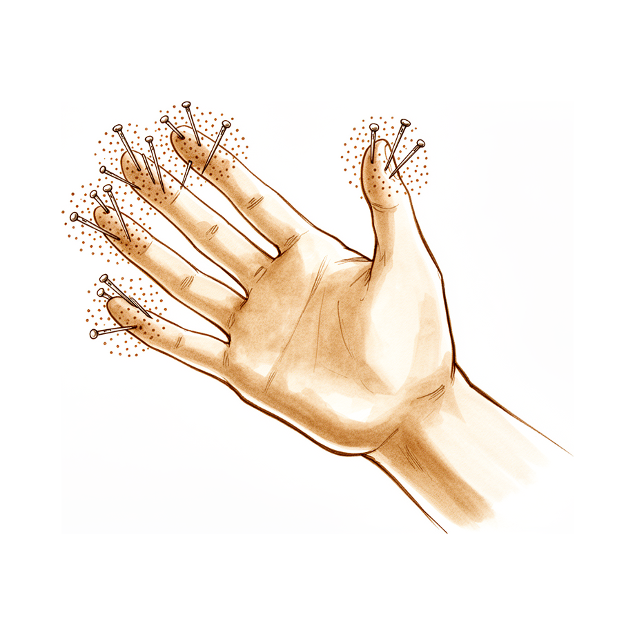
ہاتھ میں سُن پن اور جھنجھناہٹ اکثر کلائی پر کسی عصب (nerve) پر دباؤ کی وجہ سے ہوتی ہے۔ اسے کارپل ٹنل سنڈروم (carpal tunnel syndrome، کلائی میں عصب کا دبنا) کہتے ہیں، اور یہ بازو میں عصب کے دبنے کا سب سے عام مسئلہ ہے۔ جھنجھناہٹ عموماً انگوٹھے، شہادت کی انگلی اور درمیانی انگلی میں ہوتی ہے۔ کچھ لوگ اسے اوپر بازو کے نچلے حصے (forearm) میں بھی محسوس کرتے ہیں۔
علامات اکثر رات کو بڑھ جاتی ہیں اور آپ کو نیند سے جگا سکتی ہیں۔ ہاتھ کو جھٹکنے یا اسے نیچے لٹکانے سے جھنجھناہٹ کچھ دیر کے لیے کم ہو سکتی ہے۔ فون پکڑنا، گاڑی کا اسٹیئرنگ تھامنا یا ایک ہی حالت میں کتاب پڑھنا اسے بڑھا سکتا ہے۔ بہت سے لوگ اسے صبح سب سے پہلے محسوس کرتے ہیں، جب انگلیاں کھلنے سے پہلے اناڑی یا سوجی ہوئی محسوس ہوتی ہیں۔
روزمرہ کے کام مشکل ہو سکتے ہیں۔ جب آپ کی انگلیاں سُن ہوں تو بٹن لگانا، کافی کا کپ پکڑنا، ٹائپ کرنا یا چابی گھمانا عجیب اور مشکل محسوس ہو سکتا ہے۔ ہو سکتا ہے چیزیں انجانے میں آپ کے ہاتھ سے گر جائیں، کیونکہ آپ کی انگلیاں یہ واضح پیغام نہیں بھیج رہی ہوتیں کہ وہ کیا پکڑے ہوئے ہیں۔
ہاتھ اور بازو کی ہر جھنجھناہٹ کلائی سے نہیں آتی۔ کہنی کے قریب کسی عصب پر دباؤ چھوٹی انگلی تک جھنجھناہٹ پیدا کر سکتا ہے، اور گردن یا کندھے میں کسی عصب کا مسئلہ بھی علامات کو بازو میں نیچے تک بھیج سکتا ہے۔ کبھی کبھی ایک ہی عصب کے راستے میں دو جگہوں پر دباؤ مل کر اثر بڑھا دیتا ہے، اسی لیے آپ کے ڈاکٹر صرف ہاتھ کو نہیں بلکہ پورے بازو کو دیکھیں گے۔
کچھ علامات میں فوری دیکھ بھال کی ضرورت ہوتی ہے۔ اگر کوئی انگلی، ہاتھ یا بازو گرم، سرخ، سوجا ہوا اور دردناک ہو جائے، خاص طور پر بخار کے ساتھ، تو اسی دن ہسپتال کی ایمرجنسی جائیں۔ یہی بات اس وقت بھی لاگو ہوتی ہے جب آپ کی انگلیاں یا ہاتھ سفید پڑ جائیں، ٹھنڈے ہو جائیں، یا سفید یا نیلے ہو جائیں، یا کسی چوٹ کے بعد اچانک احساس یا حرکت ختم ہو جائے۔
اگر آپ کی علامات ٹھیک نہ ہو رہی ہوں، ہفتوں کے دوران بگڑ رہی ہوں، آپ کو رات کو جگا دیں، یا آپ کو کام کرنے یا اپنا ہاتھ استعمال کرنے سے روک دیں، تو اپنے فیملی ڈاکٹر (GP) سے ملیں یا ماہر ڈاکٹر سے معائنہ کروانے کی درخواست کریں۔ اگر آپ دفتری اوقات کے بعد یا ہفتے کے آخر میں کلینک سے رابطہ نہ کر سکیں تو اپنے قریب ترین ہسپتال کی ایمرجنسی جائیں۔
دراصل کیا ہو رہا ہے
آپ کے اعصاب بجلی کی تاروں کی طرح کام کرتے ہیں جو آپ کی گردن سے نیچے انگلیوں تک جاتی ہیں۔ یہ احساس اور حرکت کے لیے ہدایات لے جاتے ہیں۔ جہاں کوئی عصب کسی تنگ جگہ سے گزرتا ہے، وہاں اس پر دباؤ بڑھ سکتا ہے۔ یہی دباؤ اوپر بیان کی گئی جھنجھناہٹ اور سُن پن کی وجہ بنتا ہے۔
کلائی پر، آپ کے انگوٹھے، شہادت کی انگلی اور درمیانی انگلی کا مرکزی عصب ایک تنگ راستے سے گزرتا ہے جو کلائی کی چھوٹی ہڈیوں اور سامنے کی طرف آر پار پھیلی ایک مضبوط پٹی سے بنتا ہے۔ جب اس راستے میں جگہ تنگ ہو جاتی ہے تو عصب دب جاتا ہے۔ یہ دباؤ سب سے پہلے عصب کو خون کی فراہمی کو متاثر کرتا ہے، پھر وقت کے ساتھ خود عصب کے ریشوں کو نقصان پہنچ سکتا ہے۔ اسی لیے علامات اکثر رات کو بڑھتی ہیں، جب کلائی میں سیال جمع ہو جاتا ہے اور دباؤ بڑھ جاتا ہے۔
یہی بات دوسری جگہوں پر بھی لاگو ہوتی ہے۔ آپ کی کہنی کے اندر والی طرف ایک عصب وہاں بافت کی ایک تنگ پٹی سے دب سکتا ہے۔ بازو کے نچلے حصے میں ایک عصب ہاتھ کی طرف جاتے ہوئے کسی پٹھے سے بھنچ سکتا ہے۔ اور گردن میں کسی عصب کا مسئلہ علامات کو پورے بازو میں نیچے تک بھیج سکتا ہے، اسی لیے آپ کے ڈاکٹر پورے بازو کا معائنہ کریں گے۔
عصب پر دباؤ ہلکے سے لے کر شدید تک ہو سکتا ہے۔ شروع میں عصب میں جلن (irritation) ہوتی ہے لیکن وہ کام کرتا رہتا ہے، اور دباؤ ختم ہونے پر احساس عموماً واپس آ جاتا ہے۔ اگر زیادہ دیر تک چھوڑ دیا جائے تو خود عصب کے ریشے ٹوٹنا شروع ہو جاتے ہیں، اور سُن پن مستقل ہو سکتا ہے۔ اسی لیے انتظار کرتے رہنے کے بجائے علامات کا معائنہ کروانا بہتر ہے۔
آپ کے ڈاکٹر آپ سے آپ کی علامات کے بارے میں بات کر کے اور آپ کے ہاتھ کا معائنہ کر کے معلوم کرتے ہیں کہ عصب کہاں دب رہا ہے۔ ہاتھ کے تقریباً 90 فیصد مسائل میں تفصیل سے لی گئی طبی تاریخ صحیح تشخیص کی طرف اشارہ کرتی ہے۔ عصب کے اوپر تھپتھپانا یا آپ کی کلائی کو مڑا ہوا رکھنا آپ کی جھنجھناہٹ کو دوبارہ پیدا کر سکتا ہے، جس سے اس جگہ کی تصدیق میں مدد ملتی ہے۔ اگر صورتِ حال واضح نہ ہو تو ایسے ٹیسٹ جو یہ ناپتے ہیں کہ عصب پیغامات کتنی اچھی طرح لے جاتا ہے، یہ دکھا سکتے ہیں کہ آیا عصب واقعی دباؤ میں ہے اور آیا اس کا دباؤ ختم کرنے سے فائدہ ہوگا۔
ہم اس کے لیے کیا کر سکتے ہیں
کسی بھی طریقۂ علاج پر بات ہونے سے پہلے آپ بہت کچھ آزما سکتے ہیں۔ ہلکا مساج اور کلائی اور ہاتھ کو حرکت دینا عصب پر دباؤ کم کر سکتا ہے، اور کارپل ٹنل سنڈروم کے لیے انہی طریقوں کو پہلے قدم کے طور پر تجویز کیا جاتا ہے۔ سادہ ورزشیں جو عصب کو آسانی سے سرکنے میں مدد دیتی ہیں اور اس کے گرد سوجن کم کرتی ہیں، گھر پر کرنا آسان ہیں اور کسی بھی جگہ تھراپی کے پروگرام میں شامل کی جا سکتی ہیں۔ ایسا اسپلنٹ جو کلائی یا کہنی کو آرام کی حالت میں رکھے اور رات کو پہنا جائے، علامات کو کم کر سکتا ہے؛ کہنی پر النر عصب (ulnar nerve) کے دبنے میں، جن لوگوں کا علاج ڈراپ آؤٹ اسپلنٹ (drop-out splint) سے کیا گیا ان میں سے 24 فیصد کی علامات مکمل طور پر ختم ہو گئیں۔ ان اقدامات کو کئی ہفتوں تک مناسب طریقے سے آزمائیں۔ اگر آپ کی علامات ٹھیک نہ ہو رہی ہوں، ہفتوں کے دوران بگڑ رہی ہوں، آپ کو رات کو جگا دیں، یا آپ کو کام کرنے یا اپنا ہاتھ استعمال کرنے سے روک دیں، تو اپنے فیملی ڈاکٹر سے ملیں یا ماہر ڈاکٹر سے معائنہ کروانے کی درخواست کریں۔
ان اقدامات کے ساتھ دوائیں بھی مدد کر سکتی ہیں۔ ہو سکتا ہے سوزش کم کرنے والی دوا اور درد کی سادہ دوا عصب کے گرد جلن کو کم کر دیں۔ کچھ اعصابی مسائل میں، جیسے تھوراسک آؤٹ لیٹ سنڈروم (thoracic outlet syndrome، گردن اور کندھے کے درمیان اعصاب یا خون کی نالیوں کا دبنا)، ایسی دوائیں جو حد سے زیادہ متحرک اعصاب کو پرسکون کرتی ہیں آپ کو سونے میں مدد دے سکتی ہیں، اور کسی بھی سرجری سے پہلے طاقتور اوپیئوئڈ درد کش دواؤں کی شاذ و نادر ہی ضرورت پڑتی ہے۔ اگر مسئلے کا کچھ حصہ خون کی نالی کی کسی خرابی کی وجہ سے ہاتھ میں سوجن ہو تو ناپ کے مطابق بنا ہوا دباؤ والا دستانہ (compression glove) مدد کر سکتا ہے۔ علامات کے کچھ سوالنامے، جیسے وہ جو یہ اسکور کرتے ہیں کہ آپ کی علامات کتنی شدید ہیں اور آپ کا ہاتھ آپ کو کتنا محدود کرتا ہے، یہ جانچنے میں مدد دیتے ہیں کہ علاج کام کر رہا ہے یا نہیں۔
اگر اوپر دی گئی تدابیر کے باوجود علامات شدید رہیں تو آپ کے فیملی ڈاکٹر آپ کو ماہر ڈاکٹر کے معائنے کے لیے ریفر کر سکتے ہیں۔ ایسے ٹیسٹ جو یہ ناپتے ہیں کہ عصب پیغامات کتنی اچھی طرح لے جاتا ہے، یا خود عصب کا اسکین، اس بات کی تصدیق کر سکتے ہیں کہ دباؤ کہاں ہے اور آیا اسے ختم کرنے سے فائدہ ہوگا۔ کچھ مخصوص حالتوں میں کبھی کبھار عصب پر سے دباؤ ہٹانے کے طریقۂ علاج پر غور کیا جا سکتا ہے۔ عصب کا دباؤ ہٹانے کے زیادہ تر طریقۂ علاج کے بعد زخم کی پٹی تقریباً 10 دن تک لگی رہتی ہے اور آپ کی معائنے کی ملاقات پر بدلی یا اتاری جاتی ہے، اس کے تھوڑے عرصے بعد ہلکی حرکت شروع ہو جاتی ہے، اور اگلے ہفتوں میں طاقت اور حرکت کو دوبارہ بحال کرنے کے لیے تھراپی کی جاتی ہے۔
کیا توقع رکھیں
بازو میں عصب پر دباؤ کے مسائل عموماً دو میں سے کسی ایک راستے پر چلتے ہیں۔ اگر جلدی پتا چل جائے تو عصب میں جلن ہوتی ہے لیکن وہ ابھی صحت مند ہوتا ہے، اور دباؤ کم ہونے پر علامات اکثر ٹھیک ہو جاتی ہیں۔ اگر مہینوں یا برسوں تک چھوڑ دیا جائے تو سُن پن مستقل ہو سکتا ہے، اسی لیے انتظار کرتے رہنے کے بجائے قدم اٹھانا بہتر ہے۔
علامات کتنی دیر رہتی ہیں، یہ اس بات پر منحصر ہے کہ ان کی وجہ کیا ہے اور آپ کتنی جلدی ان کا علاج کرواتے ہیں۔ کچھ لوگوں کی جھنجھناہٹ صحیح طرح معائنہ ہونے سے پہلے لمبے عرصے تک آتی جاتی رہتی ہے۔ اگر دباؤ جلدی ختم کر دیا جائے تو عموماً ہفتوں سے مہینوں کے دوران احساس واپس آ جاتا ہے، جیسے جیسے عصب ٹھیک ہوتا ہے۔ اعصاب آہستہ آہستہ ٹھیک ہوتے ہیں، اس لیے بہتری ایک رات میں نہیں بلکہ بتدریج آتی ہے۔ کچھ لوگ محسوس کرتے ہیں کہ جھنجھناہٹ پہلے کم ہوتی ہے، جبکہ عصب کے دوبارہ جاگنے کے دوران سوئیاں چبھنے جیسا احساس کچھ عرصہ باقی رہ سکتا ہے۔
علاج کے بغیر علامات اکثر بار بار بڑھتی رہتی ہیں، خاص طور پر رات کو، اور آہستہ آہستہ بگڑ سکتی ہیں۔ مساج، ہلکی حرکت اور آرام کی حالت میں رکھنے والا اسپلنٹ جیسے سادہ اقدامات کچھ لوگوں کی مدد کرتے ہیں، اور سرجری کے بغیر علاج کے بارے میں اوپر والا حصہ بتاتا ہے کہ کیا آزمایا جائے۔ اگر یہ اقدامات کافی نہ ہوں تو دباؤ ختم کرنے کی سرجری بہت سے لوگوں کو دیرپا آرام دیتی ہے۔ جہاں عصب کو لمبے عرصے سے شدید نقصان پہنچا ہو، وہاں دوسرے طریقۂ علاج سے بنیادی مسئلہ ٹھیک ہونے کا امکان کم ہوتا ہے، اس لیے جلدی علاج عموماً بہتر اختیارات دیتا ہے۔
صحت یابی کا تجربہ ہر شخص کے لیے مختلف ہوتا ہے۔ بہت سے لوگ محسوس کرتے ہیں کہ دباؤ ختم ہونے کے کچھ ہی عرصے بعد ان کی رات کی علامات کم ہو جاتی ہیں، جبکہ احساس اور طاقت اگلے مہینوں میں زیادہ آہستہ واپس آتے ہیں۔ آپ کی دیکھ بھال کرنے والی ٹیم احساس اور حرکت کی سادہ جانچوں سے آپ کی پیش رفت پر نظر رکھے گی، اور تھراپی طاقت اور حرکت کو دوبارہ بحال کرنے میں مدد دے سکتی ہے۔ کچھ سُن پن کو بہتر ہونے میں لمبا وقت لگ سکتا ہے، اور اگر عصب کو لمبے عرصے سے نقصان پہنچا ہو تو احساس میں کچھ تبدیلی باقی رہ سکتی ہے۔
اگر آپ کی علامات ٹھیک نہ ہو رہی ہوں، ہفتوں کے دوران بگڑ رہی ہوں، آپ کو رات کو جگا دیں، یا آپ کو کام کرنے یا اپنا ہاتھ استعمال کرنے سے روک دیں، تو اپنے فیملی ڈاکٹر سے ملیں یا ماہر ڈاکٹر سے معائنہ کروانے کی درخواست کریں۔
ڈاکٹر کو کب دکھائیں
ہاتھ اور بازو کی زیادہ تر جھنجھناہٹ ایمرجنسی نہیں ہوتی، لیکن کچھ علامات ایمرجنسی ہوتی ہیں۔ اگر کوئی انگلی، ہاتھ یا بازو گرم، سرخ، سوجا ہوا اور دردناک ہو جائے، خاص طور پر بخار کے ساتھ، تو اسی دن ہسپتال کی ایمرجنسی جائیں۔ یہی بات اس وقت بھی لاگو ہوتی ہے جب آپ کی انگلیاں یا ہاتھ سفید پڑ جائیں، ٹھنڈے ہو جائیں، یا سفید یا نیلے ہو جائیں، یا کسی چوٹ کے بعد اچانک احساس یا حرکت ختم ہو جائے۔
باقی تمام صورتوں میں، پہلے اپنے فیملی ڈاکٹر سے ملیں۔ اگر آپ کی علامات ٹھیک نہ ہو رہی ہوں، ہفتوں کے دوران بگڑ رہی ہوں، آپ کو رات کو جگا دیں، یا آپ کو کام کرنے یا اپنا ہاتھ استعمال کرنے سے روک دیں، تو ماہر ڈاکٹر سے معائنہ کروانے کی درخواست کریں۔ عصب پر دباؤ کے مسائل اس وقت سب سے بہتر ٹھیک ہوتے ہیں جب ان کا جلدی پتا چل جائے، اس لیے سُن پن کے خود ختم ہونے کا مہینوں انتظار کرنے کے بجائے معائنہ کروا لینا بہتر ہے۔
آپ کے ڈاکٹر صرف ہاتھ کا نہیں بلکہ پورے بازو کا معائنہ کریں گے۔ کلائی پر عصب کے اوپر تھپتھپانا، اس جگہ پر دبانا، یا آپ کی کلائی کو مڑا ہوا رکھنا، یہ سب آپ کی جھنجھناہٹ شروع کر سکتے ہیں، اور ان میں سے ہر ایک اس جگہ کی طرف اشارہ کرتا ہے جہاں عصب دب رہا ہے۔ کبھی کبھی ایک عصب اپنی لمبائی میں ایک سے زیادہ جگہوں پر بھنچا ہوا ہوتا ہے، اسی لیے گردن، کندھے اور کہنی کا بھی معائنہ کیا جاتا ہے۔ اگر معائنے سے واضح جواب نہ ملے تو ایسے ٹیسٹ جو یہ ناپتے ہیں کہ عصب پیغامات کتنی اچھی طرح لے جاتا ہے، تشخیص کی تصدیق کر سکتے ہیں۔
Evidence & references
This is the clinical evidence summary written for health professionals. It is technical, and it lists the research this page was built from. You do not need to read it to understand your treatment or to make a decision about it.
Overview
- Carpal tunnel syndrome is the most frequent entrapment neuropathy at the upper limb [12].
- The incidence of carpal tunnel release increased by 43% between 1996 and 2005, particularly influenced by the number of elderly individuals presenting with carpal tunnel syndrome [27].
- There is a severe discordance between the estimated prevalence of mild-to-moderate carpal tunnel syndrome based on clinical signs and symptoms (73%) versus electrodiagnostic studies and ultrasound (51%) [23].
- When signs and symptoms suggest mild-to-moderate median neuropathy and surgery is being considered, patients and clinicians might consider additional testing, such as EDS or US, to increase the probability of actual median neuropathy that can benefit from surgery [5].
- Specific nerve conduction studies helped differentiate true compression symptoms from nonspecific polyneuropathies [6].
- Tinel’s sign has estimates of 50% and 77% for sensitivity and specificity, respectively [36].
- Carpal compression has estimates of 64% and 83% for sensitivity and specificity, respectively [36].
- Tinel's sign was present in 29% of subjects who did not have carpal tunnel syndrome and in 45% of those with carpal tunnel syndrome [8].
- The surgeon should observe one or more of the three major clinical signs of CTS: (1) hypoesthesia restricted to the median distribution in the hand, (2) a positive Tinel sign with light tapping over the median nerve at the wrist; and (3) a positive wrist-flexion test [14].
- An anomalous muscle invading the carpal canal has been reported more frequently than any other as a cause of median nerve compression [26].
- The systematic study of tingling caused by pressure on the nerve very often enables us to find out if the interruption to the nerve is complete or partial, determine the exact location and the extent of the injury, and detect regeneration of the axons at an early stage [4].
- The tingling response may be classified according to its location and to the magnitude threshold at which the response is triggered [2].
- Upper-extremity neuropathy can develop following nonupper extremity surgeries, particularly ACDF [7].
- Patients with a history of ulnar nerve lesions are at a significantly increased risk of developing carpal tunnel syndrome, especially within the first 2 years [15].
- As the severity of ulnar neuropathy at the elbow increases, the cross-sectional area (CSA) of the ulnar nerve correspondingly increases at the elbow [20].
- Of patients managed with a drop-out splint for ulnar nerve entrapment at the elbow, 24% demonstrated complete resolution of their symptoms, whereas another 24% subsequently required surgical decompression of the nerve [10].
- Radial tunnel syndrome is relatively uncommon but is an important cause of lateral forearm pain, with clinical examination being a crucial part of the diagnosis [13].
- In neuropathy rehabilitation, the assessment of position and tactile sensations should not be ignored in determining participation in manual skills [11].
- The authors emphasize the need for clear definitions of recurrence versus persistent compression neuropathy, agreements on supplementary diagnostics, and standardized outcome measurements to enable comparing results from different techniques and clinics [3].
- Somewhere between 11 and 17 of the 28 patients reviewed had signs of advanced (and likely permanent) nerve damage, so the probability that a second surgery will address pathophysiology is low [9].
- At least four areas of consideration are important when evaluating a patient with an injury to a nerve in the hand: (1) type of injury, (2) sensibility evaluation, (3) motor function, and (4) sudomotor function (sweating) [28].
- Normal two-point discrimination usually is 6 mm or less [28].
- If the nerve is transected, a patient would not feel light touch, would not appreciate the pin as a sharp stimulus, and would be unable to discriminate between one and two points [28].
- Patients with closed injuries or partial injuries to nerves may show spotty appreciation of light touch and pain and have markedly widened two-point discrimination [28].
- Factors that interfere with the examination of the nerves in the hand include other injuries that may be life threatening or limb threatening, patient intoxication, anxiety or lack of cooperation of the patient, and an extensive injury to the hand [28].
- If a flexor tendon function deficit is present after a finger laceration, at least one digital nerve probably has been injured as well [28].
Background & Causes
Pathophysiology and Clinical Presentation
- The histopathologic findings of chronic nerve compression span a spectrum beginning with breakdown of the blood-nerve barrier and progressing to axonal degeneration [29].
- Patient signs, symptoms, and sensory testing parallel the histopathologic changes occurring in the nerve [29].
- Abnormal postures or positions can cause nerves to be compressed or placed on tension, progressing to chronic nerve compression [29].
- Abnormal postures or positions can place muscles in shortened positions [29].
- Abnormal postures or positions can weaken muscles in elongated or shortened positions, leading to underuse and compensatory overuse of other muscles [29].
- Numbness or tingling symptoms indicate a neurologic problem rather than a mechanical one [47].
- A careful history suggests the correct diagnosis in approximately 90% of patients with hand problems [47].
- Systematic study of tingling caused by pressure on the nerve can determine if nerve interruption is complete or partial [4].
- Systematic study of tingling caused by pressure on the nerve can determine the exact location and extent of the injury [4].
- Systematic study of tingling caused by pressure on the nerve can detect regeneration of axons at an early stage [4].
- Tinel's sign may be present only in certain stages of carpal tunnel syndrome, but when present is quite specific [32].
- Clinical tests for carpal tunnel syndrome have poorer specificity when evaluated among symptomatic CTS-free subjects than among asymptomatic CTS-free subjects [60].
- Specific nerve conduction studies help differentiate true compression symptoms from nonspecific polyneuropathies [6].
- When signs and symptoms suggest mild-to-moderate median neuropathy, additional testing such as electrodiagnostic studies or ultrasound may increase the probability of actual median neuropathy that can benefit from surgery [5].
- As the severity of ulnar neuropathy at the elbow increases, the cross-sectional area of the ulnar nerve correspondingly increases at the elbow [20].
Etiology and Risk Factors
- Carpal tunnel syndrome is the most frequent among entrapment neuropathies at the upper limb [12].
- Dislocation of the ulnar nerve is not an important factor among the causes of cubital tunnel syndrome, and the important factor is the tendinous arch [56].
- Upper-extremity neuropathy can develop following nonupper extremity surgeries, particularly anterior cervical discectomy and fusion [7].
- A rise above a critical pressure brought about by vascular congestion would explain the clinical picture of predominantly nocturnal symptoms and no electro-physiological evidence with the wrist in a neutral position [63].
- Radial tunnel syndrome is relatively uncommon but is an important cause of lateral forearm pain [13].
- The authors were unable to confirm the association of bilateral disease with amyloid or the association with trigger digits in idiopathic carpal tunnel syndrome [58].
- An associated muscle that inserted into the palmar fascia was clearly not the cause of nerve compression in a case of bilateral carpal tunnel syndrome [59].
Diagnostic Considerations
- The surgeon should observe one or more of the three major clinical signs of carpal tunnel syndrome: hypoesthesia restricted to the median distribution in the hand, a positive Tinel sign with light tapping over the median nerve at the wrist, and a positive wrist-flexion test [14].
- Clinical examination is a crucial part of the diagnosis of radial tunnel syndrome [13].
- The assessment of position and tactile sensations should not be ignored in determining participation in manual skills in neuropathy rehabilitation [11].
Symptoms & Presentation
- The surgeon should observe hypoesthesia restricted to the median distribution in the hand as one of the major clinical signs of carpal tunnel syndrome [14].
- A positive Tinel sign with light tapping over the median nerve at the wrist is a major clinical sign of carpal tunnel syndrome [14].
- A positive wrist-flexion test is a major clinical sign of carpal tunnel syndrome [14].
- Tinel's sign may be present only in certain stages of carpal tunnel syndrome [32].
- When present, Tinel's sign is quite specific for carpal tunnel syndrome [32].
- Estimates for the sensitivity of Tinel's sign in detecting carpal tunnel syndrome are 50% [36].
- Estimates for the specificity of Tinel's sign in detecting carpal tunnel syndrome are 77% [36].
- Estimates for the sensitivity of carpal compression in detecting carpal tunnel syndrome are 64% [36].
- Estimates for the specificity of carpal compression in detecting carpal tunnel syndrome are 83% [36].
- Tinel's sign was present in 29% of subjects who did not have carpal tunnel syndrome [8].
- Tinel's sign was present in 45% of subjects who had carpal tunnel syndrome [8].
- When signs and symptoms suggest mild-to-moderate median neuropathy and surgery is being considered, patients and clinicians might consider additional testing such as electrodiagnostic studies or ultrasound to increase the probability of actual median neuropathy that can benefit from surgery [5].
- Systematic study of tingling caused by pressure on the nerve enables determination of if the interruption to the nerve is complete or partial [4].
- Systematic study of tingling caused by pressure on the nerve enables determination of the exact location and extent of the injury [4].
- Systematic study of tingling caused by pressure on the nerve enables detection of regeneration of the axons at an early stage [4].
- Radial tunnel syndrome is an important cause of lateral forearm pain [13].
- Neuropathy symptoms following shoulder surgery were often refractory to conservative management [18].
- Surgical decompression of neuropathy following shoulder surgery led to nearly 90% symptom resolution [18].
- Surgeons are encouraged to bear anatomic anomalies in mind when assessing patients with symptoms of median nerve compression [31].
Management
Diagnostic and Preoperative Assessment
- The systematic study of tingling caused by pressure on the nerve enables determination of whether nerve interruption is complete or partial, identification of the exact location and extent of injury, and detection of early axon regeneration [4].
- The tingling response can be classified according to its location and the magnitude threshold at which it is triggered [2].
- When signs and symptoms suggest mild-to-moderate median neuropathy and surgery is being considered, additional testing such as electrodiagnostic studies (EDS) or ultrasound (US) may increase the probability of actual median neuropathy that can benefit from surgery [5].
- Clinical examination is a crucial part of the diagnosis for radial tunnel syndrome [13].
- Preoperative assessment of nerve injury in the hand includes evaluation of the type of injury, sensibility, motor function, and sudomotor function (sweating) [28].
- Normal two-point discrimination is usually 6 mm or less [28].
- Patients with closed or partial nerve injuries may show spotty appreciation of light touch and pain and have markedly widened two-point discrimination [28].
- Magnetic resonance neurography (MRA) is reported to provide details regarding nerve anatomic relationships, fascicular pattern, intraneural swelling, and evaluation of downstream muscle injury [28].
- The carpal tunnel syndrome questionnaire assesses symptom severity (11 items) and functional status (8 items), with higher scores indicating decreased functional status [29].
- The Disabilities of Arm, Shoulder, and Hand (DASH) questionnaire uses 30 items to assess upper extremity disability based on physical symptoms and functional status [29].
- The Michigan Hand Outcomes Questionnaire (MHQ) is a 67-item questionnaire that addresses specific domains of overall hand function, physical function, cosmesis, and satisfaction [29].
- The Patient-Specific Functional Scale (PSFS) assesses items identified by patients, with each item ranked on a scale from 0 to 10 [29].
- The Patient-Reported Outcomes Measurement Information System (PROMIS) provides an online platform for assessing physical and psychosocial health domains applicable to nerve compression [29].
- Hand abduction tracings are a quantitative outcome measure to follow recovery over time for intrinsic hand function in patients with severe ulnar neuropathy following surgical intervention [22].
- Electrodiagnostic studies by an experienced examiner are critical in the differential diagnosis and localization of the level of compression for failed cubital tunnel syndrome [45].
- A proximal percussion test above the area of the previous surgical site along the course of the basilic vein results in pain and/or paresthesia in the distribution of the medial antebrachial cutaneous nerve (MABC) if a neuroma of this nerve is present [45].
- Percussion along the course of the ulnar nerve above the area of previous surgery results in paresthesia and pain in the ulnar nerve distribution if there is a problem with the ulnar nerve itself [45].
- Palpation along the course of the ulnar nerve in the distal forearm progressing to the region of the surgical site results in deep pain distal to the previous surgical site if a new compression point has developed at the site of distal surgical exposure [45].
- The physical examination of suspected complex regional pain syndrome (CRPS) patients should include a neurologic assessment and evaluation of the cervical and thoracic spine [38].
- The presence of cervical disease may exacerbate CRPS, a form of "double-crush syndrome" [38].
- Preexisting or acquired thoracic outlet or distant compression neuropathies can represent a "triple or more" crush syndrome [38].
- There are no objective laboratory tests to aid in the diagnosis of CRPS [38].
- For patients with a history of ulnar nerve lesions, there is a significantly increased risk of developing carpal tunnel syndrome, especially within the first 2 years [15].
- Upper-extremity neuropathy can develop following nonupper extremity surgeries, particularly anterior cervical discectomy and fusion (ACDF) [7].
- More than 50% of patients who did not undergo carpal tunnel release at the initial surgery for perilunate injuries required a release within the follow-up period [35].
Non-Operative Management
- Conservative treatments, specifically manual therapies like massage and mobilization, are recommended as first-line treatments for carpal tunnel syndrome [52].
- If double crush syndrome is suspected but symptoms are mild, they may respond to conservative treatment [25].
- Sensory interventions for upper limb conditions are inexpensive, noninvasive, and easy to perform, allowing implementation into conventional therapy practice in any setting [53].
- A drop-out splint for ulnar nerve entrapment at the elbow resulted in complete resolution of symptoms in 24% of patients [10].
- Complete recovery of shoulder function and relief of symptoms was achieved at 8 and 13 months after application of an orthosis for long thoracic nerve paralysis associated with thoracic outlet syndrome [24].
- Tricyclic antidepressants, neurotropics, or low doses of benzodiazepines may help patients sleep with thoracic outlet syndrome [39].
- Narcotics should rarely be prescribed preoperatively for thoracic outlet syndrome [39].
- A fitted compression glove can effectively treat some vascular malformations if the lesion tends to swell when dependent or with activity [40].
- Asymptomatic vascular malformations in the hand can usually be left alone [40].
Operative Management
- Surgical decompression for neuropathy following shoulder surgery led to nearly 90% symptom resolution [18].
- Invasive treatments for double crush syndrome should be initiated preferentially at sites with greater clinical suspicion [25].
- Surgical decompression of the ulnar nerve at the wrist associated with a recurrent branch through the flexor carpi ulnaris tendon resulted in symptom relief and normal full use of the hand and wrist after 3 months [17].
- Anterior interosseous nerve transfer, along with cubital and ulnar tunnel release, results in sustained clinical and electrophysiological improvements in patients with severe chronic ulnar nerve compression [55].
- Decompressing known areas of compression can enhance nerve regeneration in patients with incomplete nerve injuries (less than Sunderland V) [50].
- The most common intraoperative findings in patients undergoing surgery for recurrent carpal tunnel syndrome were incomplete release of the flexor retinaculum and median nerve adherence to surrounding tissue [50].
- In obese patients, failure to make an incision of adequate length may lead to insufficient decompression of the median nerve, especially at the proximal and distal sites of the surgical exposure [50].
- A minimally invasive surgical option using a synovial flap is part of the therapeutic arsenal for recurrent carpal tunnel syndromes [33].
- Endoscopic treatment for recurrent carpal tunnel syndrome requires rigorous patient selection, specifically for true recurrences without obvious etiology or trophic disorders [19].
- The radial sensory nerve is released at its point of compression between the tendons of the extensor carpi radialis longus and brachioradialis [41].
- A full tenotomy of the brachioradialis tendon and resection of a portion of the tendon are performed to completely decompress the radial sensory nerve [41].
- Neurolysis is not performed during radial sensory nerve decompression [41].
- A piece of bioresorbable adhesion barrier is placed over the nerve to decrease scarring during radial sensory nerve decompression [41].
- For injury to the superficial branch of the radial nerve complicated by CRPS, options for treatment include proximal transection of the nerve in normal tissue, exploration with neurolysis, or repair [42].
- If repair is selected for superficial radial nerve injury with CRPS, a sural nerve is preferred for interposition grafting to avoid a potential nociceptive focus in the same extremity [42].
- Nerve grafting for superficial radial nerve injury is completed with 8-0 or 9-0 nonabsorbable suture under the operating microscope [42].
- If nerve transection is selected for superficial radial nerve injury, the resection should occur adjacent to normal tissue or be relocated to an unscarred area [42].
- Excessive scar-related attachment of the nerve to underlying tendon structures or overlying skin requires neural bed modification, such as covering the nerve with a section of the adjacent cephalic vein [42].
- Postoperative reactivation of the dystrophic process in CRPS can be minimized by maintaining a continuous autonomic blockade for 3 to 5 days after surgery [42].
- Surgical management of chronic deformities after CRPS is possible, but exacerbation of a dystrophic response is a potential risk [37].
- Perioperative pain control with continuous epidural or peripheral catheters should be maintained for 3 to 5 days after surgery for CRPS-related deformities [37].
- Surgery on contracted joints in CRPS should not be performed until maximal nonoperative improvement has been achieved, with a waiting period of a minimum of 3 to 6 months after successful elimination of active dystrophic pain [37].
- Release of the metacarpophalangeal (MCP) or proximal interphalangeal (PIP) joints can be performed for persistent contractures in CRPS when nonoperative improvement has plateaued [37].
- Indications for surgery for CRPS-related joint contractures are joint pain without diffuse dystrophic symptoms and arthrofibrosis that interferes with function [37].
- Restoration of full flexion or extension is an unreasonable goal for surgical correction of CRPS-related joint contractures [37].
- The range of motion achieved during surgery for CRPS-related joint contractures is rarely maintained after surgery as some loss is expected [37].
- For vascular malformations in the hand that are markedly symptomatic, a surgical approach is warranted [40].
- The goals of treatment for vascular malformations are to completely extirpate the lesion while preserving tendons, bone, nerve, and uninvolved muscle [40].
- An aggressive surgical approach in symptomatic vascular lesions in all but the worst high-flow lesions leads to acceptable outcomes with low complication rates [40].
- Venous malformations are the most common vascular malformations in the hand, and excision is usually straightforward [40].
- Surgical excision of venous malformations usually reduces pain and improves function for a significant period of time [40].
- High-flow arteriovenous malformations can be explored and excised safely with a tourniquet on the upper arm to minimize blood loss [40].
- Arteriographic studies are preferred to accurately delineate the extent of high-flow arteriovenous malformations and the involvement of major vessels [40].
- Ischemic digits associated with high-flow arteriovenous malformations are probably best amputated [40].
- Transfer of a neurovascular island graft is useful for restoring sensibility to critical areas of a digit in cases of permanent nerve damage [43].
- Sensibility in a neurovascular island graft is never normal after transfer, and more than half of patients have persistently hyperesthetic skin [43].
- All patients with neurovascular island grafts lack precise sensory reorientation, although reorientation seems to improve with time and use [43].
- Factors to consider before neurovascular island graft surgery include hand dominance, presence of palmar scarring, status of the ipsilateral ulnar nerve, condition of the opposite hand, patient age, and surgeon experience [43].
- The neurovascular bundle should be dissected from proximally to distally to treat any vascular anomalies properly [43].
- The neurovascular bundle should not be completely freed from surrounding fatty tissue, especially at the base of the finger [43].
- The neurovascular bundle should be channeled through an incision large enough to show the entire bundle to prevent kinking, twisting, or stretching [43].
- In complete median nerve paralysis, transfer of a neurovascular island graft to the radial side of the proximal and middle phalanges of the index finger may be desirable if sensibility on the ulnar edge of the thumb pulp is reasonably good [43].
- For failed nerve compression procedures, patients can be classified as persistent, recurrent, or new [50].
- Persistent symptoms after primary carpal tunnel release may imply an incomplete decompression or an incorrect diagnosis [50].
- Recurrence of symptoms 6 months or more after initial relief usually implies a new site of ulnar nerve compression in the previous surgical field for cubital tunnel syndrome [45].
- Severe causalgia-type pain at the elbow radiating into the medial aspect of the forearm suggests a neuroma of the medial antebrachial cutaneous nerve [45].
- Severe causalgia-type pain in the ulnar nerve distribution suggests an injury to the ulnar nerve itself [45].
- If a patient has undergone more than two surgical attempts to correct a nerve compression problem, it is less likely that a third operation will be helpful [50].
- In patients with a motor deficit after failed nerve compression surgery, nerve graft, nerve transfer, or tendon transfers are considered [50].
- If pain is the primary problem after failed nerve compression surgery, a peripheral nerve stimulator or dorsal column stimulator can be considered [50].
- The probability that a second surgery will address pathophysiology is low if signs of advanced and likely permanent nerve damage are present [9].
- Surgical failures are more frequent in cases of cubital tunnel syndrome than in cases of carpal tunnel syndrome [45].
- Management of failed cubital tunnel syndrome is made difficult by the previous surgical procedure and resultant scarring of the nerve [45].
- Any operative procedure for cubital tunnel syndrome can result in an excellent outcome if basic principles related to the condition are followed [45].
- It is essential that the surgical procedure for cubital tunnel syndrome does not create any new areas of compression on the ulnar nerve [45].
- Failure to relieve symptoms in cubital tunnel syndrome may imply an incomplete decompression of the primary surgical procedure or an incorrect diagnosis [45].
- The dressing is removed 2 to 3 days after median nerve compression surgery, and general range-of-motion exercises are started [50].
- A sling can be used at night for comfort for 3 weeks after median nerve compression surgery [50].
- Patients are referred for therapy postoperatively to assist with restoration of range of motion and strength after median nerve compression surgery [50].
- The first phase of graded motor imagery can be started preoperatively so the patient is prepared for postoperative therapy [50].
- Informed consent for failed nerve compression surgery will emphasize the possibility of incomplete relief of pain and even decreased neurologic function [50].
- For recurrent carpal tunnel syndrome, electrodiagnostic studies may not be particularly helpful given that patients with successful carpal tunnel surgery will frequently have persistent electrical changes, but should be performed as a baseline [50].
- Baseline monofilament sensory testing should be performed for recurrent carpal tunnel syndrome [50].
- The sensory collapse test can be used to isolate the area of incomplete release in recurrent carpal tunnel syndrome [50].
- In patients with definite relief of symptoms for 6 months or longer followed by recurrence of the same symptoms, a secondary traction neuritis may be present [50].
- For CRPS, surgery should not be performed unless sympathectomy intervention reduces the pain considerably [42].
- An anesthesiologist should perform a continuous autonomic block before surgery for intraoperative and postoperative pain management for 2 to 5 days in CRPS cases [42].
- Technical points for CRPS nerve surgery include performing an extensile neurolysis, excising any synovitis if present, mobilizing the nerve, and covering the nerve with fat, a muscle flap, vein, synthetic conduit, or free tissue transfer [42].
- Pitfalls in CRPS nerve surgery include failure to confirm potential control of postoperative pain, incomplete mobilization of the involved nerve, and inadequate interposition of soft tissue between skin-nerve and nerve-tendon interfaces [42].
- Postoperative care for CRPS nerve surgery includes initiating early range-of-motion exercises of the fingers and wrist and maintaining adequate pain management for 3 to 7 days with parenteral or oral agents [42].
- Continuous blocks are helpful for pain relief in CRPS nerve surgery, and unrestricted motion is allowed when the patient is comfortable [42].
- For median nerve compression at the elbow and forearm, the dressing is removed 2 to 3 days after surgery and general range-of-motion exercises are started [50].
- For quadrangular space syndrome and axillary nerve decompression, diagnosis can be challenging in cases of nerve ischemia without demyelination or axonal degeneration (Sunderland I) as these are not detectable by electrodiagnostic or imaging studies [50].
- Clinically significant compression of the axillary nerve can be reliably diagnosed by an experienced clinician using manual muscle strength testing, provocative testing, presence of paresthesias, and the hierarchical sensory collapse test [50].
- Provocative testing for axillary nerve compression involves having the patient abduct and externally rotate the shoulder with the elbow flexed at 90 degrees and then internally rotate the shoulder [50].
- Serial electrodiagnostic testing can be helpful in cases of suspected direct injury to the axillary nerve to determine the degree of injury and extent of spontaneous recovery [50].
- High-resolution nerve-specific magnetic resonance imaging can be helpful in identifying swelling or signal changes in the ax
Key Considerations
Diagnosis and Clinical Assessment
- The surgeon should observe one or more of three major clinical signs of carpal tunnel syndrome: hypoesthesia restricted to the median distribution in the hand, a positive Tinel sign with light tapping over the median nerve at the wrist, and a positive wrist-flexion test [14].
Etiology and Risk Factors
- Surgeons should bear bifid median nerve anomalies in mind when assessing patients with symptoms of median nerve compression [31].
Surgical Management and Outcomes
- Neuropathy symptoms were often refractory to conservative management, while surgical decompression led to nearly 90% symptom resolution following shoulder surgery [18].
- Decompressing the deep motor branch of the ulnar nerve allowed for spontaneous nerve recovery and reinnervation of ulnar nerve innervated intrinsic muscles in cases of posttraumatic compressive neuropathy caused by heterotopic ossification [57].
- At 6 months, EMG analysis showed no residual compression and good release of the ulnar nerve in all cases following endoscopic neurolysis for ulnar nerve compression at the elbow [61].
- In a series of 250 patients with carpal tunnel syndrome over 6 years, no patient had a late diagnosis of distal ulnar nerve compression after prior carpal tunnel release [62].
- Postoperative symptoms were relieved and normal and full use of the hand and wrist was achieved after 3 months in a case of ulnar neuropathy at the wrist associated with a recurrent branch through the flexor carpi ulnaris tendon [17].
- It requires rigorous patient selection, specifically for true recurrences without obvious etiology or trophic disorders, for endoscopic treatment of recurrent carpal tunnel syndrome [19].
- This minimally invasive surgical option using a synovial flap is interesting and should be part of the therapeutic arsenal for recurrent carpal tunnel syndromes [33].
Conservative Management and Rehabilitation
- Complete recovery of shoulder function and relief of the symptoms was achieved in both cases at 8 and 13 months, respectively, after application of the orthosis for long thoracic nerve paralysis associated with thoracic outlet syndrome [24].
Outcome Measurement
- Hand abduction tracings are a quantitative outcome measure to follow recovery over time for intrinsic hand function and can be used in patients with severe ulnar neuropathy following surgical intervention [22].
Key Evidence
- [L5] The tingling response may then be classified according to its location and to the magnitude threshold at which the response is triggered. [2] (10.1016/s0894-1130(99)80068-4)
- [L5] The authors emphasize the need for clear definitions of recurrence versus persistent compression neuropathy, agreements on supplementary diagnostics, and standardized outcome measurements to enable comparing results from different techniques and clinics. [3] (10.1177/17531934241311822)
- [L5] The systematic study of tingling caused by pressure on the nerve very often enables us to find out if the interruption to the nerve is complete or partial, determine the exact location and the extent of the injury, and detect regeneration of the axons at an early stage. [4] (10.1016/j.jhsb.2004.10.007)
- [L3] When signs and symptoms suggest mild-to-moderate median neuropathy and surgery is being considered, patients and clinicians might consider additional testing, such as EDS or US, to increase the probability of actual median neuropathy that can benefit from surgery. [5] (10.1097/corr.0000000000002751)
- [L4] Specific nerve conduction studies helped differentiate true compression symptoms from nonspecific polyneuropathies. [6] (10.1016/s0363-5023(81)80059-7)
- [L3] Upper-extremity neuropathy can develop following nonupper extremity surgeries, particularly ACDF. [7] (10.1016/j.jhsg.2026.100972)
- [L5] They note that Tinel's sign was present in 29% of subjects who did not have carpal tunnel syndrome and in 45% of those with carpal tunnel syndrome. [8] (10.1016/j.jhsa.2004.02.009)
- [L4] Of these patients, 24% demonstrated complete resolution of their symptoms, whereas another 24% of the patients subsequently required surgical decompression of the nerve. [10] (10.1016/s0894-1130(12)80378-4)
- [L3] In neuropathy rehabilitation, the assessment of position and tactile sensations should not be ignored in determining participation in manual skills. [11] (10.1016/j.jht.2024.12.005)
- [Paper] Carpal tunnel syndrome is the most frequent among entrapment neuropathies at the upper limb. [12] (10.1016/j.main.2004.10.016)
- [L5] Radial tunnel syndrome is relatively uncommon but is an important cause of lateral forearm pain, with clinical examination being a crucial part of the diagnosis. [13] (10.1197/j.jht.2006.02.005)
- [L5] The surgeon should observe one or more of the three major clinical signs of CTS: (1) hypoesthesia restricted to the median distribution in the hand, (2) a positive Tinel sign with light tapping over the median nerve at the wrist; and (3) a positive wrist-flexion test. [14] (10.1016/s0363-5023(81)80163-3)
- [L2] Patients with a history of ulnar nerve lesions are at a significantly increased risk of developing carpal tunnel syndrome, especially within the first 2 years. [15] (10.1016/j.jhsg.2026.100970)
- [L5] Postoperatively the patient's symptoms were relieved and after 3 months he had normal and full use of his hand and wrist. [17] (10.1016/s0266-7681(05)80199-8)
- [L4] Neuropathy symptoms were often refractory to conservative management, while surgical decompression led to nearly 90% symptom resolution. [18] (10.1016/j.jseint.2024.05.011)
- [L4] It requires rigorous patient selection, specifically for true recurrences without obvious etiology or trophic disorders. [19] (10.1016/j.main.2015.10.029)
- [L2] As the severity of ulnar neuropathy at the elbow increases, the CSA of the ulnar nerve correspondingly increases at the elbow. [20] (10.1016/j.jhsa.2024.12.004)
- [L4] Hand abduction tracings are a quantitative outcome measure to follow recovery over time for intrinsic hand function and can be used in patients with severe ulnar neuropathy following surgical intervention. [22] (10.1016/j.jht.2023.09.005)
- [L5] There is a severe discordance between the estimated prevalence of mild-to-moderate carpal tunnel syndrome based on clinical signs and symptoms (73%) versus electrodiagnostic studies and ultrasound (51%), calling into question whether clinicians can confidently diagnose patients with mild-to-moderate CTS. [23] (10.1097/corr.0000000000002822)
- [L4] Complete recovery of shoulder function and relief of the symptoms was achieved in both cases at 8 and 13 months, respectively, after application of the orthosis. [24] (10.1016/s1058-2746(09)80005-0)
- [L4] If double crush syndrome is suspected but symptoms are mild, they may respond to conservative treatment, while invasive treatments should be initiated preferentially at sites with greater clinical suspicion. [25] (10.1186/s12891-024-07574-z)
- [L4] An anomalous muscle invading the carpal canal has been reported more frequently than any other as a cause of median nerve compression. [26] (10.1097/00130911-199906000-00005)
- [L5] We encourage surgeons to bear such anomalies in mind when assessing patients with symptoms of median nerve compression. [31] (10.1177/17531934251401431)
- [L5] Tinel's sign may be present only in certain stages of CTS, but when present is quite specific. [32] (10.1016/s0894-1130(12)80090-1)
- [L4] This minimally invasive surgical option is interesting and should be part of the therapeutic arsenal for recurrent carpal tunnel syndromes. [33] (10.1016/j.hansur.2017.10.066)
- [L3] More than 50% of patients who did not undergo carpal tunnel release at the initial surgery required a release within the follow-up period. [35] (10.1016/j.jhsg.2023.09.003)
- [L1] Next was Tinel’s, with estimates of 50% and 77%, and then carpal compression, with estimates of 64% and 83% for sensitivity and specificity, respectively. [36] (10.1197/j.jht.2004.02.015)
- [Paper] The paper argues that conservative treatments, specifically manual therapies like massage and mobilization, are recommended as first-line treatments for carpal tunnel syndrome (SCC). [52] (10.1016/j.hansur.2017.10.229)
- [L2] These interventions are inexpensive, noninvasive, and easy to perform, so can be easily implemented into conventional therapy practice in any setting. [53] (10.1016/j.jht.2024.08.051)
- [L4] Anterior interosseous nerve transfer, along with cubital and ulnar tunnel release, results in sustained clinical and electrophysiological improvements in patients with severe chronic ulnar nerve compression, which encourages its adoption as a standard treatment for severe chronic ulnar nerve compression. [55] (10.1177/17531934251381023)
- [Paper] The author states that dislocation of the ulnar nerve is not an important factor among the causes of cubital tunnel syndrome, and that the important factor is the tendinous arch. [56] (10.1016/s0363-5023(89)80109-1)
- [L4] Decompressing the patient's deep motor branch of the ulnar nerve allowed for spontaneous nerve recovery and reinnervation of his ulnar nerve innervated intrinsic muscles. [57] (10.1016/j.jhsg.2024.02.001)
- [L4] The authors were unable to confirm the association of bilateral disease with amyloid or the association with trigger digits. [58] (10.1177/17531934221085542)
- [L5] The associated muscle that inserted into the palmar fascia appeared to be a mere curiosity and was clearly not the cause of nerve compression. [59] (10.1016/0363-5023(92)90140-k)
- [L5] They demonstrate empirically that tests for carpal tunnel syndrome have poorer specificity (more false-positive results) when evaluated among symptomatic CTS-free subjects than among asymptomatic CTS-free subjects. [60] (10.1053/jhsu.2000.9420)
- [L4] At 6 months, EMG analysis showed no residual compression and good release of the ulnar nerve in all cases. [61] (10.1016/j.main.2014.10.075)
- [L5] Gelberman further notes that in 250 patients with carpal tunnel syndrome over 6 years, no patient had a late diagnosis of distal ulnar nerve compression after prior carpal tunnel release. [62] (10.1016/s0363-5023(86)80167-8)
- [L4] A rise above a critical pressure brought about by congestion would explain the clinical picture of predominantly nocturnal symptoms and no electro-physiological evidence with the wrist in a neutral (resting) position. [63] (10.1016/0266-7681(88)90164-7)
References
[2] At which stage of sensory recovery can a tingling sign be expected?. Journal of Hand Therapy. 1999. DOI: 10.1016/s0894-1130(99)80068-4
[3] Re: van der Heijden EPA, Dailiana ZH, Giele HP. State of the art review. Upper extremity revision nerve compression surgery. J Hand Surg Eur. 2024, 49: 687–97. Journal of Hand Surgery (European Volume). 2025. DOI: 10.1177/17531934241311822
[4] “Tingling” Signs with Peripheral Nerve Injuries. Journal of Hand Surgery. 2005. DOI: 10.1016/j.jhsb.2004.10.007
[5] Diagnosis of Mild-to-moderate Idiopathic Median Neuropathy at the Carpal Tunnel Based on Signs and Symptoms is Discordant From Diagnosis Based on Electrodiagnostic Studies and Ultrasound. Clinical Orthopaedics & Related Research. 2023. DOI: 10.1097/corr.0000000000002751
[6] Carpal and Guyon tunnel syndrome. The Journal of Hand Surgery. 1981. DOI: 10.1016/s0363-5023(81)80059-7
[7] Incidence of Median and Ulnar Neuropathy Following Nonupper Extremity Surgery. Journal of Hand Surgery Global Online. 2026. DOI: 10.1016/j.jhsg.2026.100972
[8] Diagnosis of compressive neuropathies in patients with fibromyalgia. The Journal of Hand Surgery. 2004. DOI: 10.1016/j.jhsa.2004.02.009
[9] Probability_and_Pathophysiology_S0363502312002821. 2002.
[10] The drop-out splint: An alternative to the conservative management of ulnar nerve entrapment at the elbow. Journal of Hand Therapy. 1990. DOI: 10.1016/s0894-1130(12)80378-4
[11] The effect of wrist position sense and tactile recognition on manual skills in patients with upper extremity neuropathy. Journal of Hand Therapy. 2025. DOI: 10.1016/j.jht.2024.12.005
[12] Le syndrome du canal carpien. Chirurgie de la Main. 2004. DOI: 10.1016/j.main.2004.10.016
[13] Radial Tunnel Syndrome: A Surgeon's Perspective. Journal of Hand Therapy. 2006. DOI: 10.1197/j.jht.2006.02.005
[14] The birth of a syndrome, or Carpal tunnel revisited. The Journal of Hand Surgery. 1981. DOI: 10.1016/s0363-5023(81)80163-3
[15] Incidence of Carpal Tunnel Syndrome After the Diagnosis of Ulnar Neuropathy. Journal of Hand Surgery Global Online. 2026. DOI: 10.1016/j.jhsg.2026.100970
[17] Ulnar Neuropathy at the Wrist Associated with a Recurrent Branch through the Flexor Carpi Ulnaris Tendon. Journal of Hand Surgery. 1996. DOI: 10.1016/s0266-7681(05)80199-8
[18] The incidence and decompression rates of median and ulnar neuropathies following shoulder surgery. JSES International. 2024. DOI: 10.1016/j.jseint.2024.05.011
[19] Traitement des récidives du syndrome du canal carpien sous endoscopie. Chirurgie de la Main. 2015. DOI: 10.1016/j.main.2015.10.029
[20] Association of Ultrasound and Electrodiagnostic Studies in Patients Evaluated for Ulnar Neuropathy. The Journal of Hand Surgery. 2025. DOI: 10.1016/j.jhsa.2024.12.004
[22] The hand diagram: A novel outcome measure following supercharged end-to-side anterior interosseous nerve to ulnar nerve transfer in severe compressive ulnar neuropathy. Journal of Hand Therapy. 2024. DOI: 10.1016/j.jht.2023.09.005
[23] CORR Insights®: Diagnosis of Mild-to-moderate Idiopathic Median Neuropathy at the Carpal Tunnel Based on Signs and Symptoms is Discordant From Diagnosis Based on Electrodiagnostic Studies and Ultrasound. Clinical Orthopaedics & Related Research. 2023. DOI: 10.1097/corr.0000000000002822
[24] Long thoracic nerve paralysis associated with thoracic outlet syndrome. Journal of Shoulder and Elbow Surgery. 1994. DOI: 10.1016/s1058-2746(09)80005-0
[25] Double entrapment neuropathy of the ulnar nerve at the elbow and the wrist : double crush syndrome?. BMC Musculoskeletal Disorders. 2024. DOI: 10.1186/s12891-024-07574-z
[26] Anatomic Anomalies and Carpal Tunnel Syndrome. Techniques in Hand & Upper Extremity Surgery. 1999. DOI: 10.1097/00130911-199906000-00005
[27] Trends_in_Carpal_Tunnel_Release_in_the_United_States_S0363502312015614. 2015.
[28] Campbell S Operative Orthopaedics 4 Volume Set. NERVE INJURIES AT THE LEVEL OF THE HAND AND WRIST > EVALUATION.
[29] Green S Operative Hand Surgery. Median Nerve Compression at the Elbow and Forearm > Authors’ Opinion.
[31] Bifid median nerve entrapment by forearm musculature – a case report and systematic literature review. Journal of Hand Surgery (European Volume). 2025. DOI: 10.1177/17531934251401431
[32] Accuracy of clinical tests used in the detection of carpal tunnel syndrome: A literature review. Journal of Hand Therapy. 1991. DOI: 10.1016/s0894-1130(12)80090-1
[33] Récidive de tunnel carpien – prise en charge par lambeau synovial. Hand Surgery and Rehabilitation. 2017. DOI: 10.1016/j.hansur.2017.10.066
[35] Predicting Acute Median Neuropathy in Perilunate Injuries. Journal of Hand Surgery Global Online. 2024. DOI: 10.1016/j.jhsg.2023.09.003
[36] Clinical diagnosis of carpal tunnel syndrome: a systematic review. Journal of Hand Therapy. 2004. DOI: 10.1197/j.jht.2004.02.015
[37] Green S Operative Hand Surgery. A Practical Guide for Complex Regional Pain Syndrome in the Acute Stage and Late Stage > CRITICAL POINTS > Management of Patients With a Painful Median Nerve and Complex Regional Pain Syndrome.
[38] Green S Operative Hand Surgery. A Practical Guide for Complex Regional Pain Syndrome in the Acute Stage and Late Stage > Physical Examination.
[39] Green S Operative Hand Surgery. CLINICAL FINDINGS.
[40] Green S Operative Hand Surgery. Vascular Malformations.
[41] Green S Operative Hand Surgery. Median Nerve Compression at the Elbow and Forearm > COMPRESSION OF THE RADIAL NERVE > Superficial Radial Nerve Compression.
[42] Green S Operative Hand Surgery. A Practical Guide for Complex Regional Pain Syndrome in the Acute Stage and Late Stage > AUTHORS’ PREFERRED METHOD OF TREATMENT: COMMON NERVE–RELATED COMPLEX REGIONAL PAIN SYNDROME.
[43] Campbell S Operative Orthopaedics 4 Volume Set. NERVE INJURIES AT THE LEVEL OF THE HAND AND WRIST > NEUROVASCULAR ISLAND GRAFTS.
[45] Green S Operative Hand Surgery. Median Nerve Compression at the Elbow and Forearm > Recurrent Cubital Tunnel Syndrome.
[47] A Lange Medical Book Current Diagnosis Treatment In Orthopedics Fifth Edition. 9Hand Surgery > DIAGNOSIS OF DISORDERS OF THE HAND.
[50] Green S Operative Hand Surgery. Median Nerve Compression at the Elbow and Forearm > Postoperative Care.
[52] Syndrome du canal carpien – traitement conservateur. Hand Surgery and Rehabilitation. 2017. DOI: 10.1016/j.hansur.2017.10.229
[53] CRITICAL APPRAISAL PAPER: “SENSORY INTERVENTIONS ON MOTOR FUNCTION, ACTIVITIES OF DAILY LIVING, AND SPASTICITY OF THE UPPER LIMB IN PEOPLE WITH STROKE: A RANDOMIZED CLINICAL TRIAL”. Journal of Hand Therapy. 2025. DOI: 10.1016/j.jht.2024.08.051
[55] Anterior interosseous nerve transfer combined with cubital and ulnar tunnel release for severe ulnar nerve compression. Journal of Hand Surgery (European Volume). 2025. DOI: 10.1177/17531934251381023
[56] Ulnar compression neuropathy secondary to the anconeus epitrochlearis muscle. The Journal of Hand Surgery. 1989. DOI: 10.1016/s0363-5023(89)80109-1
[57] Posttraumatic Compressive Neuropathy of the Deep Motor Branch of the Ulnar Nerve Caused by Heterotopic Ossification. Journal of Hand Surgery Global Online. 2024. DOI: 10.1016/j.jhsg.2024.02.001
[58] Frequency of amyloid deposition in idiopathic carpal tunnel syndrome. Journal of Hand Surgery (European Volume). 2022. DOI: 10.1177/17531934221085542
[59] Bilateral palmaris profundus causing bilateral carpal tunnel syndrome. The Journal of Hand Surgery. 1992. DOI: 10.1016/0363-5023(92)90140-k
[60] Clinical diagnostic tests for carpal tunnel syndrome. The Journal of Hand Surgery. 2000. DOI: 10.1053/jhsu.2000.9420
[61] Syndrome de compression du nerf ulnaire au coude traité par neurolyse endoscopique : à propos de 17 cas. Chirurgie de la Main. 2014. DOI: 10.1016/j.main.2014.10.075
[62] Guyon's canal decompression. The Journal of Hand Surgery. 1986. DOI: 10.1016/s0363-5023(86)80167-8
[63] The possible role of vascular congestion in carpal tunnel syndrome. The Journal of Hand Surgery: Journal of the British Society for Surgery of the Hand. 1988. DOI: 10.1016/0266-7681(88)90164-7