Patients › Hand
میوکس سسٹ
Mucous cysts – common bumps near finger joints, often linked to arthritis, and treatment options.
آپ کو کیا محسوس ہوتا ہے
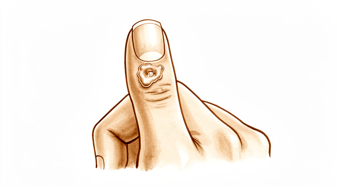
میوکس سسٹ (mucous cyst، سیال سے بھری تھیلی) ایک چھوٹی، سخت گانٹھ ہے جو انگلی کی پشت پر، عام طور پر ناخن کے بالکل پیچھے، ظاہر ہوتی ہے۔ یہ ایک گاڑھے، صاف سیال سے بھری ہوتی ہے۔ گانٹھ میں خود اکثر درد نہیں ہوتا، لیکن یہ آس پاس کے حصوں پر دباؤ ڈال کر تکلیف پیدا کر سکتی ہے۔
آپ کو خود ناخن میں بھی تبدیلیاں نظر آ سکتی ہیں۔ سسٹ ناخن کے نیچے کی جلد (nail bed) کے قریب ہوتا ہے، اس لیے بڑھتے ہوئے یہ ناخن پر دباؤ ڈال سکتا ہے۔ اس سے اکثر ناخن کی پوری لمبائی میں ایک نالی یا ابھری ہوئی لکیر بن جاتی ہے۔ سسٹ کے اوپر کی جلد بھی پتلی اور چمکدار ہو سکتی ہے۔ کچھ صورتوں میں جلد پھٹ جاتی ہے اور سسٹ کا سیال ایک چھوٹے سے راستے سے باہر رِسنے لگتا ہے۔
گانٹھ عموماً بالکل اسی جگہ ہوتی ہے جہاں ناخن سے پہلے والے آخری جوڑ پر آپ کی انگلی مڑتی ہے۔ اس سے روزمرہ کے کچھ کام مشکل ہو جاتے ہیں۔ تنگ جیب میں ہاتھ ڈالنا، مرتبان کا ڈھکن پکڑنا، یا چھوٹے سکے اٹھانا تکلیف دہ ہو سکتا ہے۔ ٹائپ کرنے یا لکھنے سے گانٹھ پر دباؤ پڑ سکتا ہے اور اس میں درد ہو سکتا ہے۔
یہ سسٹ اسی جوڑ میں جوڑ کے گھسنے والے آرتھرائٹس سے جڑا ہوتا ہے۔ ہڈی کا ایک چھوٹا سا نوکیلا ابھار، جسے آسٹیوفائٹ (osteophyte) کہتے ہیں، جوڑ میں جلن (irritation) پیدا کرتا ہے اور اسی سے وہ سیال بنتا ہے جو سسٹ کو بھرتا ہے۔ اسی وجہ سے، ہاتھ کو زیادہ استعمال کرنے کے بعد جوڑ میں دکھن ہو سکتی ہے، اور اس وقت گانٹھ کو چھونے پر زیادہ درد ہو سکتا ہے۔
اگر سسٹ کے اوپر کی جلد پھٹ جائے اور سیال باہر رِسنے لگے تو اس جگہ کو صاف رکھیں اور جلد از جلد ڈاکٹر کو دکھائیں۔ جلد سے انگلی کے جوڑ تک جانے والا راستہ انفیکشن کو اندر آنے دے سکتا ہے، اور جوڑ کے انفیکشن کا بلا تاخیر علاج ضروری ہے۔ اگر آپ کی انگلی گرم، سرخ، سوجی ہوئی اور دردناک ہو جائے، خاص طور پر بخار کے ساتھ، تو اسی دن ہسپتال کی ایمرجنسی جائیں۔ اس کے لیے فیملی ڈاکٹر (GP) کے ریفرل کی ضرورت نہیں ہے۔
خود سسٹ میں سوراخ نہ کریں اور نہ ہی اسے دبائیں۔ اس سے جوڑ میں انفیکشن داخل ہو سکتا ہے۔
اگر گانٹھ بڑھ رہی ہو، ناخن کی بگڑی ہوئی شکل آپ کو پریشان کر رہی ہو، یا علامات ہفتوں میں بہتر نہ ہو رہی ہوں، تو اپنے فیملی ڈاکٹر کو دکھائیں یا ماہر ڈاکٹر (specialist) سے معائنے کا کہیں۔
دراصل کیا ہو رہا ہے
آپ کی انگلی کا آخری جوڑ ایک چھوٹے قبضے (hinge) کی طرح کام کرتا ہے۔ کسی بھی جوڑ کی طرح، اس کے اندر ایک نرم غلاف کی تہہ ہوتی ہے جو تھوڑا سا سیال بناتی ہے تاکہ سطحیں آسانی سے پھسلتی رہیں۔ میوکس سسٹ میں اس جوڑ میں گھسنے والا آرتھرائٹس ہو چکا ہوتا ہے۔ جوڑ کی اندرونی تہہ میں جلن پیدا ہو جاتی ہے اور وہ جوڑ کی ضرورت سے زیادہ سیال بنانے لگتی ہے۔
اسی وقت آرتھرائٹس کی وجہ سے اکثر جوڑ کے کنارے پر ہڈی کا ایک چھوٹا سا نوکیلا ابھار بن جاتا ہے۔ یہ ابھار اندر سے جوڑ کے غلاف پر دباؤ ڈالتا ہے اور اسے گھسا کر اس میں سوراخ کر دیتا ہے۔ پھر سیال اس گھسی ہوئی جگہ سے رِس کر باہر نکلتا ہے اور آپ کی انگلی کی پشت پر جلد کے نیچے جمع ہو جاتا ہے۔ جو گانٹھ آپ دیکھ اور محسوس کر سکتے ہیں وہ یہی پھنسا ہوا سیال ہے، جو جوڑ کے ایک طرف، ناخن کے بالکل پیچھے ہوتا ہے۔
سسٹ بذاتِ خود کوئی رسولی نہیں بلکہ پتلی دیوار والی ایک تھیلی ہے۔ اس کی دیوار سہارا دینے والے ریشوں سے بنی ہوتی ہے، اور اس کی اپنی کوئی باقاعدہ اندرونی تہہ نہیں ہوتی۔ چونکہ دیوار پتلی ہوتی ہے اور اس کے اوپر کی جلد اکثر اس سے بھی پتلی ہوتی ہے، اس لیے ہلکی سی ٹھوکر سے یہ پھٹ سکتا ہے۔ جب ایسا ہوتا ہے تو اندر کا صاف، جیلی جیسا سیال باہر رِس آتا ہے۔
اس سے وہ علامات سمجھ میں آتی ہیں جن کے بارے میں آپ نے ابھی پڑھا۔ گانٹھ ناخن کے نیچے کی جلد کے قریب ہوتی ہے، اس لیے سسٹ کا دباؤ بڑھتے ہوئے ناخن میں نالی بنا سکتا ہے۔ جوڑ میں دکھن اس لیے ہوتی ہے کہ وہاں آرتھرائٹس بھی موجود ہے، اور سسٹ اسی کی علامت ہے۔ جوڑ کے کنارے کا ابھار ہی سیال کو بہتا رکھتا ہے، اس لیے جب تک یہ موجود ہے، سسٹ سے سیال رِس جانے یا اسے خالی کیے جانے کے بعد بھی یہ عموماً دوبارہ بھر جاتا ہے۔
اسی لیے علاج صرف گانٹھ کے بجائے جوڑ پر توجہ دیتا ہے۔ ابھار نکالنے سے رساؤ اپنے ذریعے پر ہی رک جاتا ہے، اور جب سسٹ میں مزید سیال نہیں آتا تو یہ ختم ہو جاتا ہے۔
ہم اس کے لیے کیا کر سکتے ہیں
ڈاکٹر کیرن ہِرپرا، جو Mater Private Hospital Rockhampton میں بازو اور ہاتھ کے سرجن (upper-limb surgeon) ہیں، آپ کی حالت کے مطابق سب سے کم چیر پھاڑ والے طریقوں سے آغاز کرتے ہیں۔ مریضوں کو عام طور پر ان کا فیملی ڈاکٹر (GP) ہمارے کلینک ریفر کرتا ہے؛ اگر کسی فزیوتھراپسٹ نے آپ کو ہمیں دکھانے کا مشورہ دیا ہے، تب بھی میڈیکیئر (Medicare) کی رقم کی واپسی (rebate) کا حق دار ہونے کے لیے آپ کو اپنے فیملی ڈاکٹر سے ریفرل (referral) لینا ہو گا۔ کلینک کی ملاقات پر ہم آپ سے آپ کی تکلیف کی تفصیل لیتے ہیں، آپ کی انگلی کا معائنہ کرتے ہیں اور اگر ضرورت ہو تو ایکسرے یا اسکین کرواتے ہیں۔ زیادہ تر میوکس سسٹ جوڑ کے گھسنے والے آرتھرائٹس سے جڑے ہوتے ہیں، اس لیے ہم عموماً پہلے آپریشن کے بغیر علاج آزماتے ہیں، اور جب اس سے کافی بہتری نہ آئے تو سرجری پر غور کرتے ہیں۔
پہلا قدم اکثر صرف نگرانی اور انتظار ہوتا ہے۔ کچھ سسٹ چھوٹے رہتے ہیں اور زیادہ تکلیف نہیں دیتے۔ انگلی کو بچانا مددگار ہے: گانٹھ پر بار بار ٹھوکر لگنے سے بچیں، اور ان کاموں میں احتیاط کریں جن سے اس پر دباؤ پڑتا ہو۔ اگر زیادہ استعمال کے بعد جوڑ میں دکھن ہو تو وہ کام کچھ کم کرنے سے آرام آ سکتا ہے۔ ہینڈ تھراپی (ہاتھ کی بحالی کا علاج) اکڑاؤ میں اور انگلی کو آرام سے حرکت میں رکھنے میں مدد دے سکتی ہے۔ مزید کسی فیصلے سے پہلے ان سادہ اقدامات کو کئی ہفتوں تک مناسب موقع دیں۔
درد سے آرام کا طریقہ سیدھا سادہ ہے۔ فارمیسی سے ملنے والی درد کی سادہ دوا، ہدایت کے مطابق لینے سے، جوڑ کی دکھن کو کم کر سکتی ہے۔ سوزش کم کرنے والی دوا سسٹ کے پیچھے موجود آرتھرائٹس میں آرام دے سکتی ہے۔ یہ دوائیں خود گانٹھ کا نہیں بلکہ تکلیف کا علاج کرتی ہیں۔
ایک طریقہ جو ہم پیش نہیں کرتے وہ سوئی سے سسٹ کا سیال نکالنا ہے۔ یہ کلینک میں آسانی سے ہو جانے والا طریقہ ہے، لیکن اس کے بعد تقریباً آدھی صورتوں میں سسٹ دوبارہ بن جاتا ہے۔ اس میں جوڑ کے اندر انفیکشن پہنچنے کا خطرہ بھی ہوتا ہے، اسی لیے ہم یہ مشورہ بھی دیتے ہیں کہ گھر پر بھی سسٹ میں سوراخ نہ کریں اور نہ اسے دبائیں۔
سرجری پر اس وقت غور کیا جاتا ہے جب سسٹ بار بار بھرتا رہے، ناخن کی بگڑی ہوئی شکل آپ کو پریشان کرے، یا سسٹ کے اوپر کی جلد پتلی ہو گئی ہو یا پھٹ رہی ہو۔ آپریشن صرف گانٹھ کے بجائے جوڑ کا علاج کرتا ہے۔ جوڑ کے کنارے پر ہڈی کا نوکیلا ابھار نکال دیا جاتا ہے، جس سے سیال کا رساؤ اپنے ذریعے پر ہی رک جاتا ہے، اور جب سسٹ میں مزید سیال نہیں آتا تو یہ ختم ہو جاتا ہے۔ کچھ صورتوں میں سسٹ اور آس پاس کی جلد کا ایک چھوٹا ٹکڑا (skin flap) بھی نکال دیا جاتا ہے، جو ایسے سسٹ کے لیے مناسب ہے جس نے اپنے اوپر کی جلد کو پتلا کر دیا ہو۔ ابھار کو سسٹ کی ڈنڈی کے ساتھ نکالنے سے سسٹ کے دوبارہ بننے کا امکان سب سے کم ہوتا ہے۔ ہم آپ سے بات کریں گے کہ آپ کی انگلی کے لیے کون سا طریقہ مناسب ہے، اور آپ کے ساتھ مل کر فیصلہ کریں گے۔
کیا توقع رکھیں
اگر اسے ایسے ہی چھوڑ دیا جائے تو میوکس سسٹ غائب ہونے کے بجائے عموماً بار بار واپس آتا رہتا ہے۔ جب تک جوڑ کے کنارے پر ہڈی کا ابھار موجود ہے، یہ سسٹ کو سیال سے بھرتا رہتا ہے۔ کچھ سسٹ لمبے عرصے تک چھوٹے رہتے ہیں اور زیادہ تکلیف نہیں دیتے۔ کچھ آہستہ آہستہ بڑھتے ہیں، ناخن کی نالی کو مزید گہرا کرتے ہیں، یا گانٹھ کے اوپر کی جلد کو اتنا پتلا کر دیتے ہیں کہ اس سے رساؤ شروع ہو جاتا ہے۔
علاج کے بغیر بنیادی خطرات وہی ہیں جن کے بارے میں آپ پہلے پڑھ چکے ہیں: جب تک سسٹ ناخن پر دباؤ ڈالتا رہے، ناخن پر ابھری ہوئی لکیر برقرار رہتی ہے، اور جلد پھٹ کر جوڑ تک ایک راستہ کھول سکتی ہے۔ اس راستے کو سنجیدگی سے لینا چاہیے۔ اگر آپ کی انگلی گرم، سرخ، سوجی ہوئی اور دردناک ہو جائے، خاص طور پر بخار کے ساتھ، تو اسی دن ہسپتال کی ایمرجنسی جائیں۔ اس کے لیے فیملی ڈاکٹر کے ریفرل کی ضرورت نہیں ہے۔
جوڑ پر مرکوز علاج کے ساتھ نتیجہ واضح ہے۔ ابھار نکالنے سے سیال کا رساؤ اپنے ذریعے پر ہی رک جاتا ہے، اور جب سسٹ میں مزید سیال نہیں آتا تو یہ ختم ہو جاتا ہے۔ جب ابھار نکال دیا جائے تو سسٹ بہت ہی کم دوبارہ بنتا ہے۔ جہاں سسٹ کو بھی نکالا جائے، وہاں بھی دوبارہ بننے کی شرح کم رہتی ہے: سرجری سے علاج کیے گئے ہر 100 میں سے تقریباً 2 سسٹ دوبارہ بنتے ہیں، اور جب اس جگہ کو بند کرنے کے لیے آس پاس کی جلد کا ٹکڑا (flap) استعمال کیا جائے تو ہر 100 میں سے تقریباً 1.4۔ اس طریقے سے علاج کروانے والے لوگ عموماً زخم کے نشان کی شکل سے مطمئن رہتے ہیں، اور دوبارہ یہ علاج کروانے کو تیار ہوتے ہیں۔
صحت یابی کوئی بہت بڑا مرحلہ نہیں، لیکن اسے معمولی بھی نہ سمجھیں۔ انگلی اکثر چند ہفتوں تک اکڑی ہوئی رہتی ہے اور اسے چھونے پر درد ہوتا ہے، اور گرفت یا چٹکی کی گرفت جیسے روزمرہ کے کاموں کو دوبارہ معمول کے مطابق محسوس ہونے میں کچھ وقت لگتا ہے۔ سرجری کے بعد ہینڈ تھراپی Extend Rehabilitation میں Ruby Doolan کے ساتھ ہوتی ہے۔ وہ ہینڈ تھراپسٹ (ہاتھ کی بحالی کی ماہر) ہیں: وہ آپ کی ورزشوں میں رہنمائی کرتی ہیں اور آپ کو جس اسپلنٹ کی بھی ضرورت ہو وہ بناتی ہیں۔ زیادہ تر لوگ چند ہفتوں سے چند مہینوں کے اندر ہاتھ کا معمول کا استعمال دوبارہ شروع کر دیتے ہیں۔
ناخن کے نیچے کی جلد پر سے دباؤ ہٹنے کے بعد ناخن کی نالی بھی عموماً بہتر ہو جاتی ہے۔ اس معاملے میں صبر سے کام لیں: ناخن کے بگڑے ہوئے حصے کو بڑھ کر باہر نکلنا ہوتا ہے، جس میں کئی مہینے لگتے ہیں۔
اگر علامات ہفتوں میں بہتر نہ ہو رہی ہوں، یا بگڑ رہی ہوں، تو اپنے فیملی ڈاکٹر کو دکھائیں یا ماہر ڈاکٹر سے معائنے کا کہیں۔
ڈاکٹر کو کب دکھائیں
زیادہ تر میوکس سسٹ ہنگامی نوعیت کے نہیں ہوتے، اور بہت سے سادہ دیکھ بھال سے ٹھیک ہو جاتے ہیں۔ البتہ چند علامات پر کارروائی ضروری ہے۔
اگر آپ کی انگلی گرم، سرخ، سوجی ہوئی اور دردناک ہو جائے، خاص طور پر بخار کے ساتھ، تو اسی دن ہسپتال کی ایمرجنسی جائیں۔ یہ جوڑ میں انفیکشن کی طرف اشارہ ہے، جس کا بلا تاخیر علاج ضروری ہے۔ یہی بات اس صورت میں بھی لاگو ہوتی ہے اگر سسٹ کے اوپر کی جلد پھٹ گئی ہو اور سیال رِس رہا ہو، کیونکہ یہ سوراخ انفیکشن کو اندر آنے دے سکتا ہے۔
اگر گانٹھ بڑھتی رہے، ناخن کی نالی آپ کو پریشان کر رہی ہو، یا سسٹ کے اوپر کی جلد پتلی ہو گئی ہو تو اپنے فیملی ڈاکٹر کو دکھائیں یا ماہر ڈاکٹر سے معائنے کا کہیں۔ یہی وہ علامات ہیں جن پر علاج کے بارے میں بات کرنا مفید ہے۔ اگر علامات ہفتوں میں بہتر نہ ہو رہی ہوں، یا بگڑ رہی ہوں، تو آپ کے فیملی ڈاکٹر ریفرل کا انتظام کر سکتے ہیں۔
مزید تفصیل
Advanced reading: the deeper science (optional)
یہ حصہ اس سے زیادہ تفصیل میں جاتا ہے جتنی آپ کو اپنے علاج کے فیصلوں کے لیے ضرورت ہے۔ میوکس سسٹ کے بارے میں اضافی مطالعہ ایک چھوٹی مگر مضبوط شواہد پر مبنی سرجیکل بصیرت کی وجہ سے فائدہ مند ہے: اصل مسئلہ سسٹ نہیں ہے، اور جو آپریشن سب سے بہتر کام کرتا ہے اس میں ضروری نہیں کہ سسٹ کو نکالا جائے۔
اصل وجہ ہڈی کا ابھار ہے، سسٹ نہیں
میوکس سسٹ انگلی کے سرے کے آرتھرائٹس والے جوڑ سے پیدا ہوتا ہے۔ ہڈی کا ایک نوکیلا ابھار، یعنی آسٹیوفائٹ (osteophyte)، جوڑ کی جھلی میں جلن (irritation) پیدا کرتا ہے اور اس میں سوراخ کر دیتا ہے، اور جوڑ کا سیال اس سوراخ سے باہر نکل کر جلد کے نیچے جمع ہو جاتا ہے۔ سسٹ اس عمل کا نظر آنے والا سرا ہے، اس کی شروعات نہیں۔
اس سمجھ کا سرجری پر براہِ راست اثر ہے، جو ایک تحقیق میں سامنے آیا جہاں سسٹ نکالے بغیر صرف آسٹیوفائٹ نکالنے سے زیادہ تر صورتوں میں مسئلہ مکمل طور پر ختم ہو گیا، اور اسے کم چیر پھاڑ والا ایک اچھا علاج قرار دیا گیا [1]۔
ابھار نکالنا نل بند کرنے جیسا ہے۔ سسٹ میں جب سیال آنا بند ہو جائے تو یہ ختم ہو جاتا ہے۔ یہی منطق کلائی کے گینگلیئن سسٹ پر بھی لاگو ہوتی ہے، جہاں سسٹ کا دوبارہ بننا تھیلی سے نہیں بلکہ اس کی ڈنڈی سے طے ہوتا ہے، اور اسی سے یہ بھی واضح ہوتا ہے کہ میوکس سسٹ کو صرف نکاس کرنے یا سوئی سے چھیدنے کا طریقہ اتنی باقاعدگی سے ناکام کیوں ہوتا ہے۔
جہاں سسٹ نکالا جاتا ہے، وہاں بھی نتائج اچھے ہیں
دوسرے طریقے میں سسٹ کو نکالا جاتا ہے اور خالی جگہ کو بند کرنے کے لیے ساتھ کی جلد کا ایک ٹکڑا (skin flap) کھسکا کر استعمال کیا جاتا ہے۔ یہ بھی قابلِ بھروسہ ہے: 69 مریضوں میں، آس پاس کی جلد کو آگے کھسکا کر (local advancement flap) سسٹ نکالنے کے آپریشن میں دوبارہ بننے کی شرح 1.4 فیصد تھی، اور مریض زخم کے نشان سے بہت مطمئن تھے اور دوبارہ یہ آپریشن کروانے پر آمادہ تھے [2]۔
دونوں طریقے کام کرتے ہیں، اور دونوں نیچے موجود جوڑ کا علاج کرتے ہیں۔ عملی فرق یہ ہے کہ جلد کتنی متاثر ہے: پرانا سسٹ اپنے اوپر کی جلد کو پتلا کر دیتا ہے، کبھی کبھی اس حد تک کہ اس سے رطوبت رسنے لگتی ہے، اور ایسی صورت میں پتلی جلد کو نکال کر اس کی جگہ نئی جلد لگانی پڑتی ہے، چاہے ہڈی کے ساتھ کچھ بھی کیا جائے۔
ناخن کی شکل کیوں بگڑتی ہے، اور کیا یہ ٹھیک ہو جاتا ہے
ناخن کی پوری لمبائی میں چلنے والی نالی یا ابھری ہوئی لکیر اکثر ساتھ ہوتی ہے، اور یہ لوگوں کو گانٹھ سے زیادہ پریشان کرتی ہے۔ اس کی وجہ میکانکی ہے: سسٹ بالکل ناخن کی جڑ کے اس حصے کے اوپر ہوتا ہے جو ناخن بناتا ہے (germinal matrix)، اور اس پر دباؤ ڈالتا ہے، اس لیے ناخن ایک نقص کے ساتھ بنتا ہے۔
فائدہ مند بات یہ ہے کہ یہ دباؤ ہے، تباہی نہیں۔ سسٹ کا دباؤ ختم ہونے کے بعد ناخن عموماً معمول کے مطابق بڑھتا ہے، اگرچہ بگڑے ہوئے حصے کو بڑھ کر سرے سے باہر نکلنے میں کئی مہینے لگتے ہیں۔ اس لیے ناخن کا بگاڑ سسٹ کا علاج کرنے کی ایک وجہ ہے، اس کا مستقل نتیجہ نہیں۔
اسے چھیدنے کے بارے میں محتاط رہنے کی وجہ
میوکس سسٹ کا جوڑ سے براہِ راست رابطہ ہوتا ہے۔ اسے چھیدنا، چاہے جان بوجھ کر ہو یا کسی بڑے سسٹ کے اوپر کی جلد پھٹنے سے، باہر کی دنیا سے انگلی کے جوڑ تک ایک راستہ بنا دیتا ہے، اور کسی چھوٹے جوڑ کا انفیکشن (septic arthritis) سسٹ کے مقابلے میں کہیں زیادہ سنگین مسئلہ ہے۔
یہی عملی دلیل ہے کہ ایسے سسٹ کو جو بظاہر سیال کا ایک چھالا لگے، گھر پر خود نہ نکالا جائے، اور یہی وجہ ہے کہ جس سسٹ سے خود بخود رطوبت رسنے لگے اس کا علاج صرف نگرانی کے بجائے کچھ جلدی کے ساتھ کیا جاتا ہے۔
حوالہ جات
[1] Lee H, Kim P, Jeon I, Kyung H, Ra I, Kim T. Osteophyte excision without cyst excision for a mucous cyst of the finger. J Hand Surg Eur Vol. 2013;39(3):258-61. https://doi.org/10.1177/1753193413478549
[2] Johnson SM, Treon K, Thomas S, Cox QGN. A reliable surgical treatment for digital mucous cysts. J Hand Surg Eur Vol. 2013;39(8):856-60. https://doi.org/10.1177/1753193413508540
Evidence & references
This is the clinical evidence summary written for health professionals. It is technical, and it lists the research this page was built from. You do not need to read it to understand your treatment or to make a decision about it.
Overview
- The scientific data regarding mucous cysts consist almost entirely of retrospective studies, and much of what is done or recommended is based on expert opinion [3].
- Surgical treatment of mucous cysts is not a totally benign procedure, and complications do occur contrary to literature reporting glowing results [5].
- In-office excision is a safe and effective option for treating digital mucous cysts [6].
- Complications are rare and occur only in cysts associated with fistula, justifying their early surgical treatment [7].
- Surgery provided definitive treatment with no major long-term problems [10].
- Surgical excision was the primary treatment, with no reported recurrences [11].
- Incomplete excisions will invariably result in recurrence [12].
- Surgical excision with a local advancement skin flap is a reliable treatment for digital mucous cysts, demonstrating a low recurrence rate of 1.4% and high patient satisfaction regarding the scar and willingness to undergo the procedure again [13].
- The 2% recurrence rate (2 of 113) obtained in this series illustrates the advantage of this procedure [16].
- Osteophyte excision without cyst excision may be a good treatment choice for mucous cyst of the finger, providing a less invasive method with complete resolution in most cases [20].
- The final recurrence rate of less than 1.5% may be acceptable and comparable with other techniques [21].
- Excision with local flap cover offers benefit in terms of a reduced recurrence rate and is particularly valuable where the cyst has caused marked thinning of the skin [30].
- Excision with local flap cover would seem to offer benefit in terms of a reduced recurrence rate, and is particularly valuable where the cyst has caused marked thinning of the skin [34].
- Excision of the cyst and complete removal of the marginal osteophyte eradicates mucous cysts with extremely rare recurrence [1].
- A total dorsal capsulectomy alone was a simple treatment for mucous cysts and did not lead to any recurrence [8].
Anatomy & Pathophysiology
Clinical Presentation and Location
- Mucous cysts are small, firm, cystic masses that appear just distal to the distal interphalangeal joint [23].
- The lesion is always located to one side of the midline [23].
- Mucous cysts are rarely greater than fifteen millimeters in diameter [23].
- The skin over the mucous cyst is thinned out and occasionally may be ulcerated [23].
- Pressure on the mucous cyst does not usually result in a decrease in its size [23].
- Longitudinal grooving of the nail may be noted, occasionally occurring prior to the appearance of the cyst [23].
- Mucous cysts are usually small and located to one side as they emerge from the joint beside the extensor mechanism [28, 29].
- Occasionally, mucous cysts may spread across the whole of the dorsum of the digit between the distal interphalangeal joint and nail fold [28, 29].
- The overlying skin of a mucous cyst is frequently thin, and minimal trauma may result in rupture [28, 29].
Histology and Ultrastructure
- The histological appearance of a mucous cyst is essentially identical to that of ganglions, synovial cysts, and similar lesions appearing on other areas of the extremities [23].
- The mucous cyst is surrounded by an undemarcated fibrous capsule [23].
- The basic structure of the mucous cyst is myxomatous with interspersed fibroblasts [23].
- Areas of myxomatous degeneration tend to coalesce to form a multiloculated cyst [23].
- An epithelial lining has not been reported for mucous cysts [23].
- Digital mucous cysts and wrist ganglia show no anatomic difference at the light and scanning electron microscopic levels [19].
- The wall of a digital mucous cyst consists of multidirectional strata of collagen fibers with rare fibroblast-like cells on the inner surface [19].
- There are no signs of myxomatous degeneration in the wall of a digital mucous cyst [19].
- The surface of the digital mucous cyst wall is generally flat, with some areas showing localized elevations [19].
- Multifunctional mesenchymal cells are located beneath the lining of the ganglion and digital mucous cyst [19].
- These multifunctional mesenchymal cells contain abundant rough endoplasmic reticulum, smooth-walled vacuoles, and Golgi complexes [19].
- The ganglion fluid may be produced by these multifunctional cells and reaches the ganglion cavity by flowing through the porous collagen matrix of the ganglion wall [19].
- A digital mucous cyst has a large globular dome-shaped interconnecting cavity having a wall of collagen fibers covered by a fenestrated pseudomembrane [19].
- A porous network of collagen fibers is present in the wall of the digital mucous cyst on cross section [19].
Etiology and Pathogenesis
- Degenerative arthritis of the distal interphalangeal joint is a frequent associated finding with mucous cysts [23].
- An incidence of 78 percent for degenerative arthritis of the distal interphalangeal joint has been reported in association with mucous cysts [23].
- The etiology indicated by evidence is that the lesion arises from the joint capsule [23].
- Mucous cysts are often associated with degenerative changes at the distal interphalangeal joints [28, 29].
- Theories on the pathogenesis of mucous cysts include myxoid degeneration, repeated local trauma, vascular insufficiency, and synovial herniation [28, 29].
- Studies suggest that mucous cysts arise due to excessive hyaluronic acid production from a degenerate distal interphalangeal joint [28, 29].
Classification
- The lesion is a small, firm, cystic mass which appears just distal to the distal interphalangeal joint [23].
- The lesion is always located to one side of the mid-line [23].
- The lesion is rarely greater than fifteen millimeters in diameter [23].
- The skin over the lesion is thinned out and occasionally may be ulcerated [23].
- Pressure on the lesion does not usually result in a decrease in its size [23].
- Degenerative arthritis of the distal interphalangeal joint is a frequent associated finding [23].
- An incidence of 78 per cent has been reported for degenerative arthritis of the distal interphalangeal joint associated with mucous cysts [23].
- The histological appearance is essentially identical to that of ganglions, synovial cysts, and similar lesions appearing on other areas of the extremities [23].
- The lesion is surrounded by an undemarcated fibrous capsule [23].
- The basic structure is myxomatous with interspersed fibroblasts [23].
- An epithelial lining has not been reported [23].
- In spite of their different clinical settings, digital mucous cysts and wrist ganglia show no anatomic difference at the light and scanning electron microscopic levels [19].
- The wall of the digital mucous cyst consisted of multidirectional strata of collagen fibers with rare fibroblast-like cells on the inner surface [19].
- There were no signs of myxomatous degeneration in the wall of the digital mucous cyst [19].
- The surface of the digital mucous cyst was generally flat, with some areas showing localized elevations [19].
- Multifunctional mesenchymal cells beneath the lining of the ganglion are postulated to produce collagen fibers, elastic fibers, and the interfibrillary mucopolysaccharide matrix [19].
- The porous nature of the ganglion wall was confirmed in the ultrastructural study of digital mucous cysts [19].
- A previous ultrastructural study of digital mucous cysts demonstrated a large globular dome-shaped interconnecting cavity having a wall of collagen fibers covered by a fenestrated pseudomembrane [19].
- The current study confirmed the anatomic organization of a large globular dome-shaped interconnecting cavity having a wall of collagen fibers covered by a fenestrated pseudomembrane [19].
- A porous network of collagen fibers in the wall was revealed on cross section [19].
Clinical Presentation
- The mucous cyst presents as a small, firm, cystic mass located just distal to the distal interphalangeal joint [23].
- The lesion is easily distinguished from a Heberden's node by its placement distal to the distal interphalangeal joint [23].
- The histological appearance of the mucous cyst is essentially identical to that of ganglions, synovial cysts, and similar lesions appearing on other areas of the extremities [23].
- The basic structure of the lesion is myxomatous with interspersed fibroblasts [23].
- An epithelial lining has not been reported for the mucous cyst [23].
- The wall of the digital mucous cyst consists of a porous network of collagen fibers [19].
- The fluid within the digital mucous cyst is produced by underlying mesenchymal cells [19].
Investigations
- Ultrasound is a powerful modality for evaluation of pathologic conditions in the hand and wrist, providing a cost-effective and expedient alternative and/or adjunct to MRI [33].
- Ultrasound is best used when there is a specific clinical question regarding a well-localized abnormality [33].
- Ultra-sonography provides a new ability to study the complex anatomical structures of the hand, with the technique being quick and simple to perform [38].
- A careful physical examination is essential to direct care and future testing if indicated [35].
- Diagnostic tests such as imaging and serum laboratory studies are useful in this determination but can be expensive, time consuming, and often nonspecific [35].
Treatment
Operative Techniques
- Total dorsal capsulectomy alone is a simple treatment for mucous cysts that did not lead to any recurrence [8].
- Osteophyte-sparing treatment of mucous cysts has a final recurrence rate of less than 1.5% [21].
- Surgical excision was the primary treatment in a series with no reported recurrences [11].
- Surgery provided definitive treatment for mucous cysts with no major long-term problems [10].
- The 2% recurrence rate (2 of 113) obtained in a series of 191 excised cysts illustrates the advantage of the procedure [4, 16].
- The authors of a study on osteophyte excision without cyst excision state that their principle is not to do more damage to the friable skin because the skin is not the culprit of the cyst [15].
Outcomes and Complications
- Complications of surgical treatments include infection, recurrence, nail deformity, swelling, stiffness and pain [28].
- This report provides additional basis for prophylactic extirpation of mucous cyst of the finger [2].
- This case emphasizes the utility of a pathohistological analysis in cases where doubts arise about the initial diagnosis of a benign tumorous lesion [14].
Non-Operative and Adjunctive Considerations
- Aspiration with injection of steroid is a convenient technique that can be performed in an outpatient setting but is followed by a recurrence rate of 50% [28].
- Surgical treatment has a higher cure rate than aspiration with injection of steroid [28].
- Radical treatment with complete excision of the stalk and underlying osteophytes seems to reduce recurrence rates [28].
Complications
- Complications are rare and occur only in cysts associated with fistula [7].
- Aspiration with injection of steroid is followed by a recurrence rate of 50% [28].
- The 2% recurrence rate (2 of 113) was obtained in a series of surgically treated mucous cysts [16].
- Surgical excision with a local advancement skin flap demonstrated a low recurrence rate of 1.4% [13].
- A total dorsal capsulectomy alone did not lead to any recurrence [8].
- Eccrine porocarcinomas have a substantial risk of metastasis, high risk of local recurrence, and are potentially fatal [24].
Recovery
- Surgical excision with a local advancement skin flap demonstrated a low recurrence rate of 1.4% and high patient satisfaction regarding the scar and willingness to undergo the procedure again [13].
Key Evidence
- [L4] Excision of the cyst and complete removal of the marginal osteophyte eradicates mucous cysts with extremely rare recurrence. [1] (10.2106/00004623-197355030-00013)
- [L4] This report provides additional basis for prophylactic extirpation of mucous cyst of the finger. [2] (10.1016/s0363-5023(84)80152-5)
- [L4] The scientific data regarding mucous cysts consist almost entirely of retrospective studies, and much of what is done or recommended is based on expert opinion. [3] (10.1016/j.jhsa.2010.01.029)
- [L5] Of the 191 mucous cysts excised, 113 had at least 6 months of follow-up time. [4] (10.1016/j.jhsa.2010.07.028)
- [L4] Surgical treatment of mucous cysts is not a totally benign procedure, and complications do occur contrary to literature reporting glowing results. [5] (10.1016/s0266-7681(97)80067-8)
- [Paper] In-office excision is a safe and effective option for treating digital mucous cysts. [6] (10.1177/15589447251350168)
- [L4] Complications are rare and occur only in cysts associated with fistula, justifying their early surgical treatment. [7] (10.1016/j.main.2015.06.001)
- [L4] A total dorsal capsulectomy alone was a simple treatment for mucous cysts and did not lead to any recurrence. [8] (10.1016/j.jhsa.2014.03.004)
- [L4] Surgery provided definitive treatment with no major long-term problems. [10] (10.1053/jhsu.2003.50088)
- [L5] Incomplete excisions will invariably result in recurrence. [12] (10.1016/0363-5023(93)90302-j)
- [L4] Surgical excision with a local advancement skin flap is a reliable treatment for digital mucous cysts, demonstrating a low recurrence rate of 1.4% and high patient satisfaction regarding the scar and willingness to undergo the procedure again. [13] (10.1177/1753193413508540)
- [L5] This case emphasizes the utility of a pathohistological analysis in cases where doubts arise about the initial diagnosis of a benign tumorous lesion. [14] (10.1007/s00402-008-0794-4)
- [L5] The authors state that their principle is not to do more damage to the friable skin because the skin is not the culprit of the cyst. [15] (10.1177/1753193414546990)
- [L4] The 2% recurrence rate (2 of 113) obtained in this series illustrates the advantage of this procedure. [16] (10.1016/0363-5023(94)90071-x)
- [L5] [19] (10.1016/s0363-5023(88)80143-6)
- [L4] Osteophyte excision without cyst excision may be a good treatment choice for mucous cyst of the finger, providing a less invasive method with complete resolution in most cases. [20] (10.1177/1753193413478549)
- [L4] The final recurrence rate of less than 1.5% may be acceptable and comparable with other techniques. [21] (10.5435/jaaosglobal-d-21-00164)
- [L4] [23] (10.2106/00004623-197254070-00008)
- [L4] Prompt recognition and appropriate treatment are critical because eccrine porocarcinomas have a substantial risk of metastasis, high risk of local recurrence, and are potentially fatal. [24] (10.1016/j.jhsa.2016.07.112)
- [L4] [28] (10.1177/1753193408103498)
- [L4] [29] (10.1177/1753193409103498)
- [L4] Excision with local flap cover offers benefit in terms of a reduced recurrence rate and is particularly valuable where the cyst has caused marked thinning of the skin. [30] (10.1016/0266-7681(90)90064-b)
- [L5] Ultrasound is a powerful modality for evaluation of pathologic conditions in the hand and wrist, providing a cost-effective and expedient alternative and/or adjunct to MRI, best used when there is a specific clinical question regarding a well-localized abnormality. [33] (10.1016/j.jhsa.2009.02.010)
- [L4] Excision with local flap cover would seem to offer benefit in terms of a reduced recurrence rate, and is particularly valuable where the cyst has caused marked thinning of the skin. [34] (10.1016/0266-7681_90_90064-b)
- [L4] Ultra-sonography provides a new ability to study the complex anatomical structures of the hand, with the technique being quick and simple to perform. [38] (10.1016/0266-7681(90)90133-o)
References
[1] Marginal Osteophyte Excision in Treatment of Mucous Cysts. The Journal of Bone & Joint Surgery. 1973. DOI: 10.2106/00004623-197355030-00013
[2] Infected mucous cyst of the finger. The Journal of Hand Surgery. 1984. DOI: 10.1016/s0363-5023(84)80152-5
[3] Mucous Cysts. The Journal of Hand Surgery. 2010. DOI: 10.1016/j.jhsa.2010.01.029
[4] Mucous Cysts. The Journal of Hand Surgery. 2010. DOI: 10.1016/j.jhsa.2010.07.028
[5] Complications Following Mucous Cyst Excision. Journal of Hand Surgery. 1997. DOI: 10.1016/s0266-7681(97)80067-8
[6] Safety and Cost-Effectiveness of In-Office Digital Mucous Cyst Excisions. HAND. 2025. DOI: 10.1177/15589447251350168
[7] Surgical treatment of mucous cysts by subcutaneous excision and osteophyte resection: Results in 68 cases at a mean 6.63 years’ follow-up. Chirurgie de la Main. 2015. DOI: 10.1016/j.main.2015.06.001
[8] Total Dorsal Capsulectomy for the Treatment of Mucous Cysts. The Journal of Hand Surgery. 2014. DOI: 10.1016/j.jhsa.2014.03.004
[10] Treatment of mucous cysts of the fingers: Review of 134 cases with minimum 2-year follow-up evaluation. The Journal of Hand Surgery. 2003. DOI: 10.1053/jhsu.2003.50088
[11] 10.1177-15589447261433068. n.d..
[12] Juvenile hyaline fibromatosis of the hand in an adult. The Journal of Hand Surgery. 1993. DOI: 10.1016/0363-5023(93)90302-j
[13] A reliable surgical treatment for digital mucous cysts. Journal of Hand Surgery (European Volume). 2013. DOI: 10.1177/1753193413508540
[14] Malignant Natural-Killer cell neoplasm presenting as a mucous cyst on the distal interphalangeal joint of the finger. Archives of Orthopaedic and Trauma Surgery. 2008. DOI: 10.1007/s00402-008-0794-4
[15] Re: Lee, H.-J., Kim, P.-T., Jeon, I.-H., Kyung, H.-S., Ra, I.-H. and Kim, T.-K. Osteophyte excision without cyst excision for a mucous cyst of the finger. J Hand Surg Eur. 2014, 39: 258–61. Journal of Hand Surgery (European Volume). 2014. DOI: 10.1177/1753193414546990
[16] Outcome of surgically treated mucous cysts of the hand. The Journal of Hand Surgery. 1994. DOI: 10.1016/0363-5023(94)90071-x
[19] A surface ultrastructure study of ganglia and digital mucous cysts. The Journal of Hand Surgery. 1988. DOI: 10.1016/s0363-5023(88)80143-6
[20] Osteophyte excision without cyst excision for a mucous cyst of the finger. Journal of Hand Surgery (European Volume). 2013. DOI: 10.1177/1753193413478549
[21] Osteophyte-Sparing Treatment of Mucous Cysts: Case Analysis and Surgical Technique. JAAOS: Global Research and Reviews. 2021. DOI: 10.5435/jaaosglobal-d-21-00164
[23] Etiology and Treatment of the So-Called Mucous Cyst of the Finger. The Journal of Bone & Joint Surgery. 1972. DOI: 10.2106/00004623-197254070-00008
[24] Eccrine Porocarcinoma Presenting as a Hand Cyst. The Journal of Hand Surgery. 2016. DOI: 10.1016/j.jhsa.2016.07.112
[28] Use of Wolfe Graft for the Treatment of Mucous Cysts. Journal of Hand Surgery (European Volume). 2009. DOI: 10.1177/1753193408103498
[29] Use of Wolfe Graft for the Treatment of Mucous Cysts. Journal of Hand Surgery (European Volume). 2009. DOI: 10.1177/1753193409103498
[30] Mucous cyst of the distal interphalangeal joint: Treatment by simple excision or excision and rotation flap. The Journal of Hand Surgery: Journal of the British Society for Surgery of the Hand. 1990. DOI: 10.1016/0266-7681(90)90064-b
[33] Ultrasound of the Hand and Wrist. The Journal of Hand Surgery. 2009. DOI: 10.1016/j.jhsa.2009.02.010
[34] Mucous Cyst of the Distal Interphalangeal Joint: Treatment by Simple Excision or Excision and Rotation Flap. Journal of Hand Surgery. 1990. DOI: 10.1016/0266-7681_90_90064-b
[35] Orthopaedic Knowledge Update 13 Ebook Without Multimedia. Anatomy, Evaluation, Clinical Examination, and Imaging > Evaluation and Clinical Examination: Current Concepts.
[38] Diagnostic medical ultrasound in the management of hand injuries. The Journal of Hand Surgery: Journal of the British Society for Surgery of the Hand. 1990. DOI: 10.1016/0266-7681(90)90133-o