Patients › Shoulder
کف آرتھروپیتھی
Rotator cuff arthropathy: shoulder arthritis following a long-standing, massive rotator cuff tear and its impact on function.
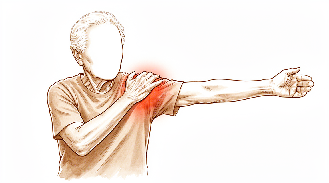
آپ کو کیا محسوس ہوتا ہے
کف آرتھروپیتھی (cuff arthropathy) کندھے کا وہ آرتھرائٹس (جوڑ کی خرابی اور سوزش) ہے جو روٹیٹر کف کے لمبے عرصے سے پھٹے رہنے کے بعد پیدا ہوتا ہے۔ روٹیٹر کف (rotator cuff) ٹینڈنوں (tendons)، یعنی پٹھے کو ہڈی سے جوڑنے والی مضبوط ڈوریوں، کا وہ گروپ ہے جو بازو اٹھاتے وقت کندھے کے جوڑ کے گولے کو درمیان میں تھامے رکھتا ہے۔ جب یہ ٹینڈن ختم ہو جاتے ہیں تو جوڑ اس طرح گھسنے لگتا ہے جس کے لیے وہ کبھی بنا ہی نہیں تھا، اور یہی گھساؤ وہ چیز ہے جو آپ محسوس کر رہے ہیں۔
درد عام طور پر خود کندھے کے اندر گہرائی میں، اس جگہ ہوتا ہے جہاں گولا پیالے سے ملتا ہے۔ یہ اکثر بازو کے اوپری حصے کی باہر والی طرف بھی محسوس ہوتا ہے۔ بازو کو سر سے اوپر اٹھانا، کسی شیلف تک ہاتھ بڑھانا، یا اس طرف سودا سلف اٹھا کر لے جانا، سب اسے بڑھا دیتے ہیں۔ بہت سے لوگوں کو رات کو درد بھڑکتا ہے، اور اس کندھے پر لیٹنے سے آپ کی آنکھ کھل سکتی ہے۔ اٹھ کر چلنے پھرنے کے بعد درد اکثر کچھ کم ہو جاتا ہے، پھر مصروف دن کے بعد دوبارہ بڑھ جاتا ہے۔
چونکہ کف اب اپنا کام نہیں کر رہا، اس لیے آپ کے بازو کی حرکت کا انداز بھی بدل جاتا ہے۔ ہو سکتا ہے آپ صرف درد کے بجائے کمزوری بھی محسوس کریں: بازو کو کندھے کی اونچائی تک یا اس سے اوپر اٹھانا مشکل ہو جاتا ہے، چاہے اسے جھلا کر اوپر لے جانے کی مدد بھی لی جائے۔ پیچھے ہاتھ لے جا کر ہار کا ہک لگانا، دھلے کپڑے لٹکانا، یا سویٹر سر کے اوپر سے پہننا عجیب یا ناممکن محسوس ہو سکتا ہے۔ کچھ لوگوں میں کندھے کے اوپری حصے کے قریب ایک ہموار، گول ابھار بن جاتا ہے جہاں گولا اوپر چڑھ گیا ہوتا ہے۔
ہفتوں اور مہینوں کے دوران یہ مسائل عموماً ایک دم ظاہر ہونے کے بجائے آہستہ آہستہ بڑھتے ہیں۔ سیٹ بیلٹ کی طرف ہاتھ بڑھانا، کیتلی سے پانی انڈیلنا، یا کوٹ پہننا جیسے سادہ کام دن کا سب سے مشکل حصہ بن سکتے ہیں۔
کچھ علامات میں فوری طبی امداد ضروری ہے۔ اگر آپ کا کندھا گرم، سرخ، سوجا ہوا اور دردناک ہو جائے، خاص طور پر بخار کے ساتھ، تو اسی دن ہسپتال کی ایمرجنسی جائیں۔ اگر آپ کا بازو سفید پڑنے لگے، ٹھنڈا ہو جائے، بالکل سفید یا نیلا ہو جائے، یا اچانک اس میں احساس یا حرکت ختم ہو جائے، تو بھی اسی دن ہسپتال کی ایمرجنسی جائیں۔ اگر آپ کی علامات کم نہ ہو رہی ہوں، ہفتوں کے دوران بڑھ رہی ہوں، یا آپ کو رات کو جگا رہی ہوں، تو اپنے فیملی ڈاکٹر (GP) سے ملیں یا ہم سے ماہر کے معائنے کا کہیں۔
دراصل کیا ہو رہا ہے
صحت مند کندھا ایسے کام کرتا ہے جیسے ایک گولا کم گہرے پیالے میں رکھا ہو۔ روٹیٹر کف کے ٹینڈن گولے کے گرد لپٹے ہوتے ہیں اور بازو اٹھاتے وقت اسے درمیان میں تھامے رکھتے ہیں۔ انہیں خیمے کی ان رسیوں کی طرح سمجھیں جو خیمے کے بانس کو سیدھا کھڑا رکھتی ہیں۔ جب یہ رسیاں نہ رہیں تو بانس جھک جاتا ہے۔
آپ کے کندھے میں، کندھے کی باہر والی طرف موجود مضبوط ڈیلٹائیڈ (deltoid) پٹھا بازو اٹھاتے وقت اب بھی کھینچتا ہے۔ گولے کو نیچے اور درمیان میں تھامنے کے لیے کف نہ ہونے کی وجہ سے یہ کھنچاؤ گولے کو اوپر کی طرف کھینچ لیتا ہے۔ پھر گولا اپنے اوپر والی ہڈی، ایکرومین (acromion)، سے رگڑ کھاتا ہے، جو آپ کے کندھے کے اوپر ہڈی کا چھجا ہے۔ یہ دونوں سطحیں کبھی ایک دوسرے کو چھونے کے لیے نہیں بنی تھیں، اس لیے یہ ایک دوسرے کو گھسا دیتی ہیں۔ یہی گھساؤ آرتھرائٹس ہے، اور اسی وجہ سے آپ کا درد جوڑ میں گہرائی میں ہوتا ہے اور اسی وجہ سے گولا اوپر چڑھ کر وہ گول ابھار بنا دیتا ہے جو شاید آپ نے محسوس کیا ہو۔
یہی گھساؤ کمزوری کی وضاحت بھی کرتا ہے۔ بازو اٹھانا اس بات پر منحصر ہے کہ گولا درمیان میں رہے، اور آپ کا گولا اب ایسا نہیں رہتا۔ اس لیے زور تو لگتا ہے لیکن بازو اس طرح اوپر نہیں اٹھتا جیسے اٹھنا چاہیے۔
اس کا ایک حل موجود ہے۔ ریورس شولڈر ریپلیسمنٹ (reverse shoulder replacement، کندھے کے جوڑ کی الٹی تبدیلی) گولے اور پیالے کی جگہ آپس میں بدل دیتی ہے: گولا پیالے والی طرف اور پیالہ بازو والی طرف بنایا جاتا ہے۔ اس تبدیلی سے جوڑ کا مرکز نیچے آ جاتا ہے اور ڈیلٹائیڈ پٹھے کو کام کرنے کے لیے ایک لمبا لیور مل جاتا ہے، کچھ ویسے ہی جیسے دروازے کو قبضوں سے زیادہ دور والے ہینڈل سے پکڑنا۔ پھر یہ پٹھا غیر موجود کف کے ٹینڈنوں کی ضرورت کے بغیر خود ہی بازو اٹھا سکتا ہے۔ نیا جوڑ اس طرح بھی بنایا جاتا ہے کہ گولا پھسل کر باہر نہ نکل سکے، جس سے حرکت کے دوران جوڑ مستحکم رہتا ہے۔
چونکہ کام ڈیلٹائیڈ سنبھال لیتا ہے، اس لیے بعد میں کندھا مختلف انداز سے حرکت کرتا ہے۔ آپ کے کندھے کی پچھلی چپٹی ہڈی (shoulder blade) پہلے کے مقابلے میں زیادہ حرکت کرتی ہے۔ زیادہ تر لوگ محسوس کیے بغیر اس کے عادی ہو جاتے ہیں، اور یہ اس عمل کا حصہ ہے جس سے نیا جوڑ حرکت بحال کرتا ہے۔
ہم اس کے لیے کیا کر سکتے ہیں
ڈاکٹر کیرن ہِرپرا، جو Mater Private Hospital Rockhampton میں بازو اور ہاتھ کے سرجن (upper-limb surgeon) ہیں، سب سے کم چیر پھاڑ والے ان طریقوں سے شروع کرتے ہیں جو آپ کی حالت کے لیے مناسب ہوں۔ مریضوں کو عام طور پر ان کے فیملی ڈاکٹر (GP) ہمارے کلینک میں ریفر کرتے ہیں؛ اگر کسی فزیوتھراپسٹ نے آپ کو ہم سے ملنے کا مشورہ دیا ہے تو بھی میڈیکیئر (Medicare) سے رقم کی واپسی کا اہل ہونے کے لیے آپ کو اپنے فیملی ڈاکٹر سے ریفرل (referral) لینا ہو گا۔ کلینک میں جانچ سے تشخیص طے ہوتی ہے، جس میں آپ کی تکلیف کی تفصیل، معائنہ، اور جہاں ضرورت ہو وہاں ایکسرے یا اسکین شامل ہیں۔
پہلا قدم عام طور پر آپریشن کے بغیر علاج ہوتا ہے۔ کندھا استعمال کرنے کا طریقہ بدلنا مدد کرتا ہے: سر سے اوپر ہاتھ بڑھانے سے پرہیز کرنا، دوسرے بازو سے اٹھانا، اور کاموں کی منصوبہ بندی اس طرح کرنا کہ آپ اس طرف بھاری بوجھ نہ اٹھائیں۔ فزیوتھراپی کا مقصد کندھے کو حرکت میں رکھنا، ان پٹھوں کو مضبوط کرنا جو اب بھی کام کر رہے ہیں، اور درد کم کرنا ہے۔ کسی مزید قدم کے بارے میں سوچنے سے پہلے ہم عام طور پر کئی مہینوں تک اسے مناسب موقع دیتے ہیں۔
اس کے ساتھ درد کی دوا مدد کر سکتی ہے۔ سادہ درد کش دوائیں اور سوزش کم کرنے والی دوائیں، یعنی وہ دوائیں جو سوجن اور جلن (irritation) کم کرتی ہیں، عام طور پر علاج کی شروعات ہوتی ہیں۔ آپ کے لیے کون سی دوا لینا محفوظ ہے، اس بارے میں آپ کا فیملی ڈاکٹر رہنمائی کر سکتا ہے۔
اگر ان اقدامات سے کافی بہتری نہ آئی ہو، تو سرجری پر غور کیا جا سکتا ہے۔ کف آرتھروپیتھی کے لیے آپریشن ریورس شولڈر ریپلیسمنٹ ہے، جو گولے اور پیالے کی جگہ آپس میں بدل دیتی ہے تاکہ ڈیلٹائیڈ پٹھا غیر موجود کف کے ٹینڈنوں کے بغیر بازو اٹھا سکے۔ ہم آپ سے بات کریں گے کہ آپریشن میں کیا کیا جاتا ہے، یہ آپ کے لیے کیا کر سکتا ہے اور کیا نہیں کر سکتا، اور کیا یہ آپ کے مقاصد کے مطابق ہے۔ یہ فیصلہ آپ ہمارے ساتھ مل کر کرتے ہیں، آپ کے درد، آپ کی حرکت اور روزمرہ زندگی میں آپ کے لیے اہم باتوں کی بنیاد پر۔
کیا توقع رکھیں
کف آرتھروپیتھی عموماً اچانک ظاہر ہونے کے بجائے مہینوں اور برسوں میں آہستہ آہستہ بڑھتی ہے۔ علاج کے بغیر درد اور کمزوری رکاوٹ بنتے رہتے ہیں۔ جوڑ کا گھساؤ خود بخود ٹھیک نہیں ہوتا، اس لیے زیادہ تر لوگوں کو لگتا ہے کہ وقت کے ساتھ ان کا کندھا انہیں کم کے بجائے زیادہ محدود کرتا ہے۔
صحیح دیکھ بھال کے ساتھ صورتحال مختلف ہوتی ہے۔ آپریشن کے بغیر علاج، جیسے فزیوتھراپی اور سادہ درد کش دوائیں، کچھ عرصے کے لیے تکلیف کم کر سکتا ہے، اور بہت سے لوگ مہینوں یا اس سے زیادہ عرصے تک ان کے ساتھ اچھی طرح گزارا کر لیتے ہیں۔ اگر سرجری کی ضرورت ہو، تو اس بیماری کے لیے ریورس شولڈر ریپلیسمنٹ کا ریکارڈ مضبوط ہے۔ یہ قابلِ اعتماد طریقے سے درد کم کرتی ہے اور حرکت بحال کرتی ہے، کیونکہ ڈیلٹائیڈ پٹھا وہ کام سنبھال لیتا ہے جو غیر موجود ٹینڈن کیا کرتے تھے۔ سرجری سے پہلے متحرک رہنے والے زیادہ تر لوگ بعد میں اپنی سرگرمیوں پر واپس آ سکتے ہیں، اور کھیل میں واپسی عام ہے۔
سرجری کے بعد صحت یابی میں وقت اور محنت لگتی ہے۔ آپ کے فزیوتھراپسٹ کی رہنمائی میں جلد شروع کی جانے والی فعال بحالی محفوظ ہے اور اچھا کام کرتی ہے، اور یہ سست اور زیادہ محتاط طریقے کے مقابلے میں جلد فائدے دے سکتی ہے۔ فزیوتھراپی پہلے چند ہفتوں کے بعد بھی اہم ہے: یہ طے کرتی ہے کہ آپ کتنی حرکت واپس حاصل کرتے ہیں اور مہینوں اور برسوں بعد روزمرہ کے کام کتنی آسانی سے کر پاتے ہیں۔ کچھ لوگ گھر پر خود کیے جانے والے ورزش کے پروگرام سے بھی اتنا ہی اچھا نتیجہ حاصل کرتے ہیں جتنا باقاعدہ نگرانی والے سیشنوں سے، اس لیے اسے اچھی طرح کرنے کا ایک سے زیادہ طریقہ ہے۔
سرجری کیا کر سکتی ہے اور کیا نہیں، اس بارے میں حقیقت پسند رہیں۔ کف آرتھروپیتھی کی وجہ سے تبدیل کیا گیا کندھا سیدھے سادے آرتھرائٹس کی وجہ سے تبدیل کیے گئے کندھے کے مقابلے میں زیادہ خراب حالت سے شروع ہوتا ہے، اس لیے اس کے نتائج ان آسان صورتوں کے مقابلے میں عموماً کچھ کم ہوتے ہیں۔ کندھے کی کسی بھی تبدیلی میں پیچیدگیاں ہو سکتی ہیں۔ سب سے عام پیچیدگیاں عدم استحکام، یعنی جوڑ کا اس طرح حرکت کرنا جیسے اسے نہیں کرنی چاہیے، اور انفیکشن ہیں۔ کچھ پیچیدگیوں کا، اگر وہ ہو جائیں، کامیابی سے علاج کیا جا سکتا ہے، اور دوبارہ آپریشن (revision)، جس میں جوڑ کی تبدیلی کا کچھ حصہ یا پورا حصہ دوبارہ کیا جاتا ہے، پھر بھی درد، حرکت اور کارکردگی میں حقیقی بہتری لاتا ہے، اگرچہ اس کے نتائج عموماً پہلی بار کے آپریشن جتنے اچھے نہیں ہوتے۔
آپ کا اپنا نقطۂ آغاز اہم ہے۔ سرجری سے پہلے آپ کا کندھا جیسا تھا، وہ اس بات کی پیش گوئی کرنے والی سب سے مضبوط باتوں میں سے ایک ہے کہ بعد میں وہ کیسا ہوگا، اور ہم اسی کو استعمال کرتے ہوئے آپ کے ساتھ مل کر آپ کی صحت یابی کی منصوبہ بندی کرتے ہیں۔
ڈاکٹر کو کب دکھائیں
کف آرتھروپیتھی عموماً آہستہ آہستہ بڑھتی ہے، اس لیے زیادہ تر لوگ ایمرجنسی کے طور پر نہیں بلکہ اس وقت آتے ہیں جب کندھا روزمرہ زندگی کو محدود کرنے لگتا ہے۔ اگر آرام اور سادہ درد کش دواؤں سے آپ کا درد کم نہ ہو رہا ہو، ہفتوں کے دوران بڑھ رہا ہو، یا آپ کو رات کو جگا رہا ہو، تو ماہر سے معائنہ کروانے کا کہیں۔ یہی بات اس صورت میں بھی لاگو ہوتی ہے جب کمزوری یا کندھے کے اوپر کا گول ابھار آپ کو کام کرنے یا اپنا بازو استعمال کرنے سے روک رہا ہو۔
کچھ علامات میں اسی دن طبی امداد ضروری ہے۔ اگر آپ کا کندھا گرم، سرخ، سوجا ہوا اور دردناک ہو جائے، خاص طور پر بخار کے ساتھ، یا آپ کا بازو سفید پڑنے لگے، ٹھنڈا ہو جائے، بالکل سفید یا نیلا ہو جائے، تو ہسپتال کی ایمرجنسی جائیں۔ بازو میں اچانک احساس یا حرکت ختم ہو جائے تو اس کا بھی ایمرجنسی میں معائنہ ضروری ہے۔
اگر آپ کے کندھے کی تبدیلی پہلے ہی ہو چکی ہے اور آپ کو ایکرومین کی جڑ میں نیا درد، نئی کمزوری، یا درد میں اچانک اضافہ یا بازو کے کام کرنے کی صلاحیت میں کمی محسوس ہو، تو ہمیں جلد بتائیں۔ اگر آپ کلینک سے رابطہ نہ کر سکیں، تو اپنے قریب ترین ہسپتال کی ایمرجنسی جائیں۔
مزید تفصیل
Advanced reading: the deeper science (optional)
یہ حصہ اس سے آگے جاتا ہے جو آپ کو اپنے علاج کے فیصلوں کے لیے جاننے کی ضرورت ہے۔ کف ٹیئر آرتھروپیتھی (cuff tear arthropathy) اور بہت بڑے ناقابلِ مرمت پھٹاؤ اس اضافی مطالعے کے لائق ہیں کیونکہ ان کے لیے بیان کیے گئے آپریشنوں کی تعداد بذاتِ خود سب سے زیادہ معلوماتی حقیقت ہے: جب بہت سے طریقے مقابلے میں ہوں تو کوئی بھی واضح طور پر بہترین نہیں ہوتا۔
ہر طریقہ کچھ نہ کچھ کام کرتا ہے، اور کوئی بھی واضح طور پر بہتر نہیں
2,000 مریضوں میں، روٹیٹر کف کے پچھلے اوپری حصے کے ناقابلِ مرمت پھٹاؤ (irreparable posterosuperior cuff tears) کے لیے جانچے گئے تمام گیارہ مختلف طریقۂ علاج سے طبی لحاظ سے اہم فائدہ دیکھا گیا، جبکہ مریضوں کی خصوصیات، ساتھ کیے گئے دیگر علاج، نتائج کی رپورٹنگ اور نگرانی کی مدت میں فرق کی وجہ سے کوئی ٹھوس موازنہ مشکل ہو گیا [1]۔ 3,363 مریضوں کے ایک الگ جائزے میں معلوم ہوا کہ جوڑ کی تبدیلی کے بغیر والے تمام چھ طریقوں سے ایک سال یا اس سے زیادہ عرصے پر حرکت کی حد اور مریضوں کے اپنے بتائے ہوئے نتائج میں شماریاتی طور پر نمایاں (statistically significant) بہتری آئی، اور دوبارہ آپریشن کی شرح کم رہی [2]۔
گیارہ طریقے، چھ آپشن، سب سے بہتری، اور کوئی بھی ثابت شدہ طور پر برتر نہیں۔ یہ انداز عام طور پر دو باتوں کی نشاندہی کرتا ہے: بیماری کے قدرتی رخ (natural history) میں ویسے بھی کچھ بہتری شامل ہوتی ہے، اور تحقیقات ایک دوسرے سے اتنی مختلف ہیں کہ ان سے علاجوں کو الگ الگ پرکھا نہیں جا سکتا۔
شروع میں بہتری، بعد میں کمی
ایک نتیجہ خاص توجہ کا مستحق ہے کیونکہ مختصر مدت کی رپورٹوں میں یہ آسانی سے نظر انداز ہو جاتا ہے۔ جوڑ کی اوپری جھلی کی دوبارہ تعمیر (superior capsule reconstruction)، جزوی مرمت، گرافٹ لگانے (graft interposition) اور ان سے ملتے جلتے طریقے کروانے والے 2,790 مریضوں میں ٹینڈن کے دوبارہ پھٹنے کی زیادہ شرح کے باوجود تمام طریقوں میں کندھے کے اسکور میں ابتدا میں بڑی بہتری آئی، اور درمیانی سے طویل مدت کی نگرانی میں کندھے کے اسکور کم ہو سکتے ہیں [3]۔
یعنی ایک ہی آپریشن ایک سال پر کامیاب نظر آ سکتا ہے اور پانچ سال پر کم کامیاب۔ جب آپ پڑھیں کہ کسی طریقے کے نتائج اچھے ہیں تو نگرانی کی مدت اتنی ہی اہم ہے جتنا خود عدد۔
4,963 مریضوں کے 2026 کے ایک تجزیے نے نتائج کے اسکور کے بجائے ناکامی کی شرح کی بنیاد پر علاجوں کی درجہ بندی کر کے اس مسئلے کو حل کرنے کی کوشش کی، اور اگرچہ اس سے کوئی ایک بہترین علاج سامنے نہیں آیا، لیکن اس نے قابلِ اعتماد ہونے کی ایک ترتیب پیش کی [4]۔ جب ابتدائی اسکور ایک دوسرے کے قریب آ جائیں تو ناکامی کی بنیاد پر درجہ بندی غالباً زیادہ دیانت دار پیمانہ ہے۔
کف ختم ہونے کے بعد جوڑ کیوں گھس جاتا ہے
یہ عمل واضح کرتا ہے کہ یہ محض ایک بڑا پھٹاؤ نہیں بلکہ ایک الگ بیماری کیوں ہے۔ جب ڈیلٹائیڈ بازو اٹھاتا ہے تو روٹیٹر کف بازو کی ہڈی کے گولے (humeral head) کو پیالے کے درمیان میں تھامے رکھتا ہے۔ اس کے بغیر ڈیلٹائیڈ کا کھنچاؤ گولے کو اوپر کی طرف ایکرومین (acromion، کندھے کی ہڈی کا اوپری چھجا) کے نچلے حصے سے ٹکرا دیتا ہے۔
اس سے ایک خاص انداز بنتا ہے: ایکسرے پر گولا اوپر چڑھا ہوا نظر آتا ہے، ایکرومین اور گولا ان جگہوں پر ایک دوسرے سے رگڑ کر گھستے ہیں جہاں انہیں کبھی ملنا ہی نہیں تھا، اور جوڑ کی سطحیں بعد میں اس کی وجہ سے خراب ہوتی ہیں۔ یہ جوڑ کی بنیادی بیماری کے بجائے میکانکی وجہ سے ہونے والا آرتھرائٹس ہے، اسی لیے میکانکی مسئلے کو حل کیے بغیر آرتھرائٹس کا علاج کام نہیں کرتا۔
یہی وجہ ہے کہ ریورس شولڈر ریپلیسمنٹ نے یہ مسئلہ حل کیا: یہ کف کی ضرورت کے بغیر ڈیلٹائیڈ کو کافی بنا دیتی ہے۔ جہاں آرتھروپیتھی پوری طرح قائم ہو چکی ہو، اس آپریشن کی تفصیل اس کے اپنے صفحے پر دی گئی ہے۔
نتائج کے اعداد کو صحیح توقع کے ساتھ پڑھنا
نتائج کو سمجھنے کے لیے ایک احتیاط: 6,698 مریضوں میں اس صورتحال میں ریورس ریپلیسمنٹ کے بعد کے نتائج دیگر وجوہات کے مقابلے میں کم رہے [5]۔ آپریشن کام کرتا ہے، لیکن کف آرتھروپیتھی کی وجہ سے تبدیل کیے گئے کندھے سے یہ توقع نہیں رکھنی چاہیے کہ وہ اس کندھے کے برابر ہو گا جو صحیح سلامت کف کے ساتھ عام آرتھرائٹس کی وجہ سے تبدیل کیا گیا ہو، کیونکہ شروع کی حالت ہی زیادہ خراب ہوتی ہے۔
حوالہ جات
[1] Kooistra B, Gurnani N, Weening A, van den Bekerom M, van Deurzen D. Low level of evidence for all treatment modalities for irreparable posterosuperior rotator cuff tears. Knee Surg Sports Traumatol Arthrosc. 2019;27(12):4038-48. https://doi.org/10.1007/s00167-019-05710-0
[2] Hughes JD, Davis B, Whicker E, Sprowls GR, Barrera L, Baradaran A, et al. Nonarthroplasty options for massive, irreparable rotator cuff tears have improved patient-reported outcomes. Knee Surg Sports Traumatol Arthrosc. 2022;31(5):1883-902. https://doi.org/10.1007/s00167-022-07099-9
[3] Davies A, Singh P, Reilly P, Sabharwal S, Malhas A. Superior capsule reconstruction, partial cuff repair, graft interposition, subacromial balloon spacers or tuberoplasty: a systematic review. J Orthop Surg Res. 2022;17(1). https://doi.org/10.1186/s13018-022-03411-y
[4] Cooke SP, Koh JL, Amirouche F. An analysis of failure rates for treatment options for large to massive irreparable rotator cuff tears. J Shoulder Elbow Arthroplasty. 2026;10(1-2):100019. https://doi.org/10.1016/j.jsea.2026.100019
[5] Yazdanpanah S, Soth BT, Eskew JR, Dancy M, Fu MC, Taylor SA, et al. Decreased clinical and functional outcomes following reverse total shoulder arthroplasty for cuff tear arthropathy: a systematic review. JSES Rev Rep Tech. 2026;6(2):100691. https://doi.org/10.1016/j.xrrt.2026.100691
Evidence & references
This is the clinical evidence summary written for health professionals. It is technical, and it lists the research this page was built from. You do not need to read it to understand your treatment or to make a decision about it.
Overview
- Favorable clinical outcomes can be achieved after hemiarthroplasty for glenohumeral arthritis complicated by massive rotator cuff tears [1].
- Shoulder arthroplasty restores shoulder function well in both osteoarthritis and rotator cuff tear arthropathy [2].
- Reverse total shoulder arthroplasty provides comparable clinical outcomes across indications of irreparable rotator cuff tear, rotator cuff tear arthropathy, and primary osteoarthritis at 2 years [3].
- Patients with rotator cuff arthropathy associated with a significant loss of deltoid function may be better managed with glenohumeral arthrodesis [4, 5].
- The short-term results of subacromial balloon spacers for management of massive rotator cuff tears demonstrate clinically relevant improvements in shoulder range of motion and substantial improvements in patient-reported outcome measures [6].
- Patients with irreparable massive rotator cuff tears without the presence of osteoarthritis have a high likelihood of achieving a painless shoulder and functional improvements after reverse shoulder arthroplasty [7].
- In patients with rotator cuff-intact glenohumeral osteoarthritis, anatomic total shoulder arthroplasty and reverse total shoulder arthroplasty demonstrated similar short-term to midterm clinical outcomes after propensity score matching, with no significant differences observed across age strata [8].
- Bipolar arthroplasty is indicated for patients with rotator cuff arthropathy [11].
- The functional results of large head or any type of hemiarthroplasty are unpredictable in rotator cuff tear arthropathy, but pain relief, the primary goal of surgery, is a predictable result of surgery [12].
- In selected and willing patients, deltoid release can be an efficacious alternative to shoulder prosthesis or to other technically complicated procedures [14].
- The most common postoperative complication in humeral head replacement was rotator cuff tearing, found in 23 (18.1%) of 127 shoulders [16, 25].
- Differences in postoperative patient-reported outcomes and improvement from baseline demonstrate a trend toward lower outcomes in patients with prior rotator cuff repair, but these differences may be below the minimal clinically important difference [17].
- Reverse shoulder arthroplasty provides optimal outcomes with low complication rates across a short term of follow-up for glenohumeral osteoarthritis with an intact rotator cuff [20].
- Irrespective of tissue source, superior capsular reconstruction serves as a reasonable joint-preserving option for massive, irreparable rotator cuff tears, with favorable short- to midterm improvements in patient-reported outcomes and range of motion [26].
- Under optimal circumstances, simultaneous shoulder arthroplasty is feasible [38].
- Tenodesis at the time of primary rotator cuff repair may be associated with a reduction in the utilization of ipsilateral shoulder revision surgery rates [41].
- Nonprosthetic glenoid arthroplasty is being expanded to individuals who do not want the risk of a glenoid prosthesis regardless of age for the treatment of glenohumeral arthritis [55].
- All six nonarthroplasty treatment options for irreparable rotator cuff tears resulted in statistically significant improvements in range of motion and patient-reported outcomes at 1 year follow-up or more, with low rates of revision and conversion to arthroplasty [56].
- Hemiarthroplasty does not provide for a successful outcome in all patients with rotator cuff arthropathy [60].
Anatomy & Pathophysiology
Bony Anatomy
- The glenoid is a convex structure of shallow depth shaped like an inverted pear [74].
- The glenoid articular surface radius of curvature is 2 to 3 mm larger than that of the humeral head [86].
- The average neck-shaft angle of the proximal humerus is 45 degrees (±5 degrees), with a range of 30 to 50 degrees [86].
- Arthritic shoulders have a flatter neck-shaft angle close to 50 degrees [86].
- The superior margin of the humeral head articular surface is normally superior to the top of the greater tuberosity by 8 to 10 mm [86].
- The distance from the lateral base of the coracoid process to the lateral margin of the greater tuberosity is called the lateral humeral offset [86].
- A significant decrease in lateral humeral offset reduces the lever arms for the deltoid and supraspinatus muscles, weakening abduction and impairing function [86].
- A significant increase in lateral humeral offset causes excessive tension on the soft tissues, resulting in loss of motion and likely accelerating polyethylene wear [86].
- Humeral articular malposition of more than 4 mm leads to increased subacromial contact [86].
- An offset of 8 mm in any direction significantly decreases passive range of motion [86].
- Proximal humeral retroversion is highly variable, ranging from 0 to 55 degrees depending on the method used for measurement [86].
- The glenoid averages 5° of retroversion in relation to the axis of the scapular body [77].
- The humeral head averages 19° of retroversion and 41° of inclination (neck-shaft angle) [77].
- The articular head of the humerus is spherical and has a diameter of 37 to 57 mm [74].
- The most superior portion of the articular surface of the humeral head averages 8 mm above the greater tuberosity [74].
- The humeral version averages 29.8 degrees (range, 10 to 55 degrees) [74].
- The head is inclined approximately 130 degrees with respect to the humeral shaft [74].
- The neck-shaft angle measures an average of 135 degrees, and the humeral head is retroverted an average of 30 degrees [75].
- The glenoid cavity is a shallow socket, approximately one third the size of the humeral head [75].
- The linear correlation between glenoid inclination and acromial angle suggests the presence of a balance between the glenoid inclination and the acromial coverage in a healthy shoulder [39].
- The critical shoulder angle should be considered as a “combined shoulder angle” with balanced contributions of glenoid inclination and acromial angle in shoulder arthritis progression [120].
Soft Tissue Anatomy
- The rotator cuff consists of four muscles: the subscapularis, supraspinatus, infraspinatus, and teres minor [75].
- The teres major is not a rotator cuff muscle [75].
- The cuff muscles serve as depressors of the humeral head to allow the deltoid to efficiently abduct the humerus [75].
- The infraspinatus and teres minor are external rotators, while the subscapularis is an internal rotator of the humerus [75].
- The greater tuberosity provides attachment for the supraspinatus, infraspinatus, and teres minor muscles [75].
- The lesser tuberosity contains the attachment of the subscapularis muscle [75].
- The rotator cuff is a sheet of conjoined tendons closely applied over the shoulder capsule and inserting mainly into the greater tuberosity of the humerus, with the subscapularis inserted into the lesser tuberosity [82].
- The coracoacromial arch is formed by the acromion process posterosuperiorly, the coracoid process anteriorly, and the coracoacromial ligament joining them [82].
- The subacromial bursa separates the tendons from the coracoacromial arch and allows them to glide [82].
- The rotator interval is defined medially by the base of the coracoid, superiorly by the supraspinatus tendon, and inferiorly by the subscapularis tendon [77].
- The rotator interval contains the coracohumeral ligament, the superior glenohumeral ligament, and the intra-articular portion of the long head of the biceps tendon [77].
- Laxity of the rotator interval results in inferior laxity (the sulcus sign), and contracture of the interval is seen with adhesive capsulitis [77].
- The coracohumeral ligament restricts external rotation in adduction and is a static restraint to inferior and posterior translation in adduction and external rotation [77].
- The superior glenohumeral ligament is a primary static restraint against anterior translation with the arm at the side [77].
- With the coracohumeral ligament, the superior glenohumeral ligament forms a pulley that provides restraint against medial subluxation of the long head of the biceps tendon [77].
- The middle glenohumeral ligament is a primary static restraint against anterior translation with the arm in external rotation and 45° of abduction [77].
- The anterior band of the inferior glenohumeral ligament is a primary static restraint against anterior-inferior dislocation of the glenohumeral joint in 90° of abduction and external rotation [77].
- The posterior band of the inferior glenohumeral ligament is a primary static restraint against posterior-inferior translation in internal rotation and adduction [77].
- The glenoid labrum provides concavity and up to 50% of marginal glenoid socket depth [77].
- The fibrocartilaginous glenoid labrum deepens the socket by 50% around the humeral head and increases stability [87].
- The glenoid articular surface and the labrum combine to create a socket that is approximately 9 mm deep in the superoinferior direction and 5 mm deep in the anteroposterior direction [87].
- Adding the glenoid labrum increases the glenoid surface to 75% of the humeral head vertically and 57% horizontally [87].
- The tendinous insertions of the rotator cuff muscles, the articular capsule, the coracohumeral ligament, and the glenohumeral ligament complex blend into a confluent sheet before insertion into the humeral tuberosities [87].
- The tendons of the infraspinatus and supraspinatus muscles join approximately 15 mm proximal to their insertion and cannot be readily separated by blunt dissection [87].
- The infraspinatus and teres minor fuse near their musculotendinous junctions [87].
- The supraspinatus and subscapularis tendons join as a sheath that surrounds the biceps tendon at the entrance of the bicipital groove [87].
- The roof of the biceps sheath consists of a portion of the supraspinatus tendon, and a sheet of the subscapularis tendon forms the floor [87].
- The coracoacromial ligament is a thick band of fibrous tissue extending from the coracoid process along the surface of the capsule to the tuberosities between the supraspinatus and subscapularis tendons [87].
- The coracoacromial ligament contributes to anterosuperior stability in rotator cuff deficiency and should be preserved with irreparable cuff tears to prevent anterosuperior escape [88].
- The subscapular bursa lies between the subscapularis tendon and the neck of the scapula and communicates with the joint cavity between the superior and middle glenohumeral ligaments [78].
- The subscapular bursa often houses loose bodies in the shoulder and is a region in which synovitis of the shoulder may be most intense [78].
- The humeroscapular motion interface lies between the inner structures of the proximal humerus, rotator cuff, coracohumeral ligament, and biceps tendon sheath and the superficial layer of the acromion, deltoid, coracoacromial ligament, coracoid process, and the conjoined tendon [80].
- Smooth, unrestricted motion at the humeroscapular motion interface is vital to shoulder mobility [80].
- The axillary nerve has an intimate relationship within the humeroscapular motion interface [80].
- The anterior and middle deltoid muscle receives sole innervation from the anterior branch of the axillary nerve [80].
- The posterior deltoid muscle innervation varies, with supply only from the anterior branch in 2.3% of cases, from the posterior branch in 8.5%, and from both branches in 89.1% [80].
- The proximal humerus receives its blood supply from the anterior and posterior humeral circumflex branches from the third division of the axillary artery [74].
- The anterior humeral circumflex artery provides vascular inflow to the humeral head by way of its terminal anterolateral branch known as the artery of Laing (also known as the arcuate artery) [74].
- Injury to the arcuate artery may result in osteonecrosis of the humeral head [74].
- Additional extraosseous collateral branches can permit humeral head perfusion despite complete ligation of the arcuate artery [74].
- The anterolateral ascending branch of the anterior humeral circumflex artery provides the primary blood supply to the humeral head [77].
- The terminal intraosseous portion of the artery enters at the proximal aspect of the intertubercular groove as the arcuate artery [77].
- Quantitative assessment has shown that 64% of the humeral head blood supply arises from the posterior humeral circumflex artery [81].
- The scapula is suspended by muscles alone and reflects the adaptive development of the shoulder [84].
- Broadening of the infraspinatus fossa has resulted in a change in the vector of muscle pull from the axillary border of the scapula to the glenoid fossa [84].
- This adaptation allows the infraspinatus and teres minor muscles to be more effective in their roles as depressors and external rotators of the humeral head [84].
- The acromion has enlarged over time, reflecting the increasing role of the deltoid muscle in shoulder function [84].
- The broader attachment of the deltoid on the acromion and its more distal insertion on the humerus have increased its mechanical advantage in shoulder motion [84].
- With the shoulder in 90 degrees of abduction, the coracoid extension over the glenohumeral joint can mechanically limit anterior translation of the humerus relative to the glenoid [84].
Pathophysiology
- Cuff tear arthropathy (CTA) is the final stage of the shoulder impingement syndrome spectrum [102].
- CTA affects patients with long-term insufficient massive rotator cuff tears, superior migration of the humeral head toward the acromion, subchondral osteoporosis, humeral head collapse, and painful debilitating shoulder arthritis [102].
- CTA was initially known as Milwaukee shoulder syndrome due to the rapidly progressive destruction of cartilage and bone, noninflammatory joint effusion containing calcium hydroxyapatite crystals, synovial hyperplasia, and multiple loose bodies [102].
- CTA affects women (3:1 female to male ratio), over 70 years old, more commonly on the dominant shoulder [102].
- Risk factors for CTA include chronic rotator cuff tears, hemorrhagic shoulder (oral anticoagulants and hematologic diseases), rheumatic disease, and crystal-induced arthropathy [102].
- Neer suggested mechanical, nutritional, and crystal-induced arthropathy pathways for CTA, but no definitive pathogenesis has been identified [102].
- Mechanical factors in CTA include insufficient cuff, superior migration of the humeral head, instability, eccentric wear of the glenoid, humeral head deformity, and decreased shoulder function [102].
- Nutritional factors in CTA include hypomobility-induced cartilage atrophy, poor nutrition (decrease in glycosaminoglycans), dehydration, and subchondral osteoporosis [102].
- Crystalline-induced arthropathy in CTA involves synovial-based matrix proteins degradation destroying rotator cuff tendons and cartilage, with end-stage calcium-phosphate crystal deposition [102].
- A massive cuff tear induces both nutritional and mechanical factors that provide a logical explanation for the development of humeral head collapse [15].
- Hydroxyapatite is strongly related to the pathogenesis of Milwaukee shoulder syndrome, which is nearly identical to cuff tear arthropathy [15].
- CPPD crystal deposition can hasten the development of cuff tear arthropathy [15].
- Cuff tear arthropathy is defined as a combination of massive cuff tear, elevation and collapse of the humeral head, and damage to the glenohumeral joint [15].
- The dysfunction of the rotator cuff results in loss of the concavity-compression mechanism, instability, and a predictable wear pattern with superior humeral migration and ultimate acetabularization of the acromion [70].
- The reverse total shoulder design counteracts a rotator cuff-deficient system by changing the center of rotation of the native glenohumeral joint to one that is more distal and medial [70].
- Distalization of the humerus lengthens the lever arm of the deltoid and increases the resting tension of the muscle, thus increasing the compression between the glenosphere and humeral prosthesis [70].
- Medialization minimizes the shear forces experienced at the bone-base plate interface [70].
- The compressive, stabilizing force generated by deltoid pressing along the lateral aspect of the proximal humerus is referred to as deltoid wrapping [70].
- Impingement-rotator cuff tears are a spectrum of disease that start with tendinitis (20 to 35 years old), progress to tendinosis (35 to 45 years old), rotator cuff tears (>45 years old), and, if not treated, evolve to cuff arthropathy (>65 years old) [118].
- Intrinsic degeneration in rotator cuff tears involves age-related (>60 years old) changes in collagen, proteoglycan, water content, and vascularity (tendinosis), usually involving the supraspinatus and infraspinatus starting on the articular side [118].
- Extrinsic rotator cuff tears result from chronic impingement on the coracoacromial arch, usually starting on the bursal side of the tendon, and may be associated with a hook-shaped acromion [118].
- Acute traumatic rotator cuff tears occur after a fall and/or dislocation of the shoulder in patients under 40 years old [118].
- The hypovascular critical zone is located on the articular side of the rotator cuff close to the insertion on the greater tuberosity [118].
- Microangiographic studies showed an area of hypovascularity near Codman’s “critical zone” just proximal to the supraspinatus insertion into the greater tuberosity [101].
- This hypoperfusion is believed to initiate degenerative changes, which subsequently lead to calcification or susceptibility to tearing [101].
- Other histologic studies showed no evidence of inadequate vascularization, and the supraspinatus, including the critical zone, was found to be well supplied with an anastomosis of vessels [101].
- One histologic study demonstrated neovascularization and neoinnervation in calcific tendonitis, with an associated substantial inflammatory response as the cause of pain [101].
- Calcific tendinitis follows a definite progression in most patients, with resolution seen in almost all of them [101].
- The precalcification stage of calcific tendinitis involves fibrocartilaginous metaplasia at the site of predilection for calcification, possibly a site with diminished blood supply [101].
- During the calcification stage, calcium is deposited into matrix vesicles, which are excreted by the cells and coalesce into larger calcium deposits [101].
- The resorption phase of calcific tendinitis involves an inflammatory response and is exquisitely painful [101].
- The precise pathogenesis of calcific tendinitis remains unclear, but an active, cell-mediated process is widely accepted [116].
- The precalcific stage of calcific tendinitis consists of predominantly fibrocartilaginous metaplasia presumably within less vascular areas of the tendon [116].
- In the formative phase of the calcific stage, matrix vesicles unite to form calcium hydroxyapatite deposits that are separated by fibrocollagenous tissue [116].
- Without a clear trigger, the resorption phase involves an inflammatory response [116].
- Pain in calcific tendinitis is variable during the proliferative phase and is correlated with the macrophage activity during the resorptive phase [116].
- Rotator cuff pathology spans a spectrum of severity that includes rotator cuff tendinopathy, partial-thickness tears, full-thickness tears, and rotator cuff arthropathy [93].
- The requisites for normal cuff function include healthy, strong cuff muscles, normal capsular laxity, intact cuff tendons, a smooth contour of the underside of the coracoacromial arch, a thin, lubricating bursa, a smooth upper surface of the cuff and tuberosities, and concent
Classification
- The hypothesis proposed by Neer concerning the pathomechanics of cuff tear arthropathy is that a massive cuff tear induces both nutritional and mechanical factors that provide a logical explanation for the development of humeral head collapse [15].
- McCarty described a shoulder condition called the Milwaukee shoulder syndrome, which was nearly identical to cuff tear arthropathy, and emphasized that hydroxyapatite was strongly related to the pathogenesis of this shoulder condition [15].
- CPPD crystal deposition has been suggested to hasten the development of cuff tear arthropathy [15].
- Primary osteoarthritis is confirmed where glenohumeral joint narrowing is observed on radiographs, together with sclerotic osteophytes on the humeral head, and acromiohumeral distance >6 mm [33].
- Secondary osteoarthritis is confirmed where glenohumeral joint narrowing is observed on radiographs, together with proximal humeral migration, as a consequence of large or massive rotator cuff tears observed on MRI or CTA [33].
- Secondary osteoarthritis includes cuff tear arthropathy (Hamada stage 5), as well as early stage of OA combined with mRCT (Hamada stages 1 and 2), and shoulders with no humeral necrosis [33].
- Fatty infiltration of the supraspinatus, infraspinatus, and subscapularis is graded using the classification of Goutallier et al [33].
- Fatty infiltration of the rotator cuff muscles is dichotomized as either functional (Goutallier classification 0, 1, or 2) or nonfunctional (Goutallier classification 3 or 4) [33].
- The Walch classification is used to assess glenoid morphology in the transverse plane [33].
- The Hamada classification is used to confirm secondary osteoarthritis due to rotator cuff tears [33].
Clinical Presentation
- Hydroxyapatite is strongly related to the pathogenesis of Milwaukee shoulder syndrome, which was described as nearly identical to cuff tear arthropathy [15].
- CPPD crystal deposition may hasten the development of cuff tear arthropathy [15].
- Overhead function is experienced only in cases when the rotating cuff was intact or properly reconstructed [13].
- Rotator cuff deficiency and instability are important factors for a poor result in shoulder arthroplasty for non-tumorous glenohumeral desintegration [128].
- The glenoid should not be resurfaced in the setting of a deficient rotator cuff, significant bone loss, or a young active patient because of the increased failure rates in these populations [19].
- Favorable clinical outcome can be achieved after hemiarthroplasty for glenohumeral arthritis complicated by massive rotator cuff tears [1].
- The functional results of large head or any type of hemiarthroplasty are unpredictable in rotator cuff tear arthropathy, but pain relief is a predictable result of surgery [12].
- Significant theoretical advantages exist for the BiPolar shoulder design which are particularly important in cases of cuff tear arthropathy [42, 62].
- Results with the Neer Shoulder prosthesis are adversely influenced by cuff tear arthritis, with a Constant Score of 45 per cent [18].
- In cadaveric studies, subacromial balloon spacers resist superior humeral head migration and reduce subacromial pressure [22].
- Arthroscopic debridement is one of the useful methods for osteoarthritis with massive rotator cuff tears, but its utility is limited [40].
- Patients with irreparable massive rotator cuff tears without presence of osteoarthritis have a high likelihood of achieving a painless shoulder and functional improvements after reverse shoulder arthroplasty [7].
- Reverse total shoulder arthroplasty can provide reliable improvement in clinical outcomes regardless of preoperative diagnosis, with few differences across diagnostic groups regarding preoperative to postoperative improvement [133].
- Within a group of patients with primary glenohumeral degenerative joint disease, shoulder arthroplasty was effective within a relatively short time frame in improving the patients' assessment of both shoulder function and overall health status [27].
- Shoulder function and outcome scores showed no significant deterioration between 5 and 20 years of follow-up after reverse total shoulder arthroplasty for rotator cuff dysfunction [35].
- Patients undergoing primary reverse shoulder arthroplasty demonstrated clinically significant improvements in both range of motion and clinical outcome scores [63].
- The most common diagnosis in a systematic review of patient satisfaction after reverse total shoulder arthroplasty was cuff tear arthropathy (25.8%) [30].
- Cuff tear arthropathy was the most common indication for reverse shoulder arthroplasty in a systematic review of outcomes following failed rotator cuff repair [31].
- Reverse total shoulder arthroplasty has continued to increase in clinical utility and popularity as an effective treatment for cuff tear arthropathy [121].
Investigations
Plain Radiography
- The purpose of shoulder imaging is to help establish the diagnosis, determine the severity of the pathoanatomy, assist in surgical planning, and enable the surgeon to illustrate the condition of the shoulder to the patient [49].
- Standardized plain films are almost always sufficient to garner the information needed for shoulder arthroplasty planning [49].
- The first key radiographic view is the anteroposterior (AP) view in the plane of the scapula, taken so that the x-ray beam passes through the glenohumeral joint [49].
- The AP view in the plane of the scapula shows the superoinferior position of the humeral head relative to the glenoid, the presence of osteophytes on the humeral head and glenoid, narrowing of the joint space, and the degree of medial displacement of the humerus in relation to the lateral acromial line [49].
- The AP view also demonstrates the quality of the humeral and glenoid bone, the presence of loose bodies, and whether there is humeral head collapse or deformity [49].
- The second key radiographic view is the axillary view taken with the arm in the functional position of elevation in the plane of the scapula [49].
- The axillary view is oriented so that both the spinoglenoid notch and the scapular neck are visible [49].
- The axillary view shows a different perspective of the humeral anatomy, the amount of glenoid bone, the shape of the glenoid, its version in relation to the plane of the scapula, and the relationship of the humeral head to the glenoid fossa [49].
- The axillary view is referred to as the "truth view" because it demonstrates the glenohumeral relationships in the functional position of elevation [49].
- CT scans have the disadvantage of being taken with the arm in the adducted position, whereas the axillary truth view is taken in elevation [49].
- When taken properly, standardized anteroposterior and axillary views indicate the thickness of the cartilage space between the humerus and the glenoid, relative positions of the humeral head and the glenoid, presence of osteophytes, degree of osteopenia, and extent of bony deformity and erosion [49].
- Joint space narrowing is most evident on the axillary truth view as opposed to images made with the arm at the side [49].
- The axillary truth view can show posterior subluxation or "functional decentering" that is not evident in images taken with the arm at the side [49].
- The degree of posterior subluxation can be measured as the position of the center of the humeral head in relation to the plane of the scapula, the position of the center of the humeral head in relation to the glenoid face, or the point of contact of the humeral articular surface on the glenoid articular surface [49].
- The point of contact of the humeral articular surface on the glenoid articular surface reflects the degree of centering of the net humeral joint reaction force on the glenoid [49].
- Malcentering of the joint reaction force leads to posterior instability, posterior glenoid wear, and "rocking horse" loosening of prosthetic glenoid components [49].
- At least two X-ray views should be obtained: an anteroposterior in the plane of the glenoid and an axillary projection with the arm in abduction to show the relationship of the humeral head to the glenoid [91].
- The standard shoulder series should include orthogonal views of the shoulder, including a true AP view in the scapular plane, an AP view, an axillary view, and a scapular Y view [104].
- The true AP view in the scapular plane visualizes the anterior greater tuberosity in profile and can reveal proximal humeral migration when the arm is held in neutral rotation with the shoulder in slight abduction [104].
- The AP view with the arm held in internal rotation visualizes the posterior aspect of the greater tuberosity and the lesser tuberosity in profile [104].
- The axillary view enables determination of the humeral head position in the glenoid fossa and may detect occult, locked posterior shoulder dislocation [104].
- The axillary view is helpful in evaluation of glenoid morphology in glenohumeral osteoarthritis and provides good visualization of the coracoid process, acromion, and distal clavicle [104].
- The scapular Y view provides visualization of the coracoacromial arch and can reveal coracoacromial spurs associated with rotator cuff pathology [104].
- The scapular Y view is a reliable alternative for evaluation of glenohumeral subluxation and dislocation and can show scapular body abnormalities and acromial shape [104].
- The acromiohumeral distance is normally 7 to 14 mm [104].
- The width of the glenohumeral joint space should be symmetric superiorly and inferiorly [104].
- The coracoclavicular distance is normally 1.1 to 1.3 cm [104].
- Neer classified acromial morphology as type I (flat), type II (curved), and type III (hooked) [104].
- Type III acromial morphology has been shown to have a correlation with the presence of rotator cuff disease, although no direct causal relationship has been demonstrated [104].
- The glenohumeral offset ratio in normal shoulders can be reliably calculated from a single radiograph [168].
- Plain radiographs are appropriate for patients presenting with shoulder pain with any history of trauma, dislocation, night pain, or chronic shoulder pain [104].
- Arthritis, calcific tendinitis, and osteolysis of the distal clavicle can be observed on plain radiograph [100].
Computed Tomography
- CT imaging is frequently used to evaluate fractures of the shoulder, to assess for bony lesions in recurrent instability cases, or for preoperative templating for shoulder arthritis [100].
- CT with three-dimensional reconstructions is the advanced imaging study of choice for determining the extent of glenoid bone loss in the setting of shoulder instability [104].
- CT is helpful for planning fracture surgery and shoulder joint replacement [91].
- Although CT scans may offer a few degrees of increased precision in the measurement of glenoid version, this precision does not necessarily improve the quality of the surgery or the clinical outcome [49].
- Three-dimensional reconstructions can reveal fine details of the shoulder anatomy, but this additional information rarely changes the planning or conduct of the arthroplasty [49].
Magnetic Resonance Imaging
- MRI is useful to identify osteonecrosis of the humeral head, or a bone tumour [91].
- MRI can identify labral tears and rotator cuff tears, although the accuracy for these is enhanced by combining the scan with arthrography [91].
- MRI is the modality of choice for evaluating the rotator cuff, biceps, and subacromial/subdeltoid bursa [100].
- T1-weighted MRI can reveal Hill-Sachs lesions and is often used with magnetic resonance arthrograms to provide a more detailed picture of the joint surfaces [100].
- T2-weighted MRI provides better visualization of full thickness rotator cuff tears [100].
- Magnetic resonance accuracy in identifying labral and rotator cuff tears in the literature ranges from 70% to 100% [98].
- The acquired multi-planar imaging of MRI allows for the detailed evaluation of the glenoid, labrum, joint capsule, and rotator cuff in different planes [98].
- MR arthrography (MRA) refers to MRI of a joint that has been injected with an intra-articular contrast agent such as diluted gadolinium or saline solution [98].
- MRA increases the sensitivity for detecting tears and other lesions by distending the joint capsule and outlining the cartilage, ligaments, and labrum with contrast [98].
- MRA has proven utility by increasing both sensitivity and specificity in detecting injuries to the capsulolabral–ligamentous complex as compared to traditional MRI [98].
- In a meta-analysis of 6 studies including 4,667 shoulders, MRA had greater diagnostic test accuracy for the detection of glenoid labral lesions than MRI, with MRA sensitivity of 88% and specificity of 93% versus MRI sensitivity of 76% and specificity of 87% [98].
- Abduction and external rotation (ABER) of the arm is an alternative position utilized to increase the sensitivity and specificity for detecting anteroinferior labroligamentous injury [98].
- Limited range of motion or pain may prohibit patients from performing the ABER provocative maneuver [98].
- MRAs can demonstrate a patulous capsule on the coronal, sagittal, and axial imaging in patients with multidirectional instability [98].
- MRAs can be helpful in evaluating lesions of the rotator interval and other associated findings that may affect the eventual surgical plan [98].
- The presence of glenoid dysplasia, increased capsular cross-sectional area, and increased glenoid retroversion have been found to be associated with increased posterior labral tears and symptomatic instability [98].
- Glenoid retroversion was significantly increased in patients with symptomatic posterior labral tears, but there was no significant association between instability and increased humeral head subluxation [98].
- The diagnosis of multidirectional instability is a clinical one, and the need for expensive and/or invasive imaging should be weighed against the information that will be gained from these studies [98].
- MR arthrography is considered the benchmark for evaluation of labral tears and is rarely indicated for evaluation of rotator cuff pathology [100].
- When MRI or MR arthrography is contraindicated, such as in patients with a pacemaker or vascular clips, CT arthrography is indicated [100].
Ultrasonography
- Ultrasonography is a simple and accurate test for identifying rotator cuff tears and calcific tendinitis [91].
- Ultrasonography can be useful in guiding injections or barbotage, such as aspirating calcific deposits in the rotator cuff [91].
- Ultrasonography is a low-cost alternative to MRI and arthrography for evaluating both skeletal and soft-tissue structures of the shoulder [100].
- Ultrasonography can provide immediate, real-time visualization of the rotator cuff, biceps tendon, and calcific deposits [100].
- Ultrasonography can be used to measure the subacromial space and detect atrophy of rotator cuff muscles [100].
- As a result of providing images in real-time, ultrasonography can evaluate impingement in various positions and motions [100].
- Ultrasonography is highly operator dependent and is not as useful for evaluating labral tears or rotator cuff tears that are very small or larger than 3 cm [100].
- The most commonly performed joint examination using ultrasonography is the shoulder examination [89].
- Accuracy of rotator cuff ultrasonography depends on the skill of the scanner operator and an awareness of pitfalls that are encountered [89].
- Ten common pitfalls of rotator cuff ultrasonography have been identified to reduce overdiagnosis or underdiagnosis of rotator cuff pathology [89].
Arthroscopy
- Arthroscopy is useful for diagnosing and treating subacromial impingement, intra-articular lesions, detachment of the glenoid labrum and rotator cuff tears [91].
Treatment
Non-Operative Management
- Nonoperative modalities for shoulder arthritis include activity modification, NSAIDs, physical therapy focusing on capsular stretching, and corticosteroid injections [107].
- Any surgical treatment for glenohumeral arthritis should be preceded by an adequate trial of conservative management that includes activity modification, physical therapy, antiinflammatory medication, and corticosteroid injections [92].
- Injectable viscosupplementation is an additional nonoperative treatment option for glenohumeral arthritis, although there is a paucity of evidence that supports its use in the shoulder and it is not currently approved by the U.S. Food and Drug Administration for injection in joints other than the knee [92].
- The minimal duration of non-operative treatment prior to surgery for irreparable posterosuperior rotator cuff tears varied from 0 months to 6 months across studies, with 28 studies not reporting on prior non-operative treatment [137].
Arthroscopic and Joint-Preserving Procedures
- Arthroscopic debridement for glenohumeral arthritis lacks high-quality evidence to support its routine use [92].
- Arthroscopic debridement with a combination of subacromial decompression, tuberoplasty, subacromial bursectomy, and biceps tenotomy produces good functional outcomes and improvement in pain at mid to long term follow up for the low-demand population greater than 65 years of age looking for pain relief over substantial increase in function [162].
- Comprehensive arthroscopic management consists of glenohumeral débridement, capsular release, and removal of humeral osteophytes [107].
- The optimal treatment of glenohumeral arthritis in young patients is not firmly established [44].
- Arthroscopic repairs of chronic, massive rotator cuff tears, whether complete or partial, are associated with significant improvements in pain, function and objective outcome scores [36].
- Superior capsule reconstruction serves as a reasonable joint-preserving option for massive, irreparable rotator cuff tears, with favorable short- to midterm improvements in patient-reported outcomes and range of motion irrespective of tissue source [26].
- Superior capsule reconstruction is a useful treatment modality for patients with irreparable rotator cuff tears, associated with significantly improved functional outcome scores and preserved or increased mean acromiohumeral distance [160].
- Shoulder scores may decline at mid- to long-term follow-up for large and massive irreparable rotator cuff tears treated with superior capsule reconstruction, partial cuff repair, graft interposition, arthroscopic debridement, or balloon spacers [21].
- Subacromial balloon spacer implantation for patients with massive irreparable rotator cuff tears may achieve satisfactory outcomes between 3 months and 3 years of follow-ups [127].
- Patients undergoing subacromial spacer implantation for the treatment of massive irreparable rotator cuff tears have satisfactory outcomes at the 2- to 3-year follow-up with a low rate of complications [58].
- Placement of the subacromial balloon spacer is a minimally invasive, technically simple procedure with favorable patient-reported outcomes at limited short-term follow-up [161].
- In selected and willing patients, deltoid release can be an efficacious alternative to shoulder prosthesis or to other technically complicated procedures for symptomatic cuff tear arthropathy [14].
Hemiarthroplasty
- Hemiarthroplasty is an option for young and active patients with severe glenohumeral arthritis, but glenoid erosion and need for early revision have been challenges in this patient population [117].
- Hemiarthroplasty is associated with a high rate of glenoid erosion and revision, and its use should be limited to a young patient with severe glenohumeral arthritis and a high level of heavy activity [117].
- The authors are optimistic for the role of shoulder hemiarthroplasty with nonprosthetic glenoid arthroplasty in the treatment of glenohumeral arthritis and have begun to expand indications to individuals who do not want the risk of a glenoid prosthesis regardless of age [55].
- Evaluation of patients suggests that bipolar arthroplasty is indicated for patients with rotator cuff arthropathy [11].
Anatomic Total Shoulder Arthroplasty
- Anatomic total shoulder arthroplasty is the benchmark for surgical treatment of primary glenohumeral arthritis with an intact rotator cuff and ample glenoid bone stock [117].
- Both keeled and pegged glenoid components yield similar pain relief, functional gains, and shoulder motion across most patient-reported outcome measures in total shoulder arthroplasty for primary osteoarthritis [37].
- Primary total shoulder arthroplasty performed through the rotator interval allows access to the glenohumeral joint without sacrificing the integrity of the subscapularis complex, maintains soft tissue balancing, allows optimal placement of the glenoid component, and allows patients to be started on a nonrestrictive physical therapy regimen [69].
Reverse Total Shoulder Arthroplasty
- Reverse total shoulder arthroplasty provides comparable clinical outcomes across indications of irreparable rotator cuff tear, rotator cuff tear arthropathy, and primary osteoarthritis at 2 years, supporting its broad applicability [3].
- Reverse total shoulder arthroplasty can address multiple issues including poor rotator cuff function, instability, and poor glenoid bone stock and is the treatment of choice in severe cuff tear arthropathy, and revision arthroplasty in elderly patients [117].
- The complication rate of reverse total shoulder arthroplasty is higher than anatomic total shoulder arthroplasty and patients commonly achieve less internal rotation postoperatively [117].
- Differences in postoperative patient-reported outcomes and improvement from baseline demonstrate a trend toward lower outcomes in patients with prior rotator cuff repair undergoing reverse shoulder arthroplasty, but these differences may be below the minimal clinically important difference [17].
- The presence of os acromiale does not appear to have a negative impact on the clinical outcomes after surgery and reverse total shoulder arthroplasty remains a safe and effective treatment option [59].
- Pre-operative glenoid bone mineral density varies significantly by indication for reverse total shoulder arthroplasty [139].
- Reverse shoulder arthroplasty provides the least benefit in forward flexion among multiple surgical treatments for massive irreparable rotator cuff tears in patients younger than 70 years of age [65].
- The postoperative rehabilitation protocol for reverse total shoulder arthroplasty generally occurs in 4 phases: 0 to 2 weeks (shoulder immobilizer with abduction pillow, non-weight-bearing), 2 to 6 weeks (active and passive ROM exercise), 6 to 16 weeks (weight-bearing as tolerated, strengthening exercises), and 16 to 24 weeks (gradual return to full activities) [66].
- In the overall study population of a systematic review on patient satisfaction after reverse total shoulder arthroplasty, the most common diagnosis was cuff tear arthropathy at 25.8%, followed by glenohumeral osteoarthritis at 20.6%, and rotator cuff tear at 19.4% [30].
- Within the subset of patients stratified by diagnosis in a systematic review on patient satisfaction after reverse total shoulder arthroplasty, the most common diagnoses were glenohumeral osteoarthritis at 34.5%, cuff tear arthropathy at 33.4%, and massive rotator cuff tear at 32.1% [30].
Arthrodesis
- Shoulder arthrodesis remains an alternative for failed prosthetic reconstructions, combined rotator cuff and deltoid deficiency, paralytic disorders, brachial plexus palsies, infection, and intractable instability in a patient who is not a candidate for reverse total shoulder arthroplasty [107].
- The position of fusion for shoulder arthrodesis is 30 degrees of abduction, 30 degrees of forward flexion, and 30 degrees of internal rotation [107].
Contraindications and Special Considerations
- Contraindications to shoulder arthroplasty include nonfunctioning deltoid and rotator cuff deficiency, intractable instability (though reverse arthroplasty may still be indicated), active infection, Charcot arthropathy, and poor patient compliance [107].
- Nonarthroplasty surgical interventions for shoulder arthritis are generally reserved for relatively young patients [107].
Complications
Postoperative Complications and Revision Rates
- In a literature review of 782 reverse total shoulder arthroplasties (RTSAs), the rate of postoperative complications was 20% [159].
- In a series of 825 RTSAs performed between 1996 and 2013, there were 84 reinterventions, including 60 revision surgeries [159].
- The complication rate for revision RTSA was 33.3%, which was almost 3-fold higher than the 13.4% rate for primary RTSA [159].
- In a cohort of 127 shoulders treated for osteonecrosis of the humeral head, the most common postoperative complication was rotator cuff tearing, occurring in 23 (18.1%) of shoulders [16, 25].
- In a study of total shoulder arthroplasty with an uncemented glenoid component, radiolucencies were noted around the glenoid component and/or screws in 45% of shoulders [64].
- Glenoid loosening is the most common long-term complication of total shoulder replacement [10].
- In a systematic review and meta-analysis of patients over 70 without a full-thickness rotator cuff tear, higher revision rates were identified following anatomic total shoulder arthroplasty (aTSA) compared to reverse total shoulder arthroplasty (rTSA) [130].
- In patients with rotator cuff-intact glenohumeral osteoarthritis, aTSA and rTSA demonstrated similar short-term to midterm clinical outcomes with no significant differences observed across age strata [8].
- In a systematic review of 2879 shoulders, differences in postoperative patient-reported outcomes and improvement from baseline demonstrated a trend toward lower outcomes in patients with prior rotator cuff repair, but these differences may be below the minimal clinically important difference [17].
- In a systematic review of 2149 patients undergoing reverse shoulder arthroplasty following failed rotator cuff repair, 760 patients had a history of prior rotator cuff repair and 1389 did not [31].
Risk Factors for Fracture and Complications
- Risk factors for acromial and scapular fractures following reverse shoulder arthroplasty include osteoporosis, inflammatory arthritis, female gender, and previous rotator cuff repair [23].
- The most commonly cited risk factors for acromial stress fractures following rTSA include osteoporosis, rheumatoid arthritis, female sex, and rotator cuff arthropathy [54].
Functional Limitations and Contraindications
- Overhead function was experienced only in cases when the rotating cuff was intact or properly reconstructed [13].
- In patients younger than 50 years, clinical outcomes after total shoulder arthroplasty tended to decline, ultimately with a large number of unsatisfactory results [110].
- In patients older than 80 years, total shoulder arthroplasty is associated with an increased risk for perioperative medical complications [110].
Disease-Specific Complications and Outcomes
- In patients with cuff tear arthritis or post-traumatic arthritis, the use of the Neer Shoulder prosthesis is more difficult and adversely influences results, with a Constant Score of 45% in both categories [18].
- In a rare case of cuff tear arthropathy associated with chondrocalcinosis, microscopic examination of the subscapularis tendon stump revealed calcium deposition closely approximating those of CPPD crystal on X-ray diffraction analysis [15].
Recovery
Arthroplasty Outcomes
- Patients with irreparable massive rotator cuff tears without osteoarthritis have a high likelihood of achieving a painless shoulder and functional improvements after reverse shoulder arthroplasty [7].
- In patients with rotator cuff-intact glenohumeral osteoarthritis, anatomic total shoulder arthroplasty and reverse total shoulder arthroplasty demonstrated similar short-term to midterm clinical outcomes with no significant differences observed across age strata [8].
- Shoulder function and outcome scores showed no significant deterioration between 5 and 20 years of follow-up for reverse total shoulder arthroplasty for rotator cuff dysfunction [35].
- All 68 shoulders achieved satisfactory long term results in total shoulder replacement for the treatment of primary glenohumeral osteoarthritis [61].
- Results with the Neer Shoulder prosthesis are influenced by etiology, with excellent and good results predictable with osteoarthritis (Constant score: 75 per cent) and rheumatoid arthritis (Constant score: 59 per cent), while cuff tear arthritis and post-traumatic arthritis make the procedure more difficult and adversely influence the results (Constant Score: 45 per cent in both categories) [18].
- Patients with prior rotator cuff repair undergoing reverse shoulder arthroplasty have worse postoperative functional scores and pain scores than those without prior repair [68].
- Patients in the proximal humerus fracture cohort were less likely to report persistent shoulder pain at all evaluated time points compared to the osteoarthritis cohort, suggesting that symptom relief following treatment of traumatic pathology may differ fundamentally from that of chronic degenerative disease [173].
Complications and Risks
- Glenoid loosening remains the most common long-term complication of total shoulder replacement [10].
- The most common postoperative complication was rotator cuff tearing, found in 23 (18.1%) of 127 shoulders in a study of osteonecrosis of the humeral head replacement [16].
- Further long-term studies are needed to assess durability for stemless versus stemmed reverse total shoulder arthroplasty as primary treatment in the elderly [140].
Joint-Preserving and Alternative Procedures
- Irrespective of tissue source, superior capsule reconstruction serves as a reasonable joint-preserving option for massive, irreparable rotator cuff tears, with favorable short- to midterm improvements in patient-reported outcomes and range of motion [26].
- Shoulder scores may decline at mid- to long-term follow-up for superior capsule reconstruction, partial cuff repair, graft interposition, arthroscopic debridement or balloon spacers for large and massive irreparable rotator cuff tears [21].
- Early results for meniscal allograft interposition arthroplasty for the arthritic shoulder appear promising, and the procedure does not preclude conversion to a total shoulder replacement or arthrodesis should this become necessary in the future [174].
Technical and Surgical Considerations
- Radiographs at 6 month follow-up demonstrate a space between the humeral head and the glenoid in all cases for surgical treatment of glenohumeral arthritis in the young patient [28].
- All glenoid components remain well fixed, with no loss of position noted, in patient-specific instrument-assisted structural glenoid bone grafting in reverse shoulder arthroplasty [67].
Key Evidence
- [L4] Favorable clinical outcome can be achieved after hemiarthroplasty for glenohumeral arthritis complicated by massive rotator cuff tears. [1] (10.1067/mse.2000.105138)
- [L3] Shoulder arthroplasty restores shoulder function well in both osteoarthritis (OA) and rotator cuff tear arthropathy (CTA). [2] (10.1016/j.otsr.2024.103852)
- [L3] rTSA provides comparable clinical outcomes across indications of irreparable rotator cuff tear, rotator cuff tear arthropathy, and primary osteoarthritis at 2 years, supporting its broad applicability. [3] (10.1016/j.jsea.2026.100080)
- [L4] Patients with rotator cuff arthropathy associated with a significant loss of deltoid function may be better managed with glenohumeral arthrodesis. [4] (10.1016/s1058-2746(95)80214-2)
- [L4] Patients with rotator cuff arthropathy associated with a significant loss of deltoid function may be better managed with glenohumeral arthrodesis. [5] (10.1016/s1058-2746(96)80227-8)
- [L4] The short-term results of subacromial balloon spacers for management of massive rotator cuff tears demonstrate clinically relevant improvements in shoulder range of motion and substantial improvements in patient-reported outcome measures. [6] (10.1016/j.arthro.2023.05.028)
- [L1] Patients with irreparable massive rotator cuff tears without presence of osteoarthritis have a high likelihood of achieving a painless shoulder and functional improvements after reverse shoulder arthroplasty. [7] (10.1016/j.jse.2017.03.039)
- [L3] In patients with rotator cuff-intact glenohumeral osteoarthritis, aTSA and rTSA demonstrated similar short-term to midterm clinical outcomes after PSM, with no significant differences observed across age strata. [8] (10.1016/j.jsea.2026.100050)
- [Paper] Glenoid loosening remains the most common long-term complication of total shoulder replacement. [10] (10.1097/00132589-200303000-00002)
- [L4] Evaluation of these patients suggests that Bipolar arthroplasty is indicated for patients with rotator cuff arthropathy. [11] (10.1016/s1058-2746(97)90083-5)
- [L4] The functional results of large head or any type of hemiarthroplasty are unpredictable in rotator cuff tear arthropathy, but pain relief, the primary goal of surgery, is a predictable result of surgery. [12] (10.1016/s1058-2746(95)80213-4)
- [L4] Overhead function was experienced only in cases when the rotating cuff was intact or properly reconstructed. [13] (10.1016/s1058-2746(95)80151-0)
- [L4] The author believes that in selected and willing patients, deltoid release can be an efficacious alternative to shoulder prosthesis or to other technically complicated procedures. [14] (10.1097/01.bte.0000159728.28049.43)
- [L5] [15] (10.1016/s1058-2746(98)90111-2)
- [L4] The most common postoperative complication was rotator cuff tearing, found in 23 (18.1%) of 127 shoulders. [16] (10.1067/mse.2000.105126)
- [L4] Differences in postoperative patient-reported outcomes and improvement from baseline demonstrate a trend toward lower outcomes in patients with prior rotator cuff repair, but these differences may be below the minimal clinically important difference. [17] (10.1177/17585732241268712)
- [L4] Results with the Neer Shoulder prosthesis are influenced by the etiology: excellent and good results are predictable with osteoarthritis (Constant score: 75 per cent) and rheumatoid arthritis (Constant score: 59 per cent) while cuff tear arthritis and post-traumatic arthritis makes the procedure more difficult and adversely influence the results (Constant Score: 45 per cent in both categories). [18] (10.1016/s1058-2746(96)80401-0)
- [L5] The glenoid should not be resurfaced in the setting of a deficient rotator cuff, significant bone loss, or a young active patient because of the increased failure rates in these populations. [19] (10.1097/bte.0b013e3181e0b319)
- [L4] Reverse shoulder arthroplasty provides optimal outcomes with low complication rates across a short term of follow-up for glenohumeral osteoarthritis with an intact rotator cuff. [20] (10.1016/j.jse.2021.06.010)
- [L1] Shoulder scores may decline at mid- to long-term follow-up. [21] (10.1186/s13018-022-03411-y)
- [L1] In cadaveric studies, subacromial balloon spacers resist superior humeral head migration and reduce subacromial pressure. [22] (10.1016/j.asmr.2020.06.011)
- [L1] Other risk factors identified included osteoporosis, inflammatory arthritis, female gender, and previous rotator cuff repair. [23] (10.1016/j.xrrt.2025.08.015)
- [L4] The most common postoperative complication was rotator cuff tearing, found in 23 (18.1%) of 127 shoulders. [25] (10.1016/s1058-2746(00)90052-1)
- [L1] Irrespective of tissue source, SCR serves as a reasonable joint-preserving option for massive, irreparable rotator cuff tears, with favorable short- to midterm improvements in patient-reported outcomes and range of motion. [26] (10.1016/j.asmr.2020.09.002)
- [L4] The data presented indicate that within a group of patients with primary glenohumeral degenerative joint disease, shoulder arthroplasty was effective within a relatively short time frame in improving the patients' assessment of both shoulder function and overall health status. [27] (10.1016/s1058-2746(95)80203-7)
- [L4] Radiographs at 6 month follow-up demonstrate a space between the humeral head and the glenoid in all cases. [28] (10.1097/01.bte.0000135965.23606.f0)
- [L4] [30] (10.1016/j.jse.2024.03.036)
- [L1] [31] (10.1177/17585732231194785)
- [L3] [33] (10.1016/j.jse.2023.07.027)
- [L1] Shoulder function and outcome scores also showed no significant deterioration between 5 and 20 years of follow-up. [35] (10.1016/j.jse.2018.10.005)
- [L2] Arthroscopic repairs of chronic, massive RCTs, whether complete or partial, are associated with significant improvements in pain, function and objective outcome scores. [36] (10.1007/s00167-020-06190-3)
- [L2] Both designs yield similar pain relief, functional gains, and shoulder motion across most patient-reported outcome measures. [37] (10.5397/cise.2025.01480)
- [L5] Under optimal circumstances simultaneous shoulder arthroplasty is feasible. [38] (10.1016/s1058-2746(96)80400-9)
- [L4] However, the linear correlation between GI and AA suggests the presence of a balance between the glenoid inclination and the acromial coverage in a healthy shoulder. [39] (10.1016/j.jseint.2024.08.157)
- [L4] Arthroscopic debridement is one of the useful methods for osteoarthritis with massive rotator cuff tears, but limited. [40] (10.1016/s1058-2746(96)80524-6)
- [L3] This suggests that tenodesis at the time of primary rotator cuff repair may be associated with a reduction in the utilization of ipsilateral shoulder revision surgery rates. [41] (10.5435/jaaosglobal-d-24-00046)
- [L4] Significant theoretical advantages exist for the BiPolar shoulder design which are particularly important in cases of cuff tear arthropathy. [42] (10.1016/s1058-2746(96)80462-9)
- [L5] The optimal treatment of glenohumeral arthritis in young patients is not firmly established. [44] (10.1097/bte.0b013e31825ce947)
- [L4] The most commonly cited risk factors for ASFs following rTSA include osteoporosis, rheumatoid arthritis, female sex, and rotator cuff arthropathy. [54] (10.1016/j.jse.2025.02.032)
- [Paper] The authors are optimistic for its role in the treatment of glenohumeral arthritis and have begun to expand indications to individuals who do not want the risk of a glenoid prosthesis regardless of age. [55] (10.1097/bte.0b013e3181976bb9)
- [L4] All six nonarthroplasty treatment options for irreparable rotator cuff tears resulted in statistically significant improvements in range of motion and patient-reported outcomes at 1 year follow-up or more, with low rates of revision and conversion to arthroplasty. [56] (10.1007/s00167-022-07099-9)
- [L1] Patients undergoing subacromial spacer implantation for the treatment of massive irreparable rotator cuff tears have satisfactory outcomes at the 2- to 3-year follow-up with a low rate of complications. [58] (10.1016/j.arthro.2018.08.006)
- [L4] The presence of os acromiale does not appear to have a negative impact on the clinical outcomes after surgery and rTSA remains a safe and effective treatment option. [59] (10.1016/j.xrrt.2025.01.002)
- [L4] Hemiarthroplasty does not provide for a successful outcome in all patients with rotator cuff arthropathy. [60] (10.1016/s1058-2746(96)80079-6)
- [L4] All 68 shoulders achieved satisfactory long term results. [61] (10.1016/s1058-2746(96)80399-5)
- [L4] Significant theoretical advantages exist for the BiPolar shoulder design which are particularly important in cases of cuff tear arthropathy. [62] (10.1016/s1058-2746(95)80108-1)
- [L1] Additionally, patients demonstrated clinically significant improvements in both range of motion and clinical outcome scores. [63] (10.1016/j.jse.2022.06.005)
- [L4] Radiolucencies were noted around the glenoid component and/or screws in 45% of shoulders. [64] (10.1097/00132589-200412000-00002)
- [L3] Reverse shoulder arthroplasty provides the least benefit in forward flexion. [65] (10.1177/03635465231204623)
- [L3] [66] (10.5435/jaaosglobal-d-22-00264)
- [L4] All glenoid components remain well fixed, with no loss of position noted. [67] (10.1097/bte.0000000000000123)
- [L1] Patients with prior rotator cuff repair undergoing reverse shoulder arthroplasty have worse postoperative functional scores and pain scores than those without prior repair. [68] (10.1016/j.xrrt.2023.01.006)
- [L5] This novel approach is advantageous because it allows access to the glenohumeral joint without sacrificing the integrity of the subscapularis complex, maintains soft tissue balancing, allows optimal placement of the glenoid component, and patients can be started on a nonrestrictive physical therapy regimen. [69] (10.1097/bte.0b013e3181b170ed)
- [L4] [70] (10.2106/jbjs.rvw.23.00238)
- [L4] Thus, CSA should indeed be considered as a “combined shoulder angle.” [120] (10.1016/j.xrrt.2026.100812)
- [L1] [121] (10.1016/j.jse.2021.07.014)
- [L1] Subacromial balloon spacer implantation for patients with massive irreparable rotator cuff tears may achieve satisfactory outcomes between 3 months and 3 years of follow-ups. [127] (10.1007/s00167-019-05834-3)
- [L4] Rotator cuff deficiency and instability are important factors for a poor result. [128] (10.1016/s1058-2746(96)80409-5)
- [L1] Higher revision rates were identified following aTSA in our study population, although admittedly this is within retrospective studies. aTSA displayed equal functional results and postoperative complications compared to rTSA in patients over 70 without a full-thickness rotator cuff tear. [130] (10.1177/24715492231206685)
- [L4] Reverse total shoulder arthroplasty can provide reliable improvement in clinical outcomes regardless of preoperative diagnosis, with few differences across diagnostic groups regarding preoperative to postoperative improvement. [133] (10.1016/j.jse.2020.10.003)
- [L4] [137] (10.1007/s00167-019-05710-0)
- [L4] Pre-operative glenoid bone mineral density (BMD) varies significantly by indication for reverse total shoulder arthroplasty. [139] (10.1016/j.jseint.2026.101720)
- [L1] Further long-term studies are needed to assess durability. [140] (10.1177/17585732251388447)
- [L4] [159] (10.1016/j.otsr.2015.06.031)
- [L1] This review demonstrates that SCR is a useful treatment modality for patients with irreparable rotator cuff tears, associated with significantly improved functional outcome scores and preserved or increased mean AHD. [160] (10.1016/j.otsr.2019.07.022)
- [L4] Placement of the subacromial balloon spacer is a minimally invasive, technically simple procedure with favorable patient-reported outcomes at limited short-term follow-up. [161] (10.1177/2325967119875717)
- [L1] Arthroscopic debridement with a combination of subacromial decompression, tuberoplasty, subacromial bursectomy, and biceps tenotomy produces good functional outcomes and improvement in pain at mid to long term follow up for the low-demand population greater than 65 years of age looking for pain relief over substantial increase in function. [162] (10.1016/j.xrrt.2021.08.012)
- [L4] The evidence indicates that a fairly constant glenohumeral offset ratio in normal shoulders can be reliably calculated from a single radiograph. [168] (10.1016/s1058-2746(09)80050-5)
- [L3] Patients in the proximal humerus fracture (PHF) cohort were less likely to report persistent shoulder pain at all evaluated time points compared to the osteoarthritis (OA) cohort, suggesting that symptom relief following treatment of traumatic pathology may differ fundamentally from that of chronic degenerative disease. [173] (10.1016/j.jsea.2026.100012)
- [L5] Early results appear promising, and the procedure does not preclude conversion to a total shoulder replacement or arthrodesis should this become necessary in the future. [174] (10.1097/00132589-200112000-00004)
References
[1] Hemiarthroplasty for cuff tear arthropathy. Journal of Shoulder and Elbow Surgery. 2000. DOI: 10.1067/mse.2000.105138
[2] Cost-effectiveness of shoulder arthroplasty for osteoarthritis and rotator cuff tear arthropathy. An economic analysis using real-world data. Orthopaedics & Traumatology: Surgery & Research. 2024. DOI: 10.1016/j.otsr.2024.103852
[3] Comparing the use of reverse total shoulder arthroplasty for glenohumeral osteoarthritis, irreparable rotator cuff tears, and rotator cuff tears with arthritis: a 2-year clinical outcome analysis using the Enovis AltiVate reverse. Journal of Shoulder and Elbow Arthroplasty. 2026. DOI: 10.1016/j.jsea.2026.100080
[4] Hemiarthroplasty of the shoulder for rotator cuff arthropathy. Journal of Shoulder and Elbow Surgery. 1995. DOI: 10.1016/s1058-2746(95)80214-2
[5] Hemiarthroplasty of the shoulder for rotator cuff arthropathy. Journal of Shoulder and Elbow Surgery. 1996. DOI: 10.1016/s1058-2746(96)80227-8
[6] Subacromial Balloon Spacer Implantation Is a Promising Alternative for Patients With Massive Irreparable Rotator Cuff Tears: A Systematic Review. Arthroscopy. 2023. DOI: 10.1016/j.arthro.2023.05.028
[7] Reverse shoulder arthroplasty for irreparable massive rotator cuff tears: a systematic review with meta-analysis and meta-regression. Journal of Shoulder and Elbow Surgery. 2017. DOI: 10.1016/j.jse.2017.03.039
[8] Comparative outcomes of anatomic and reverse total shoulder arthroplasty for rotator cuff-intact glenohumeral osteoarthritis: a propensity score–matched, age-stratified exploratory analysis. Journal of Shoulder and Elbow Arthroplasty. 2026. DOI: 10.1016/j.jsea.2026.100050
[10] Management of Glenoid Bone Deficiency During Shoulder Replacement. Techniques in Shoulder and Elbow Surgery. 2003. DOI: 10.1097/00132589-200303000-00002
[11] Bipolar shoulder arthroplasty for rotator cuff arthropathy. Journal of Shoulder and Elbow Surgery. 1997. DOI: 10.1016/s1058-2746(97)90083-5
[12] Hemiarthroplasty in rotactoc cuff tear arthropathy. Journal of Shoulder and Elbow Surgery. 1995. DOI: 10.1016/s1058-2746(95)80213-4
[13] Technical difficulties in implanting tep of glenohumeral joints. Journal of Shoulder and Elbow Surgery. 1995. DOI: 10.1016/s1058-2746(95)80151-0
[14] Distal Release of the Deltoid for the Treatment of Symptomatic Cuff Tear Arthropathy: A Preliminary Report. Techniques in Shoulder & Elbow Surgery. 2005. DOI: 10.1097/01.bte.0000159728.28049.43
[15] 12 A rare case of cuff tear arthropathy associated with chondrocalcinosis. Journal of Shoulder and Elbow Surgery. 1998. DOI: 10.1016/s1058-2746(98)90111-2
[16] Osteonecrosis of the humeral head: Results of replacement. Journal of Shoulder and Elbow Surgery. 2000. DOI: 10.1067/mse.2000.105126
[17] Is the impact of previous rotator cuff repair on the outcome of reverse shoulder arthroplasty clinically relevant? A systematic review of 2879 shoulders. Shoulder & Elbow. 2024. DOI: 10.1177/17585732241268712
[18] Clinical and radiological results with the neer shoulder prosthesis. Journal of Shoulder and Elbow Surgery. 1996. DOI: 10.1016/s1058-2746(96)80401-0
[19] Glenoid Exposure in Shoulder Arthroplasty. Techniques in Shoulder & Elbow Surgery. 2010. DOI: 10.1097/bte.0b013e3181e0b319
[20] Glenohumeral osteoarthritis with intact rotator cuff treated with reverse shoulder arthroplasty: a systematic review. Journal of Shoulder and Elbow Surgery. 2021. DOI: 10.1016/j.jse.2021.06.010
[21] Superior capsule reconstruction, partial cuff repair, graft interposition, arthroscopic debridement or balloon spacers for large and massive irreparable rotator cuff tears: a systematic review and meta-analysis. Journal of Orthopaedic Surgery and Research. 2022. DOI: 10.1186/s13018-022-03411-y
[22] Implantable Subacromial Balloon Spacers in Patients With Massive Irreparable Rotator Cuff Tears: A Systematic Review of Clinical, Biomechanical, and Financial Implications. Arthroscopy, Sports Medicine, and Rehabilitation. 2020. DOI: 10.1016/j.asmr.2020.06.011
[23] Risk factors for acromial and scapular fractures following reverse shoulder arthroplasty: a meta-analysis of over 100,000 shoulders. JSES Reviews, Reports, and Techniques. 2026. DOI: 10.1016/j.xrrt.2025.08.015
[25] Osteonecrosis of the humeral head: results of replacement. Journal of Shoulder and Elbow Surgery. 2000. DOI: 10.1016/s1058-2746(00)90052-1
[26] Clinical Outcomes of Superior Capsular Reconstruction for Massive, Irreparable Rotator Cuff Tears: A Systematic Review Comparing Acellular Dermal Allograft and Autograft Fascia Lata. Arthroscopy, Sports Medicine, and Rehabilitation. 2020. DOI: 10.1016/j.asmr.2020.09.002
[27] Effectiveness of shoulder arthroplasty in improving function & General health status. Journal of Shoulder and Elbow Surgery. 1995. DOI: 10.1016/s1058-2746(95)80203-7
[28] Surgical Treatment for Glenohumeral Arthritis in the Young Patient. Techniques in Shoulder & Elbow Surgery. 2004. DOI: 10.1097/01.bte.0000135965.23606.f0
[30] Defining patient satisfaction after reverse total shoulder arthroplasty: a systematic review. Journal of Shoulder and Elbow Surgery. 2024. DOI: 10.1016/j.jse.2024.03.036
[31] Reverse shoulder arthroplasty following failed rotator cuff repair: A systematic review and meta-analysis. Shoulder & Elbow. 2023. DOI: 10.1177/17585732231194785
[33] Reverse shoulder arthroplasty for primary glenohumeral osteoarthritis: significantly different characteristics and outcomes in shoulders with intact vs. torn rotator cuff. Journal of Shoulder and Elbow Surgery. 2024. DOI: 10.1016/j.jse.2023.07.027
[35] Long-term results of reverse total shoulder arthroplasty for rotator cuff dysfunction: a systematic review of longitudinal outcomes. Journal of Shoulder and Elbow Surgery. 2019. DOI: 10.1016/j.jse.2018.10.005
[36] Primary arthroscopic repair of massive rotator cuff tears results in significant improvements with low rate of re‐tear. Knee Surgery, Sports Traumatology, Arthroscopy. 2020. DOI: 10.1007/s00167-020-06190-3
[37] Keeled versus pegged glenoid components in total shoulder arthroplasty for primary osteoarthritis: a meta-analysis. Clinics in Shoulder and Elbow. 2026. DOI: 10.5397/cise.2025.01480
[38] Simultaneous bilateral shoulder arthroplasty, a case report. Journal of Shoulder and Elbow Surgery. 1996. DOI: 10.1016/s1058-2746(96)80400-9
[39] Critical Shoulder Angle: Impact Of Glenoid Inclination And Acromial Angle On Shoulder Arthritis?. JSES International. 2024. DOI: 10.1016/j.jseint.2024.08.157
[40] Study for osteoarthritis of glenohumeral joint, in association with massive rotator cuff tears. Journal of Shoulder and Elbow Surgery. 1996. DOI: 10.1016/s1058-2746(96)80524-6
[41] The Effect of Concomitant Biceps Tenodesis on Revision Surgery Rates After Primary Rotator Cuff Repair. JAAOS: Global Research and Reviews. 2024. DOI: 10.5435/jaaosglobal-d-24-00046
[42] Bipolar shoulder arthroplasty for rotator cuff arthropathy — A preliminary report. Journal of Shoulder and Elbow Surgery. 1996. DOI: 10.1016/s1058-2746(96)80462-9
[44] Comprehensive Arthroscopic Management (CAM) Procedure for Shoulder Osteoarthritis. Techniques in Shoulder & Elbow Surgery. 2012. DOI: 10.1097/bte.0b013e31825ce947
[49] Rockwood And Matsen S The Shoulder. Arthroscopic Management of Prearthritic and Arthritic Conditions of the Shoulder and the Postarthroplasty Shoulder > Radiographic Evaluation.
[54] Inconsistent reporting of risk factors for acromial stress fractures following reverse total shoulder arthroplasty: a systematic review. Journal of Shoulder and Elbow Surgery. 2025. DOI: 10.1016/j.jse.2025.02.032
[55] Shoulder Hemiarthroplasty With Nonprosthetic Glenoid Arthroplasty. Techniques in Shoulder & Elbow Surgery. 2009. DOI: 10.1097/bte.0b013e3181976bb9
[56] Nonarthroplasty options for massive, irreparable rotator cuff tears have improvement in range of motion and patient‐reported outcomes at short‐term follow‐up: a systematic review. Knee Surgery, Sports Traumatology, Arthroscopy. 2022. DOI: 10.1007/s00167-022-07099-9
[58] Subacromial Spacer Implantation for the Treatment of Massive Irreparable Rotator Cuff Tears: A Systematic Review. Arthroscopy. 2018. DOI: 10.1016/j.arthro.2018.08.006
[59] Clinical implications of reverse total shoulder arthroplasty with an os acromiale: a systematic review. JSES Reviews, Reports, and Techniques. 2025. DOI: 10.1016/j.xrrt.2025.01.002
[60] Hemiarthroplasty of the shoulder for rotator cuff arthropathy. Journal of Shoulder and Elbow Surgery. 1996. DOI: 10.1016/s1058-2746(96)80079-6
[61] Total shoulder replacement for the treatment of primary glenohumeral osteoarthritis. Journal of Shoulder and Elbow Surgery. 1996. DOI: 10.1016/s1058-2746(96)80399-5
[62] Bi-polar shoulder arthroplasty for rotator cuff arthropathy a preliminary report. Journal of Shoulder and Elbow Surgery. 1995. DOI: 10.1016/s1058-2746(95)80108-1
[63] Outcomes and complications of primary reverse shoulder arthroplasty with minimum of 2 years’ follow-up: a systematic review and meta-analysis. Journal of Shoulder and Elbow Surgery. 2022. DOI: 10.1016/j.jse.2022.06.005
[64] Total Shoulder Arthroplasty With an Uncemented Glenoid Component. Techniques in Shoulder and Elbow Surgery. 2004. DOI: 10.1097/00132589-200412000-00002
[65] Comparison of Multiple Surgical Treatments for Massive Irreparable Rotator Cuff Tears in Patients Younger Than 70 Years of Age: A Systematic Review and Network Meta-analysis. The American Journal of Sports Medicine. 2024. DOI: 10.1177/03635465231204623
[66] Reverse Total Shoulder Arthroplasty for Younger Patients: A Comparable Analysis of Patients Older and Younger Than 65 Years. JAAOS: Global Research and Reviews. 2023. DOI: 10.5435/jaaosglobal-d-22-00264
[67] Patient-specific Instrument-assisted Structural Glenoid Bone Grafting in Reverse Shoulder Arthroplasty. Techniques in Shoulder & Elbow Surgery. 2017. DOI: 10.1097/bte.0000000000000123
[68] Outcomes of reverse shoulder arthroplasty in patients with previous rotator cuff repair: a systematic review and meta-analysis. JSES Reviews, Reports, and Techniques. 2023. DOI: 10.1016/j.xrrt.2023.01.006
[69] Primary Total Shoulder Arthroplasty Performed Through the Rotator Interval. Techniques in Shoulder & Elbow Surgery. 2009. DOI: 10.1097/bte.0b013e3181b170ed
[70] The Influence of Component Design and Positioning on Soft-Tissue Tensioning and Complications in Reverse Total Shoulder Arthroplasty. JBJS Reviews. 2024. DOI: 10.2106/jbjs.rvw.23.00238
[74] Rockwood And Matsen S The Shoulder. Shoulder and Elbow Specialty Clinic Workers’ Survey > ANATOMY.
[75] A Lange Medical Book Current Diagnosis Treatment In Orthopedics Fifth Edition. 2Musculoskeletal Trauma Surgery > SHOULDER AND ARM INJURIES.
[77] Aaos Comprehensive Orthopaedic Review 3. Anatomy of the Shoulder, Arm, and Elbow > I. Shoulder.
[78] Rockwood And Matsen S The Shoulder. Developmental Anatomy of the Shoulder and Anatomy of the Glenohumeral Joint > Bursae.
[80] Rockwood And Matsen S The Shoulder. Developmental Anatomy of the Shoulder and Anatomy of the Glenohumeral Joint > EDITOR COMMENTARY.
[81] Rockwood And Matsen S The Shoulder. Fractures, Dislocations, and Acquired Problems of the Shoulder in Children > FRACTURES OF THE PROXIMAL HUMERUS.
[82] Apley And Solomon S Concise System Of Orthopaedics And Trauma. DISORDERS OF THE ROTATOR CUFF.
[84] Rockwood And Matsen S The Shoulder. Developmental Anatomy of the Shoulder and Anatomy of the Glenohumeral Joint > Development of Individual Regions.
[86] Campbell S Operative Orthopaedics 4 Volume Set. RECONSTRUCTIVE PROCEDURES OF THE SHOULDER AND ELBOW IN ADULTS > ANATOMY AND BIOMECHANICS.
[87] Campbell S Operative Orthopaedics 4 Volume Set. ANTERIOR CRUCIATE LIGAMENT RECONSTRUCTION WITH BONE-PATELLAR TENDON-BONE GRAFT > SHOULDER INJURIES > ANATOMY AND BIOMECHANICS.
[88] Miller S Review Of Orthopaedics. Genetics of musculoskeletal conditions and abnormalities are summarized in Table 1.27 > UPPER EXTREMITY > SHOULDER.
[89] Orthopaedic Knowledge Update Sports Medicine 6. Diagnostic Ultrasonography and Ultrasonography-Guided Procedures > Annotated References.
[91] Apley And Solomon S Concise System Of Orthopaedics And Trauma. INVESTIGATION.
[92] Rockwood And Matsen S The Shoulder. Arthroscopic Management of Prearthritic and Arthritic Conditions of the Shoulder and the Postarthroplasty Shoulder > INDICATIONS FOR ARTHROSCOPIC TREATMENT.
[93] Rockwood And Matsen S The Shoulder. Fractures, Dislocations, and Acquired Problems of the Shoulder in Children > Tendon Healing.
[98] Rockwood And Green S Fractures In Adults. 29: Principles of Nonunion and Bone Defect Treatment > Magnetic Resonance Imaging and Arthrography.
[100] Orthopaedic Knowledge Update 13 Ebook Without Multimedia. Shoulder Anatomy and Biomechanics, Clinical Evaluation, Imaging > Clinical Evaluation > Imaging.
[101] Campbell S Operative Orthopaedics 4 Volume Set. ANTERIOR CRUCIATE LIGAMENT RECONSTRUCTION WITH BONE-PATELLAR TENDON-BONE GRAFT > CALCIFIC TENDINITIS.
[102] Aaos Comprehensive Orthopaedic Review 3. Rotator Cuff Tears and Cuff Tear Arthropathy > II. Cuff Tear Arthropathy.
[104] Aaos Comprehensive Orthopaedic Review 3. Imaging of the Shoulder and Elbow > I. Shoulder.
[107] Miller S Review Of Orthopaedics. SECTION 16 PATELLAR TRACKING IN TOTAL KNEE ARTHROPLASTY > TREATMENT.
[110] Campbell S Operative Orthopaedics 4 Volume Set. RECONSTRUCTIVE PROCEDURES OF THE SHOULDER AND ELBOW IN ADULTS > OSTEOARTHRITIS.
[116] Orthopaedic Knowledge Update 13 Ebook Without Multimedia. Shoulder Instability, Rotator Cuff Disorders, Muscular Ruptures, Adhesive Capsulitis, Calcific Tendinitis > Calcific Tendinitis > Pathophysiology.
[117] Orthopaedic Knowledge Update 13 Ebook Without Multimedia. Shoulder Arthritis and Arthroplasty > Summary.
[118] Aaos Comprehensive Orthopaedic Review 3. Rotator Cuff Tears and Cuff Tear Arthropathy > I. Rotator Cuff Tears.
[120] Critical shoulder angle: a “combined shoulder angle”—balanced contributions of glenoid inclination and acromial angle in shoulder arthritis progression. JSES Reviews, Reports, and Techniques. 2026. DOI: 10.1016/j.xrrt.2026.100812
[121] Clinical outcomes of reverse total shoulder arthroplasty for elective indications versus acute 3- and 4-part proximal humeral fractures: a systematic review and meta-analysis. Journal of Shoulder and Elbow Surgery. 2022. DOI: 10.1016/j.jse.2021.07.014
[127] Subacromial balloon spacer implantation for patients with massive irreparable rotator cuff tears achieves satisfactory clinical outcomes in the short and middle of follow‐up period: a meta‐analysis. Knee Surgery, Sports Traumatology, Arthroscopy. 2020. DOI: 10.1007/s00167-019-05834-3
[128] Results of shoulder arthroplasty in non-tumorous glenohumeral desintegration. Journal of Shoulder and Elbow Surgery. 1996. DOI: 10.1016/s1058-2746(96)80409-5
[130] Anatomic Total Shoulder Arthroplasty Versus Reverse Total Shoulder Arthroplasty in Patients Aged Over 70 Without a Full-Thickness Rotator Cuff Tear: A Systematic Review and Meta-Analysis. Journal of Shoulder and Elbow Arthroplasty. 2023. DOI: 10.1177/24715492231206685
[133] Does preoperative diagnosis impact patient outcomes following reverse total shoulder arthroplasty? A systematic review. Journal of Shoulder and Elbow Surgery. 2021. DOI: 10.1016/j.jse.2020.10.003
[137] Low level of evidence for all treatment modalities for irreparable posterosuperior rotator cuff tears. Knee Surgery, Sports Traumatology, Arthroscopy. 2019. DOI: 10.1007/s00167-019-05710-0
[139] Pre-operative glenoid bone density varies by indication for reverse shoulder arthroplasty: a comparison of glenoid bone density in proximal humerus fractures, glenohumeral osteoarthritis, and rotator cuff arthropathy. JSES International. 2026. DOI: 10.1016/j.jseint.2026.101720
[140] Stemless vs. stemmed reverse total shoulder arthroplasty as primary treatment in the elderly: A meta-analysis. Shoulder & Elbow. 2025. DOI: 10.1177/17585732251388447
[159] Complications and revision of reverse total shoulder arthroplasty. Orthopaedics & Traumatology: Surgery & Research. 2016. DOI: 10.1016/j.otsr.2015.06.031
[160] The role of Superior Capsule Reconstruction in the irreparable rotator cuff tear — A systematic review. Orthopaedics & Traumatology: Surgery & Research. 2019. DOI: 10.1016/j.otsr.2019.07.022
[161] Outcomes of Subacromial Balloon Spacer Implantation for Massive and Irreparable Rotator Cuff Tears: A Systematic Review. Orthopaedic Journal of Sports Medicine. 2019. DOI: 10.1177/2325967119875717
[162] Arthroscopic debridement for management of massive, irreparable rotator cuff tears: a systematic review of outcomes. JSES Reviews, Reports, and Techniques. 2022. DOI: 10.1016/j.xrrt.2021.08.012
[168] The glenohumeral offset ratio: A radiographic study. Journal of Shoulder and Elbow Surgery. 1993. DOI: 10.1016/s1058-2746(09)80050-5
[173] Complication rates following total shoulder arthroplasty for osteoarthritis versus proximal humerus fracture: a propensity-matched cohort comparison of 9,190 patients. Journal of Shoulder and Elbow Arthroplasty. 2026. DOI: 10.1016/j.jsea.2026.100012
[174] Meniscal Allograft Interposition Arthroplasty for the Arthritic Shoulder: Description of a New Surgical Technique. Techniques in Shoulder & Elbow Surgery. 2001. DOI: 10.1097/00132589-200112000-00004