Patients › Wrist
ایس ایل اے سی اور ایس این اے سی کلائی (SLAC and SNAC wrist)
SLAC/SNAC wrist – understanding pain from arthritis at the wrist, often after injury.
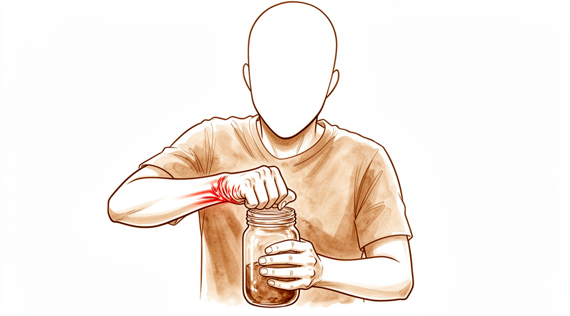
آپ کو کیا محسوس ہوتا ہے
درد آپ کی کلائی کی انگوٹھے والی طرف ہوتا ہے، جوڑ کے کناروں پر نہیں بلکہ اس کے درمیان میں۔ یہ ایک دم آنے کے بجائے عموماً برسوں میں بڑھتا ہے۔ کلائی کے درد والے بہت سے لوگ لمبے عرصے تک کسی واضح وجہ کے بغیر رہتے ہیں، کیونکہ اس قسم کا آرتھرائٹس (جوڑ کی خرابی اور سوزش) شروع میں عام ایکسرے پر ہمیشہ نظر نہیں آتا۔
کچھ خاص حرکتیں درد شروع کر دیتی ہیں۔ ہاتھ کو پیچھے کی طرف موڑ کر کلائی پر بوجھ ڈالنا، جیسے کرسی سے اٹھنے کے لیے زور لگانا یا سوئمنگ پول سے باہر نکلنا، گھسی ہوئی سطحوں کو آپس میں دبا دیتا ہے۔ کسی چیز کو مضبوطی سے پکڑ کر ساتھ ساتھ گھمانا بھی درد کی ایک عام وجہ ہے: دروازے کا ہینڈل گھمانا، کپڑا نچوڑنا، یا پیچ کس استعمال کرنا۔ آرام سے یہ عموماً کم ہو جاتا ہے، اگرچہ دکھن رات کو آپ کو جگا سکتی ہے، یا صبح جاگنے پر کچھ دیر تنگ کرتی ہے اور پھر کلائی کے کھلنے پر کم ہو جاتی ہے۔
جیسے جیسے گھساؤ پھیلتا ہے، روزمرہ کے کام مشکل ہوتے جاتے ہیں۔ ہو سکتا ہے آپ سودے کے تھیلے اٹھاتے، کیتلی سے پانی انڈیلتے، یا مرتبان کھولتے وقت اپنی گرفت کمزور محسوس کریں۔ ہاتھ پر ٹیک لگانے کے لیے کلائی کو پیچھے موڑنا، یا اپنا پورا وزن اس پر ڈالنا تکلیف دہ ہو جاتا ہے۔ کچھ لوگوں کی کلائی کی حرکت کی ہمواری اور پھرتی ختم ہو جاتی ہے، اور وہ ہر اس کام سے بچنے لگتے ہیں جس میں ہاتھ پر کسی زاویے سے بوجھ پڑے۔
اگر آپ کی علامات کم نہیں ہو رہیں، ہفتوں کے دوران بدتر ہو رہی ہیں، رات کو آپ کو جگا دیتی ہیں، یا آپ کو کام کرنے یا اپنا ہاتھ استعمال کرنے سے روکتی ہیں، تو اپنے فیملی ڈاکٹر (GP) سے ملیں یا ماہر سے معائنہ کروانے کا کہیں۔
دراصل کیا ہو رہا ہے
آپ کی کلائی آٹھ چھوٹی ہڈیوں سے بنی ہے جو دو قطاروں میں ترتیب دی گئی ہیں۔ بازو کے نچلے حصے (forearm) کے سب سے قریب والی قطار آپ کے بازو کے نچلے حصے اور ہاتھ کے درمیان ایک گدی کی طرح کام کرتی ہے، جو بوجھ کو پھیلاتی اور حرکت کو ہموار بناتی ہے۔ ان میں سے دو ہڈیوں کو لیگامنٹ (ligament)، یعنی وہ مضبوط بندھن جو ایک ہڈی کو دوسری ہڈی سے جوڑتا ہے، کی ایک مضبوط پٹی آپس میں تھامے رکھتی ہے تاکہ یہ قطار ایک اکائی کی طرح کام کر سکے؛ یہ کلائی کا وہ لیگامنٹ ہے جسے سب سے زیادہ چوٹ لگتی ہے۔
ایس ایل اے سی (SLAC) کلائی کا مسئلہ اس وقت شروع ہوتا ہے جب یہ پٹی خراب ہو جائے، عموماً کسی پرانی چوٹ کی وجہ سے۔ جن دو ہڈیوں کو یہ آپس میں جوڑے رکھتی تھی وہ ایک دوسرے سے دور سرکنے لگتی ہیں۔ ان میں سے ایک، اسکیفائیڈ (scaphoid)، پوری قطار کی حرکت کا طریقہ بدل دیتی ہے، اور جو بوجھ پہلے برابر پھیلتا تھا وہ اب جوڑ کے ایک چھوٹے سے حصے پر مرکوز ہو جاتا ہے۔ ایس این اے سی (SNAC) کلائی کا مسئلہ بھی اسی راستے پر چلتا ہے، لیکن اس کا آغاز اسکیفائیڈ کے ایسے فریکچر (ہڈی کا ٹوٹنا) سے ہوتا ہے جو کبھی نہیں جڑا، اور یہ بھی اسی طرح دونوں قطاروں کا آپس کا ربط توڑ دیتا ہے۔
یہ مرکوز بوجھ کارٹلیج (cartilage)، یعنی جوڑ میں ہڈیوں کے سروں پر چکنی، لچکدار تہہ، کو گھسا دیتا ہے؛ یہ وہی جوڑ کے گھسنے والا آرتھرائٹس (wear-and-tear arthritis) ہے جو گھٹنے میں بھی ہو سکتا ہے۔ جب آپ کلائی پر بوجھ ڈالتے ہیں تو گھسی ہوئی سطحیں آپس میں رگڑ کھاتی ہیں، اور یہی وہ درد ہے جو آپ کرسی سے اٹھنے کے لیے زور لگاتے یا پیچ کس گھماتے وقت محسوس کرتے ہیں۔ جیسے جیسے گھساؤ انگوٹھے والی طرف سے کلائی کے درمیان کی طرف پھیلتا ہے، گرفت کمزور ہوتی جاتی ہے اور حرکت میں اکڑاؤ آتا جاتا ہے۔
اس مسئلے کو مراحل (stages) میں بیان کیا جاتا ہے، اور مرحلہ اس بات پر منحصر ہوتا ہے کہ گھساؤ جوڑ کی کن سطحوں تک پہنچ چکا ہے۔ شروع میں یہ کلائی کے بیرونی کنارے تک محدود ہوتا ہے۔ بعد میں یہ ہاتھ کے درمیان والے جوڑ تک پہنچ جاتا ہے۔ یہ اس لیے اہم ہے کہ سرجری مرحلے کے مطابق چنی جاتی ہے: جہاں ایک اہم جوڑ ابھی صحت مند ہو، وہاں ہڈیوں کی گھسی ہوئی قطار نکال دینا کارگر ہو سکتا ہے، اور جہاں گھساؤ مزید پھیل چکا ہو، وہاں اس کے بجائے باقی ہڈیوں میں سے کچھ کو جوڑ کر ایک ٹھوس بلاک بنا دیا جاتا ہے۔
ہم اس کے لیے کیا کر سکتے ہیں
ڈاکٹر کیرن ہِرپرا، جو Mater Private Hospital Rockhampton میں بازو اور ہاتھ کے سرجن (upper-limb surgeon) ہیں، سب سے کم چیر پھاڑ والے ان طریقوں سے آغاز کرتے ہیں جو آپ کی حالت کے لیے موزوں ہوں۔ مریضوں کو عموماً ان کے فیملی ڈاکٹر (GP) ہمارے کلینک کے لیے ریفر کرتے ہیں؛ اگر کسی فزیوتھراپسٹ نے آپ کو ہمیں دکھانے کا مشورہ دیا ہے، تب بھی میڈیکیئر (Medicare) سے رقم کی واپسی کا حق دار ہونے کے لیے آپ کو اپنے فیملی ڈاکٹر سے ریفرل (referral) لینا ہو گا۔ ہم آپ کی طبی تاریخ لیتے ہیں، آپ کی کلائی کا معائنہ کرتے ہیں، اور جہاں ضرورت ہو گھساؤ کا مرحلہ معلوم کرنے کے لیے اسکین کرواتے ہیں۔ اس جیسے پرانے مسئلے میں ہم عموماً پہلے بغیر آپریشن کے علاج آزماتے ہیں، اور سرجری پر اس وقت غور کرتے ہیں جب اس سے کافی بہتری نہ ملی ہو۔
پہلا قدم کلائی پر بوجھ ڈالنے کا طریقہ بدلنا ہے۔ کرسی سے اٹھنے کے لیے زور لگانا، کپڑا نچوڑنا اور پیچ کس استعمال کرنا گھسی ہوئی سطحوں کو آپس میں دباتے ہیں، اس لیے ان کاموں کا طریقہ بدلنے سے دباؤ کم ہوتا ہے۔ اسپلنٹ کام کرتے یا سوتے وقت کلائی کو بے حرکت رکھ سکتا ہے۔ ہینڈ تھراپی (ہاتھ کی بحالی کا علاج) کا مقصد کلائی کو حرکت میں رکھنا اور اس کے ارد گرد کی طاقت بڑھانا ہے۔ سرجری کے بارے میں سوچنے سے پہلے ہم عموماً اسے ہفتوں سے مہینوں تک مناسب طور پر آزماتے ہیں۔
اس کے ساتھ ساتھ درد کی دوا بھی مدد کر سکتی ہے۔ جب آپ کلائی کو معمول سے زیادہ استعمال کرتے ہیں تو سوزش کم کرنے والی سادہ دوائیں دکھن کو کم کر دیتی ہیں۔ ہم آپ سے بات کریں گے کہ آپ کے لیے، اور آپ کی دوسری دواؤں کے ساتھ، کیا موزوں ہے۔
جب ان اقدامات سے کافی آرام نہ ملے، تو سرجری پر غور کیا جاتا ہے۔ مقصد جوڑ کی ان گھسی ہوئی سطحوں کو ختم کرنا ہے جو درد کا سبب ہیں، اور ساتھ ہی کلائی کی زیادہ سے زیادہ کارآمد حرکت برقرار رکھنا ہے۔ جہاں ایک اہم جوڑ ابھی صحت مند ہو، وہاں ہڈیوں کی گھسی ہوئی قطار نکال دینے سے کلائی ایک نئے، زیادہ ہموار رابطے کے ساتھ کام کرنے لگتی ہے۔ جہاں گھساؤ مزید پھیل چکا ہو، وہاں باقی ہڈیوں میں سے کچھ کو جوڑ کر ایک ٹھوس بلاک بنا دینے سے کلائی مستحکم ہو جاتی ہے اور بوجھ اٹھا سکتی ہے۔ یہ دونوں حرکت برقرار رکھنے والے طریقے ہیں، اور حرکت اور طاقت کے درمیان سودا ایک مشترکہ فیصلہ ہے جو ہم آپ کے ساتھ مل کر کرتے ہیں۔ اگر گھساؤ ان دونوں میں سے کسی کے لیے بھی بہت زیادہ پھیل چکا ہو، تو کلائی کا مکمل فیوژن (ہڈیوں کو جوڑ کر ایک کر دینا) غیر معمولی حالات کے لیے رکھا جاتا ہے، اور جہاں مریض کے لیے موزوں ہو وہاں جوڑ کی تبدیلی ایک متبادل ہو سکتی ہے۔
کیا توقع رکھیں
یہ جوڑ کے گھسنے والے آرتھرائٹس کی ایک دیرینہ قسم ہے، اس لیے یہ خود بخود جلدی ٹھیک نہیں ہوتا۔ اگر اسے ایسے ہی چھوڑ دیا جائے تو جیسے جیسے گھساؤ کلائی کے بیرونی کنارے سے درمیان کی طرف پھیلتا ہے، درد عموماً برسوں میں بڑھتا جاتا ہے۔ اس دوران گرفت کمزور ہوتی جاتی ہے اور حرکت میں اکڑاؤ آتا جاتا ہے۔ بغیر آپریشن کا علاج ہفتوں سے مہینوں میں دکھن کو کم کر سکتا ہے، لیکن یہ گھساؤ کو روکنے کے بجائے صرف علامات کو قابو میں رکھتا ہے۔
علاج کے ساتھ، آگے کی صورتحال اس بات پر منحصر ہے کہ گھساؤ کتنا پھیل چکا ہے اور آپ کے مرحلے کے لیے کون سا آپریشن موزوں ہے۔ جہاں ایک اہم جوڑ ابھی صحت مند ہو، وہاں ہڈیوں کی گھسی ہوئی قطار نکال دینے سے دیرپا نتائج مل سکتے ہیں۔ جہاں کچھ ہڈیوں کو جوڑ کر ایک ٹھوس بلاک بنا دیا جائے، وہاں زیادہ تر کلائیوں میں ہڈیاں مضبوطی سے جڑ جاتی ہیں اور کلائی رگڑ والے درد کے بغیر بوجھ اٹھا لیتی ہے۔ اکڑاؤ کم رکھنے کے لیے ہینڈ تھراپی سرجری کے بعد جلد شروع ہو جاتی ہے، کبھی کبھی پہلے ہفتے کے اندر، اور ہڈیوں کے جڑنے کے دوران کلائی کو تقریباً دو مہینے بے حرکت رکھا جاتا ہے۔ اس کے بعد بھی بہتری آتی رہتی ہے: کچھ لوگوں کی کلائی کی کارکردگی علاج کے بعد 3 سال سے زیادہ عرصے تک بہتر ہوتی رہتی ہے۔
ایمانداری کی بات یہ ہے کہ ہر کلائی پوری طرح ٹھیک نہیں ہوتی۔ فیوژن کے بعد بعض اوقات کچھ ہڈیاں نہیں جڑتیں، جس کے لیے مزید سرجری کی ضرورت پڑ سکتی ہے۔ فیوژن کو تھامنے کے لیے لگائی گئی دھات بھی کلائی کی پشت پر درد کا سبب بن سکتی ہے، اور کبھی کبھار اسے نکالنا پڑتا ہے۔ جن لوگوں کا حرکت برقرار رکھنے والا کوئی آپریشن ہو چکا ہو، ان میں سے تھوڑے سے لوگوں کو درد برقرار رہنے پر بعد میں پھر بھی کلائی کا مکمل فیوژن کروانا پڑتا ہے۔ اور کچھ کلائیوں میں ہڈیاں اچھی طرح جڑ جانے کے باوجود درد باقی رہتا ہے۔
کلائی کی کسی بھی سرجری کے بعد خطرے کی علامات پر جلدی قدم اٹھانا ضروری ہے۔ اگر آپ کی انگلی، ہاتھ یا بازو گرم، سرخ، سوجا ہوا اور دردناک ہو جائے، خاص طور پر بخار کے ساتھ، تو اسی دن ہسپتال کی ایمرجنسی جائیں؛ اس کے لیے فیملی ڈاکٹر کے ریفرل کی ضرورت نہیں۔ یہی بات اس وقت بھی لاگو ہوتی ہے اگر آپ کی انگلیاں یا ہاتھ سفید پڑ جائیں، ٹھنڈے ہو جائیں، بالکل سفید یا نیلے ہو جائیں، یا اگر اچانک ان میں احساس یا حرکت ختم ہو جائے۔
اگر آپ کی علامات کم نہیں ہو رہیں، ہفتوں کے دوران بدتر ہو رہی ہیں، رات کو آپ کو جگا دیتی ہیں، یا آپ کو کام کرنے یا اپنا ہاتھ استعمال کرنے سے روکتی ہیں، تو اپنے فیملی ڈاکٹر سے ملیں یا ماہر سے معائنہ کروانے کا کہیں۔
ڈاکٹر کو کب دکھائیں
یہ مسئلہ آہستہ آہستہ بڑھتا ہے، اس لیے زیادہ تر انتباہی علامات اچانک خطرے کے بجائے تبدیلی سے متعلق ہوتی ہیں۔ اگر آرام اور کلائی پر بوجھ ڈالنے کا طریقہ بدلنے سے آپ کی کلائی کا درد کم نہیں ہو رہا، یا ہفتوں کے دوران بدتر ہو رہا ہے، تو اپنے فیملی ڈاکٹر سے ملیں۔ اگر درد رات کو آپ کو جگا دیتا ہے، آپ کو کام کرنے سے روکتا ہے، یا آپ کی گرفت کمزور ہوتی جا رہی ہے اور روزمرہ کے کام مشکل ہوتے جا رہے ہیں، تو ماہر سے معائنہ کروانے کا کہیں۔
کلائی کی کسی بھی سرجری کے بعد، کچھ علامات میں اسی دن طبی دیکھ بھال ضروری ہے۔ اگر آپ کی انگلی، ہاتھ یا بازو گرم، سرخ، سوجا ہوا اور دردناک ہو جائے، خاص طور پر بخار کے ساتھ، تو ہسپتال کی ایمرجنسی جائیں۔ یہی بات اس وقت بھی لاگو ہوتی ہے اگر آپ کی انگلیاں یا ہاتھ سفید پڑ جائیں، ٹھنڈے ہو جائیں، بالکل سفید یا نیلے ہو جائیں، یا اگر اچانک ان میں احساس یا حرکت ختم ہو جائے۔ اگر آپ دفتری اوقات کے بعد یا ہفتے کے آخر میں کلینک سے رابطہ نہ کر سکیں، تو اپنے قریب ترین ہسپتال کی ایمرجنسی جائیں۔
خود آرتھرائٹس کی کوئی ہنگامی علامت نہیں ہوتی۔ ڈاکٹر کو دکھانے کی وجہ یہ ہے کہ گھساؤ وقت کے ساتھ پھیلتا ہے، اور سب سے زیادہ حرکت برقرار رکھنے والے طریقے اس وقت بہترین کام کرتے ہیں جب گھساؤ ابھی کلائی کے درمیان تک نہ پہنچا ہو۔
مزید تفصیل
Advanced reading: the deeper science (optional)
یہ حصہ اس سے زیادہ تفصیل میں جاتا ہے جتنی آپ کو اپنے علاج کے فیصلوں کے لیے ضرورت ہے۔ SLAC اور SNAC کلائی کے بارے میں یہ اضافی مطالعہ اس لیے فائدہ مند ہے کیونکہ دو معیاری بچاؤ کے آپریشنوں (salvage operations) میں سے انتخاب ایک حقیقی سودا ہے، ایک زیادہ حرکت دیتا ہے اور دوسرا زیادہ طاقت، اور سب سے حالیہ یکجا کیے گئے شواہد نے مجموعی طور پر ان میں سے ایک کو ترجیح دینا شروع کر دی ہے۔
دو آپریشن، دو مختلف سمجھوتے
جب کلائی کی ہڈیوں کا مجموعہ (carpus) بیٹھ کر آرتھرائٹس والے پیٹرن میں آ جائے، تو جوڑ کی سطحیں بحال نہیں کی جا سکتیں، اس لیے دونوں آپریشن آرتھرائٹس والے رابطے کی مرمت کرنے کے بجائے اسے ختم کر دیتے ہیں۔ ہڈیوں کی پہلی قطار نکالنے کا آپریشن (proximal row carpectomy) کلائی کی ہڈیوں کی پوری پہلی قطار نکال دیتا ہے، جس سے کیپیٹیٹ (capitate) ہڈی کا سر اس خالی جگہ میں بیٹھ جاتا ہے جو اسکیفائیڈ اور لونیٹ نے چھوڑی تھی۔ چار ہڈیوں کو جوڑنے کا آپریشن (four-corner fusion) اسکیفائیڈ کو نکال دیتا ہے اور باقی چار ہڈیوں کو جوڑ کر ایک ٹھوس بلاک بنا دیتا ہے۔
240 مریضوں میں ان کا براہ راست موازنہ کرنے پر سمجھوتہ واضح ہے۔ دوسری طرف کی کلائی کے فیصد کے طور پر، چار ہڈیوں کو جوڑنے کے آپریشن سے کلائی کو انگوٹھے کی طرف موڑنے (radial deviation) اور گرفت کی طاقت نمایاں طور پر زیادہ رہی، جبکہ پہلی قطار نکالنے کے آپریشن سے کلائی کو آگے موڑنا (flexion)، پیچھے موڑنا (extension) اور آگے پیچھے حرکت کی کل حد بہتر رہی، اور ساتھ ہی پیچیدگیوں کی مجموعی شرح کم رہی [1]۔
یہ اس بات سے مطابقت رکھتا ہے کہ ہر آپریشن کیسے بنایا جاتا ہے۔ چار ہڈیوں کو جوڑ کر بلاک بنانے سے وہ جڑاؤ محفوظ رہتے ہیں جو گرفت کو طاقت دیتے ہیں، لیکن ان ہڈیوں کے درمیان حرکت ختم ہو جاتی ہے۔ ایک قطار نکالنے سے کلائی کی ہڈیوں کا مجموعہ چھوٹا ہو جاتا ہے اور ایک نیا، زیادہ حرکت کرنے والا جوڑ بنتا ہے، لیکن اس کی قیمت وہ طاقت ہے جو نکالے گئے حصوں سے آتی تھی۔
بڑے یکجا تجزیے میں پہلی قطار نکالنے کے آپریشن کو ترجیح ملی ہے
زیادہ حالیہ اور کہیں بڑے شواہد نے توازن بدل دیا ہے۔ SLAC اور SNAC کلائی والے 3,174 مریضوں میں، پہلی قطار نکالنے کے آپریشن کے نتائج چار ہڈیوں کو جوڑنے کے آپریشن سے بہتر اور پیچیدگیوں کی شرح کم تھی [2]۔
اس موازنے کا زیادہ قابلِ اعتماد حصہ پیچیدگیوں کا فرق ہے، اور اس کی ایک میکانکی وضاحت ہے: چار ہڈیوں کو جوڑنے کے آپریشن میں چار ہڈیوں کی سطحوں کا آپس میں جڑنا ضروری ہے، اور ہڈیوں کا نہ جڑنا، دھات کے پرزوں کے مسائل اور انہیں نکالنے کی ضرورت ایسی پیچیدگیاں ہیں جو پہلی قطار نکالنے کے آپریشن میں ہو ہی نہیں سکتیں۔
خود فیوژن کی مختلف قسموں کا جائزہ لینے سے بھی یہ فرق ختم نہیں ہوتا۔ دو ہڈیوں کو جوڑنے (two-corner) کا چار ہڈیوں کو جوڑنے (four-corner) سے موازنہ کرنے پر، چھوٹی فیوژن کے نظریاتی فائدے کے باوجود دونوں طریقوں کے نتائج اور پیچیدگیاں ملتی جلتی رہیں، اس لیے دونوں مناسب طریقے ہیں [3]۔
جہاں فیصلہ اب بھی دوسری طرف جاتا ہے
یکجا نتیجے کا مطلب یہ نہیں کہ پہلی قطار نکالنے کا آپریشن ہر ایک کے لیے درست ہے۔ یہ اس بات پر منحصر ہے کہ کیپیٹیٹ کا سر اور ریڈیس (radius)، یعنی بازو کے نچلے حصے کی انگوٹھے والی طرف کی ہڈی، کی متعلقہ سطح خراب نہ ہوئی ہو، کیونکہ یہی دو سطحیں نیا جوڑ بنتی ہیں۔ جہاں آرتھرائٹس پہلے ہی کیپیٹیٹ تک پہنچ چکا ہو، جو اسی عمل کا زیادہ آگے کا مرحلہ ہے، وہاں پہلی قطار نکالنے کے آپریشن کے بعد جوڑ بنانے کے لیے کوئی صحیح سطح نہیں بچتی، اور فیوژن ہی راستہ بن جاتا ہے۔
عمر اور کلائی سے لیا جانے والا کام بھی اہم ہیں۔ کم عمر اور زیادہ بھاری استعمال والی کلائی کے لیے وہ طاقت زیادہ اہم ہو سکتی ہے جو فیوژن محفوظ رکھتی ہے، اور اس کے لیے پیچیدگیوں کی زیادہ شرح قبول کی جا سکتی ہے۔
جب دونوں طریقے آزمائے جا چکے ہوں
اگر بچاؤ کا آپریشن ناکام ہو جائے، یا آرتھرائٹس ان میں سے کسی کے لیے بھی بہت زیادہ پھیل چکا ہو، تو باقی طریقے کلائی کی مکمل فیوژن اور کلائی کے جوڑ کی مکمل تبدیلی ہیں۔ دونوں درد کم کرنے اور گرفت بہتر کرنے میں مؤثر ہیں، اور ان میں پیچیدگیوں کی شرح بالترتیب 17 فیصد اور 19 فیصد، یعنی ملتی جلتی ہے، جوڑ کی تبدیلی (arthroplasty) کے بعد کارکردگی میں بہتری دیکھی گئی، لیکن جوڑ کی تبدیلی کے بارے میں طویل مدتی ٹھوس ڈیٹا ابھی موجود نہیں [4]۔
یہ برابری جاننے کے لائق ہے کیونکہ فیوژن اور جوڑ کی تبدیلی کو اکثر بالکل مختلف راستوں کے طور پر پیش کیا جاتا ہے۔ ان کا فرق اس میں ہے کہ وہ آپ کو کیا دے کر جاتے ہیں، نہ کہ اس میں کہ وہ کتنی بار ناکام ہوتے ہیں: ایک مضبوط، بغیر درد کی لیکن حرکت نہ کرنے والی کلائی، یا حرکت کرنے والی ایسی کلائی جس کا مصنوعی جوڑ شاید آپ کی عمر سے پہلے ختم ہو جائے، یعنی بعد میں مزید سرجری۔
حوالہ جات
[1] Saltzman BM, Frank JM, Slikker W, Fernandez JJ, Cohen MS, Wysocki RW. Clinical outcomes of proximal row carpectomy versus four-corner arthrodesis for post-traumatic wrist arthropathy: a systematic review. J Hand Surg Eur Vol. 2014;40(5):450-7. https://doi.org/10.1177/1753193414554359
[2] Hones KM, Hao KA, Rakauskas TR, Densley S, Hampton H, Kim J, et al. Four-corner fusion versus proximal row carpectomy for scapholunate advanced collapse and scaphoid nonunion advanced collapse wrists: a systematic review and meta-analysis. J Hand Surg Am. 2024;49(7):633-8. https://doi.org/10.1016/j.jhsa.2024.01.011
[3] Hundepool CA, Duraku LS, Quanjel TJ, van Minnen LP, Jansen MC, Zuidam JM. Two-, three-, or four-corner arthrodesis for midcarpal osteoarthritis: a systematic review and meta-analysis. J Hand Surg Am. 2025;50(1):93.e1-93.e11. https://doi.org/10.1016/j.jhsa.2023.04.018
[4] Zhu XM, Perera E, Gohal C, Dennis B, Khan M, Alolabi B. A systematic review of outcomes of wrist arthrodesis and wrist arthroplasty in patients with wrist arthritis. J Hand Surg Eur Vol. 2020;46(3):297-303. https://doi.org/10.1177/1753193420953683
Evidence & references
This is the clinical evidence summary written for health professionals. It is technical, and it lists the research this page was built from. You do not need to read it to understand your treatment or to make a decision about it.
Overview
Pathophysiology and Anatomy
- Scapholunate interosseous ligament injury and extrinsic ligament complex attenuation lead to palmar flexion of the scaphoid and extension of the lunate (DISI) [20].
- The radioscaphoid joint becomes incongruous, leading to alteration in normal radioscaphoid contact forces and development of arthrosis [20].
- As the scaphoid flexes and the scapholunate diastasis increases, the capitate migrates proximally [20].
- Altered intercarpal contact forces result in arthrosis at the capitolunate joint [20].
- The styloscaphoid, radioscaphoid, and capitolunate joints are affected by SLAC wrist arthritic changes [20].
- The radiolunate joint is typically spared in SLAC wrist because of its spheroid shape [20].
- In SNAC wrist, the distal scaphoid articular surface with the radial styloid is affected first in stage I [20].
- In SNAC wrist, the proximal scaphoid pole and radioscaphoid fossa may be preserved in stage I [20].
- Bone density was greater at the capitolunate joint, the radial styloid, and the radiolunate joint in SNAC wrists compared to controls [10].
Clinical Presentation
- Symptoms of SLAC wrist include reduced grip and pinch strength [20].
- Symptoms of SLAC wrist include stiffness with extension and radial deviation [20].
- Symptoms of SLAC wrist include localized tenderness at the radioscaphoid articulation [20].
- Symptoms of SLAC wrist include decreased wrist motion on extension and radial deviation [20].
- A positive Watson shift test results when the scaphoid subluxates dorsally out of the scaphoid fossa and relocates when pressure is released [20].
- Patients with SLAC wrist were more likely to be male and have a history of trauma compared to patients with first CMC OA [12].
Diagnosis and Staging
- Radiographic classification of SLAC wrist has moderate reliability and reproducibility [15].
- Radiographic classification of SNAC wrist has limited reliability [15].
- SLAC must be distinguished from other etiologies of wrist arthritis that can mimic SLAC to appropriately inform treatment, guide prognosis, and standardize future outcome studies [6].
- It is important to differentiate between SNAC and SLAC-Wrist for classification and preoperative planning in treatment of advanced carpal collapse [2].
Treatment Indications
- Partial wrist fusion is indicated for painful posttraumatic and osteoarthritic conditions of the wrist that affect only part of the articulating system in a patient keen to have adequate pain control while preserving useful motion [16].
- Common indications for partial wrist fusion include SLAC wrist, SNAC wrist, Kienböck disease, STT arthritis, and posttraumatic radiocarpal arthritis [16].
- Chronic painful dissociative or nondissociative carpal instabilities with or without secondary arthritic change are good indications for partial wrist fusion [16].
- In inflammatory arthritis, the disease should be in a quiescent stage to avoid ongoing clinical deterioration due to disease progression for partial wrist fusion [16].
- Partial wrist fusion is contraindicated when there is active sepsis, panarthritis, and rapidly progressive joint destruction at a proliferative stage [16].
- Chronic smokers have a higher incidence of nonunion with partial wrist fusion, so alternatives for pain control such as a wrist denervation can be considered [16].
- Arthroscopic partial wrist fusions are technically demanding procedures and require extensive arthroscopic experience [16].
- Patients with preexisting extensor tendon pathology over the wrist region may have a higher incidence of tendon complications associated with partial wrist fusion [16].
- Arthrofibrosis and long-standing carpal collapse may pose additional difficulty and risk for partial wrist fusion [16].
- Scaphoid excision and four-corner fusion is indicated for the treatment of the SLAC/SNAC wrist when significant pain is refractory to nonoperative measures [9].
- Radial styloidectomy is suitable for stage I SLAC and SNAC wrist conditions [17].
- The best indications for APSI implant are SNAC wrists stages 1 and 2 as well as SLAC wrists stage 1 [4].
- Scaphoidectomy and capsulodesis is mostly indicated as a palliative procedure in elderly patients with posttraumatic SNAC or SLAC wrist with limited functional demands [25].
- Arthroscopic wrist debridement and radial styloidectomy is indicated for SLAC II or SLAC III wrists as an intermediate step to relieve pain and preserve functional motion [37].
Surgical Outcomes
- Four-corner arthrodesis with locking plate preserves satisfactory range of motion and grip strength (64% compared to the non-operated side) for SLAC and SNAC wrist [1].
- Four-corner arthrodesis with locking plate maintains the height of the carpus and prevents the premature appearance of radiocarpal osteoarthritis for SLAC and SNAC wrist [1].
- Both proximal row carpectomy and four corner fusion provide improvements in pain and subjective outcome measures for patients with symptomatic and appropriately staged SLAC or SNAC wrists [3].
- Midcarpal arthrodesis is a reliable procedure for treating SLAC- and SNAC-wrists in stages II and III [7].
- Midcarpal arthrodesis preserves some range of motion for SLAC- and SNAC-wrists [7].
- Scaphoid excision and four-corner arthrodesis reliably diminished wrist pain in patients with stage III SLAC wrist while maintaining a 54° flexion-extension arc [11].
- Successful scaphoid reconstruction in SNAC wrists shifts the load back onto the proximal pole and interrupts the otherwise inevitable degenerative pattern in scaphoid nonunions [13].
- Distal scaphoid resection is a durable procedure with good long-term results for arthritis secondary to scaphoid nonunion [31].
- 94% of patients remained satisfied after distal scaphoid resection for arthritis secondary to scaphoid nonunion [31].
- No further wrist collapse or radiocarpal arthritis developed after distal scaphoid resection for arthritis secondary to scaphoid nonunion [31].
- Findings favor Limited Carpal Fusion compared to Proximal Row Carpectomy for SNAC and SLAC wrist conditions, except for flexion-extension and grip strength in women [5].
- The authors prefer proximal row carpectomy for SLAC wrists with preserved capitate head cartilage due to socio-economic benefits, lower complication rates, and procedural ease [53].
- Capitolunate arthrodesis is a valuable alternative to four-corner fusion for selected SNAC wrist arthritis [39].
- ARARC may be a viable surgical option for patients with SLAC wrist who desire a minimally invasive procedure [116].
- Arthroscopic wrist debridement and radial styloidectomy may have advantages in relieving pain while preserving wrist motion for SLAC stage 2 or 3 disease [58].
Anatomy & Pathophysiology
Bony Anatomy
- The wrist is an anatomically and functionally highly complex joint composed of eight carpal bones arranged in two rows [54].
- The proximal carpal row contains the scaphoid, lunate, triquetrum, and pisiform [65].
- The distal carpal row contains the trapezium, trapezoid, capitate, and hamate [65].
- The distal radius articular surface has two concave facets, the scaphoid and lunate facets, separated by the scapholunate ridge [65].
- The scaphoid presents a long axis inclined by 45 degrees to the long axis of the radius [71].
- The lunate sits on the capitate and has anterior and posterior horns [71].
- The triquetrum is not in contact with the ulna head; a fibro-cartilage disc, the triangular ligament, separates the two bones [71].
- The carpus does not form a single rigid bony block because the eight small bones of the carpus all have different degrees of movement [71].
- The distal row is quite rigid, but the three proximal row bones are relatively mobile [71].
- The stability of the carpal bones does not rely upon tendon insertions, with the exception of the pisiform which is a sesamoid in flexor carpi ulnaris tendon [71].
- Carpal stability is largely due to interosseous ligaments and bony configuration of these carpal bones [71].
- The primary vascular supply to the scaphoid is a branch of the radial artery at the dorsal ridge [65].
- A group of smaller vessels enters the palmar tubercle of the scaphoid and supplies the distal 30% [65].
- The capitate head often relies on a retrograde vascular supply [65].
Ligaments
- The scapholunate interosseous ligament is the primary stabilizer of the scapholunate joint [112].
- The scapholunate interosseous ligament is composed of three distinct portions: proximal or membranous, dorsal, and palmar [112].
- The dorsal portion of the scapholunate interosseous ligament is the strongest portion and prevents translation [112].
- The palmar portion of the scapholunate interosseous ligament acts as a rotational constraint [112].
- The scapholunate interosseous ligament is C-shaped in the sagittal plane [65].
- The dorsal third of the scapholunate interosseous ligament is the thickest, strongest portion of the ligament [65].
- The radioscapholunate ligament (ligament of Testut) is a volar intra-articular neurovascular structure and provides little mechanical stability [112].
- The palmar stabilizers of the scaphoid include the radioscaphocapitate ligament, long radiolunate ligament, and short radiolunate ligament [112].
- The dorsal stabilizers of the scaphoid are the dorsal radiocarpal ligament and the dorsal intercarpal ligament [112].
- The triangular fibrocartilage complex (TFCC) is formed by the central meniscus homolog, the dorsal and volar radioulnar ligaments, the floor of the extensor carpi ulnaris tendon sheath, and the volar ulnocarpal ligaments [65].
- The dorsal and volar radioulnar ligaments are the primary stabilizers of the distal radioulnar joint [65].
- Only the peripheral 10% to 40% of the volar, ulnar, and dorsal TFCC has a vascular supply [65].
Kinematics and Biomechanics
- The wrist can essentially be considered to be a two-joint system linking the hand to the forearm around the highly mobile bones of the proximal carpal row [66].
- The two principle articulations are the radiocarpal and midcarpal joints, situated proximal and distal to the mobile proximal carpal row [66].
- The proximal carpal row has no muscular or tendinous attachments and is an intercalary segment [65].
- With ulnar deviation, the proximal row extends relative to the forearm/distal row [65].
- With radial deviation, the proximal row flexes relative to the forearm/distal row [65].
- With axial loading through the neutral wrist, approximately 80% of forces are transmitted through the distal radius and 20% through the distal ulna [65].
- Of the forces transmitted through the distal radius during neutral axial loading, 60% are transmitted through the scaphoid facet and 40% through the lunate facet [65].
- With wrist flexion, 60% of the motion is midcarpal and 40% is radiocarpal [65].
- With wrist extension, 33% of the motion is midcarpal and 66% is radiocarpal [65].
- The contact areas of the wrist accounted for only 20.6% of the available joint surface [105].
- Wrist contact areas shift from a primarily palmar location to a primarily dorsal location when the wrist changes from flexion to extension [105].
- A wrist joint should be considered biomechanically unstable when it is not able to bear loads and does not exhibit normal kinematics throughout its arc of motion [62].
SLAC Pathophysiology
- Scapholunate advanced collapse (SLAC) wrist may result from chronic pseudogout and can appear bilaterally without a clear history of injury [14].
- Scapholunate interosseous ligament injury and extrinsic ligament complex attenuation lead to palmar flexion of the scaphoid and extension of the lunate [20].
- Palmar flexion of the scaphoid and extension of the lunate results in dorsal intercalated segment instability (DISI) [20].
- The radioscaphoid joint becomes incongruous following scapholunate ligament injury, leading to alteration in the normal radioscaphoid contact forces and development of arthrosis [20].
- The experimental kinetic behavior of the chronic SLAC wrist is similar to the one with a recent scapholunate dissociation without carpal collapse associated [18].
- Measurements of the angular deformity between the scaphoid and the radius obtained in a cadaveric model help to explain the progression of degenerative changes in the SLAC wrist [26].
SNAC Pathophysiology
- Scaphoid nonunion advanced collapse (SNAC) is caused by trauma [14].
- Symptomatic scaphoid nonunion may lead to a predictable pattern of radioscaphoid and midcarpal degenerative arthritis [21].
- Degenerative changes in scaphoid nonunion can occur early, especially when instability is present [21].
- Instability patterns in scaphoid nonunion increase with time [21].
- The proximal scaphoid pole and radioscaphoid fossa may be preserved in stage I SNAC wrist [20].
- SNAC wrists differ from SLAC wrists in exhibiting a decreased sagittal lunotriquetral angle, indicating a distinct pathomechanism of carpal instability [8].
- The chronology of the progression of SNAC and SLAC wrist is largely identical [48].
- Only the initial stages of SNAC and SLAC wrist differ [48].
Classification
Definitions and Etiology
- Scapholunate advanced collapse (SLAC) may result from chronic pseudogout and can appear bilaterally without a clear history of injury [14].
- In clinical practice, SNAC pathology is observed in 60% and SLAC in 40% of cases [34].
Staging Systems
- The severity of degenerative change in SLAC and SNAC wrists is classified into three stages [34].
- The Watson and Ballet classification of SLAC wrist osteoarthritis is widely accepted, simple, and a well-recognized guide to treatment despite its significant limitations [51].
- Staging systems for SNAC wrist lack agreement [27].
- The classification of Vender et al. (1987) is widely used to describe SNAC severity, but its inter- and intra-observer reliability is poor [38].
Pathomechanics and Radiographic Features
- Carpal malalignment in SLAC wrists affects the radio- and midcarpal joints and extends to the third carpometacarpal joint, with malalignment evident in both the sagittal and coronal planes [36].
- The experimental kinetic behavior of the chronic SLAC wrist is similar to that of a recent scapholunate dissociation without carpal collapse [18].
- Measurements of angular deformity between the scaphoid and the radius help explain the progression of degenerative changes in the SLAC wrist [26].
- Carpal collapse in Kienbock’s disease is different from SLAC and does not progress to radiocarpal osteoarthritis as in post-traumatic cases [23].
Differential Diagnosis
- It is important to differentiate between SNAC and SLAC wrist for classification and preoperative planning in the treatment of advanced carpal collapse [2].
- The SOAC staging system differentiates SOAC from SLAC wrist and can guide treatment decisions [40].
Clinical Presentation
Symptoms and Physical Examination
- Symptomatic SLAC wrist usually includes pain with activity, dorsal wrist swelling, and limited wrist motion [22].
- Patients with SLAC wrist present with reduced grip and pinch strength [20].
- Patients with SLAC wrist present with stiffness with extension and radial deviation [20].
- Patients with SLAC wrist present with localized tenderness at the radioscaphoid articulation [20].
- Patients with SLAC wrist present with decreased wrist motion on extension and radial deviation [20].
- A positive Watson shift test is defined as the scaphoid subluxating dorsally out of the scaphoid fossa when pressure is directed over the palmar scaphoid tuberosity while the wrist is moved from ulnar to radial deviation, with relocation when pressure is released [20].
- SLAC wrist may result from chronic pseudogout and can appear bilaterally without a clear history of injury [14].
- SNAC wrist is caused by trauma [14].
Radiographic Findings and Staging
- Plain radiographs are usually sufficient to make the diagnosis of SLAC wrist [22].
- Joint space narrowing with sclerosis is apparent in the radioscaphoid joint in the early stages of SLAC wrist [22].
- In stage I SNAC wrist, the distal scaphoid articular surface with the radial styloid is affected first [20].
- In stage I SNAC wrist, the proximal scaphoid pole and radioscaphoid fossa may be preserved [20].
- SNAC wrists differ from SLAC wrists in exhibiting a decreased sagittal lunotriquetral angle [8].
- Carpal malalignment in SLAC wrists extends to the third carpometacarpal joint, with malalignment evident in both the sagittal and coronal planes [36].
Pathomechanics
- The radioscaphoid joint becomes incongruous, leading to alteration in the normal radioscaphoid contact forces and development of arthrosis [20].
- Measurements of the angular deformity between the scaphoid and the radius help to explain the progression of degenerative changes in the SLAC wrist [26].
- Degenerative changes can occur early, especially when instability is present [21].
Investigations
Clinical Evaluation
- A careful history and physical examination are essential in the evaluation of patients with wrist arthritis, as patients referred for surgical treatment of SLAC wrist may have pain from other conditions such as DeQuervain's tenosynovitis rather than the wrist joint itself [22].
- Patients with SLAC wrist are more likely to be male and have a history of trauma compared to patients with first CMC OA [12].
- SLAC wrist may result from chronic pseudogout and can appear bilaterally without a clear history of injury, whereas SNAC wrist is caused by trauma [14].
- SLAC must be distinguished from other etiologies of wrist arthritis that can mimic it to appropriately inform treatment, guide prognosis, and standardize future outcome studies [6].
- The Watson shift test involves directing pressure over the palmar scaphoid tuberosity while the wrist is moved from ulnar to radial deviation, with a positive test resulting when the scaphoid subluxates dorsally out of the scaphoid fossa and relocates when pressure is released [20].
- Reduced grip and pinch strength, stiffness with extension and radial deviation, localized tenderness at the radioscaphoid articulation, and decreased wrist motion on extension and radial deviation are symptoms of SLAC wrist [20].
- The history, staging, and treatment of SNAC wrist are similar to that for SLAC wrist [20].
- In SNAC wrist, the distal scaphoid articular surface with the radial styloid is affected first in stage I, while the proximal scaphoid pole and radioscaphoid fossa may be preserved [20].
- The natural inclination to study radiographs or special imaging studies prior to a thorough history and physical examination should be avoided as it introduces cognitive bias that can affect thinking and decision making [89].
- Bilateral grip and pinch strength testing is useful to uncover underlying pathology in chronic cases, with strength potentially diminished due to muscle atrophy, pain inhibition, or learned behaviors [89].
- A local injection of anesthetic to a painful joint or selected tendon sheath may help normalize dynamometer readings and narrow the diagnostic spectrum [89].
Radiographic Imaging
- Tomograms may be useful in determining the extent of arthritis but rarely change the course of treatment [22].
- Radiographic classification of SLAC wrist has moderate reliability and reproducibility, whereas classification of SNAC wrist has limited reliability [15].
- The severity of the radiocarpal arthrosis following distal radius fracture seen on radiograph is not correlated with the presence of symptoms [20].
- A routine radiographic series for evaluating a painful wrist consists of four views: posteroanterior, lateral, oblique, and ulnar-deviated posteroanterior scaphoid view [81].
- Spot views of the carpal bones for detail (carpal tunnel view) are a useful radiographic technique for evaluating a painful wrist [81].
- A series of views for instability includes anteroposterior clenched fist, posteroanterior in neutral/radial/ulnar deviation, lateral in neutral/full flexion/extension, and semipronated/semisupinated oblique views [81].
- 10° of supination can drastically alter the developed posteroanterior radiograph of the wrist due to the significant effect of rotation on radiographic landmarks [46].
- Radiographic scaphoid dorsal translation is correlated with MRI findings and might represent a lower cost option for early detection of the radioscaphoid joint eccentric wear seen in some stages of scapholunate instability [126].
Advanced Imaging (CT, MRI, Arthroscopy)
- MRI should be added for evaluation of the triangular fibrocartilage, the distal radioulnar joint, and vascularity of the various carpal bones to confirm clinical suspicion and correlate with physical examination findings [81].
- A high rate of false-positive findings on MR images of normal subjects has been reported [81].
- A dedicated wrist coil provides enhanced resolution of wrist structures for MRI [81].
- With proper technique, injuries to the triangular fibrocartilage complex can be demonstrated with MRI, appearing as linear defects or gaps filled with hyperintense fluid on coronal gradient-echo or T2-weighted pulse sequences [78].
- Evaluation of the scapholunate and lunotriquetral ligaments is more challenging on MRI, but with optimal technique and equipment, the integrity of these structures can be consistently assessed [78].
- The addition of arthrographic contrast improves the visualization of scapholunate and lunotriquetral ligaments on MR images [78].
- MRI is useful in detecting additional marrow abnormalities in osteonecrosis, as seen in the lunate in Kienböck disease or in the scaphoid after fracture [78].
- Asymmetry of marrow signal in proximal and distal fragments of a fractured scaphoid is suggestive of proximal pole ischemia on MRI [78].
- Quantitative 3-D CT demonstrates that bone density was greater at the capitolunate joint, the radial styloid, and the radiolunate joint in SNAC wrists compared to controls [10].
- Computer-aided three-dimensional analysis indicates that SNAC wrists differ from SLAC wrists in exhibiting a decreased sagittal lunotriquetral angle, indicating a distinct pathomechanism of carpal instability [8].
- CT was of diagnostic value to view the position of the scaphoid about the radius and its changes in load in patients with scapholunate instability [47].
- Arthroscopic assessment of intercarpal ligament injuries and instability is considered by many the “gold standard” for evaluation of these conditions [80].
- Arthroscopy has been found to be more accurate than arthrography in identifying the location and size of triangular fibrocartilage and interosseous ligament injuries [80].
- The inconsequent use of available modern magnetic resonance imaging (MRI) techniques and the lack of reliable preoperative diagnoses necessitated pure diagnostic arthroscopies for ulnar-sided wrist pain [118].
Pathomechanics and Differential Diagnosis
- Scapholunate interosseous ligament injury and extrinsic ligament complex attenuation lead to palmar flexion of the scaphoid and extension of the lunate (DISI) in SLAC wrist [20].
- The radioscaphoid joint becomes incongruous in SLAC wrist, leading to alteration in the normal radioscaphoid contact forces and development of arthrosis [20].
- As the scaphoid flexes and the scapholunate diastasis increases in SLAC wrist, the capitate migrates proximally [20].
- The altered intercarpal contact forces in SLAC wrist result in arthrosis at the capitolunate joint [20].
- Carpal collapse in Kienböck disease is different from scapholunate advanced collapse (SLAC) and does not progress to radiocarpal osteoarthritis as in post-traumatic cases [23].
- SOAC wrist is differentiated from SLAC wrist, and the SOAC staging system can guide treatment decisions [40].
Treatment
General Principles and Indications
- Surgical treatment for SLAC wrist includes four-corner arthrodesis, capitolunate arthrodesis, complete wrist arthrodesis, proximal row carpectomy (PRC), denervation, and radial styloidectomy [14].
- SNAC wrist has the additional surgical option of excision of the distal ununited scaphoid fragment compared to SLAC wrist [14].
- Partial wrist fusion is indicated for painful posttraumatic and osteoarthritic conditions of the wrist that affect only part of the articulating system in a patient who is keen to have adequate pain control while preserving useful motion [16].
- Chronic smokers have a higher incidence of nonunion after partial wrist fusion, so alternatives for pain control such as a wrist denervation can be considered [16].
- The treatment for SLAC or SNAC wrist is identical at any given stage of the disease, with the exception that only the initial stages of SNAC and SLAC wrist differ [48].
- Trapeziometacarpal joint arthritis and SLAC wrist may be treated either simultaneously or in stages [30].
Stage-Specific Interventions
- For SLAC 1 wrist, partial arthrodesis of the wrist with styloidectomy of the radius is indicated [48].
- Arthroscopic radial styloidectomy is indicated for Stage 1 SLAC and SNAC wrist as an isolated procedure or adjunct procedure together with scaphoid non-union or SL reconstruction procedures [55].
- Midcarpal arthrodesis is a reliable procedure for treating SLAC- and SNAC-wrists in stages II and III which preserves some range of motion [7].
- Wrist arthrodesis is generally an appropriate option for SNAC stage IV [45].
- Wrist arthrodesis is a final option in a situation where there is no other treatments to resolve pain [45].
Proximal Row Carpectomy (PRC)
- PRC is best indicated for Type 2 and Type 3 SLAC or SNAC wrists in which the injury has not yet involved the entire carpus [50].
- PRC is indicated for multiple degenerative and traumatic wrist conditions that result in persistent pain and dysfunction with activities in daily living that have been unresponsive to nonoperative measures [50].
- Proximal row carpectomy results in better outcomes and a lower complication rate compared to four-corner fusion in the treatment of scapholunate advanced collapse and scaphoid nonunion advanced collapse wrists [125].
- Among patients treated for SNAC and SLAC wrist conditions, findings are in favour of Limited Carpal Fusion compared to Proximal Row Carpectomy, except for flexion-extension and grip strength in women [5].
Four-Corner Arthrodesis (4CF)
- Four-corner arthrodesis with locking plate is a valuable surgical technique for treating SLAC and SNAC wrist because it preserves satisfactory range of motion and grip strength (64% compared to the non-operated side), maintains the height of the carpus and prevents the premature appearance of radiocarpal osteoarthritis, as long as the technical challenges of this procedure are mastered [1].
- Four-corner fusion retains 60% of wrist motion and 80% of grip strength [20].
Scaphoid-Specific Procedures
- Bone grafting of the scaphoid in symptomatic patients may help to prevent the development of arthritis [21].
- Distal scaphoid resection is a durable procedure with good long-term results, with 94% of patients remaining satisfied and no further wrist collapse or radiocarpal arthritis developing [31].
- Arthroscopic distal scaphoidectomy is indicated for symptom control in selected cases of SNAC wrist, as well as in the treatment of STT joint osteoarthritis [111].
- In SNAC wrist, distal scaphoid resection can be regarded as a “buying time” procedure in removing the primary source of mechanical symptoms in painful scaphoid nonunion [111].
- The best indication for distal scaphoidectomy in SNAC wrist is when the cartilage degeneration, osteophyte formation, and deformity are confined mainly to the distal scaphoid articular surface on both radiocarpal and midcarpal joint surfaces, manifested by severe degenerative change at the radial styloid-scaphoid articulation [111].
- A significant dorsal lunate tilt deformity (DISI) is considered a relative contraindication to distal scaphoid excision [111].
- Marked arthritis at the lunocapitate joint constitutes a relative contraindication of distal scaphoidectomy [111].
- An intact SL ligament and RSC ligament are considered a prerequisite for distal scaphoidectomy to minimize progressive midcarpal collapse and DISI [111].
- Bicolumnar fusion for SNAC wrist without bone grafting resulted in union in all cases, with postoperative Mayo wrist scores of excellent in 16 patients and good in 6 patients [107].
Combined and Adjunctive Procedures
- For SLAC 1 wrist with trapeziometacarpal arthritis, one option is to fuse the scaphocapitate with a styloidectomy, which allows for trapeziectomy or arthroplasty [48].
- For SLAC 1 wrist with trapeziometacarpal arthritis, another option is to perform a STT arthrodesis with a styloidectomy, which requires performing a prosthetic arthroplasty on the trapeziometacarpal joint [48].
- Total or subtotal scaphoidectomy is a concomitant procedure for midcarpal arthrodesis in SLAC wrist and SNAC wrist [17].
- The APSI implant has best indications for SNAC wrists stages 1 and 2 as well as SLAC wrists stage 1 [4].
Complications
- Partial wrist fusion is not a guarantee of pain relief [16].
- The potential advantage of partial wrist fusion in preservation of a useful arc of motion may be offset by the risks of nonunion [16].
- The potential advantage of partial wrist fusion in preservation of a useful arc of motion may be offset by continued pain despite successful fusion [16].
- Chronic smokers have a higher incidence of nonunion following partial wrist fusion [16].
- Patients with preexisting extensor tendon pathology over the wrist region may have a higher incidence of tendon complications associated with complex partial wrist fusion procedures [16].
- Arthrofibrosis poses additional difficulty and risk for partial wrist fusion [16].
- Long-standing carpal collapse poses additional difficulty and risk for partial wrist fusion [16].
- Total wrist fusion does not always result in complete pain relief [34].
- Total wrist fusion causes significant functional disability [34].
- Proximal row carpectomy results in a reduction of wrist motion [20].
- Proximal row carpectomy results in a reduction of grip strength [20].
- Resection of 6 mm or 10 mm of the radial styloid is associated with radial, ulnar, and palmar carpal displacement [38].
- Resection of 6 mm or 10 mm of the radial styloid is associated with increased radial translation [17].
- Resection of 6 mm or 10 mm of the radial styloid is associated with ulnar and palmar carpal displacement [17].
- Resection of 3 mm of the radial styloid was not associated with radial, ulnar, or palmar carpal displacement [38].
- The second most common complication of scaphocapitate arthrodesis is persistent wrist pain despite radiographic evidence of bony union [41].
- Persistent wrist pain despite radiographic evidence of bony union occurred in 4 of 30 patients (13%) undergoing scaphocapitate arthrodesis [41].
- In SLAC wrists with significant ligamentous lesions, the placement of a proximal scaphoid implant does not resolve the problem of ligamentous destabilization [57].
- At 43 years follow-up of a vitallium scaphoid arthroplasty, radiographs demonstrated carpal collapse [32].
- At 43 years follow-up of a vitallium scaphoid arthroplasty, radiographs demonstrated diffuse radiocarpal arthritis [32].
Recovery
- Four-corner arthrodesis with locking plate preserves satisfactory range of motion and grip strength at 64% compared to the non-operated side [1].
- Four-corner arthrodesis with locking plate maintains the height of the carpus [1].
- Four-corner arthrodesis with locking plate prevents the premature appearance of radiocarpal osteoarthritis [1].
- Limited Carpal Fusion is favored over Proximal Row Carpectomy for SNAC and SLAC wrist conditions, except for flexion-extension and grip strength in women [5].
- Midcarpal arthrodesis preserves some range of motion in the treatment of SLAC- and SNAC-wrists in stages II and III [7].
- Scaphoid excision and four-corner arthrodesis maintains a 54° flexion-extension arc in patients with stage III SLAC wrist [11].
- Functional results of four-corner fusion for SLAC and SNAC wrist are good at long-term follow-up despite radiographic changes in the radiolunate joint in 73% of patients [124].
- Distal scaphoid resection is a durable procedure with good long-term results, with 94% of patients remaining satisfied [31].
- No further wrist collapse or radiocarpal arthritis developed in patients treated with distal scaphoid resection for arthritis secondary to scaphoid nonunion [31].
- Distal scaphoid resection arthroplasty produced favorable, long-term clinical results and did not result in noteworthy wrist collapse [60].
- There has been no long term deterioration of wrist function following the use of costo-osteochondral grafts in the wrist [33].
Key Evidence
- [L4] Four-corner arthrodesis with locking plate is a valuable surgical technique for treating SLAC and SNAC wrist because it preserves satisfactory range of motion and grip strength (64% compared to the non-operated side), maintains the height of the carpus and prevents the premature appearance of radiocarpal osteoarthritis, as long as the technical challenges of this procedure are mastered. [1] (10.1016/j.hansur.2019.10.196)
- [L4] We find it important to differentiate between SNAC and SLAC-Wrist for classification and preoperative planning in treatment of advanced carpal collapse. [2] (10.1016/0266-7681(94)90353-0)
- [L4] Both procedures provide improvements in pain and subjective outcome measures for patients with symptomatic and appropriately staged SLAC or SNAC wrists. [3] (10.1177/1753193408100954)
- [L3] Les meilleures indications sont les SNAC wrists stades 1 et 2 ainsi que les SLAC wrists stades 1. [4] (10.1016/j.main.2015.10.062)
- [L3] Among patients treated for SNAC and SLAC wrist conditions, findings are in favour of Limited Carpal Fusion compared to Proximal Row Carpectomy, except for flexion-extension and grip strength in women. [5] (10.1186/s13018-023-04177-7)
- [L5] SLAC must be distinguished from other etiologies of wrist arthritis that can mimic SLAC to appropriately inform treatment, guide prognosis, and standardize future outcome studies. [6] (10.1016/j.jhsa.2015.06.110)
- [L4] Our data demonstrate that midcarpal arthrodesis is a reliable procedure for treating SLAC- and SNAC-wrists in stages II and III and, furthermore, one which preserves some range of motion. [7] (10.1097/01.sap.0000194245.94684.54)
- [L4] SNAC wrists differ from SLAC wrists in exhibiting a decreased sagittal lunotriquetral angle, indicating a distinct pathomechanism of carpal instability. [8] (10.1186/s12891-025-08652-6)
- [L4] Scaphoid excision and four-corner fusion is indicated for the treatment of the SLAC/SNAC wrist when significant pain is refractory to nonoperative measures. [9] (10.1053/otor.2003.36321)
- [L3] Bone density was greater at the capitolunate joint, the radial styloid, and the radiolunate joint in SNAC wrists compared to controls. [10] (10.2106/jbjs.22.01350)
- [L4] Scaphoid excision and four-corner arthrodesis reliably diminished wrist pain in patients with stage III SLAC wrist while maintaining a 54° flexion-extension arc. [11] (10.1016/0363-5023(94)90178-3)
- [L3] Patients with SLAC wrist were more likely to be male and have a history of trauma compared to patients with first CMC OA. [12] (10.1177/1558944718788672)
- [L4] Successful scaphoid reconstruction in SNAC wrists shifts the load back onto the proximal pole and interrupts the otherwise inevitable degenerative pattern in scaphoid nonunions. [13] (10.1016/j.jhsa.2018.06.104)
- [L5] [14] (10.1016/j.jhsa.2011.01.018)
- [L4] Radiographic classification of SLAC wrist has moderate reliability and reproducibility, whereas classification of SNAC wrist has limited reliability. [15] (10.1177/1753193413484629)
- [L5] The experimental kinetic behavior of the chronic SLAC wrist is similar to the one with a recent scapholunate dissociation without carpal collapse associated. [18] (10.1177/1558944716660555w)
- [L4] [21] (10.1016/s0363-5023(87)80198-3)
- [L5] [22] (10.1097/00130911-199712000-00003)
- [L5] The author notes that carpal collapse in KD is different from scapholunate advanced collapse (SLAC) and does not progress to radiocarpal osteoarthritis as in post-traumatic cases. [23] (10.1177/17531934231205707)
- [L4] It is mostly indicated as a palliative procedure in elderly patients with posttraumatic SNAC or SLAC wrist with limited functional demands. [25] (10.1055/s-0032-1329615)
- [L5] Measurements of the angular deformity between the scaphoid and the radius obtained in this cadaveric model help to explain the progression of degenerative changes in the SLAC wrist. [26] (10.1016/s0363-5023(87)80066-7)
- [Paper] Staging systems for SNAC wrist lack agreement. [27] (10.1007/s12593-012-0062-2)
- [L4] Trapeziometacarpal joint arthritis and SLAC wrist may be treated either simultaneously or in stages. [30] (10.1016/j.jhsa.2021.05.002)
- [L4] Distal scaphoid resection is a durable procedure with good long-term results. 94% of patients remained satisfied, and no further wrist collapse or radiocarpal arthritis developed. [31] (10.1016/s0363-5023(11)60002-6)
- [L5] At 43 years follow-up, radiographs demonstrated carpal collapse and diffuse radiocarpal arthritis, and the patient is now considering a total wrist fusion. [32] (10.1016/0363-5023(91)90015-4)
- [L4] There has been no long term deterioration of wrist function. [33] (10.1097/00130911-200109000-00008)
- [L4] [34] (10.1054/jhsb.2000.0434)
- [L3] Carpal malalignment in SLAC wrists not only affects the radio- and midcarpal joints, but also extends to the third carpometacarpal joint, with malalignment evident in both the sagittal and coronal planes. [36] (10.1016/j.jhsa.2024.09.021)
- [L4] The procedure is indicated for SLAC II or SLAC III wrists as an intermediate step to relieve pain and preserve functional motion. [37] (10.1016/j.arthro.2012.04.108)
- [L3] [38] (10.1177/1753193417739519)
- [Paper] The procedure is a valuable alternative to four-corner fusion for selected SNAC wrist arthritis. [39] (10.1007/s12593-015-0182-6)
- [L4] It also differentiates SOAC from SLAC wrist and discusses how the SOAC staging system can guide treatment decisions. [40] (10.5435/jaaos-d-25-01408)
- [Paper] The second most common complication is persistent wrist pain despite radiographic evidence of scaphocapitate bony union, occurring in 4/30 patients (13%). [41] (10.1097/00130911-200206000-00003)
- [L4] [45] (10.1142/s2424835518300049)
- [Paper] Our results highlight the significant effect of rotation on radiographic landmarks at the wrist, indicating that 10° of supination can drastically alter the developed radiograph. [46] (10.1177/15589447241255705)
- [L4] Despite these, it was of diagnostic value to view the position of the scaphoid about the radius and its changes in load. [47] (10.1016/s0363-5023(03)80339-8)
- [L5] [48] (10.1016/j.hansur.2020.08.013)
- [Paper] [50] (10.1097/bth.0b013e3181d44526)
- [L5] Despite its significant limitations, the Watson and Ballet classification of SLAC wrist osteoarthritis is widely accepted, simple, and a well-recognized guide to treatment. [51] (10.1097/corr.0000000000000451)
- [L3] The authors prefer proximal row carpectomy for SLAC wrists with preserved capitate head cartilage due to socio-economic benefits, lower complication rates, and procedural ease. [53] (10.1177/1753193408087116)
- [L1] [54] (10.1007/s00402-026-06423-z)
- [L4] Dans les séquelles de disjonction scapholunaire (SLAC wrist) où les lésions ligamentaires sont importantes la mise en place de l’implant ne résout pas le problème de la déstabilisation ligamentaire qui nécessite un traitement approprié supplémentaire. [57] (10.1016/s1297-3203(00)73492-5)
- [L4] The procedure studied may have advantages in relieving pain while preserving wrist motion for SLAC stage 2 or 3 disease. [58] (10.1177/1558944717725383)
- [L4] Distal scaphoid resection arthroplasty produced favorable, long-term clinical results and did not result in noteworthy wrist collapse. [60] (10.1016/j.jhsa.2014.05.031)
- [L5] A wrist joint should be considered biomechanically unstable when it is not able to bear loads and does not exhibit normal kinematics throughout its arc of motion. [62] (10.1053/jhsu.1999.0866)
- [L5] The contact areas accounted for only 20.6% of the available joint surface and shift from a primarily palmar location to a primarily dorsal location when the wrist changes from flexion to extension. [105] (10.1016/s0363-5023(87)80093-x)
- [L4] [107] (10.1097/bth.0b013e318249d454)
- [L4] ARARC may be a viable surgical option for patients with SLAC wrist who desire a minimally invasive procedure. [116] (10.1055/s-0034-1373839)
- [L4] The inconsequent use of available modern magnetic resonance imaging (MRI) techniques and the lack of reliable preoperative diagnoses necessitated pure diagnostic arthroscopies for ulnar-sided wrist pain. [118] (10.1177/1558944716660555hs)
- [L4] Functional results were good at long-term follow-up despite radiographic changes in the radiolunate joint in 73% of patients. [124] (10.1177/1558944716681949)
- [L1] In the treatment of scapholunate advanced collapse and scaphoid nonunion advanced collapse wrists, PRC results in better outcomes and a lower complication rate compared to 4CF. [125] (10.1016/j.jhsa.2024.01.011)
- [L4] Radiographic scaphoid dorsal translation is correlated with MRI findings and might represent a lower cost option for early detection of the radioscaphoid joint eccentric wear seen in some stages of scapholunate instability. [126] (10.1177/1558944716660555ir)
References
[1] Clinical and radiological outcomes of 40 cases of SLAC and SNAC wrist treated by four-corner arthrodesis with locking plate. Hand Surgery and Rehabilitation. 2020. DOI: 10.1016/j.hansur.2019.10.196
[2] Advanced Carpal Collapse after Longstanding Scaphoid Nonunion (SNAC-Wrist) and Longstanding Scapho-Lunate Dissociation (SLAC-Wrist)—Where are the Differences?. Journal of Hand Surgery. 1994. DOI: 10.1016/0266-7681(94)90353-0
[3] Proximal Row Carpectomy vs Four Corner Fusion for Scapholunate (Slac) or Scaphoid Nonunion Advanced Collapse (Snac) Wrists: A Systematic Review of Outcomes. Journal of Hand Surgery (European Volume). 2009. DOI: 10.1177/1753193408100954
[4] L’implant APSI – indications et résultats. Chirurgie de la Main. 2015. DOI: 10.1016/j.main.2015.10.062
[5] Limited intercarpal fusion versus proximal row carpectomy in the treatment of SLAC or SNAC wrist, results after 3.5 years. Journal of Orthopaedic Surgery and Research. 2023. DOI: 10.1186/s13018-023-04177-7
[6] Scapholunate Advanced Collapse: Nomenclature and Differential Diagnosis. The Journal of Hand Surgery. 2015. DOI: 10.1016/j.jhsa.2015.06.110
[7] Long-Term Results of Midcarpal Arthrodesis in the Treatment of Scaphoid Nonunion Advanced Collapse (SNAC-Wrist) and Scapholunate Advanced Collapse (SLAC-Wrist). Annals of Plastic Surgery. 2006. DOI: 10.1097/01.sap.0000194245.94684.54
[8] Computer-aided three-dimensional analysis of carpal alignment in scaphoid nonunion advanced collapse wrists: A comparative study with scapholunate advanced collapse and healthy wrists. BMC Musculoskeletal Disorders. 2025. DOI: 10.1186/s12891-025-08652-6
[9] Scaphoid excision and four-corner fusion in the SLAC/SNAC wrist. Operative Techniques in Orthopaedics. 2003. DOI: 10.1053/otor.2003.36321
[10] Quantitative 3-D CT Demonstrates Distal Row Pronation and Translation and Radiolunate Arthritis in the SNAC Wrist. Journal of Bone and Joint Surgery. 2023. DOI: 10.2106/jbjs.22.01350
[11] Surgical treatment of scapholunate advanced collapse. The Journal of Hand Surgery. 1994. DOI: 10.1016/0363-5023(94)90178-3
[12] The Epidemiology of Scapholunate Advanced Collapse. HAND. 2018. DOI: 10.1177/1558944718788672
[13] Long-Term Results of Scaphoid Grafting in 52 SNAC Wrists: Should We Cut Down on Salvage Procedures?. The Journal of Hand Surgery. 2018. DOI: 10.1016/j.jhsa.2018.06.104
[14] Scapholunate Advanced Collapse and Scaphoid Nonunion Advanced Collapse Arthritis—Update on Evaluation and Treatment. The Journal of Hand Surgery. 2011. DOI: 10.1016/j.jhsa.2011.01.018
[15] Reproducibility of radiographic classification of scapholunate advanced collapse (SLAC) and scaphoid nonunion advanced collapse (SNAC) wrist. Journal of Hand Surgery (European Volume). 2013. DOI: 10.1177/1753193413484629
[16] Green S Operative Hand Surgery. AUTHOR'S PREFERRED METHOD OF TREATMENT: ARTHROSCOPIC PARTIAL WRIST FUSION > Indications and Contraindications.
[17] Green S Operative Hand Surgery. AUTHOR'S PREFERRED METHOD OF TREATMENT: ARTHROSCOPIC PARTIAL WRIST FUSION > Osteotomy.
[18] Kinetics of the Wrist in Scapholunate Advanced Collapse. HAND. 2016. DOI: 10.1177/1558944716660555w
[20] Aaos Comprehensive Orthopaedic Review 3. Arthritides of the Hand and Wrist* > IV. Posttraumatic Arthritis.
[21] Degenerative change in symptomatic scaphoid nonunion. The Journal of Hand Surgery. 1987. DOI: 10.1016/s0363-5023(87)80198-3
[22] Midcarpal Arthrodesis. Techniques in Hand & Upper Extremity Surgery. 1997. DOI: 10.1097/00130911-199712000-00003
[23] Re: Luitjens J, Goller SS, Schmitt R, Erber B, Van Scoonhoven J, Hess N. Diagnostic performance of traditional radiographic indices in detection of carpal collapse in Kienbock’s disease. J Hand Surg Eur. 2023, 48: 619–24.. Journal of Hand Surgery (European Volume). 2023. DOI: 10.1177/17531934231205707
[25] Scaphoidectomy and Capsulodesis for SNAC or SLAC Stage II. Journal of Wrist Surgery. 2012. DOI: 10.1055/s-0032-1329615
[26] The effect of rotatory subluxation of the scaphoid on radio-scaphoid contact. The Journal of Hand Surgery. 1987. DOI: 10.1016/s0363-5023(87)80066-7
[27] Scaphoid Nonunion Advanced Collapse Classifications: A Reliability Study. Journal of Hand and Microsurgery. 2012. DOI: 10.1007/s12593-012-0062-2
[30] Combined Treatment of Trapeziometacarpal Joint Arthritis and Scapholunate Advanced Collapse Wrist. The Journal of Hand Surgery. 2022. DOI: 10.1016/j.jhsa.2021.05.002
[31] Distal Scaphoid Resection for Arthritis Secondary to Scaphoid Nonunion: A Twenty-year Experience. The Journal of Hand Surgery. 2011. DOI: 10.1016/s0363-5023(11)60002-6
[32] A forty-three-year follow-up of a vitallium scaphoid arthroplasty. The Journal of Hand Surgery. 1991. DOI: 10.1016/0363-5023(91)90015-4
[33] Costo-Osteochondral Grafts in the Wrist. Techniques in Hand & Upper Extremity Surgery. 2001. DOI: 10.1097/00130911-200109000-00008
[34] Midcarpal Arthrodesis with Complete Scaphoid Excision and Interposition Bone Graft in the Treatment of Advanced Carpal Collapse (SNAC/SLAC Wrist): Operative Technique and Outcome Assessment. Journal of Hand Surgery. 2000. DOI: 10.1054/jhsb.2000.0434
[36] Comparative Computer-Aided Analysis of Three-Dimensional Carpal Alignment in Scapholunate Advanced Collapse and Healthy Wrists. The Journal of Hand Surgery. 2025. DOI: 10.1016/j.jhsa.2024.09.021
[37] Arthroscopic Wrist Debridement and Radial Styloidectomy for Late‐stage Scapholunate Advanced Collapse Wrist (SS‐49). Arthroscopy. 2012. DOI: 10.1016/j.arthro.2012.04.108
[38] Radial styloidectomy for scaphoid nonunion advanced collapse – relevance of nonunion location. Journal of Hand Surgery (European Volume). 2017. DOI: 10.1177/1753193417739519
[39] Capitolunate Arthrodesis for Treatment of Scaphoid Nonunion Advanced Collapse (SNAC) Wrist Arthritis. Journal of Hand and Microsurgery. 2015. DOI: 10.1007/s12593-015-0182-6
[40] Scaphotrapeziotrapezoid Joint Osteoarthritis: Differentiating SOAC Wrist From SLAC Wrist. Journal of the American Academy of Orthopaedic Surgeons. 2026. DOI: 10.5435/jaaos-d-25-01408
[41] Scaphocapitate Arthrodesis. Techniques in Hand and Upper Extremity Surgery. 2002. DOI: 10.1097/00130911-200206000-00003
[45] Surgical Strategy for Scaphoid Nonunion Treatment. The Journal of Hand Surgery (Asian-Pacific Volume). 2018. DOI: 10.1142/s2424835518300049
[46] The Sensitivity of the Scapholunate Interval and Bony Landmarks to Wrist Rotation on Posteroanterior Radiographs. HAND. 2024. DOI: 10.1177/15589447241255705
[47] The role of CT in diagnosis of scapho-lunate instability. The Journal of Hand Surgery. 2003. DOI: 10.1016/s0363-5023(03)80339-8
[48] Trapeziometacarpal osteoarthritis and arthritis of the wrist. Hand Surgery and Rehabilitation. 2021. DOI: 10.1016/j.hansur.2020.08.013
[50] Proximal Row Carpectomy With a Dorsal Capsule Interposition Flap. Techniques in Hand & Upper Extremity Surgery. 2010. DOI: 10.1097/bth.0b013e3181d44526
[51] Classifications in Brief: Watson and Ballet Classification of Scapholunate Advanced Collapse Wrist Arthritis. Clinical Orthopaedics & Related Research. 2018. DOI: 10.1097/corr.0000000000000451
[53] Proximal Row Carpectomy Versus Four-Corner Arthrodesis as a Treatment for SLAC (Scapholunate Advanced Collapse) Wrist. Journal of Hand Surgery (European Volume). 2008. DOI: 10.1177/1753193408087116
[54] Conservative and surgical treatment of wrist osteoarthritis: a systematic review. Archives of Orthopaedic and Trauma Surgery. 2026. DOI: 10.1007/s00402-026-06423-z
[55] Green S Operative Hand Surgery. AUTHOR'S PREFERRED METHOD OF TREATMENT: ARTHROSCOPIC PARTIAL WRIST FUSION > Arthroscopic Radial Styloidectomy.
[57] Implant adaptatif du scaphoïde proximal. Chirurgie de la Main. 2000. DOI: 10.1016/s1297-3203(00)73492-5
[58] Arthroscopic Wrist Debridement and Radial Styloidectomy for Advanced Scapholunate Advanced Collapse Wrist: Long-term Follow-up. HAND. 2017. DOI: 10.1177/1558944717725383
[60] Distal Scaphoid Resection for Degenerative Arthritis Secondary to Scaphoid Nonunion: A 20-Year Experience. The Journal of Hand Surgery. 2014. DOI: 10.1016/j.jhsa.2014.05.031
[62] Position Statement: Definition of Carpal Instability. The Journal of Hand Surgery. 1999. DOI: 10.1053/jhsu.1999.0866
[65] Aaos Comprehensive Orthopaedic Review 3. Anatomy of the Hand and Wrist > VII. The Wrist.
[66] Green S Operative Hand Surgery. WRIST BIOMECHANICS > Carpal Kinematics.
[71] Exam Of The Hand Wrist 2Ed. 1.2 SKELETON OF THE WRIST.
[78] Campbell S Operative Orthopaedics 4 Volume Set. WRIST AND ELBOW.
[80] Campbell S Operative Orthopaedics 4 Volume Set. NERVE INJURIES AT THE LEVEL OF THE HAND AND WRIST > ARTHROSCOPY OF THE WRIST.
[81] Campbell S Operative Orthopaedics 4 Volume Set. NERVE INJURIES AT THE LEVEL OF THE HAND AND WRIST > RADIOGRAPHIC TECHNIQUES.
[89] Green S Operative Hand Surgery. Diagnosis and Treatment > Assessment of the Symptomatic Wrist.
[105] Load transfer characteristics of the wrist. Part I. The normal joint. The Journal of Hand Surgery. 1987. DOI: 10.1016/s0363-5023(87)80093-x
[107] Bicolumnar Fusion for Scaphoid Nonunion Advanced Collapse Without Bone Grafting. Techniques in Hand & Upper Extremity Surgery. 2012. DOI: 10.1097/bth.0b013e318249d454
[111] Green S Operative Hand Surgery. AUTHOR'S PREFERRED METHOD OF TREATMENT: ARTHROSCOPIC PARTIAL WRIST FUSION > Arthroscopic Distal Scaphoidectomy.
[112] Aaos Comprehensive Orthopaedic Review 3. Wrist Fractures and Dislocations, Carpal Dissociation, and Distal Radius Fractures > II. Carpal Ligament Injury and Perilunate Dislocation.
[116] Arthroscopic Resection Arthroplasty of the Radial Column for SLAC Wrist. Journal of Wrist Surgery. 2014. DOI: 10.1055/s-0034-1373839
[118] Preventable Repeat Wrist Arthroscopies. HAND. 2016. DOI: 10.1177/1558944716660555hs
[124] Ten-Year Minimum Follow-Up of 4-Corner Fusion for SLAC and SNAC Wrist. HAND. 2016. DOI: 10.1177/1558944716681949
[125] Four-Corner Fusion Versus Proximal Row Carpectomy for Scapholunate Advanced Collapse and Scaphoid Nonunion Advanced Collapse Wrist: A Systematic Review and Meta-Analysis. The Journal of Hand Surgery. 2024. DOI: 10.1016/j.jhsa.2024.01.011
[126] Radiologic Assessment of Scaphoid Dorsal Translation in Patients With Scapholunate Instability. HAND. 2016. DOI: 10.1177/1558944716660555ir