Patients › Rehabilitation
اے سی جوڑ کو مستحکم کرنے کا آپریشن (AC Joint Stabilisation)
Rehabilitation after AC joint stabilisation or reconstruction, with the protective phase and staged return to sport.
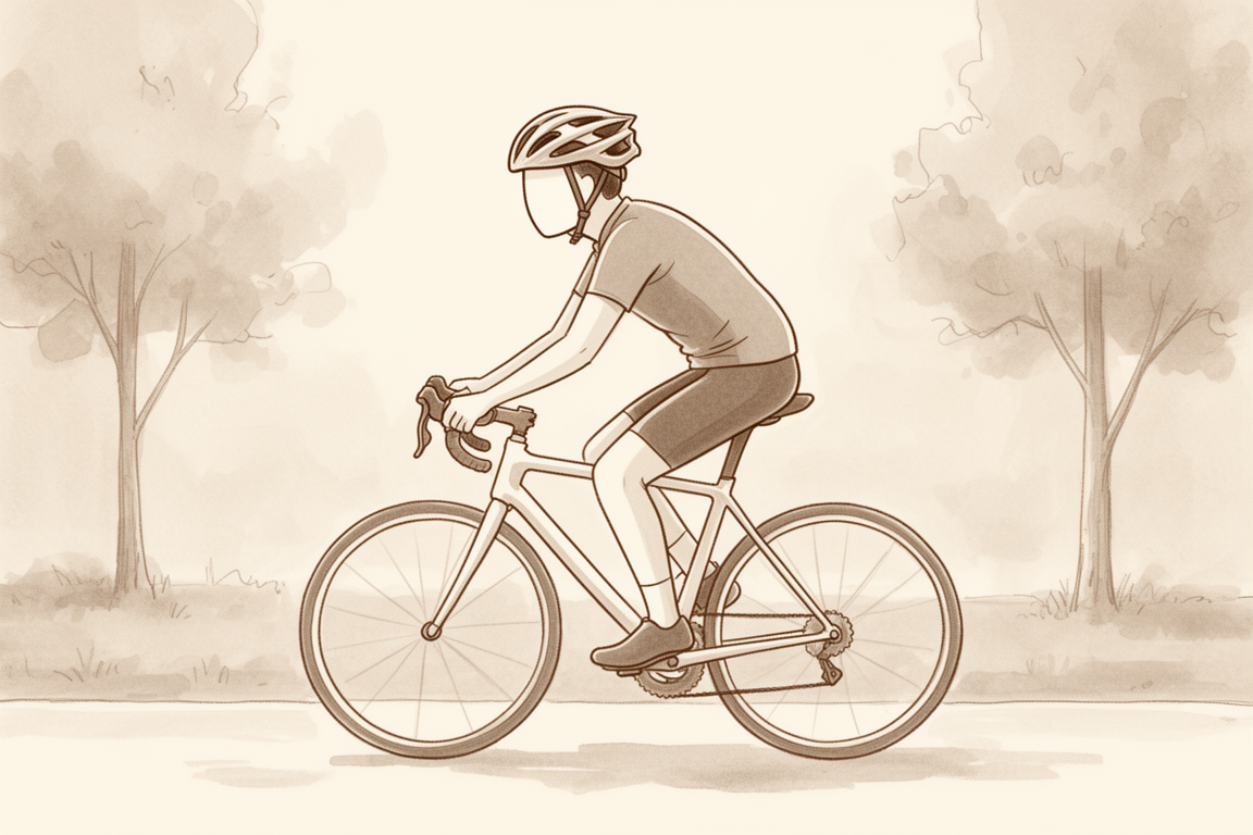
یہ پروٹوکول ایکرومیوکلیویکولر (اے سی) جوڑ (acromioclavicular joint) کو مستحکم کرنے کے آپریشن کے بعد بحالی کے بارے میں ہے، جو ڈاکٹر کیرن ہِرپرا Mater Private Hospital Rockhampton میں کرتے ہیں۔ اس آپریشن میں جوڑ کے اپنی جگہ سے نکل جانے (dislocation) کے بعد ہنسلی کی ہڈی اور کندھے کی پچھلی چپٹی ہڈی (shoulder blade) کے درمیان جوڑ کی سیدھ ایک سسپنسری آلے (suspensory device، یعنی ہڈیوں کو باندھ کر اپنی جگہ تھامنے والا آلہ) کے ذریعے بحال کی جاتی ہے، اور بعض اوقات اسے ٹینڈن (tendon)، یعنی وہ مضبوط ڈوری جو پٹھے کو ہڈی سے جوڑتی ہے، کے ایک ٹکڑے (graft) سے مزید مضبوط کیا جاتا ہے۔ فزیوتھراپی کی اپنی پہلی ملاقات پر یہ صفحہ یا اس کی پی ڈی ایف ساتھ لائیں تاکہ آپ کی بحالی میں سب ایک ساتھ رہیں۔ آپ کا فزیوتھراپسٹ نیچے دیے گئے مراحل میں آپ کی بحالی کو انفرادی طور پر آگے بڑھاتا ہے، اس پر منحصر کہ آپ کا کندھا کیسے بہتر ہو رہا ہے۔
اگر آپریشن کے بعد آپ کو اپنے زخم کے بارے میں کوئی تشویش ہو تو کلینک سے رابطہ کریں۔ اکثر یہ مددگار ہوتا ہے کہ زخم کی تصویر لے کر جائزے کے لیے ای میل کر دیں۔
کیا توقع رکھیں
اے سی جوڑ کو مستحکم کرنے کا آپریشن کندھے کے زیادہ تر دوربین کے ذریعے آپریشنوں (keyhole) سے مختلف ہے: اس میں ایک مرمت ہوتی ہے جسے بھرنے تک محفوظ رکھنا ضروری ہے، اور جو چیز اس پر سب سے زیادہ بوجھ ڈالتی ہے وہ صرف آپ کے اپنے بازو کا وزن ہے۔ کشش ثقل بازو کو (اور اس کے ساتھ کندھے کی پچھلی ہڈی کو) مسلسل نیچے، ہنسلی کی ہڈی سے دور کھینچتی ہے، اور یہی وہ سمت ہے جس کا مقابلہ اس مرمت کو کرنا ہوتا ہے۔ اس لیے ابتدائی بحالی جان بوجھ کر حفاظتی رکھی جاتی ہے: سلنگ (sling)، یعنی گلے سے لٹکی وہ پٹی جو بازو کو سہارا دیتی ہے، بازو کا وزن اٹھاتی ہے، حرکت محفوظ حدود میں رکھی جاتی ہے، اور طاقت بڑھانے کی ورزشیں اس وقت تک انتظار کرتی ہیں جب تک مرمت پختہ نہ ہو جائے۔
سلنگ حفاظت کے لیے ہے۔ اسے 6 ہفتے پہنیں، اور پہلے تین ہفتے سوتے وقت بھی پہنے رکھیں، کیونکہ لیٹے ہوئے بھی بازو کا وزن مرمت کو کھینچتا ہے۔ تین ہفتے بعد اسے بستر میں اتارا جا سکتا ہے۔ جب نہانے یا ورزش کے لیے سلنگ اتری ہو تو بازو کو سہارا دیے رکھیں: بازو کو لٹکنے دینے یا اس سے کچھ اٹھانے کے بجائے بازو کے نچلے حصے کو تکیے یا میز پر ٹکائیں۔ رات کو زیادہ تر لوگ کمر کے بل یا اس پہلو کے بل سب سے زیادہ آرام محسوس کرتے ہیں جس کا آپریشن نہیں ہوا؛ اگر آپ کمر کے بل لیٹیں تو کہنی اور بازو کے نچلے حصے کے نیچے ایک چھوٹا تکیہ کندھے کو پیچھے کی طرف ڈھلکنے سے روکتا ہے۔ مرمت بھرنے تک آپریشن والے کندھے کے بل لیٹنے سے بچیں۔
کندھے کے کسی بھی آپریشن کے بعد چھ ہفتے تک، اور جب تک آپ سلنگ پہنے ہوئے ہیں، آپ ہرگز گاڑی نہ چلائیں۔
آپ کے ورزش کے پروگرام میں تین قسم کی حرکت شامل ہے، اور آپ کی ٹیم نشان لگائے گی کہ کون سی آپ پر لاگو ہوتی ہے:
- ایکٹو حرکت (active range of motion): بغیر کسی سہارے یا مدد کے حرکت کی اجازت ہے۔
- مدد کے ساتھ ایکٹو حرکت (active-assisted range of motion): بازو کو حرکت دینے میں مدد کے لیے دوسرے بازو یا کسی چیز کا استعمال۔
- پیسو حرکت (passive range of motion): بازو پوری طرح ڈھیلا رہتا ہے، اور دوسرا بازو یا کوئی بیرونی قوت 100 فیصد کام کرتی ہے۔
سفر ایک نظر میں:
- پہلا مرحلہ: مرمت کی حفاظت، ہفتے 0 سے 6
- دوسرا مرحلہ: حرکت کی بحالی، ہفتے 6 سے 12
- تیسرا مرحلہ: طاقت بڑھانا، ہفتے 12 سے 18
- چوتھا مرحلہ: کھیل اور بھاری کام پر واپسی، ہفتہ 18 اور اس کے بعد
ہفتوں کی یہ حدیں عام اندازہ ہیں، طے شدہ نہیں؛ اس آپریشن کے شائع شدہ پروٹوکول ایک دوسرے سے مختلف ہیں، اور آپ کا فزیوتھراپسٹ آپ کو کیلنڈر کے بجائے اس بنیاد پر آگے بڑھائے گا کہ مرمت اور آپ کی حرکت کیسی جا رہی ہے۔ زیادہ تر لوگ تقریباً تین ماہ تک روزمرہ کے عام کاموں کے لیے بازو استعمال کرنے لگتے ہیں۔ ایسے کھیلوں میں واپسی جن میں کھلاڑی آپس میں ٹکراتے ہیں (contact and collision sport) عموماً تقریباً چار سے چھ ماہ لیتی ہے، اور کچھ کھیلوں اور پیشوں کے لیے یہ تیاری زیادہ عرصہ چلتی ہے۔
پہلا مرحلہ — مرمت کی حفاظت (ہفتے 0 سے 6)
Kieran Hirpara 4.0
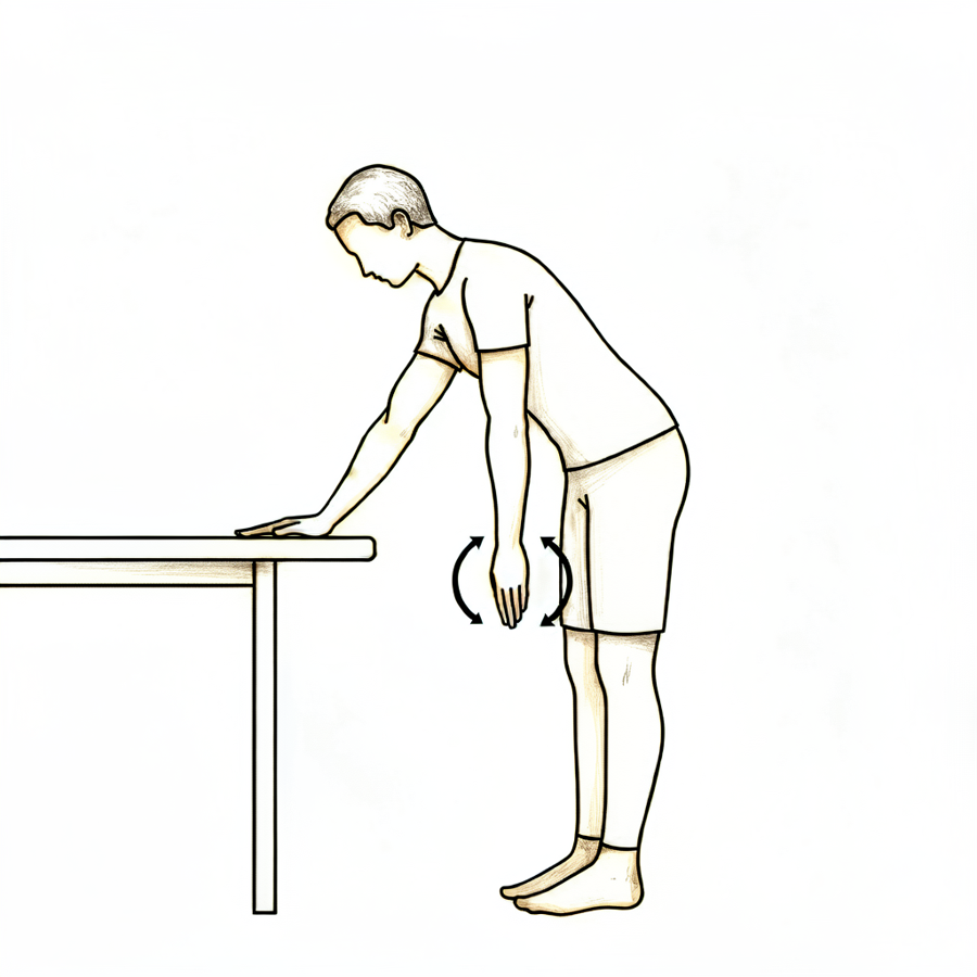
پنڈولم ورزش
سلنگ سے بازو نکال کر کمر سے آگے کی طرف جھکیں اور آپریشن والے بازو کو آہستگی سے جسم سے دور لٹکنے دیں۔ بازو کو چھوٹے، ڈھیلے دائروں میں جھولنے دیں — یہ حرکت آپ کے جسم سے آتی ہے، کندھے کے پٹھوں سے نہیں۔ آرام کی حد کے اندر رہیں، پھر بازو کو سہارا دے کر واپس سلنگ میں رکھیں۔
دن میں کئی بار
Kieran Hirpara 4.0
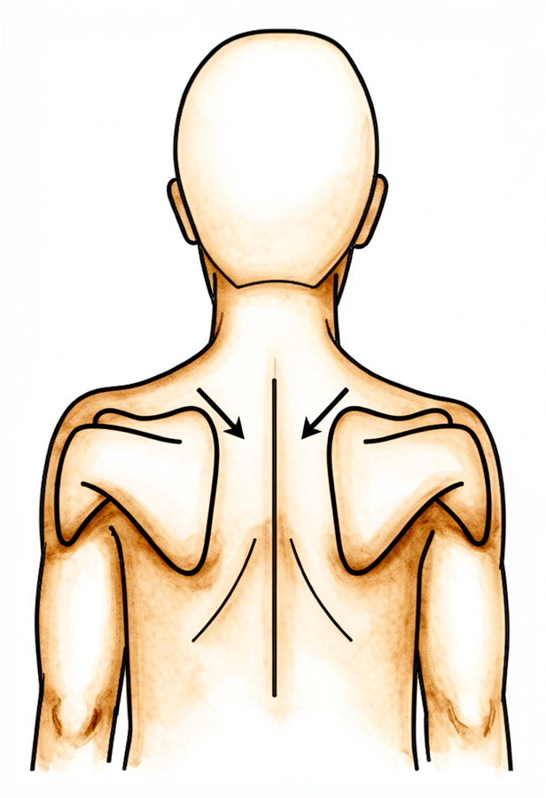
کندھے کی پچھلی ہڈیوں کو جمانا (scapular setting)
بازو کو سہارا دے کر سیدھے بیٹھیں یا کھڑے ہوں، اور آہستگی سے کندھے کی پچھلی چپٹی ہڈیوں کو پیچھے ایک دوسرے کی طرف اور نیچے، کانوں سے دور کھینچیں۔ چند سیکنڈ روکے رکھیں، پھر ڈھیلا چھوڑ دیں۔ اس سے جسم کو سیدھا رکھنے والے پٹھے مرمت پر بوجھ ڈالے بغیر کام کرتے رہتے ہیں۔
جیسا آپ کا فزیوتھراپسٹ بتائے
Kieran Hirpara 4.0
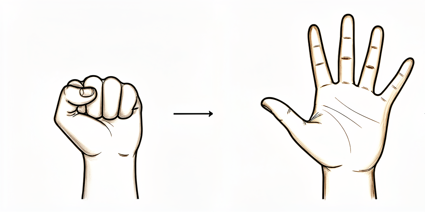
ہاتھ اور گرفت کی ورزشیں
بازو کو سلنگ میں سہارا دیے ہوئے پوری مٹھی بنائیں، پھر انگلیوں کو کھول کر سیدھا کریں۔ گرفت کی ورزش کے لیے نرم گیند یا لپیٹا ہوا موزہ دبایا جا سکتا ہے۔ پہلے دن سے ہاتھ، کلائی اور کہنی کو حرکت دیتے رہیں۔
جیسا آپ کا فزیوتھراپسٹ بتائے
Kieran Hirpara 4.0
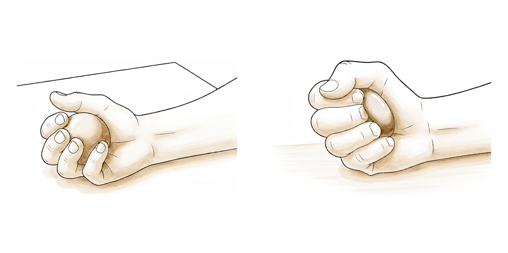
گیند دبانا
بازو کو سلنگ میں سہارا دیے ہوئے نرم گیند یا لپیٹا ہوا موزہ پکڑیں اور اسے مضبوطی سے دبائیں، پھر چھوڑ دیں۔ اس سے کندھا محفوظ رہتے ہوئے گرفت اور بازو کا نچلا حصہ کام کرتے رہتے ہیں۔
جیسا آپ کا فزیوتھراپسٹ بتائے
Kieran Hirpara 4.0
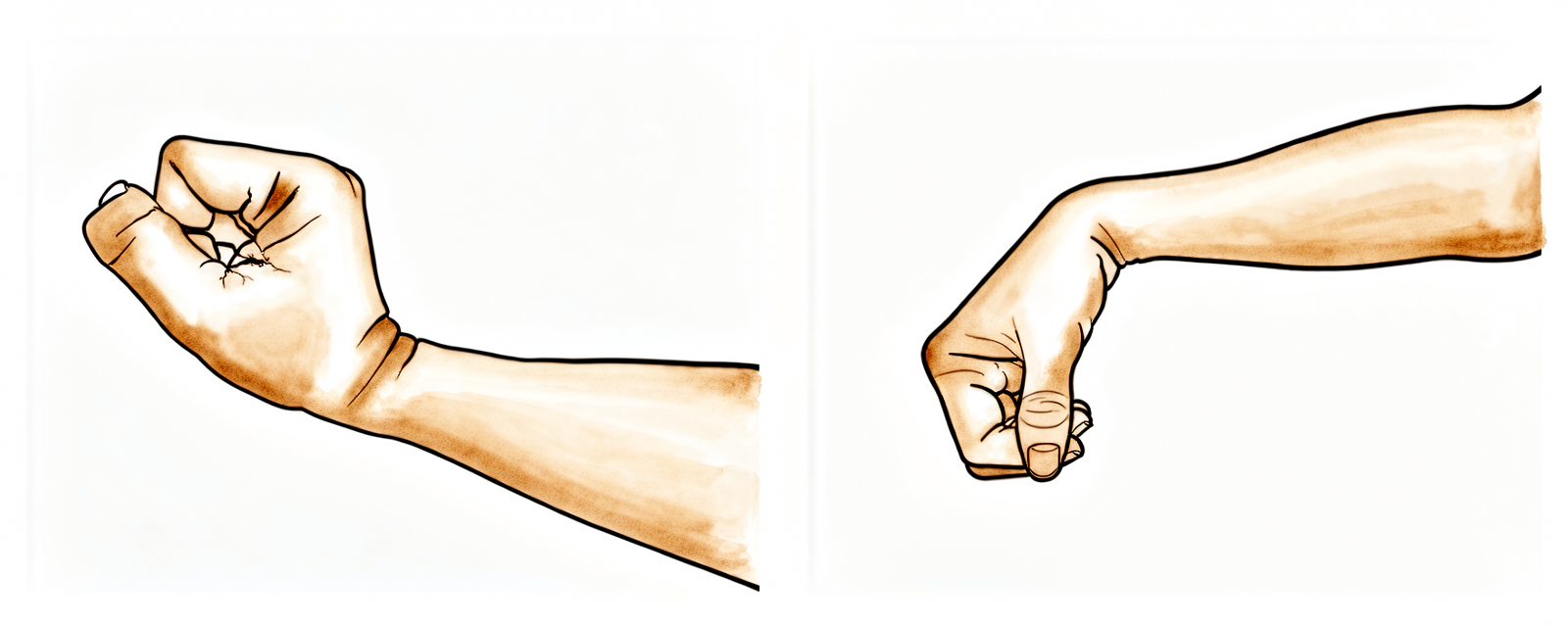
کلائی کی حرکت
بازو کو سلنگ سے نکال کر اور بازو کے نچلے حصے کو سہارا دے کر، کلائی کو آگے، پیچھے اور دائیں بائیں موڑیں۔ پہلے دن سے کلائی کو حرکت دیتے رہنے سے مرمت کے محفوظ رہنے کے دوران کلائی اکڑتی نہیں۔
جیسا آپ کا فزیوتھراپسٹ بتائے
Kieran Hirpara 4.0
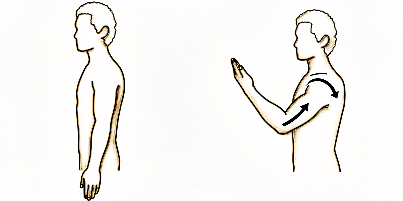
کہنی موڑنا
بازو کے اوپری حصے کو اپنے پہلو کے ساتھ سہارا دے کر کہنی کو جتنا آرام سے ہو سکے موڑیں، پھر اسے پوری طرح سیدھا کریں۔ صرف کہنی حرکت کرتی ہے — کندھے کو ڈھیلا اور سہارا دیے رکھیں تاکہ مرمت پر بوجھ نہ پڑے۔
جیسا آپ کا فزیوتھراپسٹ بتائے
Kieran Hirpara 4.0
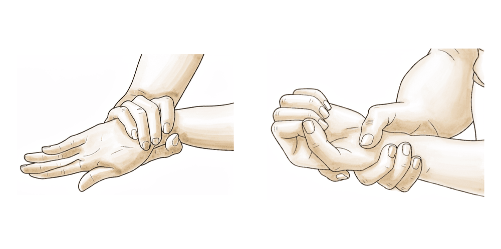
بازو کے نچلے حصے کو گھمانا
کہنی کو اپنے پہلو سے لگا کر اور زاویہ قائمہ تک موڑ کر، ہتھیلی کو اوپر چھت کی طرف پھیریں، پھر نیچے فرش کی طرف۔ بازو کا نچلا حصہ گھومتا ہے جبکہ کندھا ساکن اور سہارا دیا ہوا رہتا ہے۔
جیسا آپ کا فزیوتھراپسٹ بتائے
Kieran Hirpara 4.0
چھڑی کی مدد سے بازو کو باہر کی طرف گھمانا
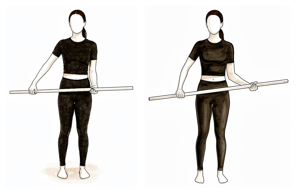
کمر کے بل لیٹیں، آپریشن والی کہنی پہلو سے لگی ہو اور زاویہ قائمہ تک مڑی ہو۔ دونوں ہاتھوں میں ایک چھڑی پکڑیں اور صحیح بازو کی مدد سے آپریشن والے بازو کے نچلے حصے کو آہستگی سے باہر کی طرف گھمائیں، اس مرحلے میں صرف اتنا جتنا آرام سے ہو سکے۔ آہستہ سے واپس لائیں اور حرکت کو چھوٹا اور سہارے کے ساتھ رکھیں۔
جیسا آپ کا فزیوتھراپسٹ بتائے
Kieran Hirpara 4.0
بغیر حرکت کے باہر کی طرف دباؤ (isometric external rotation)
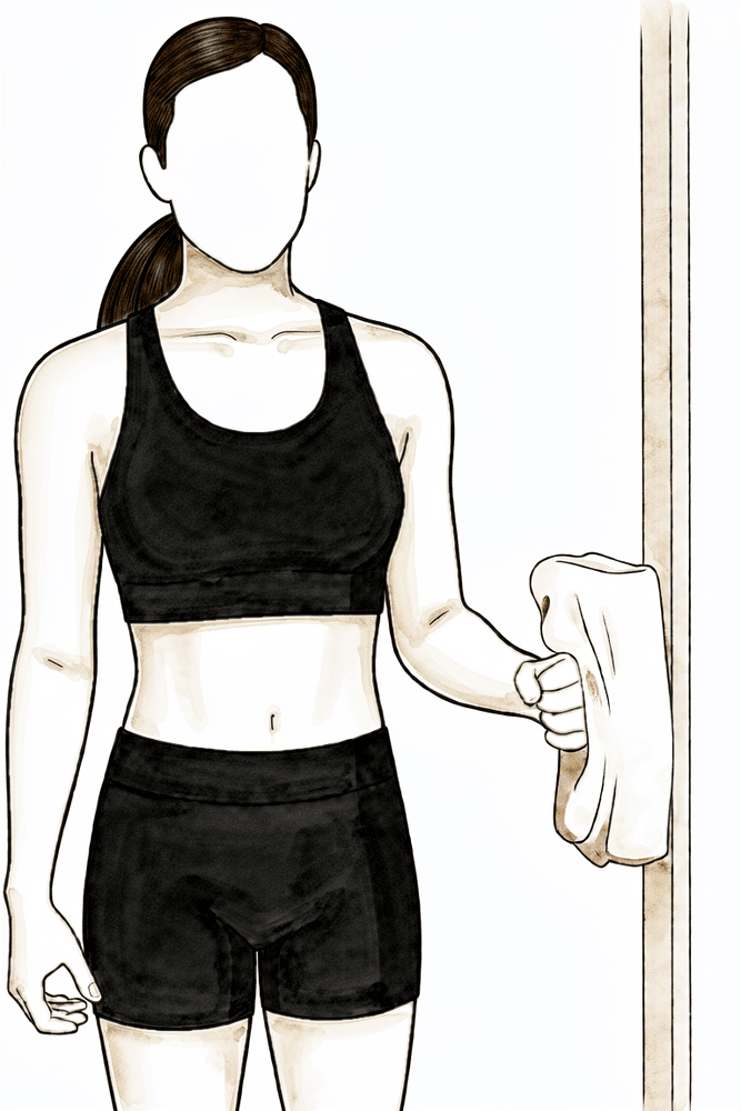
کہنی کو پہلو سے لگا کر اور زاویہ قائمہ تک موڑ کر کھڑے ہوں، ہاتھ کی پشت دیوار یا دروازے کی چوکھٹ سے لگی ہو۔ بازو کو حرکت دیے بغیر آہستگی سے اس پر باہر کی طرف دبائیں، چند سیکنڈ روکے رکھیں، پھر ڈھیلا چھوڑ دیں۔ کندھا کام کرتا ہے لیکن حرکت نہیں کرتا، اس لیے مرمت پر بوجھ نہیں پڑتا۔
پوری طاقت سے کم اور بغیر درد کے؛ جیسا آپ کا فزیوتھراپسٹ بتائے
Kieran Hirpara 4.0
بغیر حرکت کے اندر کی طرف دباؤ (isometric internal rotation)
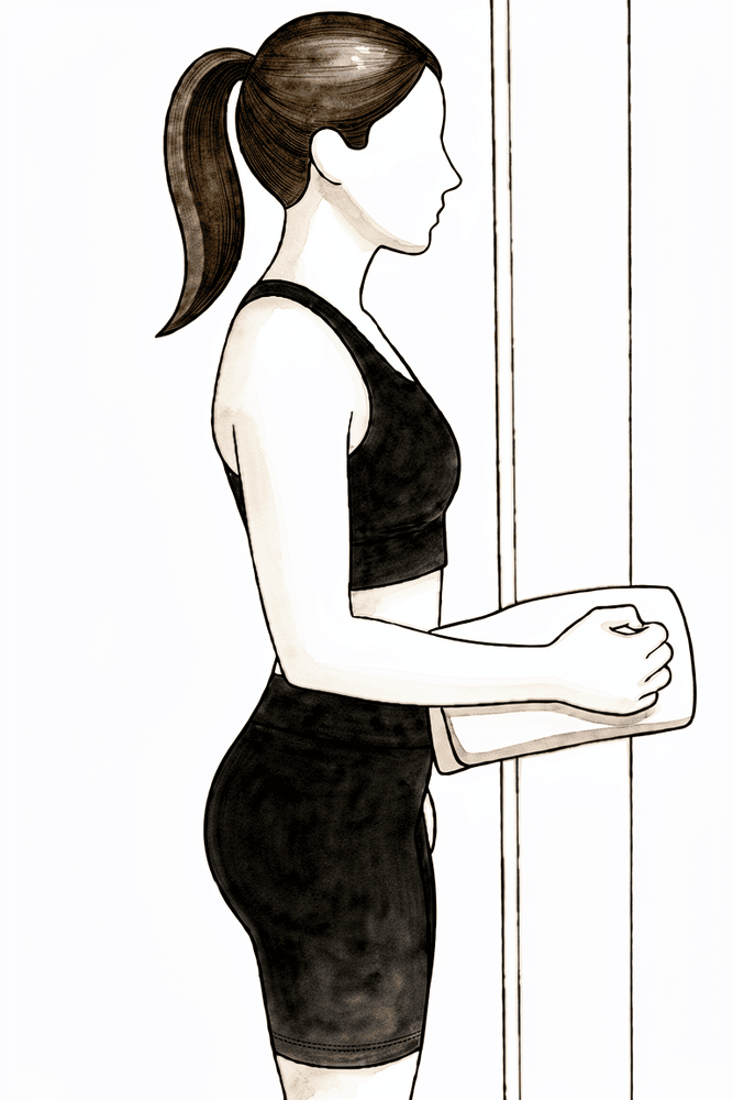
کہنی کو پہلو سے لگا کر اور زاویہ قائمہ تک موڑ کر کھڑے ہوں، ہتھیلی دیوار یا دروازے کی چوکھٹ سے لگی ہو۔ بازو کو حرکت دیے بغیر آہستگی سے اس پر اندر کی طرف دبائیں، چند سیکنڈ روکے رکھیں، پھر ڈھیلا چھوڑ دیں۔ یہ ابتدائی دنوں میں روٹیٹر کف (rotator cuff)، یعنی ٹینڈنوں کا وہ گروپ جو کندھے کو تھامتا اور گھماتا ہے، کو بغیر حرکت کے فعال رکھنے کا طریقہ ہے۔
پوری طاقت سے کم اور بغیر درد کے؛ جیسا آپ کا فزیوتھراپسٹ بتائے
پہلے چھ ہفتوں کا مقصد یہ ہے کہ مرمت بھر جائے اور بازو کا باقی حصہ حرکت کرتا رہے۔ جب بھی آپ اٹھے ہوئے اور چل پھر رہے ہوں، سلنگ بازو کا وزن اٹھاتی ہے۔ آپ کے ہاتھ، کلائی اور کہنی کو پہلے دن سے حرکت کرتے رہنا چاہیے: بازو سلنگ میں ہوتے ہوئے ہاتھ کو لکھنے اور کھانے جیسے ہلکے کاموں کے لیے استعمال کریں۔ خود کندھے کی ورزش ہلکی پنڈولم ورزشوں اور سخت حدود کے اندر مدد والی حرکتوں سے شروع ہوتی ہے: کندھے کی اونچائی سے اوپر کچھ نہیں، جسم کے آر پار ہاتھ بڑھانا نہیں، اور کمر کے پیچھے ہاتھ لے جانا نہیں۔ برف اور باقاعدگی سے درد کش دوا ورزشوں کو قابل برداشت بناتی ہیں۔ اپنی ورزشوں اور فزیوتھراپی کے سیشنوں سے پہلے درد کش دوا لیں۔ جب زخم کی دیکھ بھال کی ہدایات اجازت دیں تو آپ نہا سکتے ہیں؛ آپریشن والے بازو کے نیچے دھونے کے لیے کمر سے آگے جھکیں اور بازو کو آہستگی سے جسم سے دور لٹکنے دیں، یعنی وہی حالت جو پنڈولم ورزش کی ہے۔
اس مرحلے کے دوران آپریشن والے بازو سے تقریباً آدھے کلوگرام سے زیادہ وزنی کوئی چیز نہ اٹھائیں، اس طرف تھیلے نہ اٹھائیں، بازو پر ٹیک نہ لگائیں اور بستر یا کرسی سے اٹھنے کے لیے اس سے زور نہ لگائیں، اور بازو کو بغیر سہارے کے لٹکنے نہ دیں: ان میں سے ہر ایک کام کندھے کی پچھلی ہڈی کو ہنسلی کی ہڈی سے دور نیچے کھینچتا ہے اور مرمت پر بوجھ ڈالتا ہے۔
آپ کے فزیوتھراپسٹ کے لیے:
اہداف
- سرجری سے کی گئی اسٹیبلائزیشن کی حفاظت کریں اور نرم بافتوں (soft tissue) کو بھرنے دیں
- درد اور سوجن پر قابو؛ زخم کے بھرنے کی حفاظت
- محفوظ حدود کے اندر کندھے کے اکڑاؤ کی روک تھام
- ہاتھ، کلائی، کہنی اور گردن کی حرکت کی حد برقرار رکھنا
علاج کا طریقہ
- جب بھی اٹھے ہوئے اور چل پھر رہے ہوں تو 6 ہفتے سلنگ؛ سلنگ اتری ہو تو بازو کو سہارا
- دن میں کئی بار پنڈولم ورزشیں
- کندھے کی پچھلی ہڈی کی سیدھ (plane of the scapula) میں پیسو اور مدد کے ساتھ ایکٹو بلندی (elevation)، 90 ڈگری تک محدود
- آرام کی حد تک پیسو اور مدد کے ساتھ ایکٹو بیرونی گردش (external rotation) (شروع میں تقریباً 30 ڈگری تک)
- ہاتھ، کلائی، کہنی اور بازو کے نچلے حصے کی ایکٹو حرکت؛ گیند دبانا
- کندھے کی پچھلی ہڈی کو جمانا (retraction اور depression) اور جسمانی انداز (posture) کی درستی
- برداشت کے مطابق، نیوٹرل حالت میں پوری طاقت سے کم اور بغیر درد کے آئسومیٹرک (isometric، یعنی بغیر حرکت کے پٹھے کو دبا کر رکھنا) اندرونی اور بیرونی گردش
- ضرورت کے مطابق نرم بافتوں اور اسکیپولوتھوراسک (scapulothoracic) موبلائزیشن؛ زخم بھر جانے کے بعد زخم کے نشان کی مالش
- ضرورت کے مطابق دن میں کئی بار 15 سے 20 منٹ برف؛ ورزشوں اور سیشنوں سے پہلے درد کش دوا
احتیاطیں
- بازو کے وزن کو فکسیشن پر کھنچاؤ نہ ڈالنے دیں: بازو لٹکانا نہیں، کچھ اٹھا کر چلنا نہیں، نیچے کی طرف کھنچاؤ نہیں
- کسی بھی سمت میں 90 ڈگری سے اوپر بلندی نہیں
- تجویز کردہ مدد والے پروگرام سے آگے کندھے کی ایکٹو حرکت نہیں
- جسم کے آر پار (افقی) ایڈکشن (horizontal adduction) نہیں اور کمر کے پیچھے اندرونی گردش نہیں
- تقریباً آدھے کلوگرام سے زیادہ وزن اٹھانا نہیں؛ بازو کے ذریعے جسم کا وزن سہارنا نہیں
- چھ ہفتے تک گاڑی چلانا نہیں (یہ کندھے کے ہر آپریشن پر لاگو ہوتا ہے)، اور سلنگ پہنے ہوئے بھی نہیں
اگلے مرحلے میں جانے کی شرائط
- کندھے کی پچھلی ہڈی کی سیدھ میں تقریباً 90 ڈگری پیسو فلیکشن (flexion)
- کندھے کی پچھلی ہڈی کی سیدھ میں تقریباً 30 ڈگری پیسو بیرونی گردش
- حرکت کی حد اور آئسومیٹرک پروگرام برداشت ہو رہا ہو، اور درد اور سوجن کم ہو رہے ہوں
دوسرا مرحلہ — حرکت کی بحالی (ہفتے 6 سے 12)
Kieran Hirpara 4.0
میز پر ہاتھ پھسلانا
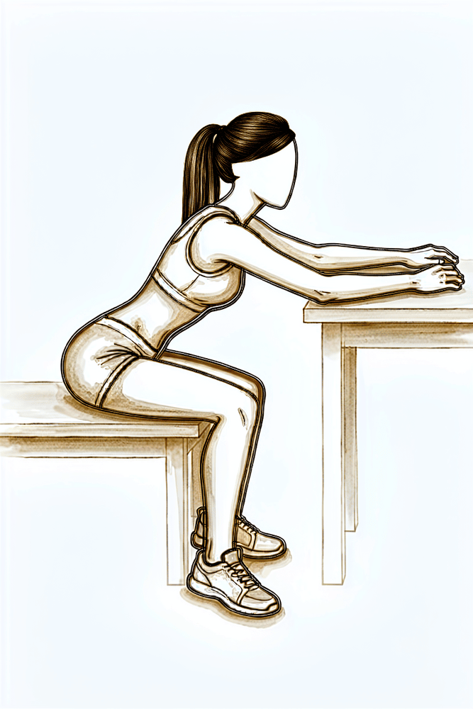
میز یا بینچ کے پاس بیٹھیں اور ہاتھ اس کی سطح پر رکھیں۔ ہاتھوں کو آگے پھسلائیں اور دھڑ کو آگے جھکنے دیں تاکہ کندھا سہارے کے ساتھ اوپر کی طرف اٹھے، پھر واپس آئیں۔ آرام کی حد کے اندر روانی سے حرکت کریں، کندھے کو کان کی طرف اچکائے بغیر۔
جیسا آپ کا فزیوتھراپسٹ بتائے
Kieran Hirpara 4.0
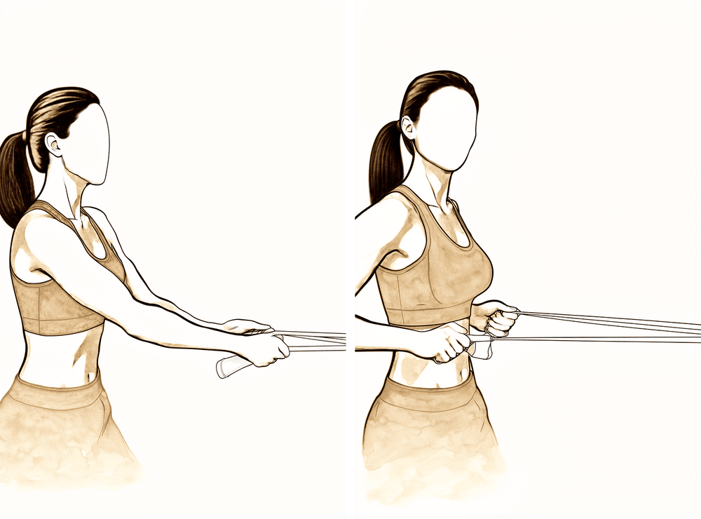
لچکدار بینڈ سے رو (row)
ورزش والے لچکدار بینڈ کو اپنے سامنے تقریباً کمر کی اونچائی پر باندھیں۔ بینڈ کو اپنی طرف کھینچیں، اور جب کہنی آپ کے پہلو سے گزرے تو کندھے کی پچھلی ہڈی کو پیچھے اور نیچے کھینچیں، پھر قابو کے ساتھ واپس جائیں۔ اس مرحلے میں مزاحمت ہلکی رکھیں۔
جیسا آپ کا فزیوتھراپسٹ بتائے
Kieran Hirpara 4.0
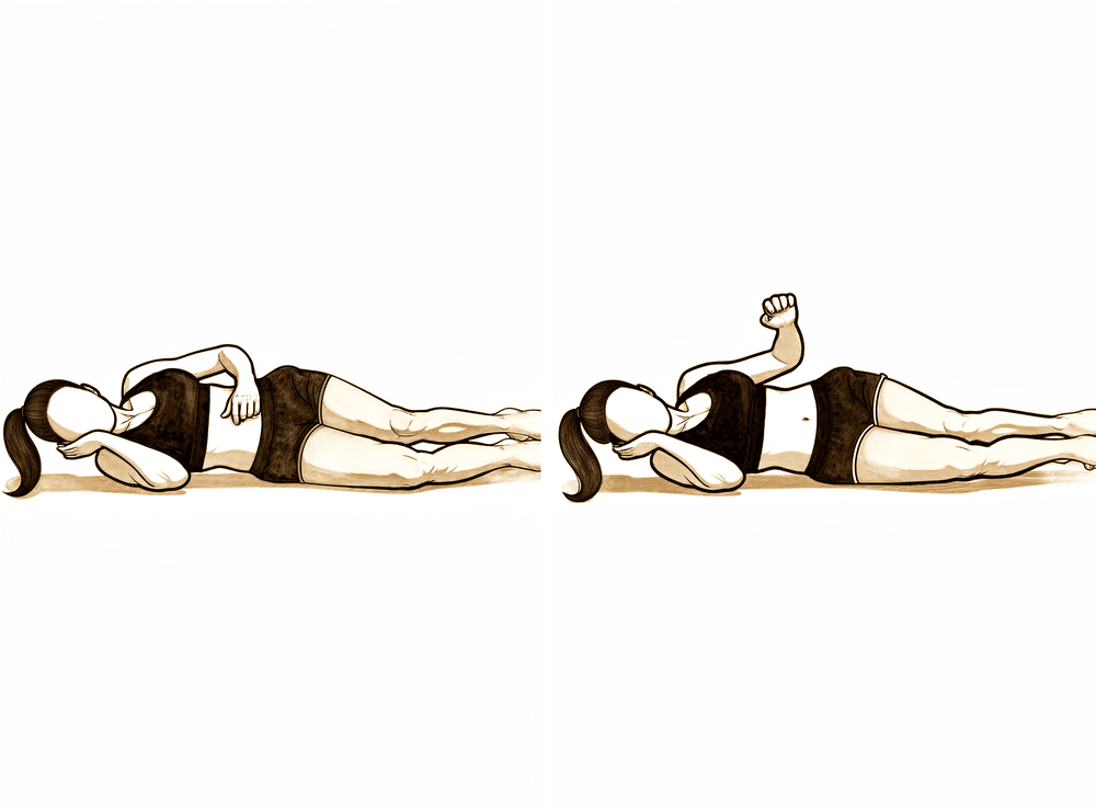
پہلو کے بل لیٹ کر بازو کو باہر کی طرف گھمانا
اس پہلو کے بل لیٹیں جس کا آپریشن نہیں ہوا، اوپر والی کہنی زاویہ قائمہ تک مڑی ہو اور جسم سے لگی ہو۔ کہنی کو پہلو سے لگائے رکھتے ہوئے بازو کے نچلے حصے کو اوپر چھت کی طرف گھمائیں، پھر قابو کے ساتھ نیچے لائیں۔ اس مرحلے میں کوئی وزن نہیں — وزن بعد میں، ہدایت کے مطابق، شامل کیا جاتا ہے۔
جیسا آپ کا فزیوتھراپسٹ بتائے
Kieran Hirpara 4.0
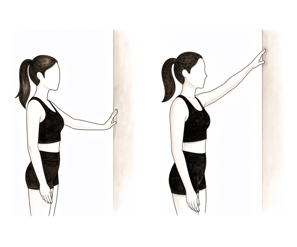
دیوار پر انگلیوں سے چلنا
دیوار کی طرف منہ کر کے کھڑے ہوں اور آپریشن والے ہاتھ کی انگلیاں اس پر رکھیں۔ انگلیوں کو آہستہ آہستہ دیوار پر اوپر چلائیں، اور کندھا اٹھتے وقت انہیں بازو کا کچھ وزن اٹھانے دیں، پھر واپس نیچے چلائیں۔ کندھے کی پچھلی ہڈی کو ڈھیلا رکھیں — کندھے کو کان کی طرف نہ اچکائیں۔
جیسا آپ کا فزیوتھراپسٹ بتائے
Kieran Hirpara 4.0
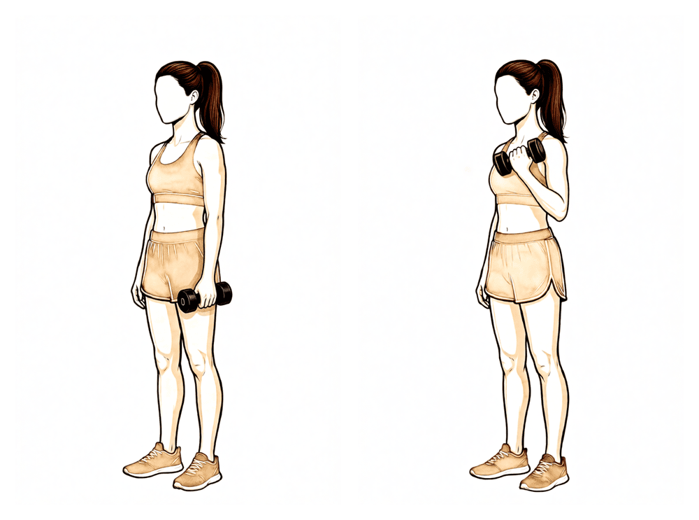
بائسیپس کرل
بازو کے اوپری حصے کو پہلو کے ساتھ رکھ کر کہنی موڑیں اور ہلکا وزن کندھے کی طرف اوپر لائیں، پھر آہستہ سے نیچے کریں۔ اس مرحلے میں وزن ہلکا رکھیں — اس سے مرمت پر زور ڈالے بغیر بازو دوبارہ مضبوط ہوتا ہے۔
جیسا آپ کا فزیوتھراپسٹ بتائے
Kieran Hirpara 4.0
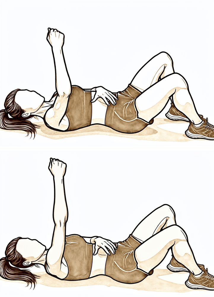
سیریٹس پنچ (serratus punch)
کمر کے بل لیٹیں اور آپریشن والا بازو اوپر چھت کی طرف سیدھا رکھیں۔ کہنی موڑے بغیر مٹھی کو تھوڑا اور اوپر لے جائیں تاکہ کندھے کی پچھلی ہڈی بستر سے اٹھ جائے، پھر قابو کے ساتھ نیچے لائیں۔ اس سے وہ پٹھا فعال ہوتا ہے جو کندھے کی پچھلی ہڈی کو پسلیوں سے لگائے رکھتا ہے۔
جیسا آپ کا فزیوتھراپسٹ بتائے
Kieran Hirpara 4.0
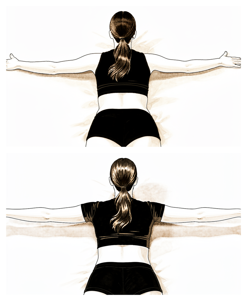
الٹا لیٹ کر 'T' ورزش (prone T)
پیٹ کے بل لیٹیں اور بازو فرش کی طرف لٹکنے دیں۔ کندھے کی پچھلی ہڈیوں کو ایک دوسرے کی طرف دبائیں اور آپریشن والے بازو کو پہلو کی طرف اٹھا کر 'T' کی شکل بنائیں، پھر آہستہ سے نیچے لائیں۔ اسے ہلکا رکھیں — اس سے جوڑ پر زیادہ بوجھ ڈالے بغیر کندھے کی پچھلی ہڈی کے پٹھوں کی تربیت ہوتی ہے۔
جیسا آپ کا فزیوتھراپسٹ بتائے
Kieran Hirpara 4.0
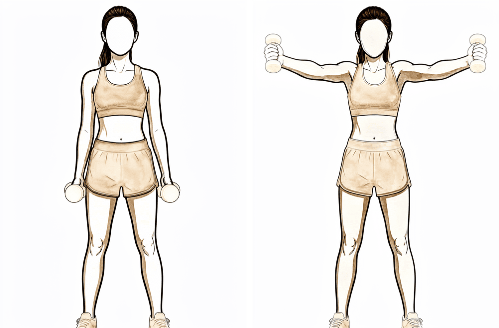
کھڑے ہو کر سکیپشن (scaption)
بازو پہلو کے ساتھ رکھ کر کھڑے ہوں، انگوٹھا اوپر کی طرف ہو۔ بازو کو آگے کی طرف ترچھے زاویے پر اٹھائیں، یعنی بالکل سامنے اور بالکل پہلو کے درمیان، اتنا ہی اونچا جتنا آرام سے ہو سکے، پھر آہستہ سے نیچے لائیں۔ کندھے کی پچھلی ہڈی کو جما کر رکھیں اور کندھے اچکانے سے بچیں۔
جیسا آپ کا فزیوتھراپسٹ بتائے
Kieran Hirpara 4.0
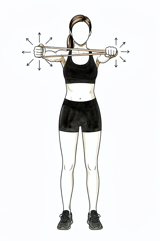
ردھمک اسٹیبلائزیشن (rhythmic stabilisation)
بازو کو اپنے سامنے تقریباً کندھے کی اونچائی پر سہارا دے کر رکھیں، اور آپ کا فزیوتھراپسٹ (یا آپ کا صحیح ہاتھ) مختلف سمتوں سے چھوٹے، ہلکے دھکے دے جبکہ آپ بازو کو ساکن اور مستحکم رکھیں — اسے حرکت نہ کرنے دیں۔ اس سے کندھے کو مستحکم رکھنے والے پٹھوں کو ردعمل دینے کی تربیت ملتی ہے۔ ساری کوشش ہلکی رکھیں۔
جیسا آپ کا فزیوتھراپسٹ بتائے
تقریباً چھ ہفتے سے آپ آہستہ آہستہ سلنگ چھوڑتے ہیں اور حرکت پر پابندیاں بتدریج اٹھائی جاتی ہیں۔ مدد والی حرکتیں ایکٹو حرکتیں بن جاتی ہیں، اور حرکت کی حد مسلسل بڑھتی ہے؛ رہنمائی کے طور پر، شائع شدہ پروٹوکول ہر ہفتے تقریباً 15 ڈگری کے قدموں میں حد بڑھاتے ہیں، اور تقریباً ہفتہ 12 تک پوری حرکت کا ہدف رکھتے ہیں۔ اس مرحلے کے دوران روٹیٹر کف (rotator cuff)، یعنی ٹینڈنوں کا وہ گروپ جو کندھے کو تھامتا اور گھماتا ہے، اور کندھے کی پچھلی ہڈی کے پٹھوں کے لیے ہلکے لچکدار بینڈ کی ورزشیں شروع ہوتی ہیں۔ مرمت کو اب بھی احتیاط کی ضرورت ہے: وزن اٹھانا تقریباً ایک کلوگرام تک رکھیں، اور زور سے دھکیلنے اور کھینچنے، پُش اپ، اور جسم کے آر پار یا سر کے اوپر وزن اٹھانے سے بچیں۔ کمر کے پیچھے ہاتھ لے جانا عموماً وہ آخری حرکت ہوتی ہے جس کی اجازت ملتی ہے۔
آپ کے فزیوتھراپسٹ کے لیے:
اہداف
- سلنگ سے بتدریج چھٹکارا
- ہر سمت میں ایکٹو حرکت کی حد کی بتدریج بحالی (تقریباً ہفتہ 12 تک پوری، یا تقریباً پوری)
- ہلکی طاقت بڑھانے کی ورزشیں شروع کرنا؛ پٹھوں کے سکڑنے (atrophy) کو کم سے کم رکھنا
- کندھے اور کندھے کی پچھلی ہڈی کی ہم آہنگ حرکت (scapulohumeral rhythm) اور اعصابی و عضلاتی کنٹرول (neuromuscular control) کی بحالی
علاج کا طریقہ
- مدد کے ساتھ ایکٹو سے ایکٹو بلندی کی طرف بڑھنا؛ فلیکشن میں دیوار اور میز پر ہاتھ پھسلانا
- کندھے کی پچھلی ہڈی کی سیدھ میں بلندی، اس ہڈی کی حرکت کے طریقے پر توجہ کے ساتھ: کندھا اچکانا نہیں
- کمر کے پیچھے اندرونی گردش بتدریج شروع کرنا (شروع میں بیلٹ کی لائن تک)
- افقی ایڈکشن صرف ایکٹو ہاتھ بڑھانے کے طور پر: ابھی پیسو اسٹریچ نہیں
- تھیرابینڈ (Theraband) سے اندرونی اور بیرونی گردش، بائسیپس کرل، رو اور سیریٹس پنچ
- کندھے کی پچھلی ہڈی اور روٹیٹر کف کی متحرک ورزشیں: پہلو کے بل لیٹ کر بیرونی گردش، الٹا لیٹ کر رو، الٹا لیٹ کر ایکسٹینشن، الٹا لیٹ کر 'T' اور 'Y'، کھڑے ہو کر سکیپشن
- جوڑ کی پوزیشن کے احساس (proprioception) اور ردھمک اسٹیبلائزیشن کی مشقیں
- ضرورت کے مطابق مینوئل تھراپی اور جوڑ کی موبلائزیشن
احتیاطیں
- آپریشن والے بازو سے تقریباً ایک کلوگرام سے زیادہ وزن اٹھانا نہیں
- زور سے دھکیلنا یا کھینچنا نہیں؛ پُش اپ نہیں
- سر کے اوپر یا جسم کے آر پار وزنی چیزیں اٹھانا نہیں
- جسم کے آر پار ایڈکشن میں آخری حد تک اسٹریچ سے بچیں؛ کمر کے پیچھے کی حرکت بتدریج آگے بڑھتی ہے
اگلے مرحلے میں جانے کی شرائط
- کندھے کی پچھلی ہڈی کی سیدھ میں کم از کم تقریباً 140 ڈگری پیسو فلیکشن اور 60 ڈگری پیسو بیرونی گردش
- اچھے طریقے کے ساتھ کشش ثقل کے خلاف کم از کم تقریباً 100 ڈگری تک ایکٹو فلیکشن
- ایکٹو حرکت کی حد اور ابتدائی طاقت کا پروگرام برداشت ہو رہا ہو
تیسرا مرحلہ — طاقت بڑھانا (ہفتے 12 سے 18)
Kieran Hirpara 4.0
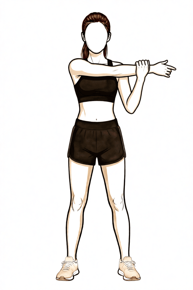
جسم کے آر پار اسٹریچ
آپریشن والے بازو کو سینے کے آر پار لائیں اور دوسرے ہاتھ کو کہنی سے اوپر رکھ کر اسے آہستگی سے مزید آر پار کھینچیں، یہاں تک کہ کندھے کے پچھلے حصے میں کھنچاؤ محسوس ہو۔ روکے رکھیں، پھر چھوڑ دیں۔ یہ اسٹریچ پہلے منع تھا اور اب حرکت کی حد کی آخری ڈگریاں واپس حاصل کرنے کے لیے استعمال ہوتا ہے۔
جیسا آپ کا فزیوتھراپسٹ بتائے
Kieran Hirpara 4.0
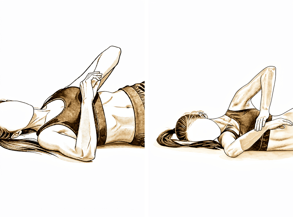
سلیپر اسٹریچ (sleeper stretch)
آپریشن والے پہلو کے بل لیٹیں، کندھا اور کہنی دونوں زاویہ قائمہ پر مڑے ہوں۔ دوسرے ہاتھ سے بازو کے نچلے حصے کو آہستگی سے بستر کی طرف نیچے لائیں، یہاں تک کہ کندھے کے پچھلے حصے میں کھنچاؤ محسوس ہو۔ روکے رکھیں، پھر ڈھیلا چھوڑ دیں — کھنچاؤ، درد نہیں۔
جیسا آپ کا فزیوتھراپسٹ بتائے
Kieran Hirpara 4.0
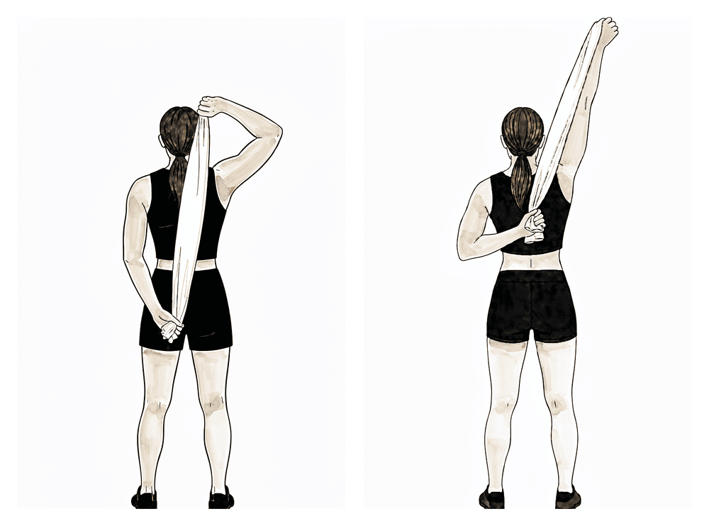
تولیے کی مدد سے کمر کے پیچھے اسٹریچ
کمر کے پیچھے ایک تولیہ پکڑیں، صحیح ہاتھ کندھے کے اوپر سے اور آپریشن والا ہاتھ کمر کے نچلے حصے پر۔ صحیح ہاتھ سے تولیے کو آہستگی سے اوپر کھینچیں تاکہ آپریشن والا ہاتھ کمر پر اوپر سرکے، یہاں تک کہ کھنچاؤ محسوس ہو، پھر چھوڑ دیں۔ کمر کے پیچھے کی حرکت سب سے آخر میں واپس آنے والی حرکتوں میں سے ہے، اس لیے اسے آہستگی سے آگے بڑھائیں۔
جیسا آپ کا فزیوتھراپسٹ بتائے
Kieran Hirpara 4.0
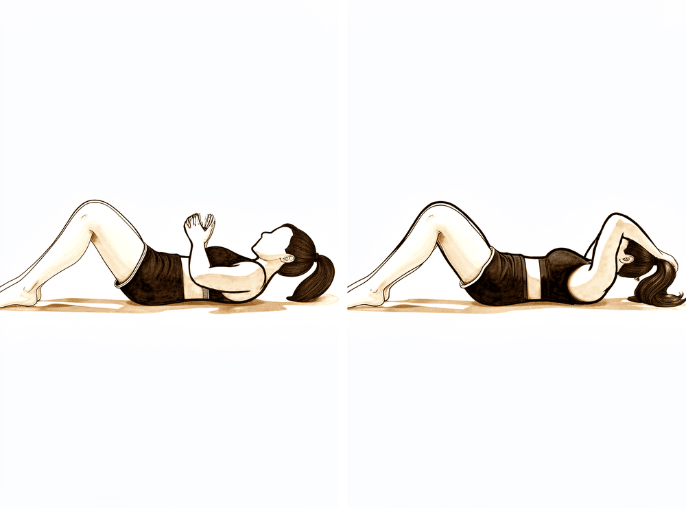
ہاتھ سر کے پیچھے
دونوں ہاتھ سر کے پیچھے باندھیں اور کہنیوں کو آہستگی سے باہر اور پیچھے کی طرف لے جائیں، یہاں تک کہ کندھے کے اگلے حصے میں کھنچاؤ محسوس ہو۔ روکے رکھیں، پھر ڈھیلا چھوڑ دیں۔ اس سے بازو اٹھا ہونے کی حالت میں باہر کی طرف گھماؤ کی آخری حد واپس ملتی ہے۔
جیسا آپ کا فزیوتھراپسٹ بتائے
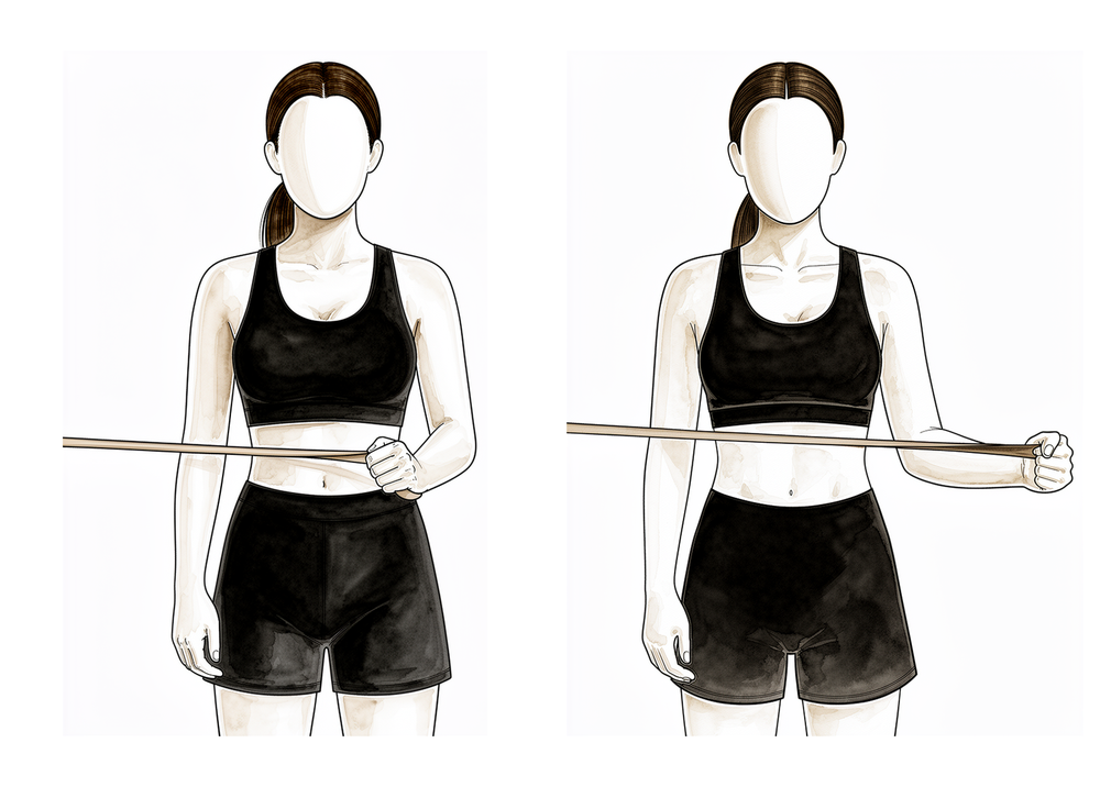
Kieran Hirpara 4.0
90 ڈگری پر باہر کی طرف گھمانا
لچکدار بینڈ پکڑ کر بازو کو کندھے کی اونچائی تک پہلو کی طرف اٹھائیں اور کہنی زاویہ قائمہ پر موڑیں۔ بینڈ کی مزاحمت کے خلاف بازو کے نچلے حصے کو اوپر گھمائیں تاکہ ہاتھ کی پشت اوپر جائے، پھر آہستہ سے واپس لائیں۔ جب حرکت اور کنٹرول اچھے ہو جائیں تو یہ روٹیٹر کف کی ایک اعلیٰ درجے کی ورزش ہے۔
جیسا آپ کا فزیوتھراپسٹ بتائے
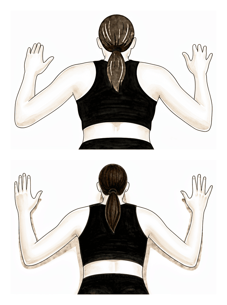
Kieran Hirpara 4.0
الٹا لیٹ کر 'W' ورزش (prone W)
پیٹ کے بل لیٹیں اور بازو لٹکنے دیں۔ کندھے کی پچھلی ہڈیوں کو ایک دوسرے کی طرف دبائیں اور کہنیوں کو پیچھے اور نیچے کھینچیں تاکہ بازو 'W' کی شکل بنائیں، پھر آہستہ سے نیچے لائیں۔ اس سے کندھے کی پچھلی ہڈی کے نچلے پٹھے مضبوط ہوتے ہیں جو جوڑ کو مستحکم رکھتے ہیں۔
جیسا آپ کا فزیوتھراپسٹ بتائے
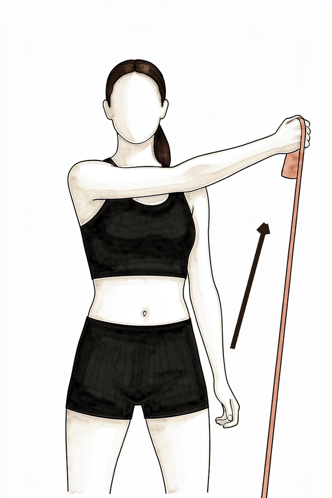
Kieran Hirpara 4.0
بینڈ کے ساتھ ترچھی حرکت (D1)
لچکدار بینڈ پکڑیں اور اسے ترچھی سمت میں کھینچیں، جسم کے سامنے سے آر پار ہوتے ہوئے اوپر اور مخالف سمت میں باہر کی طرف، پھر آہستہ سے واپس لائیں۔ یہ ترچھی حرکتیں کندھے کی اسی طرح تربیت کرتی ہیں جیسے وہ روزمرہ میں ہاتھ بڑھاتے وقت کام کرتا ہے۔
جیسا آپ کا فزیوتھراپسٹ بتائے
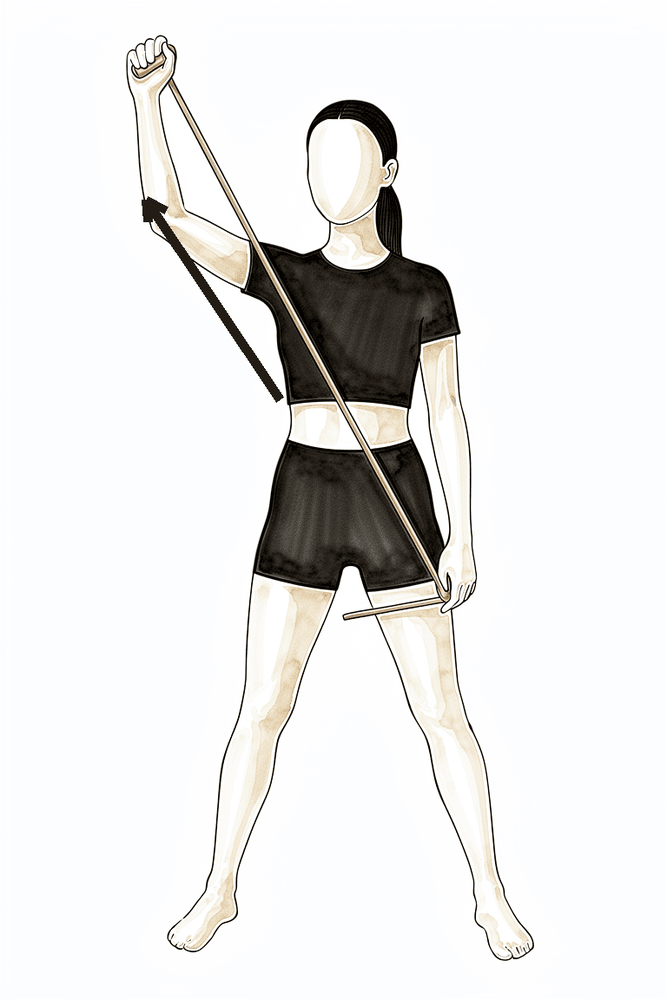
Kieran Hirpara 4.0
بینڈ کے ساتھ ترچھی حرکت (D2)
لچکدار بینڈ کو نیچے اور جسم کے آر پار پکڑیں، پھر اسے ترچھی سمت میں اوپر اور سر کے اوپر سے پہلو کی طرف باہر کھینچیں، آخر میں انگوٹھا اوپر کی طرف ہو، اور آہستہ سے واپس لائیں۔ یہ دوسری ترچھی حرکت ہے جو زیادہ وسیع حد میں کنٹرول بہتر بناتی ہے۔
جیسا آپ کا فزیوتھراپسٹ بتائے
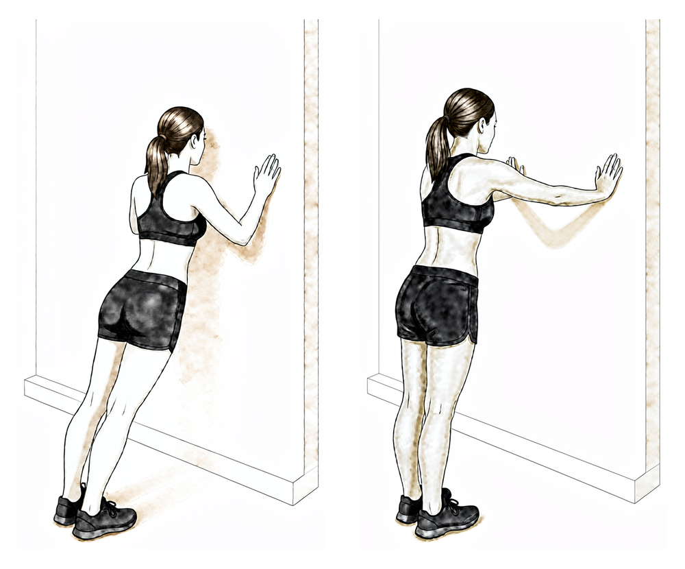
Kieran Hirpara 4.0
دیوار پر پُش اپ
دیوار کی طرف منہ کر کے کھڑے ہوں اور ہاتھ کندھے کی اونچائی پر دیوار پر رکھیں۔ کہنیاں موڑ کر سینے کو دیوار کی طرف لائیں، پھر پیچھے دھکیلیں؛ آخر میں تھوڑا اور دھکیلیں تاکہ کمر کا اوپری حصہ دیوار سے دور گولائی میں مڑ جائے۔ آگے بڑھنے سے پہلے دیوار سے شروع کریں — کندھے کی پچھلی ہڈیوں کو قابو میں رکھیں۔
جیسا آپ کا فزیوتھراپسٹ بتائے
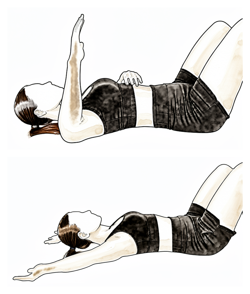
Kieran Hirpara 4.0
90/90 پر باہر کی طرف گھمانا (بینڈ)
بازو کو کندھے کی اونچائی پر پہلو کی طرف رکھ کر اور کہنی زاویہ قائمہ پر موڑ کر، بازو کے نچلے حصے کو ہلکے بینڈ یا وزن کے خلاف پیچھے اور اوپر گھمائیں، پھر آہستہ سے نیچے لائیں۔ اس سے بازو اٹھا ہونے کی (پھینکنے والی) حالت میں روٹیٹر کف کی طاقت بڑھتی ہے۔
10 سے 15 بار کے 2 سے 3 سیٹ، کم مزاحمت
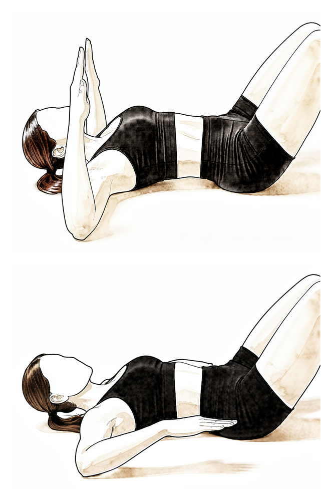
Kieran Hirpara 4.0
90/90 پر اندر کی طرف گھمانا (بینڈ)
اسی حالت سے، یعنی بازو کندھے کی اونچائی پر پہلو کی طرف رکھ کر، بازو کے نچلے حصے کو ہلکے بینڈ یا وزن کے خلاف آگے اور نیچے پاؤں کی طرف گھمائیں، پھر قابو کے ساتھ واپس لائیں۔
10 سے 15 بار کے 2 سے 3 سیٹ، کم مزاحمت
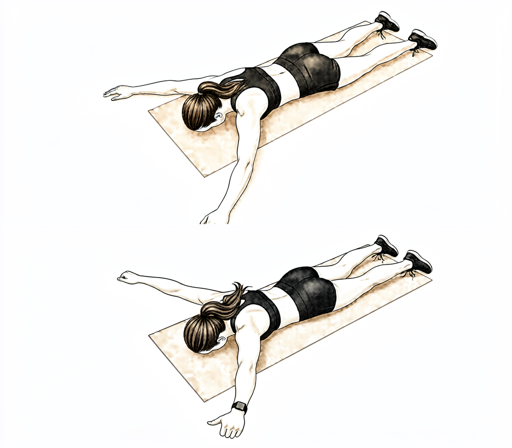
Kieran Hirpara 4.0
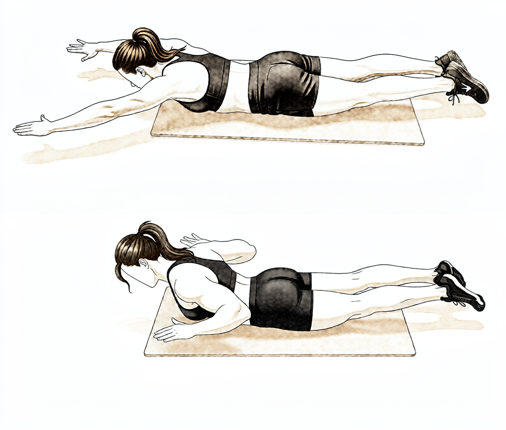
الٹا لیٹ کر 'Y' ورزش (کندھے کی پچھلی ہڈی کا نچلا حصہ)
پیٹ کے بل لیٹیں، آپریشن والا بازو سر کے اوپر کی طرف تقریباً 45° کے زاویے پر باہر پھیلا کر Y کی شکل بنائیں، انگوٹھا اوپر کی طرف ہو۔ کندھے کی پچھلی ہڈی کو نیچے اور پیچھے کھینچ کر بازو کو سطح سے تھوڑا سا اٹھائیں، پھر آہستہ سے نیچے لائیں۔ اسے ہلکا رکھیں — یہ کندھے کی پچھلی ہڈی کے نچلے پٹھوں کو نشانہ بناتی ہے۔
جیسا آپ کا فزیوتھراپسٹ بتائے — ہلکا وزن، زیادہ بار
Kieran Hirpara 4.0
الٹا لیٹ کر رو (prone row)
پیٹ کے بل لیٹیں اور آپریشن والا بازو فرش کی طرف نیچے لٹکنے دیں۔ کہنی کو اوپر چھت کی طرف کھینچیں اور کندھے کی پچھلی ہڈی کو ریڑھ کی ہڈی کی طرف دبائیں، پھر آہستہ سے نیچے لائیں۔ بغیر وزن کے شروع کریں اور حرکت کو قابو میں رکھیں۔
جیسا آپ کا فزیوتھراپسٹ بتائے
جب مرمت پختہ ہو چکی ہو اور حرکت زیادہ تر بحال ہو گئی ہو تو توجہ طاقت کی دوبارہ تعمیر کی طرف جاتی ہے۔ وہ اسٹریچ جو پہلے منع تھے (جسم کے آر پار اور کمر کے پیچھے اوپر کی طرف) اب حرکت کی حد کی آخری ڈگریاں واپس حاصل کرنے کے لیے استعمال ہوتے ہیں۔ مزاحمت والی ورزشیں لچکدار بینڈ سے ہلکے وزن کی طرف بڑھتی ہیں، اور پُش اپ پہلے دیوار کے ساتھ شروع ہوتے ہیں اور پھر آگے بڑھتے ہیں۔ جم میں وزن کی تربیت عموماً تقریباً ہفتہ 16 سے دوبارہ شروع کی جاتی ہے، شروع میں محدود حد اور ہلکے وزن کے ساتھ۔ اس مرحلے میں بھی سر کے اوپر بھاری وزن اٹھانے اور زور سے دھکیلنے اور کھینچنے سے بچا جاتا ہے۔
آپ کے فزیوتھراپسٹ کے لیے:
اہداف
- ہر سمت میں پوری ایکٹو اور پیسو حرکت کی حد
- روٹیٹر کف اور کندھے کی پچھلی ہڈی کو مستحکم رکھنے والے پٹھوں کی طاقت، برداشت اور اعصابی و عضلاتی کنٹرول میں بتدریج اضافہ
- کھیل کے مطابق بوجھ پر مرحلہ وار واپسی کی تیاری
علاج کا طریقہ
- کئی سمتوں میں آخری حد تک اسٹریچ: جسم کے آر پار اسٹریچ، کمر کے پیچھے اندرونی گردش، ہاتھ سر کے پیچھے، سلیپر اسٹریچ، 90 ڈگری ایبڈکشن (abduction) پر بیرونی گردش
- متحرک پروگرام میں بتدریج مزاحمت (تقریباً 0.5 سے 2.5 کلوگرام) شامل کرنا: پہلو کے بل لیٹ کر بیرونی گردش، الٹا لیٹ کر رو، الٹا لیٹ کر ایکسٹینشن، الٹا لیٹ کر 'T' اور 'Y'، کھڑے ہو کر سکیپشن
- تھیرابینڈ کی اگلی ورزشیں: 'T'، 'W'، ترچھی حرکتیں، 90 ڈگری پر اندرونی اور بیرونی گردش
- کلوزڈ چین (closed-chain) ورزشیں: تقریباً ہفتہ 12 سے دیوار پر پُش اپ، برداشت کے مطابق آگے بڑھانا
- تقریباً ہفتہ 16 سے مشینوں پر وزن کی تربیت: محدود حد، ہلکا وزن (رو، پُل ڈاؤن، بائسیپس اور ٹرائسیپس؛ دھکیلنے والی ورزشیں احتیاط سے دوبارہ شروع)
- ردھمک اسٹیبلائزیشن، جوڑ کی پوزیشن کا احساس، اور کندھے اور کندھے کی پچھلی ہڈی کی ہم آہنگ حرکت کی مشقیں
احتیاطیں
- بھاری وزن اٹھانے سے بچیں، خاص طور پر سر کے اوپر، اور زور سے دھکیلنے اور کھینچنے سے بھی
- طاقت کی ورزشیں بغیر درد کے رہیں اور تکلیف پیدا کرنے والی آخری حد کے بوجھ سے پہلے رک جائیں
- پوری حد میں کندھے کی پچھلی ہڈی کا کنٹرول برقرار رکھیں: اگر کندھا اچکنے یا متبادل حرکت (compensation) ظاہر ہو تو بوجھ کم کریں
اگلے مرحلے میں جانے کی شرائط
- ہر سمت میں کندھے کی ایکٹو اور پیسو حرکت روزمرہ کے کام کی حدود کے اندر
- طاقت بڑھانے کا بتدریج پروگرام بغیر تکلیف بھڑکے برداشت ہو رہا ہو
چوتھا مرحلہ — کھیل اور بھاری کام پر واپسی (ہفتہ 18 اور اس کے بعد)
آخری مرحلہ محنت طلب کام، سر کے اوپر بوجھ اور کھیل کی طرف مرحلہ وار واپسی ہے۔ طاقت کے پروگرام میں پلائیومیٹرک (plyometric، یعنی تیز اور جھٹکے دار) اور کھیل کے مطابق مشقیں شامل کی جاتی ہیں، اور وقفے وقفے سے بڑھائے جانے والے پروگرام (interval programs) گیند پھینکنے، تیراکی، گالف اور ریکٹ والے کھیلوں میں واپسی کی رہنمائی کرتے ہیں۔ ایسے کھیلوں میں واپسی جن میں کھلاڑی آپس میں ٹکراتے ہیں عموماً سرجری کے بعد تقریباً چار سے چھ ماہ لیتی ہے، اس پر منحصر کہ بازو میں پوری حرکت، طاقت اور اعتماد کب واپس آتا ہے؛ ٹکراؤ والے کھیلوں کے کھلاڑیوں کے لیے شائع شدہ پروگرام بعض اوقات زیادہ عرصہ، تقریباً نو ماہ تک، چلتے ہیں۔ طاقت والے کھیلوں کے کھلاڑی عموماً اسی جیسے عرصے میں اپنی معمول کی تربیت کی طرف واپس آتے ہیں۔ آخری اجازت کے لیے آپ کا فزیوتھراپسٹ اور سرجن رہنمائی کریں گے، اور ٹکراؤ والے کھیلوں کے کچھ کھلاڑی واپسی کے پہلے سیزن میں کندھے کا بریس پہننا پسند کرتے ہیں۔
آپ کے فزیوتھراپسٹ کے لیے:
اہداف
- پوری حرکت کی حد برقرار رکھنا
- طاقت، قوت (power) اور برداشت کو مریض کے کام اور کھیل کی ضروریات تک بڑھانا
- ٹکراؤ والے اور سر کے اوپر والے کھیلوں میں شرائط کی بنیاد پر مرحلہ وار واپسی
علاج کا طریقہ
- تیسرے مرحلے کا طاقت کا پروگرام جاری رکھیں اور آگے بڑھائیں
- کلوزڈ چین کی اگلی ورزشیں: غیر مستحکم سطحوں پر پُش اپ تک پہنچنا، دیوار پر گیند کی ورزش
- گیند پھینکنے والے اور سر کے اوپر والے کھیلوں کے کھلاڑیوں کے لیے پلائیومیٹرک ورزشیں: ریباؤنڈر پر گیند پھینکنا، وزنی گیند کی ورزش، دیوار پر گیند ڈرِبل کرنا، رفتار کم کرنے کی مشقیں (deceleration drills)
- گیند پھینکنے، گالف، ٹینس اور تیراکی کے لیے وقفے وقفے سے بڑھائے جانے والے کھیل کے پروگرام
- روزمرہ کام اور پیشے کے مطابق کائنیٹک چین (kinetic chain) کی طاقت اور برداشت
احتیاطیں
- ٹکراؤ والے کھیلوں میں واپسی صرف پوری حرکت، بحال شدہ طاقت اور سرجن کی اجازت کے ساتھ
- آگے بڑھنا علامات کے مطابق رہتا ہے: اگر درد یا جوڑ کے ڈھیلے پن (instability) کا احساس ظاہر ہو تو ایک مرحلہ پیچھے جائیں
آپ کے پروٹوکول کے بعد
اوپر دیے گئے مراحل اے سی جوڑ کو مستحکم کرنے اور اس کی تعمیر نو (reconstruction) کے شائع شدہ بحالی کے پروٹوکولز سے لیے گئے ہیں: Massachusetts General Brigham Sports Medicine، Massachusetts General Hospital کے اسپورٹس میڈیسن کا اے سی جوڑ کی تعمیر نو کا پروگرام، برطانیہ کے کندھے کے یونٹس کی اے سی جوڑ کو مستحکم کرنے کی ہدایات، اور اے سی جوڑ کی تعمیر نو کے عوامی طور پر دستیاب پروٹوکولز کا ایک منظم جائزہ (systematic review)۔ ہفتوں کی حدیں عام اندازہ ہیں، طے شدہ نہیں، اور شائع شدہ پروگرام ایک دوسرے سے مختلف ہیں؛ آپ کی آگے کی بحالی میں آپ کا فزیوتھراپسٹ، کلینک کے ساتھ مل کر، اس بنیاد پر انفرادی رہنمائی کرتا ہے کہ آپ کی مرمت اور حرکت کیسے بحال ہو رہی ہے۔ یہ صفحہ کلینک کی عمومی صحت یابی کی ہدایات کے ساتھ کام کرتا ہے؛ دیکھیں آپریشن کے بعد درد پر قابو پانا اور زخم کی دیکھ بھال۔ خود آپریشن اور اس چوٹ کے بارے میں جس کا یہ علاج کرتا ہے، دیکھیں اے سی جوڑ کو مستحکم کرنے کا آپریشن۔ اس پروٹوکول کے پیچھے موجود شواہد (تعمیر نو، ہڈیوں کی سیدھ دوبارہ بگڑ جانے (loss of reduction) سے متعلق تحقیق، اور بحالی کی تحقیقات) کا خلاصہ شواہد والے حصے میں دیا گیا ہے، جو اس صفحے کے اوپر سے پی ڈی ایف کی صورت میں دستیاب ہے۔
Evidence & references
This is the clinical evidence summary written for health professionals. It is technical, and it lists the research this page was built from. You do not need to read it to understand your treatment or to make a decision about it.
Acromioclavicular Joint Stabilisation / Reconstruction — Operative Rationale & Post-operative Rehabilitation (Evidence)
Topic scope: Post-operative rehabilitation after acromioclavicular (AC) joint stabilisation / reconstruction for a high-grade AC joint dislocation (Rockwood type III–V) — restoring the alignment between the clavicle and scapula with a coracoclavicular (CC) suspensory device (suture-button / endobutton construct), with or without a biological tendon graft (semitendinosus / pectoralis minor) and with or without a direct AC ligament reconstruction. This page also summarises the operative/non-operative evidence that frames the rehabilitation choices. It is the clinician-facing companion to the patient protocol.
Defining principle of the surgical rehab here — PROTECT the construct. Unlike a capsular release (where the enemy is re-stiffening and rehab is immediate aggressive ROM), AC joint stabilisation is a protect-the-repair pathway, closer in spirit to a rotator-cuff or instability repair. The reconstruction has to resist the constant downward pull of gravity on the weight of the arm — the exact deforming force that displaced the joint in the first place. A suspensory suture-button or tendon graft has no early intrinsic strength; biological healing of the CC/AC ligaments and tunnel incorporation takes weeks to months, and the dominant early complication is loss of reduction before that healing matures. So the rehab is deliberately conservative: the sling carries the weight of the arm (typically ~6 weeks), active elevation and any downward traction on the arm are avoided early, ROM is restricted (no elevation > 90°, no cross-body, no reaching behind the back) for the first 6 weeks, strengthening waits until the construct has matured (~12 weeks), and return to contact/collision sport is deferred to ~4–6 months. Motion progression and protection are the two levers; the single most important point distinguishing this protocol from the capsular-release inversion is that here, time and protection are the friends, not the enemy.
A. THE OPERATION & WHY REHAB IS PROTECTIVE
A high-grade AC dislocation tears the AC and coracoclavicular (conoid + trapezoid) ligaments, allowing the clavicle to rise relative to the acromion under the weight of the arm. Surgical stabilisation aims to restore the CC distance and let the ligaments heal in a reduced position. Contemporary constructs are predominantly:
- CC suspensory fixation — a suture-button / endobutton loop passed through clavicular and coracoid tunnels (e.g. flip-button, single- or double-tunnel). Restores vertical stability.
- + Biological augmentation — a free tendon graft (semitendinosus allograft/autograft, or the pectoralis minor / coracoacromial ligament in Weaver–Dunn-type procedures) to reconstruct the CC ± AC ligaments. The anatomy of the pectoralis minor tendon has been characterised specifically for this use [pec minor anatomy, JSES 2007].
- + Direct AC reconstruction — adding an AC-level construct to the CC reconstruction improves horizontal stability; combined CC + AC reconstruction gives better radiographic reduction and lower reoperation rates than isolated CC reconstruction in pooled data.
The structural properties of the reconstructed CC complex have been measured biomechanically: reconstructions restore much, but not all, of the intact ligament's stiffness and load to failure [Structural Properties, Am J Sports Med 2000]. This is the mechanical basis for protecting the construct early — the graft/button is weaker than the native ligament until it heals and incorporates.
B. EVIDENCE BY THEME
1. Operative vs non-operative — only high grades benefit from surgery
- Type I–II AC injuries are managed non-operatively. Type III is genuinely controversial and most are treated non-operatively first; type IV–V are the usual operative indications [ACJ Injuries: Evidence-based Treatment, JAAOS 2018]. A network meta-analysis of RCTs for acute Rockwood III–V found no single surgical technique clearly superior, and that surgery's advantage over non-operative care is modest and grade-dependent [network MA, JSES 2023].
- Hook-plate fixation of acute dislocations improved radiographic but NOT clinical outcomes versus non-operative treatment [Hook-plate RCT, JBJS 2017] — a caution that radiographic reduction does not automatically translate into a better patient outcome, and a reason the hook plate (which requires removal) is not the preferred construct here.
- The Rockwood classification itself has only moderate reliability between observers [Rockwood reliability, JSES 2021], which is part of why type III decision-making is debated.
2. Loss of reduction is the dominant complication — and it shapes the rehab
- Loss of reduction (the clavicle drifting back up) is the most frequent radiographic failure after suspensory-device stabilisation. Clavicular tunnel widening correlates with post-operative loss of reduction in an implant-dependent way [tunnel widening, Arthroscopy 2023] — i.e. the construct and tunnel position matter.
- Radiographic failure and reoperation rates after ACJ reconstruction are non-trivial [radiographic failure / reoperation, Bone Joint J 2016]; one suspensory-device series reported ~10–11% revision for loss of reduction / implant failure (suture fatigue, button escape, coracoid stress fracture, deep infection) [web: suspensory-device cohort].
- Adding the AC-level reconstruction and a biological graft improves radiographic reduction and lowers reoperation versus isolated CC suture-button [web: combined CC+AC reviews].
- Complications after operative treatment of high-grade injuries are well catalogued [complications, JSES 2023] — they include loss of reduction, coracoid/clavicle fracture, hardware problems and infection.
Rehab implication: because the early failure mode is mechanical loss of reduction under arm-weight loading, the early phase forbids active elevation, lifting, downward traction on the arm and weight-bearing through the arm — the patient protects the construct while the ligaments and tunnels heal.
3. Outcomes and return to sport are generally good — but timeline is conservative
- Anatomic CC reconstruction with semitendinosus graft for chronic dislocation gives good clinical and radiological results [semitendinosus reconstruction, KSSTA 2020].
- Sports activity after anatomic flip-button stabilisation is generally restored, with most athletes returning to their pre-injury sport, though return is gradual [flip-button sport, KSSTA 2016].
- Delayed (chronic) reconstruction with a modern suspensory device does not increase fixation failure or major complications versus acute fixation [web: delayed reconstruction].
4. The rehab protocol itself is consensus/expert, not RCT-derived — and it is highly variable
A systematic review of publicly available ACJ-reconstruction rehabilitation protocols found they are widely variable: sling duration ranged 3–8 weeks (the modal recommendation was 6 weeks, in 8/18 protocols), active ROM commonly began at ~6 weeks (6/20 protocols), and heavy/strenuous shoulder use was typically prohibited for a further ~6 weeks beyond the initial 6-week protection period [Cheema et al., Arthrosc Sports Med Rehabil 2021]. There is no high-level RCT defining the optimal post-op regimen — phase timings are expert/consensus.
The patient protocol's phase boundaries (0–6 / 6–12 / 12–18 weeks, sling 6 weeks, return to contact sport ~4–6 months) sit squarely within this published range and match the Massachusetts General Brigham / MGH Sports Medicine ACJ-reconstruction guideline (Phase I 0–6 wk, Phase II 7–12 wk, Phase III 13–18 wk) and similar surgeon protocols (e.g. Dickens: 6-week sling, return to all activity months 4–6).
C. PHASED POST-OP TIMELINE (consistent with the patient protocol)
| Phase | Window | Sling | ROM | Strengthening | Notes |
|---|---|---|---|---|---|
| I — Protecting the repair | Weeks 0–6 | Yes, ~6 wk whenever up; worn for sleep weeks 0–3 (arm-weight loads the repair even lying down) | Pendulum + passive/assisted elevation to 90° max in scapular plane; assisted ER to ~30°; NO active elevation, no cross-body, no behind-the-back | Hand/wrist/elbow AROM; scapular setting; sub-maximal pain-free isometric IR/ER only | No driving while in the sling (~6 wk). No lifting > ~0.5 kg, no carrying, no leaning/pushing up on the arm, no letting the arm hang unsupported — each loads the construct |
| II — Restoring movement | Weeks 6–12 | Wean off | Progress assisted → active ROM; build toward full by ~12 wk (~15°/week as a guide); behind-the-back introduced gradually (to beltline) | Light elastic-band cuff + scapular work begins (rows, IR/ER, serratus punch); side-lying ER, prone row/T/Y | Lift to ~1 kg; avoid forceful push/pull, push-ups, overhead and cross-body lifting |
| III — Strengthening | Weeks 12–18 | Off | Full ROM goal; end-range stretches (cross-body, behind-back, sleeper) now used to win final range | Progressive resistance ~0.5–2.5 kg; wall push-ups from ~wk 12; machine weights from ~wk 16 (limited range, light load) | Construct matured; still avoid heavy overhead and forceful push/pull |
| IV — Return to sport / heavy work | Week 18 onward | Off | Maintain full ROM | Plyometrics, sport-specific & interval programs; occupation-specific kinetic-chain loading | Return to contact/collision sport ~4–6 months (collision athletes / some occupations longer, up to ~9 mo); criteria-based clearance; some wear a brace first season back |
(Phase boundaries from the MGH/Mass General Brigham ACJ-reconstruction guideline; return-to-sport windows from the flip-button sport series and surgeon protocols; all within the variability documented by Cheema et al. 2021.)
D. KEY CONTROVERSIES / EVIDENCE QUALITY
- Type III — operate or not? The most-debated grade; most are trialled non-operatively first. No clear winner in RCT-level data, compounded by only-moderate reliability of the Rockwood grade itself. Moderate / conflicting.
- Which construct? Network MA shows no single technique clearly superior for acute III–V. Combined CC + AC reconstruction (± graft) gives better radiographic reduction and lower reoperation than isolated CC suture-button, but at the cost of complexity. Hook plates improve radiographs but not clinical scores and need removal. Moderate.
- Loss of reduction vs clinical outcome. Radiographic loss of reduction is common yet often clinically well-tolerated — radiographic and patient-reported outcomes diverge. This tempers how aggressively reduction should be chased. Moderate.
- The rehab protocol is consensus, not trial-derived, and published protocols vary widely (sling 3–8 wk; Cheema 2021). The patient page's timings are typical, not RCT-validated. Weak / consensus.
E. EVIDENCE-STRENGTH FLAGS (summary)
- STRONG (RCT / SR-MA): hook-plate improves radiographic but not clinical outcomes vs non-operative (RCT, JBJS 2017); network meta-analysis of RCTs for acute III–V shows no clearly superior technique (JSES 2023).
- MODERATE (cohorts / biomechanical / SR): anatomic semitendinosus CC reconstruction outcomes (KSSTA 2020); sports return after flip-button stabilisation (KSSTA 2016); loss-of-reduction / tunnel-widening drivers (Arthroscopy 2023; Bone Joint J 2016); complications of high-grade operative treatment (JSES 2023); reconstructed-CC biomechanics (AJSM 2000); combined CC+AC > isolated CC for reduction/reoperation; delayed reconstruction safety.
- WEAK / CONSENSUS ONLY: the post-operative rehabilitation protocol itself — no defining RCT; protocols are expert/consensus and highly variable (Cheema 2021; sling 3–8 wk). Rockwood classification reliability only moderate (JSES 2021).
CITATIONS
RAG corpus (180,000+ Orthopaedic articles)
- Anatomy of the pectoralis minor tendon and its use in acromioclavicular joint reconstruction. J Shoulder Elbow Surg. 2007. DOI: 10.1016/j.jse.2006.09.007
- Clavicular tunnel widening after acromioclavicular stabilization shows implant-dependent correlation with postoperative loss of reduction. Arthroscopy. 2023. DOI: 10.1016/j.arthro.2023.05.014
- Acromioclavicular joint injuries: evidence-based treatment. J Am Acad Orthop Surg. 2018. DOI: 10.5435/jaaos-d-17-00105
- Review of Weaver and Dunn on treatment of acromioclavicular injuries, especially complete acromioclavicular separation. J ISAKOS. 2019. DOI: 10.1136/jisakos-2019-000299
- Structural properties of the intact and the reconstructed coracoclavicular ligament complex. Am J Sports Med. 2000. DOI: 10.1177/03635465000280010201
- Complications after operative treatment of high-grade acromioclavicular injuries. J Shoulder Elbow Surg. 2023. DOI: 10.1016/j.jse.2023.03.019
- Sports activity after anatomic acromioclavicular joint stabilisation with flip-button technique. Knee Surg Sports Traumatol Arthrosc. 2016. DOI: 10.1007/s00167-016-4287-7
- Anatomic reconstruction of the coracoclavicular and acromioclavicular ligaments with semitendinosus tendon graft for the treatment of chronic acromioclavicular joint dislocation provides good clinical and radiological results. Knee Surg Sports Traumatol Arthrosc. 2020. DOI: 10.1007/s00167-020-06285-x
- Hook-plate fixation in patients with acute acromioclavicular joint dislocation improved radiographic but not clinical outcomes compared with nonoperative treatment. J Bone Joint Surg Am. 2017. DOI: 10.2106/jbjs.16.00582
- Radiographic failure and rates of re-operation after acromioclavicular joint reconstruction. Bone Joint J. 2016. DOI: 10.1302/0301-620x.98b4.35935
- Treatment options for acute Rockwood type III–V acromioclavicular dislocations: a network meta-analysis of randomized controlled trials. J Shoulder Elbow Surg. 2023. DOI: 10.1016/j.jse.2023.01.039
- A relook at the reliability of Rockwood classification for acromioclavicular joint injuries. J Shoulder Elbow Surg. 2021. DOI: 10.1016/j.jse.2021.01.016
Literature (URLs)
- Cheema SG, Hermanns C, Coda RG, et al. Publicly accessible rehabilitation protocols for acromioclavicular joint reconstruction are widely variable. Arthrosc Sports Med Rehabil. 2021;3(2):e427–e433. https://doi.org/10.1016/j.asmr.2020.10.007 (sling 3–8 wk, modal 6 wk; active ROM ~6 wk; further ~6 wk before heavy use)
- Acromioclavicular joint injuries: effective rehabilitation (review). PMC. https://pmc.ncbi.nlm.nih.gov/articles/PMC8169819/
- Delayed acromioclavicular joint reconstruction using a modern tunnelled suspensory device does not increase the risk of fixation failure or major complications. PubMed. https://pubmed.ncbi.nlm.nih.gov/35781084/
- Low rate of substantial loss of reduction immediately after hardware removal following ACJ stabilization using a suspensory fixation system. KSSTA. https://link.springer.com/article/10.1007/s00167-022-06978-5
- Minimum 10-year outcomes after arthroscopically assisted anatomic coracoclavicular ligament reconstruction for type III and V AC joint injuries. ScienceDirect. https://www.sciencedirect.com/science/article/pii/S2666638325001835
Published rehab protocols (URLs — basis for the phase structure)
- Massachusetts General Brigham Sports Medicine. Rehabilitation guideline for acromioclavicular joint reconstruction (including coracoclavicular ligament reconstruction). https://www.massgeneral.org/assets/MGH/pdf/orthopaedics/sports-medicine/physical-therapy/rehabilitation-protocol-for-acromioclavicular-joint-reconstruction.pdf (Phase I 0–6 wk sling, Phase II 7–12 wk, Phase III 13–18 wk)
- Dickens JD. AC joint reconstruction protocol (Duke Sports Medicine). https://www.jondickensmd.com/pdf/ac-joint-reconstruction-protocol.pdf (6-week sling worn for sleep; return to all activity months 4–6)
- Chambler A. ACJ stabilisation rehabilitation guidelines. https://www.andrewchambler.com/post/acj-stabilisation-rehabilitation-guidelines
- North Tees and Hartlepool NHS Foundation Trust. Acromioclavicular joint stabilisation — LockDown/Weaver Dunn procedure. https://www.nth.nhs.uk/resources/acromioclavicular-joint-stabilisation-lockdown-weaver-dunn-procedure/
- Stone Clinic. Acromioclavicular (AC) joint reconstruction rehab protocol. https://www.stoneclinic.com/Acromioclavicular-AC-joint-reconstruction-rehab-protocol