Patients › Rehabilitation
ہنسلی کی ہڈی کو پلیٹ سے جوڑنے کا آپریشن (Clavicle Fixation, ORIF)
Rehabilitation after plate fixation of a clavicle fracture, gated on radiographic healing at review.
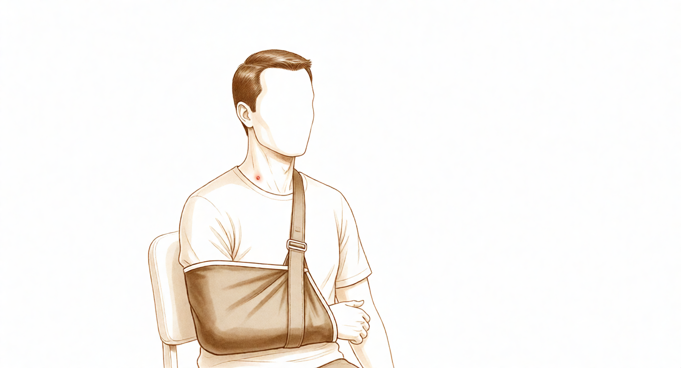
یہ پروٹوکول (protocol، یعنی بحالی کا مرحلہ وار منصوبہ) ٹوٹی ہوئی ہنسلی کی ہڈی (clavicle) کو پلیٹ سے جوڑنے کے آپریشن، یعنی اوپن ریڈکشن اینڈ انٹرنل فکسیشن (open reduction and internal fixation, ORIF)، کے بعد آپ کی صحت یابی میں رہنمائی کرتا ہے، جو ڈاکٹر کیرن ہِرپرا کے ساتھ Mater Private Hospital Rockhampton میں کیا جاتا ہے۔ نیچے ہر مرحلہ پہلے آسان زبان میں بتاتا ہے کہ کیا ہو رہا ہے اور سب سے زیادہ اہم کیا ہے، اور اس کے بعد آپ کے فزیوتھراپسٹ کے لیے لکھا گیا باقاعدہ پروٹوکول آتا ہے؛ یہ صفحہ یا اس کی پی ڈی ایف (PDF) فزیوتھراپی کی پہلی ملاقات پر ساتھ لائیں تاکہ آپ کی بحالی کا علاج ایک ہی منصوبے کے مطابق چلے۔ آپ کا فزیوتھراپسٹ آپ کی صحت یابی کی رفتار کے مطابق اس منصوبے میں تبدیلی کر سکتا ہے۔
اگر آپریشن کے بعد آپ کو اپنے زخم کے بارے میں کوئی تشویش ہو تو کلینک سے رابطہ کریں۔ اکثر یہ مددگار ہوتا ہے کہ آپ زخم کی تصویر لے کر ای میل کر دیں تاکہ اسے دیکھا جا سکے۔
کیا توقع رکھیں
آپریشن میں ہنسلی کی ہڈی کے ٹوٹے ہوئے سروں کو ایک پلیٹ اور پیچوں سے اپنی جگہ پر تھام دیا جاتا ہے تاکہ ہڈی جڑ سکے۔ پلیٹ مضبوط ہوتی ہے، لیکن یہ صرف سہارا دینے والا اسپلنٹ ہے، جڑی ہوئی ہڈی کا متبادل نہیں: ہڈی کو خود جڑنے میں عام طور پر تقریباً چھ سے بارہ ہفتے لگتے ہیں، اور اس کے بعد بھی کئی مہینوں تک وہ مضبوط ہوتی رہتی ہے (remodelling، یعنی ہڈی کا خود کو نئے سرے سے ڈھالنا)۔ بحالی کو اسی قدرتی عمل کے مطابق مرحلوں میں ترتیب دیا گیا ہے: پہلے ہفتوں میں جب فریکچر (ہڈی کا ٹوٹنا) جڑنا شروع ہوتا ہے تو پلیٹ اور پیچوں کی حفاظت کی جاتی ہے، اس کے بعد حرکت واپس لائی جاتی ہے، اور وزن اٹھانا اور کھیل سب سے آخر میں آتے ہیں، جب ہڈی انہیں برداشت کر سکے۔
چونکہ یہ فریکچر ہے، اس لیے ہر بڑا اگلا قدم (بازو کو کندھے کی اونچائی سے اوپر اٹھانا، مضبوطی کی ورزشیں، زیادہ وزن اٹھانا، اور کھیل میں واپسی) صرف کیلنڈر پر نہیں بلکہ اس بات پر بھی منحصر ہے کہ ایکسرے پر فریکچر کیسا نظر آتا ہے، جس کی تصدیق ڈاکٹر ہِرپرا کے ساتھ آپ کے معائنے میں کی جاتی ہے۔ نیچے دیے گئے ہفتوں کے اندازے عام صورت حال بتاتے ہیں، یہ طے شدہ نہیں ہیں۔
تمباکو نوشی کرنے والوں اور ذیابیطس (شوگر) کے مریضوں میں ہڈی زیادہ آہستہ جڑتی ہے، اور خاص طور پر تمباکو نوشی فریکچر کے جڑنے میں تاخیر کر سکتی ہے یا اسے جڑنے سے روک بھی سکتی ہے۔ اگر آپ تمباکو نوشی کرتے ہیں تو فریکچر کے بعد کے ہفتے اسے چھوڑنے کے لیے خاص طور پر فائدہ مند وقت ہیں۔
ہنسلی کی ہڈی جلد کے بالکل نیچے ہوتی ہے، اس لیے سوجن کم ہونے کے بعد پلیٹ کو محسوس کرنا (اور دیکھنا) عام بات ہے۔ شروع کے مہینوں میں سیٹ بیلٹ یا بستے کے پٹے کے نیچے یہ جگہ چھونے پر درد کر سکتی ہے؛ عام طور پر جیسے جیسے یہ حصہ کم حساس ہوتا جاتا ہے، یہ تکلیف ختم ہو جاتی ہے۔ اگر فریکچر کے مکمل طور پر جڑ جانے کے بعد بھی پلیٹ تکلیف دیتی رہے تو اسے نکالنا ایک ممکنہ راستہ ہے جس پر بعد کے کسی معائنے میں بات کی جا سکتی ہے؛ یہ ایک الگ فیصلہ ہے جس میں کوئی جلدی نہیں، اور یہ ہڈی کے جڑ جانے کے کافی بعد کیا جاتا ہے۔
پورے سفر پر ایک نظر:
- مرحلہ I — تحفظ: ہفتے 0 سے 3
- مرحلہ II — ابتدائی حرکت: ہفتے 3 سے 6
- مرحلہ III — مضبوطی: ہفتے 6 سے 12
- مرحلہ IV — مکمل سرگرمی اور کھیل میں واپسی: ہفتہ 12 اور اس کے بعد
سلنگ پہننا
سلنگ (sling)، یعنی گلے سے لٹکی وہ پٹی جو بازو کو سہارا دیتی ہے، شروع کے ہفتوں میں بازو کا وزن سنبھالتی ہے، تکلیف کم کرتی ہے اور جڑتی ہوئی ہڈی کی حفاظت کرتی ہے۔ اصول سادہ ہیں:
- اسے خاص طور پر گھر سے باہر پہنیں، تاکہ بازو محفوظ رہے اور لوگ اس سے نہ ٹکرائیں۔ سوتے وقت اسے پہننا ضروری نہیں۔
- پہلے تین ہفتوں میں اسے زیادہ تر وقت پہنیں۔ اس کے بعد آرام کے مطابق اس کا استعمال آہستہ آہستہ کم کیا جاتا ہے، اور زیادہ تر لوگ چھ ہفتوں تک اسے چھوڑ چکے ہوتے ہیں۔
- نہاتے وقت، ورزش کرتے وقت، اور بیٹھ کر ہلکے کام کرتے وقت جب بازو کو سہارا ملا ہو، اسے اتار دیں: کھانا، لکھنا، پڑھنا۔
- گھر پر آرام کرتے ہوئے آپ سمجھداری سے سلنگ اتار سکتے ہیں: بیٹھے ہوئے بازو تکیے پر سہارے سے رکھیں، اور ہاتھ کندھے کی اونچائی سے نیچے رکھیں۔
- چھ ہفتے تک گاڑی نہ چلائیں (یہ کندھے کے ہر آپریشن پر لاگو ہوتا ہے)، اور جب تک آپ سلنگ پہن رہے ہوں تب بھی نہیں۔ اس کے بعد گاڑی چلانا اس وقت دوبارہ شروع ہوتا ہے جب آپ گاڑی کو آرام سے اور محفوظ طریقے سے قابو میں رکھ سکیں، جس کی تصدیق ڈاکٹر ہِرپرا کے ساتھ آپ کے معائنے میں ہو۔
آپریشن کے بعد پہلے دن
اگر آپریشن کے دوران اعصابی بلاک (nerve block)، یعنی ایک انجکشن جو جسم کے ایک حصے کو سُن کر دیتا ہے، استعمال کیا گیا ہو تو اس کے بعد کچھ گھنٹوں تک بازو سُن اور بھاری محسوس ہو سکتا ہے؛ جب تک احساس معمول پر نہ آ جائے، اسے سلنگ میں محفوظ رکھیں۔ شروع کے دنوں کے لیے چند عملی مشورے:
- ورزش کرنے سے پہلے اور فزیوتھراپی کی ملاقاتوں سے پہلے اپنی درد کش دوا لیں۔
- درد اور سوجن کے لیے اس جگہ پر برف لگائیں، ایک وقت میں تقریباً 15 سے 20 منٹ، گیلے کپڑے میں لپیٹ کر؛ برف کبھی براہِ راست جلد یا زخم پر نہ لگائیں۔
- سلنگ پہنتے وقت اپنا کندھا ڈھیلا چھوڑ دیں اور بازو کا وزن سلنگ کو سنبھالنے دیں۔
- اپنے بیٹھنے اور کھڑے ہونے کے انداز کا خیال رکھیں: کان، کندھے اور کولہے ایک سیدھ میں رکھیں اور کندھوں کو آگے کی طرف جھکنے نہ دیں؛ درست انداز فریکچر کی جگہ کو محفوظ رکھتا ہے اور اکڑاؤ سے بچانے میں مدد دیتا ہے۔
- شروع سے ہی اپنی انگلیوں، کلائی، کہنی اور گردن کو حرکت دیتے رہیں۔
- اگر کوئی مسئلہ ہو تو کلینک سے رابطہ کریں یا اپنے فزیوتھراپسٹ کو بتائیں۔
مرحلہ I — تحفظ (ہفتہ 0 سے 3)
Kieran Hirpara 4.0
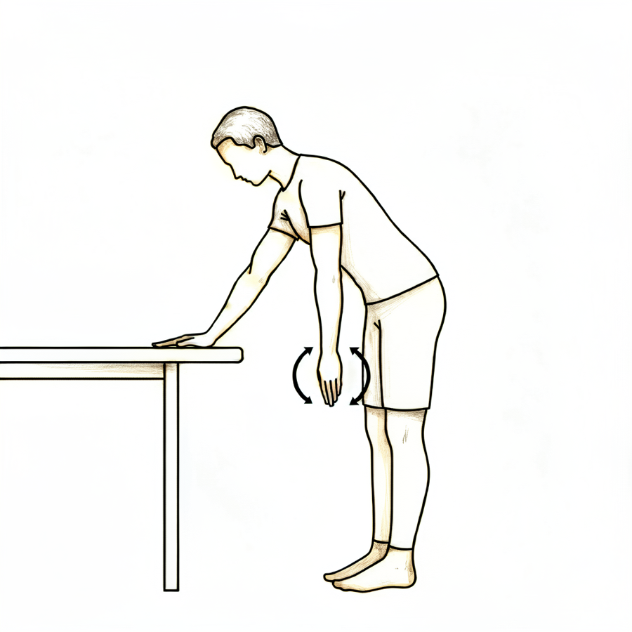
پینڈولم ورزشیں (pendulum)
آگے کی طرف جھکیں، دوسرے ہاتھ سے میز کا سہارا لیں، اور آپریشن والے بازو کو ڈھیلا لٹکنے دیں۔ اپنے جسم کو ہلکے سے ہلا کر چھوٹے دائرے بنائیں — پہلے گھڑی کی سوئیوں کی سمت میں، پھر الٹی سمت میں۔ بازو پورا وقت ڈھیلا رہتا ہے؛ حرکت آپ کے جسم سے آتی ہے، کندھے سے نہیں۔
آپ کے فزیوتھراپسٹ کی رہنمائی کے مطابق
Kieran Hirpara 4.0
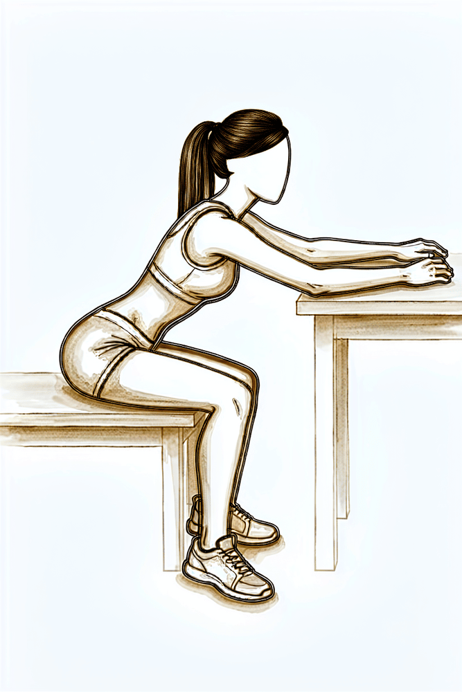
میز پر ہاتھ سرکانا (table slides)
میز کے پاس بیٹھیں اور بازو کا نچلا حصہ میز پر رکھیں۔ ہلکے سے آگے جھکیں تاکہ آپ کا ہاتھ میز پر آگے سرکے، اور بازو کا وزن میز کو سنبھالنے دیں، پھر سیدھے بیٹھ جائیں۔ آرام کی حد میں رہیں اور بازو کندھے کی اونچائی سے نیچے رکھیں۔
آپ کے فزیوتھراپسٹ کی رہنمائی کے مطابق
Kieran Hirpara 4.0
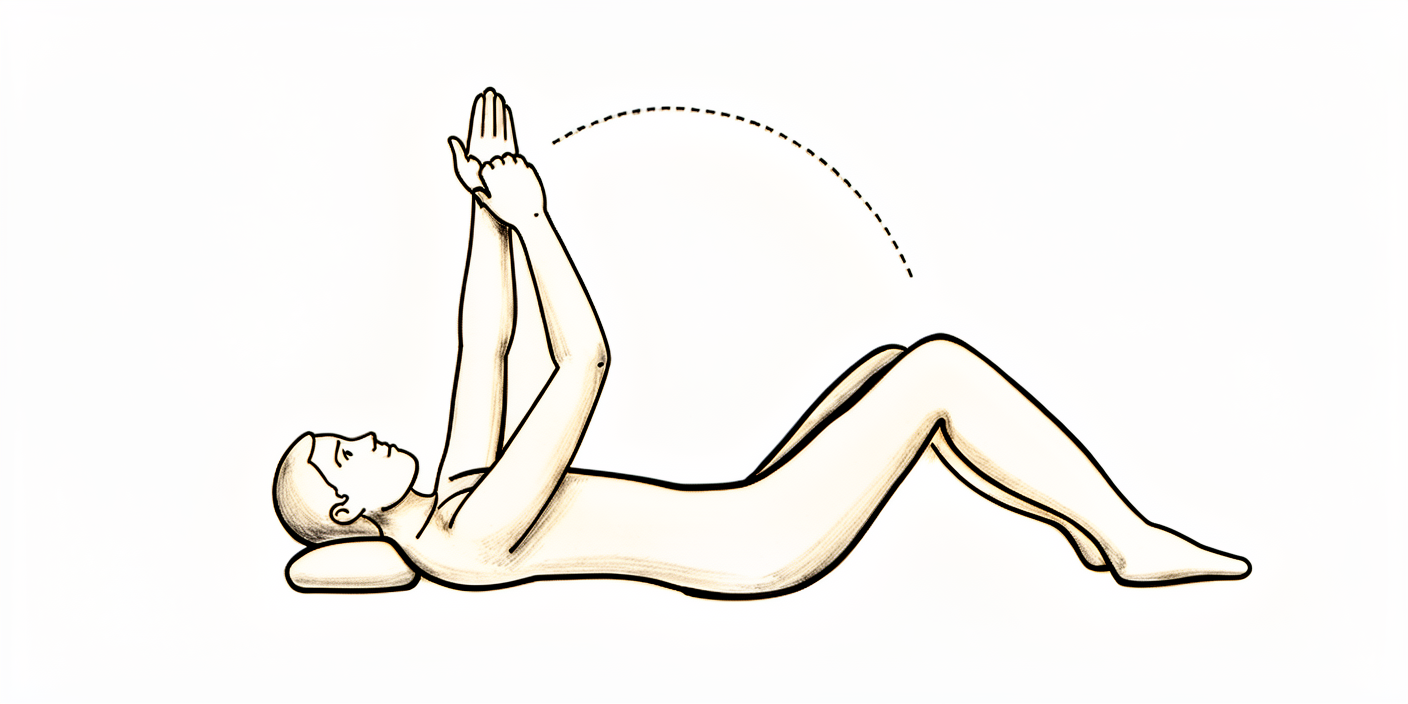
لیٹ کر مدد سے کندھے کو آگے اٹھانا (assisted flexion)
کمر کے بل لیٹ جائیں۔ دوسرے ہاتھ سے آپریشن والے بازو کو کلائی سے سہارا دیتے ہوئے اسے ہلکے سے آگے کی طرف زیادہ سے زیادہ 90 ڈگری تک اٹھائیں — یعنی چھت کی طرف اشارہ کرتا ہوا، اس سے اوپر نہیں — پھر نیچے لے آئیں۔ آپریشن والا بازو ڈھیلا رہتا ہے؛ کام آپ کا دوسرا بازو کرتا ہے۔ جب تک چھ ہفتے کے معائنے میں اجازت نہ ملے، 90 ڈگری سے آگے نہ جائیں۔
آپ کے فزیوتھراپسٹ کی رہنمائی کے مطابق
Kieran Hirpara 4.0
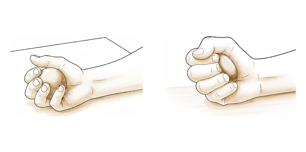
گیند دبانا
بازو کو سہارا دے کر ایک نرم گیند دبائیں یا مضبوط مٹھی بنائیں، پھر ہاتھ پورا کھول دیں۔ شروع سے ہی اپنی انگلیوں، کلائی اور ہاتھ کو حرکت دیتے رہیں۔
آپ کے فزیوتھراپسٹ کی رہنمائی کے مطابق
Kieran Hirpara 4.0
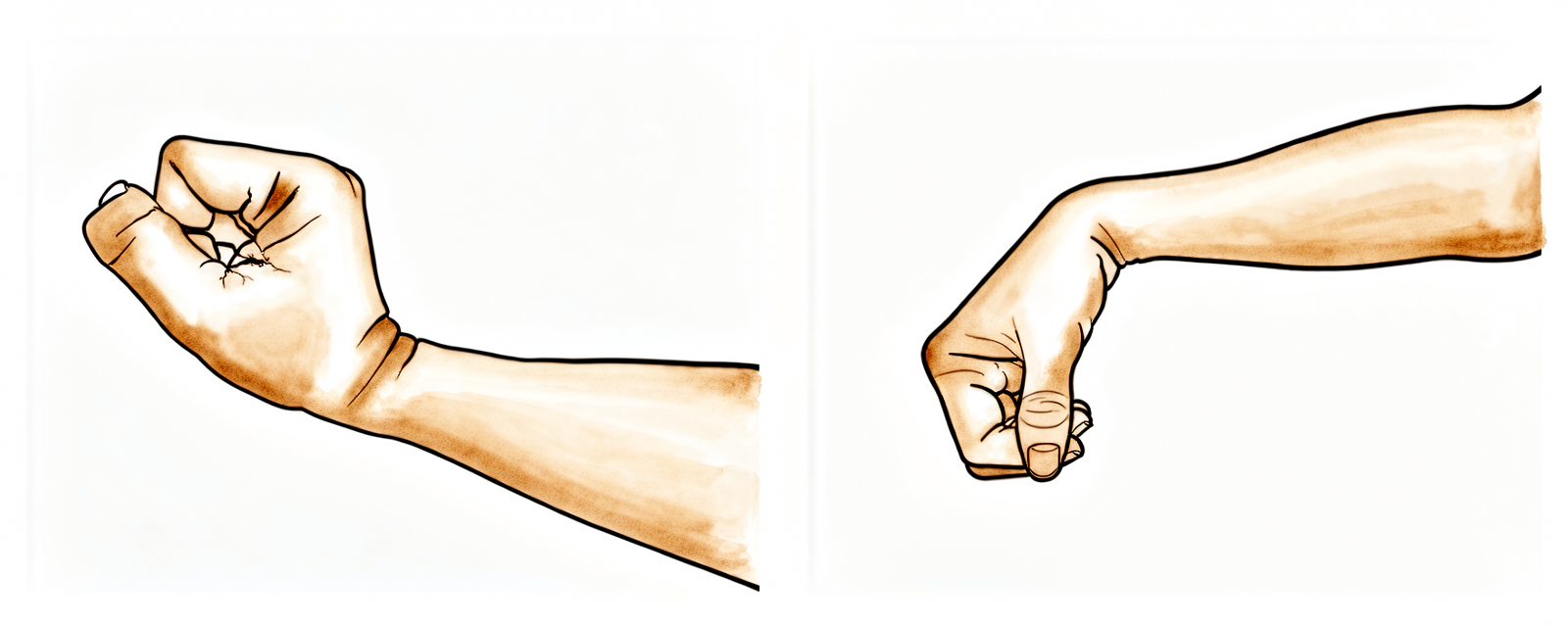
کلائی کی حرکت
بازو کو سلنگ سے نکال کر اور سہارا دے کر، کلائی کو آگے، پیچھے اور دائیں بائیں موڑ کر حرکت میں رکھیں۔ شروع سے کلائی کو کام میں رکھنے سے، جب تک کندھا محفوظ رکھا جا رہا ہے، اکڑاؤ نہیں ہوتا۔
آپ کے فزیوتھراپسٹ کی رہنمائی کے مطابق
Kieran Hirpara 4.0
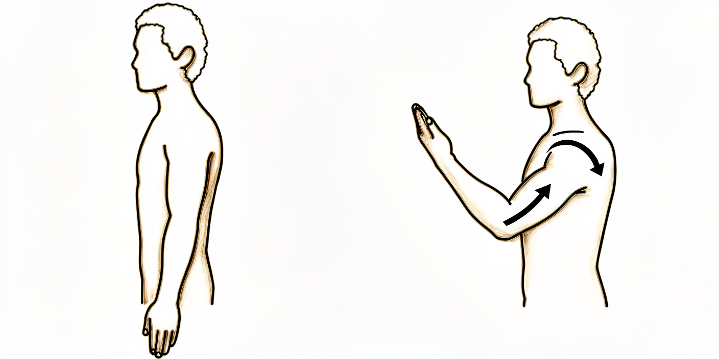
کہنی موڑنا
بازو کے اوپری حصے کو پہلو کے ساتھ سہارے سے رکھ کر کہنی کو جتنا آرام سے ہو سکے موڑیں، پھر اسے پورا سیدھا کریں۔ صرف کہنی حرکت کرے — کندھا ڈھیلا رکھیں۔ اس سے ہنسلی کی ہڈی کی حفاظت کے دوران کہنی کھلی اور آزاد رہتی ہے۔
آپ کے فزیوتھراپسٹ کی رہنمائی کے مطابق
Kieran Hirpara 4.0
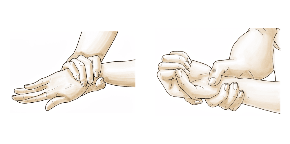
بازو کے نچلے حصے کو گھمانا
کہنی کو پہلو سے لگا کر اور زاویۂ قائمہ (right angle) پر موڑ کر ہتھیلی کو اوپر کی طرف، پھر نیچے کی طرف موڑیں۔ بازو کا اوپری حصہ ساکن رکھیں تاکہ صرف نچلا حصہ گھومے۔ دوسرے ہفتے سے آپ کا فزیوتھراپسٹ اس میں ہلکی مزاحمت شامل کر سکتا ہے۔
آپ کے فزیوتھراپسٹ کی رہنمائی کے مطابق
Kieran Hirpara 4.0
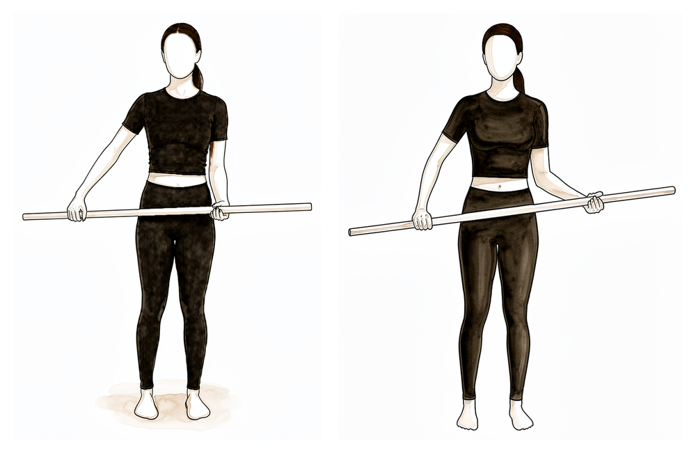
چھڑی کی مدد سے باہر کی طرف گھماؤ
کمر کے بل لیٹ جائیں، آپریشن والی کہنی پہلو سے لگی ہو اور زاویۂ قائمہ پر مڑی ہو۔ ایک چھڑی دونوں ہاتھوں میں پکڑیں اور اپنے ٹھیک بازو سے آپریشن والے ہاتھ کو ہلکے سے باہر کی طرف دھکیلیں، جس سے کندھا گھومے، پھر واپس لائیں۔ کہنی پہلو سے لگی رکھیں — یہ مدد والی حرکت ہے، زبردستی والی نہیں۔
آپ کے فزیوتھراپسٹ کی رہنمائی کے مطابق
Kieran Hirpara 4.0
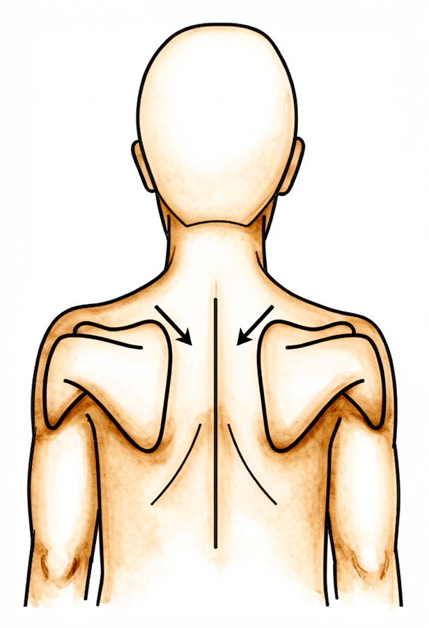
کندھے کی پچھلی ہڈی کو جمانا (scapular setting)
سیدھے بیٹھ کر یا کھڑے ہو کر، کندھے کی دونوں پچھلی چپٹی ہڈیوں (shoulder blades) کو ہلکے سے پیچھے ایک دوسرے کی طرف اور نیچے، کانوں سے دور کھینچیں۔ چند سیکنڈ روکے رکھیں، پھر ڈھیلا چھوڑ دیں۔ اس سے جڑتی ہوئی ہنسلی کی ہڈی کو حرکت دیے بغیر کندھے کی پچھلی ہڈی کے پٹھے متحرک رہتے ہیں۔
آپ کے فزیوتھراپسٹ کی رہنمائی کے مطابق
Kieran Hirpara 4.0
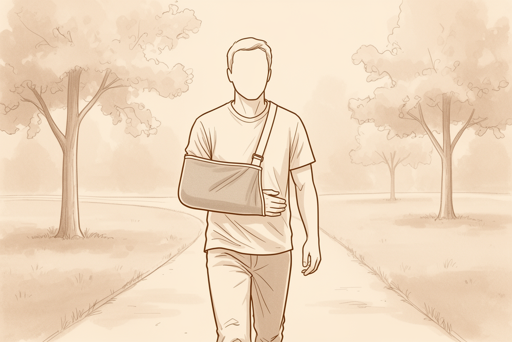
بازو سلنگ میں رکھ کر چلنا
آپریشن والا بازو سلنگ میں رکھ کر چہل قدمی کے ذریعے دل اور سانس کی ہلکی ورزش جاری رکھیں۔ اس سے فریکچر کی حفاظت کے دوران آپ کی عمومی فٹنس برقرار رہتی ہے۔
آپ کے فزیوتھراپسٹ کی رہنمائی کے مطابق
پہلے ہفتوں کا مقصد پلیٹ اور پیچوں کی حفاظت کرنا ہے جبکہ فریکچر جڑنا شروع ہوتا ہے۔ آپ سلنگ میں ہوں گے، برف سے سوجن کم کریں گے، اور ہلکی ورزشیں کریں گے جو بازو کے باقی حصوں کو حرکت میں رکھیں: ہاتھ، کلائی، کہنی اور گردن، اور ساتھ ہی پینڈولم ورزشیں اور کندھے کی اونچائی سے نیچے کندھے کی ہلکی معاون حرکت۔ سب سے اہم اصول: کہنی کو کندھے کی اونچائی سے اوپر نہ اٹھائیں، آپریشن والے بازو سے کچھ نہ اٹھائیں اور نہ اٹھا کر لے جائیں، بازو کے زور سے خود کو اوپر نہ دھکیلیں، اور چھ ہفتے تک (یہ کندھے کے کسی بھی آپریشن پر لاگو ہوتا ہے) یا جب تک سلنگ پہن رہے ہوں، گاڑی نہ چلائیں۔
آپ کے فزیوتھراپسٹ کے لیے:
اہداف
- پلیٹ اور پیچوں، جڑتی ہوئی ہڈی اور نرم بافتوں کی حفاظت
- درد اور سوجن کو کم کرنا
- اٹھان (elevation) کے 90 ڈگری سے نیچے کندھے کی غیر فعال حرکت کی حد بحال کرنا
- کہنی، کلائی، ہاتھ اور گردن (cervical) کی حرکت کی مکمل حد برقرار رکھنا
علاج کا طریقہ
- ضرورت کے مطابق ٹھنڈک کا علاج (cryotherapy) اور دیگر طریقے (modalities)؛ ورزشوں اور سیشنز سے پہلے درد کش دوا
- سلنگ کی فٹنگ چیک کریں؛ سلنگ کے استعمال (حفاظت کے لیے پہننا، خاص طور پر گھر سے باہر؛ کلینک کے معمول کے مطابق رات کو ضروری نہیں) اور جسم کے درست انداز (posture) کے بارے میں سمجھائیں
- پینڈولم ورزشیں اور میز پر ہاتھ سرکانا (table slides)
- غیر فعال حرکت (PROM): کندھے کی پچھلی چپٹی ہڈی کی سطح (plane of the scapula) میں آرام کی حد تک باہر اور اندر کی طرف گھماؤ؛ آگے اٹھانا (flexion) / اسکیپشن (scaption) / پہلو کی طرف اٹھانا (abduction) زیادہ سے زیادہ 90 ڈگری تک
- معاون فعال حرکت (AAROM): سیدھی حالت (neutral) میں چھڑی کی مدد سے باہر کی طرف گھماؤ؛ کمر کے بل لیٹ کر مدد سے آگے اٹھانا 90 ڈگری تک
- فعال حرکت (AROM): کہنی، کلائی، ہاتھ اور گردن کی ریڑھ کی ہڈی (cervical spine)؛ گرفت کی ورزش (گیند دبانا)
- ہفتہ 2 سے: مزاحمت کے ساتھ کلائی کو موڑنا/سیدھا کرنا (flexion/extension) اور بازو کے نچلے حصے کو گھمانا؛ کندھے کی پچھلی چپٹی ہڈی کو ہلکے سے جمانا اور پیچھے کھینچنا (scapular setting and retraction)
- ورزشِ قلب (cardio): بازو سلنگ میں رکھ کر چلنا؛ بازو سلنگ میں رکھ کر ایک جگہ کھڑی یا ٹیک والی ورزشی سائیکل (stationary or recumbent bike)
احتیاطیں
- کندھے کو اپنی طاقت سے اوپر نہ اٹھائیں (no active elevation)
- کندھے کو آگے یا پہلو کی طرف 90 ڈگری سے زیادہ نہ اٹھائیں، غیر فعال طور پر بھی نہیں
- آپریشن والے بازو سے نہ کچھ اٹھائیں اور نہ اٹھا کر لے جائیں؛ بازو پر وزن نہ ڈالیں
- چھ ہفتے تک گاڑی نہ چلائیں (یہ کندھے کے ہر آپریشن پر لاگو ہوتا ہے)، اور سلنگ پہنے ہوئے بھی نہیں
اگلے مرحلے میں جانے کی شرائط
- آرام سے 90 ڈگری تک غیر فعال آگے اٹھانا/اسکیپشن، اور تقریباً 30 ڈگری تک باہر کی طرف گھماؤ
- آرام کی حالت میں درد 10 میں سے 4 سے کم ہو چکا ہو
- کہنی، کلائی اور ہاتھ کی مکمل فعال حرکت
- زخم بھر چکا ہو، اور کسی پیچیدگی کی کوئی علامت نہ ہو
مرحلہ II — ابتدائی حرکت (ہفتہ 3 سے 6)
Kieran Hirpara 4.0
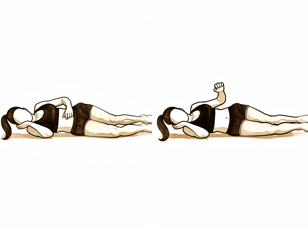
پہلو کے بل لیٹ کر باہر کی طرف گھماؤ
جس طرف آپریشن نہیں ہوا اس پہلو پر لیٹیں، آپریشن والا بازو اوپر ہو، کہنی 90 ڈگری پر مڑی ہو اور جسم سے لگی ہو۔ کہنی کو جسم سے لگائے رکھتے ہوئے بازو کے نچلے حصے کو پیٹ سے دور، اوپر کی طرف جتنا آرام سے ہو سکے گھمائیں، پھر قابو کے ساتھ نیچے لائیں۔ اس مرحلے میں کوئی وزن استعمال نہ کریں — بازو اپنی طاقت سے حرکت کرتا ہے۔
آپ کے فزیوتھراپسٹ کی رہنمائی کے مطابق
Kieran Hirpara 4.0
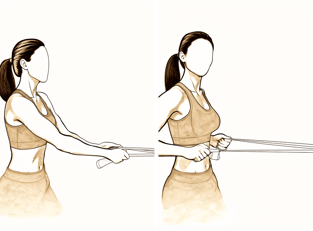
لچکدار بینڈ کے ساتھ نیچی رو (low row)
ایک لچکدار بینڈ اپنے سامنے کمر کی اونچائی پر باندھ دیں۔ سیدھے کھڑے ہو کر بینڈ کو اپنی طرف کھینچیں، کندھے کی پچھلی ہڈی کو ہلکے سے نیچے اور پیچھے لے جاتے ہوئے، پھر قابو کے ساتھ چھوڑیں۔ مزاحمت ہلکی رکھیں۔
آپ کے فزیوتھراپسٹ کی رہنمائی کے مطابق
Kieran Hirpara 4.0
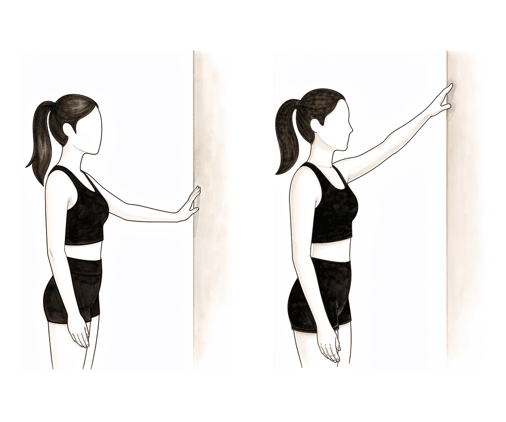
دیوار پر انگلیاں چلانا (wall slides)
دیوار کی طرف منہ کر کے کھڑے ہوں اور انگلیاں دیوار پر رکھیں۔ انگلیوں کو دیوار پر اوپر چلا کر بازو اٹھائیں، اور کچھ وزن دیوار کو سنبھالنے دیں، پھر انگلیاں واپس نیچے چلائیں۔ اس مرحلے میں بازو کندھے کی اونچائی سے نیچے رکھیں — مدد دیوار کرتی ہے، اس لیے حرکت ہلکی رہتی ہے۔
آپ کے فزیوتھراپسٹ کی رہنمائی کے مطابق
Kieran Hirpara 4.0
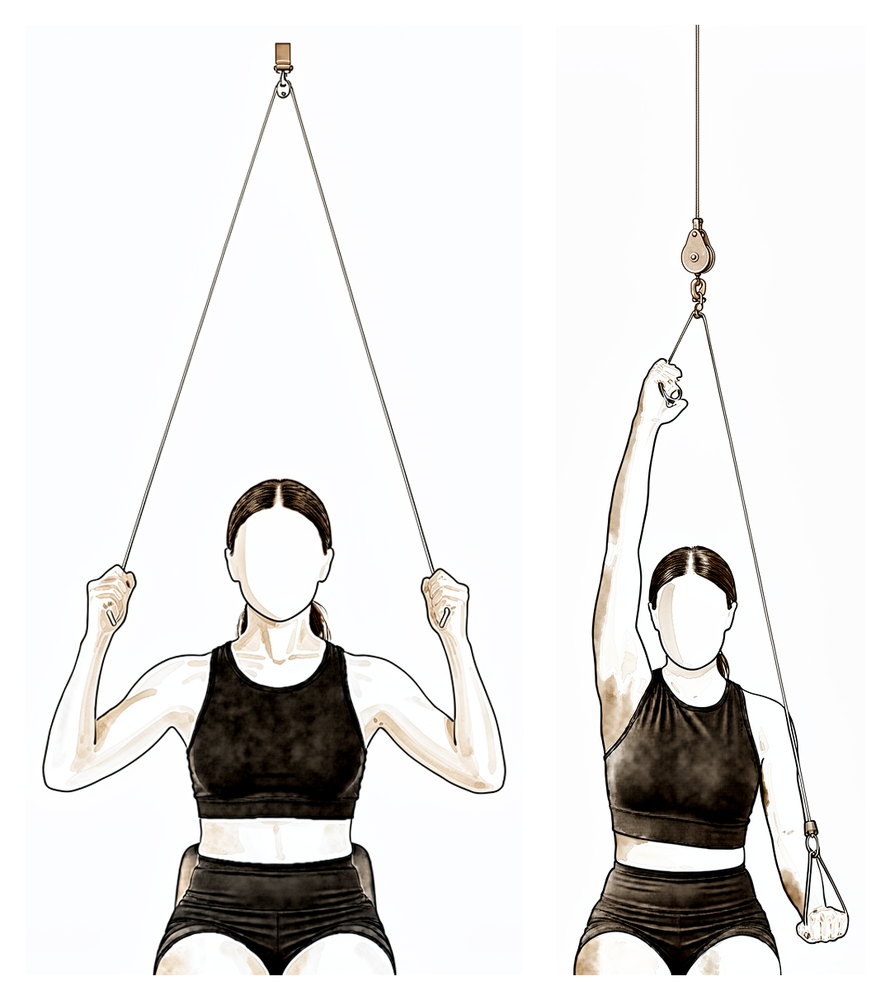
دروازے پر لگی پُلی (over-door pulley)
دروازے کے اوپر لگی پُلی کے نیچے بیٹھیں، ہر ہاتھ میں ایک ہینڈل ہو۔ اپنے ٹھیک بازو سے نیچے کھینچیں تاکہ آپریشن والا بازو جتنا آرام سے ہو سکے اوپر اٹھے، پھر آہستہ سے نیچے لائیں۔ فی الحال ہلکی حد میں اور کندھے کی اونچائی سے نیچے رہیں — کام ٹھیک بازو کرتا ہے۔
آپ کے فزیوتھراپسٹ کی رہنمائی کے مطابق
Kieran Hirpara 4.0
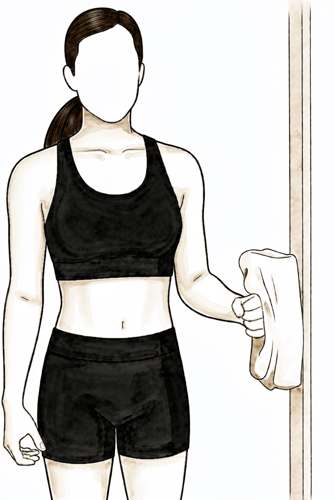
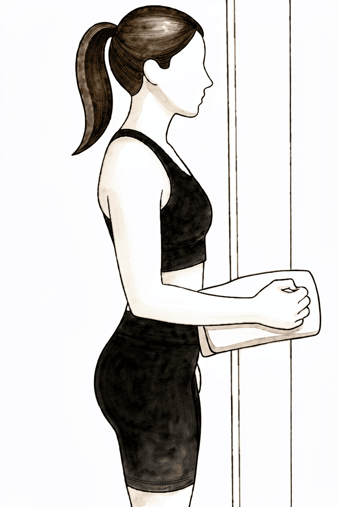
بغیر حرکت کے باہر کی طرف زور (isometric external rotation)
کہنی کو پہلو سے لگا کر اور زاویۂ قائمہ پر موڑ کر کھڑے ہوں، ہاتھ کی پشت دیوار یا دروازے کی چوکھٹ سے لگی ہو۔ بازو کو حرکت دیے بغیر ہلکے سے باہر کی طرف دبائیں، چند سیکنڈ روکیں، پھر ڈھیلا چھوڑ دیں۔ اس سے کندھے کو حرکت دیے بغیر روٹیٹر کف (rotator cuff)، یعنی ٹینڈنوں کا وہ گروپ جو کندھے کو تھامتا اور گھماتا ہے، متحرک ہوتا ہے۔
آپ کے فزیوتھراپسٹ کی رہنمائی کے مطابق
Kieran Hirpara 4.0
بغیر حرکت کے اندر کی طرف زور (isometric internal rotation)
کہنی کو پہلو سے لگا کر اور زاویۂ قائمہ پر موڑ کر کھڑے ہوں، ہتھیلی دیوار یا دروازے کی چوکھٹ سے لگی ہو۔ بازو کو حرکت دیے بغیر ہلکے سے اندر کی طرف دبائیں، چند سیکنڈ روکیں، پھر ڈھیلا چھوڑ دیں۔ یہ کف کو متحرک کرنا شروع کرنے کا ایک ہلکا طریقہ ہے۔
آپ کے فزیوتھراپسٹ کی رہنمائی کے مطابق
Kieran Hirpara 4.0
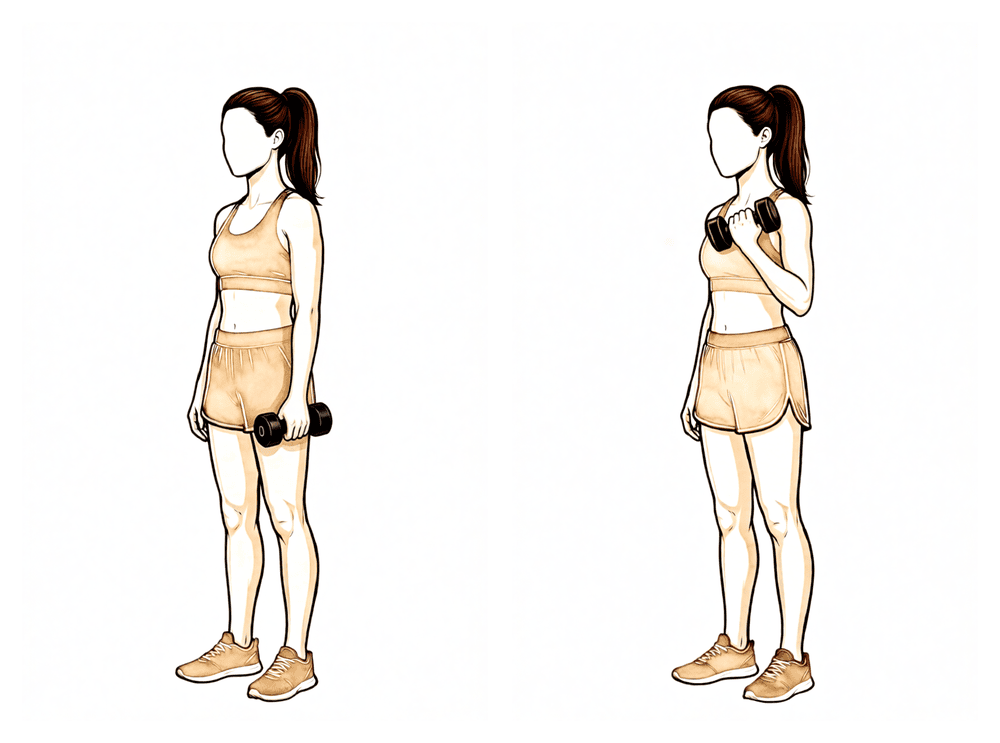
بائسیپس کرل (biceps curl)
بازو کا اوپری حصہ پہلو کے ساتھ رکھ کر کہنی موڑیں اور ایک ہلکا وزن کندھے کی طرف اٹھائیں، پھر قابو کے ساتھ نیچے لائیں۔ وزن ہلکا رکھیں — یہ بازو کی ہلکی تیاری ہے، بھاری وزن اٹھانا نہیں۔
آپ کے فزیوتھراپسٹ کی رہنمائی کے مطابق
فریکچر جڑنا شروع ہو گیا ہے، لیکن ابھی مکمل طور پر جڑا نہیں: اس مرحلے میں طاقت نہیں بلکہ حرکت واپس لائی جاتی ہے۔ آرام کے مطابق سلنگ کا استعمال آہستہ آہستہ کم کیا جاتا ہے، غیر فعال اور معاون حرکت کی حد بڑھتے بڑھتے مکمل کی طرف جاتی ہے، اور آپ کندھے کی اونچائی سے نیچے بازو کو اس کی اپنی طاقت سے حرکت دینا شروع کرتے ہیں، اور ساتھ ہی پٹھوں کو ہلکا سا متحرک کرنے والی (isometric، یعنی جوڑ کو حرکت دیے بغیر پٹھے کو کسنا) ورزشیں بھی۔ جو کچھ بھی اٹھائیں یا اٹھا کر لے جائیں، وہ کافی کے ایک کپ کے وزن سے زیادہ نہ ہو، اور کسی بھی قسم کا زور لگا کر اسٹریچ نہ کریں۔ یہ مرحلہ تقریباً چھ ہفتے پر ایکسرے اور ڈاکٹر ہِرپرا کے ساتھ معائنے پر ختم ہوتا ہے؛ صرف کیلنڈر نہیں بلکہ یہی معائنہ مضبوطی کی ورزشوں اور کندھے کی اونچائی سے اوپر حرکت کا راستہ کھولتا ہے۔
آپ کے فزیوتھراپسٹ کے لیے:
اہداف
- سلنگ آہستہ آہستہ چھڑانا (تقریباً ہفتہ 6 تک چھوڑ دی جائے)
- تمام سمتوں میں غیر فعال حرکت کی حد کو مکمل کی طرف بڑھانا
- 90 ڈگری سے نیچے درست طریقے سے فعال حرکت قائم کرنا
- ہلکی آئسومیٹرک ورزش اور کندھے کی پچھلی چپٹی ہڈی کے اردگرد کے پٹھوں (periscapular) کا کام شروع کرنا
علاج کا طریقہ
- غیر فعال حرکت (PROM): برداشت کے مطابق تمام سمتوں میں مکمل حد کی طرف بڑھائیں؛ زور لگا کر اسٹریچ نہ کریں
- معاون فعال حرکت (AAROM): کمر کے بل لیٹ کر ڈنڈے (dowel) سے آگے اٹھانا، آہستہ آہستہ سیدھا بیٹھنے کی طرف (lawn-chair progression)، دیوار اور ریلنگ پر ہاتھ سرکانا، پُلی (pulley)
- فعال حرکت (AROM): 90 ڈگری اٹھان سے نیچے، درد کے بغیر؛ لیٹ کر آگے اٹھانے سے کھڑے ہو کر اٹھانے تک؛ بیٹھ کر اور پہلو کے بل لیٹ کر باہر کی طرف گھماؤ
- سیدھی حالت (neutral) میں روٹیٹر کف (rotator cuff)، یعنی ٹینڈنوں کا وہ گروپ جو کندھے کو تھامتا اور گھماتا ہے، کی آئسومیٹرک ورزش؛ کندھے کی پچھلی ہڈی کے اردگرد کے پٹھوں کی ہلکی مضبوطی (scapular retraction، low row، mid row)؛ بائسیپس (biceps) اور ٹرائسیپس (triceps) کی ہلکی ورزش
- غلط متبادل حرکتوں پر نظر رکھیں (کندھا اوپر اچکانا (shoulder hitching)، کندھے کی پچھلی ہڈی سے کام لینا (scapular substitution))
- ورزشِ قلب (cardio): چلنا؛ ایک جگہ کھڑی ورزشی سائیکل
احتیاطیں
- کافی کے تقریباً ایک کپ سے زیادہ وزن نہ اٹھائیں اور نہ اٹھا کر لے جائیں
- جب تک چھ ہفتے کے معائنے میں فریکچر کے جڑنے کی تصدیق نہ ہو، 90 ڈگری سے زیادہ فعال اٹھان نہ کریں
- کندھے کو زور لگا کر اسٹریچ نہ کریں اور ایسی حالتوں سے بچیں جن سے درد ہو
- بازو پر وزن نہ ڈالیں
- چھ ہفتے تک گاڑی نہ چلائیں (یہ کندھے کے ہر آپریشن پر لاگو ہوتا ہے)، اور سلنگ پہنے ہوئے بھی نہیں
اگلے مرحلے میں جانے کی شرائط
- مکمل، یا تقریباً مکمل، غیر فعال حرکت کی حد
- کم سے کم غلط متبادل حرکت کے ساتھ 90 ڈگری تک فعال اٹھان، اور درد 10 میں سے 4 سے کم
- ایکسرے پر فریکچر کا تسلی بخش طور پر جڑنا، جس کی تصدیق ڈاکٹر ہِرپرا کے ساتھ معائنے میں ہو
مرحلہ III — مضبوطی (ہفتہ 6 سے 12)
Kieran Hirpara 4.0
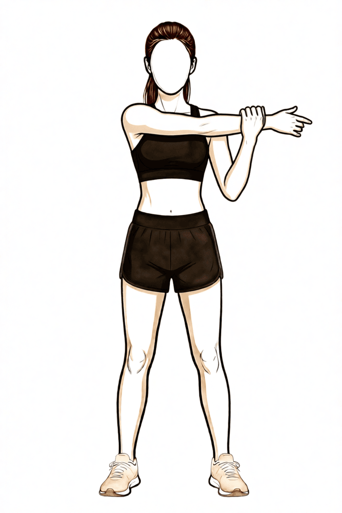
سینے کے آر پار اسٹریچ (cross-body stretch)
دوسرے ہاتھ سے آپریشن والے بازو کو سینے کے آر پار کھینچیں یہاں تک کہ کندھے کی پچھلی طرف کھنچاؤ محسوس ہو۔ تھوڑی دیر روکیں، پھر چھوڑ دیں۔ صرف واضح بے آرامی تک اسٹریچ کریں — کبھی تیز درد تک نہیں۔
آپ کے فزیوتھراپسٹ کی رہنمائی کے مطابق
Kieran Hirpara 4.0
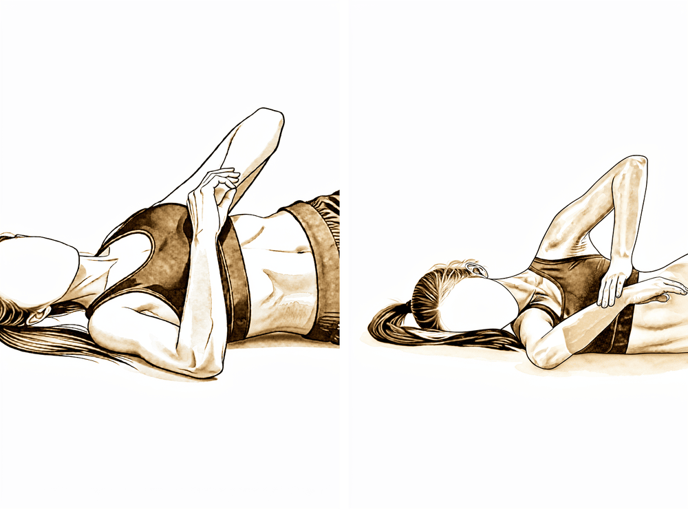
سلیپر اسٹریچ (sleeper stretch)
آپریشن والے پہلو پر لیٹیں، کندھا اور کہنی دونوں 90 ڈگری پر مڑے ہوں۔ دوسرے ہاتھ سے بازو کے نچلے حصے کو ہلکے سے بستر کی طرف دبائیں یہاں تک کہ کندھے کی پچھلی طرف کھنچاؤ محسوس ہو، پھر چھوڑ دیں۔ اسے ہلکا رکھیں — تیز درد نہیں ہونا چاہیے۔
آپ کے فزیوتھراپسٹ کی رہنمائی کے مطابق
Kieran Hirpara 4.0
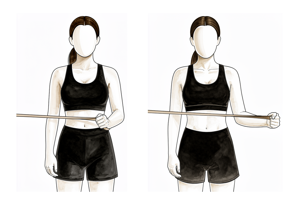
بینڈ کے ساتھ باہر کی طرف گھماؤ
کہنی کو پہلو سے لگا کر اور زاویۂ قائمہ پر موڑ کر کھڑے ہوں، اور کمر کی اونچائی پر بندھا ہوا لچکدار بینڈ پکڑیں۔ کہنی پہلو سے لگائے رکھتے ہوئے بازو کے نچلے حصے کو بینڈ کے خلاف باہر کی طرف گھمائیں، پھر آہستہ سے واپس لائیں۔ یہ پہلے کی بغیر حرکت والی ورزش سے آگے بڑھ کر روٹیٹر کف کو مضبوط کرتی ہے۔
آپ کے فزیوتھراپسٹ کی رہنمائی کے مطابق
Kieran Hirpara 4.0
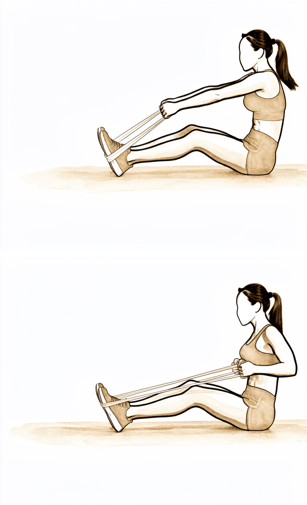
نیچی رو (low row)
اپنے سامنے کمر کی اونچائی پر بندھا ہوا بینڈ پکڑیں۔ بازو کو کافی حد تک سیدھا رکھتے ہوئے اسے پیچھے اور نیچے اپنے کولہے کی طرف کھینچیں، کندھے کی پچھلی ہڈی کو نیچے اور پیچھے دباتے ہوئے، پھر آہستہ سے واپس لائیں۔ اس سے کندھے کی پچھلی ہڈی کے وہ پٹھے مضبوط ہوتے ہیں جو کف کو سہارا دیتے ہیں۔
آپ کے فزیوتھراپسٹ کی رہنمائی کے مطابق
Kieran Hirpara 4.0
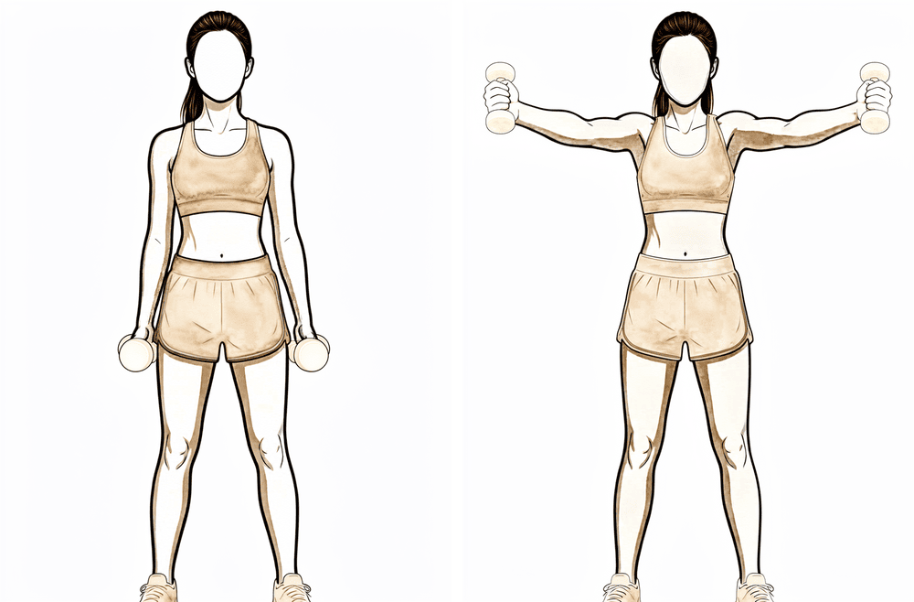
اسکیپشن میں بازو اٹھانا (full can)
انگوٹھے اوپر کی طرف رکھ کر بازو کو اپنے سامنے تقریباً 45 ڈگری کے زاویے پر اٹھائیں — سیدھا سامنے اور سیدھا پہلو کی طرف کے درمیان — پھر قابو کے ساتھ نیچے لائیں۔ جب کندھے کی اونچائی سے اوپر حرکت کی اجازت مل جائے تو سر سے اوپر آرام دہ حد تک اٹھائیں۔ وزن ہلکا رکھیں۔
آپ کے فزیوتھراپسٹ کی رہنمائی کے مطابق
Kieran Hirpara 4.0
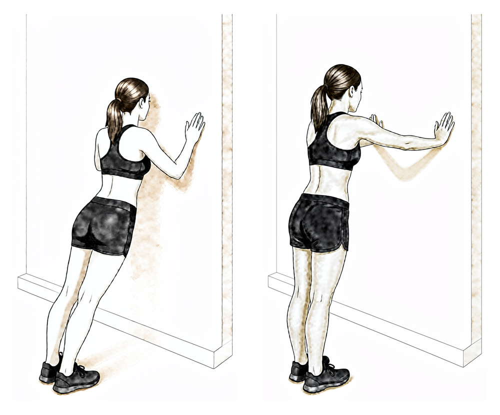
دیوار پر پُش اپ (wall push-ups)
دیوار کی طرف منہ کر کے کھڑے ہوں اور ہاتھ کندھے کی اونچائی پر دیوار پر سیدھے رکھیں۔ کہنیاں موڑ کر دیوار کی طرف جھکیں، پھر واپس دھکیلیں، اور آخر میں ہاتھوں سے ہلکا سا مزید دھکا دیں تاکہ کندھے کی پچھلی ہڈیاں پھیل جائیں۔ یہ ورزش صرف اس مرحلے کے آخر میں، اپنے فزیوتھراپسٹ کی رہنمائی کے مطابق، شروع کریں۔
آپ کے فزیوتھراپسٹ کی رہنمائی کے مطابق
Kieran Hirpara 4.0
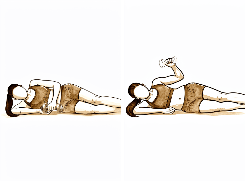
پہلو کے بل لیٹ کر وزن کے ساتھ باہر کی طرف گھماؤ
جس طرف آپریشن نہیں ہوا اس پہلو پر لیٹیں، اوپر والی کہنی زاویۂ قائمہ پر مڑی ہو اور جسم سے لگی ہو۔ ہلکا وزن پکڑ کر بازو کے نچلے حصے کو چھت کی طرف اوپر گھمائیں، پھر آہستہ سے نیچے لائیں۔ وزن ہلکا اور تکرار زیادہ رکھیں۔
آپ کے فزیوتھراپسٹ کی رہنمائی کے مطابق
Kieran Hirpara 4.0
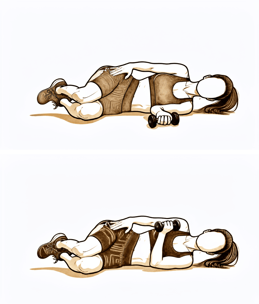
پہلو کے بل لیٹ کر وزن کے ساتھ اندر کی طرف گھماؤ
آپریشن والے پہلو پر لیٹیں، کہنی زاویۂ قائمہ پر مڑی ہو اور جسم سے لگی ہو۔ ہلکا وزن پکڑ کر کندھے کو اندر کی طرف گھماتے ہوئے اسے پیٹ کی طرف اوپر اٹھائیں، پھر آہستہ سے نیچے لائیں۔ وزن ہلکا اور تکرار زیادہ رکھیں۔
آپ کے فزیوتھراپسٹ کی رہنمائی کے مطابق
جب آپ کا معائنہ اس بات کی تصدیق کر دے کہ فریکچر اچھی طرح جڑ رہا ہے تو کندھے کی اونچائی سے اوپر حرکت شروع ہوتی ہے اور اگلے ہفتوں میں مکمل فعال حرکت کی حد بنائی جاتی ہے۔ مضبوطی کی ورزشیں ہلکے سے شروع ہوتی ہیں: پہلے پٹھوں کو متحرک کرنے کا کام، پھر لچکدار بینڈ، اور پھر روٹیٹر کف اور کندھے کی پچھلی ہڈی کے پٹھوں کے لیے ہلکے وزن۔ بارہ ہفتے تک وزن اٹھانا ہلکا ہی رہتا ہے (تقریباً 2 کلوگرام سے زیادہ نہیں)، بھاری وزن یا سر سے اوپر وزن اٹھانا بعد میں آتا ہے، اور اس مرحلے میں ایسے کھیل جن میں کھلاڑی آپس میں ٹکراتے ہیں (contact sport) نہیں کھیلے جاتے۔ تیراکی اور سائیکل چلانا عام طور پر اسی مرحلے میں، آپ کے فزیوتھراپسٹ کی رہنمائی کے مطابق، دوبارہ شروع ہوتے ہیں۔
آپ کے فزیوتھراپسٹ کے لیے:
اہداف
- تمام سمتوں میں مکمل فعال حرکت کی حد، معمول کے طریقۂ حرکت کے ساتھ
- روٹیٹر کف اور کندھے کی پچھلی ہڈی کے اردگرد کے پٹھوں کی مضبوطی شروع کرنا اور آگے بڑھانا
- روزمرہ کی معمول کی سرگرمیوں میں واپسی
علاج کا طریقہ
- تمام سمتوں میں 90 ڈگری سے اوپر فعال حرکت (AROM) بڑھائیں، غلط متبادل حرکتوں کو کم سے کم رکھتے ہوئے
- ضرورت کے مطابق اسٹریچ: کمر کا چوڑا پٹھا (latissimus)، سینے کا پٹھا (pectoral)، جوڑ کی پچھلی جھلی (posterior capsule) اور سلیپر اسٹریچ (sleeper stretch)
- مضبوطی: کف کی آئسومیٹرک ورزش سے آگے بڑھ کر بینڈ کے ساتھ مزاحمت میں باہر/اندر کی طرف گھماؤ، شروع میں کندھے کی اونچائی سے نیچے؛ کندھے کی پچھلی ہڈی کو پیچھے کھینچنا اور رو (rows)؛ اسکیپشن میں بازو اٹھانا، سیریٹس (serratus) کی ورزش اور دیوار پر پُش اپ اس مرحلے کے آخر میں
- ہلکے آزاد وزن (free weights) برداشت کے مطابق بڑھاتے ہوئے: کم وزن، زیادہ تکرار
- ورزشِ قلب (cardio): ایک جگہ کھڑی ورزشی سائیکل اور چلنا؛ اگر معائنے میں اجازت ملے تو تقریباً 8 سے 10 ہفتے سے تیراکی اور دوڑنا
احتیاطیں
- 12 ہفتے تک تقریباً 2 کلوگرام سے زیادہ وزن نہ اٹھائیں
- 12 ہفتے تک سر سے اوپر یا جسم سے دور رکھ کر بھاری وزن اٹھانے سے بچیں
- ٹکراؤ والے کھیل (contact sport) نہ کھیلیں؛ اس مرحلے کے آخر (تقریباً 10 سے 12 ہفتے) تک چھلانگ والی (plyometric) یا جھٹکے والی ورزش نہ کریں
- مضبوطی کی ورزشیں آرام دہ حد کے اندر رہیں اور ان سے ایسا درد نہیں ہونا چاہیے جو دیر تک رہے
اگلے مرحلے میں جانے کی شرائط
- فعال حرکت کی حد دوسری طرف کے کم از کم 90 فیصد
- روٹیٹر کف اور کندھے کی پچھلی ہڈی کے اردگرد کے پٹھوں کا اچھی طرح متحرک ہونا، اور مزاحمت والی ورزش پر درد 10 میں سے 3 سے زیادہ نہ ہو
- ایکسرے پر فریکچر کا جڑنا آگے بڑھ رہا ہو، جس کی تصدیق ڈاکٹر ہِرپرا کے ساتھ معائنے میں ہو
مرحلہ IV — مکمل سرگرمی اور کھیل میں واپسی (ہفتہ 12 اور اس کے بعد)
Kieran Hirpara 4.0
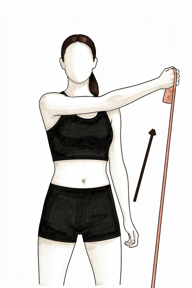
بینڈ کے ساتھ ترچھی حرکت (D1)
ایک لچکدار بینڈ پکڑیں اور اسے جسم کے آر پار ترچھا کھینچیں — ایک کولہے کے پاس نیچے اور آر پار سے، اوپر اور مخالف سمت میں باہر کی طرف، پھر قابو کے ساتھ الٹا واپس لائیں۔ روزمرہ کام جیسی یہ ترچھی حرکتیں پورے بازو کی اس مربوط حرکت کو دوبارہ بناتی ہیں جو کام اور کھیل میں استعمال ہوتی ہے۔ یہ ورزش صرف فریکچر جڑ جانے کے بعد شامل کریں۔
آپ کے فزیوتھراپسٹ کی رہنمائی کے مطابق
Kieran Hirpara 4.0
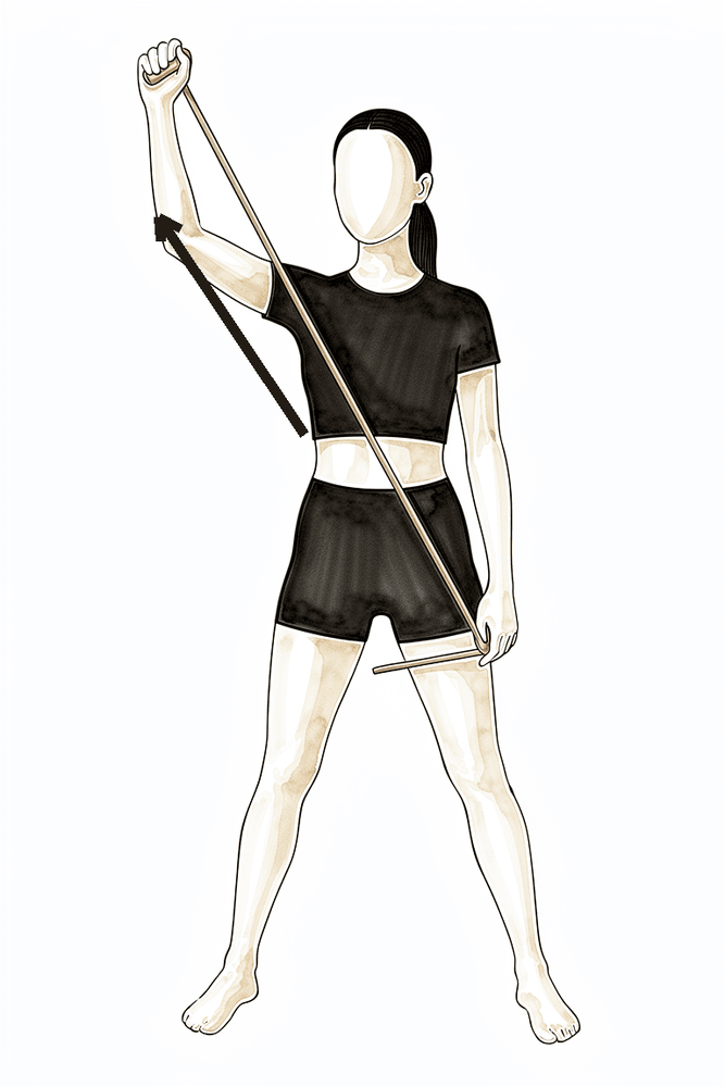
بینڈ کے ساتھ ترچھی حرکت (D2)
ایک لچکدار بینڈ پکڑیں اور اسے ترچھا کھینچیں، جسم کے آر پار نیچے سے اوپر سر کے اوپر اور باہر کی طرف، پھر قابو کے ساتھ الٹا واپس لائیں۔ اس سے کندھے کی سر سے اوپر والی، کھیل کے مطابق حرکتیں بنتی ہیں۔ یہ ورزش صرف فریکچر جڑ جانے اور سر سے اوپر کام کی اجازت ملنے کے بعد شامل کریں۔
آپ کے فزیوتھراپسٹ کی رہنمائی کے مطابق
آخری مرحلہ زیادہ وزن اٹھانے، جسمانی مشقت والے کام اور کھیل کی طرف مرحلہ وار واپسی ہے۔ مضبوطی کی ورزشیں زیادہ مزاحمت، سر سے اوپر کی حالتوں اور (کھلاڑیوں کے لیے) چھلانگ والی (plyometric)، پھینکنے والی اور کھیل کے مطابق مشقوں تک بڑھتی ہیں۔ ٹکراؤ والے کھیلوں (فٹ بال، رگبی، گھڑ سواری) کے لیے ضروری ہے کہ ایکسرے پر فریکچر جڑ چکا ہو، جس کی تصدیق ڈاکٹر ہِرپرا کے ساتھ آپ کے معائنے میں ہو؛ یہ عام طور پر جلد سے جلد تقریباً تین سے چار ماہ کے بعد ہوتا ہے، اور بعض پروٹوکول ٹکراؤ والے کھیلوں کو چھ ماہ تک مؤخر رکھتے ہیں۔ ہڈی کے جڑنے سے پہلے واپس آنے سے ہڈی کے دوبارہ ٹوٹنے کا خطرہ ہوتا ہے، اس لیے یہ ایک ایسی شرط ہے جس کا احترام کرنا واقعی ضروری ہے۔
آپ کے فزیوتھراپسٹ کے لیے:
اہداف
- مکمل، درد کے بغیر حرکت کی حد برقرار رکھنا
- طاقت دوسری (غیر متاثرہ) طرف کے کم از کم 90 فیصد
- جسمانی مشقت والے کام، تفریح اور کھیل میں مرحلہ وار واپسی
علاج کا طریقہ
- بتدریج بڑھتی مزاحمت کی ورزش، جس میں پٹھے کو لمبا کرتے ہوئے وزن سنبھالنا (eccentric loading)، سر سے اوپر کی حالتیں اور روزمرہ کام جیسی حرکتیں برداشت کے مطابق شامل ہوں
- تال کے ساتھ استحکام (rhythmic stabilisation) اور جوڑ کی پوزیشن کے احساس (proprioceptive) کی ورزش؛ سر سے اوپر کھیلنے والے کھلاڑیوں کے لیے چھلانگ والی ورزش اور وقفوں کے ساتھ پھینکنے یا ریکٹ کے پروگرام
- جسمانی مشقت والا کام کرنے والوں کے لیے کام کے مطابق تیاری؛ بغیر پابندی کھیلنے سے پہلے کھیل کے مطابق مشقیں
- کھیل میں واپسی کا فیصلہ ہر مریض کے مطابق (ٹکراؤ والا یا بغیر ٹکراؤ والا کھیل، بازو اور ہاتھ پر کتنا بوجھ پڑتا ہے) اور سرجن کے ساتھ مل کر کیا جائے
اگلے مرحلے میں جانے کی شرائط
- مکمل، درد کے بغیر فعال حرکت کی حد
- طاقت ناپنے کے آلے (dynamometry) پر دوسری (غیر متاثرہ) طرف کی کم از کم 90 فیصد طاقت، اور طاقت کے ٹیسٹ پر کوئی درد نہ ہو
- کھیل میں واپسی کا مرحلہ وار پروگرام درد یا خوف (apprehension) کے بغیر مکمل کرنا
- ٹکراؤ والے کھیل سے پہلے ایکسرے پر ہڈی کے جڑنے (radiographic union) کی تصدیق، ڈاکٹر ہِرپرا کے ساتھ معائنے میں
اپنے پروٹوکول کے بعد
اوپر دیے گئے مراحل ہنسلی کی ہڈی کے فریکچر کو جوڑنے کے آپریشن کے بعد بحالی کے شائع شدہ پروٹوکولز سے اخذ کیے گئے ہیں: Massachusetts General Brigham Sports Medicine، Mammoth Orthopedic Institute، University of Colorado (Dr Jonathan Bravman) اور Midwest Orthopaedics at Rush (Dr Brian Cole)، اور ساتھ ہی West Suffolk اور United Lincolnshire کی NHS فزیوتھراپی رہنمائی، اور کھلاڑیوں میں ہنسلی کی ہڈی کے فریکچر کے منظم جائزے (systematic review) سے کھیل میں واپسی کے شواہد۔ ہفتوں کے اندازے عام صورت حال بتاتے ہیں، یہ طے شدہ نہیں ہیں، اور آپ کا آگے بڑھنا آپ کے فزیوتھراپسٹ کی رہنمائی میں ہوتا ہے اور ڈاکٹر ہِرپرا کے ساتھ آپ کے معائنوں میں فریکچر کے جڑنے پر منحصر ہے۔ یہ صفحہ کلینک کے صحت یابی کے عمومی مشوروں کے ساتھ مل کر کام کرتا ہے؛ دیکھیں آپریشن کے بعد درد کو سنبھالنا اور زخم کی دیکھ بھال۔ خود آپریشن کے بارے میں جاننے کے لیے دیکھیں ہنسلی کی ہڈی کو جوڑنے کا آپریشن۔ اس پروٹوکول کے پیچھے موجود شواہد (آپریشن اور بغیر آپریشن کے علاج کا موازنہ کرنے والی تحقیقات کے نتائج، ہڈی جڑنے اور کھیل میں واپسی کی شرحیں، اور سرجنوں کے شائع شدہ پروٹوکولز) کا خلاصہ شواہد والے حصے میں دیا گیا ہے، جو اس صفحے کے اوپر سے پی ڈی ایف کی صورت میں دستیاب ہے۔
Evidence & references
This is the clinical evidence summary written for health professionals. It is technical, and it lists the research this page was built from. You do not need to read it to understand your treatment or to make a decision about it.
Midshaft Clavicle Fracture — Operative vs Non-operative Management & Post-operative Rehabilitation (Plate ORIF)
Topic scope: (A) the decision between non-operative management and plate fixation for displaced midshaft clavicle fractures (the randomised-trial evidence on union, function and return to sport), and (B) post-operative rehabilitation after open reduction and internal fixation (ORIF) of the clavicle with a plate and screws. Distal-third and proximal-third fractures, which involve different fixation constructs, are noted only where they bear on the rehab principles.
Defining principle of the surgical rehab here: clavicle ORIF is a protect-the-fixation / protect-the-healing-fracture pathway, NOT an early-aggressive-motion pathway. The plate is a splint, not a substitute for healed bone — it neutralises load while the fracture itself unites over roughly 6–12 weeks and remodels for months afterwards. So the rehab is staged around fracture biology: a sling and below-shoulder-height-only motion early to protect the construct, range of motion progressed as the fracture knits (overhead motion deferred until the ~6-week x-ray), and strengthening / loading / collision sport withheld until radiographic union is confirmed. This is the opposite of a debridement or capsular-release pathway, where motion is the goal from day one and there is no fracture to protect. The single most important gate throughout is the x-ray, not the calendar — every major step up depends on how the fracture is healing.
A. THE OPERATIVE-vs-NON-OPERATIVE DECISION
Most clavicle fractures heal without surgery. The debate concerns completely displaced midshaft fractures (typically ≥100% displacement or ≥~2 cm shortening), where historic "all clavicles heal" teaching was overturned by randomised data.
The landmark trial — Canadian Orthopaedic Trauma Society (COTS) 2007
The COTS multicentre RCT randomised 132 patients with displaced midshaft clavicle fractures to plate ORIF vs non-operative sling treatment. Plate fixation produced a markedly lower nonunion rate (~2% vs ~23–24% non-operative), fewer symptomatic malunions, faster time to union, and better Constant and DASH scores at one year. This trial is the basis for offering surgery to active patients with completely displaced fractures — it did not establish that all such fractures require surgery. STRONG (RCT). [COTS 2007]
What later evidence tempered
- Meta-analyses of RCTs confirm operative fixation reduces nonunion and symptomatic malunion but show that much of the early functional advantage converges by 1 year, and comes at the cost of hardware-related reoperation. The decision is therefore shared and patient-specific (activity demands, displacement, comminution, smoking, occupation) rather than automatic. STRONG (SR/MA of RCTs). [Woltz-type meta-analysis; meta-regression, JSES 2020 — DOI 10.1016/j.jse.2020.02.011]
- A modern cohort comparison of dual mini-fragment plating vs non-operative care (mean 3.4-yr follow-up) found fewer union complications with fixation but similar patient-reported outcomes at final follow-up — echoing the "fixation buys reliable union, not necessarily a better long-term shoulder" theme. MODERATE (cohort). [DOI 10.1016/j.jse.2024.10.018]
- Heterogeneity between trials (how nonunion and displacement were defined, statistical handling of time-to-union) explains some of the apparent disagreement across studies — a caution against over-reading any single union statistic. MODERATE. [DOI 10.1016/j.jse.2012.03.015; meta-regression DOI 10.1016/j.jse.2020.02.011]
Construct choice (informs the rehab, not the patient's behaviour)
- Plate vs intramedullary fixation: an RCT comparing locked intramedullary nailing with plating found both achieve union; plates remain the workhorse for comminuted/displaced patterns. MODERATE (RCT). [DOI 10.1016/j.jse.2010.05.002]
- Plate position: superior plating is biomechanically strong but the plate lies directly under thin skin and is frequently symptomatic; anteroinferior plating lowers symptomatic hardware and removal rates. This is why patients commonly feel and see the plate, and why removal is a later, elective conversation. [Hardware-removal cohort, DOI 10.1016/j.jse.2017.03.011]
- Fixation reaches union even when delayed: immediate fixation vs delayed reconstruction of displaced midshaft fractures both restore objective strength and patient-oriented outcomes — reassuring that a fracture initially treated non-operatively can still be fixed successfully if it fails to unite. MODERATE (cohort). [DOI 10.1016/j.jse.2007.01.001]
B. POST-OPERATIVE REHABILITATION (plate ORIF)
The operation holds the fracture ends in position with a plate and screws so the bone can heal. Rehab is the same staged, fracture-protective sequence used across published surgeon and NHS protocols. Key facts that shape it:
- The plate neutralises load but the bone must unite biologically — typically 6–12 weeks to radiographic union, with remodelling for months after. Strengthening and loading that precede union risk implant loosening or re-fracture. Consensus / biomechanical.
- Bone healing is slower in smokers and in diabetics, and smoking can delay or prevent union — a modifiable risk worth addressing in the post-fracture window. Established.
- Overhead motion and strengthening are gated on the x-ray, not a fixed date — published protocols restrict elevation to ≤90° until early healing is confirmed (commonly the ~6-week review).
Consensus phased post-op timeline (plate ORIF)
| Phase | Window | Sling | ROM | Strengthening | Notes |
|---|---|---|---|---|---|
| I — Protection | Week 0–3 | Most of the time; off for showers/exercises/seated tasks; not required overnight | Passive/AAROM below 90° only — flexion/scaption/abduction capped at 90°, ER/IR in scapular plane to comfort; pendulums, table slides; full elbow/wrist/hand/cervical AROM | None at shoulder (grip + wrist only) | Protect fixation; settle pain/swelling; no driving while in sling; no lifting/carrying/weight-bearing through the arm |
| II — Early motion | Week 3–6 | Weaned as comfort allows; discarded by ~6 wk | Progress passive→full all planes (no forceful stretch); active motion below 90°; AAROM lawn-chair/pulley progression | Gentle isometrics + light periscapular work only | Recover movement, not strength. Lift ≤ a coffee cup. Phase ends with x-ray + review that gates overhead motion + strengthening |
| III — Strengthening | Week 6–12 | Off | AROM progresses above 90° once union confirmed; full active range built up | Cuff + scapular strengthening: isometric → bands → light weights; lift ≤ ~2 kg until 12 wk | Swimming/cycling typically return; no contact sport; no overhead/heavy lifting |
| IV — Return to activity & sport | Week 12 + | Off | Full, pain-free, maintained | Progressive heavy/eccentric/overhead loading; sport-specific + plyometric drills | Contact/collision sport needs radiographic union — typically ~3–4 months at the earliest, some protocols stage collision as late as 6 months |
The structure above matches the topic's patient protocol and is drawn from published surgeon ORIF protocols (Massachusetts General Brigham; Mammoth Orthopedic Institute; University of Colorado / Bravman; Midwest Orthopaedics at Rush / Cole) and NHS physiotherapy guidance (West Suffolk; United Lincolnshire). These protocols broadly agree on the sling ~3 weeks, ROM ≤90° early, overhead and strengthening after the ~6-week review, return to sport gated on union sequence; exact week boundaries vary by surgeon. WEAK / CONSENSUS — no rehab RCT defines the optimal regimen.
Return to sport — the evidence
- A systematic review of return to sport after clavicle fractures (Robertson & Wood, Br Med Bull 2016, 23 studies) found ~92% return to sport, at a mean of ~96 days (~3 months). MODERATE (SR of heterogeneous cohorts). [Robertson 2016]
- A more recent systematic review and meta-analysis reported mean return to play ~3.1 months operative vs ~3.9 months non-operative, with similar overall return rates but a higher rate of return to pre-injury level after operative treatment. MODERATE. [RTP SR-MA, JSES Rev 2024]
- In elite athletes specifically (e.g. NFL series), operative management has been used to achieve predictable, timely return — though selection bias makes these cohorts hard to generalise. WEAK (selected cohorts). [DOI 10.1177/0363546510372795]
The consistent signal: most athletes return by ~3 months, operative slightly faster and more reliably to pre-injury level — but union on x-ray, not the average timeline, governs clearance for collision sport.
C. KEY CONTROVERSIES / EVIDENCE QUALITY
- Who actually needs surgery. COTS established that fixation reduces nonunion/malunion in completely displaced midshaft fractures, but the early functional gap narrows by a year and fixation adds hardware reoperations. The modern position is shared decision-making for the active, completely-displaced patient — not routine fixation of all displaced fractures. Strong evidence, nuanced application.
- How big is the nonunion benefit, really? Reported nonunion rates vary with how "nonunion" and "displacement" are defined and how time-to-union is analysed; meta-regression shows this heterogeneity drives much of the between-study disagreement. Treat single headline figures with caution. Moderate.
- Hardware prominence and removal. Because the clavicle is subcutaneous, plates are often felt and sometimes symptomatic; removal rates depend heavily on plate position (anteroinferior < superior) and design (low-profile/dual). Removal is an elective, post-union decision. Moderate (cohorts).
- The rehab protocol itself is consensus. Phase timings come from surgeon patient-guidance documents and NHS leaflets, not a rehab RCT. The ≤90°-until-6-weeks and union-gated-sport principles are widely shared; precise week boundaries are not trial-derived. Weak/consensus.
D. EVIDENCE STRENGTH FLAGS (summary)
- STRONG (RCT / SR-MA of RCTs): plate fixation reduces nonunion and symptomatic malunion in displaced midshaft fractures (COTS 2007 RCT; meta-analyses), with early functional benefit that converges by ~1 year; plate vs IM nail both achieve union (RCT).
- MODERATE (cohorts / SR of cohorts): similar long-term PROs fixation vs non-op despite fewer union complications (dual-plate cohort 2024); return to sport ~92% at ~3 months, operative slightly faster/more reliable to pre-injury level (Robertson 2016 SR; RTP SR-MA 2024); hardware removal rate and its dependence on plate position; delayed fixation still succeeds.
- WEAK / CONSENSUS: the post-operative rehabilitation protocol itself (surgeon + NHS patient-guidance documents; no defining rehab RCT); elite-athlete operative series (selection bias).
CITATIONS
RAG corpus (180,000+ Orthopaedic articles) — clavicle-specific evidence
- Canadian Orthopaedic Trauma Society. Nonoperative treatment compared with plate fixation of displaced midshaft clavicular fractures: a multicenter, randomized clinical trial. J Bone Joint Surg Am. 2007;89(1):1–10. (also corpus-adjacent reanalysis: DOI 10.1016/j.jse.2012.03.015)
- Factors explaining heterogeneity in studies comparing surgical and nonsurgical treatment of midshaft clavicle fractures: a meta-regression analysis of RCTs and high-quality observational studies. J Shoulder Elbow Surg. 2020. DOI: 10.1016/j.jse.2020.02.011
- Dual mini-fragment plate fixation of midshaft clavicle fractures demonstrates fewer union complications but similar patient-reported outcomes compared to nonoperative management: a cohort study (mean 3.4-yr follow-up). J Shoulder Elbow Surg. 2024. DOI: 10.1016/j.jse.2024.10.018
- Locked intramedullary fixation vs plating for displaced and shortened mid-shaft clavicle fractures: a randomized clinical trial. J Shoulder Elbow Surg. 2010. DOI: 10.1016/j.jse.2010.05.002
- Does delay matter? Restoration of objectively measured shoulder strength and patient-oriented outcome after immediate fixation versus delayed reconstruction of displaced midshaft clavicle fractures. J Shoulder Elbow Surg. 2007. DOI: 10.1016/j.jse.2007.01.001
- Functional outcome of surgical treatment of symptomatic nonunion and malunion of midshaft clavicle fractures. J Shoulder Elbow Surg. 2007. DOI: 10.1016/j.jse.2006.12.002
- Plate fixation of midshaft clavicular fractures: patient-reported outcomes and hardware-related complications. J Shoulder Elbow Surg. 2015. DOI: 10.1016/j.jse.2015.09.029
- What is the hardware removal rate after anteroinferior plating of the clavicle? A retrospective cohort study. J Shoulder Elbow Surg. 2017. DOI: 10.1016/j.jse.2017.03.011
- A biomechanical and clinical comparison of midshaft clavicle plate fixation: are 2 screws as good as 3 on each side of the fracture? Orthop J Sports Med. 2017. DOI: 10.1177/2325967117725293
- Evolving management of middle-third clavicle fractures in the National Football League. Am J Sports Med. 2010. DOI: 10.1177/0363546510372795
- Effect of different statistical methods on union or time to union in a published study about clavicular fractures. J Shoulder Elbow Surg. 2012. DOI: 10.1016/j.jse.2012.03.015
- Treatment of clavicle fractures: current concepts review. J Shoulder Elbow Surg. 2011. DOI: 10.1016/j.jse.2011.08.053
Literature (URLs)
- Canadian Orthopaedic Trauma Society. Nonoperative treatment compared with plate fixation of displaced midshaft clavicular fractures: a multicenter RCT. JBJS 2007. https://journals.lww.com/jbjsjournal/fulltext/2007/01000/nonoperative_treatment_compared_with_plate.1.aspx
- Plate fixation versus nonoperative treatment for displaced midshaft clavicular fractures: a meta-analysis of RCTs. PubMed. https://pubmed.ncbi.nlm.nih.gov/28632595/
- Robertson GA, Wood AM. Return to sport following clavicle fractures: a systematic review. Br Med Bull. 2016;119(1):111–128. https://academic.oup.com/bmb/article-abstract/119/1/111/1744610
- Return to play following clavicular fracture — a systematic review and meta-analysis. JSES Rev Rep Tech. 2024. https://www.sciencedirect.com/science/article/pii/S2666639124001500
- Hardware removal after clavicle plating (rates, plate position): retrospective cohort. PubMed. https://pubmed.ncbi.nlm.nih.gov/28478898/
- Have new plate designs reduced hardware removal following midshaft clavicle fixation? J Clin Med. 2025. https://www.mdpi.com/2077-0383/14/18/6351
Published rehab protocols (patient-guidance — basis for the phase structure)
- Massachusetts General Brigham Sports Medicine. Rehabilitation Protocol for Clavicle ORIF. https://www.massgeneral.org/assets/MGH/pdf/orthopaedics/sports-medicine/physical-therapy/rehabilitation-protocol-for-clavicle-ORIF.pdf
- Crall T, Perumal J. Rehabilitation Guidelines for Clavicle Fracture S/P ORIF. Mammoth Orthopedic Institute. 2018. https://www.mammothortho.com/pdf/shoulder-clavicle-fx-orif-protocol.pdf
- Bravman JT. Clavicle ORIF Rehab Protocol. University of Colorado School of Medicine. https://www.sportsandshoulderdoc.com/pt-protocols/clavicle-orif.pdf
- Cole BJ. Clavicle Fracture ORIF Rehabilitation Protocol. Midwest Orthopaedics at Rush. https://www.briancolemd.com/wp-content/themes/ypo-theme/pdf/orif-clavicle-fracture-post-op-ver2.pdf
- West Suffolk NHS Foundation Trust. Clavicle ORIF — physiotherapy advice for patients after surgery. 2023. https://www.wsh.nhs.uk/CMS-Documents/Patient-leaflets/Physiotherapy/6857-1-Clavicle-open-reduction-internal-fixation-ORIF-physiotherapy-advice.pdf
- United Lincolnshire Teaching Hospitals NHS Trust. Clavicle Fracture ORIF — physiotherapy advice for patients after surgery. June 2025. https://www.ulh.nhs.uk/wp-content/uploads/2025/07/Clavicle-fracture.pdf