Patients › Rehabilitation
بائسیپس ٹینوڈیسس
Rehabilitation after isolated biceps tenodesis, protecting the tenodesis fixation through staged return of biceps loading.
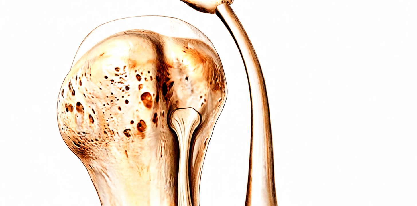
یہ پروٹوکول Mater Private Hospital Rockhampton میں ڈاکٹر کیرن ہِرپرا کے ہاتھوں بائسیپس ٹینوڈیسس (biceps tenodesis، بائسیپس پٹھے کے ٹینڈن کو بازو کی ہڈی پر نئی جگہ جوڑنا) کے بعد بحالی کے بارے میں ہے، چاہے آپریشن دوربین کے ذریعے (keyhole) کیا گیا ہو یا بغل کے سامنے کے قریب ایک چھوٹے کھلے چیرے کے ذریعے (open subpectoral technique)۔ فزیوتھراپی کے پہلے دورے پر یہ صفحہ یا اس کی پی ڈی ایف ساتھ لائیں تاکہ آپ کی بحالی میں سب کا آپس میں رابطہ رہے۔ آپ کا فزیوتھراپسٹ آپ کی بحالی کو نیچے دیے گئے مراحل میں انفرادی طور پر آگے بڑھاتا ہے، اس بات پر منحصر کہ آپ کا کندھا اور بازو کیسے بہتر ہو رہے ہیں۔
یہ پروٹوکول صرف اکیلے بائسیپس ٹینوڈیسس کے لیے ہے۔ اگر آپ کے آپریشن میں روٹیٹر کف (rotator cuff، کندھے کو تھامنے اور گھمانے والے ٹینڈنوں کا گروپ) کی مرمت بھی شامل تھی تو اس کے بجائے روٹیٹر کف کی مرمت کا پروٹوکول اپنائیں؛ مرمت شدہ ٹینڈن کی وجہ سے رفتار سست رکھی جاتی ہے۔
اگر سرجری کے بعد آپ کو اپنے زخم کے بارے میں کوئی فکر ہو تو کلینک سے رابطہ کریں۔ اکثر زخم کی تصویر لے کر جائزے کے لیے ای میل کر دینا مفید ہوتا ہے۔
کیا توقع رکھیں
بائسیپس ٹینوڈیسس میں بائسیپس کے لمبے سر کا ٹینڈن (tendon)، یعنی وہ مضبوط ڈوری جو پٹھے کو ہڈی سے جوڑتی ہے، کندھے کے اندر اپنی اصل جڑنے کی جگہ سے الگ کیا جاتا ہے اور اس کے بجائے اینکر (anchor، ہڈی میں لگایا جانے والا چھوٹا پرزہ) یا پیچ کے ذریعے بازو کے اوپری حصے کی ہڈی (humerus) پر لگا دیا جاتا ہے۔ اس نئے جوڑ کو ہڈی کے ساتھ مضبوطی سے جڑ کر ٹھیک ہونے کے لیے وقت چاہیے، اور بحالی کے شروع کے ہفتے اسی کی حفاظت کے گرد ترتیب دیے جاتے ہیں۔
بائسیپس پٹھا دو کام کرتا ہے: یہ کہنی کو موڑتا ہے اور ہتھیلی کو اوپر کی طرف گھماتا ہے (supination)۔ اسی لیے (کندھے کے آپریشن کے لیے غیر معمولی طور پر) شروع کی پابندیاں زیادہ تر کہنی سے متعلق ہیں: پہلے چند ہفتوں تک کہنی کو بیرونی مدد سے ہلایا جاتا ہے (passive، یعنی موڑنے کا کام دوسرا ہاتھ یا آپ کا فزیوتھراپسٹ کرتا ہے) تاکہ ٹھیک ہوتے ہوئے ٹینڈن کو کھینچنا نہ پڑے۔ اسی وجہ سے شروع میں آپریشن والے بازو سے وزن اٹھانا اور وزن اٹھا کر لے جانا دونوں منع ہیں۔ کندھے کی چند پوزیشنیں جو ٹینڈن پر کھنچاؤ ڈالتی ہیں، شروع میں ان پر بھی حد رکھی جاتی ہے: بازو کو تقریباً 40 ڈگری سے زیادہ باہر کی طرف گھمانا، اور بازو کو جسم کی لکیر سے پیچھے لے جانا۔
آپ تقریباً تین سے چار ہفتے سلنگ (sling)، یعنی گلے سے لٹکی وہ پٹی جو بازو کو سہارا دیتی ہے، پہنیں گے، سوتے وقت بھی، اور تقریباً تیسرے ہفتے سے آرام کے مطابق آہستہ آہستہ اسے چھوڑنا شروع کریں گے۔ کندھے کے کسی بھی آپریشن کے بعد کم از کم چھ ہفتے تک گاڑی نہ چلائیں، چاہے سلنگ اتر چکی ہو؛ آپ کا سرجن آپ کو گاڑی چلانے کی اجازت دے گا، عام طور پر چھ ہفتے والے معائنے پر۔
سفر پر ایک نظر:
- مرحلہ I — ٹینوڈیسس کی حفاظت (تقریباً پہلے چار ہفتے)
- مرحلہ II — خود حرکت (ہفتہ 4 سے 6)
- مرحلہ III — مضبوطی (ہفتہ 6 سے 12، اور 10ویں ہفتے سے بائسیپس کے لیے مزاحمت والی ورزشیں)
- مرحلہ IV — مکمل سرگرمی کی طرف واپسی (12ویں ہفتے اور اس کے بعد)
ہفتوں کی یہ حدیں عام اندازہ ہیں، طے شدہ نہیں، اور آپ کا فزیوتھراپسٹ آپ کو کیلنڈر کے بجائے اس بنیاد پر آگے بڑھائے گا کہ آپ کا بازو کیسے ٹھیک ہو رہا ہے اور کیسے حرکت کر رہا ہے۔ فزیوتھراپی عام طور پر سرجری کے بعد پہلے ایک یا دو ہفتوں کے اندر شروع ہو جاتی ہے، اور آپ کی پہلی ملاقات کی تفصیل ہسپتال سے چھٹی کے وقت ملنے والے کاغذات میں ہوتی ہے، سوائے اس کے کہ آپ نے اپنا انتظام خود کرنے کا انتخاب کیا ہو۔
مرحلہ I — ٹینوڈیسس کی حفاظت (ہفتہ 0 سے 4)
Kieran Hirpara 4.0
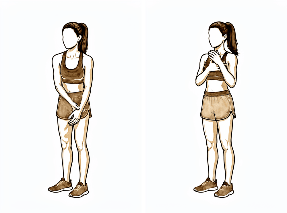
کہنی کو دوسرے ہاتھ کی مدد سے موڑنا (passive)
آپریشن والے بازو کو بالکل ڈھیلا چھوڑ دیں اور اپنے دوسرے ہاتھ سے کہنی کو اوپر کی طرف موڑیں، پھر اسے واپس سیدھا ہونے دیں۔ سارا کام آپ کا دوسرا ہاتھ کرتا ہے — بائسیپس کو بالکل بند رہنا چاہیے تاکہ ٹھیک ہوتے ہوئے ٹینڈن کو کھینچنا نہ پڑے۔ ہتھیلی کو اوپر اور نیچے کی طرف گھمانے کے لیے بھی یہی طریقہ اپنائیں۔
روزانہ
Kieran Hirpara 4.0
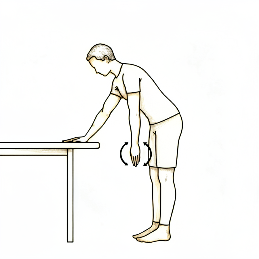
پینڈولم ورزش (جھولنے والی ورزش)
سلنگ سے بازو نکال کر کمر سے آگے کی طرف جھکیں اور آپریشن والے بازو کو جسم سے ہٹ کر آرام سے لٹکنے دیں۔ بازو کو چھوٹے، ڈھیلے دائروں میں جھولنے دیں — حرکت آپ کے جسم سے آتی ہے، کندھے یا بازو کے پٹھوں سے نہیں۔ اتنا ہی کریں جتنا آرام سے ہو سکے، پھر بازو کو واپس سلنگ میں رکھ دیں۔
جیسے آپ کا فزیوتھراپسٹ بتائے
Kieran Hirpara 4.0
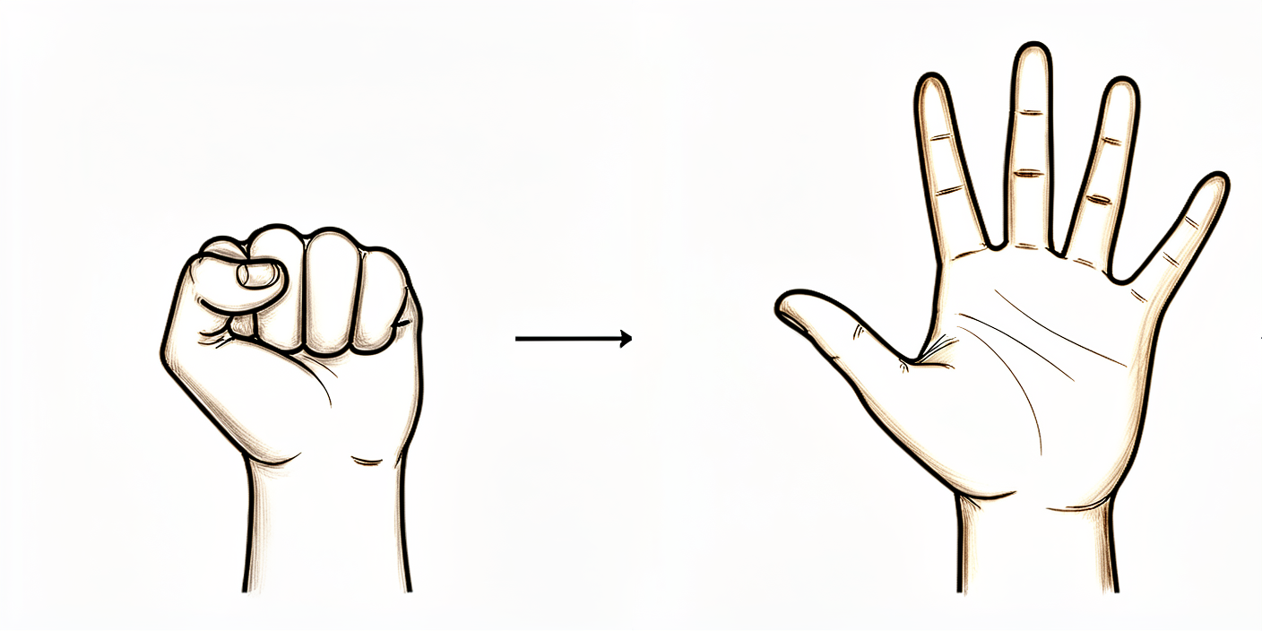
ہاتھ اور گرفت کی ورزشیں
بازو کو سہارا دے کر پوری مٹھی بند کریں، پھر انگلیوں کو کھول کر سیدھا کریں۔ گرفت کی مشق کے لیے نرم گیند یا لپیٹا ہوا موزہ دبایا جا سکتا ہے۔ ہاتھ، کلائی اور انگلیاں شروع سے ہی حرکت میں رہتی ہیں۔
جیسے آپ کا فزیوتھراپسٹ بتائے
Kieran Hirpara 4.0
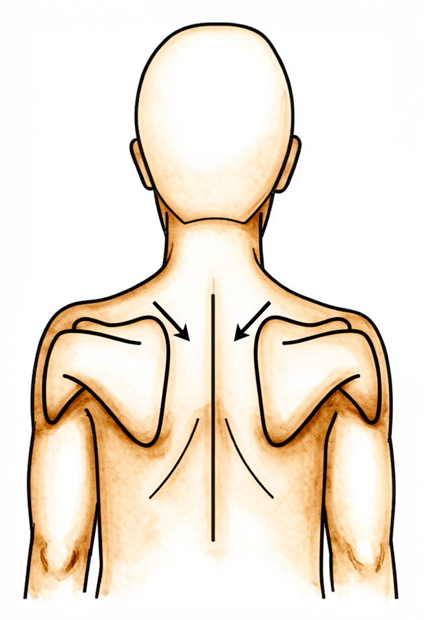
کندھے کی پچھلی ہڈی کو صحیح جگہ رکھنا (scapular setting)
بازو کو سہارا دے کر آہستہ سے اپنے کندھے کی پچھلی چپٹی ہڈیوں کو پیچھے ایک دوسرے کی طرف اور نیچے، کانوں سے دور کھینچیں۔ چند سیکنڈ روکیں، پھر ڈھیلا چھوڑ دیں۔ اس سے ٹینوڈیسس پر بوجھ ڈالے بغیر جسم کی ساخت (posture) اور کندھے کی پچھلی ہڈی پر قابو برقرار رہتا ہے۔
جیسے آپ کا فزیوتھراپسٹ بتائے
Kieran Hirpara 4.0
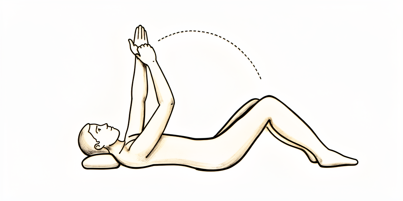
مدد کے ساتھ بازو اوپر اٹھانا (لیٹ کر)
کمر کے بل لیٹ جائیں اور اپنے ٹھیک بازو کی مدد سے آپریشن والے بازو کو سامنے کی طرف تقریباً کندھے کی اونچائی تک اٹھائیں، پھر ٹھیک بازو کی مدد جاری رکھتے ہوئے اسے واپس نیچے لائیں۔ کام آپ کا دوسرا بازو کرتا ہے تاکہ بائسیپس ڈھیلا رہے۔ اتنا ہی کریں جتنا آرام سے ہو سکے — اس سے ٹینوڈیسس ٹھیک ہونے کے دوران کندھا حرکت میں رہتا ہے۔
جیسے آپ کا فزیوتھراپسٹ بتائے
Kieran Hirpara 4.0
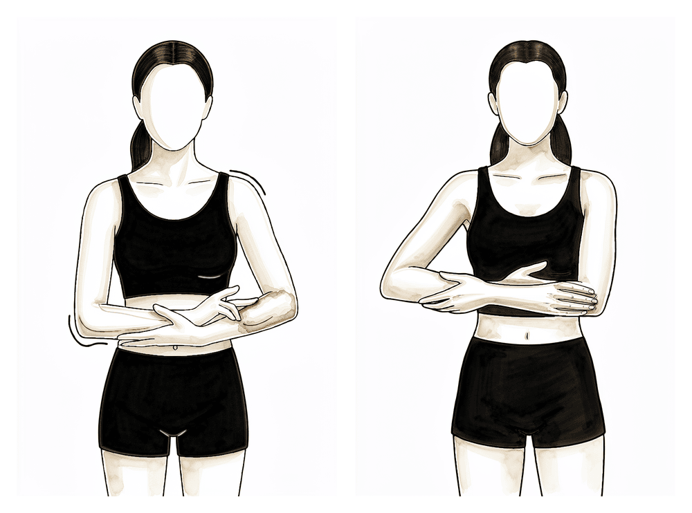
مدد کے ساتھ بازو کو پہلو کی طرف لے جانا
آپریشن والے بازو کے نچلے حصے کو اپنے دوسرے بازو میں تھام لیں اور دونوں کو آہستہ سے پہلو کی طرف اٹھائیں، کام ٹھیک بازو کو کرنے دیں، پھر قابو کے ساتھ نیچے لائیں۔ اسے آرام دہ اور شروع کی حدود کے اندر رکھیں — اس سے بائسیپس سے کام لیے بغیر کندھے کی ہلکی حرکت برقرار رہتی ہے۔
جیسے آپ کا فزیوتھراپسٹ بتائے
Kieran Hirpara 4.0
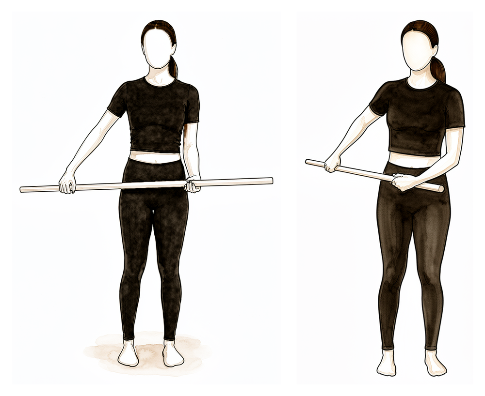
چھڑی کی مدد سے بازو کو باہر کی طرف گھمانا
کمر کے بل لیٹ جائیں، کہنی پہلو کے ساتھ لگی ہو اور سیدھے زاویے پر مڑی ہو۔ ایک چھڑی دونوں ہاتھوں میں پکڑیں اور ٹھیک بازو کی مدد سے آپریشن والے ہاتھ کو باہر کی طرف گھمائیں۔ ان شروع کے ہفتوں میں صرف تقریباً 40 ڈگری تک جائیں — یہ ایک محدود، محفوظ اسٹریچ ہے، پورا نہیں، تاکہ ٹھیک ہوتے ہوئے ٹینڈن پر کھنچاؤ نہ پڑے۔
جیسے آپ کا فزیوتھراپسٹ بتائے، تقریباً 40 ڈگری تک
Kieran Hirpara 4.0
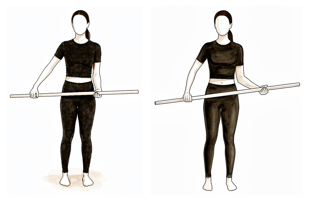
چھڑی کی مدد سے بازو کو اندر کی طرف گھمانا
اسی چھڑی کے ساتھ، اپنے ٹھیک بازو کو آپریشن والے ہاتھ کو جسم کے سامنے سے آہستہ سے اندر کی طرف تقریباً 45 ڈگری تک لے جانے دیں، پھر واپس لائیں۔ آپریشن والے بازو کو ڈھیلا رکھیں اور شروع کی حد کے اندر رہیں — اس سے ٹینوڈیسس پر بوجھ ڈالے بغیر اندر کی طرف گھمانے کی حرکت برقرار رہتی ہے۔
جیسے آپ کا فزیوتھراپسٹ بتائے، تقریباً 45 ڈگری تک
پہلے ہفتوں کا مقصد یہ ہے کہ ٹینڈن ہڈی پر جڑ کر ٹھیک ہو جائے جبکہ اس کے آس پاس سب کچھ حرکت میں رہے۔ آپ کا ہاتھ، کلائی اور انگلیاں شروع سے ہی حرکت میں رہتی ہیں۔ آپ کی کہنی کو روزانہ ہلایا جاتا ہے، لیکن بیرونی مدد سے: موڑنے کا کام اور ہتھیلی کو اوپر/نیچے گھمانے کا کام اپنے دوسرے ہاتھ کو کرنے دیں، تاکہ بائسیپس خود ڈھیلا رہے۔ کندھے کو نیچے دی گئی حدود کے اندر پینڈولم ورزشوں (جھولنے والی ورزشوں) اور مدد والی حرکتوں کے ذریعے آہستہ سے ہلایا جاتا ہے۔ درد میں آرام کے لیے برف استعمال کریں، ورزشوں اور فزیوتھراپی سیشن سے پہلے اپنی درد کش دوا لیں، اور سلنگ پہنے رکھیں، بستر میں بھی۔ آپریشن والے بازو سے نہ کچھ اٹھائیں اور نہ کچھ اٹھا کر لے جائیں، اور جب تک سلنگ کی ضرورت ہے گاڑی نہ چلائیں۔ بازو کے نچلے حصے کو سہارا دے کر ہلکے کام، جیسے لکھنا یا ٹائپ کرنا، عام طور پر آرام کے مطابق ٹھیک ہیں۔
آپ کے فزیوتھراپسٹ کے لیے:
اہداف
- ٹینوڈیسس کی فکسیشن (fixation) کی حفاظت جب تک وہ ہڈی سے جڑ کر ٹھیک ہو
- درد، سوجن اور سوزش کے ردِعمل کو کم کرنا
- کہنی اور بازو کے نچلے حصے کی بیرونی مدد سے مکمل حرکت (passive range of motion)؛ نیچے دی گئی حدود کے اندر کندھے کی بیرونی مدد سے آرام دہ حرکت
- کندھے کی پچھلی ہڈی کے کام (scapular function) اور جسم کی ساخت (posture) کو برقرار رکھنا
علاج کا طریقہ
- تقریباً 3 سے 4 ہفتے سلنگ، رات کو بھی، اور ہفتہ 3 کے آس پاس سے آہستہ آہستہ چھوڑنا
- کہنی کی بیرونی مدد سے حرکت: موڑنا/سیدھا کرنا (flexion/extension) اور بازو کے نچلے حصے کو اوپر/نیچے گھمانا (supination/pronation)
- کلائی اور ہاتھ کی خود حرکت (active)؛ گیند دبانا
- حدود کے اندر کندھے کی بیرونی مدد سے اور مدد کے ساتھ ہلکی خود حرکت (active-assisted): پینڈولم، سامنے کی طرف اٹھانا (flexion) اور کندھے کی پچھلی ہڈی کی سمت میں اٹھانا (scaption) شروع میں تقریباً 90 ڈگری تک، آرام کے مطابق بڑھاتے ہوئے؛ باہر کی طرف گھمانا (external rotation) 40 ڈگری تک؛ اندر کی طرف گھمانا (internal rotation) تقریباً 45 ڈگری تک
- بازو کو سہارا دے کر کندھے کی پچھلی ہڈی کو صحیح جگہ رکھنا اور پیچھے کھینچنا (scapular setting and retraction)، پھر اس ہڈی کی بغیر حرکت والی ورزشوں (scapular isometrics) کی طرف بڑھنا؛ گردن کی حرکت اور جسم کی ساخت پر کام
- درد اور سوجن کے لیے ٹھنڈک کا علاج (cryotherapy)؛ ورزشوں اور سیشن سے پہلے درد کش دوا
- کمر کے بل لیٹتے وقت کہنی کے نیچے لپیٹا ہوا تولیہ یا چھوٹا تکیہ، تاکہ کندھا پیچھے کی طرف نہ جائے (shoulder extension)
احتیاطیں
- کہنی کو خود موڑنا (active elbow flexion) نہیں، اور مزاحمت کے ساتھ ہتھیلی اوپر گھمانا (resisted supination) بھی نہیں؛ بائسیپس پر کوئی بوجھ نہ ہو
- کندھے کی خود حرکت نہیں؛ 40 ڈگری سے زیادہ باہر کی طرف گھمانا نہیں؛ کندھے کو نیوٹرل (neutral) پوزیشن سے آگے نہ پیچھے کی طرف لے جائیں (extension) اور نہ افقی سمت میں باہر کی طرف (horizontal abduction)
- آپریشن والے بازو سے نہ وزن اٹھانا اور نہ وزن اٹھا کر لے جانا
- بائسیپس کے اوپری حصے / ٹینوڈیسس کی جگہ پر رگڑ والا مساج (friction massage) نہیں
- چھ ہفتے تک گاڑی نہیں چلانی (یہ کندھے کے ہر آپریشن پر لاگو ہوتا ہے)
اگلے مرحلے میں جانے کی شرائط
- زخم بھر چکا ہو اور درد اچھی طرح قابو میں ہو
- کہنی کا بیرونی مدد سے مکمل موڑنا/سیدھا کرنا اور بازو کے نچلے حصے کا مکمل گھماؤ
- مقررہ حدود کے اندر کندھے کی بیرونی مدد سے آرام دہ حرکت
مرحلہ II — خود حرکت (ہفتہ 4 سے 6)
Kieran Hirpara 4.0
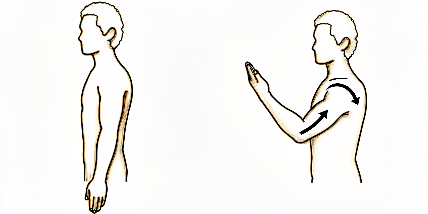
کہنی کو خود موڑنا اور سیدھا کرنا
ہاتھ میں کچھ پکڑے بغیر کہنی کو اپنی طاقت سے موڑیں اور سیدھا کریں، اور ہتھیلی کو اوپر اور نیچے کی طرف گھمائیں۔ اب بائسیپس حرکت کر رہا ہے لیکن ابھی وزن کے خلاف کام نہیں کر رہا — اس بازو سے وزن اٹھانا کم سے کم رکھیں (چائے کے ایک کپ سے زیادہ بھاری کچھ نہیں) جب تک آپ کا فزیوتھراپسٹ آپ کو اگلے قدم پر نہ لے جائے۔
جیسے آپ کا فزیوتھراپسٹ بتائے
Kieran Hirpara 4.0
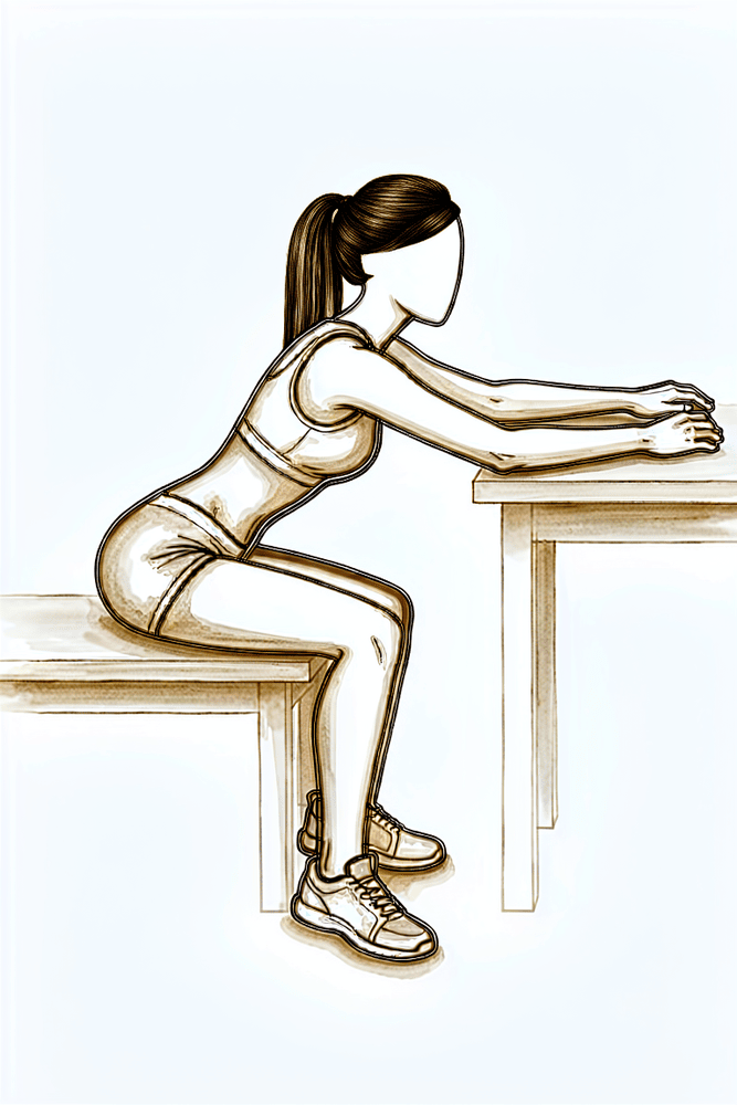
میز پر ہاتھ سرکانا
میز کے پاس بیٹھ جائیں اور اپنا ہاتھ تولیے یا کپڑے پر رکھیں۔ ہاتھ کو سطح پر آگے کی طرف سرکائیں اور آگے جھکیں تاکہ بازو آپ کے سامنے اوپر کی طرف جائے، پھر واپس سرکائیں۔ میز وزن سنبھالتی ہے، جس سے حرکت کی حد واپس آنے کے ساتھ کندھے کو خود حرکت کرنے میں مدد ملتی ہے۔
جیسے آپ کا فزیوتھراپسٹ بتائے
Kieran Hirpara 4.0
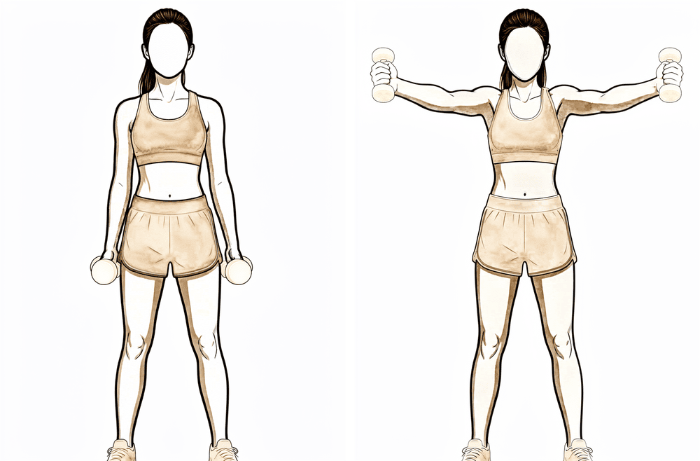
کندھے کی پچھلی ہڈی کی سمت میں بازو خود اٹھانا (scapular plane)
کھڑے ہو کر آپریشن والے بازو کو اوپر اور تھوڑا آگے کی طرف اٹھائیں — سیدھا سامنے اور پہلو کی طرف کے تقریباً درمیان — انگوٹھا اوپر کی طرف ہو، جیسے بھرا ہوا ڈبہ پکڑا ہو۔ جتنا آرام سے ہو سکے اپنی طاقت سے اٹھائیں، پھر آہستہ سے نیچے لائیں۔ اس سے کسی اضافی وزن کے بغیر کندھے کو خود اٹھانے کی صلاحیت واپس آتی ہے۔
جیسے آپ کا فزیوتھراپسٹ بتائے
Kieran Hirpara 4.0
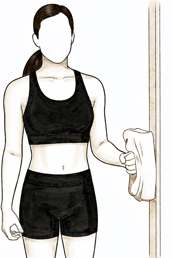
بغیر حرکت کے باہر کی طرف زور لگانا (isometric external rotation)
کہنی پہلو کے ساتھ لگا کر سیدھے زاویے پر موڑ کر کھڑے ہوں، ہاتھ کی پشت دیوار یا دروازے کی چوکھٹ سے لگی ہو۔ بازو کو حرکت دیے بغیر آہستہ سے باہر کی طرف دبائیں، چند سیکنڈ روکیں، پھر ڈھیلا چھوڑ دیں۔ اس سے بغیر حرکت کے اور بائسیپس پر کوئی بوجھ ڈالے بغیر روٹیٹر کف (rotator cuff)، یعنی ٹینڈنوں کا وہ گروپ جو کندھے کو تھامتا اور گھماتا ہے، مضبوط ہونا شروع ہوتا ہے۔
چند سیکنڈ روکیں، کئی بار؛ جیسے آپ کا فزیوتھراپسٹ بتائے
Kieran Hirpara 4.0
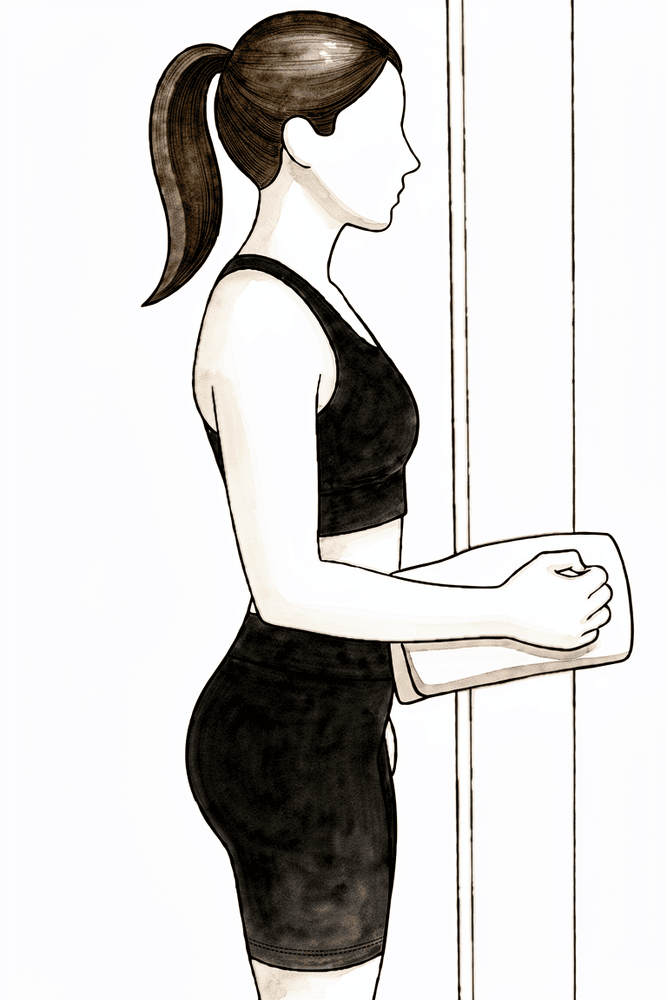
بغیر حرکت کے اندر کی طرف زور لگانا (isometric internal rotation)
کہنی پہلو کے ساتھ لگا کر سیدھے زاویے پر موڑ کر کھڑے ہوں، ہتھیلی دیوار یا دروازے کی چوکھٹ سے لگی ہو۔ بازو کو حرکت دیے بغیر آہستہ سے اندر کی طرف دبائیں، چند سیکنڈ روکیں، پھر ڈھیلا چھوڑ دیں۔ پوری طاقت سے کم زور لگائیں — مضبوط لیکن آسان، کبھی زور زبردستی نہیں۔
چند سیکنڈ روکیں، کئی بار؛ جیسے آپ کا فزیوتھراپسٹ بتائے
Kieran Hirpara 4.0
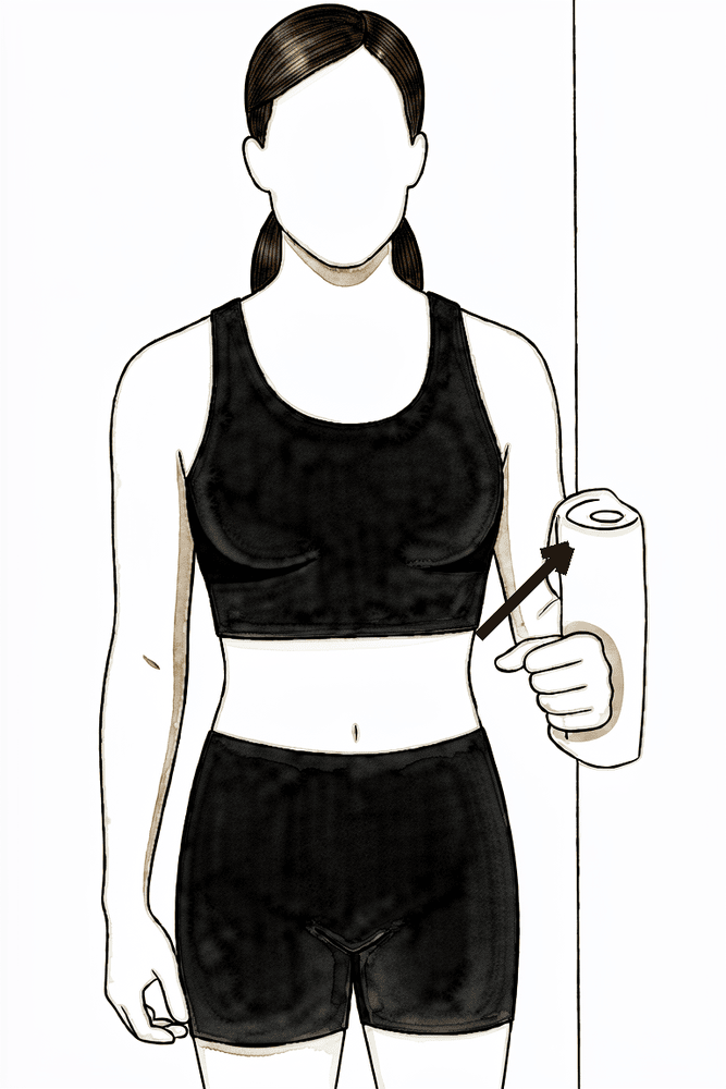
بغیر حرکت کے بازو کو باہر کی طرف زور لگانا (isometric abduction)
دیوار کی طرف پہلو کر کے کھڑے ہوں، کہنی پہلو کے ساتھ ہو۔ بازو کے اوپری حصے کی باہر والی طرف کو آہستہ سے دیوار میں دبائیں جیسے اسے پہلو کی طرف اٹھا رہے ہوں، اسے حرکت دیے بغیر، چند سیکنڈ روکیں، پھر ڈھیلا چھوڑ دیں۔ اس سے حرکت والی ورزشوں پر بوجھ شروع ہونے سے پہلے کندھے کے پٹھے بغیر حرکت کے مضبوط ہوتے ہیں۔
چند سیکنڈ روکیں، کئی بار؛ جیسے آپ کا فزیوتھراپسٹ بتائے
Kieran Hirpara 4.0
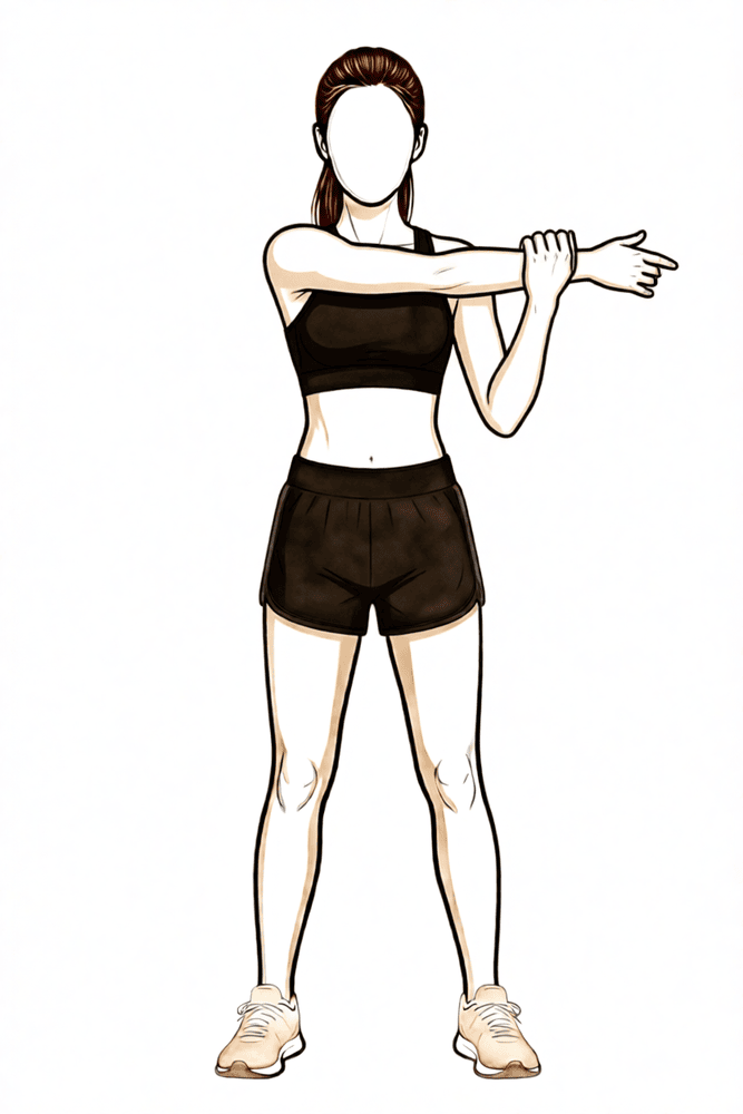
سینے کے آر پار اسٹریچ
آپریشن والے بازو کو اپنے سینے کے آر پار لائیں اور دوسرے بازو سے اسے آہستہ سے مزید آگے کھینچیں یہاں تک کہ کندھے کے پیچھے ایک آرام دہ کھنچاؤ محسوس ہو۔ روکیں، پھر ڈھیلا چھوڑ دیں۔ اس سے حرکت کی حد واپس آنے کے ساتھ کندھے کے پیچھے کی جکڑن کم ہوتی ہے۔
جیسے آپ کا فزیوتھراپسٹ بتائے
Kieran Hirpara 4.0
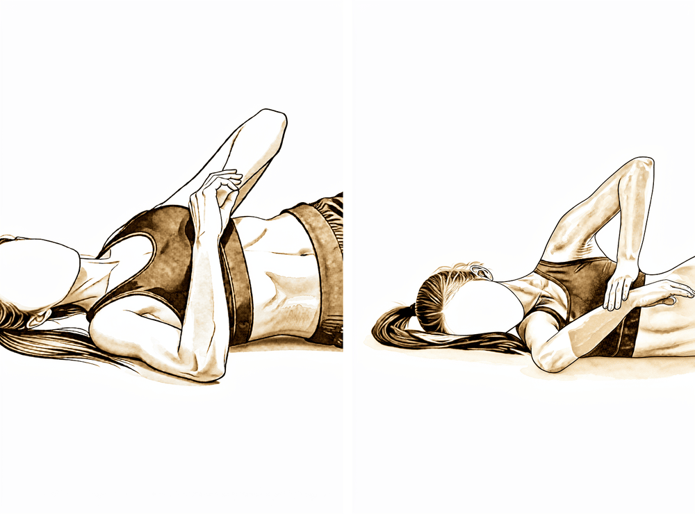
سلیپر اسٹریچ (sleeper stretch)
آپریشن والی طرف کروٹ لے کر لیٹیں، وہ بازو سامنے ہو اور کہنی سیدھے زاویے پر مڑی ہو۔ اپنے دوسرے ہاتھ سے بازو کے نچلے حصے کو آہستہ سے بستر کی طرف دبائیں یہاں تک کہ کندھے کے پیچھے کھنچاؤ محسوس ہو۔ اسے ہلکا رکھیں — زبردستی کرنے کے بجائے اسٹریچ میں ڈھیلے پڑ جائیں۔
جیسے آپ کا فزیوتھراپسٹ بتائے
Kieran Hirpara 4.0
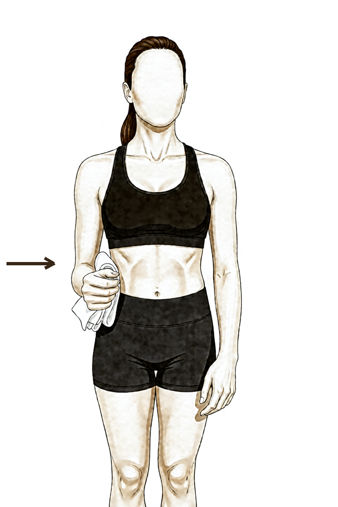
بغیر حرکت کے اندر کی طرف دبانا (adduction)
کہنی پہلو کے ساتھ لگا کر کھڑے ہوں اور بازو کے اوپری حصے اور جسم کے درمیان لپیٹا ہوا تولیہ رکھیں۔ اپنے بازو کو تولیے کے خلاف آہستہ سے اندر کی طرف دبائیں بالکل حرکت کیے بغیر — ہلکا زور، تقریباً اپنی طاقت کا ایک چوتھائی، اور کوئی درد نہ ہو۔ روکیں، پھر ڈھیلا چھوڑ دیں۔
تقریباً 5 سیکنڈ روکیں، ہلکا زور (تقریباً 25 فیصد طاقت)، جیسے آپ کا فزیوتھراپسٹ بتائے
سلنگ اترنے کے بعد بازو اپنی طاقت سے حرکت کرنا شروع کرتا ہے۔ کندھا مدد والی حرکتوں سے ہر سمت میں خود حرکت کی طرف بڑھتا ہے، اور کہنی اب خود مڑتی اور گھومتی ہے، لیکن اب بھی بغیر وزن کے۔ بائسیپس حرکت کر رہا ہے، ابھی کام نہیں کر رہا: وزن اٹھانا کم سے کم رکھیں (اس بازو سے چائے کے ایک کپ سے زیادہ بھاری کچھ نہیں)، اور دھکیلنا، کھینچنا اور وزن اٹھا کر لے جانا، یہ سب دوسرے بازو پر چھوڑ دیں۔ اس مرحلے میں ہلکا دفتری کام عام طور پر آرام سے ہو جاتا ہے۔ گاڑی چلانا چھ ہفتے سے دوبارہ شروع ہوتا ہے، جب آپ کا سرجن اجازت دے دے اور آپ گاڑی کو آرام سے اور محفوظ طریقے سے قابو میں رکھ سکیں۔
آپ کے فزیوتھراپسٹ کے لیے:
اہداف
- کندھے اور کہنی کی مکمل خود حرکت کی بتدریج بحالی
- حرکت کے دوران کندھے کی پچھلی ہڈی کا معمول کا طریقہ کار (scapular mechanics)
- کندھے کی بغیر حرکت والی ورزشیں (isometrics) پوری طاقت سے کم زور کے ساتھ شروع کرنا
- کمر کی سطح پر بازو کا ہلکا روزمرہ استعمال
علاج کا طریقہ
- کندھے کی مدد کے ساتھ خود حرکت سے ہر سمت میں مکمل خود حرکت کی طرف بڑھنا (مثال کے طور پر lawn-chair progression، دیوار اور ریلنگ پر ہاتھ سرکانا، لیٹ کر سامنے کی طرف اٹھانے سے کھڑے ہو کر scaption تک)
- کہنی کو خود موڑنا/سیدھا کرنا اور بازو کے نچلے حصے کو اوپر/نیچے گھمانا، بغیر مزاحمت کے
- کندھے کی بغیر حرکت والی ورزشیں، پوری طاقت سے کم: اندر کی طرف گھمانا، باہر کی طرف گھمانا، باہر اٹھانا (abduction)، اندر لانا (adduction)
- کندھے کی پچھلی ہڈی کو مستحکم کرنے (scapular stabilisation) اور جسم کی ساخت پر کام جاری رکھنا
- زخم پختہ ہونے کے ساتھ زخم کے نشان کا مساج؛ ٹینوڈیسس کی جگہ پر آڑی رگڑ (cross-friction) نہیں
- ضرورت کے مطابق کندھے کی پچھلی جھلی کا اسٹریچ (posterior capsule stretching: cross-body، sleeper stretch)
- فٹنس کے لیے چہل قدمی یا ایک جگہ کھڑی ورزش والی سائیکل؛ متاثرہ بازو پر وزن نہیں ڈالنا
احتیاطیں
- بائسیپس کی مزاحمت والی ورزش نہیں؛ کہنی موڑنے والے اور ہتھیلی اوپر گھمانے والے پٹھوں پر بوجھ نہیں
- آپریشن والے بازو سے وزن اٹھانا نہیں؛ ابھی دوڑنا نہیں
- سخت اسٹریچنگ یا ہاتھ سے کیے جانے والے علاج (manual therapy) سے ٹھیک ہوتے ہوئے ٹینڈن پر ضرورت سے زیادہ دباؤ ڈالنے سے بچیں
اگلے مرحلے میں جانے کی شرائط
- کندھے، کہنی اور بازو کے نچلے حصے کی مکمل، بغیر درد کے خود حرکت
- حرکت اور ہلکے کام کے دوران کندھے کی پچھلی ہڈی کا درست طریقہ کار
- درد اچھی طرح قابو میں ہو
مرحلہ III — مضبوطی (ہفتہ 6 سے 12)
Kieran Hirpara 4.0
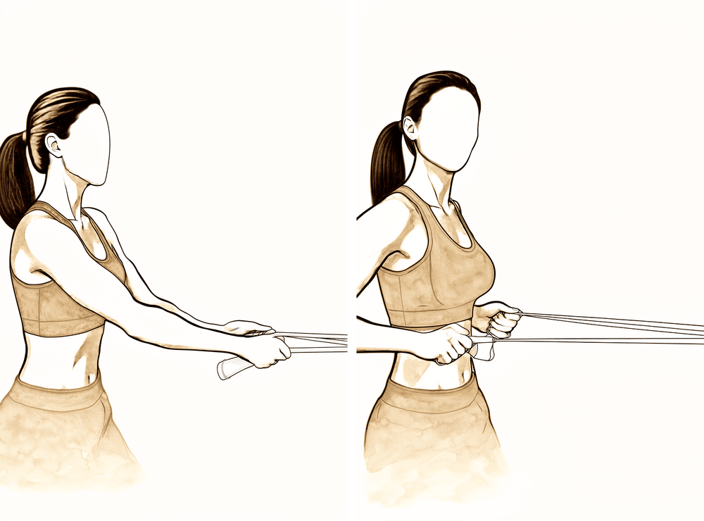
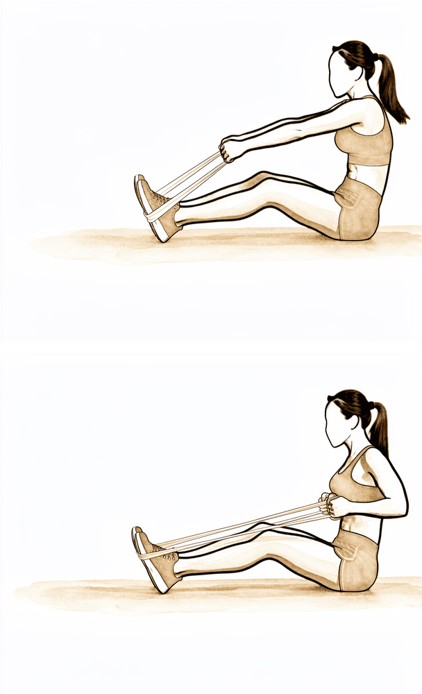
لچکدار بینڈ کے ساتھ رو (row)
ورزش کا لچکدار بینڈ اپنے سامنے تقریباً کمر کی اونچائی پر باندھ دیں۔ بینڈ کو اپنی طرف کھینچیں، اور جب کہنی آپ کے پہلو سے گزرے تو کندھے کی پچھلی ہڈی کو پیچھے اور نیچے کھینچیں، پھر قابو کے ساتھ واپس جائیں۔ یہ کندھے کی پچھلی ہڈی کے پروگرام کا حصہ ہے — وزن کم اور تعداد زیادہ رکھیں۔
جیسے آپ کا فزیوتھراپسٹ بتائے
Kieran Hirpara 4.0
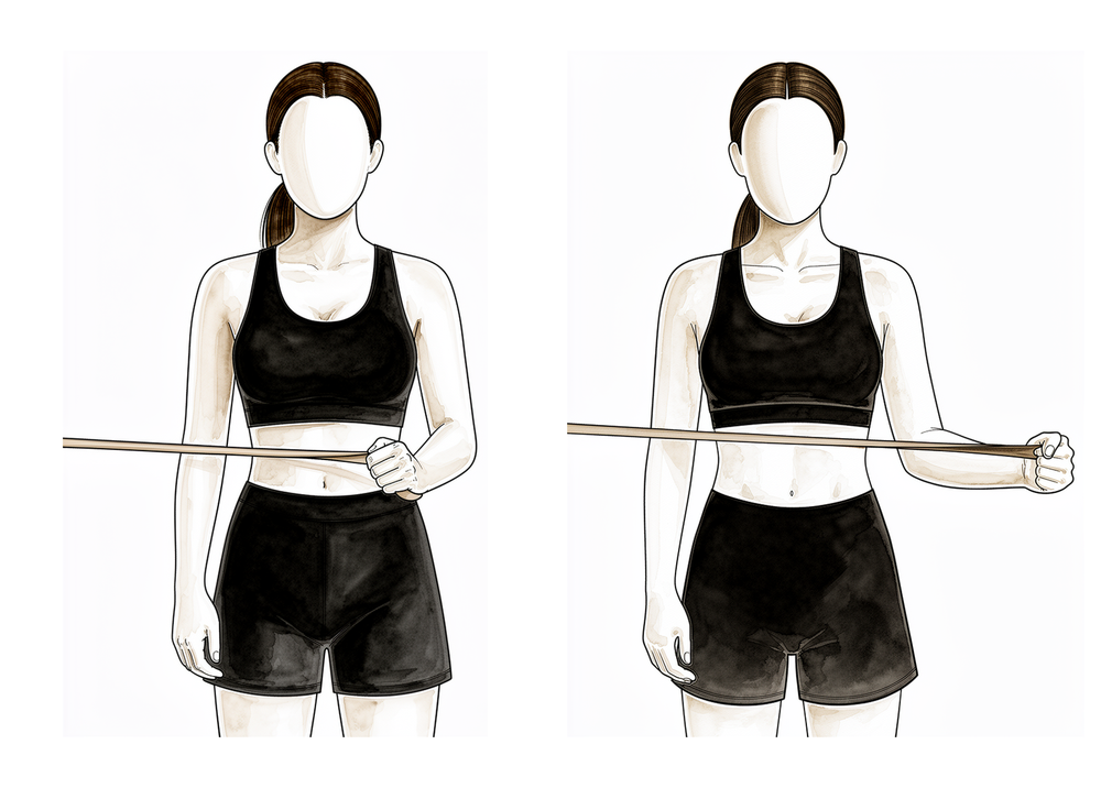
بینڈ کے ساتھ باہر کی طرف گھمانا
لچکدار بینڈ کمر کی اونچائی پر باندھ دیں۔ کہنی پہلو کے ساتھ لگا کر سیدھے زاویے پر موڑ کر، بینڈ کو باہر کی طرف کھینچیں اور بازو کے نچلے حصے کو جسم سے دور گھمائیں، پھر آہستہ سے واپس لائیں۔ ہلکے سے شروع کریں اور تعداد زیادہ رکھیں — اس سے روٹیٹر کف مضبوط ہوتا ہے۔
جیسے آپ کا فزیوتھراپسٹ بتائے — کم وزن، زیادہ تعداد
Kieran Hirpara 4.0
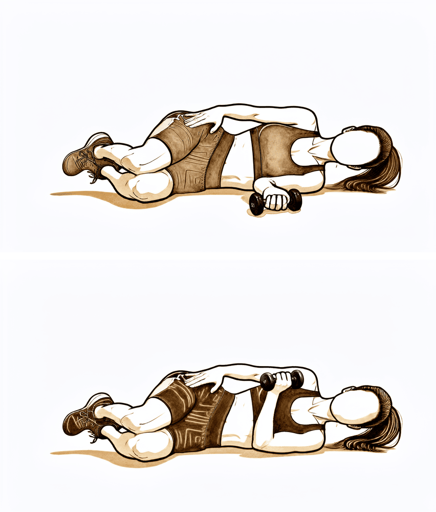
مزاحمت کے ساتھ اندر کی طرف گھمانا
آپریشن والی طرف کروٹ لے کر لیٹیں، ہاتھ میں ہلکا وزن ہو اور کہنی پہلو کے ساتھ مڑی ہو۔ بازو کے نچلے حصے کو اپنے جسم کی طرف اوپر گھما کر وزن اٹھائیں، پھر آہستہ سے نیچے لائیں۔ اس سے روٹیٹر کف کے اندر کی طرف گھمانے والے پٹھے مضبوط ہوتے ہیں — شروع میں وزن ہلکا رکھیں۔
جیسے آپ کا فزیوتھراپسٹ بتائے — کم وزن، زیادہ تعداد
Kieran Hirpara 4.0
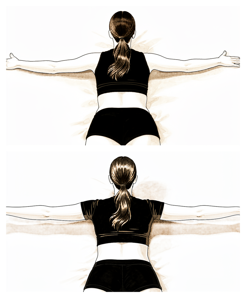
الٹا لیٹ کر بازو کو T کی شکل میں اٹھانا
پیٹ کے بل لیٹ جائیں اور بازو کنارے سے نیچے لٹکنے دیں۔ آپریشن والے بازو کو پہلو کی طرف کندھے کی اونچائی تک اٹھائیں اور کندھے کی پچھلی ہڈی کو دبائیں، تاکہ بازو T کی شکل بنائیں، پھر آہستہ سے نیچے لائیں۔ یہ کندھے کی پچھلی ہڈی کے پروگرام کا حصہ ہے — وزن سے زیادہ قابو اہم ہے۔
جیسے آپ کا فزیوتھراپسٹ بتائے
Kieran Hirpara 4.0
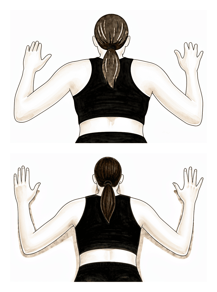
الٹا لیٹ کر بازو کو W کی شکل میں کھینچنا
پیٹ کے بل لیٹ جائیں اور کہنیوں کو پیچھے اور نیچے کمر کی طرف کھینچیں، انہیں موڑتے ہوئے تاکہ بازو W کی شکل بنائیں، اور کندھے کی پچھلی ہڈیوں کو ایک دوسرے کی طرف دبائیں، پھر آہستہ سے نیچے لائیں۔ اس سے کندھے کی پچھلی ہڈی کے نچلے پٹھے مضبوط ہوتے ہیں جو جسم کی اچھی ساخت کو سہارا دیتے ہیں۔
جیسے آپ کا فزیوتھراپسٹ بتائے
Kieran Hirpara 4.0
نیچی رو (low row)
بازو پہلو کے ساتھ رکھ کر بینڈ یا کسی سطح کے خلاف نیچے اور پیچھے کی طرف دبائیں، کندھے کی پچھلی ہڈی کو نیچے اور اندر کی طرف کھینچیں، پھر چھوڑ دیں۔ یہ کندھے کی پچھلی ہڈی کے پروگرام کا حصہ ہے، جو اس ہڈی کے نچلے حصے کے گرد قابو پیدا کرتا ہے۔
جیسے آپ کا فزیوتھراپسٹ بتائے
Kieran Hirpara 4.0
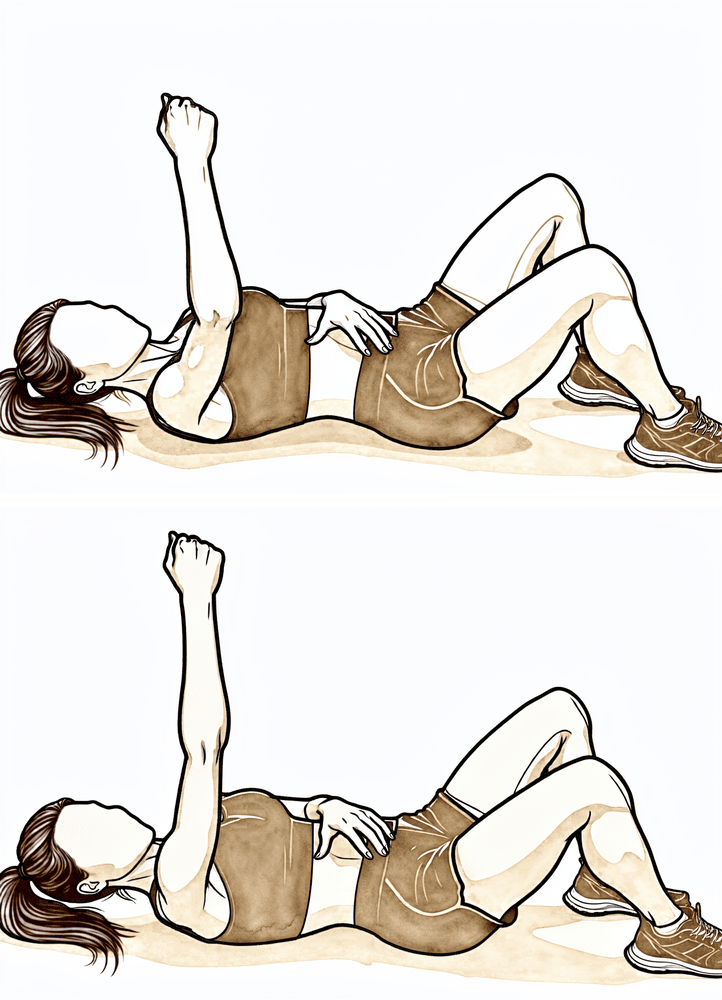
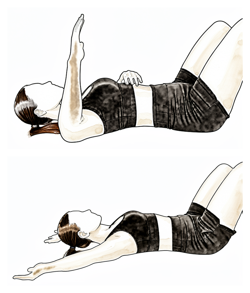
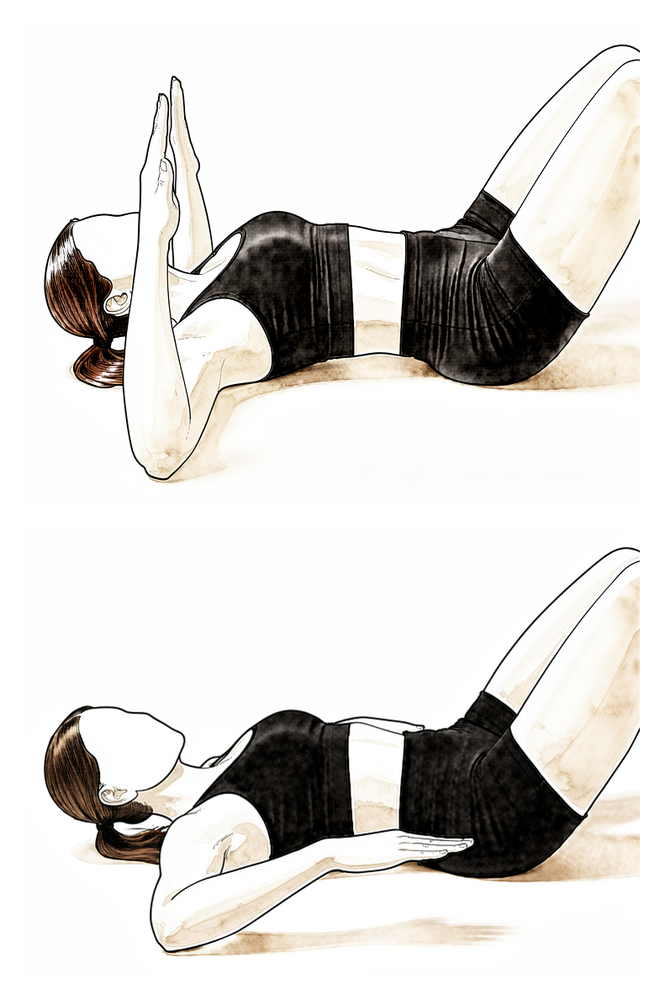
سیریٹس پنچ (serratus punch)
کمر کے بل لیٹ جائیں اور آپریشن والے بازو کو سیدھا چھت کی طرف اٹھائیں۔ تھوڑا سا مزید اوپر مکا ماریں تاکہ کندھے کی پچھلی ہڈی فرش سے اٹھ جائے، پھر اسے واپس نیچے لائیں۔ اس سے سیریٹس پٹھا مضبوط ہوتا ہے جو کندھے کی پچھلی ہڈی کو پسلیوں کے ساتھ لگائے رکھتا ہے۔
جیسے آپ کا فزیوتھراپسٹ بتائے
Kieran Hirpara 4.0
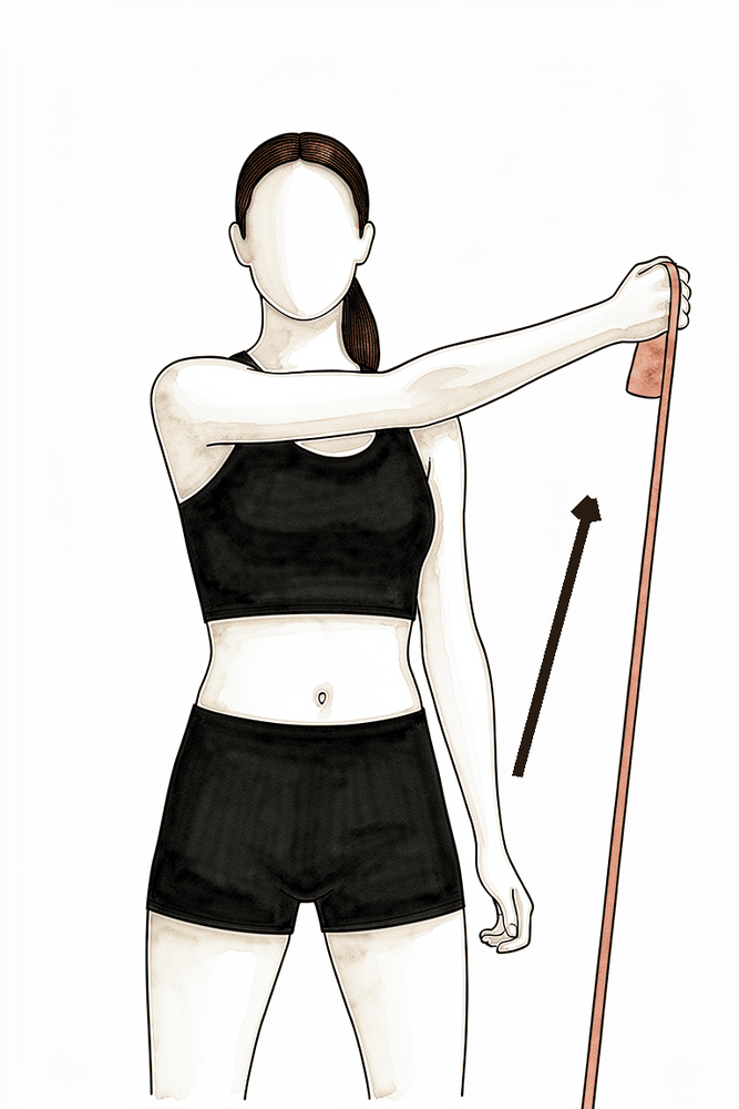
بینڈ کے ساتھ ترچھی حرکت (D1)
لچکدار بینڈ کے ساتھ، بازو کو جسم کے سامنے سے ترچھی لکیر میں اوپر اور باہر اپنے سے دور کھینچیں، پھر قابو کے ساتھ واپس لائیں۔ یہ ترچھی حرکتیں کندھے کی ہم آہنگ اور روزمرہ کام آنے والی طاقت دوبارہ بناتی ہیں۔
جیسے آپ کا فزیوتھراپسٹ بتائے
Kieran Hirpara 4.0
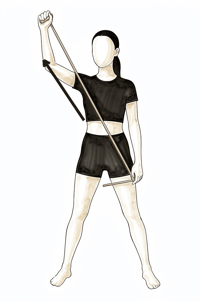
بینڈ کے ساتھ ترچھی حرکت (D2)
لچکدار بینڈ کے ساتھ، بازو کو نیچے سے جسم کے سامنے سے ترچھی لکیر میں اوپر اور باہر سر کے اوپر تک لے جائیں، پھر قابو کے ساتھ واپس نیچے لائیں۔ یہ دوسری ترچھی حرکت ہے جو زیادہ پوری، کھیل جیسی حدود میں طاقت بناتی ہے۔
جیسے آپ کا فزیوتھراپسٹ بتائے
Kieran Hirpara 4.0
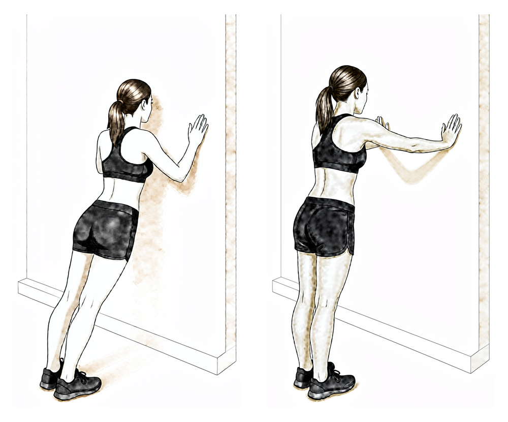
دیوار پر پُش اپ پلس (push-up plus)
دونوں ہاتھ کندھے کی اونچائی پر دیوار پر رکھ کر کھڑے ہوں۔ کہنیاں سیدھی رکھتے ہوئے سینے کو دیوار سے دور دھکیلیں تاکہ کندھے کی پچھلی ہڈیاں آگے کی طرف گول ہو جائیں، پھر ڈھیلے ہو کر واپس آئیں۔ یہ پُش اپ پلس کے مرحلہ وار سلسلے کا آغاز ہے — پہلے دیوار، پھر کاؤنٹر، پھر گھٹنوں کے بل، پھر فرش پر، جیسے بتایا جائے۔
جیسے آپ کا فزیوتھراپسٹ بتائے
Kieran Hirpara 4.0
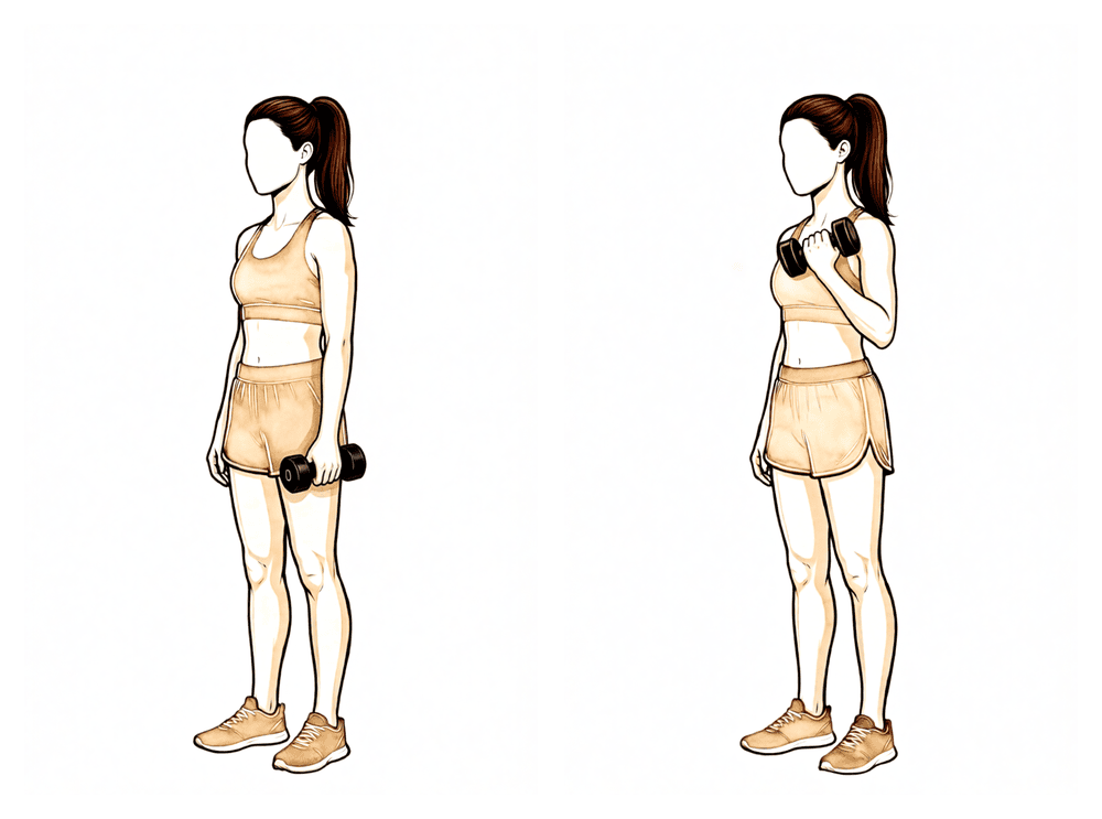
بائسیپس کرل (biceps curl)
10ویں ہفتے سے پہلے نہ کریں۔ ہاتھ میں ہلکا وزن لے کر اور کہنی پہلو کے ساتھ رکھ کر، کہنی موڑ کر وزن اٹھائیں، پھر آہستہ سے نیچے لائیں۔ مزاحمت کے ساتھ ہتھیلی اوپر کی طرف گھمانا بھی اسی طرح شامل کیا جاتا ہے۔ ہلکے سے شروع کریں اور آہستہ آہستہ بڑھائیں — یہ جان بوجھ کر وہ آخری ورزش ہے جو ٹھیک ہوتے ہوئے ٹینڈن پر بوجھ ڈالتی ہے۔
10ویں ہفتے سے — کم وزن، زیادہ تعداد
Kieran Hirpara 4.0
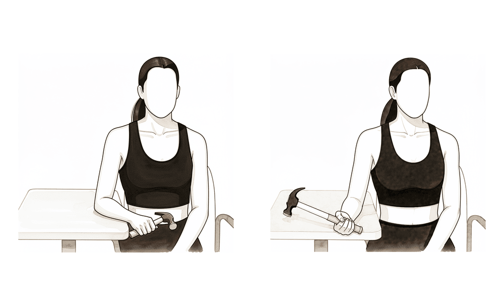
مزاحمت کے ساتھ بازو کے نچلے حصے کو گھمانا
10ویں ہفتے سے پہلے نہ کریں۔ بازو کا نچلا حصہ میز پر رکھیں اور ہاتھ کنارے سے باہر ہو، ہاتھ میں ہلکا وزن پکڑیں۔ آہستہ سے ہتھیلی اوپر کی طرف گھمائیں، پھر نیچے کی طرف، اور حرکت کو قابو میں رکھیں۔ ہتھیلی اوپر گھمانے سے بائسیپس پر براہِ راست کام پڑتا ہے، اس لیے یہ ورزش کرل کے ساتھ ہی شروع ہوتی ہے — ہلکے سے شروع کریں اور آہستہ آہستہ بڑھائیں۔
10ویں ہفتے سے — کم وزن، زیادہ تعداد
Kieran Hirpara 4.0
بازو کو 90/90 پوزیشن میں باہر کی طرف گھمانا (بینڈ)
بازو کو کندھے کی اونچائی پر پہلو کی طرف رکھ کر اور کہنی سیدھے زاویے پر موڑ کر، ہلکے بینڈ یا وزن کے خلاف بازو کے نچلے حصے کو پیچھے اور اوپر کی طرف گھمائیں، پھر آہستہ سے نیچے لائیں۔ اس سے اٹھی ہوئی (گیند پھینکنے والی) پوزیشن میں روٹیٹر کف کی طاقت بنتی ہے۔
10 سے 15 بار کے 2 سے 3 سیٹ، کم مزاحمت
Kieran Hirpara 4.0
بازو کو 90/90 پوزیشن میں اندر کی طرف گھمانا (بینڈ)
اسی پوزیشن سے، بازو کندھے کی اونچائی پر پہلو کی طرف رکھ کر، ہلکے بینڈ یا وزن کے خلاف بازو کے نچلے حصے کو آگے اور نیچے اپنے پاؤں کی طرف گھمائیں، پھر قابو کے ساتھ واپس لائیں۔
10 سے 15 بار کے 2 سے 3 سیٹ، کم مزاحمت
Kieran Hirpara 4.0
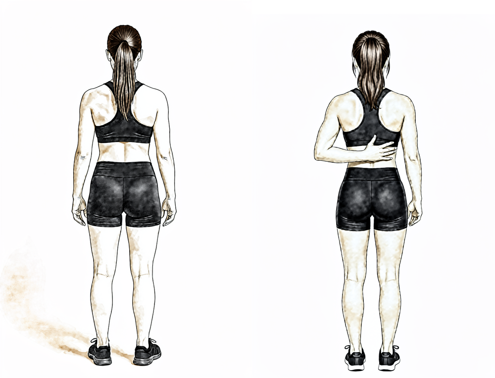
ہاتھ کمر کے پیچھے لے جانا
کھڑے ہو کر آپریشن والے ہاتھ کی پشت کو کمر کے نچلے حصے پر رکھیں اور اپنے پٹھوں سے آہستہ سے اسے ریڑھ کی ہڈی کے ساتھ اوپر سرکائیں جتنا آرام سے جا سکے، پھر نیچے لائیں۔ صرف آرام دہ حد کے اندر حرکت کریں اور کبھی زبردستی نہ کریں۔
جیسے آپ کا فزیوتھراپسٹ بتائے
Kieran Hirpara 4.0
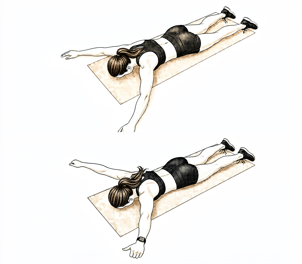
الٹا لیٹ کر Y کی شکل (کندھے کی پچھلی ہڈی کا نچلا حصہ)
پیٹ کے بل لیٹ جائیں، آپریشن والا بازو سر کے اوپر اور تقریباً 45° پر باہر کی طرف ہو تاکہ Y کی شکل بنے، انگوٹھا اوپر کی طرف ہو۔ کندھے کی پچھلی ہڈی کو نیچے اور پیچھے کھینچ کر بازو کو سطح سے تھوڑا سا اٹھائیں، پھر آہستہ سے نیچے لائیں۔ اسے ہلکا رکھیں — اس سے کندھے کی پچھلی ہڈی کے نچلے پٹھوں پر کام ہوتا ہے۔
جیسے آپ کا فزیوتھراپسٹ بتائے — ہلکا، زیادہ تعداد
Kieran Hirpara 4.0
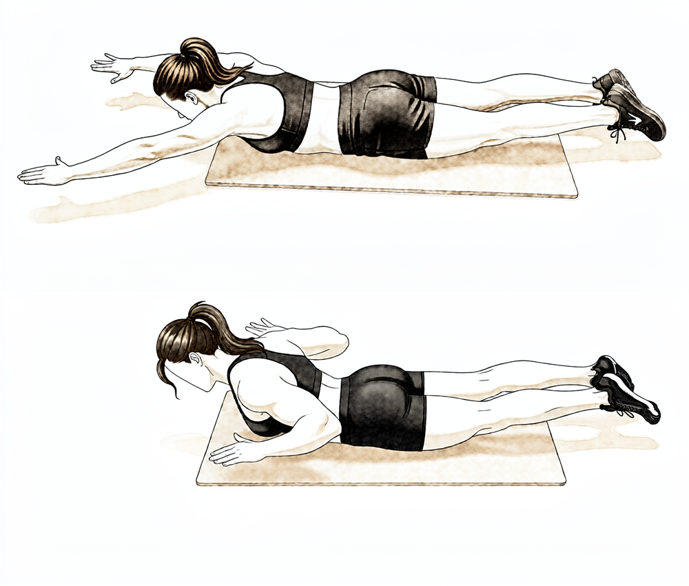
الٹا لیٹ کر رو (prone row)
پیٹ کے بل لیٹ جائیں اور آپریشن والا بازو فرش کی طرف نیچے لٹکنے دیں۔ کہنی کو چھت کی طرف اوپر کھینچیں اور کندھے کی پچھلی ہڈی کو ریڑھ کی ہڈی کی طرف دبائیں، پھر آہستہ سے نیچے لائیں۔ بغیر وزن کے شروع کریں اور حرکت کو قابو میں رکھیں۔
جیسے آپ کا فزیوتھراپسٹ بتائے
مضبوطی کی ورزشیں آہستہ سے شروع ہوتی ہیں اور کندھے کی پچھلی ہڈی سے باہر کی طرف بڑھتی ہیں: پہلے روٹیٹر کف اور کندھے کی پچھلی ہڈی کے پٹھے، لچکدار بینڈ اور ہلکے وزن کے ساتھ، کم بوجھ اور زیادہ تعداد میں۔ بائسیپس کی مزاحمت والی ورزش (کرل اور مزاحمت کے ساتھ ہتھیلی اوپر گھمانا) 10ویں ہفتے تک انتظار کرتی ہے، پھر ہلکے سے شروع ہو کر آہستہ آہستہ بڑھتی ہے۔ شائع شدہ پروٹوکول اسے ہفتہ 6 اور ہفتہ 10 کے درمیان کسی بھی وقت شروع کرتے ہیں؛ یہ پروٹوکول اس حد کے زیادہ محتاط سرے پر چلتا ہے تاکہ ٹینوڈیسس کی فکسیشن کو بوجھ پڑنے سے پہلے زیادہ سے زیادہ وقت ملے۔ اس مرحلے کے دوران روزمرہ کی معمول کی سرگرمیاں زیادہ تر پہلے جیسی ہو جانی چاہییں، اور تقریباً 8ویں ہفتے کے آس پاس سے آپ عام طور پر دوڑنا، سائیکل چلانا اور گالف دوبارہ شروع کر سکتے ہیں، جیسے آپ کا فزیوتھراپسٹ بتائے۔
آپ کے فزیوتھراپسٹ کے لیے:
اہداف
- طاقت، برداشت اور اعصاب و پٹھوں کے باہمی قابو (neuromuscular control) کو معمول پر لانا
- کمر کی سطح سے سینے کی سطح تک اور پھر سر سے اوپر کی طرف کام کی صلاحیت بحال کرنا
علاج کا طریقہ
- پورے مرحلے میں کندھے اور کہنی کی مکمل حرکت برقرار رکھنا
- روٹیٹر کف کی حرکت والی مضبوطی کی ورزشیں (isotonic): ہلکی مزاحمت کے ساتھ اندر اور باہر کی طرف گھمانا، نیوٹرل سے شروع کر کے 90 ڈگری تک بازو باہر اٹھانے (abduction) کی طرف بڑھنا
- کندھے کی پچھلی ہڈی کا پروگرام: الٹا لیٹ کر کی جانے والی ورزشوں کا سلسلہ (prone series) پورے مرحلے میں بڑھاتے ہوئے؛ مزاحمت کے ساتھ سیریٹس پنچ، نیچی رو، پُش اپ پلس کا مرحلہ وار سلسلہ (دیوار، کاؤنٹر، گھٹنے، فرش)
- 10ویں ہفتے سے مزاحمت کے ساتھ بائسیپس کرل، ہتھیلی اوپر اور نیچے گھمانا (supination and pronation)؛ پہلے چھوٹا لیور (short lever)، کم بوجھ، زیادہ تعداد، احتیاط سے بڑھاتے ہوئے
- تال دار استحکام (rhythmic stabilisation) اور ترچھی (D1/D2) حرکتیں؛ ہاتھ ٹکا کر استحکام کی ورزشیں (closed-chain stabilisation)
- تقریباً 8ویں ہفتے کے آس پاس سے درست طریقے کے ساتھ دوڑنے، سائیکل چلانے اور گالف کی طرف واپسی
احتیاطیں
- مرحلے کے شروع میں لمبے لیور کے ساتھ مزاحمت والا کہنی موڑنا اور ہتھیلی اوپر گھمانا نہ کریں
- جب تک طاقت واپس نہ آ جائے، نہ بھاری وزن اٹھائیں اور نہ وزن اٹھا کر لے جائیں
- اس مرحلے میں نہ تیراکی کریں اور نہ کوئی چیز پھینکیں
- مضبوطی کی ورزشیں تب تک انتظار کرتی ہیں جب تک حرکت کی حد تقریباً مکمل نہ ہو جائے، اور بغیر درد کے رہتی ہیں
اگلے مرحلے میں جانے کی شرائط
- کندھے اور اس کی پچھلی ہڈی کی معمول کی ہم آہنگ حرکت (scapulohumeral rhythm) کے ساتھ مکمل، بغیر درد کے خود حرکت
- کندھے کی پچھلی ہڈی کی سمت (scapular plane) میں 90 ڈگری تک بازو باہر اٹھانے پر روٹیٹر کف کی طاقت 5/5، اور کندھے کی پچھلی ہڈی کے پٹھوں کی طاقت (scapulothoracic strength) بھی 5/5
- علامات بھڑکے بغیر مضبوطی کی ورزشیں برداشت ہو جائیں
مرحلہ IV — مکمل سرگرمی کی طرف واپسی (12ویں ہفتے اور اس کے بعد)
آخری مرحلہ بھاری کام، سر سے اوپر کی سرگرمیوں اور کھیل کی طرف بتدریج واپسی ہے۔ زیادہ تر لوگوں کو تین سے چار ماہ کے درمیان جم، جسمانی مشقت والے کام اور تفریحی کھیل کی اجازت مل جاتی ہے، اور بوجھ آہستہ آہستہ بڑھایا جاتا ہے؛ سر سے اوپر کے کھیل کھیلنے والوں اور ایسے کھیل کھیلنے والوں کو جن میں کھلاڑی آپس میں ٹکراتے ہیں (contact sport) — جیسے پھینکنا، تیراکی، ریکٹ والے کھیل — زیادہ لمبے عرصے میں مرحلہ وار واپس لایا جاتا ہے، اکثر چار سے پانچ ماہ یا اس سے زیادہ۔ اس کے بعد بھی کئی ماہ تک بائسیپس کی طاقت اور اعتماد بڑھتا رہتا ہے، اس لیے اس مرحلے میں ترقی کا اندازہ تاریخ سے نہیں بلکہ اس سے لگایا جاتا ہے کہ بازو کیا کر سکتا ہے۔
آپ کے فزیوتھراپسٹ کے لیے:
اہداف
- جسم کی دوسری حرکتوں سے کمی پوری کیے بغیر (compensatory movement patterns) مکمل طاقت اور زور
- تیز رفتار اور کھیل سے متعلق مخصوص حرکتوں کے ساتھ استحکام اور قابو
- معمول کے کھیل اور کام کی سرگرمیوں پر واپسی
علاج کا طریقہ
- کئی جوڑوں پر مشتمل مرکب مضبوطی کی ورزشیں، بوجھ کو مستقل بڑھاتے ہوئے
- اچھل کود والی تربیت (plyometric training) کندھے کی اونچائی سے نیچے دونوں بازوؤں کے ساتھ شروع کر کے ایک بازو اور سر سے اوپر کی طرف بڑھنا
- ضرورت کے مطابق پھینکنے، تیراکی یا ریکٹ والے کھیلوں کے لیے وقفوں کے ساتھ کھیل میں واپسی کے پروگرام (interval return-to-sport programs)
- دھڑ (core) اور کولہے کی طاقت اور قابو پر کام تاکہ کندھے کو کمی پوری نہ کرنی پڑے
احتیاطیں
- کندھے کے اگلے حصے پر زور ڈالنے والی ورزشیں (مثال کے طور پر bench press، upright row) آہستہ آہستہ بڑھائیں
- کھیل میں واپسی اجازت ملنے کے بعد ہوتی ہے، جب طاقت دوسرے بازو کے مقابلے میں تقریباً 90 فیصد ہو اور تیز رفتار، کھیل سے متعلق مخصوص حرکتوں پر بغیر درد کے قابو ہو
پروٹوکول کے بعد
اوپر دیے گئے مراحل اکیلے بائسیپس ٹینوڈیسس کے لیے شائع شدہ بحالی کے پروٹوکول سے اخذ کیے گئے ہیں: Massachusetts General Brigham Sports Medicine، the University of Virginia Department of Orthopaedic Surgery، The Ohio State University Wexner Medical Center اور UW Health (University of Wisconsin)۔ ہفتوں کی یہ حدیں عام اندازہ ہیں، طے شدہ نہیں، اور آپ کی آگے کی بحالی کی رہنمائی آپ کا فزیوتھراپسٹ کلینک کے ساتھ مل کر انفرادی طور پر کرتا ہے، اس بنیاد پر کہ آپ کا بازو کیسے ٹھیک ہوتا ہے۔ یہ صفحہ کلینک کی صحت یابی سے متعلق عمومی ہدایات کے ساتھ کام کرتا ہے؛ دیکھیں آپریشن کے بعد درد پر قابو اور زخم کی دیکھ بھال۔ خود آپریشن کے لیے دیکھیں بائسیپس ٹینوڈیسس۔ اس پروٹوکول کے پیچھے موجود شواہد (فکسیشن اور ٹھیک ہونا، ٹینوڈیسس اور ٹینوٹومی (tenotomy، ٹینڈن کو صرف کاٹ دینا) میں سے انتخاب، پیچیدگی اور ناکامی کی شرح، اور بحالی کی رفتار کیسے طے کی جاتی ہے) کا خلاصہ شواہد والے حصے میں دیا گیا ہے، جو اس صفحے کے اوپر سے پی ڈی ایف کی صورت میں دستیاب ہے۔
Evidence & references
This is the clinical evidence summary written for health professionals. It is technical, and it lists the research this page was built from. You do not need to read it to understand your treatment or to make a decision about it.
Biceps Tenodesis — Post-operative Rehabilitation Evidence Summary
Topic scope: the evidence behind post-operative rehabilitation after isolated biceps tenodesis (re-anchoring the long head of biceps to the humerus) — fixation methods and their healing implications, when to protect versus load the construct (active elbow flexion and resisted supination), shoulder ROM restrictions, return-to-activity timing, and the failure/complication profile (Popeye deformity, fixation failure). The combined cuff-repair pathway defers to the rotator-cuff-repair protocol and is out of scope here.
Defining principle of the surgical rehab here: a tenodesis is a healing construct that must be protected — the tendon has been detached from its native anchor and fixed into bone, and that bone–tendon interface needs weeks to heal. Because the biceps bends the elbow and turns the palm up (supination), the early restrictions are unusually centred on the elbow, not the shoulder: active elbow flexion and resisted supination are the loads that pull directly on the fresh tenodesis, so they are deferred while the construct heals. The shoulder itself is generally less restricted — only positions that tension the tendon (external rotation past ~40°, extension/horizontal abduction behind the body) are limited early. This is the inverse of a rotator-cuff or labral repair, where the shoulder is the protected structure.
A. THE PROCEDURE & WHY THE REHAB IS SHAPED THIS WAY
In a biceps tenodesis the long head of biceps is released from its origin on the superior glenoid/labrum and re-fixed into the proximal humerus, most commonly with an interference screw, a suture anchor, or a cortical/unicortical button (or combinations). The location is either suprapectoral (higher, usually all-arthroscopic, in or above the bicipital groove) or subpectoral (lower, usually open, below the pectoralis major tendon). Both achieve the same goal — remove the painful intra-articular biceps as a pain generator while preserving the muscle's length-tension relationship to avoid a Popeye deformity and cramping.
The rehab is built around the time for the tendon to incorporate into the bone tunnel/socket. The biceps' two actions — elbow flexion and forearm supination — are precisely the movements that load the tenodesis, so resisted/active use of these is staged in last. Shoulder motion is restored earlier because most shoulder positions do not pull hard on the construct.
B. EVIDENCE BY THEME
1. Tenodesis vs tenotomy — the decision upstream of rehab
Tenotomy (simply releasing the tendon) and tenodesis give broadly equivalent pain relief and function, but tenodesis trades a slightly more demanding recovery for a lower rate of Popeye deformity and cramping/fatigue, particularly relevant in younger, leaner and more active patients. A prospective double-blinded RCT and multiple reviews support this trade-off [Castricini RCT; Frost/Hackney review; Slenker review]. Moderate–strong (RCT + SR). This is why a tenodesis — and therefore a protected construct needing staged rehab — is chosen in the first place.
2. Fixation strength and what it permits
Interference screws and suture anchors both provide clinically adequate fixation; biomechanical load-to-failure figures vary between studies and clinical outcomes do not differ meaningfully by fixation type [biomechanical cadaver series; clinical comparisons]. The practical point for rehab: the construct is strong at time zero but the biological bond to bone is what is healing over the first 6–12 weeks — which is why loaded elbow flexion is deferred regardless of the hardware used. Moderate (biomechanical + clinical cohorts).
3. Does the rehab pace actually need to be slow? — the key controversy
The traditional protocol protects the elbow for ~6 weeks before active flexion and reserves resisted biceps work for ~10–12 weeks. However, Mazzocca et al. challenged this: in 105 open subpectoral tenodeses (dual-fixation button + interference screw) rehabilitated with immediate, unrestricted motion and no postoperative restrictions, the failure rate was only 2.2% (2 of 98) at minimum 2-year follow-up, with excellent ASES/DASH scores. Both failures occurred early (5 and 9 weeks). The conclusion: with a robust dual-fixation construct, early mobilisation is reasonable and may improve outcomes [Mazzocca, JSES 2018, DOI 10.1016/j.jse.2018.02.061]. A subsequent comparison of early versus delayed active ROM reached similar reassurance [PMID 34458384]. Moderate (single-arm cohort + comparative). This protocol nonetheless follows the more protective published pace — it is the safer default across mixed fixation methods and does not assume a dual-fixation construct.
4. Complications and failure rates — what rehab is protecting against
A review of 1,526 shoulders found a low overall complication rate: persistent anterior shoulder pain ~11–13%, Popeye deformity ~4.6–4.7%, with no meaningful difference by fixation type or location; reported fixation failure/re-rupture is ~0.8% [Nho/Virk review, DOI 10.1016/j.jse.2018.09.005]. The small early-failure window (the Mazzocca failures at 5 and 9 weeks) is exactly the period the protective phases cover. Moderate (large pooled review).
5. Suprapectoral vs subpectoral — does it change rehab?
A systematic review and meta-analysis of arthroscopic suprapectoral versus open subpectoral tenodesis found comparable clinical outcomes and complication profiles [DOI 10.1177/2325967120945322]. The rehabilitation pathway is therefore the same for both techniques — this protocol covers arthroscopic and open subpectoral alike. Moderate (SR-MA).
C. PHASED POST-OP TIMELINE (isolated tenodesis)
Consistent with the synthesis protocol. The hallmark is protect the elbow (no active flexion / no resisted supination) while keeping the hand, wrist, scapula and most of the shoulder moving.
| Phase | Window | Sling | ROM | Strengthening | Notes |
|---|---|---|---|---|---|
| I — Protecting the tenodesis | Week 0–4 | Yes, incl. at night; wean from ~wk 3 | Passive elbow flexion/extension + forearm rotation; active hand/wrist; gentle shoulder PROM/AAROM — flexion/scaption to ~90°, ER to 40°, IR to ~45°; pendulums | None (biceps stays unloaded) | No active elbow flexion, no resisted supination; no shoulder extension / horizontal abduction past neutral; no lifting/carrying. No driving while in the sling. |
| II — Active movement | Week 4–6 | Off | Progress shoulder AAROM → AROM all planes; active (unloaded) elbow flexion/extension + supination/pronation begins | Submaximal shoulder isometrics | Biceps moving but not working — keep lifting minimal (≤ a cup of tea). Driving once out of sling and able to control the car safely. |
| III — Strengthening | Week 6–12 | Off | Maintain full ROM | Cuff + scapular strengthening from wk 6; resisted biceps curls / resisted supination from ~week 10 | Resisted biceps deliberately last; published protocols introduce it wk 6–10, this protocol uses the protective end. Running/cycling/golf from ~wk 8. |
| IV — Return to full activity | Week 12+ | Off | Full | Progressive load, power, sport-specific | Gym/manual work/recreational sport ~3–4 months; overhead/throwing/contact staged over ~4–5 months+. |
Branch point — combined cuff repair: if a rotator cuff repair was performed at the same time, the rotator-cuff-repair protocol takes priority (sling ~6 weeks, ROM restrictions, slower strengthening, ~5 months total). The surgeon confirms post-operatively which pathway applies.
D. KEY CONTROVERSIES / EVIDENCE QUALITY
- How fast is safe? The strongest single piece of rehab-specific evidence (Mazzocca 2018) suggests immediate unrestricted motion is safe with a robust dual-fixation construct (2.2% failure). But this is a single-arm cohort with one specific construct; it does not license fast rehab across all fixation methods. The protective default remains the prudent generalisation. Moderate.
- Tenodesis vs tenotomy is well studied (RCT + SRs) and favours tenodesis for cosmesis/cramping — but this is the indication decision, not a rehab-timing trial. Moderate–strong.
- The rehab protocol timings themselves are consensus/expert, drawn from published academic physiotherapy guidelines (MGH Brigham, UVA, Ohio State, UW Health) rather than head-to-head rehab RCTs. Week ranges are typical, not trial-derived. Weak/consensus.
- Resisted-biceps start week varies (6–10) across published protocols; the choice of week 10 here is a deliberate protective bias, not a trial-supported threshold. Weak/consensus.
E. EVIDENCE-STRENGTH FLAGS (summary)
- MODERATE–STRONG (RCT / SR-MA): tenodesis vs tenotomy equivalence with lower Popeye/cramping after tenodesis; suprapectoral vs subpectoral outcome equivalence (SR-MA).
- MODERATE (cohorts / large pooled review): immediate-unrestricted-motion safety with dual fixation (Mazzocca, 2.2% failure); low overall complication profile (1,526-shoulder review — Popeye ~4.6%, fixation failure ~0.8%); fixation-type clinical equivalence.
- WEAK / CONSENSUS: the post-operative rehabilitation protocol itself (academic PT guidelines, no defining rehab RCT); the specific week-10 resisted-biceps threshold.
CITATIONS
RAG corpus (180,000+ Orthopaedic articles)
- Mazzocca AD, et al. Immediate physical therapy without postoperative restrictions following open subpectoral biceps tenodesis: low failure rates and improved outcomes at a minimum 2-year follow-up. J Shoulder Elbow Surg. 2018. DOI: 10.1016/j.jse.2018.02.061
- Complications of biceps tenodesis based on location, fixation, and indication: a review of 1526 shoulders. J Shoulder Elbow Surg. 2019. DOI: 10.1016/j.jse.2018.09.005
- Biceps tenotomy versus tenodesis: a review of clinical outcomes and biomechanical results. J Shoulder Elbow Surg. 2011. DOI: 10.1016/j.jse.2010.08.019
- Biceps Tenodesis Versus Tenotomy in the Treatment of Lesions of the Long Head of the Biceps Tendon in Patients Undergoing Arthroscopic Shoulder Surgery: A Prospective Double-Blinded Randomized Controlled Trial. Am J Sports Med. 2020. DOI: 10.1177/0363546520912212
- Outcomes and Complications After Primary Arthroscopic Suprapectoral Versus Open Subpectoral Biceps Tenodesis for SLAP Tears or Biceps Abnormalities: A Systematic Review and Meta-analysis. Orthop J Sports Med. 2020. DOI: 10.1177/2325967120945322
- Arthroscopic Proximal Biceps Tenodesis at the Articular Margin: Evaluation of Outcomes, Complications, and Revision Rate. Arthroscopy. 2014. DOI: 10.1016/j.arthro.2014.08.024
- Clinical and Biomechanical Evaluation of an All-Arthroscopic Suprapectoral Biceps Tenodesis. Orthop J Sports Med. 2014. DOI: 10.1177/2325967114553558
- All-Arthroscopic Suprapectoral Versus Open Subpectoral Tenodesis of the Long Head of the Biceps Brachii Without the Use of Interference Screws. Arthroscopy. 2016. DOI: 10.1016/j.arthro.2016.07.007
Literature (URLs)
- Early Versus Delayed Active Range of Motion After Open Subpectoral Biceps Tenodesis. PubMed. https://pubmed.ncbi.nlm.nih.gov/34458384/
- Complications of biceps tenodesis based on location, fixation, and indication: a review of 1526 shoulders. PubMed. https://pubmed.ncbi.nlm.nih.gov/30573431/
- Mazzocca — immediate PT without restrictions (minimum 2-year follow-up). PubMed. https://pubmed.ncbi.nlm.nih.gov/29804912/
- Interference Screw vs. Suture Anchor Fixation for Open Subpectoral Biceps Tenodesis: Does it Matter? PMC. https://www.ncbi.nlm.nih.gov/pmc/articles/PMC2553411/
Published rehab protocols (patient-guidance — basis for the phase structure)
- Massachusetts General Brigham Sports Medicine. Rehabilitation Guidelines for Biceps Tenodesis. https://www.massgeneral.org/assets/MGH/pdf/orthopaedics/sports-medicine/physical-therapy/rehabilitation-protocol-for-biceps-tenodesis.pdf
- UVA Department of Orthopaedic Surgery, University of Virginia. Isolated Biceps Tenodesis Post-operative Rehabilitation Protocol. https://med.virginia.edu/orthopaedic-surgery/wp-content/uploads/sites/242/2021/06/Isolated-Biceps-Tenodesis.pdf
- The Ohio State University Wexner Medical Center. Biceps Tenodesis Clinical Practice Guideline. https://medicine.osu.edu/-/media/files/medicine/departments/sports-medicine/medical-professionals/shoulder-and-elbow/bicep-tenodesis-2020.pdf
- UW Health Sports Medicine, University of Wisconsin. Rehabilitation Guidelines for Biceps Tenodesis. https://bynder.uwhealth.org/m/8a7c2438102f495f/original/Rehab-Guideline-Biceps-Tenodesis.pdf