Patients › Hand
فلیکسر ٹینڈن کی مرمت (انگلی موڑنے والے ٹینڈن کو جوڑنا)
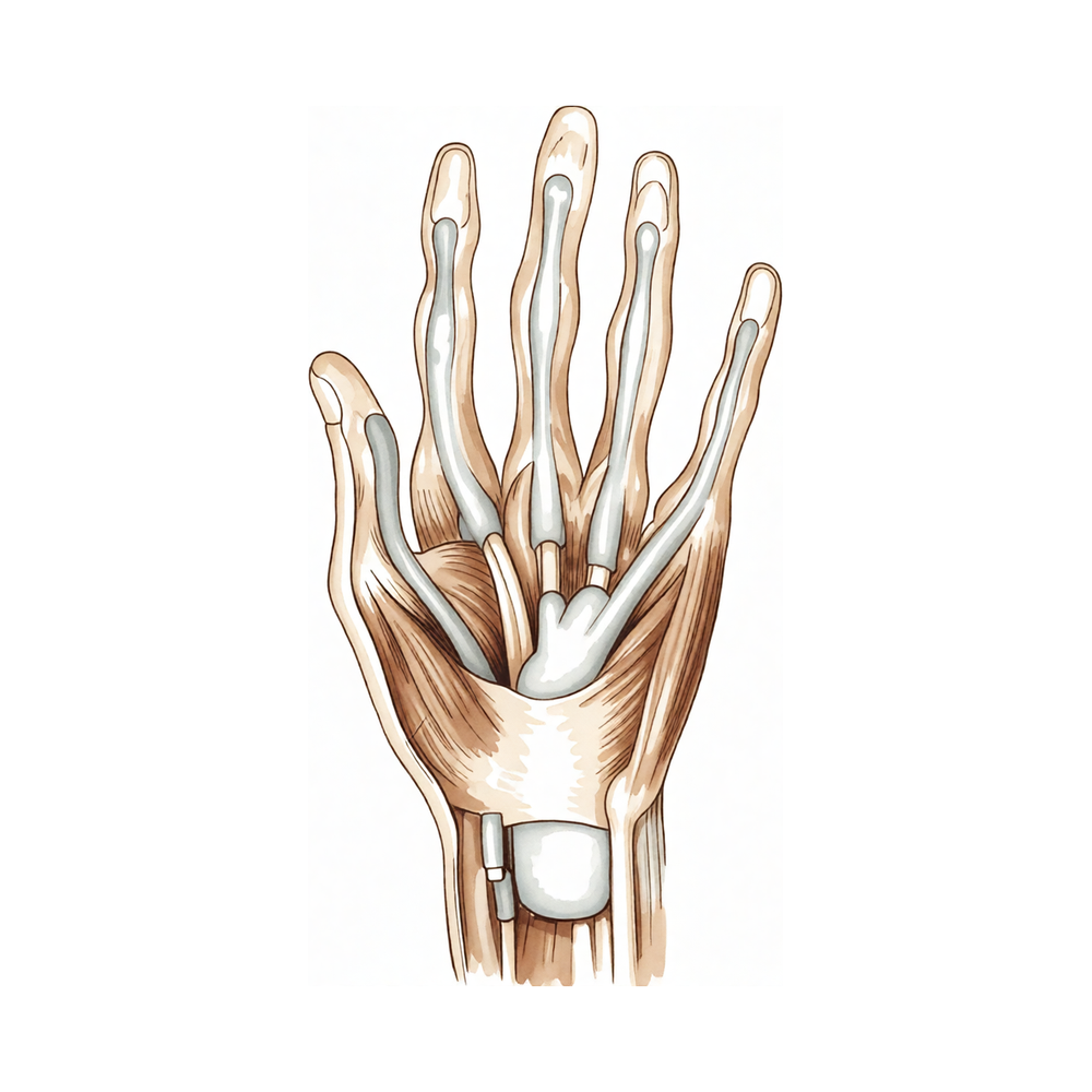
یہ آپریشن کیوں تجویز کیا گیا ہے
ڈاکٹر کیرن ہِرپرا، جو Mater Private Hospital Rockhampton میں بازو اور ہاتھ کے سرجن (upper-limb surgeon) ہیں، علاج کو آپ کی مخصوص چوٹ کے مطابق طے کرتے ہیں۔ مریض عام طور پر اپنے فیملی ڈاکٹر (GP) کے ذریعے ہمارے کلینک بھیجے جاتے ہیں؛ اگر کسی فزیوتھراپسٹ نے آپ کو ہم سے ملنے کا مشورہ دیا ہے، تب بھی میڈیکیئر (Medicare) کی رقم کی واپسی کا حق دار ہونے کے لیے آپ کو اپنے فیملی ڈاکٹر سے ریفرل (referral) لینا ہوگا۔ آپ کی ملاقات پر ہم آپ سے مسئلے کی تفصیل پوچھتے ہیں، آپ کے ہاتھ کا معائنہ کرتے ہیں، اور اگر ضرورت ہو تو یہ جاننے کے لیے ایکسرے یا اسکین کا انتظام کرتے ہیں کہ کیا چیز زخمی ہوئی ہے۔
فلیکسر ٹینڈن (flexor tendon) کی مرمت کا مطلب ہے آپ کی انگلی یا انگوٹھے کے کٹے ہوئے ٹینڈن (tendon)، یعنی وہ مضبوط ڈوری جو پٹھے کو ہڈی سے جوڑتی ہے، کو ٹانکوں سے دوبارہ جوڑنا۔ یہ ٹینڈن آپ کی انگلیوں کو موڑتے ہیں، اور جب ان میں سے کوئی کٹ جائے تو انگلی خود سے نہیں مڑ سکتی۔ چونکہ یہ اچانک لگنے والی چوٹ ہے، اس لیے ہو سکتا ہے فوراً سرجری کا مشورہ دیا جائے، اور پہلے آپریشن کے بغیر علاج نہ آزمایا جائے۔ کٹا ہوا ٹینڈن خود سے دوبارہ پوری مضبوطی تک نہیں جڑتا، اور مرمت کے بغیر امکان ہے کہ انگلی اکڑی ہوئی یا کمزور رہ جائے۔
آپریشن کا مقصد مرمت کو اتنا مضبوط بنانا ہے کہ آپ جلد حرکت شروع کر سکیں، جس سے ٹینڈن کو اپنے غلاف کے اندر چپکنے سے روکنے میں مدد ملتی ہے۔ مقصد یہ ہے کہ آپ کی انگلی کی حرکت کی حد اور کام کرنے کی صلاحیت معمول پر واپس آ جائے۔
آپریشن سے پہلے
فلیکسر ٹینڈن کی مرمت عام طور پر چوٹ کے جلد بعد کی جاتی ہے، اس لیے زیادہ انتظامات کی ضرورت نہیں ہوتی۔ آپ کے سرجن آپ کو بتائیں گے کہ کب کھانا پینا بند کرنا ہے۔ ہم سات گھنٹے خالی پیٹ رہنے کا کہتے ہیں تاکہ اگر آپریشن کی فہرست وقت سے پہلے چل رہی ہو تو آپ کا آپریشن پہلے کیا جا سکے۔ ہو سکتا ہے آپ کو سرجری سے پہلے اپنی کچھ معمول کی دوائیں روکنی پڑیں، اور آپ کے سرجن آپ کو ٹھیک ٹھیک ہدایات دیں گے کہ کون سی دوائیں اور کتنے عرصے کے لیے۔ جو کچھ بھی آپ لیتے ہیں اس کی لکھی ہوئی فہرست ساتھ لائیں۔ بعد میں آپ کو گاڑی میں گھر لے جانے کے لیے کسی کا انتظام کریں، اور آپریشن کے دن ڈھیلے، آرام دہ کپڑے پہنیں۔ اگر آپ کو دوسری طبی بیماریاں ہیں تو ہو سکتا ہے آپ کو خون کے ٹیسٹ یا بے ہوشی کے ڈاکٹر (anaesthetist) سے معائنے کی ضرورت ہو، لیکن زیادہ تر لوگوں کو اس کی ضرورت نہیں ہوتی۔ ہو سکتا ہے آپریشن کی منصوبہ بندی میں مدد کے لیے ایکسرے، الٹراساؤنڈ یا ایم آر آئی جیسی جانچ پہلے ہی کروائی جا چکی ہو۔
آپریشن کے دن
آپ ہسپتال کے سرجری کے داخلے کے شعبے میں پہنچتے ہیں، جہاں آپ کا اندراج کیا جاتا ہے اور آپ کو آپریشن تھیٹر کے لیے تیار کیا جاتا ہے۔ وہیں آپ کی ملاقات بے ہوشی کے ڈاکٹر سے ہوتی ہے۔ یہ آپریشن جنرل اینستھیزیا (مکمل بے ہوشی) میں کیا جاتا ہے۔ بعض اوقات آپریشن کے بعد درد میں آرام کے لیے اس کے ساتھ اعصابی بلاک (nerve block)، یعنی ایک انجکشن جو جسم کے ایک حصے کو سُن کر دیتا ہے، بھی لگایا جاتا ہے؛ بے ہوشی کے ڈاکٹر اسی دن آپ سے اس بارے میں بات کریں گے۔ پھر آپ کو آپریشن تھیٹر لے جایا جاتا ہے، جہاں آپریشن کیا جاتا ہے۔
آپ ریکوری روم میں جاگتے ہیں، جہاں بے ہوشی کا اثر ختم ہونے تک نرسیں آپ کی نگرانی کرتی ہیں۔ جب آپ کی حالت مستحکم ہو جائے تو آپریشن اور آپ کی صحت یابی کے لحاظ سے آپ یا تو وارڈ میں جاتے ہیں یا گھر چلے جاتے ہیں۔
آپریشن میں کیا کیا جاتا ہے
آپ کے سرجن پھٹے ہوئے ٹینڈن تک پہنچنے کے لیے انگلی یا ہتھیلی پر چیرا لگاتے ہیں۔ ٹینڈن کے کٹے ہوئے سرے تلاش کر کے انہیں دوبارہ آپس میں ملایا جاتا ہے۔ ٹینڈن کو مضبوط ٹانکوں سے سیا جاتا ہے جو اس کے درمیانی حصے سے گزرتے ہیں، اور ساتھ ہی مرمت کے بیرونی کنارے کے گرد باریک ٹانکے لگائے جاتے ہیں۔ یہ بیرونی ٹانکے مرمت کو واقعی زیادہ مضبوط بناتے ہیں۔ مرمت کے آر پار ٹانکے کے کئی دھاگے استعمال کیے جاتے ہیں، کیونکہ زیادہ دھاگوں والی مرمت کم دھاگوں والی مرمت سے زیادہ مضبوط ہوتی ہے۔
سرجن ٹینڈن اور اس کے آس پاس کے نازک حصوں کے گرد بہت احتیاط سے کام کرتے ہیں، کیونکہ یہی حصے انگلی مڑتے وقت ٹینڈن کو پھسلنے دیتے ہیں۔ مرمت کس طرح کی جاتی ہے، اس کا انحصار اس بات پر ہے کہ ٹینڈن کس جگہ سے کٹا ہے۔ انگلی کے کچھ حصوں (zones) میں کام کرنا دوسروں کے مقابلے میں زیادہ مشکل ہوتا ہے، اور چنا گیا طریقہ اسی بات کو مدِنظر رکھتا ہے۔
ٹینڈن کی مرمت کے بعد جلد کو ٹانکوں سے بند کیا جاتا ہے اور زخم کی پٹی لگائی جاتی ہے۔ آپ یہ پٹی تقریباً 10 دن تک لگائے رکھیں گے، جیسا کہ صحت یابی والے حصے میں بتایا گیا ہے۔
پورے آپریشن میں مقصد ایسی مرمت ہے جو اتنی مضبوط ہو کہ آپ اپنی انگلی کو جلد حرکت دینا شروع کر سکیں، جس سے ٹینڈن کے اپنے غلاف کے اندر چپکنے کا امکان کم ہو جاتا ہے۔
آپریشن کے بعد
آپ ریکوری روم میں جاگتے ہیں، جہاں بے ہوشی کا اثر ختم ہونے تک نرسیں آپ پر گہری نظر رکھتی ہیں۔ آپ کے ہاتھ پر زخم کی پٹی اور ایک اسپلنٹ ہوگا جو آپ کی انگلی کو ساکت رکھتا ہے۔ درد میں آرام کا منصوبہ آپ کے آپریشن تھیٹر سے نکلنے سے پہلے آپ کے ساتھ بنایا جاتا ہے، اور آپ کسی بھی وقت مزید دوا مانگ سکتے ہیں۔ پہلے 24 گھنٹے کسی کو آپ کے ساتھ رہنا چاہیے۔ جیسے ہی آپ خود کو سنبھلا ہوا محسوس کریں، آپ اٹھ کر چل پھر سکتے ہیں، اور آپ کو دکھایا جائے گا کہ چلتے پھرتے اپنے ہاتھ کو کیسے محفوظ رکھنا ہے۔ آپ کی ٹیم آپ کو بتائے گی کہ آپ اسی دن گھر جائیں گے یا ایک رات ہسپتال میں رہیں گے۔ ہم زخم کی پٹی تقریباً 10 دن تک لگی رہنے دیتے ہیں؛ براہ کرم اس سے پہلے اسے نہ اتاریں، جب تک ہم آپ کو ایسا کرنے کو نہ کہیں۔ جب ہم آپ کو دیکھتے ہیں تو اسے بدلتے یا اتار دیتے ہیں۔
صحت یابی
پہلے چند دن آپ کی انگلی میں درد اور سوجن رہے گی، اور تکلیف عام طور پر شروع میں سب سے زیادہ ہوتی ہے۔ باقاعدگی سے درد کی دوا لینا، بیٹھتے یا آرام کرتے وقت ہاتھ کو تکیوں پر اونچا رکھنا، اور آہستہ آہستہ چلنا پھرنا، یہ سب اسے کم کرنے میں مدد دیتے ہیں۔ ہفتے گزرنے کے ساتھ سوجن آہستہ آہستہ کم ہو جاتی ہے۔
آپ کا ہاتھ ایک اسپلنٹ میں ہوگا جو مرمت کی حفاظت کرتا ہے۔ سرجری کے بعد ہینڈ تھراپی Extend Rehabilitation میں روبی ڈولن (Ruby Doolan) کے ساتھ ہوتی ہے۔ روبی آپ کی ورزشوں کی رہنمائی کریں گی اور جو بھی اسپلنٹ آپ کو درکار ہو وہ بنائیں گی۔ آپ اپنی انگلی کو جلد حرکت دینا شروع کریں گے، کیونکہ ہلکی حرکت ٹینڈن کو اپنے غلاف کے اندر چپکنے سے روکنے میں مدد دیتی ہے۔ شروع میں ورزشیں بہت معمولی لگتی ہیں، اکثر صرف انگلی کو آپ کے تھراپسٹ کی مقرر کردہ حدود کے اندر تھوڑا سا موڑنا۔ یہ ورزشیں اتنی ہی اہم ہیں جتنا کہ خود آپریشن، اس لیے انہیں ہدایت کے مطابق کرنا ضروری ہے۔
گھر پر آپ روزمرہ کے زیادہ تر کام اپنے دوسرے ہاتھ سے کر سکتے ہیں۔ آپ کو اسپلنٹ کو خشک رکھنا ہوگا، اور جب تک آپ کا تھراپسٹ اجازت نہ دے، آپریشن والے ہاتھ سے کچھ اٹھانے، پکڑنے یا لے جانے سے پرہیز کرنا ہوگا۔ شروع میں سونا کچھ مشکل ہو سکتا ہے؛ ہاتھ کو تکیے پر رکھنے سے اکثر مدد ملتی ہے۔
جیسے جیسے حرکت واپس آتی ہے، ورزشیں آگے بڑھتی ہیں، اور سوجن کم ہونے کے بعد آپ کو روزمرہ کے کام آسان لگیں گے۔ آپ کے سرجن اور روبی آپ کو بتائیں گے کہ مرمت کب ہر نئے مرحلے کے لیے کافی مضبوط ہو گئی ہے، بشمول یہ کہ آپ کب دوبارہ گاڑی چلا سکتے ہیں؛ بازو اور ہاتھ کی سرجری کے بعد گاڑی چلانے کے بارے میں ہماری رہنمائی میں لاگو ہونے والے قواعد بیان کیے گئے ہیں۔
صحت یابی ہر شخص میں مختلف ہوتی ہے، اس لیے آپ کا وقت مختلف ہو سکتا ہے۔ آپ کے سرجن اور آپ کا تھراپسٹ پورے راستے میں آپ کی رہنمائی کریں گے۔
کیا مسائل پیدا ہو سکتے ہیں
زیادہ تر مریض ٹھیک ہو جاتے ہیں، لیکن کبھی کبھار مسائل پیدا ہو سکتے ہیں۔ آپ کے سرجن اور ٹیم آپ کی قریب سے نگرانی کرتے ہیں تاکہ کسی بھی مسئلے کو جلد پکڑا جا سکے۔
بعض اوقات مرمت شدہ ٹینڈن کھنچ کر الگ ہو سکتا ہے۔ آپ کو انگلی میں اچانک "پٹ" کی آواز یا جھٹکا محسوس ہو سکتا ہے، یا آپ دیکھ سکتے ہیں کہ جو انگلی آپ کی ورزشوں کے دوران مڑ رہی تھی وہ پھر سے خود سے مڑنا بند کر دیتی ہے۔ اگر ایسا ہو تو اپنے اگلے معائنے کا انتظار کرنے کے بجائے فوراً کلینک سے رابطہ کریں۔
مرمت کے گرد زخم بھرنے سے بننے والا سخت ریشہ (scar tissue) بن سکتا ہے اور ٹینڈن کو اس کے غلاف کے اندر چپکا سکتا ہے۔ ہو سکتا ہے انگلی صرف آدھی مڑے، یا حرکت دینے کی کوشش پر اکڑی ہوئی اور اٹکی ہوئی محسوس ہو۔ بعض اوقات یہ صرف تھراپی سے ٹھیک ہو جاتا ہے۔ اگر نہ ہو تو ایک چھوٹا آپریشن اس سخت ریشے کو کاٹ کر ٹینڈن کو آزاد کر سکتا ہے۔ اگر اکڑاؤ بہتر نہیں ہو رہا تو اپنی معائنے کی ملاقاتوں میں اس کا ذکر کریں۔
انفیکشن کم ہوتا ہے لیکن اس پر جلد توجہ کی ضرورت ہوتی ہے۔ ایسے درد پر نظر رکھیں جو کم ہونے کے بجائے بڑھتا ہی جائے، زخم سے باہر کی طرف پھیلتی سرخی، گرمائش، سوجن، یا زخم سے رطوبت کا رسنا۔ ٹینڈن کے غلاف میں گہرا انفیکشن بروقت علاج کے باوجود انگلی کو اکڑا ہوا چھوڑ سکتا ہے، اس لیے جلد معائنہ اہم ہے۔ اسی دن کلینک کو فون کریں، یا اگر کسی سے رابطہ نہ ہو سکے تو ہسپتال کی ایمرجنسی جائیں۔
ٹھیک ہونے کے بعد کچھ انگلیوں میں دوسرے مسائل ہو جاتے ہیں۔ انگلی مڑتے وقت کلک کر سکتی ہے یا اٹک سکتی ہے، یا ایک انگلی پوری طرح نہیں مڑ پاتی کیونکہ اس کی حرکت دوسری انگلیوں سے جڑی ہوتی ہے۔ چھوٹی انگلی میں دوسری انگلیوں کے مقابلے میں کم حرکت رہ سکتی ہے۔ اگر آپ کلک کرنا، اٹکنا یا غیر برابر مڑنا محسوس کریں تو اپنے اگلے معائنے میں اس کا ذکر کریں۔
چند مریضوں کو کسی نہ کسی قسم کی مزید سرجری کی ضرورت پڑتی ہے، چاہے وہ الگ ہو جانے والی مرمت، زخم بھرنے سے بننے والے سخت ریشے، یا کسی اور مسئلے کے لیے ہو۔ اگر ایسا ضروری ہوا تو آپ کے سرجن آپ کو بتائیں گے کہ اس میں کیا شامل ہے۔
اگر آپ تفصیل جاننا چاہتے ہیں تو اس صفحے پر پیچیدگیوں کے جدول میں عام شرحیں درج ہیں۔
ہمیں کب فون کریں
اگر آپ کو بخار ہو، یا زخم زیادہ سرخ، گرم یا سوجا ہوا ہو جائے، یا اس سے رطوبت رسنے لگے تو ہمیں فون کریں۔ ایسے درد کے لیے جو کم ہونے کے بجائے بڑھتا ہی جائے، ہمیں اسی دن فون کریں۔ اگر آپ کو اچانک شدید درد ہو، انگلی میں اچانک "پٹ" ہو اور وہ خود سے مڑنا بند کر دے، پنڈلی میں سوجن یا درد ہو، یا سانس پھولے تو ہسپتال کی ایمرجنسی جائیں۔ اگر آپ کی انگلیوں میں احساس ختم ہو جائے، یا آپ انہیں بالکل حرکت نہ دے سکیں تو بھی ہسپتال کی ایمرجنسی جائیں۔ اگر کلینک میں کسی سے رابطہ نہ ہو سکے تو ہسپتال کی ایمرجنسی جائیں۔
مزید تفصیل
Advanced reading: the deeper science (optional)
یہ حصہ اس سے آگے جاتا ہے جتنا آپ کو اپنے علاج کے فیصلوں کے لیے جاننے کی ضرورت ہے۔ فلیکسر ٹینڈن کی مرمت کے بارے میں مزید پڑھنا اس لیے فائدہ مند ہے کہ یہ ایک ہی سخت سودے پر چلتی ہے: جو حرکت ٹینڈن کو چپکنے سے روکتی ہے، وہی حرکت مرمت کو کھینچ کر الگ بھی کر سکتی ہے۔ اس آپریشن کا تقریباً ہر تکنیکی فیصلہ ان دو ناکامیوں کے درمیان گنجائش پیدا کرنے کی کوشش ہے۔
یہ سودا، ناپ تول کر
زون II کی 569 مرمتوں پر ایک میٹا اینالیسس (meta-analysis) نے سرجری کے بعد ابتدائی فعال حرکت (early active motion، جس میں مریض خود اپنے پٹھوں سے انگلی موڑتا ہے) کا موازنہ ابتدائی غیر فعال حرکت (early passive motion، جس میں انگلی کو باہر سے موڑا جاتا ہے) سے کیا [1]۔ ابتدائی فعال حرکت والے گروپ نے کل فعال حرکت (total active motion) زیادہ حاصل کی، اور یہی وہ نتیجہ ہے جو طے کرتا ہے کہ انگلی کام کی ہے یا نہیں [1]۔
اسی تجزیے میں اس کی قیمت بھی سامنے آئی۔ فعال طور پر انگلی موڑنے اور سیدھی کرنے والے گروپ میں مرمت ٹوٹنے کا زیادہ خطرہ دیکھا گیا، جب ٹینڈن کی مرمت 2 دھاگوں والے مرکزی ٹانکے (2-strand core suture) سے کی گئی تھی [1]۔
یہی شرط اصل بات ہے، اور اسے نظرانداز کرنا آسان ہے۔ ابتدائی فعال حرکت اپنے آپ میں خطرناک نہیں، اور 2 دھاگوں والی مرمت اپنے آپ میں کمزور نہیں۔ مسئلہ ان دونوں کا ایک ساتھ ہونا ہے۔ مرمت کی مضبوطی اور بحالی کی ورزشوں کی شدت ایک ہی فیصلہ ہے جو دو لوگ کرتے ہیں، اور انہیں ایک دوسرے کے مطابق رکھنا ضروری ہے۔ اگر لگے کہ آپ کا ہینڈ تھراپسٹ اور آپ کے سرجن استعمال ہونے والے مخصوص ٹانکے کے بارے میں آپس میں بات کر رہے ہیں، تو یہ نظام بالکل ویسے ہی کام کر رہا ہے جیسا اسے کرنا چاہیے۔
تحقیق کو سمجھنا اتنا مشکل کیوں ہے جتنا نہیں ہونا چاہیے
انگلیوں کے 1,878 فلیکسر ٹینڈن مرمتوں پر ایک منظم جائزے (systematic review) کا مقصد 2 دھاگوں والے اور زیادہ دھاگوں والے مرکزی ٹانکوں کا موازنہ کرنا تھا، اور یہ اپنے مفروضے کی حتمی تصدیق نہیں کر سکا، اس لیے نہیں کہ جواب منفی تھا، بلکہ اس لیے کہ نتائج اور تحقیق کے طریقوں کو بیان کرنے کے انداز میں بہت زیادہ فرق تھا [2]۔
ہاتھ کی سرجری میں یہ بار بار آنے والا مسئلہ ہے۔ مختلف تحقیقی مقالے حرکت کی مختلف پیمائشیں، اچھے نتیجے کی مختلف تعریفیں اور بعد کے معائنوں کے مختلف اوقات استعمال کرتے ہیں، جس کی وجہ سے انہیں اکٹھا کرنا تقریباً بے معنی ہو جاتا ہے۔ جہاں آپ یہ پُراعتماد دعوے دیکھیں کہ مرمت کا ایک طریقہ دوسرے سے بہتر ہے، وہاں اکٹھے کیے گئے شواہد کی بنیاد پر دیانت دارانہ موقف یہ ہے کہ ابھی رپورٹنگ اتنی یکساں نہیں کہ یہ بات کہی جا سکے۔
اسپلنٹ کا طریقہ بدل رہا ہے، اور شواہد پیچھے ہیں
ریلیٹو موشن آرتھوسس (relative motion orthoses)، یعنی ایسے اسپلنٹ جو مرمت شدہ انگلی کو اس کی ساتھ والی انگلیوں سے تھوڑا مختلف انداز میں رکھتے ہیں تاکہ ٹینڈن کا پھسلنا ان میں بٹ جائے، نے ایکسٹینسر ٹینڈن (extensor tendon، انگلی سیدھی کرنے والا ٹینڈن) کی بحالی کو بدل دیا ہے۔ 529 مریضوں پر ایک منظم جائزے کا نتیجہ یہ تھا کہ اب اچھے شواہد موجود ہیں کہ یہ طریقہ زون V سے VI کی ایکسٹینسر مرمتوں میں محفوظ ہے، لیکن فلیکسر مرمتوں کے لیے شواہد ابھی محدود ہیں [3]۔
لہٰذا فلیکسر کی طرف یہ ایک امید افزا سمت ہے، کوئی طے شدہ معیار نہیں، اور یہ پوچھنا مناسب ہے کہ کون سا طریقۂ کار استعمال ہو رہا ہے اور کیوں۔
تکنیک کس طرف جا رہی ہے
تحقیق کا ایک رخ اس سودے کو قبول کر کے اس کے گرد راستہ نکالتا ہے۔ زون II کی ایک تکنیک، جس میں باہر رکھا گیا تناؤ کم کرنے والا ٹانکا (externalised detensioning suture) استعمال ہوتا ہے، یعنی جلد کے باہر ایک عارضی ٹانکا جو مرمت سے بوجھ ہٹاتا ہے، نے مرمت کو کافی مضبوط کیا اور مرمت ٹوٹنے کے کم خطرے کے ساتھ ابتدائی فعال حرکت ممکن بنائی، لیکن اس کی قیمت یہ تھی کہ یہ ٹانکا نکالے جانے تک انگلی کے سرے والے جوڑ کی حرکت محدود رہتی ہے [4]۔
یہ اس شعبے کا منصفانہ خلاصہ ہے: بنیادی کشمکش ختم نہیں ہوئی، بس اسے زیادہ ہوشیاری سے سنبھالا جا رہا ہے۔
آپ کی صحت یابی کے لیے اس کا کیا مطلب ہے
تھراپی آپریشن کے بعد کی دیکھ بھال نہیں، بلکہ آپریشن ہی کا حصہ ہے۔ ملاقاتوں پر حاضر ہونا، اور ٹھیک اتنی ہی ورزش کرنا جتنی بتائی گئی ہے، اس سے زیادہ نہیں، وہ چیز ہے جو آپ کو اوپر بیان کیے گئے سودے کی صحیح طرف رکھتی ہے۔ پہلے چھ ہفتوں میں مرمت کا ٹوٹنا عام طور پر دوسرے آپریشن کا مطلب ہوتا ہے، جس کا نقطۂ آغاز پہلے آپریشن سے بدتر ہوتا ہے۔
حوالہ جات
[1] Xu H, Huang X, Guo Z, Zhou H, Jin H, Huang X. Outcome of surgical repair and rehabilitation of flexor tendon injuries in zone II of the hand: systematic review and meta-analysis. J Hand Surg Am. 2023;48(4):407.e1-407.e11. https://doi.org/10.1016/j.jhsa.2021.11.013
[2] Hardwicke JT, Tan JJ, Foster MA, Titley OG. A systematic review of 2-strand versus multistrand core suture techniques and functional outcome after digital flexor tendon repair. J Hand Surg Am. 2014;39(4):686-95.e2. https://doi.org/10.1016/j.jhsa.2013.12.037
[3] Shaw AV, Verma Y, Tucker S, Jain A, Furniss D. Relative motion orthoses for early active motion after finger extensor and flexor tendon repairs: a systematic review. J Hand Ther. 2023;36(2):332-46. https://doi.org/10.1016/j.jht.2023.02.011
[4] Suszynski TM, Coutinho D, Kaufmann RA. Flexor tendon repair in zone II augmented with an externalized detensioning suture: protected flexor tendon repair. J Hand Surg Am. 2023;48(10):1065.e1-1065.e4. https://doi.org/10.1016/j.jhsa.2023.01.018
Evidence & references
This is the clinical evidence summary written for health professionals. It is technical, and it lists the research this page was built from. You do not need to read it to understand your treatment or to make a decision about it.
Overview
- Consistent, successful management of flexor tendon injuries relies on understanding the anatomy, characteristics and repair of tendons in the different zones, potential complications, rehabilitation protocols, recent advances in treatment, and future directions [1].
- Future directions for flexor tendon injury management include tissue engineering and biologic modification of the repair site [1].
- Flexor tendon repair in Zone II is a technically demanding procedure [2].
- Outcomes for flexor tendon repair in Zone II have become more predictable and satisfying [2].
- No gold standard has been determined for the optimal flexor tendon repair algorithm [3].
- Flexor tendon repairs are usually chosen based on familiarity, popularity, and technical difficulty [3].
- Zone I flexor tendon injuries traditionally have not yielded results as good as other flexor tendon injuries [5].
- Full motion is rarely regained in Zone I flexor tendon injuries [5].
- Good or excellent results are reported in only up to 67% of Zone I flexor tendon injury cases [5].
- Functional outcomes for flexor tendon injuries remain unreliable despite improvements in surgical technique and rehabilitation [6].
- Adhesion formation is the most common complication after flexor tendon injuries [6].
- Joint contractures are the most common complication after flexor tendon injuries [6].
- Tendon grafting is the treatment of choice for flexor tendon injuries in zones I and II when direct repair is not possible or delayed [7].
- Secondary reconstruction remains an important and useful technique for complicated flexor tendon injuries [9].
- Secondary reconstruction remains an important and useful technique for flexor tendon injuries that have failed primary repair [9].
- Limited evidence currently exists for relative motion orthoses for early active motion after flexor tendon repairs in zones IV and VII [10].
- Many of the principles of flexor tendon repair and rehabilitation can be applied to zones III–V [11].
- Primary flexor tendon repair in the digital sheath area has become standard practice [12].
- Current practice for primary flexor tendon repair involves a shift toward strong multistrand core sutures [12].
- Current practice for primary flexor tendon repair involves a shift toward modified pulley preservation [12].
- A modified protocol for primary flexor tendon repair in zones 1 and 2 utilizes a 6-strand core suture without circumferential suturing [16].
- A modified protocol for primary flexor tendon repair in zones 1 and 2 utilizes selective pulley division [16].
- A modified protocol for primary flexor tendon repair in zones 1 and 2 utilizes partial FDS resection to facilitate early active motion and improve outcomes [16].
Anatomy & Pathophysiology
General Principles
- Consistent, successful management of flexor tendon injuries relies on understanding the anatomy, characteristics, and repair of tendons in the different zones [1].
- An understanding of the biomechanics of the flexor tendon system is essential to proper evaluation and treatment of disorders of the upper extremity [48].
- Flexor tendons function as cables transmitting forces to move and stabilize joints [48].
- Recent developments in hand surgery have resulted from a better understanding of the dynamic anatomy and function of the hand [36].
- The concept of functional rather than static anatomy is central to the study of the hand [36].
Hand Architecture
- The hand is an organ designed to obtain information and an organ of execution [36].
- The hand functions efficiently only if the proximal joints of the limb are stable and yet mobile [36].
- The hand is located at the extremity of the upper limb, which functions as its vector [36].
- The shoulder is the most mobile joint in the body and allows orientation of the upper limb as required [36].
- The movements of the clavicle amplify those of the shoulder [36].
- The elbow brings the hand closer to or moves it away from the body through flexion–extension movements [36].
- The combined movements of the wrist and forearm place the hand in a position for grasping [36].
- For gripping, the wrist is usually in flexion when close to the trunk and in extension when placed at a distance [36].
- Forearm rotation (pronation–supination) plays an important role in bringing food to the mouth [36].
- The hand’s blood and nerve supplies are continuous with those of the rest of the limb [36].
- Some hand muscles, the extrinsic muscles, arise in the arm and forearm [36].
- The open hand forms a balanced graceful oval in its longitudinal axis [36].
- The proximal carpometacarpal half of the hand is flattened and presents two faces with unique anatomical and functional significance [36].
- The posterior or dorsal aspect of the hand is convex [36].
- The anterior, palmar or volar aspect of the hand is concave [36].
- The distal half of the hand is separated into five digits which flex toward the palm [36].
- Digits converge in closing by flexing and adducting, and diverge in opening by extending and abducting [36].
- The thumb has a more proximal and lateral position, allowing movement inward and outward from the palm [36].
- The four fingers are the distal extension of the carpometacarpal part of the hand [36].
- The hinges of finger movements are at the thenar crease and at the transverse distal palmar crease [36].
- When digits are fully extended and touching, their tips almost describe a regular curve with peripheral digits being the shortest [36].
- When fingers are extended and separated, their tips lie on the circumference of a circle whose center is the head of the third metacarpal [36].
- The hand consists of 19 bones, 17 articulations, and 19 muscles situated entirely within the hand [36].
- The hand contains about the same number of tendons activated by the forearm muscles as it has intrinsic muscles [36].
Metacarpal and Longitudinal Arches
- The metacarpal arch is endowed with a great deal of adaptability because of the mobility of the peripheral metacarpals [41].
- The peripheral metacarpals form the sides of the cup or palmar gutter and can deepen the concavity as they approach each other [41].
- The thumb metacarpal is independent and articulates with the trapezium [41].
- The middle metacarpals are united to the carpus by the intrinsic interlocking encasement of the bones themselves [41].
- The index metacarpal is the most firmly fixed [41].
- The ring metacarpal is a transitional element to the fifth metacarpal and has about 10 degrees of mobility in flexion and extension [41].
- The fifth metacarpal is semi-independent, articulates with the hamate, and is restrained on its radial side by its articulation with the base of the fourth metacarpal [41].
- The fifth metacarpal has a range of flexion–extension of approximately 20 degrees [41].
- The second to fifth metacarpals are bound together by various fibrous structures, the most distal of which is the deep transverse intermetacarpal ligament [41].
- The deep transverse intermetacarpal ligament is better named the interglenoid ligament because it ties together the anterior glenoid ligaments of the metacarpophalangeal articulations [41].
- The anterior glenoid ligaments of the metacarpophalangeal articulations are known as the volar plates [41].
- The longitudinal arches are composed of a fixed portion, the carpometacarpal, and a mobile portion, the digits [41].
- There is a longitudinal arch for every ray of the hand [41].
- The longitudinal arches diverge distally according to their different obliquities, with the thumb ray being the most divergent [41].
- The keystones of the longitudinal arches are the metacarpophalangeal articulations [41].
- The thick anterior glenoid capsules, or volar plates, of the metacarpophalangeal joints prevent hyperextension [41].
- The volar plates are interconnected by the transverse interglenoid ligament [41].
- The stability of the metacarpophalangeal joints is essential to the support of the longitudinal arch as well as of the transverse metacarpal arch [41].
- The five rays of the hand differ in mobility and independence, with considerable mobility for the thumb, much less for the fifth ray, and even less for the others [41].
- The index ray has a certain degree of independence at the phalangeal level owing to the arrangement of its flexor and extensor muscles [41].
Intrinsic Muscles
- There are seven interosseous muscles, four dorsal and three volar [38].
- The dorsal interossei are abductors [38].
- The anatomic axis of the hand coincides with the axis of the third metacarpal [38].
- The dorsal interossei lie to the radial side of the index and middle fingers and the ulnar side of the middle and ring fingers [38].
- The little finger is abducted by the abductor digiti quinti [38].
- The volar interossei are adductors [38].
- The volar interossei lie to the ulnar side of the index finger and the radial side of the ring and little fingers [38].
- The middle finger has two dorsal interossei (abductors) and no volar interossei (adductors) because the central axis of the hand lies within it [38].
- Each dorsal interosseous muscle, with the exception of the third, has two muscle heads [38].
- The superficial head of the dorsal interosseous muscles arises most dorsally from the shaft of the contiguous metacarpals [38].
- The superficial head inserts deeply by a medial tendon onto the lateral tubercle of the base of the proximal phalanx [38].
- The superficial head abducts and weakly flexes the proximal phalanx [38].
- The superficial head has no direct effect on the middle or distal phalanges [38].
- The deep head of each dorsal interosseous muscle forms a lateral tendon, or lateral band, at the level of the MP joint [38].
- The deep head flexes and weakly abducts the proximal phalanx while extending the middle and distal phalanges [38].
- At the level of the middle of the proximal phalanx, transverse fibers arch dorsally from each lateral band to join each other over the dorsum of the finger [38].
- Transverse fibers flex the proximal phalanx [38].
- Oblique fibers (spiral fibers) from the lateral bands sweep over the distal third of the proximal phalanx to insert onto the lateral tubercles at the base of the middle phalanx [38].
- The oblique fibers extend the middle phalanx (PIP joint) [38].
- The lateral bands are joined by the lateral slips of the extensor tendon to form the conjoined lateral band [38].
- The two conjoined lateral bands to each finger unite at the distal third of the middle phalanx to form the terminal tendon [38].
- The terminal tendon inserts at the base of the distal phalanx to extend it [38].
- The flexor digiti quinti brevis is structurally and functionally similar to the deep head of the dorsal interossei [38].
- The flexor digiti quinti brevis forms the ulnar lateral band of the little finger [38].
- The three volar interossei arise from adjacent surfaces of contiguous metacarpal shafts [38].
- Each volar interosseous muscle has only one muscle head [38].
- None of the volar interossei insert onto the proximal phalanx [38].
- The volar interossei form the ulnar lateral band of the index finger and the radial lateral band of the ring and little fingers [38].
- The volar interossei send oblique or spiral fibers that insert onto the base of the middle phalanx at its lateral tubercle [38].
- The abductor digiti quinti and flexor digiti quinti brevis are similar in both structure and function to the superficial and deep heads of the dorsal interossei, respectively [38].
- The abductor digiti quinti and flexor digiti quinti brevis arise from the fifth metacarpal [38].
- The abductor digiti quinti inserts onto the ulnar lateral tubercle at the base of the proximal phalanx of the little finger [38].
- The flexor digiti quinti forms the ulnar lateral band [38].
- The opponens digiti quinti lies deepest among the hypothenar muscles [38].
- The opponens digiti quinti arises from the pisohamate ligament and the hook of the hamate [38].
- The opponens digiti quinti inserts onto the ulnar side of the diaphysis of the fifth metacarpal [38].
- The opponens digiti quinti flexes and supinates the fifth metacarpal [38].
Cutaneous Units
- There are functional cutaneous units in the hand similar to those described in the face [37].
- One dorsal cutaneous unit extends from the wrist to the proximal interphalangeal joints of the fingers and the interphalangeal joint of the thumb [37].
- The dorsal covering of the interphalangeal articulations of the digits forms a unique cutaneous unit characterized by a considerable excess of skin when the digits are in extension [37].
- The fine tight skin of the dorsal aspect of the middle phalanx forms another cutaneous unit [37].
- The dorsal integument of the distal phalanx is very special because of the nail bed with its matrix [37].
- The palm forms a cutaneous unit extending from the distal transverse crease of the wrist up to the transverse crease at the base of the digits [37].
- The palmar integument may be subdivided into two separate zones by the oppositional crease of the thumb [37].
- The oppositional crease of the thumb constitutes the oblique axis of the hand [37].
- The skin of the radial portion of the palm covers the thenar eminence and the external part of the palm [37].
- The radial portion of the palmar skin is relatively well vascularized and is the mobile portion [37].
- The skin of the ulnar and distal portion covers the hypothenar eminence where the skin has poor mobility [37].
- The distal part of the palm beyond the transverse distal palmar crease is a true hinge just at the level of the metacarpophalangeal articulations [37].
- The central triangular part of the palm has skin that is fixed and poorly vascularized [37].
- The central triangular part of the palm covers almost directly the superficial palmar aponeurosis, which inserts into it [37].
- The integument of the palmar face of the digits may be subdivided into phalangeal units separated by digital flexion folds [37].
- There are three digital flexion folds for the digits and two for the thumb [37].
- When a digit is completely flexed, the integument of adjacent phalanges comes into contact in the zones of the flexion creases [37].
- Areas of cutaneous contact in the flexed digits are in the form of a diamond [37].
- The sides of the diamond-shaped cutaneous contact areas do not undergo variations in length during movements of flexion and extension [37].
- Incisions made along the lines of the diamond-shaped cutaneous contact areas present a minimal chance of retraction [37].
- The web spaces are formed from the union of two nonsymmetrical cutaneous surfaces [37].
- The dorsal slope of the web space has a gradual incline and its supple skin is not adherent to the subjacent region [37].
- The palmar surface of the web space is flat and precipitously interrupted [37].
- The palmar skin of the web space is densely adherent to the commissural skeleton [37].
- The commissural skeleton is formed by the interdigital palmar (natatory) ligament between the fingers [37].
- The commissural skeleton is formed by the distal transverse ligament at the level of the thumb web [37].
- The distal transverse ligament at the level of the thumb web is by far the deepest and the most mobile [37].
Vascular Anatomy
- The arteries of the thumb vary in both size and number, making surgical reconstruction delicate [42].
- The layout of the palmar arteries of the thumb is the result of innumerable variations regarding origin, transit, connections, and size [42].
- In anatomical studies, only 15% of dissections of the palmar arteries of the thumb fall into the classical "typical" category [42].
- The princeps pollicis artery is the terminal branch of the radial artery [42].
- The princeps pollicis artery crosses the first intermetacarpal space [42].
- The princeps pollicis artery runs along the ulnar side of the first metacarpal bone and along the volar surface of the adductor muscle [42].
- The princeps pollicis artery emerges onto the subcutaneous palmar tissue at the level of the cutaneous flexion crease of the metacarpophalangeal joint [42].
- The princeps pollicis artery divides into two terminal rami, namely the collateral palmar arteries of the thumb [42].
- The collateral palmar arteries of the thumb run along the digital tunnel symmetrically and are of equal caliber [42].
- The collateral palmar arteries of the thumb head distally to finally unite in the pulp arcade [42].
- During their transit in the digital tunnel, the collateral palmar arteries break off into numerous collateral branches, either cutaneous, articular, or osseous [42].
- An arcade located deep in the flexor tendon joins together the two arteries at the level of the distal metaphysis of the first phalanx [42].
- Vessels originating from the subtendinous arcade enter the vincula and irrigate the flexor tendon [42].
- It is rare to find arteries of surgical interest on the volar surface of the thumb between the opposition crease and the metacarpophalangeal flexion crease [42].
- In the first segment of the thumb, the artery is located deeply and is more easily accessible from the dorsal surface [42].
- In the second segment of the thumb, the two arteries run alongside the flexor tendon and behind the collateral nerves [42].
- In the second segment of the thumb, the main artery is the ulnar collateral artery [42].
- The subtendinous anastomosis situated at the level of the neck of the first phalanx acts as a moderator between the two arteries in the second segment [42].
- In cases where the palmar ulnar collateral artery is absent, the dorsal artery takes its place by means of a branch through the subtendinous arcade [42].
- In the pulp segment of the thumb, the two arteries are of similar size and run through the thick fatty subcutaneous padding [42].
- In the pulp segment of the thumb, the two arteries cross over and convert into the ends of the digital nerves at the level of the median axis [42].
- The dorsal arteries of the thumb originate from the palmar arteries (princeps, commissural, or anastomoses of the superficial arcade) at the level of the first metacarpal [42].
- The dorsal arteries of the thumb run laterally along the metacarpophalangeal joint and continue obliquely from volar to dorsal [42].
- The dorsal arteries of the thumb head in a distal direction remaining on the side of the two distal phalanges [42].
- At the level of the neck of the first phalanx, an anastomosis can be found which originates from the palmar arteries for the dorsal aspect [42].
- The dorsal arteries
Classification
- Management of flexor tendon injuries requires consideration of surgical timing, injury location, approach, and soft tissue handling [15].
- Consistent management of flexor tendon injuries relies on understanding the anatomy, characteristics, and repair of tendons in different zones [1].
- Many principles of flexor tendon repair and rehabilitation can be applied to zones III–V [11].
- Differences in the management of flexor tendon injuries between children and adults include differences in classification [21].
Clinical Presentation
- Flexor tendon injuries are complex, and management requires consideration of surgical timing, injury location, approach, and soft tissue handling [15].
- Spontaneous flexor tendon ruptures of the hand occur more often than one might recognize [28].
- The majority of spontaneous flexor tendon ruptures involve the profundus tendon of the small finger in the palm [28].
- Closed flexor tendon disruptions include traumatic avulsion, spontaneous midsubstance rupture, attrition rupture, infiltrative tenosynovial rupture, and iatrogenic causes [50].
- Pediatric flexor tendon injuries differ from adults in diagnosis and rehabilitation [18].
- Pediatric flexor tendon injuries often require surgical exploration due to uncooperative patients [18].
- Differences in the management of flexor tendon injuries between children and adults include epidemiology, anatomy, classification, diagnosis, incisions and skin closure, the size of the flexor tendons, technical aspects of zones I and II repairs, core suture purchase length, rehabilitation, results, and complications of primary flexor tendon repair [21].
Investigations
- Clinical evaluation of the injured or dysfunctional hand and wrist requires combining patient history with a careful physical examination to pinpoint or narrow the scope of possible pathologic processes [27].
- Diagnostic tests such as imaging and serum laboratory studies are useful in determining pathology but can be expensive, time consuming, and often nonspecific [27].
- A systematic method to approaching the physical examination of the hand is essential due to the number of structures in a small space [27].
- Patients often have difficulty accurately describing their symptoms and may incorrectly attribute pathology to a perceived deficit [27].
Treatment
General Principles and Current Practice
- Consistent, successful management of flexor tendon injuries relies on understanding anatomy, repair characteristics by zone, complications, rehabilitation protocols, and recent advances including tissue engineering and biologic modification [1].
- The central tenet of modern flexor tendon surgery is to increase tendon healing and avoid adhesion formation by making a repair strong enough to move within a few days of injury [19].
- Primary flexor tendon repair in the digital sheath area has become standard practice with a shift toward strong multistrand core sutures and modified pulley preservation [12].
- No gold standard has been determined for the optimal flexor tendon repair algorithm, and repairs are usually chosen based on familiarity, popularity, and technical difficulty [3].
- Despite significant advances in flexor tendon repair and reconstruction, the drive towards perfection continues [14].
Zone-Specific Considerations
- Flexor tendon repair in Zone II is a technically demanding procedure, but outcomes have become more predictable and satisfying [2].
- Zone I flexor tendon injuries traditionally have not yielded results as good as other flexor tendon injuries, with full motion rarely regained and good/excellent results reported in only up to 67% of cases [5].
- Pediatric flexor tendon injuries differ from adults in diagnosis and rehabilitation, often requiring surgical exploration due to uncooperative patients [18].
Surgical Techniques and Modifications
- Increasing the number of suture strands, using locking-loop configurations, and optimizing suture purchase length significantly improve the mechanical strength and gap resistance of flexor tendon repairs [69].
- A modified protocol for primary flexor tendon repair in zones 1 and 2 utilizes a 6-strand core suture without circumferential suturing, selective pulley division, and partial FDS resection to facilitate early active motion and improve outcomes [16].
- The authors of a 2013 study no longer perform flexor tendon repair with tourniquet, sedation, or muscle paralysis [57].
- The volar plate flap technique may take its place in flexor tendon surgery, with initial clinical experience described as encouraging [23].
Grafting and Reconstruction
- Secondary reconstruction remains an important and useful technique for complicated flexor tendon injuries or those that have failed primary repair [9].
- Single-stage tendon grafting for reconstruction of zone I and II flexor tendon injuries is a challenging procedure requiring careful patient selection, strict indications, and adherence to sound surgical principles [54].
- Recent studies demonstrate that tendon grafts will tolerate early motion therapy if the proximal and distal tenorrhaphy junctures are strong enough to withstand the forces of active finger motion [54].
- The most common donor tendon for palm-to-fingertip reconstruction is the palmaris longus tendon, and the most common donor tendon for forearm-to-fingertip reconstruction is the plantaris tendon [49].
- The palmaris longus tendon is present in only 75% to 85% of people [49].
- The plantaris tendon is present in about 80% of people [49].
- Intrasynovial grafts are associated with fewer adhesions in animal models [49].
- When there is not sufficient profundus tendon available for primary repair, the traditional method is a modification of the classic Bunnell tendon-to-bone pull-out technique [49].
- Suture anchors are commonly used in patients with good bone quality and may be combined with a pull-out suture for the increased strength of a multistrand repair [49].
Rehabilitation and Motion Protocols
- Based on a lack of superior benefits following true active motion regimens, there is not sufficient evidence to support true active motion as an effective or preferable choice for flexor tendon rehabilitation at this time [59].
- There is currently limited evidence informing use of relative motion flexion orthoses following flexor tendon repair [67].
- Limited evidence currently exists for zones IV and VII extensor and for flexor tendon repairs regarding relative motion orthoses [10].
Complications and Management
- Despite improvements in surgical technique and rehabilitation, functional outcomes for flexor tendon injuries remain unreliable, with adhesion formation and joint contractures being the most common complications [6].
- Prompt recognition of problems and treatment with hand therapy, splinting, and/or surgery may help minimize recovery time and improve function [26].
- Patient selection, cooperation, and a rational goal are as key to success as the operative procedure itself for flexor tenolysis [29].
Biological Factors
- Understanding the role that growth factors play in tendon repair should enable a more targeted approach to be developed to improve the results of flexor tendon repair, although currently no strategies are routinely used in clinical practice [22].
Complications
General Outcomes and Adhesions
- Adhesion formation and joint contractures are the most common complications following flexor tendon injuries [6].
- The overall rate of reoperation after flexor tendon repair in all zones is 6% [56].
- The median time to reoperation after flexor tendon repair was 140 days in New York state [56].
- The reported rate of tendon adhesions is 4% [56].
- The rate of tenolysis performed after flexor tendon repair is 3.6% in New York state [56].
- Full motion is rarely regained in Zone I flexor tendon injuries, with good or excellent results reported in only up to 67% of cases [5].
- The nature of the original injury is the chief determinant of outcome and is out of the control of the surgeon [71].
Repair Rupture
- Repair rupture rates range from 4% to 10% in finger flexors [13].
- Repair rupture rates range from 3% to 17% in the FPL of thumbs [13].
- The rate of repair rupture reported in the literature is 4% [56].
- The rate of reoperation for repair rupture is 2.3% in New York state [56].
- Outcomes of Zone 2 repairs have a very low to zero incidence of rupture [70].
Rehabilitation Protocol Risks
- Passive rehabilitation protocols have a higher risk of decreased postoperative digit range of motion [20].
- Early active motion protocols have a higher risk of rupture [20].
Specific Mechanical and Structural Complications
- Other possible complications include triggering, pulley failure, quadriga, and lumbrical plus deformity [56].
- Over-tightening the graft during flexor tendon pulley reconstruction commonly results in poor finger flexion and resultant stiffness [64].
- Poor tensioning of the graft during flexor tendon pulley reconstruction yields a result similar to the patient's initial presentation and is classified as a failure [64].
- Rupture of the reconstructed pulley can occur but is not common [64].
- Late fracture of the phalanx beneath the pulley is a complication of flexor tendon pulley reconstruction [64].
Infection and Inflammation
- Synovitis, stiffness, re-rupture, and infection are potential complications during flexor tendon pulley reconstruction [64].
- Infection is more common during flexor tendon pulley reconstruction procedures than in other contexts, often associated with 2-stage reconstructions and tendon implant placement [64].
- An infected implant is usually preceded by synovitis, which is due to excessive activity or poor implant gliding resulting in implant buckling [64].
- Synovitis can resolve without infection if managed appropriately with immobilization [64].
- Even otherwise healthy patients can expect some residual digital stiffness following flexor tendon sheath infection despite aggressive and prompt antibiotic therapy and surgical intervention [35].
Management and Prevention
- Careful soft-tissue handling, apposition of tendon edges with a strong multistrand repair, minimizing repair gapping and bulk with a peripheral suture, and appropriate implementation of early motion are critical to decrease the risk of adverse events after flexor tendon repair [56].
- Repeated administration of sodium hyaluronate at the tendon repair site may be effective in improving postoperative active finger motion after primary hand flexor tendon repair in the mid-term [31].
Recovery
- Rehabilitation after surgical repair of flexor tendon injuries is a controversial topic where motion at the repair site decreases risk for adhesions but increases risk for rupture [53].
- The partial-range active flexion protocol is recommended as a safe, efficient, and generalizable framework for rehabilitation after flexor tendon repair [32].
- The partial-range active flexion protocol is particularly useful where therapist assistance is unavailable [32].
- A modified protocol for primary flexor tendon repair in zones 1 and 2 utilizes a 6-strand core suture without circumferential suturing, selective pulley division, and partial FDS resection to facilitate early active motion [16].
- The modified protocol described for zones 1 and 2 aims to improve outcomes through the facilitation of early active motion [16].
- Adhesion formation and joint contractures are the most common complications following flexor tendon injuries despite improvements in surgical technique and rehabilitation [6].
- Repair ruptures were documented in most reports with rates ranging from 4%-10% in finger flexors [13].
- Repair ruptures were documented in most reports with rates ranging from 3%-17% in FPL of thumbs [13].
- Future research is suggested to increase understanding of repair strength, optimal age ranges for early active motion, and cost-effectiveness in pediatric flexor tendon injuries [34].
Key Evidence
- [L5] Consistent, successful management of flexor tendon injuries relies on understanding the anatomy, characteristics and repair of tendons in the different zones, potential complications, rehabilitation protocols, recent advances in treatment, and future directions, including tissue engineering and biologic modification of the repair site. [1] (10.5435/jaaos-d-16-00316)
- [L5] Flexor tendon repair in Zone II is a technically demanding procedure, but outcomes have become more predictable and satisfying. [2] (10.1016/j.hcl.2004.11.001)
- [L5] No gold standard has been determined for the optimal flexor tendon repair algorithm, and repairs are usually chosen based on familiarity, popularity, and technical difficulty. [3] (10.1016/j.jhsa.2014.06.025)
- [L5] Zone I flexor tendon injuries traditionally have not yielded results as good as other flexor tendon injuries, with full motion rarely regained and good/excellent results reported in only up to 67% of cases. [5] (10.1016/j.hcl.2004.12.004)
- [L5] Despite improvements in surgical technique and rehabilitation, functional outcomes for flexor tendon injuries remain unreliable, with adhesion formation and joint contractures being the most common complications. [6] (10.1016/j.hcl.2009.11.004)
- [L5] Tendon grafting is the treatment of choice for flexor tendon injuries in zones I and II when direct repair is not possible or delayed. [7] (10.1016/j.hcl.2004.12.003)
- [L5] Secondary reconstruction remains an important and useful technique for complicated flexor tendon injuries or those that have failed primary repair. [9] (10.1016/j.jhsa.2007.08.018)
- [L1] Limited evidence currently exists for zones IV and VII extensor and for flexor tendon repairs. [10] (10.1016/j.jht.2023.02.011)
- [L5] Many of the principles of flexor tendon repair and rehabilitation can be applied to zones III–V. [11] (10.1016/j.hcl.2004.11.007)
- [L5] Primary flexor tendon repair in the digital sheath area has become standard practice with a shift toward strong multistrand core sutures and modified pulley preservation. [12] (10.1016/j.hcl.2013.02.003)
- [L4] Repair ruptures were documented in most reports with rates ranging from 4%-10% in finger flexors and 3%-17% in FPL of thumbs. [13] (10.1016/j.hcl.2004.11.005)
- [L5] Despite significant advances in flexor tendon repair and reconstruction, the drive towards perfection continues. [14] (10.1177/17531934251404821)
- [L5] Flexor tendon injuries are complex, and management requires consideration of surgical timing, injury location, approach, and soft tissue handling. [15] (10.1016/j.jhsa.2024.05.013)
- [L5] The authors describe a modified protocol for primary flexor tendon repair in zones 1 and 2 that utilizes a 6-strand core suture without circumferential suturing, selective pulley division, and partial FDS resection to facilitate early active motion and improve outcomes. [16] (10.1016/j.hcl.2017.03.001)
- [L5] Pediatric flexor tendon injuries differ from adults in diagnosis and rehabilitation, often requiring surgical exploration due to uncooperative patients. [18] (10.1016/j.hcl.2004.11.004)
- [L4] The central tenet of modern flexor tendon surgery is to increase tendon healing and avoid adhesion formation by making a repair strong enough to move within a few days of injury. [19] (10.1016/j.hcl.2013.03.001)
- [L4] Passive protocols have a higher risk of decreased postoperative digit range of motion, while early active motion protocols have a higher risk of rupture. [20] (10.1016/j.jhsa.2013.06.025)
- [L5] [21] (10.1177/1753193413498207)
- [L5] Understanding the role that growth factors play in tendon repair should enable a more targeted approach to be developed to improve the results of flexor tendon repair, although currently no strategies are routinely used in clinical practice. [22] (10.1177/1753193413509231)
- [L4] Initial clinical experience is encouraging and the volar plate flap technique may take its place in flexor tendon surgery. [23] (10.1016/j.jhsa.2015.11.004)
- [L5] Prompt recognition of problems and treatment with hand therapy, splinting, and/or surgery may help minimize recovery time and improve function. [26] (10.5435/00124635-200607000-00001)
- [L4] Spontaneous flexor tendon ruptures of the hand occur more often than one might recognize, with the majority involving the profundus tendon of the small finger in the palm. [28] (10.1016/j.jhsa.2007.06.012)
- [L5] The article outlines preoperative, operative, and postoperative considerations for flexor tenolysis, emphasizing that patient selection, cooperation, and a rational goal are as key to success as the operative procedure itself. [29] (10.1016/j.hcl.2004.11.008)
- [L1] Repeated administration of sodium hyaluronate at the tendon repair site may be effective in improving postoperative active finger motion after primary hand flexor tendon repair in the mid-term. [31] (10.1016/j.jhsa.2021.07.012)
- [L5] The author recommends the partial-range active flexion protocol as a safe, efficient, and generalizable framework for rehabilitation after flexor tendon repair and other hand disorders, particularly where therapist assistance is unavailable. [32] (10.1177/17531934211037112)
- [L5] Future research is suggested to increase understanding of repair strength, optimal age ranges for early active motion, and cost-effectiveness. [34] (10.1016/j.jht.2014.12.002)
- [L5] Despite aggressive and prompt antibiotic therapy and surgical intervention, even otherwise healthy patients can expect some residual digital stiffness following flexor tendon sheath infection. [35] (10.5435/jaaos-20-06-373)
- [L5] An understanding of the biomechanics of the flexor tendon system is essential to proper evaluation and treatment of disorders of the upper extremity, as the tendons function as cables transmitting forces to move and stabilize joints. [48] (10.1016/j.hcl.2004.11.002)
- [L5] This article reviews different causes, diagnoses, and treatment options of closed flexor tendon disruptions, including traumatic avulsion, spontaneous midsubstance rupture, attrition rupture, infiltrative tenosynovial rupture, and iatrogenic causes. [50] (10.1016/j.jhsa.2014.04.005)
- [L5] Rehabilitation after surgical repair of flexor injuries is a controversial topic where motion at the repair site decreases risk for adhesions but increases risk for rupture. [53] (10.1016/j.jhsa.2019.02.010)
- [L5] [54] (10.1016/j.jhsa.2015.04.016)
- [L5] [56] (10.5435/jaaos-22-12-791)
- [L5] The authors no longer perform flexor tendon repair with tourniquet, sedation, or muscle paralysis. [57] (10.1016/j.hcl.2013.02.009)
- [L1] Based on a lack of superior benefits following true active motion regimens, there is not sufficient evidence to support true active motion as an effective or preferable choice for flexor tendon rehabilitation at this time. [59] (10.1016/j.jht.2018.06.001)
- [L5] [64] (10.1016/j.jhsa.2010.07.029)
- [L4] There is currently limited evidence informing use of relative motion flexion orthoses following flexor tendon repair. [67] (10.1016/j.jht.2022.11.004)
- [L5] Increasing the number of suture strands, using locking-loop configurations, and optimizing suture purchase length significantly improve the mechanical strength and gap resistance of flexor tendon repairs. [69] (10.1016/j.jhsa.2009.12.044)
- [L5] Outcomes of Zone 2 repairs are not dissimilar to those in other zones with very low to zero incidence of rupture. [70] (10.1177/17531934211053757)
- [L5] The nature of the original injury is the chief determinant of outcome and is out of the control of the surgeon. [71] (10.1016/j.hcl.2004.11.003)
References
[1] Flexor Tendon Injuries. Journal of the American Academy of Orthopaedic Surgeons. 2018. DOI: 10.5435/jaaos-d-16-00316
[2] Acute Flexor Tendon Repairs in Zone II. Hand Clinics. 2005. DOI: 10.1016/j.hcl.2004.11.001
[3] Flexor Tendon Repairs: Techniques, Eponyms, and Evidence. The Journal of Hand Surgery. 2014. DOI: 10.1016/j.jhsa.2014.06.025
[5] Zone I Flexor Tendon Injuries. Hand Clinics. 2005. DOI: 10.1016/j.hcl.2004.12.004
[6] Complications After Flexor Tendon Injuries. Hand Clinics. 2010. DOI: 10.1016/j.hcl.2009.11.004
[7] Delayed Treatment of Flexor Tendon Injuries Including Grafting. Hand Clinics. 2005. DOI: 10.1016/j.hcl.2004.12.003
[9] Secondary Flexor Tendon Reconstruction, A Review. The Journal of Hand Surgery. 2007. DOI: 10.1016/j.jhsa.2007.08.018
[10] Relative motion orthoses for early active motion after finger extensor and flexor tendon repairs: A systematic review. Journal of Hand Therapy. 2023. DOI: 10.1016/j.jht.2023.02.011
[11] Treatment of Acute Flexor Tendon Injury: Zones III–V. Hand Clinics. 2005. DOI: 10.1016/j.hcl.2004.11.007
[12] Current Practice of Primary Flexor Tendon Repair. Hand Clinics. 2013. DOI: 10.1016/j.hcl.2013.02.003
[13] Clinical Outcomes Associated with Flexor Tendon Repair. Hand Clinics. 2005. DOI: 10.1016/j.hcl.2004.11.005
[14] The IFSSH consensus and current guidelines on flexor tendon repairs and reconstruction. Journal of Hand Surgery (European Volume). 2026. DOI: 10.1177/17531934251404821
[15] Flexor Tendon Injuries. The Journal of Hand Surgery. 2024. DOI: 10.1016/j.jhsa.2024.05.013
[16] Primary Flexor Tendon Repair with Early Active Motion. Hand Clinics. 2017. DOI: 10.1016/j.hcl.2017.03.001
[18] Pediatric Flexor Tendon Injuries. Hand Clinics. 2005. DOI: 10.1016/j.hcl.2004.11.004
[19] Primary Flexor Tendon Surgery. Hand Clinics. 2013. DOI: 10.1016/j.hcl.2013.03.001
[20] Flexor Tendon Repair Rehabilitation Protocols: A Systematic Review. The Journal of Hand Surgery. 2013. DOI: 10.1016/j.jhsa.2013.06.025
[21] Flexor tendon injuries in the child. Journal of Hand Surgery (European Volume). 2013. DOI: 10.1177/1753193413498207
[22] The growth factors involved in flexor tendon repair and adhesion formation. Journal of Hand Surgery (European Volume). 2013. DOI: 10.1177/1753193413509231
[23] Use of the Volar Plate of the Distal Interphalangeal Joint as a Distally Based Flap in Flexor Tendon Surgery. The Journal of Hand Surgery. 2016. DOI: 10.1016/j.jhsa.2015.11.004
[26] Complications After Treatment of Flexor Tendon Injuries. Journal of the American Academy of Orthopaedic Surgeons. 2006. DOI: 10.5435/00124635-200607000-00001
[27] Orthopaedic Knowledge Update 13 Ebook Without Multimedia. Anatomy, Evaluation, Clinical Examination, and Imaging > Evaluation and Clinical Examination: Current Concepts.
[28] Spontaneous Flexor Tendon Ruptures of the Hand: Case Series and Review of the Literature. The Journal of Hand Surgery. 2007. DOI: 10.1016/j.jhsa.2007.06.012
[29] Flexor Tenolysis. Hand Clinics. 2005. DOI: 10.1016/j.hcl.2004.11.008
[31] Effectiveness of Sodium Hyaluronate and ADCON-T/N for the Prevention of Adhesions in Hand Flexor Tendon Surgery: A Systematic Review and Meta-Analysis. The Journal of Hand Surgery. 2022. DOI: 10.1016/j.jhsa.2021.07.012
[32] Rehabilitation after flexor tendon repair and others: a safe and efficient protocol. Journal of Hand Surgery (European Volume). 2021. DOI: 10.1177/17531934211037112
[34] Flexor tendon injuries in children: Rehabilitative options and confounding factors. Journal of Hand Therapy. 2015. DOI: 10.1016/j.jht.2014.12.002
[35] Flexor Tendon Sheath Infections of the Hand. Journal of the American Academy of Orthopaedic Surgeons. 2012. DOI: 10.5435/jaaos-20-06-373
[36] Exam Of The Hand Wrist 2Ed. INTRODUCTION.
[37] Exam Of The Hand Wrist 2Ed. Functional cutaneous units.
[38] Green S Operative Hand Surgery. Interosseous and Hypothenar Muscles.
[41] Exam Of The Hand Wrist 2Ed. The arches of the hand > The metacarpal arch.
[42] Exam Of The Hand Wrist 2Ed. Techniques of investigation of the arterial supply by J P Melki > Vascularization of the thumb > Palmar aspect.
[48] Biomechanics of the Flexor Tendons. Hand Clinics. 2005. DOI: 10.1016/j.hcl.2004.11.002
[49] Green S Operative Hand Surgery. Obtaining Tendon Grafts.
[50] Closed Flexor Tendon Ruptures. The Journal of Hand Surgery. 2014. DOI: 10.1016/j.jhsa.2014.04.005
[53] Postsurgical Rehabilitation of Flexor Tendon Injuries. The Journal of Hand Surgery. 2019. DOI: 10.1016/j.jhsa.2019.02.010
[54] Single-Stage Flexor Tendon Grafting: Refining the Steps. The Journal of Hand Surgery. 2015. DOI: 10.1016/j.jhsa.2015.04.016
[56] Update on Zone II Flexor Tendon Injuries. Journal of the American Academy of Orthopaedic Surgeons. 2014. DOI: 10.5435/jaaos-22-12-791
[57] Wide-awake Flexor Tendon Repair and Early Tendon Mobilization in Zones 1 and 2. Hand Clinics. 2013. DOI: 10.1016/j.hcl.2013.02.009
[59] Flexor tendon rehabilitation in the 21st century: A systematic review. Journal of Hand Therapy. 2019. DOI: 10.1016/j.jht.2018.06.001
[64] Flexor Tendon Pulley Reconstruction. The Journal of Hand Surgery. 2010. DOI: 10.1016/j.jhsa.2010.07.029
[67] Relative motion flexion following zone I-III flexor tendon repair: Concepts, evidence and practice.. Journal of Hand Therapy. 2023. DOI: 10.1016/j.jht.2022.11.004
[69] Technical and Biological Modifications for Enhanced Flexor Tendon Repair. The Journal of Hand Surgery. 2010. DOI: 10.1016/j.jhsa.2009.12.044
[70] Flexor tendon repair: recent changes and current methods. Journal of Hand Surgery (European Volume). 2021. DOI: 10.1177/17531934211053757
[71] Flexor Tendons: Anatomy and Surgical Approaches. Hand Clinics. 2005. DOI: 10.1016/j.hcl.2004.11.003