Patients › Shoulder
ایس ایل اے پی پھٹاؤ (SLAP) اور بائسیپس کے مسائل
Superior labral (SLAP) tears and disorders of the long head of biceps — assessment and treatment.
آپ کو کیا محسوس ہوتا ہے
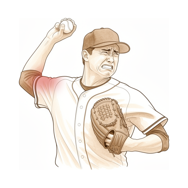
ایس ایل اے پی پھٹاؤ (SLAP tear)، یعنی کندھے کے ساکٹ کے اوپری کنارے پر کارٹلیج کے حلقے کا آگے سے پیچھے تک پھٹ جانا، کا درد کندھے کے اندر گہرائی میں، جوڑ کے اوپری حصے میں ہوتا ہے۔ ہو سکتا ہے آپ باہر کی طرف کسی ایک دکھتی جگہ کی نشاندہی نہ کر سکیں۔ یہ اکثر گرنے یا بازو کے اچانک کھنچ جانے کے بعد شروع ہوتا ہے، یا آہستہ آہستہ بھی آ سکتا ہے، جو سر سے اوپر بازو گھما کر گیند پھینکنے والے لوگوں میں عام ہے، جیسے کرکٹ کے کھلاڑی یا بیس بال کے بولر (pitchers)۔
بہت سے لوگ سر سے اوپر کی سرگرمی کے دوران یا اس کے بعد گہری دکھن محسوس کرتے ہیں۔ کچھ لوگ بتاتے ہیں کہ اوپر ہاتھ بڑھاتے وقت ان کا بازو بے جان یا بھاری ہو جاتا ہے، اور ساتھ ہی تھکاوٹ یا اٹکنے کا احساس ہوتا ہے۔ اگر مسئلہ خود بائسیپس (biceps) کے ٹینڈن (tendon)، یعنی وہ مضبوط ڈوری جو پٹھے کو ہڈی سے جوڑتی ہے، میں ہو، تو بازو سر سے اوپر اٹھاتے وقت آپ کو کلک یا تڑخنے کا احساس ہو سکتا ہے۔ اگر ٹینڈن پھٹ کر ٹوٹ گیا ہو تو بازو کے اوپری حصے کے سامنے ایک صاف نظر آنے والی گانٹھ بن سکتی ہے، جسے اکثر پوپائے کی علامت (Popeye sign) کہتے ہیں۔
روزمرہ زندگی میں وہ کام تکلیف دیتے ہیں جو کندھے کے اوپری حصے پر بوجھ ڈالتے ہیں: اونچی شیلف تک ہاتھ بڑھانا، بھاری تھیلا اٹھانا، گیند پھینکنا، یا بازو کندھے کی اونچائی سے اوپر رکھ کر کام کرنا۔ درد عموماً صبح جاگتے ہی نہیں بلکہ سرگرمی کے بعد بھڑکتا ہے۔ آپ کی طاقت اور حرکت اکثر برقرار رہتی ہے، اور یہی ایک وجہ ہے کہ اس مسئلے کو کندھے کی کوئی دوسری بیماری سمجھ لیا جاتا ہے۔
ایک بات جاننے کے قابل ہے: ایس ایل اے پی پھٹاؤ شاذ و نادر ہی اکیلا ہوتا ہے۔ زیادہ تر صورتوں میں اسی کندھے میں اس کے ساتھ کوئی اور مسئلہ بھی ہوتا ہے، جیسے روٹیٹر کف (rotator cuff)، یعنی ٹینڈنوں کا وہ گروپ جو کندھے کو تھامتا اور گھماتا ہے، میں گھساؤ، یا لیبرم (labrum)، جوڑ کے کنارے کا کارٹلیج کا حلقہ، کا پھٹاؤ۔ یہی ایک وجہ ہے کہ درد مبہم محسوس ہو سکتا ہے اور اس کی ٹھیک جگہ بتانا مشکل ہو سکتا ہے، اور اسی لیے آپ کے سرجن کسی ایک جگہ کے بجائے پورے کندھے کا معائنہ کریں گے۔
اگر آپ کی علامات کم نہ ہو رہی ہوں، ہفتوں کے دوران بڑھ رہی ہوں، آپ کو رات کو جگا دیتی ہوں، یا آپ کو کام کرنے یا اپنا بازو استعمال کرنے سے روکتی ہوں، تو اپنے فیملی ڈاکٹر (GP) سے ملیں یا ماہر سے معائنہ کروانے کا کہیں۔
دراصل کیا ہو رہا ہے
آپ کا کندھا گیند اور ساکٹ والا جوڑ ہے۔ ساکٹ کے کنارے کے گرد نرم کارٹلیج (cartilage) کا ایک حلقہ ہوتا ہے جسے لیبرم کہتے ہیں، جو کچھ حد تک ربڑ کے گسکٹ کی طرح کام کرتا ہے، ساکٹ کو گہرا کرتا ہے اور جوڑ کی گیند کو اپنی جگہ بٹھائے رکھتا ہے۔ بائسیپس کے لمبے سرے (long head) کا ٹینڈن سیدھا اسی حلقے کے اوپری حصے میں جڑتا ہے، اس لیے یہ دونوں ایک ہی مسلسل ڈھانچہ ہیں۔ جب لیبرم کا اوپری حصہ پھٹتا ہے تو وہ بائسیپس کے جڑاؤ کا کچھ حصہ بھی ساتھ لے جاتا ہے۔ ایس ایل اے پی پھٹاؤ یہی ہے: گسکٹ کے اوپری حصے میں، جہاں بائسیپس جڑتا ہے، وہاں کا پھٹاؤ۔
یہ پھٹاؤ عام طور پر کسی زور کی وجہ سے ہوتا ہے۔ پھیلے ہوئے بازو کے بل گرنا، بازو کو اچانک جھٹکا لگنا، یا بار بار گیند پھینکنے کا زور لیبرم کے اوپری حصے کو ساکٹ کے کنارے سے پیچھے کی طرف اکھاڑ سکتا ہے، جیسے کسی مرتبان سے لیبل اکھڑ جاتا ہے۔ سر سے اوپر بازو گھما کر گیند پھینکنے والے لوگوں میں، برسوں کھیلنے کے دوران کندھا پیچھے سے تنگ ہو جاتا ہے۔ یہ تنگی گیند پھینکنے کی حرکت کے دوران جوڑ کی گیند کو تھوڑا اوپر اور پیچھے کی طرف جھکا دیتی ہے، جس سے ہر بار لیبرم کا اوپری حصہ مروڑ کھاتا ہے۔ یہی زور خود بائسیپس کے ٹینڈن پر بھی پڑتا ہے، جس میں سوزش ہو سکتی ہے، جو گھس کر ریشہ ریشہ ہو سکتا ہے، یا غیر مستحکم ہو سکتا ہے، یہاں تک کہ وہ کندھے کے سامنے اپنی نالی میں کلک کرے یا تڑخے۔
یہی مسائل ان علامات کی وضاحت کرتے ہیں جن کے بارے میں آپ نے اوپر پڑھا۔ گہری دکھن جوڑ کے اوپری حصے میں پھٹے ہوئے لیبرم اور اس ٹینڈن سے آتی ہے جس میں جلن (irritation) پیدا ہو گئی ہو۔ اٹکنا اور کلک ہونا اس لیے ہوتا ہے کہ بازو اٹھاتے وقت پھٹا ہوا کنارہ حرکت کرتا ہے۔ اور چونکہ بائسیپس جوڑ کی گیند کو ساکٹ میں جمائے رکھنے میں مدد دیتا ہے، اس لیے وہاں کا پھٹاؤ سر سے اوپر کے کام کے دوران آپ کے بازو کو کمزور یا بے جان محسوس کروا سکتا ہے۔
ایس ایل اے پی پھٹاؤ کے درجے اس بنیاد پر بھی طے کیے جاتے ہیں کہ پھٹاؤ کتنا پھیلا ہوا ہے، کنارے کے گھس کر ریشہ ریشہ ہونے سے لے کر ایسے پھٹاؤ تک جو لیبرم کو چیر کر نیچے خود بائسیپس کے ٹینڈن میں چلا جائے۔ اونچے درجوں میں، جہاں بائسیپس کا ٹینڈن بھی پھٹا ہوا ہو، عموماً لیبرم کی مرمت کے بجائے بائسیپس کا علاج زیادہ ممکنہ منصوبہ بن جاتا ہے۔
ہم اس کے لیے کیا کر سکتے ہیں
ڈاکٹر کیرن ہِرپرا، جو Mater Private Hospital Rockhampton میں بازو اور ہاتھ کے سرجن (upper-limb surgeon) ہیں، آپ کی حالت کے مطابق سب سے کم چیر پھاڑ والے طریقوں سے آغاز کرتے ہیں۔ مریضوں کو عام طور پر ان کا فیملی ڈاکٹر (GP) ہمارے کلینک بھیجتا ہے؛ اگر کسی فزیوتھراپسٹ نے آپ کو ہم سے ملنے کا مشورہ دیا ہے تو بھی میڈیکیئر (Medicare) کی رقم کی واپسی کا حق دار ہونے کے لیے آپ کو اپنے فیملی ڈاکٹر سے ریفرل (referral) لینا ہوگا۔ ملاقات پر ہم آپ کی طبی تاریخ لیتے ہیں، کندھے کا معائنہ کرتے ہیں، اور جہاں مددگار ہوں وہاں اسکین کا انتظام کرتے ہیں۔ لیبرم اور بائسیپس کے اسکین مکمل طور پر قابلِ بھروسہ نہیں ہوتے، اس لیے ہم انہیں اکیلے نہیں بلکہ آپ کی بتائی ہوئی تفصیل اور آپ کے معائنے کے ساتھ ملا کر دیکھتے ہیں۔
زیادہ تر ایس ایل اے پی پھٹاؤ میں پہلے آپریشن کے بغیر علاج کو پورا موقع دیا جاتا ہے۔ اس کا مطلب ہے گیند پھینکنے یا سر سے اوپر کے دوسرے کھیلوں سے آرام، سوزش کم کرنے والی دوا، اور فزیوتھراپی۔ فزیوتھراپی کے پروگرام کا مقصد روٹیٹر کف اور کندھے کی پچھلی چپٹی ہڈی (shoulder blade) کے گرد کے پٹھوں کو مضبوط کرنا، اور کندھے کے پچھلے حصے کے تنگ ڈھانچوں کو اسٹریچ کرنا ہے۔ گیند پھینکنے والے لوگوں میں، کام کا ایک حصہ خود گیند پھینکنے کی حرکت کی خامیوں کو درست کرنا ہے، کیونکہ اکثر یہی چیز پھٹاؤ پر بار بار بوجھ ڈالتی ہے۔ اس علاج کے بارے میں رائے قائم کرنے سے پہلے اسے کم از کم 3 ماہ دیں۔ بہت سے لوگ اس سے ٹھیک ہو جاتے ہیں، اور اگر پہلا کورس کافی نہ ہو تو سامنے آنے والے مخصوص مسائل پر مرکوز دوسرا کورس پھر بھی مدد کر سکتا ہے۔ اعلیٰ سطح کے کھلاڑی اس دوران اکثر مقابلوں میں حصہ لیتے رہ سکتے ہیں اور اپنا سیزن پورا کر سکتے ہیں۔
اگر صرف فزیوتھراپی سے آرام نہ آیا ہو، تو ہم کندھے کے جوڑ میں، یا اس نالی میں جہاں سے بائسیپس کا ٹینڈن گزرتا ہے، کورٹیسون (اسٹیرائیڈ) کا انجکشن پیش کر سکتے ہیں۔ کورٹیسون سوزش کم کرنے والی ایک طاقتور دوا ہے۔ یہ درد کم کر سکتا ہے اور یہ جاننے میں مدد دے سکتا ہے کہ آیا درد کی اصل وجہ واقعی یہی پھٹاؤ ہے، اور کچھ لوگوں میں یہ علامات کو اتنا کم کر دیتا ہے کہ سرجری کی ضرورت نہیں رہتی۔
سرجری کی بات اس وقت آتی ہے جب آپریشن کے بغیر 3 ماہ کے علاج کے بعد بھی علامات برقرار رہیں۔ منصوبہ آپ کی عمر، آپ کتنے سرگرم ہیں، پھٹاؤ کی شکل، اور کندھے میں موجود دوسرے مسائل پر منحصر ہے۔ کچھ پھٹاؤ میں، خاص طور پر کم عمر اور سرگرم لوگوں میں، لیبرم کی مرمت کر کے اسے دوبارہ ساکٹ سے جوڑ دیا جاتا ہے۔ کچھ دوسرے لوگوں میں، خاص طور پر 30 سال سے زیادہ عمر کے ان لوگوں میں جن کے جسمانی تقاضے کم ہوں، بائسیپس کے ٹینڈن کا علاج بہتر کام کرتا ہے: ٹینڈن کو پھٹے ہوئے لیبرم سے الگ کر کے بازو میں نیچے دوبارہ جوڑ دیا جاتا ہے، یا صرف الگ کر دیا جاتا ہے۔ آپ کے لیے کون سا راستہ مناسب ہے، یہ فیصلہ ہم آپ کے ساتھ مل کر، ہر ایک میں شامل باتوں کو تولتے ہوئے کرتے ہیں۔ اگر کندھے میں کوئی سسٹ (سیال سے بھری تھیلی) کسی عصب کو دبا رہا ہو، تو سرجری پر جلد غور کیا جاتا ہے، کیونکہ انتظار کرنے سے مستقل کمزوری رہ سکتی ہے۔
کیا توقع رکھیں
علاج کے بغیر، آپ کے کندھے کے اوپری حصے کی گہری دکھن عموماً خود بخود ٹھیک ہونے کے بجائے سر سے اوپر کے کام کے بعد بار بار بھڑکتی رہتی ہے۔ گیند پھینکنے سے آرام، سوزش کم کرنے والی دوا اور فزیوتھراپی بہت سے لوگوں کو ٹھیک کر دیتی ہیں، اور اس صفحے کے پچھلے حصوں میں اس علاج کی تفصیل دی گئی ہے۔ اگر پہلا کورس کافی نہ ہو، تو آپ کے مخصوص مسائل پر مرکوز دوسرا کورس پھر بھی مدد کر سکتا ہے۔
اگر آپ سرجری کا راستہ اختیار کریں، تو نتیجہ آپ کی عمر، آپ کی سرگرمی، اور کندھے میں موجود دوسرے مسائل پر منحصر ہے۔ زیادہ تر لوگ پھٹاؤ کی مرمت کے بعد اپنی پہلے والی سر سے اوپر کی سرگرمیوں میں واپس آ جاتے ہیں۔ 50 سال سے کم عمر کے وہ لوگ جن کے اس پھٹاؤ کے ساتھ روٹیٹر کف کا مسئلہ بھی ہو، ان میں نتائج اور سرگرمی میں واپسی عموماً ایک ہموار راستے پر چلتے ہیں۔
ایمانداری سے کہیں تو تصویر مکمل نہیں ہے۔ لیبرم کی مرمت کروانے والے کچھ لوگوں کو بعد میں مزید سرجری کی ضرورت پڑتی ہے۔ کم عمر فوجی مریضوں کے ایک گروپ میں، جسے طویل مدت تک دیکھا گیا، مرمت کروانے والوں میں سے 40 فیصد کو بعد میں اس کے بجائے بائسیپس کے ٹینڈن کا علاج کروانا پڑا، جبکہ جن کا پہلے ہی بائسیپس کا علاج کیا گیا تھا ان میں سے کسی کو بھی دوبارہ آپریشن (revision) کی ضرورت نہیں پڑی۔ مجموعی طور پر، صرف لیبرم کی مرمت کروانے والے ہر 100 میں سے تقریباً 10 لوگوں کو بعد میں ایک اور آپریشن کروانا پڑتا ہے، اور اس کی وجہ اکثر یہ ہوتی ہے کہ کندھے کا کوئی دوسرا مسئلہ سامنے آ جاتا ہے۔ 40 سال سے زیادہ عمر، تمباکو نوشی، موٹاپا، اور خود بائسیپس کے ٹینڈن میں جلن (irritation)، یہ سب دوبارہ آپریشن کی ضرورت پڑنے کا امکان بڑھاتے ہیں۔
اگر آپ اپنے اختیارات پر غور کر رہے ہیں تو کچھ اور باتیں بھی جاننے کے قابل ہیں۔ پھٹے ہوئے حصے کی مرمت کیے بغیر صرف اسے تراش دینے کا اثر عموماً وقت کے ساتھ کم ہوتا جاتا ہے: درد میں جو آرام ایک سال پر زیادہ تر لوگوں کو ملتا ہے، دو سال تک کم لوگوں کو ملتا ہے، اور اس وقت تک آدھے سے بھی کم لوگ اپنے پہلے والے کھیل کی سطح پر واپس آ پاتے ہیں۔ زیادہ جسمانی تقاضوں والے جن لوگوں کو پھٹاؤ اور بائسیپس میں جلن دونوں ہوں، ان میں مرمت اور بائسیپس کے علاج کو ایک ساتھ کرنے کا تعلق کسی ایک مسئلے کا اکیلے علاج کرنے کے مقابلے میں بدتر نتائج سے پایا گیا ہے۔ آپ کے لیے کون سا راستہ مناسب ہے، یہ فیصلہ آپ کے سرجن آپ کے ساتھ مل کر، آپ کی عمر، آپ کے کھیل اور خود پھٹاؤ کی بنیاد پر کرتے ہیں۔
اگر آپ کی علامات کم نہ ہو رہی ہوں، ہفتوں کے دوران بڑھ رہی ہوں، آپ کو رات کو جگا دیتی ہوں، یا آپ کو کام کرنے یا اپنا بازو استعمال کرنے سے روکتی ہوں، تو اپنے فیملی ڈاکٹر سے ملیں یا ماہر سے معائنہ کروانے کا کہیں۔
ڈاکٹر کو کب دکھائیں
اپنے فیملی ڈاکٹر سے ملیں یا ماہر سے معائنہ کروانے کا کہیں اگر سر سے اوپر کی سرگرمی سے چند ہفتوں کے آرام کے بعد بھی آپ کے کندھے کی گہری دکھن کم نہ ہو رہی ہو، یا اگر ہر بار گیند پھینکنے، کندھے کی اونچائی سے اوپر وزن اٹھانے، یا اوپر ہاتھ بڑھانے پر یہ بھڑک اٹھتی ہو۔ اور جلدی رابطہ کریں اگر بازو سر سے اوپر اٹھاتے وقت بے جان یا بھاری ہو جائے، اگر آپ کو کندھے کے سامنے نیا کلک یا تڑخنا محسوس ہو، یا اگر بازو اتنا کمزور محسوس ہو کہ آپ کام نہ کر سکیں یا کھیل نہ سکیں۔ اگر کسی چوٹ کے بعد آپ کے بازو کے اوپری حصے کے سامنے کوئی گانٹھ ابھر آئی ہو، یا آپ کا کندھا اٹکنے یا اچانک ڈھیلا پڑ کر جواب دینے لگا ہو، تو اس کا معائنہ کروانا بھی مناسب ہے۔ اس مسئلے میں ایمرجنسی والی کوئی بات نہیں، لیکن کندھے کا معائنہ جتنی جلدی ہوگا، آپ اور آپ کے فیملی ڈاکٹر اتنی ہی جلدی کسی منصوبے پر متفق ہو سکیں گے۔
مزید تفصیل
Advanced reading: the deeper science (optional)
یہ حصہ آپ کے اپنے علاج کے فیصلوں کے لیے ضروری معلومات سے آگے جاتا ہے۔ ایس ایل اے پی پھٹاؤ کے بارے میں یہ اضافی مطالعہ اس لیے مفید ہے کہ یہ کندھے کی وہ تشخیص ہے جس میں معائنے کے ٹیسٹ اور اسکین دونوں سب سے کم قابلِ بھروسہ ہیں، اور اس لیے بھی کہ جو آپریشن اب ترجیح پا چکا ہے وہ پھٹاؤ کی مرمت کرنے والا آپریشن نہیں ہے۔
معائنے کا ٹیسٹ تشخیص کا بوجھ نہیں اٹھاتا
ایکٹیو کمپریشن ٹیسٹ (Active Compression، O'Brien test) وہ طریقہ ہے جسے ایس ایل اے پی زخموں کے ساتھ سب سے زیادہ جوڑا جاتا ہے۔ 3,091 مریضوں میں جانچنے پر اس ٹیسٹ میں مسئلے کی ابتدائی چھانٹی اور تصدیق دونوں کی صلاحیت محدود پائی گئی، اور مصنفین صاف کہتے ہیں کہ وہ طبی فیصلوں میں اس کے استعمال کی حمایت نہیں کرتے [1]۔
یہ تقریباً اتنا ہی منفی نتیجہ ہے جتنا کوئی تشخیصی جائزہ دیتا ہے، اور یہ اسی ٹیسٹ پر لاگو ہوتا ہے جسے ریفرل خطوط میں سب سے زیادہ مثبت لکھا جاتا ہے۔
اور ایم آر آئی اسے رد نہیں کر سکتا
امیجنگ بہتر ہے لیکن نامکمل ہے۔ 2,916 مریضوں میں ایم آر آئی کی حساسیت (sensitivity) درمیانی اور مخصوصیت (specificity) اور درستگی بہترین پائی گئی، جس سے یہ ایس ایل اے پی زخم کی تصدیق کے لیے قیمتی ہے، جبکہ یہ اسے حتمی طور پر رد نہیں کر سکتا، اور آرتھروسکوپی (دوربین سے جوڑ کے اندر دیکھ کر علاج) ہی معیاری کسوٹی (reference standard) ہے [2]۔
دونوں نتائج کو ملا کر دیکھیں تو عملی صورت حال یہ ہے: مثبت ایم آر آئی معلوماتی ہے، منفی ایم آر آئی فیصلہ کن نہیں، اور معائنہ دونوں صورتوں میں بہت کم اضافہ کرتا ہے۔ یہی بڑی وجہ ہے کہ ان کندھوں میں ایس ایل اے پی پھٹاؤ کی ضرورت سے زیادہ تشخیص ہوتی ہے جن کے درد کی وجہ کچھ اور ہوتی ہے، اور یہ بھی کہ لیبرم کے اوپری حصے میں اتفاقاً نظر آنے والی تبدیلی کا علاج کرنا ایک تسلیم شدہ غلطی کیوں ہے، خاص طور پر زیادہ عمر کے کندھوں میں جہاں اس جگہ ریشے ریشے ہونا عمر کے ساتھ متوقع تبدیلی ہے۔
ٹینوڈیسس نے مرمت کی جگہ لے لی ہے، اور اس کی وجوہات سمجھنے کے قابل ہیں
جہاں صرف دوسری قسم (type II) کا پھٹاؤ ہو اور وہ واقعی علامات پیدا کر رہا ہو، وہاں دو آپریشنوں میں مقابلہ ہے: لیبرم کو دوبارہ ساکٹ سے جوڑنا، یا بائسیپس کے ٹینڈن کو الگ کر کے نیچے دوبارہ جوڑنا، تاکہ وہ کھنچاؤ ختم ہو جائے جو پھٹے ہوئے لیبرم پر زور ڈالتا ہے۔
881 مریضوں میں، ابتدائی علاج کے طور پر ایس ایل اے پی کی مرمت اور بائسیپس ٹینوڈیسس دونوں قابلِ قبول ہیں؛ مرمت اب بھی سب سے زیادہ کی جاتی ہے، لیکن ٹینوڈیسس اتنا ہی مؤثر اور ایک پرکشش متبادل ہے [3]۔ 908 مریضوں پر مشتمل ایک فیصلہ جاتی تجزیے (decision analysis) نے اس سے آگے بات کی: زیادہ متوقع فائدے کی بنیاد پر ٹینوڈیسس کو مرمت پر ترجیح دی جاتی ہے، اور میٹا اینالیسس (meta-analysis) میں اچھے نتائج زیادہ کثرت سے دیکھے گئے [4]۔
میکانیکی منطق یہ ہے کہ مرمت بناوٹ کو بحال کر دیتی ہے لیکن بائسیپس کو بھرتی ہوئی مرمت پر کھینچتا رہنے دیتی ہے، جو زیادہ عمر کے یا اکڑے ہوئے کندھے میں اکثر مسلسل درد اور بازو کو باہر کی طرف گھمانے کی صلاحیت میں کمی کا سبب بنتا ہے۔ ٹینوڈیسس اصل بناوٹ کو چھوڑ دیتا ہے اور بگاڑ پیدا کرنے والی قوت کو ختم کر دیتا ہے۔ نتائج قوت کو ختم کرنے کے حق میں رہے ہیں۔
استثنا سر سے اوپر بازو چلانے والا نوجوان کھلاڑی ہے، جہاں بائسیپس کے جڑاؤ کو قربان کرنا کام کاج کے لحاظ سے ایک بڑا فیصلہ ہے اور مرمت کا کردار اب بھی باقی ہے۔
بائسیپس اور لیبرم ایک ہی موضوع کیوں ہیں
بائسیپس کا لمبا سرا براہِ راست لیبرم کے اوپری حصے سے جڑتا ہے، دونوں ایک دوسرے سے ملے ہوئے ہیں۔ اس لیے اس جوڑ پر پھٹاؤ بیک وقت لیبرم کی چوٹ بھی ہے اور بائسیپس کے جڑاؤ کی چوٹ بھی، اسی لیے بائسیپس پر آپریشن لیبرم کے زخم کا علاج کرتا ہے، اور اسی لیے علامات بائسیپس کے ٹینڈن کی خرابی (tendinopathy) سے اس قدر ملتی ہیں کہ معائنے میں دونوں میں فرق کرنا اکثر ممکن نہیں ہوتا۔
حوالہ جات
[1] Davis C, Immormino J, Higgins BM, Clark K, Engebose S, Garcia AN, et al. Diagnostic utility of the Active Compression Test for the superior labrum anterior posterior lesion: a systematic review with meta-analysis. Shoulder Elbow. 2018;11(5):321-31. https://doi.org/10.1177/1758573218811656
[2] Nosratpour M, Zarei H, Zaker Moshfegh M, Mahdavi M, Moteshakereh SM, Shirvani P, et al. Diagnostic accuracy of magnetic resonance imaging for detecting superior labrum anterior-posterior lesions: a systematic review and meta-analysis. JSES Int. 2025;9(6):1972-87. https://doi.org/10.1016/j.jseint.2025.05.023
[3] de SA D, Arakgi ME, Lian J, Crum RJ, Lin A, Lesniak BP. Labral repair versus biceps tenodesis for primary surgical management of type II superior labrum anterior to posterior tears: a systematic review. Arthroscopy. 2019;35(6):1927-38. https://doi.org/10.1016/j.arthro.2018.12.015
[4] Recker AJ, Waters TL, Bullock G, Rosas S, Scholten DJ, Nicholson K, et al. Biceps tenodesis has greater expected value than repair for isolated type II SLAP tears: a decision analysis and meta-analysis. Arthroscopy. 2022;38(10):2887-2900. https://doi.org/10.1016/j.arthro.2022.05.005
Evidence & references
This is the clinical evidence summary written for health professionals. It is technical, and it lists the research this page was built from. You do not need to read it to understand your treatment or to make a decision about it.
Overview
- For operative treatment of proximal biceps pathology in overhead athletes, biceps tenodesis has consistent and reliable results, whereas return to play after SLAP repair can be unpredictable [1].
- Biceps tenodesis is a safe, effective, and technically straightforward alternative to primary SLAP repair in patients with type II and IV SLAP tears [2].
- Both arthroscopic repair and biceps tenotomy and tenodesis interventions had benefits in type II SLAP lesions [3].
- SLAP repairs are generally favored in younger, active patients, whereas treating the biceps is preferred in lower-demand patients aged >30 years [4].
- SLAP repair and biceps tenodesis both present viable treatment options but come with specific advantages and disadvantages, with the decision ultimately made individually with the patient [5].
- Biceps tenodesis has been increasingly used for the management of SLAP lesions, with recent studies reporting high rates of return to sport, high satisfaction, and good to excellent patient-reported outcomes in carefully selected athletes [8].
- Increased patient age correlates with the likelihood of treatment with biceps tenodesis or tenotomy versus SLAP repair [9].
- Short-term follow-up of 20 subpectoral biceps tenodesis procedures using an all-suture anchor fixation has not shown any failure of fixation or residual biceps discomfort [10].
- Biceps tenodesis may be considered a valid primary or revision surgery for patients suffering from symptomatic type II SLAP tears due to no detrimental effect on glenohumeral stability [11].
- Biceps tenodesis is a predictable, safe, and effective treatment for failed arthroscopic SLAP tears at a minimum 2-year follow-up [12].
- Appropriate treatment for biceps pathology, whether conservative or surgical, should be based on established pathology [16].
- The number of isolated SLAP repairs performed has decreased over time, and management of failed SLAP repair has shifted toward biceps tenodesis or tenotomy over revision SLAP repair in more recent years [17].
- Primary subpectoral open biceps tenodesis for SLAP tears or pathology of the long head of the biceps tendon provides significant improvement in shoulder outcomes with a reliable return to activity level with low risk for complications [18].
- Primary biceps tenodesis offers increased effectiveness when compared with both primary SLAP repair and nonoperative treatment and lower costs than primary SLAP repair [29].
- In a young active population, primary arthroscopic biceps tenodesis is a viable surgical alternative to labral repair for type II SLAP lesions [30].
- Treatment of proximal biceps pathology is largely based on expert opinion and patient preferences rather than robust randomized evidence [31].
- Adjuvant biceps procedures are not required when repairing isolated supraspinatus tears, unless biceps pathology is observed intraoperatively, for which tenodesis grants better function and strength than tenotomy [32].
- The treatment option of biceps tenodesis is an appealing alternative to SLAP repair, but the indications and technique of biceps tenodesis in the elite pitcher still need to be defined [59].
- High-demand patients with biceps tendonitis in the setting of a SLAP lesion with labral instability who undergo combined tenodesis and labral repair have significantly worse outcomes than patients who undergo either isolated labral repair for type II SLAP tears or isolated biceps tenodesis for a SLAP tear and biceps tendonitis [62].
- Biceps tenotomy is well accepted by most patients with good overall results [70].
- The choice between biceps tenotomy and tenodesis for pathology of the proximal biceps tendon can continue to be based on surgeon and patient preference [71].
Anatomy & Pathophysiology
Bony and Soft Tissue Anatomy
- The long head of the biceps originates from the bicipital tubercle at the superior rim of the glenoid and along the posterior superior rim of the glenoid and labrum [34].
- The short head of the biceps originates from the coracoid tip lateral to and in common with the coracobrachialis [34].
- The biceps tendon is an intra-articular but extrasynovial structure within the glenohumeral joint [45].
- 40% to 60% of the biceps tendon attaches to the supraglenoid tubercle 5 mm medial to the superior glenoid rim, with the remainder attaching directly to the superior glenoid labrum [45].
- The biceps tendon typically attaches entirely (type I) or predominantly posterior (type II) on the superior labrum [45].
- The biceps tendon may have equal anterior and posterior contributions (type III) or, less commonly, predominantly anterior (type IV) labral attachment [45].
- The glenoid labrum consists of parallel collagen fibers that course around the circumference of the glenoid [45].
- The superior labrum inserts on the superior glenoid rim, medial to the articular cartilage margin, through a transitional zone of fibrocartilage [45].
- A normal synovial recess exists between the meniscoid or triangular superior labrum and the articular cartilage extension over the superior glenoid rim [45].
- The glenoid labrum is composed of fibrocartilaginous tissue [46].
- The inner portion of the labrum is avascular, and the superior labrum is less vascular compared with the inferior and posterior labrum [46].
- The superior labrum is usually triangular but can have a meniscoid shape, and commonly attaches medial to the articular margin of the glenoid rim [46].
- The LHB anchor has some inherent physiologic motion, and overconstraint from repair can contribute to stiffness [46].
- The biceps tendon passes through the bicipital groove, or intertubercular groove, between the greater and lesser tuberosities [45].
- Stability of the biceps within the bicipital groove is afforded by the biceps sling, or pulley, consisting of fibers from the subscapularis tendon, supraspinatus tendon, coracohumeral ligament, and superior glenohumeral ligament [45].
- The LHB pulley is a capsuloligamentous complex comprising the superior glenohumeral ligament, the coracohumeral ligament, and fibers from the subscapularis and supraspinatus tendons that stabilizes the proximal LHB as the tendon enters the bicipital groove [46].
- The bicipital tendon does not move up and down in the groove; rather, the humerus moves down and up with adduction and abduction relative to the tendon [34].
- The bicipital tendon is retained within the groove by a pulley made up of fibers from the coracohumeral and superior glenohumeral ligaments, with some reinforcement from adjacent tendons [34].
- The biceps tendon is innervated by thinly myelinated sensory neurons, with most innervation occurring at the LHB origin [46].
- Blood is supplied to the LHB tendon from the thoracoacromial and brachial arteries via the osteotendinous and musculotendinous junctions, respectively [46].
- A hypovascular zone exists at the proximal portion of the LHB tendon, close to the superior glenoid attachment [45].
- The biceps muscle has two distal tendinous insertions: a lateral insertion to the posterior part of the tuberosity of the radius and a medial aponeurotic insertion into the deep fascia of the volar forearm [34].
- Loss of the long head attachment is manifested mainly as loss of supination strength (20%) with a smaller loss (8%) of elbow flexion strength [34].
Anatomic Variants
- Anatomic variants in the superior labrum include a sublabral foramen or absence of the superior labrum, often seen together with a cordlike middle glenohumeral ligament (MGHL) [46].
- In a cohort of 73 shoulders, 3.3% had a sublabral foramen, 8.6% had a sublabral foramen with cordlike MGHL (Buford complex), and 1.5% had an absent anterosuperior labrum [46].
- The sublabral recess represents a small potential space under the biceps anchor and the anterosuperior labrum, often present at the 12 o'clock position [91].
- The Buford complex is a normal variant consisting of a cordlike middle glenohumeral ligament that originates directly from the superior labrum at the base of the biceps tendon, resulting in an absence of anterosuperior labral tissue [91].
- The sublabral foramen involves a cordlike middle glenohumeral ligament that attaches directly to the anterosuperior labrum, creating a hole between the ligament and the glenoid [91].
- Inappropriate surgical attachment of a cord-like middle glenohumeral ligament to a void on the anterosuperior glenoid results in painful restriction of external rotation and elevation [91].
- The incidence of the cordlike middle glenohumeral ligament in isolation is 18%, which is more common than its combination with the Buford complex (1% to 2%) [91].
- The superior labrum has a high degree of normal variation, typically either rounded or meniscoid, with the meniscal component overlying but not attached to the glenoid articular surface [91].
Biceps-Labral Complex Zones
- The biceps-labral complex (BLC) is conceptualized as having three distinct zones: Inside, Junction, and Extra-articular [46].
- The Inside zone of the BLC consists of the superior glenoid labrum and the LHB anchor, which is closely associated with the SGL [46].
- The Junction zone includes the intra-articular portion of the LHB, as well as the stabilizing biceps pulley [46].
- The Extra-articular zone consists of the bicipital tunnel and is further divided into three zones: zone 1 bony groove, zone 2 “No Man’s Land,” and zone 3 subpectoralis [46].
- Zone 1 and zone 2 of the bicipital tunnel contain synovial tissue, which may generate pain [46].
- Zone 2 of the bicipital tunnel cannot be visualized by arthroscopy from above or with an open approach from below the zone [46].
Pathophysiology of SLAP Lesions
- SLAP tears can be caused by forceful traction to the arm, direct compression loads, and repetitive overhead throwing [36].
- Increased external rotation of the shoulder in the late cocking phase increases torsional force at the LHB root, resulting in a peel-back injury to the posterosuperior labrum [36].
- Injuries can result from repetitive contact of the posterosuperior labrum with the undersurface of the rotator cuff in the late cocking phase, known as internal impingement [36].
- SLAP tears are seen more frequently in the late cocking position, occurring because of an adaptive posterior capsular contracture [36].
- Throwing athletes demonstrate increased shoulder external rotation and decreased internal rotation in abduction, which causes posterosuperior migration of the humeral head in the late cocking phase [36].
- Increased external rotation results in greater torsional loads across the superior labrum from the more posteriorly oriented LHB tendon, causing the labrum and LHB tendon to displace medially over the glenoid rim [36].
- In a cadaveric model simulating a fall on the outstretched hand, 5 type II SLAP lesions were found in shoulders simulating a forward fall, while only 2 were observed in shoulders simulating a backward fall [41].
- Shearing forces are a major factor in the pathogenesis of type II SLAP lesions, in association with predisposing anatomic factors [41].
- Traction on the biceps tendon in a biomechanical model can reproducibly create type II SLAP lesions [109].
- The production of type II SLAP lesions differed significantly between reduced shoulders (2 out of 8 tests) and shoulders with inferior subluxation (7 out of 8 tests) [109].
- Glenohumeral translations were increased after simulation of type II SLAP lesions that ranged in severity from subperiosteal elevation to complete detachment of the superior labrum and biceps anchor [23].
- SLAP lesions lead to increased glenohumeral translation and concurrently LHB tension and load in at most anterior direction [133].
- Type II SLAP lesions are thought to result from a peelback mechanism during maximum external rotation of the arm during the throwing motion that involves eccentric biceps contraction and a huge magnitude of tensile forces on the biceps anchor [58].
- SLAP tears may represent an adaptive process, because the peel-back of the SGL permits increased humeral external rotation needed to participate in overhead sporting activity [88].
Pathophysiology of Biceps Tendon Disorders
- Pathology of the LHB tendon includes tendinitis, tendinopathy, tears, subluxation, entrapment, delamination, and dislocation out of the bicipital groove [36].
- Because of the relatively anterior position of the bicipital groove along the humeral head combined with humeral retroversion, the tendon is exposed to medial instability, which can increase the risk of tendon degeneration [36].
- Variations of bicipital groove morphology can also increase the risk of LHB tendon pathology [36].
- Isolated LHB tendon pathology can occur but frequently is associated with other shoulder pathologies, especially rotator cuff pathology [36].
- Primary LHB tendinitis usually occurs in younger patients who participate in overhead activities such as volleyball and baseball [36].
- With LHB tendon instability, the patient describes a clicking or snapping with overhead motions [36].
- A subscapularis tear is associated with LHB medial instability and a supraspinatus tear is associated with posterolateral instability [36].
- Biceps tendinitis is rarely the primary cause of shoulder pain and is usually secondarily involved as a part of an impingement syndrome or degenerative lesions of the rotator cuff [48].
- In the context of rotator cuff disease, the etiology of anterior shoulder pain with macroscopic changes in the biceps tendon is related to the complex interaction of the tendon and surrounding soft tissues, rather than a single entity [14].
- Traumatic rupture of a normal long head of the biceps tendon is uncommon [15].
- Most cases of traumatic tenosynovitis or dislocation occur as part of the impingement syndrome and a rotator cuff tear [15].
- Acute rupture of the biceps tendon has been reported in association with superior labral injury but is rare [15].
- Repetitive throwing resulted in tears of the anterior superior or posterior superior labrum, or both, in 73 throwing athletes [15].
- Seven of 35 (20%) baseball pitchers had partial tearing of the tendon of the long head of the biceps [15].
- The most common mechanism of injury for superior labral lesions was a fall or a direct blow to the shoulder [15].
- Twenty-eight percent of superior labral lesions were isolated and were not associated with rotator cuff pathology [15].
- Biceps tendon lesions may be inflammatory, degenerative, or traumatic as a result of repetitive microtrauma or macrotrauma [60].
- The injury site for biceps tendon lesions may include the attachment to the supraglenoid tubercle, SLAP, the tendon (intraarticular or extraarticular), and the bicipital arch [60].
- The bicipital arch consists of the conglomerate of the superior glenohumeral ligament and the coracohumeral ligament attachment at the superior bicipital groove [60].
- In a study of 200 consecutive patients undergoing arthroscopic cuff repair, 45% had anterior, posterior, or both anterior and posterior biceps instability [60].
- Larger rotator cuff tears correlated with a higher incidence and degree of biceps instability [60].
- An hourglass-shaped biceps deformity is associated with inflammation and triggering through the proximal pulley [60].
- Persistence of triggering can result in pulley instability [60].
- Biceps tendinitis concurrent with rotator cuff disease is classified as an inflammatory disorder [26].
- Primary bicipital tendinitis is classified as an inflammatory disorder [26].
- Subluxation and dislocation of the biceps tendon are classified as instability disorders [26].
- Traumatic rupture and superior labral tears (SLAP lesions) are classified as traumatic disorders [26].
- The distinction between inflammatory, unstable, and traumatic biceps disorders is not always clear, as degenerated and inflamed tendons are more prone to trauma and repeated trauma may result in changes indistinguishable from inflammation [26].
- Subluxation of the long biceps tendon is defined as a partial and/or transient incomplete loss of contact between the tendon and its bony groove [90].
- Type I subluxation involves a partial or complete tear of the circular sling of the superior glenohumeral and coracohumeral ligaments, resulting in loss of restraint above the entrance to the groove [90].
- Type II subluxation involves a lesion located below the entrance to the bony groove where the tendon slips over the medial rim and rides on the border of the lesser tuberosity [90].
- The causal lesion for Type II subluxation is a detachment of the outermost fibres of the subscapularis tendon [90].
- Type III subluxation involves malunion and nonunion of the lesser tuberosity that compromises the medial bony restraint of the long biceps tendon [90].
- Type I dislocation is extraarticular dislocation combined with a partial tear of the subscapularis tendon [82].
- In Type I dislocation, the biceps tendon is displaced over the anterior wall of the groove and slips or glides medially over the torn fibres of the subscapularis tendon [82].
- Type II dislocation is intraarticular dislocation of the long biceps tendon combined with a complete tear of the subscapularis tendon [82].
- In Type II dislocation, the biceps tendon is interposed into the joint space and displaced inferomedially, with entrapment occurring with each internal rotational movement of the humerus [82].
- Dislocation of the long biceps tendon over a completely intact subscapularis tendon is very rare, occurring in only 2 of 70 patients (3%) in one series [82].
- The intraarticular dislocation of the biceps tendon is often associated with extensive tearing of the rotator cuff [82].
- Approximately half of intraarticular biceps dislocations have a traumatic etiology [82].
Classification
SLAP Lesion Classification
- Snyder et al. characterized superior glenoid labral injuries anterior and posterior as superior labrum anterior and posterior (SLAP) lesions [15].
- Snyder et al. classified superior labral injuries into four types based on arthroscopic findings [15].
- Type I SLAP lesions involved fraying and degeneration of the superior labrum with an intact biceps anchor [15].
- Type II SLAP injuries consisted of detachment of the labrum and biceps anchor from the superior glenoid [15].
- Type III SLAP lesions involved a bucket-handle detachment of the labrum [15].
- Type IV SLAP injuries involved a bucket-handle tear of the superior labrum with extension into the biceps [15, 26].
- In a series of 140 cases, the most common mechanism of injury for SLAP lesions was a fall or a direct blow to the shoulder [15].
- In a series of 140 cases, 28% of superior labral lesions were isolated and not associated with rotator cuff pathology [15].
- Type II SLAP tears are the most common subtype of SLAP lesions initially described by the Snyder classification [58].
- Type II SLAP lesions are thought to result from a peelback mechanism during maximum external rotation of the arm during the throwing motion involving eccentric biceps contraction and tensile forces on the biceps anchor [58].
- A cadaveric model simulating a forward fall on an outstretched hand resulted in type II SLAP lesions in 5 of 5 shoulders [41].
- A cadaveric model simulating a backward fall on an outstretched hand resulted in type II SLAP lesions in 2 of 5 shoulders [41].
- Glenohumeral translations were increased after simulation of type II SLAP lesions ranging in severity from subperiosteal elevation to complete detachment of the superior labrum and biceps anchor [23].
Biceps Tendon Disorder Classification
- Biceps tendon disorders are classified into inflammatory, unstable, or traumatic categories based on the original initiating event [26].
- The distinction between inflammatory, unstable, and traumatic biceps tendon disorders is not always clear, as degenerated and inflamed tendons are prone to trauma and repeated trauma can result in changes indistinguishable from inflammation [26].
- Inflammatory biceps tendon disorders include biceps tendinitis concurrent with rotator cuff disease and primary bicipital tendinitis [26].
- Instability biceps tendon disorders include subluxation and dislocation [26].
- Subluxation of the biceps tendon is classified into Type I (superior subluxation), Type II (unstable at proximal portion of groove), and Type III (subluxation following melanin or nonunion of lesser tuberosity) [26].
- Dislocation of the biceps tendon is classified into Type I (extraarticular, combined with partial tear of subscapularis) and Type II (intraarticular, combined with full-thickness tear of subscapularis) [26].
- Traumatic biceps tendon disorders include traumatic rupture and superior labral tears (SLAP lesions) [26].
- Traumatic rupture of the biceps tendon is classified into Type I (partial) and Type II (complete) [26].
- Superior labral tears (SLAP lesions) in the traumatic classification are categorized as Type I (significant fraying), Type II (complete detachment of biceps tendon and superior labrum from glenoid), Type III (“bucket-handle” tear of superior labrum), and Type IV (central superior labrum tear with extension into the biceps) [26].
- Most cases of traumatic tenosynovitis or dislocation of the biceps tendon occur as part of the impingement syndrome and a rotator cuff tear [15].
Clinical Presentation
History and Mechanism
- A history of acute trauma, consisting of sudden traction or compression to the affected extremity, may be present in patients with SLAP tears [50].
- SLAP tears can be associated with a previous subluxation or dislocation event [50].
- Insidious onset of symptoms associated with SLAP tears is most common in overhead throwing athletes [50].
- The most common mechanism of injury for SLAP lesions is a fall or a direct blow to the shoulder [15].
- Increased external rotation of the shoulder in the late cocking phase increases torsional force at the long head of biceps root, resulting in a peel-back injury to the posterosuperior labrum [36].
- Increased external rotation results in greater torsional loads across the superior labrum from the more posteriorly oriented long head of biceps tendon, causing the labrum and tendon to displace medially over the glenoid rim [36].
- In a cadaveric model simulating a forward fall on the outstretched hand, 5 type II SLAP lesions were found in 5 shoulders [41].
- In a cadaveric model simulating a backward fall on the outstretched hand, 2 type II SLAP lesions were observed in 5 shoulders [41].
- Most cases of traumatic tenosynovitis or dislocation of the biceps occur as part of the impingement syndrome and a rotator cuff tear [15].
Symptoms
- Pain caused by a SLAP tear often is localized deep within the glenohumeral joint [50].
- SLAP tears can be associated with mechanical symptoms, fatigue, or a “dead arm” sensation of the extremity during overhead activities [50].
- SLAP tears can be associated with frank weakness of the rotator cuff in a concomitant paralabral cyst [50].
- Patients with tenosynovitis of the long head of biceps tendon often report pain in the anterior aspect of the shoulder that radiates down the arm into the anterior biceps [49].
- Symptoms of long head of biceps tendon tenosynovitis may be exacerbated with overhead activity and activity that requires elbow flexion [49].
- Pain caused by long head of biceps tendon tendinitis usually is localized more distally than pain typically caused by rotator cuff impingement [49].
- Less commonly, patients with long head of biceps tendon pathology may report mechanical symptoms as a result of the tendon snapping or catching in the anterior shoulder [49].
- In patients with a long head of biceps tendon rupture, ecchymosis in the proximal aspect of the arm and a Popeye deformity frequently are observed [49].
- Muscle belly cramping may be reported in patients with a long head of biceps tendon rupture [49].
- Patients with biceps tendonitis or subluxation generally always have pain at the bicipital groove [37].
- Biceps-related pain felt in the bicipital groove should migrate laterally with external rotation of the arm [37].
- Pain from rotator cuff tendonitis generally radiates to the deltoid insertion and does not move with arm rotation [37].
- In bicipital tendonitis, pain is almost always accompanied by rotator cuff symptoms [37].
- Patients with bicipital tendonitis complain of pain in the anterior aspect of the arm, particularly with extension and internal rotation [37].
- Rest pain is seen later in the disease progression of bicipital tendonitis and there can be a significant component of night pain [37].
- When longstanding, pain from the long head of the biceps tendon can spontaneously resolve after a full-thickness tendon rupture [37].
- Patients with biceps tendon subluxation often notice a painful snapping or clicking sensation in the shoulder, especially with overhead positions going from internal to external rotation [37].
- Frank dislocations of the long head of the biceps are usually traumatic in origin and associated with complete tears of the subscapularis [37].
- With long head of biceps tendon instability, the patient describes a clicking or snapping with overhead motions [36].
- A subscapularis tear is associated with long head of biceps tendon medial instability [36].
- A supraspinatus tear is associated with long head of biceps tendon posterolateral instability [36].
- The proximal long head of biceps tendon has been recognized as a source of substantial anterior shoulder pain [36].
- Clinical entity of proximal long head of biceps tendon pain can be difficult to diagnose because it is known to occur with other pathologies including SLAP lesions, rotator cuff disorders, impingement, bursitis, and acromioclavicular joint disorders [36].
- There is no single pattern of pain that distinguishes biceps conditions from other shoulder abnormalities [24].
- Biceps tendon pain in the absence of tears is associated with microscopic changes consistent with tendinopathy [65].
- These microscopic changes are often missed by MRI [65].
Physical Examination
- Clinical diagnosis of a SLAP tear or symptomatic long head of biceps tendinopathy through physical examination is often challenging because examination findings are similar to other pathologies within the glenohumeral joint [94].
- No single physical examination finding produces a consistently accurate SLAP tear diagnosis [94].
- The clinician should assess the patient for shoulder asymmetry and atrophy of the rotator cuff muscles when examining the shoulder [94].
- Isolated atrophy of the infraspinatus can indicate the presence of suprascapular neuropathy caused by a spinoglenoid cyst, which is often associated with a superior labral tear [94].
- Range of motion and rotator cuff strength must be assessed and both are usually preserved in SLAP tears [94].
- Long head of biceps-specific tests such as the Speed and Yergason tests can elicit shoulder pain in patients with SLAP tears [94].
- Apprehension, relocation, and load-and-shift tests should be performed to assess for shoulder stability [94].
- Overt instability in the setting of an isolated SLAP tear is rare [94].
- Glenohumeral internal rotation deficit should be assessed in overhead athletes [94].
- Extreme deficits greater than 25° to 30° in glenohumeral internal rotation can predispose patients to internal impingement and SLAP tears [94].
- The O’Brien active compression test is the most commonly used maneuver to evaluate for a possible SLAP tear [94].
- Clinical examination alone has been shown to be unreliable in diagnosing SLAP tears when multiple physical examination tests have been compared with intraoperative findings [94].
- The 3-pack examination includes the active compression test, the throwing test, and bicipital tunnel palpation [94].
- A negative active compression test coupled with the absence of pain on bicipital tunnel palpation correlated with a negative predictive value of 93% to 96% for hidden extra-articular bicipital tunnel disease [94].
- The active compression test was reported to have a sensitivity of 95.7% when assessing the ability of physical examination techniques to detect bicipital tunnel pathology [94].
- Tenderness to palpation was reported to have a sensitivity of 97.8% when assessing the ability of physical examination techniques to detect bicipital tunnel pathology [94].
- A deformity of the long head of biceps tendon such as a Popeye sign indicates tendon rupture [94].
- The most common physical examination finding for long head of biceps pathology is tenderness caused by palpating the tendon within the bicipital groove [94].
- An examiner can test for synovitis localized in the bicipital groove by palpating the long head of biceps tendon medial to the pectoralis major insertion during internal rotation with resistance [94].
- The examiner should test the contralateral side and compare physical examination findings with those identified on the affected side to help confirm the diagnosis [94].
- Multiple physical examination maneuvers have been established to identify long head of biceps tendinitis and associated pathologies, but none has a sufficiently high positive predictable value [94].
- Both the Yergason and Speed tests are specific but not sensitive in detecting long head of biceps tendinitis, rupture, and SLAP lesions [94].
- A painful click or tenderness to palpation at full abduction and external rotation indicates medial long head of biceps instability [94].
- If the long head of biceps tendon is dislocated, it can be rolled under the examiner’s fingers [94].
- The physical examination and clinical diagnosis of symptomatic biceps tendinopathy is often difficult because the findings are similar to those of other pathologic entities that affect the glenohumeral joint [52].
- One of the most common physical examination findings in patients with disorders of the long head of biceps is point tenderness elicited by palpation of the tendon within the bicipital groove [52].
- In the subpectoral long head of biceps tendon test, the examiner palpates the tendon just medial to the pectoralis major tendon insertion while the patient internally rotates the arm against resistance [52].
- A greater amount of pain on the affected side during the subpectoral long head of biceps tendon test suggests that synovitis is localized to the bicipital groove [52].
- Gross deformity of the biceps muscle (ie, Popeye sign) is indicative of long head of biceps tendon rupture [52].
- The area of the intertubercular groove, which is located 7 cm below the acromion with the arm internally rotated 10°, is the most common site of pain in long head of biceps pathology [49].
- Pain can be elicited via direct palpation of the intertubercular groove, especially with gentle internal and external rotation of the shoulder during palpation [49].
- Provocative examination maneuvers for biceps-related pathology and SLAP tears may help differentiate various etiologies of shoulder pain, however, most tests are associated with poor specificity for the diagnosis of either pathology [49].
- The Speed test is sensitive for the diagnosis of long head of biceps tendon pathology [49].
- The Yergason test is sensitive for the diagnosis of long head of biceps tendon pathology [49].
- Speed and Yergason tests demonstrate poor sensitivity, moderate specificity, and poor accuracy [50].
- Including two sensitive tests (active compression and crank tests) and a specific test (Speed test) increases the overall accuracy for SLAP diagnosis [50].
- Physical examination should include assessment of rotator cuff strength and infraspinatus atrophy to identify patients who may have suprascapular nerve compression from a paralabral ganglion cyst [50].
- An instability examination should be performed for SLAP tears [50].
- Assessment of throwing athletes includes the total arc of rotation to identify those with a glenohumeral internal rotation deficit [50].
- A positive subpectoral biceps test was associated with gross pathologic changes of the biceps in 93% of patients [7].
- The O’Brien active compression test is performed by positioning the affected extremity in 90° of forward elevation, slight adduction, and maximum internal rotation; the patient performs resisted forward elevation; the test is repeated in maximum external rotation [50].
- The O’Brien active compression test is positive if pain occurs deep within the shoulder in maximum internal rotation, then improves with maximum external rotation [50].
- The crank test is performed by elevating the affected extremity to 160° in the scapular plane; axial force is applied to the extremity while the humerus is passively rotated [50].
- The crank test is positive if pain, clicking, or catching is reproduced [50].
- The biceps load I and II test is performed by abducting the affected extremity to 90° to 120° and maximally externally rotating; the forearm is maximally supinated; and the elbow is flexed against resistance [50].
- The biceps load I and II test is positive if pain or apprehension worsens with resisted elbow flexion [50].
- The anterior slide test is performed by placing the hand of the affected extremity on the hip with the thumb posterior; one hand of the examiner is placed on the elbow of the affected extremity, exerting a slight anterior and axial force to the extremity; the patient is asked to resist this force to the elbow [50].
- The anterior slide test is positive if pain, a pop, or a click is reproduced [50].
- The Speed test is performed by elevating the affected extremity to 90° in full supination with the elbow extended; the patient resists downward pressure on the extremity by the examiner [50].
- The Speed test is positive if pain is experienced in the anterior shoulder or glenohumeral joint [50].
- The dynamic labral shear test is performed by externally rotating and progressively abducting the affected arm while horizontally extended [50].
- A positive dynamic labral shear test is characterized by reproducible painful click deep in the shoulder in the mid-arc of abduction [50].
- The Yergason test is performed by adducting the affected extremity against the side with the elbow flexed to 90° in full pronation; the patient then supinates against resistance [50].
- The Yergason test is positive if pain is experienced in the bicipital groove or glenohumeral joint [50].
- The sensitivity of the Speed test for detecting biceps pathology and SLAP lesions is 0.54 [86].
- The specificity of the Speed test for detecting biceps pathology and SLAP lesions is 0.81 [86].
- The positive predictive value of the Speed test for detecting biceps pathology and SLAP lesions is 0.56 [86].
- The negative predictive value of the Speed test for detecting biceps pathology and SLAP lesions is 0.79 [86].
- The sensitivity of the Yergason test for detecting biceps pathology and SLAP lesions is 0.41 [86].
- The specificity of the Yergason test for detecting biceps pathology and SLAP lesions is 0.79 [86].
- The positive predictive value of the Yergason test for detecting biceps pathology and SLAP lesions is 0.48 [86].
- The negative predictive value of the Yergason test for detecting biceps pathology and SLAP lesions is 0.74 [86].
- The sensitivity of the O’Brien test for detecting biceps pathology and SLAP lesions is 0.38 [86].
- The specificity of the O’Brien test for detecting biceps pathology and SLAP lesions is 0.61 [86].
- The positive predictive value of the O’Brien test for detecting biceps pathology and SLAP lesions is 0.31 [86].
- The negative predictive value of the O’Brien test for detecting biceps pathology and SLAP lesions is 0.67 [86].
- The 3-pack tests were highly sensitive (73% to 98%) for biceps-labrum complex disease [33].
- Diagnosis of long head biceps tendon and subscapularis pathology in association with shoulder rotator cuff pathology can be challenging due to limitations in MRI and arthroscopic visualization [54].
- Surgeons should maintain a high level of suspicion and utilize specific techniques to prevent missing pathology in long head biceps tendon and subscapularis [54].
- If calcific tendinitis of the long head of the biceps brachii at its origin is suspected, it may be helpful to consider the presence of a concurrent SLAP lesion and its management [6].
- The concomitant presence of SLAP and pulley lesions is significantly rare, occurring in only about 10% of all patients with SLAP and pulley lesions [39].
Diagnostic Imaging
- MRI is the imaging modality of choice for SLAP tears [50].
- Diagnostic accuracy of MRI may be improved by positioning the arm in abduction and external rotation [50].
- Magnetic resonance arthrography improves the diagnostic performance of an MRI for the detection of a SLAP tear [50].
- Diagnostic accuracy of MRI ranges widely in the literature [50].
- Overdiagnosis of SLAP tears is common as normal anatomy can be misconstrued as pathologic [50].
- Accurate diagnosis is predicated on clinical examination and concordant MRI findings and cannot be confirmed until the time of surgery [50].
- Ultrasonography can be useful in the dynamic
Investigations
Physical Examination
- No single physical examination finding is completely accurate for the diagnosis of a SLAP tear [25].
- A combined physical examination approach aids in the diagnosis of SLAP or long head of biceps (LHB) pathology [25].
- The "3-pack" examination, consisting of the active compression test, throwing test, and bicipital tunnel palpation, is highly sensitive (73% to 98%) for biceps-labrum complex disease [33].
- Clinical diagnosis and physical examination of a SLAP tear or symptomatic LHB tendinopathy is often challenging because findings are similar to other pathologies within the glenohumeral joint [25].
Imaging
- Plain radiographs (scapular Y, AP, and axillary lateral views) should be obtained to assess the glenohumeral joint for abnormalities [92].
- MRI may be used to assess the LHB tendon, associated fluid and possible synovitis, the morphology of the bicipital groove, and the presence of bony osteophytes [92].
- Studies have demonstrated poor correlation between MRI and arthroscopic findings regarding LHB pathology [92].
- MRI has poor to moderate sensitivity for inflammation, partial-thickness tendon tears, and tendon ruptures of the LHB [92].
- Magnetic resonance arthrography (MRA) is more specific and sensitive for LHB pathology and SLAP tears than MRI [25, 92].
- MRA was found to have a sensitivity of 82% to 89% and a specificity of 87% to 98% in the evaluation of the biceps pulley [47].
- Both MRI and MRA should be performed in the sagittal oblique and axial planes because LHB subluxation and dislocation are often associated with partial-thickness and full-thickness subscapularis tendon tears [92].
- Ultrasonography is accurate and cost-effective in the diagnosis of LHB dislocation, subluxation, and rupture [92].
- Ultrasonography is not as accurate as other modalities in diagnosing partial-thickness tendon tears of the LHB [92].
- Biceps-radial MR images excellently agreed with arthroscopic findings regarding LHB tendon instability and pulley lesions, whereas conventional MR images poorly or moderately agreed [150].
- Most abnormal MRI findings were not different in frequency between symptomatic and asymptomatic shoulders [43].
- In patients with chronic long head biceps tendinopathy, MRI and intraoperative assessment did not show significant structural abnormalities within the tendon despite significant histopathologic changes [21].
- Biceps tendon pain in the absence of tears is associated with microscopic changes consistent with tendinopathy, which are often missed by MRI [65].
- The use of MRI before a trial of conservative management in patients with atraumatic shoulder pain, minimal to no strength deficits, and suspected cuff tendinopathy other than full-thickness tears provides negative value in management [152].
- Bicipital groove morphology measured by MRI has no value as a predictor of biceps tendon or rotator cuff pathology at the time of surgery [131].
- Preoperative MRI scans of the shoulder interpreted by orthopaedic surgeons with a described systematic approach resulted in improved accuracy in diagnosing subscapularis tendon tears compared with previous studies [139].
- Needle arthroscopy has been shown to be more accurate than magnetic resonance imaging in diagnosing pathology within the biceps tendon and rotator cuff [145].
- It was not possible to establish a correlation between the discrepancy of the biceps muscle length measured by MRI and the presence of fatty infiltration in the anterior compartment of the arm [147].
Arthroscopic Diagnosis
- Arthroscopic diagnosis of SLAP tears is confirmed using the Snyder criteria, which includes separation of the chondrolabral junction, erythema at the LHB anchor junction, and a minimum 5 mm of labral excursion [53].
- In approximately 80% of intra-articular biceps tears evaluated, a "hidden lesion" was observed extending beyond the bicipital groove to the distal extra-articular portion [67].
Treatment
Non-Operative Management
- Nonoperative treatment of SLAP tears is the mainstay of treatment, particularly in throwers [57].
- Physical therapy for superior labral tears consists of rotator cuff strengthening, periscapular muscular strengthening, and posteroinferior capsular stretching [118].
- Injection of local anesthetic with corticosteroid into the glenohumeral joint or bicipital groove is diagnostic and potentially therapeutic [118].
- Aspiration of the spinoglenoid notch cyst can be done to treat suprascapular nerve compression [118].
- In the general population, predictive factors for failure of nonsurgical management include history of trauma, positive compression-rotation test, and participation in overhead sports [118].
- In baseball players, advanced age, prolonged symptoms, pitching, presence of exostosis of the posterior band of the inferior glenohumeral ligament (Bennett lesion), and presence of partial articular rotator cuff tear have been associated with failure of conservative management [118].
- Initial management for biceps tendinitis includes strengthening exercises and local corticosteroid injection into the biceps sheath [115].
- Surgical release (with or without tenodesis) is usually reserved for refractory cases of biceps tendinitis [115].
Operative Management: SLAP Repair
- Surgical management of SLAP tears should be considered in patients with persistent symptoms following a 3-month period of nonsurgical treatment [53].
- Type I SLAP tears are usually managed with a débridement back to a stable base [53].
- Type II lesions should be repaired when the history and examination suggest a SLAP tear and the arthroscopic examination confirms existence of a type II tear [53].
- Degenerative type II tears associated with concomitant shoulder lesions in older patients do not require repair but can be better addressed with débridement, tenodesis, or tenotomy [53].
- Type III SLAP tears are managed with either repair of the bucket handle or, depending on size and tissue quality, a resection of the unstable labral fragment and repair of the MGHL if it is attached to the torn fragment [53].
- If less than 30% of the tendon is involved in a Type IV tear, these tears are usually managed with débridement [53].
- Tears of more than 30% of the LHB tendon in Type IV lesions are usually managed with LHB tenodesis [53].
- SLAP repairs have had more beneficial results in patients younger than 40 years and if they are not associated with a rotator cuff repair [25].
- SLAP repairs are generally favored in younger, active patients [4].
- A revision surgery rate of 6.3%, with a 4.3% rate of revision SLAP repair, has been reported [53].
- Revision surgery and failure after index SLAP repair correlated with the use of absorbable poly-l/d-lactic acid suture anchors [53].
- Bulky suture knots should be avoided to prevent shoulder pain, impingement, and chondral injury [53].
- Knotless horizontal mattress suture fixation resulted in significantly better range of motion compared to vertical knot fixation, although no significant difference was noted in functional outcomes scores [53].
- Concomitant repair of rotator cuff tears and SLAP tears have shown good clinical outcomes with high patient satisfaction [53].
- In patients aged 50 years and older with a degenerative SLAP tear, a combined LHB tenotomy or tenodesis and rotator cuff repair has shown superior outcomes compared with rotator cuff and SLAP repair combined [53].
- Bioabsorbable tacks are no longer used because of concerns about synovitis and cartilage damage caused by the degradation and release of loose bodies [53].
- Paralabral ganglion cysts associated with SLAP tears can successfully be treated arthroscopically [53].
- Subacromial procedures performed in conjunction with a superior labral repair should be done with caution because they may increase the risk of postoperative stiffness [118].
- It is generally preferable to perform a biceps procedure, rather than a SLAP repair, when a concomitant rotator cuff repair is performed [118].
- Overconstraining of the biceps anchor should be avoided during superior labral repair [118].
- In patients older than 40 years, biceps tenodesis may be preferred over SLAP repair secondary to concerns for complications such as retear and excessive stiffness [118].
Operative Management: Biceps Tenodesis and Tenotomy
- Treating the biceps is preferred in lower-demand patients aged >30 years [4].
- Short-term follow-up of 20 procedures of subpectoral biceps tenodesis using an all-suture anchor fixation has not shown any failure of fixation or residual biceps discomfort [10].
- Primary subpectoral open biceps tenodesis for SLAP tears or pathology of the LHBT provides significant improvement in shoulder outcomes with a reliable return to activity level with low risk for complications [18].
- Biceps tenodesis remains a reliable treatment for pathologic abnormality of the long head of the biceps [22].
- Patients undergoing treatment for LHBT or SLAP pathology with either biceps tenodesis or tenotomy can be expected to experience similar improvements in patient-reported and functional outcomes [28].
- High-quality randomized controlled trials comparing biceps tenotomy versus tenodesis during shoulder arthroscopy have largely demonstrated statistical noninferiority of clinical outcomes [113].
- Patient age should not be used as the sole criterion when deciding between biceps tenotomy and tenodesis [138].
- Tenodesis renders better results than tenotomy in repairs of isolated supraspinatus tears with pathologic biceps [32].
- Biceps tenotomy has the advantage of being a fast and relatively simple procedure, with fewer restrictions on postoperative rehabilitation and the avoidance of potential complications associated with further surgical dissection and hardware placement involved in the tenodesis [61].
- The benefits of biceps tenodesis over tenotomy are the avoidance of a “Popeye deformity,” which can occur in up to 70% of patients after a tenotomy, and the avoidance of persistent biceps spasm and fatigue that can be seen in up to 40% of patients [61].
- Recent literature suggests no difference in the outcome from biceps tenodesis and tenotomy procedures [61].
- Some authors recommend tenodesis in young or athletic patients in order to restore the length-tension relationship and maximize the function of the elbow, although there is limited evidence to support this in the literature [61].
- Tenodesis is favored over tenotomy in active patients for cosmesis and prevention of biceps cramping [60].
- The method of fixation for biceps tenodesis seems to be less important than the quality of the tissue fixed [60].
- Subpectoral tenodesis has been recommended to prevent the groove pain reported in some series [60].
- The potential for plexus and musculocutaneous nerve injury or humeral diaphyseal stress fractures has been reported with subpectoral tenodesis techniques and must be considered [60].
- Biceps tenodesis to treat type 2 SLAP tears has been reported to be successful in approximately two thirds of athletes, comparable to primary SLAP repair [60].
- Pitchers treated with tenodesis tend to have persistence of some anterior shoulder pain [60].
- Subpectoral biceps tenodesis utilizing a dual suture anchor technique is a treatment option for SLAP lesions, partial thickness tears, subluxation, and tenosynovitis of the long head of the biceps with high rates of postoperative patient satisfaction, a low failure rate, and improved outcome scores [100].
- Although revision to subpectoral biceps tenodesis may be an effective strategy to address failed prior biceps surgery, the potential complication of persistent pain must be emphasized [73].
- Tenotomy without tenodesis is associated with subjective cramping and potential for cosmetic deformity (“Popeye deformity”), but weakness is not associated with tenotomy [115].
- Tenodesis may result in “groove pain” if the technique of the tenodesis retains a portion of the tendon in the intertubercular groove; a subpectoral tenodesis technique reduces the risk of groove pain [115].
- In biceps tendon instability, acceptable outcomes have not been achieved with pulley repair or reconstruction [118].
- Biceps tenotomy benefits include technical ease of the procedure and postoperative rehabilitation, and advantages in elderly, less active patients, who are less likely to be negatively affected by cosmetic deformity, cramping, or fatigue of the biceps muscle [118].
- Biceps tenodesis involves removal of the intra-articular portion of the tendon (a pain generator), with more distal reinsertion of the tendon to maintain the length-tension relationship of the biceps muscle [118].
- Concern exists that proximal tenodesis may be associated with a higher incidence of persistent pain due to the preservation of a potentially pathologic tendon and tenosynovium within the bicipital groove [118].
- Distal tenodesis, below the groove in a suprapectoral or subpectoral region, removes the biceps tendon from the joint and bicipital groove, thus mitigating the risk of persistent postoperative pain [118].
Operative Management: Overhead Athletes
- For operative treatment, biceps tenodesis has consistent and reliable results, whereas return to play after SLAP repair can be unpredictable [1].
- Return to sport for throwers after SLAP repair or biceps tenodesis remains completely unpredictable due to massive variability in outcomes and a lack of robust comparative literature [42].
- Outcomes for both SLAP repair and BT exhibit massive variability when treating SLAP tears in overhead throwers [69].
- Surgical treatment of SLAP tears, whether by repair or biceps tenotomy/tenodesis, has overall been successful in the general population, but surgical results are far less favorable in overhead athletes [57].
- High-level athletes with a SLAP tear are usually able to compete and finish the season [53].
- Earlier intervention can be offered to patients with evidence of suprascapular nerve compression from a spinoglenoid cyst, because delay in surgical intervention may lead to irreversible infraspinatus atrophy and weakness [53].
Complications
Biceps Tenodesis and Tenotomy
- Primary subpectoral open biceps tenodesis for SLAP tears or long head of the biceps tendon pathology carries a low risk for complications [18].
- Short-term follow-up of 20 subpectoral biceps tenodesis procedures using all-suture anchor fixation showed no failure of fixation or residual biceps discomfort [10].
- Revision to subpectoral biceps tenodesis for failed prior biceps surgery carries the potential complication of persistent pain [73].
- Arthroscopic biceps tenodesis is associated with an increased incidence of postoperative stiffness compared with open biceps tenodesis [146].
SLAP Repair
- After type II SLAP repair, roughly 1 in 10 patients may undergo reoperation [77].
- Risk factors for revision surgery after SLAP repair include age >40 years, female sex, obesity, smoking, and diagnosis of biceps tendinitis or long head of the biceps tearing [20].
- Return to sport for throwers after SLAP repair remains unpredictable due to massive variability in outcomes and a lack of robust comparative literature [42].
- The number of isolated SLAP repairs performed has decreased over time, with management of failed SLAP repair shifting toward biceps tenodesis or tenotomy over revision SLAP repair in more recent years [17].
Outcomes and Predictability
- Return to play after SLAP repair can be unpredictable for overhead athletes [1].
- Outcomes after SLAP repair and biceps tenodesis are unpredictable for throwing athletes with SLAP lesions [42].
Recovery
Outcomes and Return to Sport
- Biceps tenodesis has consistent and reliable results for operative treatment, whereas return to play after SLAP repair can be unpredictable in overhead athletes [1].
- Patients undergoing treatment for long head of the biceps tendon or SLAP pathology with either biceps tenodesis or tenotomy can be expected to experience similar improvements in patient-reported and functional outcomes [28].
- Biceps tenodesis has no significant difference in rates of return to play in athletes, as well as in functional outcome scores and rates of revision surgery in younger patients compared to SLAP repair [141].
Revision and Reoperation
Stability and Biomechanics
- After biceps tenotomy, SLAP repair does not affect glenohumeral translation [75].
Surgical Technique and Complications
- Superior clinical outcomes are seen in nonsmokers, those with only 1 tendon affected, and those who undergo tenotomy instead of tenodesis for a damaged long head of biceps tendon [153].
Key Evidence
- [L5] For operative treatment, biceps tenodesis has consistent and reliable results, whereas return to play after SLAP repair can be unpredictable. [1] (10.1016/j.csm.2015.08.009)
- [L4] Based on these results, biceps tenodesis is a safe, effective, and technically straightforward alternative to primary SLAP repair in patients with type II and IV SLAP tears. [2] (10.1177/0363546514540273)
- [L1] Both arthroscopic repair and biceps tenotomy and tenodesis interventions had benefits in type II SLAP lesions. [3] (10.1186/s13018-019-1096-y)
- [L5] SLAP repairs are generally favored in younger, active patients, whereas treating the biceps is preferred in lower-demand patients aged >30 years. [4] (10.1016/j.jse.2024.09.040)
- [L5] SLAP repair and biceps tenodesis both present viable treatment options but come with specific advantages and disadvantages, with the decision ultimately made individually with the patient. [5] (10.1016/j.arthro.2019.02.026)
- [L4] The authors conclude that if calcific tendinitis of the long head of the biceps brachii at its origin is suspected, it may be helpful to consider the presence of a concurrent SLAP lesion and its management. [6] (10.1007/s00167-007-0323-y)
- [L3] A positive subpectoral biceps test was associated with gross pathologic changes of the biceps in 93% of patients. [7] (10.1016/j.arthro.2019.02.017)
- [L5] Biceps tenodesis has been increasingly used for the management of SLAP lesions, with recent studies reporting high rates of return to sport, high satisfaction, and good to excellent patient-reported outcomes in carefully selected athletes. [8] (10.5435/jaaos-d-21-01199)
- [L3] Increased patient age correlates with the likelihood of treatment with biceps tenodesis or tenotomy versus SLAP repair. [9] (10.1177/0363546514534939)
- [L5] Short-term follow-up of 20 procedures has not shown any failure of fixation or residual biceps discomfort. [10] (10.1007/s00167-014-3348-z)
- [L5] Biceps tenodesis may be considered a valid primary or revision surgery for patients suffering from symptomatic type II SLAP tears due to no detrimental effect on glenohumeral stability. [11] (10.1016/j.jse.2013.07.036)
- [L4] Biceps tenodesis is a predictable, safe, and effective treatment for failed arthroscopic SLAP tears at a minimum 2-year follow-up. [12] (10.1177/0363546513520122)
- [L4] In the context of rotator cuff disease, the etiology of anterior shoulder pain with macroscopic changes in the biceps tendon is related to the complex interaction of the tendon and surrounding soft tissues, rather than a single entity. [14] (10.1016/j.jse.2008.05.044)
- [Paper] [15] (10.1016/s0278-5919(05)70266-0)
- [Paper] The article outlines that appropriate treatment for biceps pathology, whether conservative or surgical, should be based on established pathology. [16] (10.1016/j.csm.2009.12.003)
- [L3] In addition, the number of isolated SLAP repairs performed has decreased over time, and management of failed SLAP repair has shifted toward biceps tenodesis or tenotomy over revision SLAP repair in more recent years. [17] (10.1016/j.arthro.2016.01.053)
- [L4] Primary subpectoral open biceps tenodesis for SLAP tears or pathology of the LHBT provides significant improvement in shoulder outcomes with a reliable return to activity level with low risk for complications. [18] (10.1016/j.arthro.2019.06.035)
- [L3] Risk factors for revision surgery after SLAP repair include age >40 years, female sex, obesity, smoking, and diagnosis of biceps tendinitis or long head of the biceps tearing. [20] (10.1177/0363546517691950)
- [L4] In patients with chronic long head biceps tendinopathy who underwent open subpectoral tenodesis, MRI and intraoperative assessment did not show significant structural abnormalities within the tendon despite significant histopathologic changes. [21] (10.1016/j.arthro.2018.01.021)
- [L3] Biceps tenodesis remains a reliable treatment for pathologic abnormality of the long head of the biceps. [22] (10.1177/0363546515570024)
- [L5] Glenohumeral translations were increased after simulation of type II SLAP lesions that ranged in severity from subperiosteal elevation to complete detachment of the superior labrum and biceps anchor. [23] (10.1016/j.jse.2003.09.004)
- [L5] There is no single pattern of pain that distinguishes biceps conditions from other shoulder abnormalities. [24] (10.1016/j.csm.2015.08.004)
- [L1] Patients undergoing treatment for LHBT or SLAP pathology with either biceps tenodesis or tenotomy can be expected to experience similar improvements in patient-reported and functional outcomes. [28] (10.1016/j.jse.2020.11.012)
- [L3] Primary biceps tenodesis offers increased effectiveness when compared with both primary SLAP repair and nonoperative treatment and lower costs than primary SLAP repair. [29] (10.1016/j.arthro.2018.01.029)
- [L3] In a young active population, primary arthroscopic biceps tenodesis is a viable surgical alternative to labral repair for type II SLAP lesions. [30] (10.1007/s00167-020-05971-0)
- [L5] Treatment of proximal biceps pathology is largely based on expert opinion and patient preferences rather than robust randomized evidence. [31] (10.1097/corr.0000000000002448)
- [L3] Adjuvant biceps procedures are not required when repairing isolated supraspinatus tears, unless biceps pathology is observed intraoperatively, for which tenodesis grants better function and strength than tenotomy. [32] (10.1016/j.jse.2018.03.030)
- [L5] [37] (10.1097/00132589-200109000-00002)
- [L4] The concomitant presence of SLAP and pulley lesions is significantly rare, occurring in only about 10% of all patients with SLAP and pulley lesions. [39] (10.1016/j.arthro.2011.01.005)
- [L5] [41] (10.1016/j.jse.2003.09.008)
- [L5] Return to sport for throwers after SLAP repair or biceps tenodesis remains completely unpredictable due to massive variability in outcomes and a lack of robust comparative literature. [42] (10.1016/j.arthro.2025.03.022)
- [L3] Most abnormal MRI findings were not different in frequency between symptomatic and asymptomatic shoulders. [43] (10.1016/j.jse.2019.04.001)
- [L5] [49] (10.5435/jaaos-d-17-00085)
- [L5] [52] (10.5435/00124635-201011000-00002)
- [L5] Diagnosis of long head biceps tendon and subscapularis pathology in association with shoulder rotator cuff pathology can be challenging due to limitations in MRI and arthroscopic visualization; surgeons should maintain a high level of suspicion and utilize specific techniques to prevent missing pathology. [54] (10.1016/j.arthro.2017.09.005)
- [L5] [58] (10.1016/j.arthro.2025.05.022)
- [L5] The treatment option of biceps tenodesis is an appealing alternative to SLAP repair, but the indications and technique of biceps tenodesis in the elite pitcher still need to be defined. [59] (10.1016/j.arthro.2018.01.001)
- [L3] High-demand patients with biceps tendonitis in the setting of a SLAP lesion with labral instability who undergo combined tenodesis and labral repair have significantly worse outcomes than patients who undergo either isolated labral repair for type II SLAP tears or isolated biceps tenodesis for a SLAP tear and biceps tendonitis. [62] (10.1007/s00167-015-3774-6)
- [L5] Biceps tendon pain in the absence of tears is associated with microscopic changes consistent with tendinopathy, which are often missed by MRI. [65] (10.1016/j.csm.2015.08.002)
- [L4] In approximately 80% of the intra-articular biceps tears evaluated in this study, a 'hidden lesion' was observed going beyond the bicipital groove and extending to the distal extra-articular portion. [67] (10.1177/0363546514554193)
- [L1] Outcomes for both SLAP repair and BT exhibit massive variability when treating SLAP tears in overhead throwers. [69] (10.1016/j.arthro.2025.01.061)
- [L4] Biceps tenotomy is well accepted by most patients with good overall results. [70] (10.1016/j.jse.2011.01.014)
- [L3] The choice between biceps tenotomy and tenodesis for pathology of the proximal biceps tendon can continue to be based on surgeon and patient preference. [71] (10.1177/2325967115570848)
- [L4] Although this may be an effective strategy to address failed prior biceps surgery, the potential complication of persistent pain must be emphasized. [73] (10.1177/0363546519892922)
- [L5] After biceps tenotomy, SLAP repair does not affect glenohumeral translation. [75] (10.1016/j.jse.2011.11.005)
- [L3] After type II SLAP repair, roughly 1 in 10 patients may undergo reoperation. [77] (10.1007/s00167-020-06397-4)
- [L5] [86] (10.1016/j.csm.2015.08.008)
- [L4] Subpectoral biceps tenodesis utilizing a dual suture anchor technique is a treatment option for SLAP lesions, partial thickness tears, subluxation, and tenosynovitis of the long head of the biceps with high rates of postoperative patient satisfaction, a low failure rate, and improved outcome scores. [100] (10.1007/s00402-017-2810-z)
- [Paper] [109] (10.1016/s1058-2746(98)90031-3)
- [L1] High-quality randomized controlled trials comparing biceps tenotomy versus tenodesis during shoulder arthroscopy have largely demonstrated statistical noninferiority of clinical outcomes. [113] (10.1177/03635465261440392)
- [L1] We do not find any value in bicipital groove morphology measured by MRI as a predictor of biceps tendon or rotator cuff pathology at the time of surgery. [131] (10.1016/j.jse.2010.04.044)
- [L5] SLAP lesions lead to increased glenohumeral translation and concurrently LHB tension and load in at most anterior direction. [133] (10.1007/s00167-011-1423-2)
- [L4] Patient age should not be used as the sole criterion when deciding between biceps tenotomy and tenodesis. [138] (10.1016/j.arthro.2016.04.022)
- [L3] Preoperative MRI scans of the shoulder interpreted by orthopaedic surgeons with the described systematic approach resulted in improved accuracy in diagnosing subscapularis tendon tears compared with previous studies. [139] (10.1016/j.arthro.2012.04.142)
- [L1] This study found that biceps tenodesis has no significant difference in rates of return to play in athletes, as well as in functional outcome scores and rates of revision surgery in younger patients compared to SLAP repair. [141] (10.1016/j.jisako.2023.09.007)
- [L5] Needle arthroscopy leads to less fluid inflow, potentially improving postoperative pain and shoulder range of motion, and has been shown to be more accurate than magnetic resonance imaging in diagnosing pathology within the biceps tendon and rotator cuff. [145] (10.1016/j.eats.2024.103414)
- [L3] [146] (10.1016/j.arthro.2014.03.024)
- [L3] It was not possible to establish a correlation between the discrepancy of the biceps muscle length measured by MRI and the presence of fatty infiltration in the anterior compartment of the arm. [147] (10.1055/s-0040-1714231)
- [L3] The biceps-radial MR images excellently agreed with the arthroscopic findings regarding LHBT instability and pulley lesions, whereas the conventional MR images poorly or moderately agreed. [150] (10.1016/j.jse.2023.06.037)
- [L4] The use of MRI before a trial of conservative management in patients with atraumatic shoulder pain, minimal to no strength deficits on physical examination, and suspected cuff tendinopathy other than full-thickness tears provides negative value in the management of these patients, at both the individual and population level. [152] (10.1016/j.jse.2019.04.003)
- [L4] Superior clinical outcomes are seen in nonsmokers, those with only 1 tendon affected, and those who undergo tenotomy instead of tenodesis for a damaged long head of biceps tendon. [153] (10.1016/j.jse.2019.12.011)
References
[1] Proximal Biceps in Overhead Athletes. Clinics in Sports Medicine. 2016. DOI: 10.1016/j.csm.2015.08.009
[2] Subpectoral Biceps Tenodesis for the Treatment of Type II and IV Superior Labral Anterior and Posterior Lesions. The American Journal of Sports Medicine. 2014. DOI: 10.1177/0363546514540273
[3] Is arthroscopic repair superior to biceps tenotomy and tenodesis for type II SLAP lesions? A meta-analysis of RCTs and observational studies. Journal of Orthopaedic Surgery and Research. 2019. DOI: 10.1186/s13018-019-1096-y
[4] Principles of the superior labrum and biceps complex: an expert consensus from the NEER Circle. Journal of Shoulder and Elbow Surgery. 2025. DOI: 10.1016/j.jse.2024.09.040
[5] Editorial Commentary: Which to Fix—the Biceps or the Labrum? The Shoulder SLAP Tear Is Still Controversial. Arthroscopy. 2019. DOI: 10.1016/j.arthro.2019.02.026
[6] A SLAP lesion associated with calcific tendinitis of the long head of the biceps brachii at its origin. Knee Surgery, Sports Traumatology, Arthroscopy. 2007. DOI: 10.1007/s00167-007-0323-y
[7] Clinical Outcomes After Biceps Tenodesis or Tenotomy Using Subpectoral Pain to Guide Management in Patients With Rotator Cuff Tears. Arthroscopy. 2019. DOI: 10.1016/j.arthro.2019.02.017
[8] Surgical Treatment of Superior Labral/Biceps Pathology in the Overhead Thrower. Journal of the American Academy of Orthopaedic Surgeons. 2023. DOI: 10.5435/jaaos-d-21-01199
[9] Surgical Trends in the Treatment of Superior Labrum Anterior and Posterior Lesions of the Shoulder. The American Journal of Sports Medicine. 2014. DOI: 10.1177/0363546514534939
[10] Subpectoral biceps tenodesis: a new technique using an all‐suture anchor fixation. Knee Surgery, Sports Traumatology, Arthroscopy. 2014. DOI: 10.1007/s00167-014-3348-z
[11] Role of the superior labrum after biceps tenodesis in glenohumeral stability. Journal of Shoulder and Elbow Surgery. 2014. DOI: 10.1016/j.jse.2013.07.036
[12] The Efficacy of Biceps Tenodesis in the Treatment of Failed Superior Labral Anterior Posterior Repairs. The American Journal of Sports Medicine. 2014. DOI: 10.1177/0363546513520122
[14] Biceps tendinitis in chronic rotator cuff tears: A histologic perspective. Journal of Shoulder and Elbow Surgery. 2008. DOI: 10.1016/j.jse.2008.05.044
[15] BICEPS TENDINITIS AND SUBLUXATION. Clinics in Sports Medicine. 2001. DOI: 10.1016/s0278-5919(05)70266-0
[16] Rehabilitation of Biceps Tendon Disorders in Athletes. Clinics in Sports Medicine. 2010. DOI: 10.1016/j.csm.2009.12.003
[17] Subsequent Shoulder Surgery After Isolated Arthroscopic SLAP Repair. Arthroscopy. 2016. DOI: 10.1016/j.arthro.2016.01.053
[18] Outcomes of Primary Biceps Subpectoral Tenodesis in an Active Population: A Prospective Evaluation of 101 Patients. Arthroscopy. 2019. DOI: 10.1016/j.arthro.2019.06.035
[20] Risk Factors for Revision Surgery After Superior Labral Anterior-Posterior Repair: A National Perspective. The American Journal of Sports Medicine. 2017. DOI: 10.1177/0363546517691950
[21] Radiologic and Histologic Evaluation of Proximal Bicep Pathology in Patients With Chronic Biceps Tendinopathy Undergoing Open Subpectoral Biceps Tenodesis. Arthroscopy. 2018. DOI: 10.1016/j.arthro.2018.01.021
[22] All-Arthroscopic Suprapectoral Versus Open Subpectoral Tenodesis of the Long Head of the Biceps Brachii. The American Journal of Sports Medicine. 2015. DOI: 10.1177/0363546515570024
[23] Glenohumeral translations are increased after a type II superior labrum anterior-posterior lesion: a cadaveric study of severity of passive stabilizer injury. Journal of Shoulder and Elbow Surgery. 2004. DOI: 10.1016/j.jse.2003.09.004
[24] Examination of the Biceps Tendon. Clinics in Sports Medicine. 2016. DOI: 10.1016/j.csm.2015.08.004
[25] Orthopaedic Knowledge Update Sports Medicine 6. Superior Labrum and Biceps Pathology > Summary.
[26] Classifications And Scores Of The Shoulder. 6.6 Classification of biceps tendon disorders according to Yamaguchi and Bindra [140].
[28] Biceps tenodesis versus tenotomy: a systematic review and meta-analysis of level I randomized controlled trials. Journal of Shoulder and Elbow Surgery. 2021. DOI: 10.1016/j.jse.2020.11.012
[29] Treatment for Symptomatic SLAP Tears in Middle‐Aged Patients Comparing Repair, Biceps Tenodesis, and Nonoperative Approaches: A Cost‐Effectiveness Analysis. Arthroscopy. 2018. DOI: 10.1016/j.arthro.2018.01.029
[30] Arthroscopic treatment of type II superior labral anterior to posterior (SLAP) lesions in a younger population: minimum 2‐year outcomes are similar between SLAP repair and biceps tenodesis. Knee Surgery, Sports Traumatology, Arthroscopy. 2020. DOI: 10.1007/s00167-020-05971-0
[31] Clinical Faceoff: Tenotomy Versus Tenodesis for the Treatment of Proximal Biceps Pathology. Clinical Orthopaedics & Related Research. 2022. DOI: 10.1097/corr.0000000000002448
[32] Tenodesis renders better results than tenotomy in repairs of isolated supraspinatus tears with pathologic biceps. Journal of Shoulder and Elbow Surgery. 2018. DOI: 10.1016/j.jse.2018.03.030
[33] Orthopaedic Knowledge Update Sports Medicine 6. Superior Labrum and Biceps Pathology > Annotated References.
[34] Rockwood And Matsen S The Shoulder. Developmental Anatomy of the Shoulder and Anatomy of the Glenohumeral Joint > Biceps Brachii.
[36] Orthopaedic Knowledge Update Sports Medicine 6. Superior Labrum and Biceps Pathology > Pathophysiology.
[37] Tenodesis or Tenotomy of the Biceps Tendon: Why and When to Do It. Techniques in Shoulder & Elbow Surgery. 2001. DOI: 10.1097/00132589-200109000-00002
[39] Is There an Association Between SLAP Lesions and Biceps Pulley Lesions?. Arthroscopy. 2011. DOI: 10.1016/j.arthro.2011.01.005
[41] Contribution to the study of the pathogenesis of type II superior labrum anterior-posterior lesions: a cadaveric model of a fall on the outstretched hand. Journal of Shoulder and Elbow Surgery. 2004. DOI: 10.1016/j.jse.2003.09.008
[42] Editorial Commentary: Outcomes After SLAP Repair and Biceps Tenodesis Are Unpredictable for Throwing Athletes With SLAP Lesions. Arthroscopy. 2025. DOI: 10.1016/j.arthro.2025.03.022
[43] Bilateral magnetic resonance imaging findings in individuals with unilateral shoulder pain. Journal of Shoulder and Elbow Surgery. 2019. DOI: 10.1016/j.jse.2019.04.001
[45] Aaos Comprehensive Orthopaedic Review 3. Superior Labrum Anterior to Posterior Tears and Lesions of the Proximal Biceps Tendon > II. Anatomy.
[46] Orthopaedic Knowledge Update Sports Medicine 6. Superior Labrum and Biceps Pathology > Anatomy.
[47] Orthopaedic Knowledge Update Sports Medicine 6. Magnetic Resonance Imaging of the Glenohumeral Joint > The Biceps Tendon and Rotator Cuff Interval.
[48] Rockwood And Matsen S The Shoulder. Developmental Anatomy of the Shoulder and Anatomy of the Glenohumeral Joint > BICEPS TENDON.
[49] Management of Biceps Tendon Pathology: From the Glenoid to the Radial Tuberosity. Journal of the American Academy of Orthopaedic Surgeons. 2018. DOI: 10.5435/jaaos-d-17-00085
[50] Aaos Comprehensive Orthopaedic Review 3. Superior Labrum Anterior to Posterior Tears and Lesions of the Proximal Biceps Tendon > V. Diagnosis.
[52] Long Head of the Biceps Tendinopathy: Diagnosis and Management. Journal of the American Academy of Orthopaedic Surgeons. 2010. DOI: 10.5435/00124635-201011000-00002
[53] Orthopaedic Knowledge Update Sports Medicine 6. Superior Labrum and Biceps Pathology > Surgical Management.
[54] Editorial Commentary: You May Not Have Seen It, but It Has Seen You: Diagnosis of Long Head Biceps Tendon and Subscapularis Pathology in Association With Shoulder Rotator Cuff Pathology Can Be Challenging. Arthroscopy. 2017. DOI: 10.1016/j.arthro.2017.09.005
[57] Rockwood And Matsen S The Shoulder. Arthroscopic Management of Prearthritic and Arthritic Conditions of the Shoulder and the Postarthroplasty Shoulder > SLAP Tears.
[58] Biceps Tenodesis/Tenotomy Disrupts Biomechanical Glenohumeral Stability in the Setting of Superior Labrum Anteroposterior Tear and Repair. Arthroscopy. 2025. DOI: 10.1016/j.arthro.2025.05.022
[59] Editorial Commentary: The Shoulder Biceps Tendon and Baseball Continue Their Controversial Relationship. Arthroscopy. 2018. DOI: 10.1016/j.arthro.2018.01.001
[60] Campbell S Operative Orthopaedics 4 Volume Set. ANTERIOR CRUCIATE AND ANTROLATERAL LIGAMENT RECONSTRUCTION (BOX 51.8) > BICEPS TENDON LESIONS.
[61] Rockwood And Matsen S The Shoulder. Fractures, Dislocations, and Acquired Problems of the Shoulder in Children > Biceps Tendon.
[62] Combined SLAP repair and biceps tenodesis for superior labral anterior–posterior tears. Knee Surgery, Sports Traumatology, Arthroscopy. 2015. DOI: 10.1007/s00167-015-3774-6
[65] How Accurate Are We in Detecting Biceps Tendinopathy?. Clinics in Sports Medicine. 2016. DOI: 10.1016/j.csm.2015.08.002
[67] Analysis of “Hidden Lesions” of the Extra-articular Biceps After Subpectoral Biceps Tenodesis. The American Journal of Sports Medicine. 2014. DOI: 10.1177/0363546514554193
[69] Biceps Tenodesis and SLAP Repair Show Similar Outcomes in Overhead Throwing Athletes With Baseball Pitchers Exhibiting Worse Rates of Return to Sport: A Systematic Review. Arthroscopy. 2025. DOI: 10.1016/j.arthro.2025.01.061
[70] Patient acceptance of long head of biceps brachii tenotomy. Journal of Shoulder and Elbow Surgery. 2012. DOI: 10.1016/j.jse.2011.01.014
[71] Biceps Tenotomy Versus Tenodesis in Active Patients Younger Than 55 Years. Orthopaedic Journal of Sports Medicine. 2015. DOI: 10.1177/2325967115570848
[73] Management of Failed Proximal Biceps Surgery: Clinical Outcomes After Revision to Subpectoral Biceps Tenodesis. The American Journal of Sports Medicine. 2019. DOI: 10.1177/0363546519892922
[75] The influence of superior labrum anterior to posterior (SLAP) repair on restoring baseline glenohumeral translation and increased biceps loading after simulated SLAP tear and the effectiveness of SLAP repair after long head of biceps tenotomy. Journal of Shoulder and Elbow Surgery. 2012. DOI: 10.1016/j.jse.2011.11.005
[77] Isolated type II SLAP tears undergo reoperation more frequently. Knee Surgery, Sports Traumatology, Arthroscopy. 2021. DOI: 10.1007/s00167-020-06397-4
[82] Classifications And Scores Of The Shoulder. 6.9 Classification of dislocation of the long head of the biceps tendon according to Walch [54]*.
[86] Biceps Tenotomy Versus Tenodesis. Clinics in Sports Medicine. 2016. DOI: 10.1016/j.csm.2015.08.008
[88] Orthopaedic Knowledge Update Sports Medicine 6. Superior Labrum and Biceps Pathology > Introduction.
[90] Classifications And Scores Of The Shoulder. 6.8 Classification of subluxation of the long head of the biceps tendon according to Walch [54]*.
[91] Rockwood And Matsen S The Shoulder. Arthroscopic Management of Prearthritic and Arthritic Conditions of the Shoulder and the Postarthroplasty Shoulder > SLAP Lesions.
[92] Orthopaedic Knowledge Update Sports Medicine 6. Superior Labrum and Biceps Pathology > Imaging.
[94] Orthopaedic Knowledge Update Sports Medicine 6. Superior Labrum and Biceps Pathology > Physical Examination.
[100] Clinical and sonographic evaluation of subpectoral biceps tenodesis with a dual suture anchor technique demonstrates improved outcomes and a low failure rate at a minimum 2-year follow-up. Archives of Orthopaedic and Trauma Surgery. 2017. DOI: 10.1007/s00402-017-2810-z
[109] The mechanism of creation of superior labrum, anterior, and posterior lesions in a dynamic biomechanical model of the shoulder: The role of inferior subluxation. Journal of Shoulder and Elbow Surgery. 1998. DOI: 10.1016/s1058-2746(98)90031-3
[113] Statistical Robustness of Randomized Controlled Trials Comparing Biceps Tenotomy Versus Tenodesis: A Reverse Continuous Fragility Index Analysis. The American Journal of Sports Medicine. 2026. DOI: 10.1177/03635465261440392
[115] Miller S Review Of Orthopaedics. PROXIMAL BICEPS TENDON PATHOLOGY.
[118] Aaos Comprehensive Orthopaedic Review 3. Superior Labrum Anterior to Posterior Tears and Lesions of the Proximal Biceps Tendon > VI. Treatment.
[131] Bicipital groove morphology on MRI has no correlation to intra-articular biceps tendon pathology. Journal of Shoulder and Elbow Surgery. 2010. DOI: 10.1016/j.jse.2010.04.044
[133] Increased glenohumeral translation and biceps load after SLAP lesions with potential influence on glenohumeral chondral lesions: a biomechanical study on human cadavers. Knee Surgery, Sports Traumatology, Arthroscopy. 2011. DOI: 10.1007/s00167-011-1423-2
[138] Patients Have Strong Preferences and Perceptions for Biceps Tenotomy Versus Tenodesis. Arthroscopy. 2016. DOI: 10.1016/j.arthro.2016.04.022
[139] A Systematic Approach for Diagnosing Subscapularis Tendon Tears With Preoperative Magnetic Resonance Imaging Scans. Arthroscopy. 2012. DOI: 10.1016/j.arthro.2012.04.142
[141] Similar outcomes between biceps tenodesis and SLAP repair for SLAP tears in younger patients – A meta-analysis. Journal of ISAKOS. 2024. DOI: 10.1016/j.jisako.2023.09.007
[145] Percutaneous Biceps Tenodesis Using Needle Arthroscopy and Regional Anesthesia: The Infinity Technique. Arthroscopy Techniques. 2025. DOI: 10.1016/j.eats.2024.103414
[146] Increased Incidence of Postoperative Stiffness After Arthroscopic Compared With Open Biceps Tenodesis. Arthroscopy. 2014. DOI: 10.1016/j.arthro.2014.03.024
[147] Avaliação da tenotomia ou tenodese bicipital na infiltração gordurosa do músculo bíceps. Revista Brasileira de Ortopedia. 2020. DOI: 10.1055/s-0040-1714231
[150] Efficacy of biceps-radial-slice magnetic resonance images for the diagnosis of biceps and pulley lesions. Journal of Shoulder and Elbow Surgery. 2023. DOI: 10.1016/j.jse.2023.06.037
[152] A value-based care analysis of magnetic resonance imaging in patients with suspected rotator cuff tendinopathy and the implicated role of conservative management. Journal of Shoulder and Elbow Surgery. 2019. DOI: 10.1016/j.jse.2019.04.003
[153] Medium-term outcomes of a cohort of revision rotator cuff repairs. Journal of Shoulder and Elbow Surgery. 2020. DOI: 10.1016/j.jse.2019.12.011