Patients › Hand
انگلی کے سرے والے جوڑ کا آرتھرائٹس (DIPJ arthritis)
آپ کو کیا محسوس ہوتا ہے
انگلی کے سرے والے جوڑ، یعنی ناخن کے سب سے قریب والے جوڑ، کا آرتھرائٹس (جوڑ کی خرابی اور سوزش) ایسا درد پیدا کرتا ہے جو بالکل انگلی کے آخر میں ہوتا ہے۔ جوڑ اکثر سوج جاتا ہے اور اسے چھونے پر درد ہوتا ہے، اور اس کے گرد ہڈی کی سخت گانٹھیں بن سکتی ہیں جنہیں ہیبرڈن نوڈز (Heberden's nodes) کہتے ہیں۔ یہ گانٹھیں اس جوڑ میں آرتھرائٹس کی ایک عام علامت ہیں۔
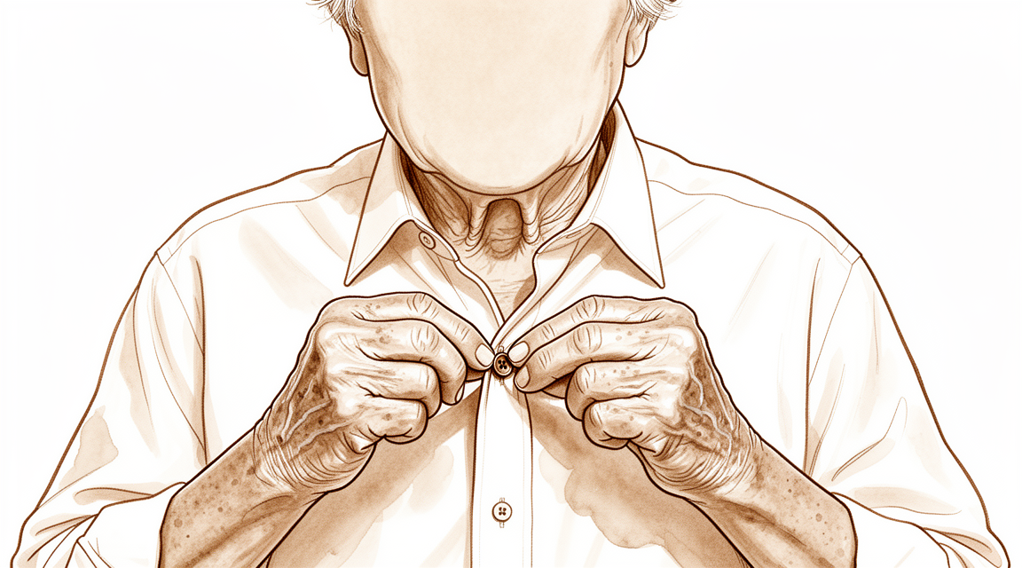
درد عموماً انگلی استعمال کرنے کے بعد بھڑک اٹھتا ہے، اور صبح جاگنے پر بھی تکلیف ہو سکتی ہے۔ وہ باریک کام مشکل ہو جاتے ہیں جن میں انگلی کا بالکل سرا استعمال ہوتا ہے: قمیض کے بٹن بند کرنا، چھوٹے سکے اٹھانا، سوئی میں دھاگا ڈالنا، یا تالا کھولنے کے لیے چابی پکڑ کر گھمانا۔ ہاتھ سے لکھنے میں بھی دکھن ہو سکتی ہے۔ وقت کے ساتھ جوڑ اکڑ سکتا ہے اور اس کے مڑنے کی صلاحیت کچھ کم ہو سکتی ہے، اور انگلی آہستہ آہستہ اپنی سیدھ سے ہٹ سکتی ہے۔
کچھ لوگوں کو ناخن کے پاس سیال سے بھری ایک چھوٹی گانٹھ نظر آتی ہے، جسے میوکس سسٹ (mucous cyst) کہتے ہیں۔ یہ سسٹ نیچے موجود آرتھرائٹس والے جوڑ سے بنتا ہے، اس لیے اس کا سیال نکالنے کے بعد بھی یہ اکثر دوبارہ بن جاتا ہے۔
اگر آرتھرائٹس انگلی کی کسی چوٹ کے بعد شروع ہوا ہو، جیسے میلٹ فنگر (انگلی کا سرا نیچے لٹک جانا) جس میں انگلی کو سیدھا کرنے والا ٹینڈن (tendon)، یعنی وہ مضبوط ڈوری جو پٹھے کو ہڈی سے جوڑتی ہے، پھٹ گیا ہو یا ہڈی کا ایک ٹکڑا اپنے ساتھ کھینچ کر الگ کر دیا ہو، تو جوڑ اس سے زیادہ تیزی سے گھس سکتا ہے جتنا ویسے گھستا۔ اس گھساؤ سے وہی درد اور اکڑاؤ ہوتا ہے، اور چوٹ خود ٹھیک ہو جانے کے باوجود انگلی کی حرکت کم ہو سکتی ہے۔
چند خطرے کی علامات پر نظر رکھیں۔ اگر آپ کی انگلی گرم، سرخ، سوجی ہوئی اور دردناک ہو جائے، خاص طور پر بخار کے ساتھ، تو اسی دن ہسپتال کی ایمرجنسی جائیں۔ یہ انفیکشن ہو سکتا ہے جس کا اسی دن علاج ضروری ہے، اور اس کے لیے پہلے فیملی ڈاکٹر (GP) سے ریفرل (referral) لینے کی ضرورت نہیں۔ اگر درد ہفتوں میں کم نہ ہو رہا ہو، بڑھ رہا ہو، یا آپ کو رات کو جگا دے تو اپنے فیملی ڈاکٹر کو دکھائیں یا ماہر ڈاکٹر (specialist) سے معائنے کا کہیں۔
دراصل کیا ہو رہا ہے
ہر انگلی چھوٹی ہڈیوں کی ایک زنجیر ہے۔ بالکل سرے والا جوڑ، جو ناخن کے ذرا پیچھے ہے، وہ جگہ ہے جہاں آخری ہڈی اپنے سے اوپر والی ہڈی سے ملتی ہے۔ دونوں ہڈیوں کے سروں پر ایک چکنی، پھسلنی تہہ چڑھی ہوتی ہے جو انہیں ایک دوسرے پر پھسلنے دیتی ہے۔ اسے ایک قدرتی جھٹکا جذب کرنے والی چیز سمجھیں جو سطح کو چکنا بھی رکھتی ہے۔
آرتھرائٹس میں یہ پھسلنی تہہ گھس کر پتلی ہو جاتی ہے یا ٹوٹ پھوٹ جاتی ہے۔ پھر ہڈیاں ایک دوسرے سے رگڑ کھاتی ہیں، اور جوڑ اس کے جواب میں اپنے کناروں کے گرد اضافی ہڈی بنا لیتا ہے۔ ہڈی کے یہی ابھار وہ سخت گانٹھیں ہیں جو آپ ناخن کے پاس محسوس کر سکتے ہیں، یعنی وہی جن کا ذکر اوپر والے حصے میں کیا گیا ہے۔ رگڑ سے دکھن ہوتی ہے، اور گھسی ہوئی سطحیں اور ابھار مل کر بتاتے ہیں کہ جوڑ کیوں سوجتا ہے، اکڑتا ہے اور آہستہ آہستہ اپنی سیدھ سے ہٹ جاتا ہے۔
جوڑ کو دونوں طرف بافتوں کی چھوٹی پٹیاں بھی تھامے رکھتی ہیں، اور ٹینڈن کے ریشوں کی ایک چپٹی پٹی بھی جو جوڑ کے اوپر سے گزرتی ہے اور جوڑ کے ذرا آگے جڑتی ہے۔ جب آرتھرائٹس جوڑ کو بدلتا ہے تو یہ پٹیاں اور ٹینڈن اپنا توازن کھو سکتے ہیں۔ یہ ایک وجہ ہے کہ انگلی درمیانی جوڑ پر ضرورت سے زیادہ مڑ سکتی ہے جبکہ اس کا سرا نیچے کی طرف مڑ جاتا ہے، اور شکل کی یہ تبدیلی عموماً آرتھرائٹس بڑھنے کے ساتھ پیدا ہوتی ہے۔
کبھی کبھی یہ گھساؤ صرف عام گھساؤ کے بجائے کسی چوٹ کے بعد ہوتا ہے۔ زور کی ضرب، کچلے جانے کی چوٹ، یا میلٹ فنگر جیسی ٹینڈن کی چوٹ جوڑ کی سطحوں کو نقصان پہنچا سکتی ہے۔ پھر جوڑ اس سے پہلے گھس جاتا ہے جتنا ویسے گھستا، اسی لیے درد اور اکڑاؤ کسی چوٹ کے بظاہر ٹھیک ہو جانے کے برسوں بعد ظاہر ہو سکتے ہیں۔
اس جوڑ کا آرتھرائٹس ناخن کے پہلو میں سیال سے بھری ایک چھوٹی گانٹھ بھی بنا سکتا ہے، یعنی وہی میوکس سسٹ جس کا ذکر پہلے ہوا۔ یہ سسٹ خود اسی جوڑ سے نکلتا ہے جس میں جلن (irritation) ہو رہی ہو، اسی لیے سیال نکالنے کے بعد یہ اکثر دوبارہ بن جاتا ہے۔
اچھی خبر یہ ہے کہ یہ جوڑ بھاری گرفت کا بہت کم کام کرتا ہے۔ اس کا کام انگلی کے سرے پر باریک کنٹرول ہے، اسی لیے علاج کا مقصد درد کو کم کرنا اور اس باریک کنٹرول کو کام کرتے رکھنا ہوتا ہے۔
ہم اس کے لیے کیا کر سکتے ہیں
ڈاکٹر کیرن ہِرپرا، جو Mater Private Hospital Rockhampton میں بازو اور ہاتھ کے سرجن (upper-limb surgeon) ہیں، آپ کی حالت کے مطابق سب سے کم چیر پھاڑ والے طریقوں سے علاج شروع کرتے ہیں۔ مریضوں کو عام طور پر ان کے فیملی ڈاکٹر (GP) ہمارے کلینک ریفر کرتے ہیں؛ اگر کسی فزیوتھراپسٹ نے آپ کو ہم سے ملنے کا مشورہ دیا ہے، تب بھی میڈیکیئر (Medicare) ری بیٹ کا اہل ہونے کے لیے آپ کو اپنے فیملی ڈاکٹر سے ریفرل (referral) لینا ہوگا۔ آپ کی پہلی ملاقات پر ہم آپ کی طبی تاریخ لیتے ہیں، آپ کی انگلی کا معائنہ کرتے ہیں اور اگر ضرورت ہو تو ایکسرے یا اسکین کرواتے ہیں۔ چونکہ یہ گھساؤ کا ایک پرانا مسئلہ ہے، اس لیے ہم عام طور پر پہلے آپریشن کے بغیر علاج آزماتے ہیں، اور سرجری پر بات صرف اس وقت کرتے ہیں جب اس سے خاطر خواہ بہتری نہ آئی ہو۔
پہلا قدم اکثر ایک چھوٹا اسپلنٹ (splint، انگلی کو سہارا دینے والی پٹی) ہوتا ہے جو انگلی کے سرے والے جوڑ کو سیدھا رکھتا ہے۔ اسپلنٹ لگانے سے جوڑ کا درد کم ہوتا ہے اور انگلی کو سیدھا ہونے میں مدد ملتی ہے۔ یہ آسان اور محفوظ ہے، اور آپ اسے اپنے معمول کے دن بھر پہن سکتے ہیں۔ ہم عام طور پر اس کے ساتھ سوزش کم کرنے والی دوا دیتے ہیں، یعنی ایسی گولیاں جو سوجن اور درد کو کم کرتی ہیں۔ ہینڈ تھراپی (ہاتھ کی بحالی کے ماہر کا علاج) بھی جلن کم ہونے کے دوران انگلی کے سرے کو باریک کاموں کے لیے کارآمد رکھنے میں مدد کر سکتی ہے۔ مزید کوئی فیصلہ کرنے سے پہلے ان اقدامات کو پورا موقع دیں۔
اگر اس کے بعد بھی درد اور اکڑاؤ آپ کو محدود رکھیں تو سرجری ایک اختیار بن جاتی ہے۔ معیاری آپریشن فیوژن (دو ہڈیوں کو جوڑ کر ایک کر دینا) ہے، جسے آرتھروڈیسس (arthrodesis) بھی کہتے ہیں، جس میں جوڑ کی گھسی ہوئی سطحیں نکال دی جاتی ہیں اور دونوں ہڈیوں کو اس طرح جوڑا جاتا ہے کہ وہ جڑ کر ایک ہو جائیں۔ اس کے بعد جوڑ مڑتا نہیں، لیکن وہ بغیر درد کے اور مستحکم ہوتا ہے، اور انگلی کو ایسی حالت میں جمایا جاتا ہے جو روزمرہ کے کاموں کے لیے اچھی طرح کام کرے۔ فیوژن اپنی سیدھ سے ہٹی ہوئی انگلی کو بھی سیدھا کرتا ہے، اور یہی وہ علاج ہے جو میوکس سسٹ کو دوبارہ بننے سے روکتا ہے۔ اگر انگلی کے سرے میں کچھ حرکت برقرار رکھنا آپ کے لیے درد کو مکمل طور پر ختم کرنے سے زیادہ اہم ہو تو ایک اور آپریشن بھی ہے جس میں ہڈی کی گانٹھیں تراش دی جاتی ہیں اور جوڑ کو اندر سے صاف کیا جاتا ہے، جبکہ جوڑ حرکت کے قابل رہتا ہے۔ ہم آپ کے ساتھ بات کریں گے کہ کون سا اختیار آپ کی انگلی کے لیے اور آپ اپنے ہاتھ سے جو کام لینا چاہتے ہیں اس کے لیے مناسب ہے، اور آپ کے ساتھ مل کر فیصلہ کریں گے۔
کیا توقع رکھیں
انگلی کے سرے والے جوڑ کا آرتھرائٹس عموماً برسوں میں آہستہ آہستہ بڑھتا ہے۔ درد آتا جاتا رہتا ہے: انگلی استعمال کرنے کے بعد بھڑک اٹھتا ہے، پھر آرام سے دوبارہ کم ہو جاتا ہے۔ علاج کے بغیر جوڑ اکثر مزید اکڑ جاتا ہے اور وقت کے ساتھ انگلی اپنی سیدھ سے مزید ہٹ سکتی ہے۔ کچھ لوگوں کو لگتا ہے کہ جوڑ کے اکڑنے کے ساتھ درد خود ہی کم ہو جاتا ہے، لیکن بہت سے لوگوں میں یہ تکلیف دیتا رہتا ہے، خاص طور پر قمیض کے بٹن بند کرنے یا چابی گھمانے جیسے باریک کاموں میں۔
زیادہ تر لوگوں کو پہلے بیان کیے گئے سادہ اقدامات سے اچھا آرام ملتا ہے: اسپلنٹ، سوزش کم کرنے والی دوا اور ہینڈ تھراپی۔ یہ درد کو کم کرتے ہیں جبکہ آپ روزمرہ کے کاموں کے لیے انگلی استعمال کرتے رہتے ہیں۔ اگر پورا موقع دینے کے بعد بھی ان اقدامات سے فائدہ نہ ہو تو سرجری عموماً درد کو اچھی طرح کم کر دیتی ہے۔ فیوژن کیا ہوا انگلی کے سرے والا جوڑ بغیر درد کے اور مستحکم ہوتا ہے، اور لوگ عموماً اپنے ہاتھ کا معمول کا استعمال دوبارہ شروع کر دیتے ہیں، اگرچہ سرا اب مڑتا نہیں۔ اگر کچھ حرکت برقرار رکھنا آپ کے لیے زیادہ اہم ہو تو جوڑ کی صفائی والا آپریشن انگلی کو مڑنے کے قابل رہنے دیتا ہے، اور اس سے درد میں آرام اچھا ملتا ہے لیکن ہمیشہ مکمل نہیں۔
سرجری کے بعد صحت یابی میں دن نہیں بلکہ ہفتوں سے مہینوں تک کا وقت لگتا ہے۔ شروع میں ہڈیوں کے جڑنے تک انگلی کو ساکن رکھا جاتا ہے، اور جب آپ اپنی گرفت اور باریک کنٹرول دوبارہ بحال کریں گے تو آپ کے ہینڈ تھراپسٹ آپ کی رہنمائی کریں گے۔ زیادہ تر لوگوں کو آخر میں ایسی انگلی ملتی ہے جو دکھن کے بغیر اپنا کام کرتی ہے، اگرچہ اس کا سرا پہلے سے زیادہ سیدھا رہتا ہے۔
چند باتیں صاف صاف بتانا ضروری ہے۔ فیوژن ہمیشہ پہلی کوشش میں نہیں جڑتا، اور کچھ لوگوں کو دھات نکالنے یا فیوژن دوبارہ کرنے کے لیے مزید آپریشن کی ضرورت پڑتی ہے۔ تمباکو نوشی ہڈی کے جڑنے کو سست کرتی ہے، اس لیے سرجری سے پہلے اسے چھوڑ دینا آپ کی انگلی کو اچھی طرح جڑنے کا بہترین موقع دیتا ہے۔ اگر درد ہفتوں میں کم نہ ہو رہا ہو، بڑھ رہا ہو، یا آپ کو رات کو جگا دے تو اپنے فیملی ڈاکٹر کو دکھائیں یا ماہر ڈاکٹر سے معائنے کا کہیں۔
ڈاکٹر کو کب دکھائیں
انگلی کے سرے والے جوڑ کا زیادہ تر آرتھرائٹس آہستہ آہستہ بڑھتا ہے، اس لیے شروع کرنے کے لیے فیملی ڈاکٹر کے پاس معمول کی ملاقات کافی ہے۔ اگر درد ہفتوں میں کم نہ ہو رہا ہو، بڑھ رہا ہو، یا آپ کو رات کو جگا دے تو اپنے فیملی ڈاکٹر کو دکھائیں۔ اگر انگلی کی وجہ سے آپ کام نہ کر سکیں یا قمیض کے بٹن بند کرنے جیسے باریک کام نہ کر سکیں تو بھی یہی کریں۔
کچھ علامات میں جلد علاج کی ضرورت ہوتی ہے۔ اگر آپ کی انگلی گرم، سرخ، سوجی ہوئی اور دردناک ہو جائے، خاص طور پر بخار کے ساتھ، تو اسی دن ہسپتال کی ایمرجنسی جائیں۔ یہ صورت انفیکشن کی طرف اشارہ کرتی ہے، اور اس کے لیے پہلے فیملی ڈاکٹر سے ریفرل لینے کی ضرورت نہیں۔
اگر چوٹ کے بعد جوڑ کے اوپر جلد پھٹ گئی ہو، ہڈی نظر آ رہی ہو، یا انگلی کی شکل واضح طور پر بگڑی ہوئی دکھائی دے تو ہسپتال کی ایمرجنسی جائیں۔ اگر انگلی سفید پڑ جائے، ٹھنڈی ہو جائے، یا اس کا رنگ سفید یا نیلا ہو جائے، یا کسی ضرب کے بعد اچانک اس میں احساس یا حرکت ختم ہو جائے تو بھی یہی کریں۔
بار بار واپس آنے والا میوکس سسٹ، یا اپنی سیدھ سے ہٹتی ہوئی انگلی، ماہر ڈاکٹر سے معائنے کے لائق ہے۔
مزید تفصیل
Advanced reading: the deeper science (optional)
یہ حصہ آپ کے اپنے علاج کے فیصلوں کے لیے ضروری معلومات سے آگے جاتا ہے۔ انگلی کے سرے والے جوڑ کے آرتھرائٹس کے بارے میں یہ اضافی مطالعہ اس لیے فائدہ مند ہے کہ فیوژن، یعنی معیاری آپریشن جسے عموماً سیدھا سادہ بتایا جاتا ہے، میں پیچیدگیوں کی شرح اس کی شہرت سے زیادہ ہے، اور اس لیے بھی کہ حرکت برقرار رکھنے والا ایک متبادل موجود ہے جس کا ذکر شاذ و نادر ہی ہوتا ہے۔
فیوژن اتنا بے مسئلہ نہیں جتنا سننے میں لگتا ہے
انگلی کے سرے والے جوڑ کے فیوژن کو ایک قابلِ اعتماد آپریشن کے طور پر پیش کیا جاتا ہے، اور درد سے نجات کے لحاظ سے یہ واقعی ہے۔ لیکن اس کی پیچیدگیوں کی تصویر اتنی بے ضرر نہیں۔ 173 مریضوں میں خطرے کے عوامل کا جائزہ یہ بات صاف کہتا ہے: انگلی کے سرے والے جوڑ (distal interphalangeal joint) کے فیوژن (arthrodesis) سے اکثر پیچیدگیاں پیدا ہوتی ہیں، اور جوڑ کے گھسنے والا آرتھرائٹس، دوبارہ فیوژن اور تمباکو نوشی کو خطرے کے عوامل کے طور پر شناخت کیا گیا [1]۔
اس فہرست میں تمباکو نوشی کا ہونا عمل کرنے کے لائق بات ہے، کیونکہ یہ وہ واحد عامل ہے جو مریض کے اپنے اختیار میں ہے۔ ہڈی کا جڑنا خون کی فراہمی پر منحصر ہے، اور یہ ایک چھوٹا جوڑ ہے جس کے گرد نرم بافتوں کی پتلی تہہ ہے اور جو انگلی کی خون کی فراہمی کے بالکل آخری سرے پر واقع ہے۔
جو امپلانٹ ہڈی کو بہتر جوڑتا ہے وہ ایسے مسائل بھی پیدا کرتا ہے جو سستے طریقوں میں نہیں ہوتے
جوڑنے کے طریقے کی بحث کا جواب غیر معمولی طور پر واضح ہے، اور وہ کسی ایک کی جیت نہیں بلکہ ایک سودا ہے۔ 1,125 مریضوں میں، بغیر سر والے کمپریشن پیچ (headless compression screws) سے ہڈی جڑنے (union) کی شرح بڑھتی دکھائی دیتی ہے لیکن ان سے ایسی پیچیدگیاں وابستہ ہیں جو دوسرے مستند اور سستے طریقوں میں نہیں دیکھی جاتیں، اور ہڈی جڑنے کے علاوہ یہ دکھانے کے لیے ناکافی شواہد ہیں کہ پیچ بہتر ہے [2]۔
پیچ سے مخصوص پیچیدگیاں جسم کی بناوٹ سے پیدا ہوتی ہیں۔ پیچ انگلی کے سرے کی لمبائی کے محور میں چلتا ہے، اس لیے یہ ناخن کے نیچے کی جلد (nail bed) کے قریب سے گزرتا ہے اور ناخن کی شکل بگاڑ سکتا ہے، اور انگلی کے سرے کی بہت چھوٹی ہڈی (distal phalanx) میں اسے تھامنے کے لیے کافی ہڈی نہیں ہوتی۔ تار سستے ہیں اور ان خاص مسائل سے بچاتے ہیں، جبکہ ان سے ہڈی کا جڑنا تھوڑا کم یقینی ہوتا ہے۔
جہاں صرف ہڈی کا جڑنا ترجیح ہو، دوبارہ فیوژن ہو، یا مریض تمباکو نوشی کرتا ہو، وہاں پیچ کا فائدہ سب سے زیادہ اہم ہے۔ جہاں ہڈی چھوٹی ہو اور ناخن اہم ہو، وہاں یہ اتنا واضح طور پر درست انتخاب نہیں۔
حرکت برقرار رکھنے والا متبادل
فیوژن واحد اختیار نہیں، اور اس کے متبادل پر زیادہ بات نہیں ہوتی۔ جن مریضوں کی بنیادی شکایت پورے جوڑ کا آرتھرائٹس نہیں بلکہ ہڈی کی گانٹھیں اور ان سے ہونے والا درد ہو، ان میں ابھرے ہوئے ہڈی کے ابھار (osteophytes) نکالے جا سکتے ہیں اور جوڑ کو اپنی جگہ برقرار رکھا جا سکتا ہے۔
انگلی کے سرے والے جوڑ کی کھلی کائلیکٹومی (cheilectomy، جوڑ کے کناروں سے بڑھی ہوئی ہڈی کو تراشنا) کو ان مریضوں میں فیوژن کا محفوظ اور مؤثر متبادل بتایا گیا ہے جنہیں جوڑ کے گھسنے والے آرتھرائٹس کی علامات ہوں اور جو جوڑ کی حرکت برقرار رکھنا چاہتے ہوں، اور یہ نتیجہ 78 مریضوں پر مبنی ہے [3]۔
یہ اس لیے اہم ہے کہ انگلی کے سرے والا جوڑ گرفت کی طاقت میں کم حصہ ڈالتا ہے لیکن باریک کاموں اور ہاتھ کی ظاہری شکل میں کافی حصہ ڈالتا ہے۔ جس شخص کی علامات کی اصل وجہ خود ہیبرڈن نوڈز (Heberden's nodes، انگلی کے سرے والے جوڑ پر ہڈی کی گانٹھیں) ہوں، اس کے لیے انہیں ہمیشہ کے لیے اکڑے ہوئے انگلی کے سرے سے بدلنا سننے سے کہیں بڑی قربانی ہے، اور درمیان کا ایک راستہ موجود ہے۔
سسٹ جو اکثر اس کے ساتھ ہوتا ہے
اس جوڑ کا آرتھرائٹس اکثر میوکس سسٹ پیدا کرتا ہے، یعنی آرتھرائٹس والے جوڑ سے نکلنے والی سیال سے بھری ایک چھوٹی سوجن، جو عموماً ناخن کے پہلو میں ہوتی ہے۔ چونکہ اس کی وجہ نیچے کا جوڑ ہے، اس لحاظ سے یہ کلائی کے گینگلیئن سسٹ کی طرح برتاؤ کرتا ہے: اس کا سیال نکالنے سے سوجن کا علاج ہوتا ہے، اس کی جڑ کا نہیں۔ اس پر الگ سے بات کی گئی ہے، لیکن یہ تعلق جاننے کے لائق ہے، کیونکہ بار بار لوٹنے والا سسٹ کوئی الگ تھلگ مسئلہ نہیں بلکہ نیچے موجود آرتھرائٹس کی نشانی ہے۔
حوالہ جات
[1] Runkel A, Bonaventura B, Sundermann B, Zajonc H, Eisenhardt S, Leibig N. Risk factors in distal interphalangeal joint arthrodesis in the hand: a retrospective study. J Hand Surg Eur Vol. 2022;47(9):907-14. https://doi.org/10.1177/17531934221111641
[2] Dickson D, Mehta S, Nuttall D, Ng C. A systematic review of distal interphalangeal joint arthrodesis. J Hand Microsurg. 2014;6(2):74-84. https://doi.org/10.1007/s12593-014-0163-1
[3] Lin EA, Papatheodorou LK, Sotereanos DG. Cheilectomy for treatment of symptomatic distal interphalangeal joint osteoarthritis: a review of 78 cases. J Hand Surg Am. 2017;42(11):889-93. https://doi.org/10.1016/j.jhsa.2017.07.006
Evidence & references
This is the clinical evidence summary written for health professionals. It is technical, and it lists the research this page was built from. You do not need to read it to understand your treatment or to make a decision about it.
Overview
- Osteoarthritis is the most common indication for distal interphalangeal joint arthrodesis [19].
- In a cohort of 149 cases, postoperative complications for distal interphalangeal and thumb interphalangeal joint arthrodesis occurred at a rate similar to that reported in existing literature [19].
- Finger distal interphalangeal and thumb interphalangeal joint arthrodesis generally results in favorable outcomes in terms of bony union regardless of underlying medical condition or technical details of the surgical operation [6].
- The X-fuse implant provides excellent stability with minimal hardware problems and a high rate of union in patients with finger distal interphalangeal and thumb interphalangeal joint arthritis [1].
- In select patients, percutaneous distal interphalangeal joint arthrodesis is advantageous in comparison with open fusion techniques [2].
- Reamed percutaneous distal interphalangeal joint arthrodesis is associated with higher union rates achieved in a shorter time and reduced postoperative pain compared with the non-reamed percutaneous technique [8].
- Successful fusions of the distal interphalangeal joint at an angle up to 35 degrees can be achieved using small diameter headless compression screws [14].
- The Mini-Acutrak screw is suitable for distal interphalangeal joint fusion in all fingers with the exception of the small finger [22].
- The nonaxial multiple small screws technique is a feasible option for distal interphalangeal and thumb interphalangeal joint arthrodesis, especially when a small finger is indicated and a significant flexion angle is required [24].
- Given the lack of difference in biomechanical performance between K-wires and compression screws, consideration should be given to other factors such as cost and complication profiles when choosing an implant for distal interphalangeal joint fusion [34].
- The smile incision and reverse shotgun approach may be a good surgical option for distal interphalangeal joint arthrodesis when more volar part joint preparation and more volar implant insertion sites are necessary [17].
- Results from a lateral approach and plate fixation for distal interphalangeal joint arthrodesis are equivalent to traditional methods but with fewer major complications [11].
- For persistently symptomatic, unstable distal interphalangeal joints, arthrodesis should be considered [4].
- In the distal interphalangeal joint, there is probably good reason to proceed to arthrodesis immediately for synovial chondromatosis [3].
- The swan neck deformity can progress significantly with time because of increasing distal interphalangeal joint flexion contracture [5].
- Silicone interpositional arthroplasty of the distal interphalangeal joint is an acceptable alternative to arthrodesis, achieving excellent pain relief and a range of movement of 30–40 degrees with a low overall complication rate of 5% [13].
- Swanson hinge implant arthroplasty can be the surgical treatment of choice for the patient who desires continued mobility, albeit limited, of the involved osteoarthritic distal interphalangeal joint [9].
- Open distal interphalangeal joint cheilectomy is a safe and effective alternative to distal interphalangeal joint arthrodesis in patients with symptomatic osteoarthritis who wish to preserve joint motion [28].
- Denervation with cheilectomy of the distal interphalangeal joint presents a compelling motion-preserving alternative to arthrodesis for symptomatic distal interphalangeal joint osteoarthritis [10].
- A surgical technique for treating symptomatic distal interphalangeal joint arthritis reduces pain while preserving distal interphalangeal joint motion [18].
- The authors recommend simultaneous surgical intervention in case of severe painful osteoarthritis of the proximal interphalangeal and distal interphalangeal joints of the same digit [7].
- The combination of distal interphalangeal arthrodesis and proximal interphalangeal Swanson arthroplasty resulted in a favorable outcome in terms of simultaneous bony union and flexibility [12].
- Combined distal interphalangeal arthrodesis and proximal interphalangeal procedures present unique challenges regarding hardware conflict, where K-wires offer the easiest compatibility and headless screws must ideally not reach proximal to the midpoint of the middle phalanx [90].
- A customized structural bone graft addresses issues of bone stock loss and medullary absence in failed distal interphalangeal joint silicone arthroplasty, achieving reliable union rates and high patient satisfaction [21].
Anatomy & Pathophysiology
Bony Anatomy & Morphology
- The skeleton of the hand consists of 27 bones, of which 19 are long bones [47].
- The skeleton is divided into five rays, each ray making up a polyarticulated chain comprising the metacarpals and phalanges [47].
- The thumb ray is made up of only three bones—a metacarpal and two phalanges [47].
- The other four digital rays are formed by four skeletal segments—a metacarpal and three phalanges [47].
- The thumb metacarpal is the shortest, and the index metacarpal is by far the longest [47].
- The proximal and middle phalanges of the long and ring fingers are longer than those of the index finger [47].
- The epiphyseal plates are located at the proximal ends of the phalanges and the first metacarpal [47].
- An examination of 100 specimens of the terminal joints of human fingers was described in a 1982 study [75].
- A substantial number of distal phalanges are too small to accommodate commonly available headless compression screws, particularly in females and the small finger [39].
- Understanding of DIP joint morphology may lend insight into the biomechanics and disease progression within the DIP joints [23].
Soft Tissue Anatomy & Extensor Mechanism
- Distal interphalangeal joint extension is achieved through the conjoined lateral bands that are composed of tendinous slips from the extrinsic and intrinsic tendons [46].
- The principal bony insertion of the extrinsic digital extensors is on the dorsal proximal aspect of the middle phalanx [46].
- The deep head of each dorsal interosseous muscle forms a lateral tendon, or lateral band, at the level of the MP joint [59].
- The lateral bands are joined by the lateral slips of the extensor tendon to form the conjoined lateral band [59].
- The two conjoined lateral bands to each finger unite at the distal third of the middle phalanx to form the terminal tendon [59].
- The terminal tendon inserts at the base of the distal phalanx to extend it [59].
- The flexor digitorum profundus inserts on the proximal volar aspect of the distal phalanx, flexing the distal interphalangeal joint as well as the proximal interphalangeal and metacarpophalangeal joints [46].
- The flexor digitorum profundus provides digital flexion at both the proximal and distal interphalangeal joints [66].
- The A5 annular pulley is located over the distal interphalangeal joint [66].
- The vinculum breve of the flexor digitorum profundus and check-rein ligaments may account for the greater amount of passive hyperextension that can be achieved at the distal than the proximal interphalangeal joint [80].
Biomechanics & Kinematics
- The wrist influences the position of the metacarpophalangeal joint; the metacarpophalangeal joint also affects the position of the proximal interphalangeal joint, which in turn affects the distal interphalangeal joint [67].
- Almost all movements in the hand are around oblique and variable axes, resulting in combined movements permitting optimal orientation of the phalanges at the time of prehension [67].
- The axes of flexion are so arranged that flexion of all the metacarpophalangeal and proximal interphalangeal joints causes the fingers to converge toward the scaphoid [70].
- In a combinatorial relationship, intrinsic muscles produce steep inclination of extensor forces at the DIP joint [73].
- There were no significant differences in comparisons among loads (200, 400, 600, and 800 g) regarding the inclination slope of intrinsic extensor forces at the DIP joint [73].
- Immobilization of the distal interphalangeal joint of any finger reduces the overall grip strength of the hand, with the effect becoming progressively more pronounced from the index to the little fingers [37].
- Positioning the middle finger DIP joint in either extension or 20° of flexion did not significantly affect grip strength or dexterity [49].
- Biomechanically, dynamic tenodesis for the DIP joint using the remaining FDP tendon results in a flexion angle greater than 30 degrees [76].
Pathophysiology & Etiology
- Osteoarthritis occurs with the highest prevalence in the distal interphalangeal joint of the hand and has been divided into an erosive and a nonerosive form [26].
- The pathogenesis of the early stages of osteoarthritis is poorly understood, but considerable emphasis has been placed on the role of cartilage and subchondral bone as well as soft tissue structures such as collateral ligaments and tendons [26].
- Osteoarthritis is a complex disease resulting in the failure of articular cartilage due to a combination of genetic, metabolic, biochemical, and biomechanical factors [26].
- Cartilage loss and reactive new bone formation at joint margins is accompanied by the proliferation of osteoarticular tissue in the capsule [26].
- Heberden's nodes are more common in women who engage in cooking in school and the incidence increases with the number of tasks involved [25].
- Cooking as an occupation is an aetiological factor in the pathogenesis of Heberden's nodes [25].
- Work-load, sports, and repeated minimal trauma contribute to the development of Heberden's nodes [25].
- Repetitive injuries to a joint lead to an increased risk of developing posttraumatic arthritis [48].
- Athletes have a higher incidence of degenerative joint changes compared to the general population [48].
- Mallet finger is caused by forced flexion of an extended fingertip [51].
- Less common hyperextension or hyperextension/axial loading tends to cause the larger fractures and subluxation in mallet injuries [51].
- One-third of all mallet fingers are associated with a fracture [51].
- Mallet fractures associated with large fragments may result in volar subluxation of the distal phalanx as the collateral ligaments remain attached to the fracture fragment [51].
- Fracture fragments that are less than 43% of the distal phalanx articular surface usually do not allow volar subluxation [51].
- Fracture fragments involving larger than 52% of the joint surface consistently allow subluxation [51].
- Palmar subluxation of a DIP joint without preexisting arthritic deformity is expected when more than one half of the dorsal articular surface is injured [20].
- The mechanism of displaced intra-articular osteochondral fracture causing irreducible dislocation of the DIP joint probably involved hyperextension of the DIP joint [15].
- The palmar plate was torn at the attachment to the distal phalanx in cases of displaced intra-articular osteochondral fracture [15].
- Attempts to perform a closed reduction of displaced intra-articular osteochondral fractures can lead to further displacement of the fracture, with folding of the fragment on the articular cartilage hinge [15].
- The cause of irreducible palmar dislocation of the DIP joint can be an entrapment of the extensor tendon in front of the head of the middle phalanx [41].
- Irreducibility was more commonly seen in dorsal than in volar dislocations of the DIP joint [85].
- Volar dislocations of the DIP joint carried a higher risk of instability immediately after reduction compared to dorsal dislocations [85].
- Some loss of motion in small joints of the fingers after hyperflexion injuries would be expected [79].
Classification
- Osteoarthritis of the distal interphalangeal joint has been divided into an erosive and a nonerosive form [26].
- Radiographic evaluation represents the most standardized method to quantify disease progression in distal interphalangeal joint osteoarthritis, with different systems developed for defining and grading radiographic features [26].
- The interrater reliability of the Kellgren & Lawrence and OARSI classification systems for post-traumatic osteoarthritis in the distal interphalangeal joint after mallet finger fractures is considerably lower than initially assumed [86].
Clinical Presentation
- Osteoarthritis occurs with the highest prevalence in the distal interphalangeal joint of the hand [26].
- Heberden's nodes are more common in women who engage in cooking in school, with incidence increasing with the number of tasks involved [25].
- There is no confirmed difference in the incidence of disease between right and left hands in elementary school cooks [25].
- Radiographic evaluation represents the most standardized method to quantify disease progression in distal interphalangeal joint osteoarthritis [26].
- The pathogenesis of the early stages of distal interphalangeal joint osteoarthritis is poorly understood [26].
- Considerable emphasis has been placed on the role of cartilage and subchondral bone in the pathogenesis of distal interphalangeal joint osteoarthritis [26].
- Soft tissue structures such as collateral ligaments and tendons play a role in the pathogenesis of distal interphalangeal joint osteoarthritis [26].
- Cartilage loss and reactive new bone formation at joint margins are accompanied by the proliferation of osteoarticular tissue in the capsule [26].
- Radiological osteoarthritis after a mallet finger fracture is similar to the natural degenerative process in the distal interphalangeal joint [27].
- Radiological osteoarthritis after a mallet finger fracture is accompanied by a decrease in range of motion of the distal interphalangeal joint [27].
- The decrease in range of motion following mallet finger fracture does not clinically affect patient-reported outcomes [27].
- Palmar subluxation of a distal interphalangeal joint without preexisting arthritic deformity is expected when more than one half of the dorsal articular surface is injured [20].
- Primary synovial chondromatosis of the distal interphalangeal joint is an extremely rare entity that requires accurate diagnosis to distinguish from other arthropathies [50].
- Adnexal tumours may masquerade as a distal interphalangeal joint ganglion and should enter the differential diagnosis when examining tumours of the digits [89].
- Floating distal interphalangeal joint injuries can be misdiagnosed initially due to minimal deformity [33].
- Osteoarthritis may develop following floating distal interphalangeal joint injuries [33].
- The mechanism of displaced intra-articular osteochondral fracture causing irreducible dislocation of the distal interphalangeal joint probably involved hyperextension of the joint [15].
- The palmar plate is torn at the attachment to the distal phalanx in displaced intra-articular osteochondral fractures of the distal interphalangeal joint [15].
- Attempts to perform a closed reduction of a displaced intra-articular osteochondral fracture can lead to further displacement of the fracture with folding of the fragment on the articular cartilage hinge [15].
- The cause of irreducibility in palmar dislocation of the distal interphalangeal joint can be entrapment of the extensor tendon in front of the head of the middle phalanx [41].
- Interrater reliability for measurements of the distal interphalangeal joint motions of the index and long digits is high [16].
- Interrater reliability for distal interphalangeal joint motion measurements is slightly higher for the dorsal method of placement than for the lateral method [16].
Investigations
- Radiographic evaluation is the most standardized method to quantify disease progression in distal interphalangeal joint osteoarthritis [26].
- Different systems have been developed for defining and grading radiographic features of distal interphalangeal joint osteoarthritis [26].
- Interrater reliability was high for measurements of the proximal and distal interphalangeal joint motions of the index and long digits [16].
- Interrater reliability was slightly higher for the dorsal method of goniometer placement than for the lateral method [16].
- Primary synovial chondromatosis of the distal interphalangeal joint requires accurate diagnosis to distinguish from other arthropathies [50].
- A distinct collagen septum exists between the extensor tendon and skin at the distal interphalangeal joint [53].
- The existence of a distinct collagen septum between the extensor tendon and skin at the distal interphalangeal joint was confirmed using MRI and histology [53].
Treatment
Non-Operative
- Splinting of the distal interphalangeal joint reduces pain and improves extension at the joint [29].
- Splinting for DIP joint arthritis does not give rise to non-compliance, increased stiffness, or restriction of range of motion [29].
- Splinting and anti-inflammatory medications are the mainstay of nonoperative treatment for DIP joint arthritis [48].
- Injection with Collagenase Clostridium histolyticum is an option for the treatment of DIP joint contractures in Dupuytren disease, though the potential risk for recurrence should be carefully weighed prior to its use [93].
Operative: Arthrodesis
- Arthrodesis is generally the most accepted surgical option for treatment of degenerative and traumatic conditions involving the distal interphalangeal joint of the fingers or the interphalangeal joint of the thumb [38].
- The ideal position of DIP arthrodesis is thought to be slightly flexed to improve power, fine pinch, and grip [38].
- In select patients, percutaneous DIP joint arthrodesis is advantageous in comparison with open fusion techniques [2].
- Reamed percutaneous DIPJ arthrodesis is associated with higher union rates achieved in a shorter time and reduced postoperative pain compared with the non-reamed percutaneous technique [8].
- The X-fuse implant provides excellent stability with minimal hardware problems and a high rate of union in patients with finger DIPJ and thumb IPJ arthritis [1].
- Finger DIP and thumb IP joint arthrodesis generally resulted in favourable outcome in terms of bony union regardless of the underlying medical condition or technical details of the surgical operation [6].
- Osteoarthritis was the most common indication for DIP and thumb IP arthrodesis, and postoperative complications occurred at a rate similar to that reported in existing literature [19].
- Successful fusions of the DIP joints at an angle up to 35 degrees were achieved using small diameter headless compression screws, providing benefits including early mobilization and favourable functional outcome scores [14].
- The Mini-Acutrak screw is suitable for DIPJ fusion in all fingers with the exception of the small finger [22].
- The nonaxial multiple small screws technique could be used as a feasible option in DIPJ and thumb IPJ arthrodesis, especially when a small finger is indicated and a significant flexion angle is required [24].
- Given the lack of difference in biomechanical performance between K-wires and compression screws, consideration should be given to other factors such as cost and complication profiles when choosing an implant for DIPJ fusion [34].
- DIP joint preparation is not necessary before performing arthrodesis in fingers with stage IV chondropathy [44].
- The use of a buried break-away compression screw avoids complications related to bulky hardware in DIP arthrodesis [44].
- Dorsal plate fixation allows for the performance of a distal finger joint arthrodesis in slight flexion [38].
- The smile incision and reverse shotgun approach may be a good surgical option for DIPJ arthrodesis when more volar part joint preparation and more volar implant insertion sites are necessary [17].
- Lateral approach and plate fixation for DIP joint arthrodesis yields results equivalent to traditional methods but with fewer major complications [11].
- For persistently symptomatic, unstable DIP joints, arthrodesis should be considered [4].
- In the DIP joint, there is probably good reason to proceed to arthrodesis immediately in cases of synovial chondromatosis [3].
- A customized structural bone graft addresses issues of bone stock loss and medullary absence in failed DIPJ silicone arthroplasty, achieving reliable union rates and high patient satisfaction [21].
- All joints had fused radiologically in a series of proximodistal interphalangeal arthrodesis of the little finger [32].
- Arthrodesis of the DIP joint provides stability and pain relief and can correct deformity [48].
- A painless, stable, distal interphalangeal joint that is in a functional position is more important than the ability to move the joint [48].
- The authors recommend simultaneous surgical intervention in case of severe painful OA of the PIP and DIP joints of the same digit [7].
- The combination of DIP arthrodesis and PIP Swanson arthroplasty resulted in a favourable outcome in terms of simultaneous bony union and flexibility [12].
- For patients prioritizing hand aesthetics or with unstable joints, distal interphalangeal joint arthrodesis is preferable [31].
Operative: Arthroplasty and Motion-Preserving
- Silicone interpositional arthroplasty of the DIP joint is an acceptable alternative to arthrodesis, achieving excellent pain relief and a range of movement of 30–40 degrees with a low overall complication rate of 5% [13].
- Swanson hinge implant arthroplasty can be the surgical treatment of choice for the patient who desires continued mobility, albeit limited, of the involved osteoarthritic DIP joint [9].
- Open DIP joint cheilectomy is a safe and effective alternative to DIP joint arthrodesis in patients with symptomatic osteoarthritis who wish to preserve joint motion [28].
- Denervation with cheilectomy of the distal interphalangeal joint presents a compelling motion-preserving alternative to arthrodesis for symptomatic DIP joint osteoarthritis [10].
- A surgical technique for DIP joint arthritis reduces pain while preserving DIP joint motion [18].
- Volar plate arthroplasty of the distal interphalangeal joint allows for a painless functional arc of motion without residual joint subluxation [40].
Complications
- The X-fuse implant provides excellent stability with minimal hardware problems [1].
- The X-fuse implant has a high rate of union in patients with finger DIPJ and thumb IPJ arthritis [1].
- Reamed percutaneous DIPJ arthrodesis is associated with reduced postoperative pain compared with the non-reamed percutaneous technique [8].
- The swan neck deformity progressed significantly with time because of increasing DIPJ flexion contracture [5].
- Postoperative complications occurred at a rate similar to that reported in the existing literature in a retrospective cohort of 149 cases of DIP and thumb IP joint arthrodesis [19].
- Arthrodesis of the distal interphalangeal joint often leads to complications [30].
- Silicone interpositional arthroplasty of the DIP joint has a low overall complication rate of 5% [13].
- Bony union of the joint was delayed in a series using the Herbert screw for DIPJ arthrodesis [56].
- Follow-up to 5 years of DIP joint flexible implant arthroplasty showed only one complication [92].
Recovery
Arthrodesis Outcomes and Complications
- Postoperative complications in DIP and thumb IP joint arthrodesis occurred at a rate similar to that reported in existing literature [19].
- Finger DIP and thumb IP joint arthrodesis generally resulted in favourable outcomes in terms of bony union regardless of underlying medical condition or technical details of the surgical operation [6].
- Reamed percutaneous DIPJ arthrodesis is associated with higher union rates achieved in a shorter time compared with the non-reamed percutaneous technique [8].
- Successful fusions of the DIP joints at an angle up to 35 degrees were achieved using small diameter headless compression screws [14].
- Small diameter headless compression screws for DIP arthrodesis provide benefits including early mobilization and favourable functional outcome scores [14].
- Although bony union of the joint was delayed in a minimally invasive technique using the Herbert Screw, firm bone union was ultimately obtained in all joints [56].
Arthroplasty Outcomes
- Silicone interpositional arthroplasty of the DIP joint achieves a range of movement of 30–40 degrees [13].
Non-Operative Management
- Splinting of the DIP joint does not give rise to non-compliance, increased stiffness, or restriction of range of motion [29].
Key Evidence
- [L4] The X-fuse implant seems to provide excellent stability with minimal hardware problems with a high rate of union in a wide array of patients with finger DIPJ and thumb IPJ arthritis. [1] (10.1097/bth.0b013e31829ba688)
- [L4] In select patients, this percutaneous DIP joint arthrodesis is advantageous in comparison with open fusion techniques. [2] (10.1007/s11552-010-9265-9)
- [L5] In the DIP joint there is probably good reason to proceed to arthrodesis immediately. [3] (10.1016/s0363-5023(09)91115-7)
- [L4] For persistently symptomatic, unstable DIP joints, arthrodesis should be considered. [4] (10.1016/j.jhsb.2005.09.003)
- [L5] The swan neck deformity in this individual progressed significantly with time because of increasing DIPJ flexion contracture. [5] (10.1016/j.jht.2009.11.005)
- [L3] In our cohort finger DIP and thumb IP joint arthrodesis generally resulted in favourable outcome in terms of bony union regardless of the underlying medical condition or technical details of the surgical operation. [6] (10.1142/s2424835520500216)
- [L3] The authors recommend simultaneous surgical intervention in case of severe painful OA of the PIP and DIP joints of the same digit. [7] (10.1177/17531934231191255)
- [L3] Reamed percutaneous DIPJ arthrodesis is associated with higher union rates achieved in a shorter time and reduced postoperative pain compared with the non-reamed percutaneous technique. [8] (10.1177/15589447261487482)
- [L5] Swanson hinge implant arthroplasty can be the surgical treatment of choice for the patient who desires continued mobility, albeit limited, of the involved osteoarthritic DIP joint. [9] (10.1016/s0894-1130(98)80061-6)
- [L4] It presents a compelling motion-preserving alternative to arthrodesis for symptomatic DIP joint osteoarthritis. [10] (10.1016/j.jhsa.2026.01.027)
- [L4] The results obtained in this small series are equivalent to the traditional methods of DIP joint arthrodesis but with fewer major complications. [11] (10.1016/j.jhsa.2007.09.004)
- [L4] The combination of DIP arthrodesis and PIP Swanson arthroplasty resulted in a favourable outcome in terms of simultaneous bony union and flexibility. [12] (10.1177/17531934231215790)
- [L4] The study confirms that silicone interpositional arthroplasty of the DIP joint is an acceptable alternative to arthrodesis, achieving excellent pain relief and a range of movement of 30–40 degrees with a low overall complication rate of 5%. [13] (10.1177/1753193411422679)
- [L4] In this series, successful fusions of the DIP joints, at an angle up to 35 degrees were achieved using small diameter headless compression screws, which provided benefits including early mobilization and favourable functional outcome scores. [14] (10.1142/s2424835518500406)
- [L5] [15] (10.1016/s0363-5023(82)80018-x)
- [L5] Interrater reliability was high for measurements of the PIP and DIP joint motions of the index and long digits and slightly higher for the dorsal method of placement than for the lateral method. [16] (10.1016/s0894-1130(01)80021-1)
- [L4] This technique may be a good surgical option for DIPJ arthrodesis when more volar part joint preparation and more volar implant insertion sites are necessary. [17] (10.1186/s12891-024-08016-6)
- [L4] This surgical technique reduces pain while preserving DIP joint motion. [18] (10.1177/1558944716660555i)
- [L3] Osteoarthritis was the most common indication for arthrodesis and postoperative complications occurred at a rate similar to that reported in the existing literature. [19] (10.1186/s12891-024-07361-w)
- [L5] Palmar subluxation of a DIP joint without preexisting arthritic deformity is expected when more than one half of the dorsal articular surface is injured. [20] (10.1016/j.jhsa.2007.09.006)
- [L4] A customized structural bone graft using the described technique addresses issues of bone stock loss and medullary absence in failed DIPJ silicone arthroplasty, achieving reliable union rates and high patient satisfaction. [21] (10.1177/17531934231151217)
- [L4] The authors conclude that the Mini-Acutrak screw is suitable for DIPJ fusion in all fingers with the exception of the small finger. [22] (10.1016/j.jhsa.2005.09.009)
- [L5] Our understanding of morphology may lend insight into the biomechanics and disease progression within the DIP joints. [23] (10.1007/s11552-014-9605-2)
- [L4] Thus, the NMSS technique could be used as a feasible option in DIPJ and thumb IPJ arthrodesis, especially when a small finger is indicated and a significant flexion angle is required. [24] (10.1186/s12891-022-05473-9)
- [L4] [25] (10.1016/0266-7681(93)90167-e)
- [L5] [26] (10.1016/j.jhsa.2010.09.003)
- [L4] Radiological OA after an MFF is similar to the natural degenerative process in the DIP joint and is accompanied by a decrease in range of motion of the DIP joint, which does not clinically affect PROMs. [27] (10.1016/j.jhsa.2023.03.027)
- [L4] Open DIP joint cheilectomy is a safe and effective alternative to DIP joint arthrodesis in patients with symptomatic osteoarthritis who wish to preserve joint motion. [28] (10.1016/j.jhsa.2017.07.006)
- [L2] It does not give rise to non-compliance, increased stiffness or restriction of range of motion. [29] (10.1016/j.jht.2013.08.004)
- [L3] Arthrodesis of the distal interphalangeal joint often leads to complications. [30] (10.1177/17531934221111641)
- [L3] For patients prioritizing hand aesthetics or with unstable joints, distal interphalangeal joint arthrodesis is preferable. [31] (10.1177/1753193420917818)
- [L4] All joints had fused radiologically. [32] (10.1016/j.hansur.2016.06.003)
- [Case_report] Floating DIP joint injuries can be misdiagnosed initially due to minimal deformity; open reduction and internal fixation is a viable treatment option for chronic cases, though osteoarthritis may develop. [33] (10.1016/j.jhsa.2010.05.025)
- [L5] Given the lack of difference in biomechanical performance between K-wires and compression screws, consideration should be given to other factors such as cost and complication profiles when choosing an implant for DIPJ fusion. [34] (10.1177/1558944715627211)
- [L4] Immobilization of the distal interphalangeal joint of any finger reduces the overall grip strength of the hand, with the effect becoming progressively more pronounced from the index to the little fingers. [37] (10.1177/1753193418765068)
- [L4] [38] (10.1016/j.jhsa.2018.03.049)
- [L4] A substantial number of distal phalanges are too small to accommodate commonly available headless compression screws, particularly in females and the small finger. [39] (10.1007/s11552-014-9679-x)
- [L4] The technique allows for a painless functional arc of motion without residual joint subluxation. [40] (10.1053/jhsu.2001.26325)
- [L5] The cause of irreducibility was an entrapment of the extensor tendon in front of the head of the middle phalanx. [41] (10.1016/s0363-5023(87)80116-8)
- [L4] Our results show that DIP joint preparation is not necessary before performing arthrodesis in fingers with stage IV chondropathy and that the use of a buried break-away compression screw avoids complications related to bulky hardware. [44] (10.1016/j.main.2015.03.002)
- [L4] [48] (10.1142/s0218810417500149)
- [L2] Positioning the middle finger DIP joint in either extension or 20° of flexion did not significantly affect grip strength or dexterity, allowing other considerations such as appearance to be prioritized. [49] (10.1016/j.jhsa.2014.06.021)
- [Case_report] Primary synovial chondromatosis of the distal interphalangeal joint is an extremely rare entity that requires accurate diagnosis to distinguish from other arthropathies. [50] (10.1177/15589447211049520)
- [L5] [51] (10.1177/1753193414554772)
- [L5] We confirmed the existence of a distinct collagen septum between the extensor tendon and skin at the DIP joint using MRI and histology. [53] (10.1016/j.jhsa.2008.11.030)
- [L4] Although bony union of the joint was delayed, firm bone union was ultimately obtained in all joints. [56] (10.1097/00130911-200212000-00008)
- [L5] [73] (10.1002/jor.22021)
- [L5] An examination of 100 specimens of the terminal joints of human fingers is described. [75] (10.1016/s0363-5023(82)80084-1)
- [L5] Biomechanically, dynamic tenodesis for the DIP joint using the remaining FDP tendon is a valuable procedure because it results in a flexion angle greater than 30 degrees. [76] (10.1016/j.jhsg.2020.08.007)
- [L5] Therefore, some loss of motion in small joints of the fingers after hyperflexion injuries would be expected. [79] (10.1142/s2424835516720206)
- [L5] This may account for the greater amount of passive hyperextension that can be achieved at the distal than the proximal interphalangeal joint. [80] (10.1016/0266-7681(91)90058-v)
- [L4] Irreducibility was more commonly seen in dorsal than in volar dislocations, while volar dislocations carried a higher risk of instability immediately after reduction. [85] (10.1177/1753193415616957)
- [L4] The interrater reliability of the Kellgren & Lawrence and OARSI classification systems for post-traumatic osteoarthritis in the distal interphalangeal joint after mallet finger fractures is considerably lower than initially assumed. [86] (10.1016/j.jhsa.2024.03.012)
- [L5] Adnexal tumours may masquerade as a DIPJ ganglion, and should enter the differential diagnosis of the hand surgeon when examining tumours of the digits. [89] (10.1177/1753193415620179)
- [L5] Combined DIP arthrodesis and PIP procedures present unique challenges regarding hardware conflict; K-wires offer the easiest compatibility, while headless screws must ideally not reach proximal to the midpoint of the middle phalanx. [90] (10.1016/j.jhsa.2024.08.006)
- [L4] Follow-up to 5 years showed results that were good to excellent, with only one complication, and a high degree of patient satisfaction. [92] (10.1016/0363-5023(89)90184-6)
- [L4] Injection with CCH is an option for the treatment of DIP joint contractures in Dupuytren disease, though the potential risk for recurrence should be carefully weighed prior to its use. [93] (10.1016/j.jhsa.2018.07.004)
References
[1] Distal Interphalangeal Joint Arthrodesis Using Nitinol Intramedullary Fixation Implants. Techniques in Hand & Upper Extremity Surgery. 2013. DOI: 10.1097/bth.0b013e31829ba688
[2] Treatment of Symptomatic Distal Interphalangeal Joint Arthritis with Percutaneous Arthrodesis: A Novel Technique in Select Patients. HAND. 2010. DOI: 10.1007/s11552-010-9265-9
[3] Synovial chondromatosis in the distal interphalangeal joint. The Journal of Hand Surgery. 1990. DOI: 10.1016/s0363-5023(09)91115-7
[4] Management of Tophaceous Gout of the Distal Interphalangeal Joint. Journal of Hand Surgery. 2006. DOI: 10.1016/j.jhsb.2005.09.003
[5] Swan Neck Deformity after Distal Interphalangeal Joint Flexion Contractures: A Biomechanical Analysis. Journal of Hand Therapy. 2010. DOI: 10.1016/j.jht.2009.11.005
[6] Factors Influencing Bone Union in Finger Distal Interphalangeal and Thumb Interphalangeal Joint Arthrodesis. The Journal of Hand Surgery (Asian-Pacific Volume). 2020. DOI: 10.1142/s2424835520500216
[7] Does distal interphalangeal joint arthrodesis affect proximal interphalangeal joint arthroplasty outcomes in the same finger?. Journal of Hand Surgery (European Volume). 2023. DOI: 10.1177/17531934231191255
[8] Reamed Percutaneous Arthrodesis for Distal Interphalangeal Joint Arthritis Improves Bone Fusion and Pain Compared With a Non-Reamed Technique. HAND. 2026. DOI: 10.1177/15589447261487482
[9] Distal interphalangeal joint implant arthroplasty in a musician. Journal of Hand Therapy. 1998. DOI: 10.1016/s0894-1130(98)80061-6
[10] Denervation with Cheilectomy of the Distal Interphalangeal Joint: Technique and Medium-Term Results. The Journal of Hand Surgery. 2026. DOI: 10.1016/j.jhsa.2026.01.027
[11] Alternative to the Distal Interphalangeal Joint Arthrodesis: Lateral Approach and Plate Fixation. The Journal of Hand Surgery. 2008. DOI: 10.1016/j.jhsa.2007.09.004
[12] Simultaneous anterograde screw arthrodesis of distal interphalangeal joint and silastic proximal interphalangeal joint replacement for osteoarthritis. Journal of Hand Surgery (European Volume). 2023. DOI: 10.1177/17531934231215790
[13] Joint replacement in 131 painful osteoarthritic and post-traumatic distal interphalangeal joints. Journal of Hand Surgery (European Volume). 2011. DOI: 10.1177/1753193411422679
[14] A Functional Angle of Up to 35° at the Distal Interphalangeal Joint Can Be Achieved with Headless Compression Screw Fusion. The Journal of Hand Surgery (Asian-Pacific Volume). 2018. DOI: 10.1142/s2424835518500406
[15] Displaced intra-articular osteochondral fracture—Cause for irreducible dislocation of the distal interphalangeal joint. The Journal of Hand Surgery. 1982. DOI: 10.1016/s0363-5023(82)80018-x
[16] Goniometry of the proximal and distal interphalangeal joints, part II: Placement preferences, interrater reliability, and concurrent validity. Journal of Hand Therapy. 2001. DOI: 10.1016/s0894-1130(01)80021-1
[17] Smile incision and reverse shotgun approach in distal interphalangeal joint arthrodesis. BMC Musculoskeletal Disorders. 2024. DOI: 10.1186/s12891-024-08016-6
[18] Treatment of Symptomatic Distal Interphalangeal Joint Arthritis. HAND. 2016. DOI: 10.1177/1558944716660555i
[19] Arthrodesis of distal interphalangeal and thumb interphalangeal joint: a retrospective cohort study of 149 cases. BMC Musculoskeletal Disorders. 2024. DOI: 10.1186/s12891-024-07361-w
[20] A Biomechanical Study of Distal Interphalangeal Joint Subluxation After Mallet Fracture Injury. The Journal of Hand Surgery. 2008. DOI: 10.1016/j.jhsa.2007.09.006
[21] Salvage of failed Swanson’s arthroplasty of the distal interphalangeal joint. Journal of Hand Surgery (European Volume). 2023. DOI: 10.1177/17531934231151217
[22] Use of a Headless Compressive Screw for Distal Interphalangeal Joint Arthrodesis in Digits: Clinical Outcome and Review of Complications. The Journal of Hand Surgery. 2006. DOI: 10.1016/j.jhsa.2005.09.009
[23] Curvatures of the DIP Joints of the Hand. HAND. 2014. DOI: 10.1007/s11552-014-9605-2
[24] Distal interphalangeal joint arthrodesis with nonaxial multiple small screws: a biomechanical analysis with axial headless compression screw and clinical result of 15 consecutive cases. BMC Musculoskeletal Disorders. 2022. DOI: 10.1186/s12891-022-05473-9
[25] The Aetiological Significance of Work-Load in the Development of Osteoarthritis of the Distal Interphalangeal Joint. Journal of Hand Surgery. 1993. DOI: 10.1016/0266-7681(93)90167-e
[26] Osteoarthritis of the Distal Interphalangeal Joint. The Journal of Hand Surgery. 2010. DOI: 10.1016/j.jhsa.2010.09.003
[27] Posttraumatic Osteoarthritis of the Distal Interphalangeal Joint: A Follow-Up Study of 12 Years After Nonsurgical Treatment of Mallet Finger Fractures. The Journal of Hand Surgery. 2023. DOI: 10.1016/j.jhsa.2023.03.027
[28] Cheilectomy for Treatment of Symptomatic Distal Interphalangeal Joint Osteoarthritis: A Review of 78 Patients. The Journal of Hand Surgery. 2017. DOI: 10.1016/j.jhsa.2017.07.006
[29] Splinting of the Distal Interphalangeal Joint Reduces Pain and Improves Extension at the Joint; Results Front the Splint-OA Study. Journal of Hand Therapy. 2014. DOI: 10.1016/j.jht.2013.08.004
[30] Risk factors in distal interphalangeal joint arthrodesis in the hand: a retrospective study of 173 cases. Journal of Hand Surgery (European Volume). 2022. DOI: 10.1177/17531934221111641
[31] Silicone arthroplasty versus screw arthrodesis in distal interphalangeal joint osteoarthritis. Journal of Hand Surgery (European Volume). 2020. DOI: 10.1177/1753193420917818
[32] Proximodistal interphalangeal arthrodesis of the little finger: A series of 7 cases. Hand Surgery and Rehabilitation. 2016. DOI: 10.1016/j.hansur.2016.06.003
[33] Floating Distal Interphalangeal Joint Injury: Case Report. The Journal of Hand Surgery. 2010. DOI: 10.1016/j.jhsa.2010.05.025
[34] Biomechanical Analysis of Internal Fixation Methods for Distal Interphalangeal Joint Arthrodesis. HAND. 2016. DOI: 10.1177/1558944715627211
[37] Effect of immobilization of the distal interphalangeal joint of fingers on grip strength. Journal of Hand Surgery (European Volume). 2018. DOI: 10.1177/1753193418765068
[38] Dorsal Plate Fixation for Distal Interphalangeal Joint Arthrodesis of the Fingers and Thumb. The Journal of Hand Surgery. 2018. DOI: 10.1016/j.jhsa.2018.03.049
[39] Dimensional Analysis of the Distal Phalanx with Consideration of Distal Interphalangeal Joint Arthrodesis Using a Headless Compression Screw. HAND. 2014. DOI: 10.1007/s11552-014-9679-x
[40] Volar plate arthroplasty of the distal interphalangeal joint. The Journal of Hand Surgery. 2001. DOI: 10.1053/jhsu.2001.26325
[41] Irreducible palmar dislocation of the distal interphalangeal joint of the finger. The Journal of Hand Surgery. 1987. DOI: 10.1016/s0363-5023(87)80116-8
[44] Comparison of distal interphalangeal fusion with and without joint preparation in cases of stage IV chondropathy. Chirurgie de la Main. 2015. DOI: 10.1016/j.main.2015.03.002
[46] A Lange Medical Book Current Diagnosis Treatment In Orthopedics Fifth Edition. 9Hand Surgery > Image DISORDERS OF THE MUSCULATURE OF THE HAND.
[47] Exam Of The Hand Wrist 2Ed. 1.1 SKELETON OF THE HAND > The osseous skeleton.
[48] Arthrodesis of Little Finger Distal Interphalangeal Joint in Flexion to Regain Sporting Ability. The Journal of Hand Surgery (Asian-Pacific Volume). 2017. DOI: 10.1142/s0218810417500149
[49] Simulated Distal Interphalangeal Joint Fusion of the Index and Middle Fingers in 0° and 20° of Flexion: A Comparison of Grip Strength and Dexterity. The Journal of Hand Surgery. 2014. DOI: 10.1016/j.jhsa.2014.06.021
[50] Primary Distal Interphalangeal Joint Tenosynovial Chondromatosis of the Small Finger: A Case Report With Literature Review. HAND. 2022. DOI: 10.1177/15589447211049520
[51] Mallet fingers with bone avulsion and DIP joint subluxation. Journal of Hand Surgery (European Volume). 2014. DOI: 10.1177/1753193414554772
[53] Dorsal Digital Septum of the Distal Interphalangeal Joint. The Journal of Hand Surgery. 2009. DOI: 10.1016/j.jhsa.2008.11.030
[56] Distal Interphalangeal Joint Arthrodesis Using a Minimally Invasive Technique with the Herbert Screw. Techniques in Hand and Upper Extremity Surgery. 2002. DOI: 10.1097/00130911-200212000-00008
[59] Green S Operative Hand Surgery. Interosseous and Hypothenar Muscles.
[66] A Lange Medical Book Current Diagnosis Treatment In Orthopedics Fifth Edition. 9Hand Surgery > FLEXOR TENDON INJURY.
[67] Exam Of The Hand Wrist 2Ed. 1.3 MOVEMENTS OF THE HAND AND WRIST > Gliding mechanisms.
[70] Exam Of The Hand Wrist 2Ed. The digital rays.
[73] Combinatorial roles of extrinsic and intrinsic muscles in extension strength of the distal interphalangeal joint. Journal of Orthopaedic Research. 2011. DOI: 10.1002/jor.22021
[75] The distal interphalangeal joints of human fingers. The Journal of Hand Surgery. 1982. DOI: 10.1016/s0363-5023(82)80084-1
[76] The Effect of Flexor Digitorum Profundus Dynamic Tenodesis on the Distal Interphalangeal Joint: A Cadaver Study. Journal of Hand Surgery Global Online. 2020. DOI: 10.1016/j.jhsg.2020.08.007
[79] Simultaneous Volar Dislocation of Distal Interphalangeal Joint and Volar Fracture-Subluxation of Proximal Interphalangeal Joint of Little Finger: A New Mechanism of Injury. The Journal of Hand Surgery (Asian-Pacific Volume). 2016. DOI: 10.1142/s2424835516720206
[80] Accessory Roles of the Vinculum Breve of the Flexor Digitorum Profundus and Check-Rein Ligaments at the Distal Interphalangeal Joint. Journal of Hand Surgery. 1991. DOI: 10.1016/0266-7681(91)90058-v
[85] Differences between dorsal and volar dislocations of the distal interphalangeal joint of fingers: a report of 30 cases. Journal of Hand Surgery (European Volume). 2016. DOI: 10.1177/1753193415616957
[86] Rater Agreement of Post-Traumatic Osteoarthritis of the Distal Interphalangeal Joint 12 Years After a Mallet Finger Fracture. The Journal of Hand Surgery. 2024. DOI: 10.1016/j.jhsa.2024.03.012
[89] An adnexal tumour masquerading as a distal interphalangeal joint ganglion. Journal of Hand Surgery (European Volume). 2016. DOI: 10.1177/1753193415620179
[90] Combined Distal Interphalangeal Joint Arthrodesis With Proximal Interphalangeal Joint Arthroplasty or Arthrodesis: Technical Considerations. The Journal of Hand Surgery. 2025. DOI: 10.1016/j.jhsa.2024.08.006
[92] Distal interphalangeal joint flexible implant arthroplasty. The Journal of Hand Surgery. 1989. DOI: 10.1016/0363-5023(89)90184-6
[93] Collagenase Clostridium histolyticum for the Treatment of Distal Interphalangeal Joint Contractures in Dupuytren Disease. The Journal of Hand Surgery. 2019. DOI: 10.1016/j.jhsa.2018.07.004