Patients › Wrist
اسکیفائیڈ ہڈی کا فریکچر (Scaphoid fracture)
Scaphoid fractures — recognition, the high non-union risk, casting and percutaneous/open fixation.
آپ کو کیا محسوس ہوتا ہے
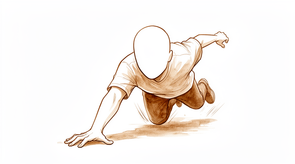
اسکیفائیڈ (scaphoid) کا فریکچر (ہڈی کا ٹوٹنا) عموماً اس وقت ہوتا ہے جب آپ پھیلے ہوئے ہاتھ کے بل گرتے ہیں، یا کھیل کے دوران۔ اسکیفائیڈ آپ کی کلائی کی انگوٹھے والی طرف، انگوٹھے کی جڑ سے ذرا نیچے ایک چھوٹی ہڈی ہے۔ یہ فریکچر اکثر کھڑے کھڑے معمولی اونچائی سے گرنے سے ہوتا ہے، اگرچہ مردوں میں یہ زیادہ تر کسی زیادہ زوردار ضرب کے بعد ہوتا ہے، جیسے کھیل یا کار کا حادثہ۔ کچھ لوگوں کو چوٹ لگنے کے لمحے ایک تیز درد کے سوا کچھ محسوس نہیں ہوتا۔
چوٹ لگتے ہی آپ کی کلائی کی انگوٹھے والی طرف درد ہوتا ہے۔ اس طرف سوجن اور نیل پڑ سکتا ہے۔ انگوٹھے کی جڑ سے ذرا نیچے کی جگہ، یعنی ایک چھوٹا سا گڑھا جسے اناٹومیکل اسنف باکس (anatomical snuffbox) کہتے ہیں، اکثر دبانے پر درد کرتی ہے۔ آپ کی کلائی اکڑی ہوئی محسوس ہو سکتی ہے، اور اسے پوری طرح موڑنے، اس پر ٹیک لگانے، یا انتہائی زاویوں پر اس پر بوجھ ڈالنے سے درد ہو سکتا ہے۔ انگوٹھے اور شہادت کی انگلی کو آپس میں ملا کر چٹکی بنانا، یا انگوٹھے کی لمبائی کی سمت میں اسے دبانا بھی تکلیف دہ ہو سکتا ہے۔
یہ علامات حیرت انگیز حد تک معمولی ہو سکتی ہیں۔ اس گڑھے میں سوجن تھوڑی سی ہو سکتی ہے، اور کچھ لوگ کلائی کو اتنی اچھی طرح ہلا لیتے ہیں کہ وہ سمجھتے ہیں کہ یہ صرف موچ ہے۔ یہ بھی ایک وجہ ہے کہ اسکیفائیڈ کے فریکچر نظر سے رہ جاتے ہیں۔ 30 سے 40 فیصد تک فریکچر پہلے ایکسرے پر نظر نہیں آتے، اور کچھ کی تصدیق تبھی ہوتی ہے جب کلائی کا دوبارہ معائنہ کیا جائے اور دوبارہ تصاویر (اسکین) لی جائیں، اکثر چوٹ کے 10 سے 14 دن بعد۔
پہلے چند دنوں سے لے کر چند ہفتوں تک، درد عموماً شروع میں سب سے زیادہ ہوتا ہے۔ کلائی کو ہلانے، گرفت کرنے، یا اس ہاتھ کے سہارے کرسی سے اٹھنے پر درد ہو گا۔ پہلے چند دنوں میں رات کے وقت دکھن عام ہے۔ ہڈی جڑنا شروع ہونے پر درد عموماً کم ہو جاتا ہے، اگرچہ انگوٹھے کے نیچے اس گڑھے میں چھونے پر درد دوسرے ہفتے تک بھی رہ سکتا ہے۔
اگر آپ کی علامات کم نہیں ہو رہیں، ہفتوں کے دوران بدتر ہو رہی ہیں، آپ کو رات کو جگا دیتی ہیں، یا آپ کو کام کرنے یا اپنا ہاتھ استعمال کرنے سے روکتی ہیں، تو اپنے فیملی ڈاکٹر (GP) کو دکھائیں یا ماہر ڈاکٹر سے معائنے کے لیے کہیں۔ اگر آپ کلینک سے رابطہ نہ کر سکیں تو اپنے قریب ترین ہسپتال کی ایمرجنسی جائیں۔
دراصل کیا ہو رہا ہے
اسکیفائیڈ آپ کی کلائی کی انگوٹھے والی طرف ایک چھوٹی، خم دار ہڈی ہے۔ یہ خود کلائی کے جوڑ کے اندر ہوتی ہے، اور ان چھوٹی ہڈیوں کی دو قطاروں کو آپس میں ملاتی ہے جن سے آپ کی کلائی بنتی ہے۔ اسے ریل گاڑی کے اگلے اور پچھلے حصے کو جوڑنے والی کڑی سمجھیں: جب یہ سالم ہو تو دونوں حصے ایک ساتھ ہمواری سے مڑتے اور سیدھے ہوتے ہیں۔ جب یہ ٹوٹ جائے تو آپ کی کلائی کے دونوں حصے ایک دوسرے سے بے ربط ہو کر حرکت کرنے لگ سکتے ہیں۔
زیادہ تر فریکچر ہڈی کے درمیانی حصے کے آر پار ہوتے ہیں، جسے ویسٹ (waist) کہتے ہیں۔ یہی وہ جگہ ہے جہاں گرتے وقت کلائی کے زبردستی پیچھے کی طرف مڑنے پر ہڈی پر سب سے زیادہ موڑنے والا زور پڑتا ہے۔ اسکیفائیڈ کے تقریباً 70 سے 80 فیصد فریکچر ویسٹ پر ہوتے ہیں۔ باقی بازو کے نچلے حصے (forearm) کے زیادہ قریب یا انگوٹھے کے زیادہ قریب ہوتے ہیں۔
اس ہڈی کو خون کی فراہمی غیر معمولی طریقے سے ہوتی ہے۔ خون اس میں دور والے سرے سے، یعنی انگوٹھے کے قریب سے، داخل ہوتا ہے اور ہڈی کے ساتھ ساتھ الٹی سمت میں جاتا ہے۔ فریکچر ہڈی کے اس ٹکڑے کی خون کی فراہمی کاٹ سکتا ہے جو بازو کے نچلے حصے کے زیادہ قریب ہے۔ خون کے بغیر اس ٹکڑے کو جڑنے میں مشکل ہوتی ہے۔ اسی لیے فریکچر کی جگہ اتنی اہم ہے، اور اسی لیے کچھ فریکچروں کا علاج دوسروں سے مختلف کیا جاتا ہے چاہے وہ ایکسرے پر ایک جیسے نظر آئیں۔
ہڈی دوبارہ آپس میں جڑ کر ٹھیک ہوتی ہے، بالکل اسی طرح جیسے ٹوٹا ہوا بازو۔ فریکچر کے آر پار نئی ہڈی بنتی رہتی ہے یہاں تک کہ دونوں ٹکڑے پھر سے ایک ہو جائیں۔ لیکن ایسا ہونے کے لیے ضروری ہے کہ ٹکڑے ساکن رکھے جائیں اور انہیں خون کی اچھی فراہمی ملے۔ اگر فریکچر اپنی جگہ سے ہٹ گیا ہو، یا اگر کلائی استعمال کرتے وقت دونوں ٹکڑے ایک دوسرے کے مقابل حرکت کریں، تو ہو سکتا ہے ہڈی نہ جڑے۔ اگر یہ نہ جڑے تو برسوں میں کلائی آہستہ آہستہ گھس سکتی ہے، کیونکہ ہڈیوں کی دونوں قطاریں ایک دوسرے سے بے ربط کام کرتی رہتی ہیں۔
مختصراً مکینیکل مسئلہ یہی ہے: ایک ایسے جوڑ میں ٹوٹی ہوئی جوڑنے والی کڑی جس کا دار و مدار اسی کڑی پر ہے۔
ہم اس کے لیے کیا کر سکتے ہیں
ڈاکٹر کیرن ہِرپرا، جو Mater Private Hospital Rockhampton میں بازو اور ہاتھ کے سرجن (upper-limb surgeon) ہیں، علاج کو آپ کی مخصوص چوٹ کے مطابق طے کرتے ہیں۔ اسکیفائیڈ کے کچھ فریکچر آپریشن کے بغیر اچھی طرح جڑ جاتے ہیں، اور کچھ کو چوٹ کے جلد بعد سرجری کی ضرورت ہوتی ہے، اس لیے بروقت معائنہ اہم ہے۔ ہم دیکھتے ہیں کہ فریکچر کہاں ہے، کیا ٹکڑے اپنی جگہ سے ہٹ گئے ہیں، اور کام اور کھیل کے لیے آپ کے ہاتھ کو کیا کچھ کرنا ہوتا ہے۔ مریضوں کو عموماً ان کے فیملی ڈاکٹر ہمارے کلینک کے لیے ریفر کرتے ہیں؛ اگر کسی فزیوتھراپسٹ نے آپ کو ہمیں دکھانے کا مشورہ دیا ہے، تب بھی میڈیکیئر (Medicare) سے رقم کی واپسی کا حق دار ہونے کے لیے آپ کو اپنے فیملی ڈاکٹر سے ریفرل (referral) لینا ہو گا۔
اگر آپ کا فریکچر مستحکم ہے یا بمشکل اپنی جگہ سے سرکا ہے، تو اکثر پلستر یا اسپلنٹ مناسب انتخاب ہوتا ہے۔ یہ جڑنے کے دوران ہڈی کو ساکن رکھتا ہے، اور اس دوران ہم بار بار تصاویر (اسکین) لے کر جڑنے کی جانچ کرتے ہیں۔ جو فریکچر چوٹ کے 6 ماہ کے اندر پکڑے جائیں وہ صرف پلستر سے بھی جڑ سکتے ہیں، چاہے تشخیص دیر سے ہوئی ہو۔ ہڈی جڑ جانے کے بعد ہینڈ تھراپسٹ (ہاتھ کی بحالی کے ماہر) مرحلہ وار حرکت کی طرف آپ کی واپسی میں رہنمائی کرتا ہے۔ شواہد سے ایک احتیاط: پہلے مہینے کے اندر لی جانے والی سوزش کم کرنے والی درد کش گولیوں کا تعلق ہڈی کے نہ جڑنے کے زیادہ امکان سے پایا گیا ہے، اس لیے ہم آپ سے ایسی درد کش دوا کے بارے میں بات کریں گے جو ہڈی کے جڑنے میں رکاوٹ نہ ڈالے۔
کچھ چوٹوں میں سرجری شروع ہی سے تجویز کی جاتی ہے، بعد کے متبادل کے طور پر نہیں۔ ہڈی کے اس سرے کے قریب کے فریکچر جو بازو کے نچلے حصے کے سب سے قریب ہے، اکثر پلستر میں نہیں جڑتے، اور ان کا آپریشن کرنے سے وہ کہیں زیادہ بھروسے کے ساتھ جڑ جاتے ہیں۔ جن فریکچروں میں ٹکڑے اپنی جگہ سے ہٹ گئے ہوں، یا جہاں ہڈی غیر مستحکم ہو، ان کا علاج بھی سرجری سے کیا جاتا ہے تاکہ جڑنے کے دوران ٹکڑے صحیح جگہ پر تھمے رہیں۔ ایک چھوٹا پیچ اندر سے ہڈی کو ساکن رکھتا ہے، جس کی بدولت کچھ لوگ جلد پلستر پہننا چھوڑ دیتے ہیں اور جلد کام پر واپس جاتے ہیں، اوسطاً تقریباً 7 ہفتے پہلے۔ دوسروں کے لیے یہ فیصلہ واقعی ہم آپ کے ساتھ مل کر کرتے ہیں: مستحکم فریکچر پلستر میں جڑ سکتا ہے، لیکن اس کا مطلب پلستر میں زیادہ ہفتے اور کام پر سست واپسی ہے، اور سرجری کے اپنے چھوٹے خطرات ہیں۔ ہم آپ کے ساتھ مل کر ان فائدوں اور نقصانات کو تولتے ہیں۔
آپ جو بھی راستہ اختیار کریں، پہلے چند ہفتے ملتے جلتے ہوتے ہیں۔ ہڈی جڑنا شروع ہونے کے دوران درد کش دوا آپ کو آرام میں رکھتی ہے۔ کلائی کی حفاظت کا مطلب ہے اسے ساکن رکھنے کے منصوبے پر پوری احتیاط سے عمل کرنا، کیونکہ ٹوٹے ہوئے ٹکڑوں کے درمیان حرکت ہی جڑنے کو روکتی ہے۔ ہینڈ تھراپی Extend Rehabilitation میں Ruby Doolan کے ساتھ مناسب مرحلے پر شروع ہوتی ہے، جب ہڈی کا جڑنا کافی مستحکم ہو جائے۔ وہ آپ کی ورزشوں کی رہنمائی کرتی ہیں اور آپ کو جس اسپلنٹ کی بھی ضرورت ہو وہ تیار کرتی ہیں۔ اگر آپ کی علامات کم نہیں ہو رہیں، ہفتوں کے دوران بدتر ہو رہی ہیں، آپ کو رات کو جگا دیتی ہیں، یا آپ کو کام کرنے یا اپنا ہاتھ استعمال کرنے سے روکتی ہیں، تو اپنے فیملی ڈاکٹر کو دکھائیں یا ماہر ڈاکٹر سے معائنے کے لیے کہیں۔
کیا توقع رکھیں
ہڈی کا جڑنا اس بات پر منحصر ہے کہ فریکچر کہاں ہے اور علاج کتنی جلدی شروع ہوتا ہے۔ جن فریکچروں کا علاج چوٹ کے 4 ہفتوں کے اندر ہو، وہ بعد میں علاج ہونے والے فریکچروں کے مقابلے میں زیادہ بھروسے کے ساتھ جڑتے ہیں، اور جن کا علاج چوٹ کے 31 دن سے زیادہ بعد ہو ان کے نہ جڑنے کا امکان زیادہ ہوتا ہے۔ پلستر میں ہڈی کو ساکن رکھا جاتا ہے جبکہ فریکچر کے آر پار نئی ہڈی بنتی ہے۔ سرجری میں ایک چھوٹا پیچ ٹکڑوں کو اندر سے آپس میں تھامے رکھتا ہے، اور کچھ لوگ جلد پلستر پہننا چھوڑ دیتے ہیں اور اوسطاً تقریباً 7 ہفتے پہلے کام پر واپس جاتے ہیں۔ جو مستحکم فریکچر بمشکل سرکے ہوں، ان میں دونوں راستے ایسی کلائی تک پہنچا سکتے ہیں جو اچھی طرح کام کرے، اور طویل مدت میں کسی ایک کا دوسرے پر کوئی ثابت شدہ فائدہ نہیں۔
دونوں صورتوں میں پہلے چند ہفتے تقریباً ایک جیسے ہوتے ہیں۔ آپ کی کلائی کی حفاظت کی جاتی ہے، درد آہستہ آہستہ کم ہوتا ہے، اور آپ ہاتھ پر بوجھ ڈالنے سے پرہیز کرتے ہیں۔ جیسے جیسے ہڈی جڑتی ہے، ہینڈ تھراپسٹ مرحلہ وار حرکت اور گرفت کی طرف آپ کی واپسی میں رہنمائی کرتا ہے۔ زیادہ تر لوگ بھاری کام پر واپس جانے سے پہلے روزمرہ کے کاموں پر واپس آ جاتے ہیں۔ کھیل سب سے آخر میں آتا ہے، جب ہڈی کے جڑنے کی تصدیق ہو جائے اور طاقت واپس آ جائے۔ اگر ہڈی جڑ جائے تو تقریباً تمام کلائیوں کا نتیجہ اچھا ہوتا ہے، چاہے ہڈی تھوڑی بدلی ہوئی شکل میں جڑے۔
سب سے بڑا مسئلہ جو پیدا ہو سکتا ہے وہ ہڈی کا آہستہ جڑنا یا بالکل نہ جڑنا ہے، جسے ہڈی کا نہ جڑنا (nonunion) کہتے ہیں۔ یہ سرجری سے علاج کیے گئے 10 فیصد سے زیادہ فریکچروں میں ہوتا ہے، اور اس کا امکان اس وقت زیادہ ہوتا ہے جب علاج دیر سے شروع ہو یا جب فریکچر ہڈی کے اس سرے کے قریب ہو جو بازو کے نچلے حصے کے سب سے قریب ہے۔ اگر ہڈی نہ جڑے تو برسوں میں کلائی میں آہستہ آہستہ گھساؤ والا آرتھرائٹس (wear-and-tear arthritis) پیدا ہو سکتا ہے۔ جن لوگوں کے نہ جڑے ہوئے فریکچر کا کبھی علاج نہیں ہوا، ان میں سے جنہیں پانچ سال یا اس سے زیادہ پہلے چوٹ لگی تھی، ان میں سے 97 فیصد میں آرتھرائٹس پیدا ہو چکا تھا۔ جڑا ہوا فریکچر دوبارہ بھی ٹوٹ سکتا ہے: ایک درج شدہ کیس میں فریکچر جڑنے کے 7 ماہ کے اندر دوبارہ ٹوٹ گیا۔ جب تک کلائی ساکن رکھی جاتی ہے اکڑاؤ عام ہے، اور ہڈی کے مضبوطی سے جڑ جانے کے بعد تھراپی سے عموماً بہتر ہو جاتا ہے۔
اگر آپ کی علامات کم نہیں ہو رہیں، ہفتوں کے دوران بدتر ہو رہی ہیں، آپ کو رات کو جگا دیتی ہیں، یا آپ کو کام کرنے یا اپنا ہاتھ استعمال کرنے سے روکتی ہیں، تو اپنے فیملی ڈاکٹر کو دکھائیں یا ماہر ڈاکٹر سے معائنے کے لیے کہیں۔ اگر آپ کلینک سے رابطہ نہ کر سکیں تو اپنے قریب ترین ہسپتال کی ایمرجنسی جائیں۔
ڈاکٹر کو کب دکھائیں
اگر آپ کی کلائی واضح طور پر اپنی شکل سے ہٹی ہوئی ہو، اگر چوٹ کی جگہ پر کھلا زخم ہو، یا اگر آپ کی انگلیاں سفید پڑ جائیں، ٹھنڈی ہو جائیں، بالکل سفید یا نیلی ہو جائیں، تو فوری طبی مدد حاصل کریں۔ چوٹ کے بعد ہاتھ میں اچانک احساس یا حرکت ختم ہو جانے کی صورت میں بھی اسی دن ہسپتال کی ایمرجنسی میں دکھانا ضروری ہے۔
اسکیفائیڈ کی زیادہ تر چوٹوں میں صورتحال اتنی نمایاں نہیں ہوتی، اور یہی اصل دھوکا ہے۔ کلائی پھر بھی حرکت کر سکتی ہے، اور سوجن معمولی ہو سکتی ہے۔ اگر آپ کا درد کم نہیں ہو رہا، یا اگر ہڈی جڑنے کے ساتھ ساتھ ہر ہفتے سوجن، حرکت یا گرفت میں بہتری نہیں آ رہی، تو اپنے فیملی ڈاکٹر کو دکھائیں یا ماہر ڈاکٹر سے معائنے کے لیے کہیں۔ جو علامات ہفتوں کے دوران بدتر ہوں، آپ کو رات کو جگا دیں، یا آپ کو کام کرنے یا اپنا ہاتھ استعمال کرنے سے روکیں، ان کا بھی باقاعدہ معائنہ ہونا چاہیے۔ اگر آپ کلینک سے رابطہ نہ کر سکیں تو اپنے قریب ترین ہسپتال کی ایمرجنسی جائیں۔
مزید تفصیل
Advanced reading: the deeper science (optional)
یہ حصہ اس سے زیادہ تفصیل میں جاتا ہے جتنی آپ کو اپنے علاج کے فیصلوں کے لیے ضرورت ہے۔ اسکیفائیڈ کے فریکچر کے بارے میں یہ اضافی مطالعہ اس لیے فائدہ مند ہے کیونکہ ہڈی پر فریکچر کی جگہ تقریباً کسی بھی دوسری خصوصیت سے زیادہ اہم ہے، اور اس لیے بھی کہ آپریشن کے حق میں دلیل وہ نہیں جو زیادہ تر لوگ سمجھتے ہیں۔
ہڈی پر فریکچر کی جگہ خطرے کو سات گنا تک بدل دیتی ہے
اسکیفائیڈ کو خون کی زیادہ تر فراہمی ان خون کی نالیوں سے ملتی ہے جو اس کے دور والے (انگوٹھے کی طرف کے) سرے کے قریب داخل ہوتی ہیں، اس لیے خون ہڈی کے ساتھ ساتھ الٹی سمت میں قریبی سرے (proximal pole)، یعنی بازو کے نچلے حصے کی طرف والے سرے، کی جانب بہتا ہے۔ ہڈی کے درمیانی حصے سے گزرنے والا فریکچر اس سے آگے کے پورے حصے کی خون کی فراہمی میں رکاوٹ ڈالتا ہے۔
اس کے نتیجے کو اعداد میں ناپا گیا ہے۔ شائع شدہ تحقیقات کو یکجا کرنے پر، اسکیفائیڈ کے قریبی سرے کے تازہ فریکچروں میں سے 34 فیصد، جب ان کا علاج بغیر آپریشن کے کیا جائے، ہڈی کے نہ جڑنے (nonunion) تک پہنچ جاتے ہیں، اور ان فریکچروں میں نہ جڑنے کا نسبتی خطرہ (relative risk) 7.5 ہے، ان فریکچروں کے مقابلے میں جو ہڈی پر زیادہ دور (distal) ہوں اور جن کا علاج اسی طرح کیا گیا ہو [1]۔
اسی لیے دو فریکچر جو ایکسرے پر ایک جیسے نظر آتے ہیں، ان کے لیے بالکل مختلف مشورے ہو سکتے ہیں۔ پلستر میں رکھے گئے دور والے (distal) فریکچر کے جڑنے کا اچھا امکان ہوتا ہے۔ قریبی سرے کا فریکچر، جس کا بالکل اسی طرح علاج کیا جائے، تقریباً ایک تہائی صورتوں میں نہیں جڑتا۔
سرجری نہ جڑنے کی شرح کم کرتی ہے لیکن آخری نتیجہ بہتر نہیں کرتی
عام درمیانی حصے کے فریکچر میں سرجری کا پلستر کے علاج سے موازنہ ایسا نتیجہ دیتا ہے جسے غور سے پڑھنا چاہیے۔ سرجری سے نہ جڑنے کی شرح تین گنا کم رہی، کارکردگی جلد واپس آئی، اور عارضی طور پر گرفت کی طاقت اور حرکت کی حد بہتر رہی، لیکن اس کے ساتھ زیادہ پیچیدگیاں بھی تھیں۔ درد، چھونے پر درد، خرچ یا کارکردگی کے نتیجے میں کوئی نمایاں فرق نہیں تھا [2]۔
یوں آپریشن سے ہڈی کے جڑنے کا زیادہ بھروسہ اور صحت یابی میں تیزی ملتی ہے، اور اس کی قیمت سرجری کی پیچیدگیاں ہیں، اور آخرکار دونوں گروپ ایک ہی جگہ پہنچ جاتے ہیں۔ اس لیے یہ فیصلہ اس بات کا ہے کہ کسی خاص شخص کے لیے زیادہ تیز اور زیادہ یقینی راستے کی کتنی اہمیت ہے؛ جسمانی مشقت والا کام کرنے والا شخص یا کھلاڑی ان مہینوں کو اس شخص سے مختلف انداز میں تولتا ہے جو پلستر کے ساتھ گزارا کر سکتا ہے۔
زیادہ تر نقصان سے بچاؤ تشخیص کے مرحلے پر ہوتا ہے
چونکہ نظر سے رہ جانے والا اسکیفائیڈ کا فریکچر ہی نہ جڑنے کی حالت تک پہنچتا ہے، اس لیے تشخیص کا طریقہ کار اہم ہے۔ انگوٹھے کی جڑ کے پاس کلائی کے گڑھے (anatomical snuffbox) میں چھونے پر درد سب سے حساس طبی معائنہ تھا، اور کئی معائنوں کو ملا کر دیکھنے سے معائنے کے بعد فریکچر ہونے کے امکان (post-test probability) کا اندازہ بہتر ہو گیا، جس کی مدد سے کلائی کو غیر ضروری طور پر ساکن رکھنے، ہسپتال کے غیر ضروری چکروں اور غیر ضروری تصاویر کو محدود کیا جا سکتا ہے [3]۔
یہ آخری حصہ ہی کارآمد ہے۔ معائنے کے نتائج کو ملا کر دیکھنا صرف فریکچر پکڑنے کے لیے نہیں؛ اس سے ان لوگوں کی بھی پہچان ہوتی ہے جو محفوظ طریقے سے پلستر پہننا چھوڑ سکتے ہیں، اسی لیے منظم طریقے سے دوبارہ معائنے کے لیے آنا فائدہ مند ہے چاہے کلائی بہتر محسوس ہو رہی ہو۔
پہلے سے نہ جڑے ہوئے فریکچر میں زیادہ پیچیدہ پیوند زیادہ بہتر پیوند نہیں
جہاں فریکچر جڑ نہیں سکا، وہاں معیاری علاج ہڈی کا پیوند (bone graft) لگانا ہے، اور خون کی نالیوں والا پیوند (vascularised graft)، یعنی ایسا پیوند جو اپنی خون کی فراہمی ساتھ لاتا ہے، بظاہر بہتر لگتا ہے۔
شواہد اس کی تائید نہیں کرتے۔ موجودہ شواہد سے پتا چلتا ہے کہ اسکیفائیڈ کے نہ جڑنے میں خون کی نالیوں والا پیوند، خون کی نالیوں کے بغیر پیوند کے مقابلے میں نمایاں طور پر بہتر نتائج نہیں دیتا، اگرچہ مصنفین بتاتے ہیں کہ مریضوں کے انتخاب میں ممکنہ جانبداری (selection bias) کی وجہ سے اس نتیجے پر اعتماد کم ہو جاتا ہے [4]۔ ہڈی جڑنے کے اعداد و شمار قریب قریب ہیں: خون کی نالیوں والے پیوند میں 84 فیصد اور خون کی نالیوں کے بغیر پیوند میں 80 فیصد، اور مختلف رپورٹوں کے درمیان کافی فرق ہے جس کی وجہ مریض، فریکچر، علاج اور تحقیق کے طریقہ کار سے متعلق عوامل ہیں [5]۔
چونکہ خون کی نالیوں والا پیوند لمبا اور تکنیکی طور پر زیادہ مشکل آپریشن ہے، اس لیے یہ تقریباً برابری جاننے کے لائق ہے۔ یہاں انتخاب کی جانبداری ایک خاص سمت میں کام کرتی ہے: خون کی نالیوں والے پیوند زیادہ مشکل کیسوں میں استعمال ہوتے ہیں، اس لیے یہ موازنہ غالباً انہیں ان کی اصل سے کم دکھاتا ہے۔ لیکن اس سے یہ ثابت نہیں ہوتا کہ ان میں سے کسی ایک کو معمول کے طور پر بہتر سمجھ کر چنا جائے۔
حوالہ جات
[1] Eastley N, Singh H, Dias JJ, Taub N. Union rates after proximal scaphoid fractures; meta-analyses and review of available evidence. J Hand Surg Eur Vol. 2012;38(8):888-97. https://doi.org/10.1177/1753193412451424
[2] Symes TH, Stothard J. A systematic review of the treatment of acute fractures of the scaphoid. J Hand Surg Eur Vol. 2011;36(9):802-10. https://doi.org/10.1177/1753193411412151
[3] Mallee WH, Henny EP, van Dijk CN, Kamminga SP, van Enst WA, Kloen P. Clinical diagnostic evaluation for scaphoid fractures: a systematic review and meta-analysis. J Hand Surg Am. 2014;39(9):1683-1691.e2. https://doi.org/10.1016/j.jhsa.2014.06.004
[4] Duncumb JW, Robinson PG, Williamson TR, Murray IR, Campbell D, Molyneux SG, et al. Bone grafting for scaphoid nonunion surgery: a systematic review and meta-analysis. Bone Joint J. 2022;104-B(5):549-58. https://doi.org/10.1302/0301-620X.104B5.BJJ-2021-1114.R1
[5] Ferguson DO, Shanbhag V, Hedley H, Reichert I, Lipscombe S, Davis TRC. Scaphoid fracture non-union: a systematic review of surgical treatment using bone graft. J Hand Surg Eur Vol. 2015;41(5):492-500. https://doi.org/10.1177/1753193415604778
Evidence & references
This is the clinical evidence summary written for health professionals. It is technical, and it lists the research this page was built from. You do not need to read it to understand your treatment or to make a decision about it.
Overview
Epidemiology and Natural History
- Nondisplaced scaphoid fractures heal in the vast majority of cases with strict immobilization [18].
- Displaced scaphoid fractures have a propensity for nonunion due to displacement and rotation [18].
- Pediatric scaphoid fractures have excellent outcomes [1].
Diagnostic and Evidence Status
- Even some well-established and widely used principles of scaphoid fracture management are supported by an insufficient amount of evidence, with many decisions based on small case series [2].
- The management of scaphoid fractures remains a source of controversy with no established gold standard for immobilization, acute surgical repair, or nonunion management despite decades of study [40].
- Currently, there is insufficient evidence to support the most effective treatment for acute scaphoid fractures [66].
- Scaphoid fracture and nonunion management continues to be an area of expanding evidence with opportunities to improve knowledge and familiarization with current evidence-based data [33].
- The definition of instability of scaphoid fractures and the indications for conservative treatment must be considered carefully [28].
Nonoperative Treatment
- Nondisplaced scaphoid fractures can be effectively treated nonoperatively with union rates approaching or exceeding those of operative intervention [62].
- This study did not demonstrate a true long-term benefit of internal fixation, compared with nonoperative treatment, for acute nondisplaced or minimally displaced scaphoid fractures [11].
- Cast immobilization for scaphoid fractures presenting 21 days or more after injury is a reasonable option [38].
Operative Treatment
- Operative intervention is recommended for displaced fractures [62].
- Early internal fixation is increasingly favored even for nondisplaced fractures [4].
- Internal fixation of scaphoid fractures is indicated in certain acute situations and in chronic nonunion cases [29].
- Appropriately performed acute percutaneous internal fixation is now a standard treatment option for a selected group of patients with acute scaphoid fracture [31].
- For all indications, the scaphoid staple has a high union rate and a low complication rate [12].
- In patients with subacute scaphoid fractures, volar percutaneous fixation performed with appropriate indications achieved excellent outcomes [76].
- The authors prefer to treat nondisplaced acute scaphoid fractures in the athlete on an individualized basis [71].
Nonunion and Malunion
- Despite improvements in diagnosis and surgical techniques, nonunion rates remain high [4].
- The clinical outcomes of malunited scaphoids after reconstruction for scaphoid fractures nonunion did not differ significantly from well-united scaphoids at a minimum 5-year follow-up [10].
- Virtually all scaphoid fractures which unite have a good outcome, regardless of malunion [13].
- The rationale, indications, contraindications, technique and results of bone grafting scaphoid nonunions with grafts harvested from the medial femoral condyle are presented [69].
- Patients with recent scaphoid fractures that failed treatment may also be treated with distal scaphoid resection [70].
- The procedure is considered pertinent for certain recent scaphoid nonunions [202].
Postoperative Care
- The optimal protocol for postoperative immobilization following operative treatment of scaphoid fractures remains controversial [81].
Anatomy & Pathophysiology
Bony Anatomy
- The scaphoid is a small, irregular S-shaped tubular bone located in the proximal carpal row on the radial aspect of the wrist [99].
- The scaphoid lies entirely within the wrist joint at a 45-degree plane to the longitudinal and horizontal axis of the wrist [99].
- The scaphoid articulates with the trapezium/trapezoid on its distal surface, the radius on its proximal/lateral surface, and the capitate and lunate on its medial surface [99].
- The proximal articular surface of the scaphoid is convex and articulates with the radius [99].
- The capitate head articulates with a sulcus on the scaphoid located across the radial articular surface, providing a socket-like fit [99].
- The scaphoid gently pronates and flexes distally such that the distal pole sits ulnarly angulated relative to the proximal pole [99].
- The distal articular surface of the scaphoid has two distinct facets for the trapezium and trapezoid, forming the STT joint [99].
- Over 80% of the scaphoid surface is covered with articular cartilage [99].
- The scaphoid is divided into three regions: proximal pole, waist, and distal pole (tubercle) [139].
- The proximal pole of the scaphoid articulates with the scaphoid fossa of the distal radius and the lunate [139].
- The scaphoid is oriented in the carpus with an intrascaphoid angle averaging 40 ± 3 degrees in the coronal plane and 32 ± 5 degrees in the sagittal plane [139].
- The scaphoid is the only carpal bone that bridges the proximal and distal carpal rows and acts as a tie-rod [139].
- The scaphoid acts as a midcarpal joint “bridge” linking and synchronizing the motions of the proximal and distal carpal rows as part of the key intercalated segment [99].
- Motion of the scaphoid includes rotation proximally and gliding distally, while providing stability to the midcarpal joint [99].
- The scaphoid is ridged across its nonarticular dorsoradial surface, along which the critical dorsal ridge vessels traverse [99].
- The dorsal ridge of the scaphoid is the insertion point for both the dorsal component of the scapholunate and intercarpal ligaments [99].
- There are no tendon attachments to the scaphoid [99].
Ligamentous Anatomy
- The ligamentous attachments of the scaphoid are predominantly found on the nonarticular dorsoradial surface [99].
- The short intrinsic ligaments provide stability to the scaphoid through attachments to the other carpal bones, in particular the lunate, and merge with the extrinsic ligaments and capsule of the wrist [99].
- The radioscapocapitate ligament does not attach to the bone itself but crosses the waist, acting as a sling across it allowing it to rotate [99].
- The radioscaphocapitate (RSC) ligament originates from the volar radial aspect of the radius, crosses the volar concavity of the scaphoid waist, and proceeds ulnarly toward the capitate [139].
- The RSC ligament acts as a fulcrum around which the scaphoid rotates [139].
- The scaphoid can fracture around the RSC ligament fulcrum at the waist [139].
- The scaphocapitate ligament originates from the distal scaphoid and inserts into the border between the trapezoid facet and the capitate facet [139].
- The scaphocapitate ligament inserts into the volar waist of the capitate distal to the RSC ligament [139].
- The scaphocapitate ligament, along with the scaphotrapezial ligament, functions as a primary restraint of the distal pole [139].
- The scapholunate interosseous ligament (SLIL) is a stout ligament connecting the scaphoid to the lunate and is the primary stabilizer [139].
- The dorsal aspect of the SLIL is composed of transverse collagen fibers, whereas the palmar ligament is composed of oblique collagen fibers inserting to the volar capsular ligaments [139].
- The dorsal portion of the SLIL is twice as strong as the palmar portion [139].
- Only 20 to 30 degrees of motion is possible at an intact scapholunate interval [139].
- The dorsal region of the SLIL resists palmar-dorsal translation and gap, whereas the volar portion resists rotation [139].
Vascular Anatomy
- The blood supply of the scaphoid is predominantly retrograde [139].
- 70% to 80% of the intraosseous and proximal pole vascular supply of the scaphoid is from branches of the radial artery entering distally through the dorsal ridge [139].
- Volar branches from the radial artery or superficial palmar arch enter in the region of the tubercle and provide blood supply to 20% to 30% of the bone in the region of the distal pole [139].
- The proximal pole also receives blood supply from the radioscapholunate ligament (ligament of Testut) and direct scapholunate branches from the palmar and dorsal transverse carpal arches [139].
- Venous drainage from the proximal pole of the scaphoid is via the dorsal ridge into the venae comitantes of the radial artery [139].
- The waist of the scaphoid has minimal or no perforating vasculature [99].
- No vessels perforate the proximal dorsal cartilaginous area or through the scapholunate ligament [99].
- Proximal fractures are associated with at least temporary disruption of the interosseous blood supply to the proximal pole [99].
- Only 67% of scaphoid bones have arterial foramina throughout their length, including the distal, middle, and proximal thirds [107].
- 13% of scaphoid bones have blood supply predominantly in the distal third [107].
- 20% of scaphoid bones have most of the arterial foramina in the waist area with no more than a single foramen near the proximal third [107].
- One third of scaphoid fractures occurring in the proximal third may be without adequate blood supply [107].
- The prevalence of osteonecrosis can be 35% in fractures at the proximal pole level [107].
- Vessels enter the scaphoid from the radial artery laterovolarly, dorsally, and distally [107].
- The laterovolar and dorsal systems share in the blood supply to the proximal two thirds of the scaphoid [107].
- 70% to 80% of the interosseous circulation is provided through branches of the radial artery entering through the dorsal ridge [107].
- 20% to 30% of the bone in the distal tuberosity region receives its blood supply from volar branches of the radial artery [107].
Mechanism of Injury
- Acute scaphoid fractures account for 2% to 3% of all fractures, approximately 10% of all hand fractures, and between 60% and 80% of all carpal fractures [74].
- The incidence of scaphoid fractures is quoted in the literature with a range from 1.5 to 121 fractures per 100,000 persons per year [74].
- The mean age for scaphoid fractures in the literature ranges from 25 to 35 years [74].
- Males are significantly younger at the time of injury compared to females [74].
- A male predominance is seen with a male to female ratio of approximately 2.5:1 [74].
- Scaphoid fractures usually occur after a fall on to the outstretched hand or during sports [74].
- Sports injuries are associated with a true scaphoid fracture [74].
- Low-energy falls from standing height occur more frequently in females, while males are more likely to sustain fractures after high-energy injuries such as sports or motor-vehicle collisions [74].
- Fractures of the scaphoid are increasingly documented after punching or assault-related injuries [74].
- The usual mechanism of injury is forced hyperextension of the wrist [17].
- Patients classically present with wrist pain following a fall onto the outstretched hand, with almost 90% recalling a hyperextension injury [30].
- Hyperextension past 95 degrees is the usual position of injury for scaphoid fractures [116].
- Other mechanisms such as axial loading and hyperflexion of the wrist have been postulated to produce scaphoid fractures [116].
- With the hyperextension mechanism, a fracture of the scaphoid usually begins at the volar waist with a tensile failure [116].
- Forces propagate to the dorsal surface with compression loading until failure occurs in hyperextension injuries [116].
- Fracture of the scaphoid occurs when the wrist is dorsiflexed to at least 95 degrees and radially deviated to at least 10 degrees [123].
- In this position, the proximal pole of the scaphoid is held firmly between the radius, capitate, radioscaphocapitate ligament, and the palmar capsule [123].
- With the wrist radially deviated, the radioscaphocapitate ligament is relaxed and unable to relieve the increasing force being applied to the radiopalmar aspect of the scaphoid [123].
- When axial loading and/or dorsal compression of the scaphoid occurs in this position, the scaphoid fractures most frequently through the waist [123].
- The waist is subject to maximal bending movement and has a characteristically lower trabecular volume [123].
- Wrist deviation may predict the location of the fracture, with the line of the midcarpal joint crossing the proximal pole in radial deviation and the distal pole in ulnar deviation [123].
- Fractures of the waist are usually the result of shear forces across the scaphoid [123].
- Tubercle fractures appear to be caused by either compression or avulsion [123].
- The size of a proximal pole fracture is dependent on the level of the proximal extent of the joint facet with the capitate [123].
- Smaller proximal pole fractures can be caused by an avulsion of the attachment of the scapholunate ligament [123].
- The scaphoid usually fractures on tension at the radial-palmar side during a fall on the outstretched palm [107].
- The proximal pole locks in the scaphoid fossa of the radius, and the distal pole moves excessively dorsal during injury [107].
- 60% to 80% of scaphoid fractures occur at the scaphoid waist or midportion [107].
- 17% of patients with scaphoid fractures have other fractures of the carpus and forearm [107].
- Associated injuries include transscaphoid perilunar dislocations, fractures of the trapezium, Bennett fractures, fractures of the radial head, dislocations of the lunate, and fractures at the distal end of the radius [107].
- In a cadaveric study, wrists placed in extreme dorsiflexion and ulnar deviation produced fractures through the scaphoid waist as the scaphoid impinged on the dorsal rim of the radius [116].
- Proximal scaphoid fractures resulted from dorsal subluxation during forced hyperextension in cadaveric studies [116].
- Carpal dislocations and scapholunate ligament tears were reproduced with wrist extension and ulnar deviation, combined with intercarpal supination [116].
Fracture Patterns and Epidemiology
- Scaphoid fractures account for almost 75% of all carpal fractures [17].
- Scaphoid fractures are rare in children and in the elderly [17].
- Fractures occur in three anatomical locations: distal tubercle, waist, and proximal pole [17].
- Waist fractures represent 70% of scaphoid fractures [14].
- Proximal pole fractures represent 20% of scaphoid fractures [14].
- Distal pole fractures represent 10% of scaphoid fractures [14].
- Fractures tend to occur at the waist partly because the RSC ligament acts as a fulcrum over which the scaphoid waist fractures [14].
- Some fractures, especially distal oblique and waist fractures, are unstable, which predisposes to non-union or malunion [17].
- The estimated incidence rate for the U.S. population is 1.47 fractures per 100,000 person-years [90].
- Scaphoid fractures made up 2.36% of wrist fractures overall in the U.S. population [90].
- 66.4% of scaphoid fractures occurred in males [90].
- The incidence rate ratio for gender, using females as the referent group, was 2.04 [90].
- Scaphoid fracture occurrence showed a peak incidence in the second and third decades [90].
- The incidence rate of scaphoid injury for those aged 10 to 19 years was 3.38 per 100,000 [90].
- The incidence rate of scaphoid injury for those aged 20 to 29 years was 2.34 per 100,000 person-years [90].
- Scaphoid fractures are the most common carpal injury in the pediatric population [77].
- Scaphoid fractures account for approximately 3% of hand and carpal fractures and 0.34% of all fractures in children [77].
- The majority of pediatric scaphoid fractures now occur at the waist, similar to adult patterns [77].
- In a review of 1000 consecutive hand injuries, only 18% involved fractures of the carpus, with the scaphoid being the most commonly injured carpal bone at 58% [131].
Pathophysiology and Kinematics
- The scaphoid is critical to the coordination of normal carpal kinematics, and its fracture has significant biomechanical consequences to the wrist [98].
- With an unstable displaced scaphoid fracture, the kinematics of the wrist is altered [123].
- Joint compressive forces, trapezium–scaphoid shear stress, and capitolunate rotation moments act upon the scaphoid, leading to a dissociation of the proximal and distal carpal rows [123].
- This dissociation permits the natural tendency of the two carpal rows to fail by collapsing, assuming a lunate-extended posture [123].
- The scaphoid will assume an anteverted position, the lunate and triquetrum may subluxate forward and rotate dorsally, and the capitate and hamate subluxate dorsally and proximally, producing the dorsal intercalated segment instability (DISI) deformity [123].
- The collapse pattern seen with chronic scaphoid nonunion is known as scaphoid nonunion advanced collapse (SNAC) and appears as a DISI deformity [123].
- The proximal and distal fracture fragments can collapse giving a characteristic flexed or “humpback” position on radiographs with an intrascaphoid angle of greater than approximately 30 degrees [123].
- After a simulated scaphoid waist fracture, the proximal and distal segments of the scaphoid moved independently [216].
- The distal scaphoid assumed a relatively flexed stance and displayed increased motion after osteotomy [216].
- The proximal scaphoid fragment and lunate assumed a relatively extended stance and displayed less motion after osteotomy [216].
- These kinematic abnormalities produced significant interfragmentary motion that would be expected to complicate normal fracture healing [216].
- The spontaneous collapse of the two scaphoid fragments produced a dorsal angulation or “humpback" deformity that simulated the clinical situation of displaced scaphoid nonunions [216].
- The scaphoid serves an important role maintaining normal alignment of the carpal bones and producing normal wrist motion [216].
- Scaphoid nonunions have a dramatic impact on carpal kinematics, partially uncoupling the proximal and distal carpal rows [102].
- Problem fractures and non-unions of the scaphoid are associated with major alterations in wrist kinematics and a higher incidence of premature carpal collapse and degenerative arthritis [61].
- Untreated displaced fractures of the waist will usually angulate as the volar bone is reabsorbed, yielding a “humpback” flexion deformity of the scaphoid [116].
- The resultant radial column shortening and the extension of the proximal scaphoid pole releases the lunate to rotate into DISI under the influence of the attached triquetrum [116].
- Untreated scaphoid nonunion will predictably progress to arthritic change, termed scaphoid nonunion advanced collapse (SNAC) [116].
- Arthritic change arises at the radial styloid articulation with the distal scaphoid pole (stage I) and is followed by degeneration
Classification
General Classification Schemes and Reliability
- Three common classifications used for scaphoid fractures are the Mayo classification, Russe classification, and Herbert classification [224].
- The Mayo and Russe classifications are based on anatomic planes of the scaphoid [224].
- The Herbert classification defines stable and unstable fractures [224].
- Some series have shown limited prognostic value and poor inter- and intraobserver reliability of scaphoid fracture classification schemes [224].
- The Herbert classification may be particularly helpful in determining treatment options [224].
- Scaphoid nonunions are not easy to categorize and have been described by anatomic location or with clinically specific terms such as stable, fibrous, sclerotic, unstable, humpback, synovial, cystic, pseudarthrosis, or avascular [212].
- There is a need for a validated prognostic classification system for scaphoid nonunions that can allow comparisons between outcome studies [120].
- Several classification schemes have been proposed for scaphoid nonunion, generally based on factors such as time since injury, mobility of the fragments, cystic or flexion deformity, and degenerative change [214].
Herbert Classification
- In the Herbert classification, Type A fractures are acute stable fractures [213].
- In the Herbert classification, Type A1 fractures are fractures of the tubercle [213].
- In the Herbert classification, Type A2 fractures are undisputed "crack" fractures of the waist [213].
- In the Herbert classification, Type B fractures are acute unstable fractures [213].
- In the Herbert classification, Type B1 fractures are oblique fractures of the distal third [213].
- In the Herbert classification, Type B2 fractures are displaced or mobile fractures of the waist [213].
- In the Herbert classification, Type B3 fractures are proximal pole fractures [213].
- In the Herbert classification, Type B4 fractures are fracture dislocations of the carpus [213].
- In the Herbert classification, Type B5 fractures are comminuted fractures [213].
- In the Herbert classification, Type C fractures are delayed union [213].
- In the Herbert classification, Type D fractures are established non-union [213].
- In the Herbert classification, Type D1 fractures are fibrous non-union [213].
- In the Herbert classification, Type D2 fractures are sclerotic non-union (pseudoarthrosis) [213].
- Type A Herbert classification fractures include fractures of the tubercle (A1) and an incomplete fracture of the waist (A2) [224].
- Type B Herbert classification fractures include subtypes B1 (oblique fractures of the distal third), B2 (displaced or mobile fractures of the waist), B3 (proximal pole fractures), B4 (fracture dislocations), and B5 (comminuted fractures) [224].
- Type C Herbert classification fractures show delayed union after more than 6 weeks of plaster immobilization [224].
- Type D Herbert classification fractures are established nonunions, either fibrous (D1) or sclerotic (D2) [224].
Schernberg Classification
- The Schernberg classification classifies scaphoid fractures as being through the proximal pole (type 1), proximal part of the body (type 2), distal part of the body (type 3), tuberosity (type 4) or distal segment (type 5) [211].
- The Schernberg classification includes partial fractures of the tuberosity as type 6 [211].
Slade and Geissler Classification
- The Slade and Geissler classification of scaphoid fracture nonunion includes Type 1 as delayed presentation for 4–12 weeks [214].
- The Slade and Geissler classification of scaphoid fracture nonunion includes Type 2 as fibrous union with minimal fracture line [214].
- Slade and Dodds described a classification of scaphoid nonunion into six grades [222].
- In the Slade and Dodds classification, grades I, II, and III correspond to delayed presentation, fibrous nonunion, and minimal sclerosis [222].
- In the Slade and Dodds classification, grades IV and V correspond to bone loss without significant flexion deformity [222].
- In the Slade and Dodds classification, grade VI corresponds to more advanced resorption [222].
Pediatric Classification
- D’Arienzo proposed a three-part classification system for pediatric scaphoid fractures based on the age of the child and the presumed degree of ossification [217].
- Type 1 pediatric scaphoid fractures occur in children younger than age 8 years [217].
- Type 1 pediatric scaphoid fractures may be purely chondral or may involve part of the ossific nucleus [217].
- Type 2 pediatric scaphoid fractures are osteochondral fractures and occur in patients aged 8 to 11 years [217].
- Type 3 pediatric scaphoid fractures occur in adolescents aged ≥12 years [217].
- In Type 3 pediatric scaphoid fractures, the scaphoid is almost completely ossified [217].
- Pediatric scaphoid fractures may be classified according to anatomic location: tuberosity, transverse distal pole, avulsion distal pole, waist, and proximal pole [217].
- In children, fractures of the distal third of the scaphoid are the most common [217].
Nonunion Classification Characteristics
- A revised classification of scaphoid nonunions focuses on the width of the devitalized scaphoid zone and circumstances that complicate the healing process when additional structural or biologic enhancements are needed [212].
- Scaphoid nonunions can be divided roughly into two groups: early nonunions without substantial bone resorption, and older nonunions with substantial bone resorption [212].
- Complicating factors in treating scaphoid nonunions include perfusion, deformity, and instability (bony or ligamentous) [212].
- Type I scaphoid nonunions are defined as delayed or fibrous union with no deformity [214].
- Scaphoid nonunions with minimal fracture sclerosis (<1 mm) require only rigid fixation to heal if there is adequate perfusion [214].
- Scaphoid nonunions with minimal bone resorption of the anterior cortical bone and minimal fracture sclerosis (<2 mm confirmed by CT scan) still have the potential for healing in the early stages with screw fixation alone [214].
Clinical Presentation
Epidemiology and Mechanism
- Scaphoid fractures account for 2% to 3% of all fractures, approximately 10% of all hand fractures, and between 60% and 80% of all carpal fractures [74].
- The annual incidence of true radiographically confirmed acute scaphoid fractures is approximately 29 per 100,000 persons per year [74].
- The mean age for scaphoid fracture in the literature ranges from 25 to 35 years [74].
- There is a male predominance with a male-to-female ratio of approximately 2.5:1 [74].
- Scaphoid fractures usually occur after a fall onto the outstretched hand or during sports [74].
- Males are more likely to sustain scaphoid fractures after high-energy injuries such as sports or motor-vehicle collisions, while females more frequently sustain them after low-energy falls from standing height [74].
- Scaphoid fractures are increasingly documented after punching or assault-related injuries [74].
- Almost 90% of patients recall a hyperextension injury [30].
- In the pediatric population, scaphoid fractures account for approximately 3% of hand and carpal fractures and 0.34% of all fractures in children [77].
- Pediatric scaphoid fracture patterns are shifting toward the waist, similar to adults, due to increased participation in high-energy extreme sports and increasing BMIs [77].
Symptoms and Signs
- Patients classically present with pain on the radial side of the wrist [14].
- There may be swelling on the radial side of the wrist [14].
- There is usually a history of trauma, such as falling on an outstretched hand, collision of the wrist against a person or heavy obstacle, or a direct blow against an object [14].
- Limited range of motion and pain when applying extended wrist loading or positioning the wrist in extreme positions of flexion or extension may be present [14].
- In the acute phase, pain, swelling, ecchymosis, and tenderness around the region of the scaphoid may be present [30].
- Slight fullness in the anatomical snuffbox may be present [17].
- Precisely localized tenderness in the anatomical snuffbox is an important diagnostic sign [17].
- Swelling in the snuffbox is more often present with fractures than without after 2 weeks [47].
- Wrists with chronic injury may have swelling in the dorsoradial wrist [14].
- Pain on longitudinal compression of the thumb (scaphoid axial compression test) is a sign of scaphoid fracture [14].
- The main complaint is radial-sided wrist pain with localized tenderness over the scaphoid in the region of the anatomical snuffbox [30].
Physical Examination Findings
- "Snuffbox tenderness" applies predominantly to waist fractures, which represent 70% of scaphoid fractures [14].
- Proximal pole fractures are the second most common type, accounting for 20% of scaphoid fractures [14].
- Distal pole fractures are the least common, accounting for 10% of scaphoid fractures [14].
- Fractures tend to occur at the waist partly because the radioscaphocapitate (RSC) ligament acts as a fulcrum over which the scaphoid waist fractures [14].
- The distal pole of the scaphoid should be palpated at the scaphoid tubercle on the palmar aspect of the wrist [14].
- With radial deviation of the wrist, the prominence of the distal pole moves palmarly toward the examiner’s thumb [14].
- The proximal pole is palpated dorsally in line with the second ray just distal to the dorsal radius lip [14].
- The scapholunate ligament is in line between the second and third rays just distal to the dorsal radius lip and corresponds to the 3-4 wrist arthroscopy portal [14].
- The proximal pole is located just radial to the scapholunate ligament/3-4 portal area [14].
- Anatomical snuffbox tenderness has a sensitivity of 87–100% and a specificity of 3–98% [30].
- Axial compression of the thumb has a sensitivity of 48–100% and a specificity of 22–97% [30].
- Scaphoid tubercle tenderness has a sensitivity of 82–100% and a specificity of 17–57% [30].
- Pain on ulnar deviation has a sensitivity of 67–100% and a specificity of 17–60% [30].
- Pain on radial deviation has a sensitivity of 67–90% and a specificity of 31–42% [30].
- Reduced range of movement of the thumb has a sensitivity of 65–66% and a specificity of 38–59% [30].
- Thumb–index finger pinch has a sensitivity of 75–79% and a specificity of 44–76% [30].
- No single clinical sign has been found to be adequately sensitive or specific for scaphoid fracture [30].
- Anatomical snuffbox tenderness is oversensitive and has poor specificity [30].
- In a study of 246 patients with suspected scaphoid fracture, anatomical snuffbox tenderness had a sensitivity of 90% and a specificity of 40% [30].
- In the same study of 246 patients, scaphoid tubercle tenderness had a sensitivity of 87% and a specificity of 57% [30].
- A combination of anatomical snuffbox tenderness, scaphoid tubercle tenderness, and anatomical snuffbox pain on longitudinal compression of the thumb generated a sensitivity of 100% and a specificity of 74% [30].
- The combination of anatomical snuffbox tenderness, scaphoid tubercle tenderness, and scaphoid axial compression test has a sensitivity of 87% to 100% and a specificity of 74% [14].
- Clinical signs are inadequate indicators of scaphoid fracture when used alone and should be combined to achieve a more accurate clinical diagnosis [42].
- The sensitivity of the scaphoid compression test is 70.5% and the specificity is 21.8% [119].
- Pain on thumb–index finger pinch and anatomical snuffbox pain on pronation of the forearm are most suggestive of a true scaphoid fracture [30].
- The best predictors of fracture within 72 hours of injury are the absence of pain on ulnar deviation of the wrist and pain on thumb–index finger pinch [30].
- Scaphoid tubercle tenderness is most predictive at week 2 [30].
- A clinical scaphoid score (CSS) of 4 or higher requires an MRI, using a scoring system of 3 points for anatomical snuffbox tenderness with the wrist in ulnar deviation, 2 points for tenderness over the scaphoid tubercle, and 1 point for pain upon longitudinal compression of the thumb [30].
- Most scaphoid fractures are missed due to failure to consider the possibility of the injury and search for clinical signs [45].
Diagnostic Challenges and Missed Fractures
- Up to 30% to 40% of scaphoid fractures are not identified on initial assessment and investigation with standard four-view radiographs [30].
- Patients with a suspected occult scaphoid fracture are reevaluated after 1 to 2 weeks of immobilization in a forearm cast or splint [80].
- An examination by a specialist after the injury has become less painful allows for a more accurate physical examination and substantially increases the sensitivity of detecting a scaphoid fracture [80].
- The combination of conventional radiographs and two clinical examinations does not provide adequate diagnostic certainty for scaphoid fractures, as a true fracture was identified in only about 40% of patients [6].
- The combination of conventional radiographs and clinical reassessment does not increase the accuracy of these diagnostic tests compared with conventional radiographs alone [19].
- The incidence of fractures of the scaphoid in patients diagnosed initially as a clinical fracture of the scaphoid is very low [15].
- Even at 2 weeks, it is very difficult to separate clinically those patients with proven fractures from those in whom there was clinical suspicion of a fracture which could not be substantiated on either scintigraphy or radiography [22].
- Some patients with fractures were almost completely asymptomatic, whereas some who did not have a fracture had signs and symptoms very suggestive of a fracture [22].
- Radiography cannot be used as the gold standard for diagnosing a scaphoid fracture [22].
- Fractures not visible on original radiographs show up on repeat radiographs at 2 weeks in only 2% of cases [22].
- In a retrospective review of 108 patients with a diagnosis of "clinical" fracture of the scaphoid, no fracture was demonstrated on radiographs after a period of immobilization [22].
- Dorsal avulsion fractures of the scaphoid are indistinguishable from X-ray negative scaphoid fractures because both present with similar clinical signs and isotope bone scans without a specific view [21].
- If initial radiographs are negative, a fracture of the trapezium is more likely to be present than one of the scaphoid, despite high levels of clinical suspicion for scaphoid injuries [48].
- The high rates of delayed presentation and incomplete evaluation and treatment suggest a strong need for better patient and doctor education on the subject of scaphoid injuries and nonunions [32].
- Pediatric scaphoid fractures often present late because pain and swelling can be subtle in the anatomic snuffbox [77].
Investigations
Clinical Examination
- Patients with scaphoid fractures typically present with pain on the radial side of the wrist, swelling, and a history of trauma such as falling on an outstretched hand [14].
- "Snuffbox tenderness" is predominantly associated with waist fractures, which represent 70% of scaphoid fractures [14].
- Proximal pole fractures account for 20% of scaphoid fractures, while distal pole fractures account for 10% [14].
- The full physical examination of the scaphoid should include palpation of the waist, distal pole, and proximal pole [14].
- A combination of anatomic snuffbox tenderness, scaphoid tubercle tenderness, and the scaphoid axial compression test has a sensitivity of 87% to 100% and a specificity of 74% for scaphoid fracture [14].
- Anatomical snuffbox tenderness has a sensitivity range of 87–100% and a specificity range of 3–98% [30].
- Axial compression of the thumb has a sensitivity range of 48–100% and a specificity range of 22–97% [30].
- Scaphoid tubercle tenderness has a sensitivity range of 82–100% and a specificity range of 17–57% [30].
- A combination of anatomic snuffbox tenderness, scaphoid tubercle tenderness, and anatomic snuffbox pain on longitudinal compression of the thumb generated a sensitivity of 100% and a specificity of 74% in the first 24 hours after injury [30].
- Pain on thumb–index finger pinch and anatomic snuffbox pain on pronation of the forearm were the most suggestive clinical signs of a true scaphoid fracture [30].
- The absence of pain on ulnar deviation of the wrist and pain on thumb–index finger pinch were the best predictors of fracture within 72 hours of injury [30].
- Scaphoid tubercle tenderness was the most predictive clinical sign at week 2 [30].
- A clinical scaphoid score (CSS) of 4 or higher, based on anatomic snuffbox tenderness with ulnar deviation, scaphoid tubercle tenderness, and pain upon longitudinal compression of the thumb, indicates the need for MRI [30].
- Clinical examination along with early MRI scan should form the basis of diagnosing a suspected scaphoid fracture [50].
Radiography
- Standard radiographic views for scaphoid fractures include posteroanterior, lateral, oblique, and scaphoid views [59].
- A true scaphoid pisiform capitate (SPC) lateral radiograph allows for a true assessment of carpal alignment [59].
- The scaphoid view is taken with the wrist in ulnar deviation to take the scaphoid out of its usual position of flexion and pronation [59].
- A clenched pencil view is useful for assessing associated dynamic scapholunate widening and SNAC/SLAC wrist changes [59].
- Radiographs may not show a fracture in the first few days after injury, but the break is usually much clearer two weeks later due to bone resorption and slight displacement [17].
- Up to 30% to 40% of scaphoid fractures are not identified on initial assessment with standard four-view radiographs [30].
- Radiographs are initially nondiagnostic in more than 30% of cases [124].
- The combination of conventional radiographs and two clinical examinations does not provide adequate diagnostic certainty, as a true fracture was identified in only about 40% of patients [6].
- Due to low agreement between observers and poor diagnostic performance, 6-week radiographs are not adequate for evaluating suspected scaphoid fractures [26].
- Plain radiography is approximately 50% sensitive for the detection of a scaphoid fracture [112].
- Dorsal avulsion fractures of the scaphoid are indistinguishable from X-ray negative scaphoid fractures without a specific view [21].
- There is no consensus regarding the imaging modality and measurements to use to define a scaphoid fracture as 'nondisplaced' [20].
Advanced Imaging
- If there is a strong clinical suspicion of a scaphoid fracture which cannot be confirmed by conventional radiology, bone scintigraphy is a valuable diagnostic tool [7].
- The best diagnostic strategy for clinically suspected scaphoid fractures consists of initial radiography followed by bone scintigraphy in patients with negative radiographs [16].
- The specificity of bone scanning for scaphoid fractures is 100%, as no false-negative scans occurred [23].
- Bone scintigraphy has a high sensitivity of 95%, but specificity varies between 60% and 95% [49].
- MRI is the definitive way to confirm or exclude a diagnosis of scaphoid fracture if the technique is available [17].
- MRI demonstrated fracture of the scaphoid in approximately one-third of patients with a negative X-Ray [46].
- MRI at presentation can detect scaphoid fractures missed by plain radiography and identify associated ligamentous or bony lesions [65].
- Early magnetic resonance imaging provides an immediate diagnosis for suspected scaphoid fractures when initial radiographs are inconclusive [68].
- The use of early MRI in patients with clinically suspected scaphoid fracture results in the accurate and reliable identification of a significant number of radiological occult injuries [60].
- MRI has the highest sensitivity, specificity, and accuracy (all >95%), with high positive and negative predictive values, at less than 24 hours [124].
- A normal MRI study as early as 2 days after injury has a negative predictive value of 100% [112].
- MRI is the optimal second test for assessing a possible scaphoid fracture after a negative radiograph [156].
- According to existing literature, MRI is the best diagnostic radiological test for triage of suspected scaphoid fractures [167].
- MRI is not 100% specific for diagnosing an occult scaphoid fracture, with a specificity of 96% in healthy volunteers [194].
- Routine MRI of suspected scaphoid fractures carries a notable risk of overdiagnosis and potential overtreatment, with nearly 70% of MRI findings categorized as distracting and potentially misleading [181].
- MRI-detected scaphoid fractures are not universally benign, with delayed or nonunion seen in over 6% despite appropriate initial immobilization [58].
- CT is more sensitive for diagnosing a scaphoid fracture and is particularly useful in confirming the alignment of bone fragments if surgery is planned [17].
- CT is preferred when the fracture is visible for further assessment and surgical planning [156].
- Three-dimensional computed tomography is useful for analyzing displacement of scaphoid fracture [94].
- Multidetector CT sensitivity was 86% and specificity was 100% for detecting occult scaphoid fractures [153].
- CT is a good way to screen occult fractures but may not be any better than MRI or bone scanning in detecting scaphoid fractures without some over treatment [151].
- High-frequency ultrasound is recommended to investigate occult suspected scaphoid fractures because of its ability to allow early diagnosis and eliminate the need for more invasive or expensive diagnostic tests in most cases [63].
- In patients with an occult scaphoid fracture, the sensitivity of intrasound vibration was 18% [52].
- Low field MRI can be used to show scaphoid fractures and allows diagnosis of additional or simulating lesions [159].
- All advanced imaging modalities are better for ruling out rather than ruling in a scaphoid fracture [124].
Treatment
General Principles and Evidence Quality
- Scaphoid fractures are managed largely on the basis of anecdotal evidence and traditional remedies [3].
- Early treatment of acute scaphoid fractures is important, with union rates significantly greater when treatment is instituted prior to 4 weeks from injury [72].
Non-Operative Management
- A restricted period of cast immobilisation is recommended for the initial treatment of non-displaced scaphoid fractures [34].
- Non-operative treatment of non-displaced scaphoid fractures may be preferred over early surgical treatment, as the benefits of early surgical treatment are less obvious [34].
- Subacute scaphoid fractures presenting within 6 months from injury can be expected to successfully heal with casting alone, even if the initial diagnosis is delayed [5].
- There was no difference in functional outcome at 12 months for fractures of the waist of the scaphoid with ≤ 2 mm displacement treated operatively or nonoperatively [35].
- Non- and minimally displaced scaphoid waist fractures are best treated conservatively [104].
- Aggressive conservative management avoids unnecessary surgery in all acute scaphoid fractures [147].
- The outcome of non-operative treatment of undisplaced scaphoid waist fractures can be predicted with reasonable accuracy by assessing fracture union on a week 4 CT scan [175, 176].
- Among patients with nonoperatively managed scaphoid fractures, those prescribed NSAIDs within 1 month of diagnosis demonstrated an increased risk of nonunion and subsequent salvage procedures [146].
- The authors recommend considering nonoperative management for asymptomatic scaphoid nonunion in children [148].
Operative Management: Acute Fractures
- Displaced fractures have a propensity for nonunion due to displacement and rotation [18].
- Minimally invasive fixation has been demonstrated to have a higher union rate than cast treatment and has relatively few complications [78].
- Surgical treatment for non-displaced and minimally displaced acute scaphoid fractures may be slightly favourable compared to conservative treatment for standardised functional outcome on the short term (within 2 years), with a significantly faster return to work (SMD of 7 weeks) [54].
- The proximal scaphoid fracture did not heal by conservative management in more than 30% of cases, while the operative regimen failed only once in 7 cases [79].
- One study reported a faster return to play with internal fixation compared with a playing cast alone [85].
- The author indicates surgery for all proximal pole fractures and displaced (>1 mm) waist fractures [85].
- Distal pole fractures are almost always treated nonoperatively in a short-arm cast, unless a person's occupation or special needs requires earlier and unrestricted mobility [85].
- For nondisplaced waist fractures, the author has a discussion with the patient about the advantages and disadvantages of operative and nonoperative approaches [85].
- For nonoperative care of nondisplaced waist fractures, the author treats in a short-arm cast [85].
- Mini-open internal fixation (APM: dorsal approach) is preferred for active, young, manual workers, athletes, or workers in high-demand occupations for nondisplaced waist fractures [85].
- Mini-open internal fixation via dorsal approach is indicated for nondisplaced proximal pole fractures [85].
- Open reduction and internal fixation, with or without bone graft, is indicated for displacement of more than 1 mm [85].
- Open reduction and internal fixation, with or without bone graft, is indicated for a lateral intrascaphoid angle of more than 35 degrees [85].
- Open reduction and internal fixation, with or without bone graft, is indicated for bone loss or comminution [85].
- Open reduction and internal fixation, with or without bone graft, is indicated for perilunate fracture-dislocation [85].
- Open reduction and internal fixation, with or without bone graft, is indicated for dorsal intercalated segmental instability alignment (DISI with radiolunate angle >15 degrees) [85].
- Surgical management is recommended for displaced scaphoid fractures, proximal pole fractures, comminuted fractures, and fractures that are part of a greater perilunate injury [89].
- Percutaneous fixation for undisplaced or minimally displaced waist fractures may reduce the time in cast, increase the rate of return to function, and increase the rate of union [89].
- The inherent instability of scapho-lunate dissociation occurring with scaphoid fracture has a high scaphoid non-union rate, and if identified, treatment by open reduction and internal fixation is recommended [172].
Operative Management: Nonunion and Salvage
- Nonunion rates remain high despite improvements in diagnosis and surgical techniques [4].
- The frequency of non-union after surgical management for closed scaphoid fractures exceeds 10% and remained consistent during the study period [24].
- Patients treated nonoperatively or with salvage procedures had similar long-term outcomes as those treated with a corrective scaphoid osteotomy [37].
- The use of 2 headless compression screws for the treatment of scaphoid nonunions is safe and effective [127].
- The success of percutaneous fixation of acute fractures can be extended to the care of scaphoid nonunions [154].
- Uncomplicated scaphoid nonunions that are nondisplaced and nonangulated are candidates for the minimally invasive bone grafting and compression screw fixation procedure [158].
- Management of scaphoid nonunions by internal bone grafting is a simple and effective method that can be applied successfully in selected cases [184].
- The combination of scaphoid plate fixation and pure cancellous bone grafting for scaphoid nonunion with segmental defects yields reliable union rates and good patient outcomes [186].
- The use of electrical stimulation for scaphoid nonunion may be indicated in patients who have failed previous bone grafting procedures or if surgery is refused or unsafe [178].
- Treatment with LIPUS had no effect on reducing time to union in patients who underwent surgical fixation of established scaphoid nonunions [166].
- Real time ultrasonography is a useful adjunct in the assessment of scaphoid non-union, particularly when radiographs are inconclusive between 8 to 12 weeks of immobilization [144].
- CT scans should be regularly used in cases when there is concern as to the state of union of a healing scaphoid fracture to prevent patients with partial union undergoing unnecessary surgery for presumed non-union [168].
- The non-union rate of the scaphoid was relatively higher in the series treated by closed reduction [164].
Complications
Nonunion and Delayed Union
- The frequency of non-union after surgical management for closed scaphoid fractures exceeds 10% [24].
- Persistent nonunion is common after surgery for scaphoid non-union, and surgeries for persistent nonunion are even less successful [82].
- Scaphoid nonunions are unlikely to remain aligned or free of arthritis after 10 years [191].
- The reported incidence of non-union of scaphoid fractures varies from 2.7% to 8.9% when immobilization is started within a month from injury [210].
- An incidence of 5.1% non-union was found in fractures of the proximal third of the scaphoid in adults when immobilization began within the first 24 hours after injury [210].
- A delay of immobilization up to four weeks does not increase the frequency of non-union [210].
- With a delay of treatment of more than four weeks, the incidence of non-union is considerably increased [210].
- Lindstrom (1975) reported a frequency of 19.2% nonunion after a delay of three to 16 weeks [210].
- Eddeland (1975) reported 88.1% non-union following a delay of more than four weeks [210].
- A study found 40.0% non-union when the delay in treatment exceeded four weeks [210].
- Displacement of scaphoid fractures results in a threefold increase in the incidence of non-union and a two-week prolongation of time to bony union in displaced waist fractures [210].
- The highest incidence of non-union was found in fractures of the proximal pole [210].
- Increased likelihood for nonunion was found when the fracture was treated greater than 31 days from injury [91].
- Increased likelihood for nonunion was found when fracture volume was less than 38% of the entire scaphoid [91].
- Neglected scaphoid fractures seldom heal with acceptable alignment, which often leads to complications in their management [3].
- In cases of non-union or malunion, reconstruction of the scaphoid is exceedingly difficult, increasing the chances of morbidity associated with an extended period of hand immobilisation [3].
- One patient presented with a persistent scaphoid nonunion 44 months after injury that may become symptomatic in the future [83].
- A fractured proximal pole scaphoid documented by serial CT scanning refractured within 7 months of documented healing [88].
Malunion and Arthrosis
- Problem fractures and non-unions of the scaphoid are associated with major alterations in wrist kinematics and a higher incidence of premature carpal collapse and degenerative arthritis than previously appreciated [61].
- An alteration of the carpal dynamics, due to deformation and shortening of the scaphoid, is the most likely cause of post-traumatic arthrosis after primary healing of scaphoid fractures [92, 93].
- Nearly half of all patients with malunited acute scaphoid fractures demonstrated radiographic findings of early arthritis on CT imaging [87].
- Patients with malunited acute scaphoid fractures had overall good clinical results on midterm follow-up despite radiographic findings of early arthritis [87].
- Patients treated nonoperatively or with salvage procedures had similar long-term outcomes as those treated with a corrective scaphoid osteotomy for malunion [37].
Diagnostic Complications and Missed Injuries
- Dorsal avulsion fractures of the scaphoid are indistinguishable from X-ray negative scaphoid fractures without a specific view, as both present with similar clinical signs and isotope bone scans [21].
- It is very difficult to separate clinically those patients with proven fractures from those in whom there was clinical suspicion of a fracture which could not be substantiated on either scintigraphy or radiography, even at 2 weeks [22].
- Fractures not visible on the original radiographs may show up after 2 weeks of immobilization in only 2% of cases [22].
- The specificity of bone scanning for scaphoid fractures is 100%, as no false-negative scans occurred and all patients with negative scans failed to demonstrate signs of a fracture on follow-up [23].
- Scintigraphy is very sensitive but not specific for a fracture [22].
- High rates of delayed presentation and incomplete evaluation and treatment suggest a strong need for better patient and doctor education on the subject of scaphoid injuries and nonunions [32].
Pediatric Specifics
- Cast immobilization for scaphoid fractures presenting 21 days or more after injury is a reasonable option in pediatric patients [38].
- Historically, scaphoid fractures in children predominantly involved the distal pole, requiring neither surgical care nor extended followup [203].
- Changing patient characteristics appear to be altering fracture epidemiology and treatment in children [203].
Treatment-Related Complications and Outcomes
- The scaphoid staple has a high union rate and a low complication rate for all indications [12].
- Nondisplaced fractures of the scaphoid heal with cast immobilization in most cases, but operative treatment is being offered with greater frequency to active patients to reduce the period of cast immobilization [73].
- No instances of non-union, necrosis of the proximal fragment of the scaphoid or scapholunate dissociation were noted during a mean follow-up period of 18 months for isolated scaphoid fracture with anterosuperior dislocation of the proximal fragment [86].
- The long-term prognosis after distal articular fractures of the scaphoid has not been studied [8, 9].
- The scaphoid bone bruise is a benign injury with predictable recovery and is unlikely to result in long-term morbidity in the form of nonunion [67].
Recovery
General Outcomes and Prognosis
- Good clinical outcomes can be achieved after scaphoid fractures in prospective NFL athletes [200].
- From an 8- to 11-year perspective, patients with distal scaphoid fractures report normal self-assessed hand function as well as good wrist motion and strength [39].
- The long-term prognosis after distal articular fractures of the scaphoid has not been studied and its further assessment should be based on accurate classification of the anatomy and mechanism of the injury [8, 9].
Malunion and Arthrosis
- Nearly half of all patients with malunited acute scaphoid fractures demonstrated radiographic findings of early arthritis on CT imaging but overall good clinical results on midterm follow-up [87].
Nonunion and Delayed Healing
- Increased likelihood for nonunion was found when the fracture was treated greater than 31 days from injury and when fracture volume was less than 38% of the entire scaphoid [91].
- Patients with comorbid psychiatric conditions experienced increased rates of delayed scaphoid union [219].
- A fractured proximal pole scaphoid that was documented by serial CT scanning refractured within 7 months of documented healing [88].
- Although at his last visit the patient was symptom free and no osteoarthritic changes were seen on the radiographs, it is possible that in the long run osteoarthritic changes will occur because a non-union of the scaphoid has been present for more than 1 year [228].
- Scaphoid nonunions demonstrate findings indicative of progression to union on CT at a mean of 6 weeks and as early as 3 weeks postoperatively [227].
Timing and Treatment Impact on Recovery
- We found no difference in functional outcome at 12 months for fractures of the waist of the scaphoid with ≤ 2 mm displacement treated operatively or nonoperatively [35].
- No instances of non-union, necrosis of the proximal fragment of the scaphoid or scapholunate dissociation were noted during a mean follow-up period of 18 months [86].
- The fractures of the carpal scaphoid were all united within the usual time limits and there was no residual disability in any of the elbows [75].
Risk Factors and Complications
- This study suggests that the scaphoid bone bruise is a benign injury with predictable recovery and is unlikely to result in long-term morbidity in the form of nonunion [67].
Key Evidence
- [L1] Pediatric scaphoid fractures have excellent outcomes. [1] (10.1177/1558944717735948)
- [L5] Even some well-established and widely used principles of scaphoid fracture management are supported by an insufficient amount of evidence, with many decisions based on small case series. [2] (10.1177/1753193420977241)
- [L4] [3] (10.1016/j.injury.2009.07.078)
- [L5] This article reviews current concepts regarding the treatment of scaphoid fractures and nonunions, highlighting that despite improvements in diagnosis and surgical techniques, nonunion rates remain high and early internal fixation is increasingly favored even for nondisplaced fractures. [4] (10.1016/j.jhsa.2008.04.026)
- [L4] Subacute scaphoid fractures presenting within 6 months from injury can be expected to successfully heal with casting alone, even if the initial diagnosis is delayed. [5] (10.1055/s-0035-1564983)
- [L5] The combination of conventional radiographs and two clinical examinations does not provide adequate diagnostic certainty for scaphoid fractures, as a true fracture was identified in only about 40% of patients. [6] (10.1097/corr.0000000000002413)
- [Paper] If there is a strong clinical suspicion of a scaphoid fracture which cannot be confirmed by conventional radiology, bone scintigraphy is a valuable diagnostic tool. [7] (10.1016/j.injury.2005.02.009)
- [L4] The long-term prognosis after distal articular fractures of the scaphoid has not been studied and its further assessment should be based on accurate classification of the anatomy and mechanism of the injury. [8] (10.1016/0266-7681_88_90061-7)
- [L4] The long-term prognosis after distal articular fractures of the scaphoid has not been studied and its further assessment should be based on accurate classification of the anatomy and mechanism of the injury. [9] (10.1016/0266-7681(88)90061-7)
- [L4] The clinical outcomes of malunited scaphoids after reconstruction for scaphoid fractures nonunion did not differ significantly from well-united scaphoids at a minimum 5-year follow-up. [10] (10.1016/j.otsr.2014.09.026)
- [L1] This study did not demonstrate a true long-term benefit of internal fixation, compared with nonoperative treatment, for acute nondisplaced or minimally displaced scaphoid fractures. [11] (10.2106/jbjs.g.00673)
- [L4] For all indications, the scaphoid staple has a high union rate and a low complication rate. [12] (10.1177/1558944716658747)
- [L3] Virtually all scaphoid fractures which unite have a good outcome, regardless of malunion. [13] (10.1177/1753193408093327)
- [L4] The incidence of fractures of the scaphoid in patients diagnosed initially as a clinical fracture of the scaphoid is very low. [15] (10.1016/s0266-7681_85_80065-6)
- [L3] The best diagnostic strategy in the management of clinically suspected scaphoid fractures consists of initial radiography followed by bone scintigraphy in patients with negative radiographs. [16] (10.1016/0266-7681(93)90074-p)
- [L5] Nondisplaced scaphoid fractures heal in the vast majority of cases with strict immobilization, whereas displaced fractures have a propensity for nonunion due to displacement and rotation. [18] (10.1016/s0749-0712(21)01437-2)
- [L2] The combination of conventional radiographs and clinical reassessment does not increase the accuracy of these diagnostic tests compared with the accuracy of conventional radiographs alone and is therefore also limited in diagnosing scaphoid fractures. [19] (10.1097/corr.0000000000002310)
- [L5] There is no consensus regarding the imaging modality and measurements to use to define a scaphoid fracture as 'nondisplaced.' [20] (10.1016/j.jhsa.2012.10.025)
- [L4] Without this specific view, these fractures are indistinguishable from X-ray negative scaphoid fractures because both present with similar clinical signs and isotope bone scans. [21] (10.1016/0266-7681(93)90198-o)
- [L3] [22] (10.1016/0266-7681(94)90251-8)
- [L4] The specificity of bone scanning for scaphoid fractures is 100%, as no false-negative scans occurred and all patients with negative scans failed to demonstrate signs of a fracture on follow-up. [23] (10.1016/s0363-5023(79)80006-4)
- [L3] The frequency of non-union after surgical management for closed scaphoid fractures exceeds 10% and remained consistent during the study period. [24] (10.1016/j.jhsa.2015.06.019)
- [L2] Due to low agreement between observers for the recognition of scaphoid fractures and poor diagnostic performance, 6-week radiographs are not adequate for evaluating suspected scaphoid fractures. [26] (10.1007/s00402-016-2438-4)
- [L5] The definition of instability of scaphoid fractures and the indications for conservative treatment must be considered carefully. [28] (10.1142/s0218810415400018)
- [L5] Internal fixation of scaphoid fractures is indicated in certain acute situations and in chronic nonunion cases. [29] (10.1016/s0749-0712(21)00118-9)
- [L4] Appropriately performed acute percutaneous internal fixation is now a standard treatment option for a selected group of patients with acute scaphoid fracture. [31] (10.5435/00124635-200708000-00004)
- [L2] The high rates of delayed presentation and incomplete evaluation and treatment suggest a strong need for better patient and doctor education on the subject of scaphoid injuries and nonunions. [32] (10.1016/j.jhsa.2011.06.016)
- [L5] Scaphoid fracture and nonunion management continues to be an area of expanding evidence with opportunities to improve knowledge and familiarization with current evidence-based data. [33] (10.1016/j.jhsg.2024.06.013)
- [L4] [34] (10.1016/j.injury.2008.10.028)
- [L1] We found no difference in functional outcome at 12 months for fractures of the waist of the scaphoid with ≤ 2 mm displacement treated operatively or nonoperatively. [35] (10.1302/0301-620x.104b8.bjj-2022-0085.r2)
- [L4] Patients treated nonoperatively or with salvage procedures had similar long-term outcomes as those treated with a corrective scaphoid osteotomy. [37] (10.1177/1558944716643295)
- [L4] Cast immobilization for scaphoid fractures presenting 21 days or more after injury is a reasonable option. [38] (10.1016/j.jhsa.2023.10.020)
- [L2] From an 8- to 11-year perspective, patients with distal scaphoid fractures report normal self-assessed hand function as well as good wrist motion and strength. [39] (10.1016/j.jhsa.2017.06.016)
- [L5] The management of scaphoid fractures remains a source of controversy with no established gold standard for immobilization, acute surgical repair, or nonunion management despite decades of study. [40] (10.1016/j.hcl.2019.04.001)
- [L2] The results suggest that these clinical signs are inadequate indicators of scaphoid fracture when used alone and should be combined to achieve a more accurate clinical diagnosis. [42] (10.1016/s0266-7681(98)80050-8)
- [L4] Most scaphoid fractures were missed due to failure to consider the possibility of the injury and search for clinical signs. [45] (10.1016/j.injury.2019.05.009)
- [L3] MRI demonstrated fracture of the scaphoid in approximately one-third of the patients with a negative X-Ray. [46] (10.1097/00130911-200209000-00004)
- [L3] [47] (10.1016/0266-7681(94)90249-6)
- [L3] If initial radiographs are negative, a fracture of the trapezium is more likely to be present than one of the scaphoid, despite high levels of clinical suspicion for scaphoid injuries. [48] (10.1007/s10140-019-01702-2)
- [L4] [49] (10.1016/j.jhsb.2006.04.007)
- [L3] Clinical examination along with early MRI scan should form the basis of diagnosing a suspected scaphoid fracture. [50] (10.1177/1753193420979465)
- [L2] In patients with an occult scaphoid fracture (evidence on the bone scan), the sensitivity was 18% and the specificity was unchanged. [52] (10.1016/s0363-5023(98)80118-4)
- [L1] Surgical treatment for non-displaced and minimally displaced acute scaphoid fractures may be slightly favourable compared to conservative treatment for standardised functional outcome on the short term (within 2 years), with a significantly faster return to work (SMD of 7 weeks). [54] (10.1136/jisakos-2015-000024)
- [L3] MRI-detected scaphoid fractures are not universally benign, with delayed or nonunion seen in over 6% despite appropriate initial immobilization, with most of these patients with nonunion requiring surgery to achieve union. [58] (10.1302/0301-620x.106b4.bjj-2023-1171.r1)
- [L2] The use of early MRI in patients with clinically suspected scaphoid fracture results in the accurate and reliable identification of a significant number of radiological occult injuries and early identification of patients without acute injuries. [60] (10.1177/1753193412471008)
- [L5] Problem fractures and non-unions of the scaphoid are associated with major alterations in wrist kinematics and a higher incidence of premature carpal collapse and degenerative arthritis than previously appreciated. [61] (10.2106/00004623-199274030-00014)
- [L1] Nondisplaced scaphoid fractures can be effectively treated nonoperatively with union rates approaching or exceeding those of operative intervention, while operative intervention is recommended for displaced fractures. [62] (10.2106/jbjs.rvw.15.00073)
- [L3] We recommend that high-frequency ultrasound be used to investigate occult suspected scaphoid fractures because of its ability to allow early diagnosis and to eliminate the need for a more invasive or expensive diagnostic test in most cases. [63] (10.1016/j.jhsa.2003.12.012)
- [L3] MRI at presentation can detect scaphoid fractures missed by plain radiography and identify associated ligamentous or bony lesions. [65] (10.1016/s0266-7681(97)80455-x)
- [L1] Currently, there is insufficient evidence to support the most effective treatment for acute scaphoid fractures. [66] (10.1007/s11552-010-9276-6)
- [L3] This study suggests that the scaphoid bone bruise is a benign injury with predictable recovery and is unlikely to result in long-term morbidity in the form of nonunion. [67] (10.1016/j.jhsb.2006.09.018)
- [L5] Early magnetic resonance imaging (MRI) provides an immediate diagnosis for suspected scaphoid fractures when initial radiographs are inconclusive, which is cost-effective and minimizes complications. [68] (10.1016/j.jhsa.2013.03.055)
- [Paper] The rationale, indications, contraindications, technique and results of bone grafting scaphoid nonunions with grafts harvested from the medial femoral condyle are presented. [69] (10.1016/j.main.2010.09.009)
- [L4] Patients with recent scaphoid fractures that failed treatment may also be treated with distal scaphoid resection. [70] (10.1016/j.jhsg.2024.03.013)
- [L4] The authors prefer to treat nondisplaced acute scaphoid fractures in the athlete on an individualized basis. [71] (10.1016/s0749-0712(21)00181-5)
- [L5] Early treatment of acute scaphoid fractures is important, with union rates significantly greater when treatment is instituted prior to 4 weeks from injury. [72] (10.1016/s0749-0712(21)00580-1)
- [L5] Nondisplaced fractures of the scaphoid heal with cast immobilization in most cases, but operative treatment is being offered with greater frequency to active patients to reduce the period of cast immobilization. [73] (10.5435/00124635-200007000-00003)
- [L4] The fractures of the carpal scaphoid were all united within the usual time limits and there was no residual disability in any of the elbows. [75] (10.1016/s0020-1383(73)80017-8)
- [L3] In patients with subacute scaphoid fractures, volar percutaneous fixation performed with appropriate indications achieved excellent outcomes. [76] (10.1016/j.otsr.2025.104186)
- [L4] The proximal scaphoid fracture did not heal by conservative management in more than 30% of cases, while the operative regimen failed only once in 7 cases. [79] (10.1016/s0266-7681(96)80318-4)
- [L4] The optimal protocol for postoperative immobilization following operative treatment of scaphoid fractures remains controversial. [81] (10.1177/15589447221093675)
- [L4] Persistent nonunion is common after surgery for scaphoid non-union, and surgeries for persistent nonunion are even less successful. [82] (10.1016/j.jhsa.2015.06.022)
- [L4] One patient (44 months after injury) has a persistent scaphoid nonunion that may become symptomatic in the future. [83] (10.1016/s0363-5023(84)80225-7)
- [L4] No instances of non-union, necrosis of the proximal fragment of the scaphoid or scapholunate dissociation were noted during a mean follow-up period of 18 months. [86] (10.1016/j.main.2011.06.002)
- [L4] Nearly half of all patients with malunited acute scaphoid fractures demonstrated radiographic findings of early arthritis on CT imaging but overall good clinical results on midterm follow-up. [87] (10.1016/j.jhsa.2020.04.002)
- [L5] A fractured proximal pole scaphoid that was documented by serial CT scanning refractured within 7 months of documented healing. [88] (10.1016/s0363-5023(05)80016-4)
- [L4] [90] (10.1016/j.jhsa.2010.05.017)
- [L3] Increased likelihood for nonunion was found when the fracture was treated greater than 31 days from injury and when fracture volume was less than 38% of the entire scaphoid. [91] (10.1055/s-0039-3402769)
- [L4] It is concluded that an alteration of the carpal dynamics, due to deformation and shortening of the scaphoid, is the most likely cause of post-traumatic arthrosis after primary healing of scaphoid fractures. [92] (10.1016/0266-7681(90)90041-2)
- [L4] It is concluded that an alteration of the carpal dynamics, due to deformation and shortening of the scaphoid, is the most likely cause of post-traumatic arthrosis after primary healing of scaphoid fractures. [93] (10.1016/0266-7681_90_90041-2)
- [L4] Three-dimensional computed tomography is useful for analyzing displacement of scaphoid fracture. [94] (10.1016/0363-5023(91)90019-8)
- [L5] The scaphoid is critical to the coordination of normal carpal kinematics, and its fracture has significant biomechanical consequences to the wrist. [98] (10.1016/s0749-0712(21)01439-6)
- [L4] Scaphoid nonunions have a dramatic impact on carpal kinematics, partially uncoupling the proximal and distal carpal rows. [102] (10.1016/j.jhsa.2008.03.008)
- [L2] Non- and minimally displaced scaphoid waist fractures are best treated conservatively. [104] (10.1016/j.jhsa.2015.03.007)
- [L3] [119] (10.1016/0266-7681(94)90250-x)
- [L4] There is a need for a validated prognostic classification system for scaphoid nonunions that can allow comparisons between outcome studies. [120] (10.1177/1753193417739510)
- [L4] The use of 2 headless compression screws for the treatment of scaphoid nonunions is safe and effective. [127] (10.1016/j.jhsa.2014.02.030)
- [L4] Real time ultrasonography is a useful adjunct in the assessment of scaphoid non-union, particularly when radiographs are inconclusive between 8 to 12 weeks of immobilization. [144] (10.1016/0266-7681(94)90216-x)
- [L2] Among patients with nonoperatively managed scaphoid fractures, those prescribed NSAIDs within 1 month of diagnosis demonstrated an increased risk of nonunion and subsequent salvage procedures. [146] (10.1016/j.jhsg.2026.100958)
- [L3] This aggressive conservative management also avoids unnecessary surgery in all acute scaphoid fractures. [147] (10.1142/s2424835518500029)
- [L4] The authors recommend considering nonoperative management for asymptomatic scaphoid nonunion in children. [148] (10.1055/s-0037-1602799)
- [Commentary] CT is a good way to screen occult fractures but may not be any better than MRI or bone scanning in detecting scaphoid fractures without some over treatment. [151] (10.1177/1753193412446273)
- [L2] Although MRI remains the best diagnostic tool after radiography for detecting occult scaphoid fractures, MDCT sensitivity was 86% and specificity was 100% in this study. [153] (10.1007/s11604-010-0520-3)
- [L4] The success of percutaneous fixation of acute fractures can be extended to the care of scaphoid nonunions. [154] (10.1097/bth.0b013e3181877644)
- [Paper] MRI is the optimal second test for assessing a possible scaphoid fracture after a negative radiograph, while CT is preferred when the fracture is visible for further assessment and surgical planning. [156] (10.1016/j.hcl.2019.03.001)
- [L4] Uncomplicated scaphoid nonunions that are nondisplaced and nonangulated are candidates for the minimally invasive bone grafting and compression screw fixation procedure described. [158] (10.1016/j.jhsa.2008.03.004)
- [L4] Low field MRI can be used to show scaphoid fractures and allows diagnosis of additional or simulating lesions. [159] (10.1016/s0266-7681(05)80172-x)
- [L4] Non-union rate of the scaphoid was relatively higher in the series treated by closed reduction. [164] (10.1016/j.hansur.2017.10.196)
- [L1] Treatment with LIPUS had no effect on reducing time to union in patients who underwent surgical fixation of established scaphoid nonunions. [166] (10.2106/jbjs.23.00783)
- [L5] According to the existing literature, MRI is the best diagnostic radiological test for triage of suspected scaphoid fractures, but bone scanning, CT, and ultrasound may also be useful, particularly when MRI is not readily available. [167] (10.1016/j.jhsa.2008.04.016)
- [L3] CT scans should be regularly used in cases when there is concern as to the state of union of a healing scaphoid fracture to prevent patients with partial union undergoing unnecessary surgery for presumed non-union. [168] (10.1016/j.jhsb.2005.05.007)
- [L4] The inherent instability of this injury has a high scaphoid non-union rate and, if identified, treatment by open reduction and internal fixation is recommended. [172] (10.1016/0266-7681(92)90117-k)
- [L2] The outcome of non-operative treatment of undisplaced scaphoid waist fractures can be predicted with reasonable accuracy by assessing fracture union on a week 4 CT scan. [175] (10.1016/s0363-5023(09)60110-6)
- [L2] The outcome of non-operative treatment of undisplaced scaphoid waist fractures can be predicted with reasonable accuracy by assessing fracture union on a week 4 CT scan. [176] (10.1016/s0363-5023(09)60111-8)
- [L5] The use of electrical stimulation for scaphoid nonunion may be indicated in patients who have failed previous bone grafting procedures or if surgery is refused or unsafe. [178] (10.1016/s0749-0712(21)01453-0)
- [L5] Routine MRI of suspected scaphoid fractures carries a notable risk of overdiagnosis and potential overtreatment, with nearly 70% of MRI findings categorized as distracting and potentially misleading, suggesting that stopping the pursuit of occult fractures may prevent unnecessary treatment. [181] (10.1097/corr.0000000000002914)
- [L4] Management of scaphoid nonunions by internal bone grafting is a simple and effective method that can be applied successfully in selected cases. [184] (10.1097/bth.0000000000000137)
- [L4] The combination of scaphoid plate fixation and pure cancellous bone grafting for scaphoid nonunion with segmental defects yields reliable union rates and good patient outcomes. [186] (10.1016/j.jhsa.2018.05.023)
- [L4] [191] (10.1016/j.jhsa.2014.08.030)
- [Paper] MRI is not 100% specific for diagnosing an occult scaphoid fracture, with a specificity of 96% in healthy volunteers. [194] (10.1016/s0363-5023(10)60085-8)
- [L4] Good clinical outcomes can be achieved after scaphoid fractures in prospective NFL athletes. [200] (10.1016/j.arthro.2017.08.259)
- [L4] The procedure is considered pertinent for certain recent scaphoid nonunions. [202] (10.1016/j.jhsa.2014.06.089)
- [L3] [203] (10.1016/s0363-5023(11)60062-2)
- [L4] [210] (10.1016/0266-7681(88)90058-7)
- [L4] [211] (10.1016/s0266-7681(05)80048-8)
- [L5] [212] (10.1097/01.blo.0000205886.66081.9d)
- [L4] [213] (10.1016/0266-7681(88)90178-7)
- [L5] [216] (10.1016/0363-5023(89)90022-1)
- [L5] [217] (10.5435/00124635-200902000-00004)
- [L3] Patients with comorbid psychiatric conditions experienced increased rates of delayed scaphoid union. [219] (10.1177/15589447221142894)
- [L4] [222] (10.1177/1753193419841253)
- [Paper] [224] (10.1016/j.hcl.2009.08.007)
- [L4] Scaphoid nonunions demonstrate findings indicative of progression to union on CT at a mean of 6 weeks and as early as 3 weeks postoperatively. [227] (10.1016/j.jhsa.2016.07.051)
- [L5] Although at his last visit the patient was symptom free and no osteoarthritic changes were seen on the radiographs, it is possible that in the long run osteoarthritic changes will occur because a non-union of the scaphoid has been present for more than 1 year. [228] (10.1016/s0266-7681(98)80228-3)
References
[1] Management Modalities and Outcomes Following Acute Scaphoid Fractures in Children: A Quantitative Review and Meta-Analysis. HAND. 2017. DOI: 10.1177/1558944717735948
[2] Questions regarding the evidence guiding treatment of displaced scaphoid fractures. Journal of Hand Surgery (European Volume). 2020. DOI: 10.1177/1753193420977241
[3] Management of late-diagnosed scaphoid fractures. Injury. 2010. DOI: 10.1016/j.injury.2009.07.078
[4] Treatment of Scaphoid Fractures and Nonunions. The Journal of Hand Surgery. 2008. DOI: 10.1016/j.jhsa.2008.04.026
[5] The Missed Scaphoid Fracture–Outcomes of Delayed Cast Treatment. Journal of Wrist Surgery. 2015. DOI: 10.1055/s-0035-1564983
[6] CORR Insights®: What Is the Diagnostic Performance of Conventional Radiographs and Clinical Reassessment Compared With HR-pQCT Scaphoid Fracture Diagnosis?. Clinical Orthopaedics & Related Research. 2022. DOI: 10.1097/corr.0000000000002413
[7] Outcome of routine bone scintigraphy in suspected scaphoid fractures. Injury. 2005. DOI: 10.1016/j.injury.2005.02.009
[8] Articular Fractures of the Distal Scaphoid. Journal of Hand Surgery. 1988. DOI: 10.1016/0266-7681_88_90061-7
[9] Articular fractures of the distal scaphoid. The Journal of Hand Surgery: Journal of the British Society for Surgery of the Hand. 1988. DOI: 10.1016/0266-7681(88)90061-7
[10] Clinical outcome of scaphoid malunion as a result of scaphoid fracture nonunion surgical treatment: A 5-year minimum follow-up study. Orthopaedics & Traumatology: Surgery & Research. 2015. DOI: 10.1016/j.otsr.2014.09.026
[11] Nonoperative Compared with Operative Treatment of Acute Scaphoid Fractures. The Journal of Bone & Joint Surgery. 2008. DOI: 10.2106/jbjs.g.00673
[12] The Scaphoid Staple: A Systematic Review. HAND. 2016. DOI: 10.1177/1558944716658747
[13] The Clinical Outcome of Scaphoid Fracture Malunion at 1 Year. Journal of Hand Surgery (European Volume). 2009. DOI: 10.1177/1753193408093327
[14] Green S Operative Hand Surgery. Examination and Imaging of the Scaphoid.
[15] Clinical Fracture of the Carpal Scaphoid— An Illusionary Diagnosis. Journal of Hand Surgery. 1985. DOI: 10.1016/s0266-7681_85_80065-6
[16] The Value of Radiographs and Bone Scintigraphy in Suspected Scaphoid Fracture. Journal of Hand Surgery. 1993. DOI: 10.1016/0266-7681(93)90074-p
[17] Apley And Solomon S Concise System Of Orthopaedics And Trauma. FRACTURES OF THE DISTAL RADIUS IN CHILDREN > FRACTURE OF THE SCAPHOID.
[18] INCIDENCE, MECHANISM, AND NATURAL HISTORY OF SCAPHOID FRACTURES. Hand Clinics. 2001. DOI: 10.1016/s0749-0712(21)01437-2
[19] What Is the Diagnostic Performance of Conventional Radiographs and Clinical Reassessment Compared With HR-pQCT Scaphoid Fracture Diagnosis?. Clinical Orthopaedics & Related Research. 2022. DOI: 10.1097/corr.0000000000002310
[20] Diagnosis of Scaphoid Fracture Displacement. The Journal of Hand Surgery. 2013. DOI: 10.1016/j.jhsa.2012.10.025
[21] Dorsal Avulsion Fractures of the Scaphoid: Diagnostic Implications and Applied Anatomy. Journal of Hand Surgery. 1993. DOI: 10.1016/0266-7681(93)90198-o
[22] Scintigraphy in the Evaluation of the “Clinical” Scaphoid Fracture. Journal of Hand Surgery. 1994. DOI: 10.1016/0266-7681(94)90251-8
[23] Bone scanning in the assessment of fractures of the scaphoid. The Journal of Hand Surgery. 1979. DOI: 10.1016/s0363-5023(79)80006-4
[24] An Epidemiologic Perspective on Scaphoid Fracture Treatment and Frequency of Nonunion. The Journal of Hand Surgery. 2015. DOI: 10.1016/j.jhsa.2015.06.019
[26] 6-week radiographs unsuitable for diagnosis of suspected scaphoid fractures. Archives of Orthopaedic and Trauma Surgery. 2016. DOI: 10.1007/s00402-016-2438-4
[28] Scaphoid Fracture - Overview and Conservative Treatment. Hand Surgery. 2015. DOI: 10.1142/s0218810415400018
[29] INTERNAL FIXATION OF SCAPHOID FRACTURES. Hand Clinics. 1997. DOI: 10.1016/s0749-0712(21)00118-9
[30] Rockwood And Green S Fractures In Adults. 42: Fractures of the Distal Radius and Ulna > Signs and Symptoms of Scaphoid Fractures.
[31] Percutaneous Fixation of Scaphoid Fractures. Journal of the American Academy of Orthopaedic Surgeons. 2007. DOI: 10.5435/00124635-200708000-00004
[32] Delays and Poor Management of Scaphoid Fractures: Factors Contributing to Nonunion. The Journal of Hand Surgery. 2011. DOI: 10.1016/j.jhsa.2011.06.016
[33] Scaphoid Fractures and Nonunion: A Survey-based Review of Hand Surgeon’s Practice and the Evidence. Journal of Hand Surgery Global Online. 2024. DOI: 10.1016/j.jhsg.2024.06.013
[34] Non-operative treatment of non-displaced scaphoid fractures may be preferred. Injury. 2009. DOI: 10.1016/j.injury.2008.10.028
[35] One-year outcome of surgery compared with immobilization in a cast for adults with an undisplaced or minimally displaced scaphoid fracture. The Bone & Joint Journal. 2022. DOI: 10.1302/0301-620x.104b8.bjj-2022-0085.r2
[37] Long-Term Outcomes of Scaphoid Malunion. HAND. 2016. DOI: 10.1177/1558944716643295
[38] Clinically Significant Treatment Delay in Pediatric Scaphoid Fractures. The Journal of Hand Surgery. 2024. DOI: 10.1016/j.jhsa.2023.10.020
[39] Long-Term Outcomes After Distal Scaphoid Fractures: A 10-Year Follow-Up. The Journal of Hand Surgery. 2017. DOI: 10.1016/j.jhsa.2017.06.016
[40] Current Concepts and Controversies in Scaphoid Fracture Management. Hand Clinics. 2019. DOI: 10.1016/j.hcl.2019.04.001
[42] Combining the Clinical Signs Improves Diagnosis of Scaphoid Fractures. Journal of Hand Surgery. 1998. DOI: 10.1016/s0266-7681(98)80050-8
[45] Why scaphoid fractures are missed. A review of 52 medical negligence cases. Injury. 2019. DOI: 10.1016/j.injury.2019.05.009
[46] Diagnosis of Occult Carpal Scaphoid Fracture: A Comparison of Magnetic Resonance Imaging and Computed Tomography Techniques. Techniques in Hand and Upper Extremity Surgery. 2002. DOI: 10.1097/00130911-200209000-00004
[47] Clinical Signs in Scaphoid Fractures. Journal of Hand Surgery. 1994. DOI: 10.1016/0266-7681(94)90249-6
[48] Trapezium fracture: a common clinical mimic of scaphoid fracture. Emergency Radiology. 2019. DOI: 10.1007/s10140-019-01702-2
[49] Diagnostic Strategy for Suspected Scaphoid Fractures in the Presence of Other Fractures in the Carpal Region. Journal of Hand Surgery. 2006. DOI: 10.1016/j.jhsb.2006.04.007
[50] Reliability of clinical tests for prediction of occult scaphoid fractures and cost benefit analysis of a dedicated scaphoid pathway. Journal of Hand Surgery (European Volume). 2020. DOI: 10.1177/1753193420979465
[52] Is intrasound vibration useful in the diagnosis of occult scaphoid fractures?. The Journal of Hand Surgery. 1998. DOI: 10.1016/s0363-5023(98)80118-4
[54] Surgical treatment of non- and minimally-displaced acute scaphoid fractures favours over-conservative treatment but only in the short term: an updated meta-analysis. Journal of ISAKOS. 2016. DOI: 10.1136/jisakos-2015-000024
[58] The rate of nonunion in the MRI-detected occult scaphoid fracture. The Bone & Joint Journal. 2024. DOI: 10.1302/0301-620x.106b4.bjj-2023-1171.r1
[59] Green S Operative Hand Surgery. Diagnostic Imaging of Scaphoid Fractures.
[60] Early magnetic resonance imaging in patients with a clinically suspected scaphoid fracture may identify occult wrist injuries. Journal of Hand Surgery (European Volume). 2012. DOI: 10.1177/1753193412471008
[61] Compression-staple fixation for fractures, non-unions, and delayed unions of the carpal scaphoid.. The Journal of Bone & Joint Surgery. 1992. DOI: 10.2106/00004623-199274030-00014
[62] Acute Scaphoid Fractures. JBJS Reviews. 2016. DOI: 10.2106/jbjs.rvw.15.00073
[63] Ultrasound for the early diagnosis of clinically suspected scaphoid fracture. The Journal of Hand Surgery. 2004. DOI: 10.1016/j.jhsa.2003.12.012
[65] Occult Fractures of the Scaphoid. Journal of Hand Surgery. 1997. DOI: 10.1016/s0266-7681(97)80455-x
[66] Treatment of Acute Scaphoid Fractures: A Systematic Review and Meta-Analysis. HAND. 2010. DOI: 10.1007/s11552-010-9276-6
[67] SCAPHOID BONE BRUISING – PROBABLY NOT THE PRECURSOR OF ASYMPTOMATIC NON-UNION OF THE SCAPHOID. Journal of Hand Surgery (European Volume). 2007. DOI: 10.1016/j.jhsb.2006.09.018
[68] The Role of Magnetic Resonance Imaging in Scaphoid Fractures. The Journal of Hand Surgery. 2013. DOI: 10.1016/j.jhsa.2013.03.055
[69] Medial femoral condyle vascularized bone grafts for scaphoid nonunions. Chirurgie de la Main. 2010. DOI: 10.1016/j.main.2010.09.009
[70] Distal Scaphoid Excision for Chronic and Nonchronic Scaphoid Fracture Nonunion. Journal of Hand Surgery Global Online. 2024. DOI: 10.1016/j.jhsg.2024.03.013
[71] PERCUTANEOUS AND ARTHROSCOPIC SCREW FIXATION OF SCAPHOID FRACTURES IN THE ATHLETE. Hand Clinics. 1999. DOI: 10.1016/s0749-0712(21)00181-5
[72] MANAGEMENT OF ACUTE SCAPHOID FRACTURES. Hand Clinics. 2000. DOI: 10.1016/s0749-0712(21)00580-1
[73] Acute Fractures of the Scaphoid. Journal of the American Academy of Orthopaedic Surgeons. 2000. DOI: 10.5435/00124635-200007000-00003
[74] Rockwood And Green S Fractures In Adults. 42: Fractures of the Distal Radius and Ulna > Assessment of Scaphoid Fractures.
[75] Carpal scaphoid fracture associated with fracture of the radial head. Injury. 1973. DOI: 10.1016/s0020-1383(73)80017-8
[76] Does the screw trajectory affect surgical outcomes in percutaneous fixation for subacute scaphoid fracture?. Orthopaedics & Traumatology: Surgery & Research. 2025. DOI: 10.1016/j.otsr.2025.104186
[77] Orthopaedic Knowledge Update 13 Ebook Without Multimedia. Pediatric Forearm, Wrist, and Hand Trauma > Scaphoid Fractures.
[78] Green S Operative Hand Surgery. SCAPHOID FRACTURES AND NONUNION.
[79] Pros and cons of conservative and operative treatment in fractures of the carpal scaphoid: results in 414 scaphoid fractures between 1984 and 1994. Journal of Hand Surgery. 1996. DOI: 10.1016/s0266-7681(96)80318-4
[80] Rockwood And Green S Fractures In Adults. 42: Fractures of the Distal Radius and Ulna > Suspected Scaphoid Fractures.
[81] Postoperative Immobilization of Scaphoid Fractures: A Comprehensive Review of the Literature. HAND. 2022. DOI: 10.1177/15589447221093675
[82] Importance of Computed Tomography in Determining Displacement of Scaphoid Fractures. The Journal of Hand Surgery. 2015. DOI: 10.1016/j.jhsa.2015.06.022
[83] Transscaphoid-lunate dislocation: A report of two cases. The Journal of Hand Surgery. 1984. DOI: 10.1016/s0363-5023(84)80225-7
[85] Green S Operative Hand Surgery. Special Considerations > Managing Scaphoid Fractures in Athletes.
[86] Isolated scaphoid fracture with anterosuperior dislocation of the proximal fragment. Chirurgie de la Main. 2011. DOI: 10.1016/j.main.2011.06.002
[87] Scaphoid Malunion Clinical and Radiographic Outcomes at a Minimum of 4 Years Follow-Up. The Journal of Hand Surgery. 2020. DOI: 10.1016/j.jhsa.2020.04.002
[88] Refracture of a proximal pole scaphoid fracture: A case report. The Journal of Hand Surgery. 1995. DOI: 10.1016/s0363-5023(05)80016-4
[89] Rockwood And Green S Fractures In Adults. 42: Fractures of the Distal Radius and Ulna > Future Directions Related to Scaphoid Fractures.
[90] Incidence Estimates and Demographics of Scaphoid Fracture in the U.S. Population. The Journal of Hand Surgery. 2010. DOI: 10.1016/j.jhsa.2010.05.017
[91] Factors Associated with Scaphoid Nonunion following Early Open Reduction and Internal Fixation. Journal of Wrist Surgery. 2020. DOI: 10.1055/s-0039-3402769
[92] Incidence of post-traumatic arthrosis after primary healing of scaphoid fractures: A clinical and radiological study. The Journal of Hand Surgery: Journal of the British Society for Surgery of the Hand. 1990. DOI: 10.1016/0266-7681(90)90041-2
[93] Incidence of Post-Traumatic Arthrosis after Primary Healing of Scaphoid Fractures: A Clinical and Radiological Study. Journal of Hand Surgery. 1990. DOI: 10.1016/0266-7681_90_90041-2
[94] Analysis of scaphoid fracture displacement by three-dimensional computed tomography. The Journal of Hand Surgery. 1991. DOI: 10.1016/0363-5023(91)90019-8
[98] EFFECTS OF SCAPHOID FRACTURES ON THE BIOMECHANICS OF THE WRIST. Hand Clinics. 2001. DOI: 10.1016/s0749-0712(21)01439-6
[99] Rockwood And Green S Fractures In Adults. 42: Fractures of the Distal Radius and Ulna > Pathoanatomy and Applied Anatomy Related to Scaphoid Fractures.
[102] Interfragmentary Motion in Patients With Scaphoid Nonunion. The Journal of Hand Surgery. 2008. DOI: 10.1016/j.jhsa.2008.03.008
[104] Conservative Treatment Versus Arthroscopic-Assisted Screw Fixation of Scaphoid Waist Fractures—A Randomized Trial With Minimum 4-Year Follow-Up. The Journal of Hand Surgery. 2015. DOI: 10.1016/j.jhsa.2015.03.007
[107] Campbell S Operative Orthopaedics 4 Volume Set. NERVE INJURIES AT THE LEVEL OF THE HAND AND WRIST > FRACTURES OF THE SCAPHOID.
[112] Campbell S Operative Orthopaedics 4 Volume Set. OVERCORRECTION OSTEOTOMY AND LIGAMENTOUS REPAIR OR RECONSTRUCTION > SCAPHOID AND CARPAL FRACTURES.
[116] Green S Operative Hand Surgery. Biomechanics of Scaphoid Fractures and Implications of Nonunion.
[119] What Value the Scaphoid Compression Test?. Journal of Hand Surgery. 1994. DOI: 10.1016/0266-7681(94)90250-x
[120] Scaphoid nonunion: what is the role of the Zaidemberg 1,2 intercompartmental supraretinacular arterial flap?. Journal of Hand Surgery (European Volume). 2017. DOI: 10.1177/1753193417739510
[123] Rockwood And Green S Fractures In Adults. 42: Fractures of the Distal Radius and Ulna > Carpal Fractures > Fracture of the Scaphoid.
[124] Miller S Review Of Orthopaedics. 2. Scaphoid fractures > Diagnosis.
[127] Scaphoid Nonunions Treated With 2 Headless Compression Screws and Bone Grafting. The Journal of Hand Surgery. 2014. DOI: 10.1016/j.jhsa.2014.02.030
[131] Green S Operative Hand Surgery. FRACTURES OF CARPAL BONES OTHER THAN THE SCAPHOID.
[139] Green S Operative Hand Surgery. SCAPHOID FRACTURES AND NONUNION > PERTINENT ANATOMY OF THE SCAPHOID.
[144] Real Time Ultrasonography in the Assessment of Movement at the Site of a Scaphoid Fracture Non-Union. Journal of Hand Surgery. 1994. DOI: 10.1016/0266-7681(94)90216-x
[146] Early Nonsteroidal Anti-Inflammatory Drug Prescriptions and Nonunion After Scaphoid Fractures: A TriNetX Matched Cohort Study. Journal of Hand Surgery Global Online. 2026. DOI: 10.1016/j.jhsg.2026.100958
[147] A Prospective Study of Acute Undisplaced and Minimally Displaced Scaphoid Fractures Managed by Aggressive Conservative Approach. The Journal of Hand Surgery (Asian-Pacific Volume). 2018. DOI: 10.1142/s2424835518500029
[148] Spontaneous Healing of a Pediatric Scaphoid Proximal Pole Fracture Nonunion. Journal of Wrist Surgery. 2017. DOI: 10.1055/s-0037-1602799
[151] Commentary on ‘Early CT for suspected occult scaphoid fractures’ by Stevenson et al. J Hand Surg Eur. 2012, 37: 447-51. Journal of Hand Surgery (European Volume). 2012. DOI: 10.1177/1753193412446273
[153] Diagnostic accuracy of multidetector computed tomography for patients with suspected scaphoid fractures and negative radiographic examinations. Japanese Journal of Radiology. 2011. DOI: 10.1007/s11604-010-0520-3
[154] Percutaneous Management of Scaphoid Nonunions. Techniques in Hand & Upper Extremity Surgery. 2009. DOI: 10.1097/bth.0b013e3181877644
[156] Imaging for Acute and Chronic Scaphoid Fractures. Hand Clinics. 2019. DOI: 10.1016/j.hcl.2019.03.001
[158] Mini-Incision Fixation of Nondisplaced Scaphoid Fracture Nonunions. The Journal of Hand Surgery. 2008. DOI: 10.1016/j.jhsa.2008.03.004
[159] Low Field MRI and Scaphoid Fracture. Journal of Hand Surgery. 1995. DOI: 10.1016/s0266-7681(05)80172-x
[164] Fracture luxation trans-scapho rétrolunaire dorsale bilatérale – à propos de deux cas. Hand Surgery and Rehabilitation. 2017. DOI: 10.1016/j.hansur.2017.10.196
[166] Low-Intensity Pulsed Ultrasound Versus Sham in the Treatment of Operatively Managed Scaphoid Nonunions. Journal of Bone and Joint Surgery. 2024. DOI: 10.2106/jbjs.23.00783
[167] Imaging for Suspected Scaphoid Fracture. The Journal of Hand Surgery. 2008. DOI: 10.1016/j.jhsa.2008.04.016
[168] Partial Union of Acute Scaphoid Fractures. Journal of Hand Surgery. 2005. DOI: 10.1016/j.jhsb.2005.05.007
[172] Scapho-Lunate Dissociation Occurring with Scaphoid Fracture. Journal of Hand Surgery. 1992. DOI: 10.1016/0266-7681(92)90117-k
[175] Triage of Suspected Scaphoid Fractures: Computed Tomography versus Magnetic Resonance Imaging. The Journal of Hand Surgery. 2009. DOI: 10.1016/s0363-5023(09)60110-6
[176] Undisplaced Scaphoid Fractures: The Use of a Week 4 CT Scan to Predict the Outcome of Non-operative Treatment and Reduce the Length of Immobilisation. The Journal of Hand Surgery. 2009. DOI: 10.1016/s0363-5023(09)60111-8
[178] ELECTRICAL AND ULTRASOUND STIMULATION FOR SCAPHOID FRACTURES. Hand Clinics. 2001. DOI: 10.1016/s0749-0712(21)01453-0
[181] Editor’s Spotlight/Take 5: Routine MRI Among Patients With a Suspected Scaphoid Fracture Risks Overdiagnosis. Clinical Orthopaedics & Related Research. 2023. DOI: 10.1097/corr.0000000000002914
[184] Internal Bone Grafting for the Treatment of Scaphoid Nonunions. Techniques in Hand & Upper Extremity Surgery. 2017. DOI: 10.1097/bth.0000000000000137
[186] Outcomes of Unstable Scaphoid Nonunion With Segmental Defect Treated With Plate Fixation and Autogenous Cancellous Graft. The Journal of Hand Surgery. 2019. DOI: 10.1016/j.jhsa.2018.05.023
[191] Persistent Fracture Line After Scaphoid Fracture Fixation. The Journal of Hand Surgery. 2014. DOI: 10.1016/j.jhsa.2014.08.030
[194] False Positive MRI's for Scaphoid Fracture in Healthy Volunteers. The Journal of Hand Surgery. 2010. DOI: 10.1016/s0363-5023(10)60085-8
[200] Clinical and Radiologic Outcomes After Scaphoid Fracture: Injury and Treatment Patterns in National Football League Combine Athletes Between 2009 and 2014. Arthroscopy. 2017. DOI: 10.1016/j.arthro.2017.08.259
[202] Benefits of a Double Antirotation Screw Fixation Performed With Arthroscopy for Scaphoid Fractures: A Prospective Series of 9 Cases. The Journal of Hand Surgery. 2014. DOI: 10.1016/j.jhsa.2014.06.089
[203] Scaphoid Fractures in Children: Contemporary Injury Patterns and Factors Influencing Healing. The Journal of Hand Surgery. 2011. DOI: 10.1016/s0363-5023(11)60062-2
[210] Consequences of late immobilization of scaphoid fracture. The Journal of Hand Surgery: Journal of the British Society for Surgery of the Hand. 1988. DOI: 10.1016/0266-7681(88)90058-7
[211] Pattern of Scaphoid Fracture Union Detected by Macroradiography. Journal of Hand Surgery. 1995. DOI: 10.1016/s0266-7681(05)80048-8
[212] Minimally Invasive Management of Scaphoid Nonunions. Clinical Orthopaedics and Related Research. 2006. DOI: 10.1097/01.blo.0000205886.66081.9d
[213] Scaphoid fractures treated by Herbert screw fixation. The Journal of Hand Surgery: Journal of the British Society for Surgery of the Hand. 1988. DOI: 10.1016/0266-7681(88)90178-7
[214] Scaphoid Fractures and Nonunion. 2021.
[216] The effects of simulated unstable scaphoid fractures on carpal motion. The Journal of Hand Surgery. 1989. DOI: 10.1016/0363-5023(89)90022-1
[217] Pediatric Scaphoid Fractures. Journal of the American Academy of Orthopaedic Surgeons. 2009. DOI: 10.5435/00124635-200902000-00004
[219] Delayed Scaphoid Fracture Union in Patients With Comorbid Psychiatric Diagnoses: A Retrospective Analysis of 20 340 Patients. HAND. 2022. DOI: 10.1177/15589447221142894
[222] Wrist arthroscopy for the treatment of scaphoid delayed or nonunions and judging the need for bone grafting. Journal of Hand Surgery (European Volume). 2019. DOI: 10.1177/1753193419841253
[224] Acute Scaphoid Fractures. Hand Clinics. 2010. DOI: 10.1016/j.hcl.2009.08.007
[227] Early Detection of Healing of Scaphoid Fracture Nonunions Using Computed Tomography. The Journal of Hand Surgery. 2016. DOI: 10.1016/j.jhsa.2016.07.051
[228] Spontaneous Healing of a non-Union of the Scaphoid. Journal of Hand Surgery. 1998. DOI: 10.1016/s0266-7681(98)80228-3